Note: Descriptions are shown in the official language in which they were submitted.
W02012/103470 CA 02827498 2013-07-26
PCT/US2012/022959
METHOD OF DETECTING NEUROLOGICAL DISEASE
Cross Reference to Related Applications
The present application claims the benefit of
U.S. Provisional Patent Application Serial No.
61/437,138 filed on January 28, 2011.
Field of the Invention
The field of the invention relates to using eye
movements as a way to provide differential diagnosis
of progressive supranuclear palsy from Parkinson's
disease.
Background of the Invention
The eye movements of people with neurological
disease differ significantly from those of healthy
people. In addition, eye movements among people with
seemingly similar but different neurological diseases
can also be different from each other. Because of
the importance of accurate diagnosis of neurological
diseases, a need exists for better methods of
evaluating such differences.
The eyes do not stay perfectly still during
visual fixation. Fixational eye movements and
saccadic intrusions continuously change the position
of the gaze.
Microsaccades are rapid, small-magnitude
involuntary saccades that occur several times each
second during fixation; microsaccades counteract
visual fading and generate strong neural transients
in the early visual system. Microsaccades may also
drive perceptual flips in binocular rivalry.
Microsaccade rates and directions are moreover
1
W02012/103470 CA 02827498 2013-07-26
PCT/US2012/022959
modulated by attention, and thus generate rich
spatio-temporal dynamics. Further, fixational eye
movements as a whole enhance fine spatial acuity.
The most common type of saccadic intrusion is
referred to as a square wave jerk (SWJ). SWJs are
characterized by one small horizontal saccadic
movement that moves the eye away from the fixation
target, followed by a corrective saccade towards the
target shortly thereafter. SWJs are prevalent in
some neurological diseases such as progressive
supranuclear palsy (PSP). However, they are also
common in normal healthy subjects and in patients
with Parkinson's disease (PD).
Patients with PSP and those with early stages of
PD often appear to present similarly. It would be
beneficial to be able to differentially diagnose one
disease from the other in a non-invasive manner. The
following disclosure provides one such differential
diagnostic method.
Summary
A method and apparatus are disclosed for
detecting the eye movements of a patient to provide a
differential diagnosis of progressive supranuclear
palsy (PSP) from Parkinson's disease (PD) in that
patient. The method includes the step of identifying
a plurality of at least partially repetitive eye
movements of a patient to be diagnosed with one or
the other of PSP or PD that over time define square
wave jerks within a sequence of eye movements. Each
square wave jerk of the plurality of square wave
jerks is defined by a first horizontal saccadic
movement that moves the eye away from a fixation
target that is followed by a corrective saccadic
2
W02012/103470 CA 02827498 2013-07-26
PCT/US2012/022959
movement towards the target shortly thereafter. A
vertical component associated with the plurality of
square wave jerks is measured. The vertical
component is compared with a predetermined threshold
value, such as the mean of control data 1 standard
deviation, and the presence of PSP or PD is
identified based upon the comparison of the vertical
component with the threshold value whereby a vertical
component statistically different (mean 1 standard
deviation) from that of a normal subject or that PD
patients identifies the patient as having PSP,
whereas a vertical component that is (a) not
statistically different (mean 1 standard deviation)
from that of normal healthy subjects or PD patients
or (b) is statistically different (mean 1 standard
deviation) from that of PSP patients, identifies the
patient as having PD.
Also contemplated is another method for
providing a differential diagnosis of progressive
supranuclear palsy (PSP) from Parkinson's disease
(PD) in that patient. Here, the method includes the
step of identifying a plurality of at least partially
repetitive eye movements of a patient to be diagnosed
with one or the other of PSP or PD that over time
define square wave jerks within a sequence of eye
movements. Each square wave jerk of the plurality of
square wave jerks is defined by a first horizontal
saccadic movement that moves the eye away from a
fixation target that is followed by a corrective
saccadic movement towards the target shortly
thereafter. The saccade rate [number of saccades per
unit time, e.g., number per second (N/s)] is
determined and that rate is compared with a
predetermined threshold value, such as the mean of
3
W02012/103470 CA 02827498 2013-07-26
PCT/US2012/022959
control data 1 standard deviation, and the presence
of PSP or PD is identified based upon the comparison
of the saccade rate with the threshold value whereby
a saccade rate statistically different (mean 1
standard deviation) from that of normal subjects or
from that PD patients identifies the patient as
having PSP, whereas a saccade rate that is
statistically different (mean 1 standard deviation)
from that of normal subjects or PSP patients
identifies the patient as having PD.
As used herein, the word "subject" with or
without modifiers such as "healthy" and "normal"
refers to a person free from apparent symptoms of PSP
or PD, data from whom are used as control values. A
group of "subjects" is sometimes referred to herein
as a "healthy population".
Brief Description of the Drawings
Fig. 1 is a block diagram of a system for
detecting and characterizing square wave jerks in the
eye movements of a subject to diagnose neurological
disease, shown generally in accordance with an
illustrated embodiment of the invention;
Fig. 2 is a number of graphs of saccades of
patients and healthy subjects;
Fig. 3 is a flow chart of steps that may be
followed by the system of Fig. 1;
Fig. 4 graphically depicts steps in identifying
square wave jerks that may be followed by the system
of Fig. 1;
Fig. 5 compares statistics of patients and
healthy subjects that can be provided by the system
of Fig. 1;
4
W02012/103470 CA 02827498 2013-07-26
PCT/US2012/022959
Fig. 6 is a graph of saccade magnitude
(abscissa) versus saccade speed (ordinate) for PD
patients (o) and PSP patients (x); and
Fig. 7 in two panels, as Fig. 7A and Fig. 7B,
provides two graphs that show saccade data from PSP
patients (x), PD patients (o) and normal healthy
subjects (A), and in which ovals surrounding the data
points indicate the region of the mean 1 standard
deviation.
Detailed Description of an Illustrated Embodiment
A process is described herein that automatically
identifies SWJs in the eye movements of a person,
during visual fixation of a small target. The
results show that SWJs are common in both PSP
patients and normal healthy subjects. Other results
show that SWJs are also common in Parkinson's disease
(PD) patients. However, several SWJ parameters
(e.g., SWJ rates, magnitudes, percentage of small
saccades that are part of SWJs, average inter-
saccadic intervals for the SWJs, saccadic rates,
saccadic peak velocities within SWJs, standard
deviation of the direction difference between pairs
of saccades in the SWJs, standard deviation of the
difference between the horizontal and the direction
of the saccades in the SWJs) have been found to be
different in the PSP group.
The vertical components of SWJs of PD and
healthy subjects are similar; i.e., not statistically
significantly different (mean 1 standard deviation).
On the other hand, the saccade rate between PD
patients and normal healthy subjects is just
different enough that the two can be distinguished.
That is, the saccade rates of PD patients and normal
W02012/103470 CA 02827498 2013-07-26
PCT/US2012/022959
healthy subjects are statistically significantly
different (mean 1 standard deviation).
= Thus, the objective characterization of SWJs can
provide a powerful tool in the differential diagnosis
of oculomotor diseases such as PSP and PD.
Although people spend about 80% of their waking
lives fixating their gaze, the contribution of
impaired fixational eye movements to vision loss has
been overlooked as a potential clinical malady. This
gap in knowledge has prevented the field from
developing new treatments and diagnostics to
ameliorate visual deficits due to impaired fixational
eye movements.
In general, a healthy subject or a patient will
fixate on a target while his/her eye movements are
recorded with an eye tracking system. Any eye
tracking system available can be used for this
purpose: video tracking, scleral search coil, etc.
The temporal and spatial resolution of the eye
tracking systems is ideally high enough to allow the
detection of small saccades during fixation. A
sampling rate of 500 Hz or higher is recommended,
although small saccades can nevertheless be detected
with lower rates at the expense of non-optimal
performance.
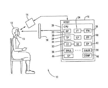
Fig. 1 shows an example of an eye tracking
system 10 for detecting eye movement under an
illustrated embodiment of the invention. Fig. 3 is a
flow chart of steps that can be followed by the
system 10. Included within the system 10 can be an
eye tracking device 14, such as the EyeLink II by SR
Research (sr-research.com/fixed tech_spec.php) or
other equivalent eye tracking systems such as the
IVIEWTM HI-SPEED 1250 tracking system by
6
W02012/103470 CA 02827498 2013-07-26
PCT/US2012/022959
SensoMotoroic Instruments (smivision.com/en/eye-gaze-
tracking-systems/products/iview-x-hi-speed.html).
Also included within the system 10 can be a
display 16 and host 18. The host 18 includes a
central processing unit (CPU) 20 embodied as hardware
and a number of associated processors (described
below), that can also be embodied as hardware. In
this case, the processors can each be defined by a
respective hardware processor executing one or more
programs loaded from a non-transitory computer
readable medium (memory).
The objective of the data collection of the
system 10 is to automatically and objectively detect
square wave jerks (SWJs) present in the eye movement
trace. SWJs are characterized by one small
horizontal saccade that moves the eye away from the
fixation target, followed by a corrective saccade
towards the target shortly thereafter.
Fig. 2 provides examples of eye movement
recordings for PSP patients and healthy subjects. A
first graph 100 shows an example of horizontal eye
position in degrees of visual angle versus time.
Graphs 102, 104, 106, 108 provide other examples of
eye position versus time for a group of PSP patients,
whereas graphs 110, 112, 114, 116, 118 and 120
provide examples of eye position for healthy test
subjects.
A display processor 22 within a controller 20 of
the system 10 presents the fixation target to a
subject 12 on the display 16. As the subject 12
fixates in the target on the display 16, the eye
tracking device 14 detects and records 200 the
position and movement of the eyes 13 of the subject
12. A tracking processor 28 within the host 18 can
7
W02012/103470 CA 02827498 2013-07-26
PCT/US2012/022959
receive the position of the eyes 13 and store it for
later transfer to a saccade processor 30.
The saccade processor 30 can receive the eye
position measurements, can detect 202 substantially
all the consecutive pairs of saccades (up to a
certain maximum magnitude, for instance, 5 degrees).
Any method to detect small saccades can be used by
the saccade processor 30. Two main algorithms have
been used in the literature: the Martinez-Conde and
Macknik algorithm [Martinez-Conde, Macknik, Hubel
(2000) Nature Neuroscience 3:251-258] and the Engbert
algorithm [Engbert, Kliegl (2003) Vision Res 43:1035-
1045].
The first step of the Martinez-Conde and Macknik
process that can be used by the saccade processor 30
is the differentiation of the data (horizontal and
vertical position), so that each element represents
the instantaneous velocity of the eye in horizontal
and vertical space, then data can then be smoothed
with a 31 milliseconds (ms) wide unweighted boxcar
filter to reduce noise. Then, the direction and size
of the motion between each two samples is calculated.
The size of the motion represents the velocity of
movement in polar coordinates and the direction is
differentiated to obtain the rate-of-turn indicator.
The saccade processor 30 determines that the eye is
moving when the polar velocity is more than 3 per
second (s) and the rate-of-turn is smaller than 15 .
Finally, only detected eye movements of more than 3
arc minutes (arcmin) and less than 2 are considered
saccades.
Under the Engbert process, the saccade processor
30 can first transform the time series of eye
8
WO 2012/103470 CA 02827498 2013-07-26
PCT/US2012/022959
positions into velocities in accordance with the
equation
2'n=r2 Arf-1 gt-1 ¨
iren =
6At
that represents a moving average of velocities over 5
data samples in order to suppress noise. As a
consequence of the random orientations of the
velocity vectors during fixation, the resulting mean
value of noise is effectively zero. A multiple of
the standard deviation of the velocity distribution
is used as the detection threshold. Detection
thresholds are computed independently for horizontal
and vertical components and separately for each
trial, relative to the noise level.
Typical values for the threshold are 4, 5 or 6
times the standard deviation of the velocity.
Therefore, the process used by the saccade processor
30 is robust with respect to different noise levels
between different trials and subjects. Additionally,
minimum saccade duration of 8 or 12 ms is required to
further reduce noise. Finally, only binocular
saccades are used, that is, saccades with at least 1
sample of overlap between the two eyes.
The principal advantage of the Engbert algorithm
is that it adapts to the level of noise of the data.
However, although this improves its performance in
noisy situations it can produce non-optimal results
in low noise conditions where the Martinez-Conde and
Macknik algorithm behaves better.
As the saccades 32, 34 are identified (or
after), a pairing processor 40 can determine and
combine consecutive pairs of associated saccades 32,
9
W02012/103470 CA 02827498 2013-07-26
PCT/US2012/022959
34 into potential SWJs 36,, 38. The pairing processor
40 can get a first pair of consecutive saccades 204
and measure a direction difference, a relative
magnitude difference and an inter-saccade difference
206. The pairing processor 40 can use three criteria
210, 212, 214 to determine whether a pair of saccades
32, 34 is a SWJ 36, 38. If a pair of saccades 32, 34
does not meet each of the three criteria, then the
pair can be discarded.
The First criterion requires that the two
consecutive saccades 32, 34 should have (loosely)
opposite directions. In a perfect SWJ this
difference would be exactly 180 . Allowing for some
variability, a pair of saccades meets this criterion
210 if the direction difference is in the range 180
80 . (See FIGs. 4A and 4D).
The second criterion 212 is that the two
consecutive saccades 32, 34 should have similar
magnitudes as shown in FIGs. 4B and 4E. A
disimilarity index can be objectively calculated as
the magnitude difference between the 1st and the 2nd
saccade divided by the average magnitude of both
saccades (expressed in percent terms) by the
following equation,
magnitude of ist 1.--,accade ¨magnitude of 2nd ffaccade
average magnitude of lgt" and2 _______________________________ 100ndsaccath3
An ideal SWJ (where the two saccades have equal
magnitudes) would have an index of 0%. A pair of
saccades is considered a SWJ if the index is in the
range 100%.
The third criterion 214 can require that the two
consecutive saccades should have Inter-Saccadic
W02012/103470 CA 02827498 2013-07-26
PCT/US2012/022959
Interval (ISI) (between the end of the 1st saccade
and the beginning of the 2nd saccade) in the range
70ms - 650ms. (See FIGs. 40 .and 4F).
Once the saccade processor 30 has processed a
saccade pair, the processor 30 can determine if there
are any more saccade pairs 218. If so, then the
saccade processor 30 retrieves the next pair 208 of
the sequence and the process repeats.
The specific numeric values for the three
different criteria were optimized based on a data
from a set of PS? patients (see FIGs. 4D, 4E, 4F).
The system 10 can also be used with other criteria
values where the other criteria values show a better
performance.
As a last step, an elimination processor 42 can
locate sequences of potential SWJs sharing saccades
220 and eliminate the SWJs 36, 38 that share saccades
32, 34. The result of the previous step is a
sequence of pairs of saccades that meet the initially
defined SWJ criteria. However, it is possible in
some cases that these pairs are linked by a shared
saccade. To solve this problem and have saccades
that are only part of a unique SWJ, the following
rule can be used: if the number of SWJs linked by
shared saccades is odd, then the SWJs in even
positions in the sequence of SWJs are discarded.
That is one way to ensure that all the saccades are
part of only one SWJ.
If the number is even, then it is impossible to
achieve this result and at least one saccade will not
be part of any SWJ. In this case the odd or even
SWJs can be discarded depending upon which choice
provides a shorter average inter-saccadic interval.
11
W02012/103470 CA 02827498 2013-07-26
PCT/US2012/022959
As such, the elimination processor 42 can
retrieve a first sequence 222 of potential SWJs and
determine the number of potential SWJs that share a
saccade. If the number of SWJs with shared saccades
is even 228, then the elimination processor 42
selects the SWJs in the even or odd positions in the
sequence according to the inter-saccadic intervals
230. If not, then the elimination processor 42
selects the SWJs in the odd positions of the sequence
226. If there are any more sequences 232, the
process repeats. If not, then the elimination
process ends 234.
Following identification of SWJs 36, 38, that
meet the appropriate criteria, the remaining SWJs 36,
38 can be transferred to a statistics processor 44.
Within the statistics processor 44, the SWJs 36, 38
of PSP patients can be compared with healthy
subjects. Fig. 5 provides SWJ parameter comparison
between a population of healthy subjects and a
population of PSP patients.
The system 10 can be used to automatically
compute several SWJ parameters that can help to
determine whether a person is healthy (a subject) or
has certain neurological diseases. In all the panels
of Fig. 5, the upper rows (labeled "001" through
"014") correspond to respective healthy subjects and
the lower rows (labeled "PSP001" through "PSP010")
correspond to respective PSP patients. The
horizontal bar (labeled "Normal subjs.avg.") is the
average of the healthy subjects' population and the
horizontal bar (labeled "Patients.avg.") is the
average of the PSP patients' population respectively.
The parameters represented in each panel of Fig.
are (from left to right and top to bottom): (A)
12
W02012/103470 CA 02827498 2013-07-26
PCT/US2012/022959
Number of SWJ per second; (B) Percentage of small
saccades that are part of SWJs; (C) Average magnitude
of the saccades that are part of SWJs; (D) Average
inter-saccadic interval for the SWJs; (E) Number of
saccades per second; (F) Average peak velocity of the
saccades in SWJs; (G) Standard deviation of the
direction difference and (H) Standard deviation of
the difference between the horizontal and the
direction of the saccades in the SWJs. All the
parameters (with the exception of the inter-saccadic
interval) are significantly different between the two
populations (two-tailed t-test).
Any of a number of the statistics of Fig. 5 can
be the basis of a screening test for detecting PSP.
For example, the standard deviation of direction
difference in Fig. 5 (G) shows a greater than a two
to one difference between the PSP patients and normal
subjects. In this case, the standard deviation of
direction difference of a subject 12 tested with the
system 10 can be compared within a comparator 46 with
the standard deviation of direction difference of a
normal subjects to detect PSP. Other statistics of
Fig. 5 can be used in a similar manner.
In another embodiment of the invention,
differences in SWJ characteristics between normal
patients (healthy subjects) and patients with
Progressive Supranuclear Palsy (PSP) can also be used
to identify patients with Parkinson's disease (PD).
In their early stages, these two afflictions cannot
be definitively or differentially diagnosed under
previously known methods because their symptoms are
so similar. It is critical nevertheless to
differentiate between them as early as possible as
their neurological bases and treatment regimens are
13
W02012/103470 CA 02827498 2013-07-26
PCT/US2012/022959
quite different. It has been found that the
characteristics of SWJs in these two diseases are
different, and that the measurement of SWJ
characteristics in these two patient populations
therefore provide as a sensitive method to
differentially diagnose these diseases earlier than
other tests.
It is also believed that the measurement of SWJ
characteristics similarly serve as a sensitive test
for other neurological disorders as well. Examples
include such afflictions as stroke, Friedrich's
ataxia, cerebellar disease, multiple sclerosis,
cerebral lesions, strabismus, and nystagmus.
It has been found that the method described
herein accurately differentially diagnoses PSP from
PD, in part, because PD patients are more like normal
patients for the types of eye movements used in the
diagnosis. This is a major advance because there is
no previous method to accurately and non-invasively
differentially diagnose PSP from PD.
PSP is a much more debilitating disease than PD,
although at the early stages they appear similar.
Because of the similar symptoms, patients with PSP
are often misdiagnosed as having PD and are given L-
DOPA or other PD drugs in levels appropriate for PD,
but way too low for PSP patients. As a result, PSP
patients misdiagnosed with PD typically suffer much
more than they otherwise would, had they been
accurately diagnosed. The method described below
demonstrates that accurate diagnosis of PSP can be
made from a simple non-invasive eye movement
analysis.
In this regard, it has been found that PSP is
distinguishable from Parkinson's disease because the
14
W02012/103470 CA 02827498 2013-07-26
PCT/US2012/022959
vertical component of SWJ's is smaller for PSP
patients than for Parkinson's patients. This effect
exists between PSP patients and healthy people
(subjects), as well.
Detection in this regard can include an eye
movement processing apparatus (including one or more
of device 14 and special purpose processors 20, 30,
40) that detects SWJs as discussed above. One or
more programmed processors, such as those shown in
Fig. 1 (e.g., processors 42, 44), can then analyze a
vertical component of each of the SWJs.
In this regard, the vertical component can be
manifested as a tilting of the horizontal saccades by
a few degrees. As above, a standard deviation of
directional difference can be determined in the
vertical component for the deviation away from the
fixation target versus the corrective saccade towards
the target. This can be compared with a normal
subject and/or with normal subjects as a basis for
determining a set of threshold values for PSP and
Parkinson's disease.
In addition, the number of vertical deviations
per time period as well the magnitude of the vertical
deviation can be collected. The velocity of the
vertical deviation can be measured.
The data points of Fig. 6 illustrate individual
saccades. It is seen the PD patients have faster
saccades than PSP patients. These data provide the
"main sequence slope" utilized in Fig. 7 in which
saccade rate is plotted vs. main sequence slope in
Fig. 7A and the normalized saccade vertical component
is plotted vs. main sequence slope in Fig. 7B.
The data used in FIGs. 6 and 7 were subjected to
statistical analysis. The significance testing
W02012/103470 CA 02827498 2013-07-26 PCT/US2012/022959
statistics in the table below were calculated using
ANOVA and corrected for multiple comparisons using
Tukey's Honest Significant Difference. As is seen,
differences between control (normal healthy) patients
and PSP patients were highly significant, as were
differences between PSP and PD patients for vertical
component and main sequence slope, whereas there was
little difference in saccade rate between PSP and PD
patients. There were also not significant
differences between control and PD patients in
vertical component and main sequence slope.
Multiple Comparisons Between Groups
(adjusted p-values)
Vertical Main Saccade rate
component sequence
slope
Control-PSP 0.0000025 0.0000022 0.0071293
Control-PD 0.4250289 0.9916481 0.0320478
PSP-PD 0.0233523 0.0002209 0.9880972
The algorithm used successfully characterized
patients in each group (PSP, PD or control) based on
three dynamical eye movement parameters in fixation
saccades: vertical component, rate and velocity. PSP
patients are distinguished from controls based on
lack of vertical component, slower velocity or higher
rate. PSP and PD patients are distinguished from
each other because PSP patients have slower and more
horizontal saccades. PD patients and controls
(normal healthy subjects) can be distinguished
because PDs have higher saccade rates.
The collected vertical data can be compared with
that of PSP patients as well as that of normal
16
W02012/103470 CA 02827498 2013-07-26
PCT/US2012/022959
subjects. A first set of threshold values can be
used to identify patients with Parkinson's disease.
In a similar manner, a second set of threshold values
can be used to identify patients with PSP. Other
threshold values can be used to identify patients
with stroke, Friedrich's ataxia, cerebellar disease,
multiple sclerosis, cerebral lesions, strabismus, and
nystagmus.
A specific embodiment of method and apparatus
for detecting and characterizing square wave jerks in
the eye movements of a subject, which can provide a
powerful tool in the differential diagnosis of
oculomotor and neurological disease, has been
described for the purpose of illustrating the manner
in which the invention is made and used. The method
disclosed herein has extended the previously filed
method of application Serial No. 12/740,008 to
include the capability of distinguishing PSP patients
and control subjects from each other as well as from
and patients affected with Parkinson's Disease (PD).
It should be understood that the implementation
of other variations and modifications of the
invention and its various aspects will be apparent to
one skilled in the art, and that the invention is not
limited by the specific embodiments described.
Therefore, it is contemplated to cover the present
invention and any and all modifications, variations,
or equivalents that fall within the true spirit and
scope of the basic underlying principles disclosed
and claimed herein. The use of the article "a" or
"an" is intended to include one or more.
17
W02012/103470 CA 02827498 2013-07-26
PCT/US2012/022959
Aspects of the Invention
1. A method for providing a differential
diagnosis of a patient with progressive supranuclear
palsy (PSP) from a patient with Parkinson's disease
(PD) that comprises the steps of:
a) identifying in a patient to be diagnosed with
one or the other of PSP or PD a plurality of at least
partially repetitive eye movements that over time
define square wave jerks within a sequence of eye
movements, each square wave jerk of the plurality of
square wave jerks being defined by a first horizontal
saccadic movement that moves the eye away from a
fixation target that is followed by a corrective
saccadic movement towards the target shortly
thereafter;
b) measuring a vertical component associated
with the plurality of square wave jerks;
c) comparing the vertical component with a
threshold value for normal healthy subjects or other
patients with PSP or other patients with PD;
d) identifying the presence of PSP or PD in the
patient to be diagnosed based upon the comparison of
the vertical component with the threshold value
whereby a vertical component statistically different
from the threshold value of normal subjects or PD
patients identifies the patient to be diagnosed as
having PSP, whereas a vertical component that is (a)
not statistically different from the threshold value
of normal subject or (b) is statistically different
from the threshold value of PSP patients identifies
the patient as having PD.
18
W02012/103470 CA 02827498 2013-07-26
PCT/US2012/022959
2. The method for providing a differential
diagnosis as in claim 1 further comprising obtaining
a sequence of saccadic movements.
3. The method for providing a differential
diagnosis as in claim 2 further comprising
identifying pairs of consecutive saccadic movements
of the sequence.
4. The method for providing a differential
diagnosis as in claim 3 further comprising
determining whether each saccadic movement of each
identified pair is opposite the direction of the
other saccadic movement and, if not, then discarding
the pair.
5. The method for providing a differential
diagnosis as in claim 4 further comprising
determining whether a magnitude of each saccadic
movement of each identified pair is comparable and,
if not, then discarding the pair.
6. The method for providing a differential
diagnosis as in claim 5 further comprising
determining whether the pair of saccadic movements of
each identified pair are temporally related by a
predetermined time period and, if not, then
discarding the pair.
7. The method for providing a differential
diagnosis as in claim 6 further comprising collecting
any remaining pairs of saccadic movements as square
wave jerks.
19
W02012/103470 CA 02827498 2013-07-26
PCT/US2012/022959
8. The method for providing a differential
diagnosis as in claim 7 further comprising comparing
a set of parameters of the square wave jerks in a
potential patient against the corresponding
parameters of a healthy population.
9. The method for providing a differential
diagnosis as in claim 8 further comprising defining
the opposite direction of the saccadic movements in
the pairs as 180 degrees, plus or minus 80 degrees.
10. The method for providing a differential
diagnosis as in claim 9 further comprising defining
the magnitude of each saccadic movements in the pairs
comparable as the dissimilarity index is in the range
10096.
11. A method for providing a differential
diagnosis of a patient with progressive supranuclear
palsy (PSP) from a patient with Parkinson's disease
(PD) that comprises the steps of:
a) identifying in a patient to be diagnosed with
one or the other of PSP or PD a plurality of at least
partially repetitive eye movements that over time
define square wave jerks within a sequence of eye
movements, each square wave jerk of the plurality of
square wave jerks being defined by a first horizontal
saccadic movement that moves the eye away from a
fixation target that is followed by a corrective
saccadic movement towards the target shortly
thereafter;
b) determining the saccade rate of the patient;
W02012/103470 CA 02827498 2013-07-26
PCT/US2012/022959
c) comparing the saccade rate with a threshold
value for normal healthy subjects or other patients
with PD;
d) identifying the presence of PSP or PD in the
patient to be diagnosed based upon the comparison of
the saccade rate with the threshold value whereby a
saccade rate statistically different from the
threshold value of normal subjects or PD patients
identifies the patient to be diagnosed as having PSP,
whereas a saccade rate that is statistically
different from the threshold value of normal subjects
or PSP patients identifies the patient as having PD.
12. The method for providing a differential
diagnosis as in claim 11 further comprising obtaining
a sequence of saccadic movements.
13. The method for providing a differential
diagnosis as in claim 12 further comprising
identifying pairs of consecutive saccadic movements
of the sequence.
14. The method for providing a differential
diagnosis as in claim 13 further comprising
determining whether each saccadic movement of each
identified pair is opposite the direction of the
other saccadic movement and, if not, then discarding
the pair.
15. The method for providing a differential
diagnosis as in claim 14 further comprising
determining whether a magnitude of each saccadic
movement of each identified pair is comparable and,
if not, then discarding the pair.
21
WO 2012/103470 CA 02827498 2013-07-26
PCT/US2012/022959
16. The method for providing a differential
diagnosis as in claim 15 further comprising
determining whether the pair of saccadic movements of
each identified pair are temporally related by a
predetermined time period and, if not, then
discarding the pair.
17. The method for providing a differential
diagnosis as in claim 16 further comprising
collecting any remaining pairs of saccadic movements
as square wave jerks.
18. The method for providing a differential
diagnosis as in claim 17 further comprising comparing
a set of parameters of the square wave jerks in a
potential patient against the corresponding
parameters of a healthy population.
19. The method for providing a differential
diagnosis as in claim 18 further comprising defining
the opposite direction of the saccadic movements in
the pairs as 180 degrees, plus or minus 80 degrees.
20. The method for providing a differential
diagnosis as in claim 19 further comprising defining
the magnitude of each saccadic movements in the pairs
comparable as the dissimilarity index is in the range
100%.
21. An apparatus that detects and characterizes
eye movements of a subject, for the differential
diagnosis of a patient with progressive supranuclear
22
W02012/103470 CA 02827498 2013-07-26
PCT/US2012/022959
palsy (PSP) from a patient with Parkinson's disease
(PD) comprising:
a processor that processes a sequence of
repetitive, involuntary eye movements, each of the
plurality of involuntary eye movements defined by a
first horizontal saccadic movement that moves the eye
away from a fixation target followed by a corrective
saccadic movement towards the target shortly
thereafter;
a processor that measures vertical components
associated with the plurality of repetitive eye
movements;
a processor that compares the vertical
components with a threshold value; and
a processor that differentially identifies
progressive supranuclear palsy from Parkinson's
disease based upon the comparison of the vertical
component with the threshold value.
22. The apparatus for the differential
diagnosis of a patient with progressive supranuclear
palsy (PSP) from a patient with Parkinson's disease
(PD) as in claim 21, wherein the eye movements
identified by the processor further comprises a
sequence of saccadic movements.
23. The apparatus for the differential
diagnosis of a patient with progressive supranuclear
palsy (PSP) from a patient with Parkinson's disease
(PD) as in claim 22 wherein the eye movements
identified by the processor further comprises pairs
of consecutive saccadic movements of the sequence.
23
W02012/103470 CA 02827498 2013-07-26
PCT/US2012/022959
24. The apparatus that detects and
characterizes eye movements as in claim 23, wherein
the processor that identifies eye movements
determines whether each saccadic movement of each
identified pair is opposite the direction of the
other saccadic movement and, if not, then discards
the pair.
25. The apparatus that detects and
characterizes eye movements as in claim 24, wherein
the processor that identifies eye movements
determines whether a magnitude of each saccadic
movement of each identified pair is comparable and,
if not, then discards the pair.
26. The apparatus that detects and
characterizes eye movements as in claim 25, wherein
the processor that identifies eye movements
determines whether the pair of saccadic movements of
each identified pair are temporally related by a
predetermined time period and, if not, then
discarding the pair.
27. The apparatus that detects and
characterizes eye movements as in claim 26, wherein
the processor that identifies eye movements collects
any remaining pairs of saccadic movements as square
wave jerks.
28. The apparatus that detects and
characterizes eye movements as in claim 27, wherein
the processor that identifies eye movements compares
a set of parameters of the eye movements in a
24
W02012/103470 CA 02827498 2013-07-26
PCT/US2012/022959
potential patient against the corresponding
parameters of a healthy population.
29. The apparatus that detects and
characterizes eye movements as in claim 28, wherein
the processor that identifies eye movements defines
the opposite direction of the saccadic movements in
the pairs as 180 degrees, plus or minus 80 degrees.
30. A system that detects and characterizes eye
movements of a patient, for the differential
diagnosis of a patient with progressive supranuclear
palsy (PSP) from a patient with Parkinson's disease
(PD) comprising:
eye movement processing apparatus that
identifies a plurality of square wave jerks within a
sequence of repetitive eye movements, each of the
plurality of square wave jerks defined by a first
horizontal saccadic movement that moves the eye away
from a fixation target followed by a corrective
saccadic movement towards the target shortly
thereafter;
a processor that measures vertical components
associated with the plurality of square wave jerks;
a processor that compares the vertical
components with a threshold value or the saccade
rate; and
a processor that identifies PSP or PD based upon
the comparison of the vertical component with the
threshold value .