Note: Descriptions are shown in the official language in which they were submitted.
CA 02827552 2013-08-16
WO 2012/113407 PCT/DK2012/050062
1
Method of modifying the gingival part of a virtual model of a set of
teeth.
This invention generally relates to a method of and a system for generating a
virtual model of a set of teeth for manufacturing a physical model of the set
of
teeth, and to a physical model of a set of teeth.
The invention may be used for instance in relation to dental implants and
other applications where a restoration is to be inserted into a gingival part
of
a physical model of the set of teeth.
When designing a dental restoration for a patient, a dental technician often
uses a physical model of the patient's set of teeth. In many cases it is
preferred that the designed restoration, such as an abutment and the
corresponding crown or bridge, is shaped such that it gently displaces a
portion of the patient's gingiva. When a physical model of the patient's set
of
teeth is made in a material which is not easily displaced/deformed by the
dental technician, such a design of the restoration will result in that the
restoration cannot be inserted into the physical model of the patient's set of
teeth.
Disclosed is a method of generating and modifying a virtual model of a set of
teeth, said set of teeth comprising a region configured for insertion of a
restoration, the region being located in a gingival part of the set of teeth,
where the method comprises:
- obtaining at least one three dimensional representation of the set of teeth;
- generating a virtual model of the set of teeth from said three dimensional
representation, where the virtual model of the set of teeth comprises a
CA 02827552 2013-08-16
WO 2012/113407 PCT/DK2012/050062
2
gingival part, said gingival part comprising a virtual region corresponding to
said region configured for insertion of a restoration and at least part of the
gingiva surrounding said region;
- obtaining a virtual model of said restoration; and
- modifying the gingival part of the virtual model of the set of teeth such
that
the virtual model of the restoration can be virtually inserted into said
virtual
region with no overlap between the volume of said virtual model of the
restoration and the volume of said gingival part of the virtual model of the
set
of teeth.
At least a portion of the gingival part of the virtual model of the set of
teeth
may correspond to the gingival part of the patient's set of teeth.
Disclosed is a method of generating a virtual model of a set of teeth for
manufacturing a physical model of the set of teeth, where the method
comprises:
- obtaining a three dimensional representation of the set of teeth;
- obtaining a virtual model of a restoration, where at least a sub-gingival
part
of the virtual model of the restoration is configured to have the shape of an
anatomically correct restoration; and
- generating a virtual model of the set of teeth from said three dimensional
representation, the virtual model of the set of teeth comprising a gingival
part,
where the gingival part comprises a gingiva and a region configured for
insertion of a restoration, and where the gingival part is configured to
provide
that when the restoration is inserted in said region the volume of the
restoration and the volume of the gingival part do not overlap.
CA 02827552 2013-08-16
WO 2012/113407 PCT/DK2012/050062
3
In the context of the present invention, the phrase "sub-gingival part of"
used
in relation to the restoration may refer to the portion of the restoration
which
resides below the surface of the gingiva when the restoration is inserted in
the virtual or physical model of the set of teeth. The sub-gingival part may
be
the portion of the restoration arranged below the margin line when the
restoration is inserted in the gingival part of the model of the set of teeth.
The sub-gingival part of the restoration may take in different shapes. In some
embodiments, the sub-gingival part of a restoration has a cross sectional
dimension which changes along the longitudinal direction of the restoration.
The cross sectional dimension of the sub-gingival portion of the restoration
may increase towards a margin line of the restoration, such that the diameter
of the restoration at the margin line is larger than the diameter further
below
the surface of the gingival part. The cross sectional dimension may be the
diameter or area of the restoration in a cross sectional plane which may be
perpendicular with the insertion direction of the restoration.
The method may in general relate to a gingival part which is configured to
provide that when a restoration is inserted in a region of the gingival part,
the
volume of the restoration and the volume of the gingival part do not overlap.
The virtual model of the restoration may comprise the entire restoration or a
part of the restoration.
In some embodiments, the gingival part of the virtual model generated from
the three dimensional representation directly provides that the volume of the
restoration and the volume of the gingival part do not overlap. This may be
the case e.g. when the method provides that the gingival part is configured
for insertion of the restoration in the same step as the virtual model is
generated from the three dimensional representation.
CA 02827552 2013-08-16
WO 2012/113407 PCT/DK2012/050062
4
In some embodiments, the virtual model of the set of teeth is generated in
one step and where the gingival part of the virtual model subsequently is
modified to provide that the volume of the restoration and the volume of the
gingival part do not overlap. This may be the case e.g. when the method
provides that the gingival part is configured for insertion of the restoration
after the virtual model is generated from the three dimensional
representation.
In some embodiments, the gingival part of the virtual model of the set of
teeth
is modified to provide that the adjoining surfaces of the virtual model of the
restoration and the gingival part of the virtual model of the set of teeth
follow
each other.
In some embodiments, an offset is provided between the adjoining surfaces
of the virtual model of the restoration and the gingival part of the virtual
model
of the set of teeth. The offset may be substantially uniform over a part of
said
sub-gingival part of the restoration.
In the context of the present invention, the phrase "adjoining surfaces" may
be used in relation to virtual surfaces which are adjoining e.g. when the
virtual model of the restoration is arranged in its anatomical correct
position
relative to the virtual model of the set of teeth. The phrase may also be used
in relation to the surfaces of a restoration formed from the virtual model of
the
restoration when this arranged at a physical model of the set of teeth.
In some embodiments, modifying the gingival part of the virtual model of the
set of teeth comprises digitally cutting a portion of the gingiva away such
that
the volume of the restoration and the volume of the gingiva do not overlap.
The digital cutting away may correspond to a removal of material from a
physical model of the set of teeth. Preferably, the digitally cutting away is
CA 02827552 2013-08-16
WO 2012/113407 PCT/DK2012/050062
made with the virtual model of the restoration virtually arranged in the
gingival part of the virtual model of the set of teeth.
One advantage that may be provided by the present invention is the
5 possibility of digitally combining a restoration with a gingival part of
a virtual
model of a set of teeth, where the anatomically correct shape of the
restoration is taken into account.
One advantage that may be provided by the present invention is that a
anatomically correct restoration can be inserted into a physical model
manufactured from a virtual model of the set of teeth generated using the
method according to the present invention. The restoration may be arranged
in its anatomically correct position and at the anatomically correct
orientation
relative to the gingival part and any neighboring teeth in the model. This may
allow e.g. a dental technician to test the form and arrangement of the actual
restoration in a physical model of the set of teeth.
Disclosed is a method of generating and modifying a virtual model of a set of
teeth for manufacturing a physical model of the set of teeth, where the
method comprises:
- obtaining a three dimensional representation of the set of teeth;
- generating a virtual model of the set of teeth from said three dimensional
representation, the virtual model of the set of teeth comprising a gingival
part
comprising a gingiva; and
- modifying the gingival part to enable insertion of a restoration in a region
of
the virtual model configured for insertion of a restoration.
CA 02827552 2013-08-16
WO 2012/113407 PCT/DK2012/050062
6
Disclosed is a method of generating a virtual model of a set of teeth for
manufacturing a physical model of the set of teeth, where the method
comprises:
- obtaining a virtual model of the set of teeth, the model comprising a
gingival
part comprising a gingiva; and
- obtaining a virtual model of a restoration configured to be arranged in
its
anatomical correct position relative to said gingival part of the model;
where the surface of the gingiva defines a first surface at said restoration;
and
- modifying the gingiva at said restoration such that the surface of the
modifying gingiva defines a second surface at said restoration, wherein the
second surface is configured to avoid an overlap between the volume of the
restoration and the volume of the gingival part of the model.
Disclosed is a method of adjusting a virtual model of a set of teeth, where
the
virtual model of the set of teeth is for manufacturing a physical model of the
set of teeth, where the method comprises:
- obtaining a pre-adjustment configuration of a virtual model of the set of
teeth, the virtual model of the set of teeth comprising a gingival part; and
- obtaining a virtual model of a restoration configured to be arranged in
its
anatomical correct position relative to said gingival part of the model,
where the volume of the gingival part of the virtual model of the set of teeth
and the volume of the restoration overlaps when the restoration is arranged
in the anatomical correct position ; and
- adjusting a portion of the gingival part of the virtual model of the set
of teeth
arranged at said restoration providing a post-adjustment configuration of the
virtual model of the set of teeth, in which post-adjustment configuration the
gingival part of the model is configured to avoid overlap between the volume
CA 02827552 2013-08-16
WO 2012/113407 PCT/DK2012/050062
7
of the virtual model of the restoration and the volume of the gingival part of
the model of the set of teeth.
Disclosed is a method of adjusting a virtual model of a set of teeth, where
the
virtual model is for manufacturing a physical model of the set of teeth, where
the method comprises:
- obtaining a pre-adjustment configuration of a virtual model of the set of
teeth, the model comprising a gingival part; and
- adjusting a portion of the gingival part of the model arranged at said
restoration providing a post-adjustment configuration of the virtual model of
the set of teeth, in which post-adjustment configuration the gingival part of
the model is configured to avoid overlap between the volume of a restoration
configured to be arranged in its anatomical correct position in the gingival
part of the model.
In some embodiments, the adjusting of the gingival part comprises
configuring the shape of the gingival part such that the overlap between the
volumes is avoided.
In some embodiments, the method comprises configuring the material of the
gingiva at the restoration to be sufficiently soft such that a restoration may
deform the gingival part. This may be done by removing relative harder
material from a physical model of the set of teeth and replacing it by a
relative softer material, such as replacing gypsum with a dental silicone
material.
Disclosed is a method of generating a physical model of a set of teeth, where
the method comprises:
- obtaining at least one three dimensional representation of the set of teeth;
CA 02827552 2013-08-16
WO 2012/113407 PCT/DK2012/050062
8
- generating and modifying a virtual model of the set of teeth from said at
least one three dimensional representation, the virtual model of the set of
teeth comprising a gingival part; and
- modifying the gingival part to enable insertion of a restoration in a
region of
the virtual model of the set of teeth configured for insertion of a
restoration.
- manufacturing said physical model from said virtual model of the set of
teeth.
In some embodiments, the method comprises configuring the gingival part to
avoid an overlap with a restoration when the restoration is inserted in the
model. Such an overlap may make it difficult or even impossible to arrange
the restoration in its anatomical correct position in the gingival part of the
model.
Disclosed is a physical model of a set of teeth, wherein the physical model is
manufactured from a virtual model generated by the method according to the
present invention.
In the context of the present invention, the phrase "a restoration" may refer
to
a full dental restoration or a part of a restoration, such as an abutment or a
crown arranged on said abutment. An abutment may be a cutmized abutment
or a stock-abutment.
At least one step of the method is computer implemented. In some
embodiments at least the generating and modifying of the virtual model of the
set of teeth is computer implemented.
CA 02827552 2013-08-16
WO 2012/113407 PCT/DK2012/050062
9
In some embodiments, a first three dimensional representation of the set of
teeth is obtained by scanning the patient's set of teeth with a scan body
arranged in said implant region. Data relating to the scan body may hence
become part of the virtual model of the set of teeth.
In some embodiments, said virtual model of the set of teeth is generated at
least in part from said first three dimensional representation. The virtual
model of the set of teeth may thus comprise a section corresponding to the
gingival part of the set of teeth.
In some embodiments, a second three dimensional representation of the set
of teeth is obtained by scanning the patient's set of teeth with the emergence
profile at said implant region being visible. In this case, may data relating
to
the emergence of the gingiva be derived or become part of a virtual model of
the set of teeth.
In some embodiments, said virtual model of the set of teeth is generated at
least in part from said second three dimensional representation. The virtual
model of the set of teeth may then comprise a section corresponding to the
gingival part.
In some embodiments, one of said first or second three dimensional
representation of the set of teeth is obtained by scanning a relatively larger
section of the patient's set of teeth, and the other of said first or second
three
dimensional representation then is obtained by scanning a relatively smaller
section around the implant region.
In some embodiments, the method comprises generating a first virtual model
of the set of teeth from said first three dimensional representation of the
set
of teeth.
CA 02827552 2013-08-16
WO 2012/113407 PCT/DK2012/050062
In some embodiments, the method comprises generating a second virtual
model of the set of teeth from said second three dimensional representation
of the set of teeth.
5 In some embodiments, the method comprises combining the first and second
virtual models of the set of teeth to generate said virtual model of the set
of
teeth. Such a virtual model of the set of teeth may then comprise both the
emergence profile and data relating to the implant position and orientation.
10 In some embodiments, a virtual model of the scan body is provided and
virtually aligned with the first virtual model of the set of teeth to
determine the
orientation and position of the implant.
In some embodiments, the restoration is designed based on the virtual model
of the set of teeth.
In some embodiments, the restoration is a pre-manufactured restoration such
as a pre-manufactured abutment.
In some embodiments, the modified virtual model of the set of teeth is for
manufacturing a physical model of the set of teeth.
In some embodiments, at least a sub-gingival part of the virtual model of the
restoration is configured to have the shape of an anatomically correct
restoration.
The region of the virtual model configured for insertion of a restoration may
comprise a region of the gingival part configured to comprise an implant
analog, a hole, a healing abutment, a scan-body or in principle any dental
indication. The region may be bounded by an area of the gingiva surface
CA 02827552 2013-08-16
WO 2012/113407 PCT/DK2012/050062
11
having a circumference which is bounded partly by the nearest neighbor
teeth.
In some embodiments, the method comprises configuring the gingiva mask
to comprise an opening, where the opening is configured to allow a
restoration to access the gingival part arranged below the gingiva mask.
In some embodiments, a virtual hole is provided in said gingival part of the
virtual model of the set of teeth.
The virtual hole may be such that a corresponding hole in the physical model
of the set of teeth is configured to mate with a part of said restoration
configured to fit into the gingival part of the physical model of the set of
teeth.
The virtual hole may be configured to allow an implant analog to inserted
manually in the corresponding hole of the physical model of the set of teeth.
The virtual hole and/or said implant analog may be configured such that said
the implant analog can be inserted only in the correct anatomical position and
orientation in the gingival part of the model.
The opening of the gingiva mask may be aligned with an implant analog
arranged in the gingival part below the gingiva mask
In some embodiments, the implant analog is configured to have a shape with
reduced cross sectional rotation symmetry, such as an N-fold symmetry,
wherein N is an integer number below 25.
The implant analog may have no rotation symmetry in its cross sectional
plane.
In some embodiments, the gingival part of the virtual model of the set of
teeth
is configured to provide that a corresponding ejection hole in the physical
model of the set of teeth is in fluid connection with said hole such that a
CA 02827552 2013-08-16
WO 2012/113407 PCT/DK2012/050062
12
restoration or an implant analog can be accessed through said ejection hole
to be ejected from the gingival part of the physical model of the set of
teeth.
In some embodiments, the implant analog is configured to comprise a stop
section with a smaller cross sectional area at its distal end, said stop
section
preferably being arranged centrally around the longitudinal axis of the
implant
analog.
In some embodiments, the implant analog is configured to comprise a stop
surface at its distal end, said stop surface preferably being arranged
centrally
around the longitudinal axis of the implant analog.
In some embodiments, the stop surface is reduced in size/diameter
compared to other parts of the implant analog to provide that space is
provided for rounded corners of the hole side wall.
The virtual hole defined in the gingival part of the virtual model of the set
of
teeth may be configured to provide that the corresponding hole in the
physical model of the set of teeth has rounded edges at its distal end or at
any kink along the longitudinal direction of the hole.
In some embodiments, a height inspection groove is defined in the implant
analog to allow for a visually or a contact based inspection of whether the
implant analog is arranged in the correct postion in the gingival part of the
model.
The height inspection groove may extend around the entire circumference of
the implant analog forming a band shaped height inspection groove which
can be seen from all directions in a cross sectional plane intersecting the
height inspection groove.
CA 02827552 2013-08-16
WO 2012/113407 PCT/DK2012/050062
13
A window or a through hole may be provided in the ginival part to allow visual
and/or physical contact to the inplant analog from the outside of the model.
The window or through hole may be provided in the virtual model or after the
manufacturing of the physical model.
In the context of the present invention, the phrase "below" is only used to
describe the relative orientation of the parts of the model and does not
present a limitation on which part is closer to the ground than the other
parts.
One part being below another part may be used to describe an arrangement
of the parts relative to the occlusion plane of a set of teeth.
The phrase "below" may be used to describe that a sub-gingival part of a
virtual model of a restoration is arranged behind the surface of the gingival
part relative to a viewpoint situated at a position corresponding to the
center
of a patient's mouth. That is, in a patient's mouth the object which is
arranged
below the surface of the gingival part of the set of teeth may not be visible.
In the context of the present invention, the phrases "proximal end" and
"distal
end" may refer to two opposite ends of a e.g. a holed in the gingival part of
the model, where the distal end may refer to the part of said hole which is
the
furthest away from the entrance of the hole. The distal end may also be
referred to as the bone end.
In some embodiments, the virtual model comprises a restoration configured
to be inserted in the virtual model at the region configured for insertion of
a
restoration.
In some embodiments, the restoration comprises a full restoration or a part of
a restoration, such as an abutment or a crown arranged on said abutment, an
implant bar, or in principle any other indication used in relation to dental
restorations.
CA 02827552 2013-08-16
WO 2012/113407 PCT/DK2012/050062
14
In some embodiments, restoration is to be arranged in its anatomical correct
position relative to said gingival part of the model.
In some embodiments, configuring the gingival part provides that the
restoration can be positioned in a physical model manufactured from the
virtual model, the restoration even in the case where the surface of the
gingival part of the virtual model generated from three dimensional
representation and an adjoining surface of the restoration overlaps.
In the context of the present invention, the phrase "the model" may be used
in relation to both the physical and the virtual manifestation of the set of
teeth. In some embodiments, there is a one-to-one relationship between the
virtual model and the physical model of the set of teeth.
In some embodiments, configuring the gingival part comprises modifying the
gingival part of the virtual model generated from the three dimensional
representation.
The virtual representation of the set of teeth may be provided by scanning
the set of teeth, such as by scanning the set of teeth by means of an
intraoral
scanner or by scanning an impression of the set of teeth.
In some embodiments, a unit, such as a healing abutment, a scan-body or an
implant analog, is arranged in the region configured for the insertion of a
restoration during the scanning of the set of teeth. The generated virtual
model of the set of teeth may hence show such a unit. The method may
comprise digitally removing this unit from the virtual model or it may
comprise
generating a virtual model of the set of teeth where such a unit is not part
of
the virtual model.
CA 02827552 2013-08-16
WO 2012/113407 PCT/DK2012/050062
In some embodiments, configuring the gingival part comprises configuring
the material of the gingival part at the restoration to be sufficiently soft
such
that a restoration may deform the gingival part. In a physical model
manufactured from the virtual model, the material may be sufficiently soft
5 such that an operator, such as a dental technician, may arrange the
restoration in its anatomical correct postion without having to use excessive
force.
In some embodiments, configuring or modifying the gingival part comprises
virtually removing a portion of said gingival part in the region configured
for
10 insertion of a restoration.
The configuring the gingival part of the model may comprise digitally cutting
a
portion of the gingiva away such that the volume of the restoration and the
volume of the gingiva do not overlap.
After a removal of material from the model, the gingival part in a physical
model manufactured from the virtual model is configured to follow the
adjoining surface of the restoration such that a correct positioning of the
restoration is enabled. This may correspond to cutting the gingiva to the
restoration, i.e. that the gingival part of the model adapts to the
restoration. In
this embodiment, the entire gingival part of the model may be manufactured
in a relatively hard material since there is no overlap between the volumes of
the gingival part of the model and the restoration when the latter is
positioned
correctly in the model.
In the context of the present invention, the phrase "to follow the adjoining
surface of the restoration" may refer to the case the restoration is arranged
in
relation to the gingival part such that at least a part of the sub-gingival
portion
of the restoration has a surface which is substantially parallel to the
adjoining
surface of the gingival part. The adjoining surfaces of the sub-gingival
portion
of the restoration and the gingival part may be spaced apart by a
CA 02827552 2013-08-16
WO 2012/113407 PCT/DK2012/050062
16
substantially constant distance over a least part of their common area, such
that there is a substantially constant offset between the sub-gingival portion
of the restoration and the gingival part over that area. The adjoining
surfaces
may be taken to be the area of the side-walls of a hole in the gingival part,
where said hole is configured for the insertion of a restoration. The side
walls
may be the surface of such a hole that is located along the direction of
insertion of the restoration into the gingival part.
In some embodiments, the method comprises virtually adding material to the
gingival part of the virtual model in the region configured for insertion of a
restoration. The virtual addition of the material may occur after a virtual
removal of material in the region configured for insertion of a restoration.
In some embodiments, the gingival part of the virtual model of the set of
teeth
defines a first surface at the region configured for insertion of a
restoration.
In some embodiments, the first surface follows at least a section of said
emergence profile of the gingiva in the region.
In some embodiments, modifying the virtual model of the set of teeth
comprises replacing said first surface by a second surface, where said
second surface is shaped such that the virtual model of said restoration can
be virtually arranged in said virtual implant region with no overlap with the
modified virtual model of the set of teeth.
In some embodiments, at least a section of said second surface is defined by
offsetting part of the surface of the virtual model of the restoration. The
offset
may be such that the second surface encloses the surface of the virtual
model of the restoration.
CA 02827552 2013-08-16
WO 2012/113407 PCT/DK2012/050062
17
In some embodiments, the method comprises subtracting the virtual model of
the restoration or the volume enclosed by the offset surface from the virtual
model of the set of teeth. The second surface may then be identical to or be
based on the virtual surface of the gingival part from which a volume is
subtracted.
In some embodiments, the gingival part of the virtual model of the set of
teeth
after the virtual removal of a portion of the gingiva defines the second
surface
at the region configured for insertion of a restoration. The second surface
may be referred to as a cutting surface. Cutting the gingival part at the
second surface may correspond to cutting the gingival part to the restoration.
In some embodiments, the gingival part of the virtual model of the set of
teeth
after virtually adding material to the gingiva defines a third surface at the
region configured for insertion of a restoration.
The third surface may be substantially identical to said first surface.
The first, second and third surfaces are provided on the virtual model of the
set of teeth. In a virtual model, the surfaces may be defined as a result of
the
generation of the gingival part of the model. In a physical model, the
surfaces
may be realized when the volume of the gingival part of the model is
manufactured.
In some embodiments, the gingival part of the virtual model of the set of
teeth
is divided into a first and a second gingival region by the second surface,
where said second gingival region is arranged between the second surface
and the third surface, the second surface forming an interface between the
first and the second gingival region.
The first gingival region may be configured to be manufactured in a first
material in a physical model manufactured from the virtual model.
CA 02827552 2013-08-16
WO 2012/113407 PCT/DK2012/050062
18
In some embodiments, the method comprises manufacturing the physical
model of the set of teeth such that the portion of the physical model
corresponding to the first gingival region of the virtual model of the set of
teeth is manufactured in a first material.
The second gingival region may be configured to be manufactured in a
second material in a physical model manufactured from the virtual model.
In some embodiments, the method comprises manufacturing the physical
model of the set of teeth such that the portion of the physical model
corresponding to the second gingival region of the virtual model of the set of
teeth is manufactured in a second material.
In some embodiments, the second material is configured to be softer than the
first material at ambient conditions. The indention hardness of the second
material may be smaller than that of the first material.
The second material is configured to be comprised in a removable unit in a
physical model manufactured from the virtual model. The addition of material
to the gingival part of the model may comprise generating a gingiva mask.
The gingiva mask may be produced in a relatively hard material and may be
moved before arranging the restoration in the model such that an overlap
between the gingival part and the restoration may be avoided by removing
the gingiva mask before positioning the restoration.
The gingiva mask may be configured to comprise a first retention structure
configured to mate with a second retention structure arranged on the gingival
part of the model, such that the gingiva mask is arranged correct when said
first and second retention structures mate.
CA 02827552 2013-08-16
WO 2012/113407 PCT/DK2012/050062
19
In some embodiments, the gingival part of the model comprises an undercut
region, in which said second gingival region is partly confined.
In some embodiments, a void is provided between the adjoining surfaces of
the restoration and the gingival part of the model.
In some embodiments, the method comprises that the teeth of the model are
manufactured in a relatively harder, less flexible material and at least the
gingival of the model around the restoration is manufactured in a relatively
softer, more flexible material.
It may be an advantage to manufacture the teeth of the model in a relatively
harder material and the gingival part of the model in a relatively softer
material, because then the different materials resemble the real materials in
the mouth, and this facilitates the testing or modeling of the restoration.
The second material may be configured to be softer than the material used to
manufacture the restoration.
The material of the physical model may be gypsum which often is used for
physical models of teeth, or a relatively harder material used for 3D printing
of a physical model. The second material may be a softer and more
compressible dental silicone.
Disclosed is a cover for enclosing a volume in cooperation with a physical
model of a set of teeth, where said cover is for use when filling said volume
with a second gingival material, said cover comprising
- an implant engaging portion;
- a top portion comprising a model facing surface and a through channel,
where one part of the model facing surface is configured for contacting the
physical model of the set of teeth and and second part if configured for
enclosing said volume in collaboration with the surface of the physical model,
CA 02827552 2013-08-16
WO 2012/113407 PCT/DK2012/050062
and where said through channel provides a liquid connection to the enclosed
volume.
In some embodiments, the method comprises providing a cover which in
5 cooperation with the first gingival region is configured to enclose the
second
gingival region.
The cover may comprise an opening configured to allow the injection of said
second material into said second gingival region in a physical model
manufactured from the virtual model of the teeth.
10 The cover may be configured to have a surface facing said second
gingival
region, which surface may be shaped as said third surface.
When the cover is arranged in relation to the physical model, the model
facing surface defines the surface of the second gingival region when the
15 enclosed volume between the cover and the physical model is filled with
a
material which is sufficiently soft and compressible such that an operator
with
reasonable effort can deform it by pressing the restoration into the material.
The cover defines said third surface.
20 In some embodiments, the implant engaging portion of the cover is
dimensioned according to the implant analog in the physical model.
In some embodiments, the model facing surface of the cover is linked to the
abutment, such that for a particular abutment, the surface is such that the
generated third surface is shaped according to the corresponding surfaces of
the abutment.
In some embodiments, the method comprises designing and configuring the
model to be manufactured by means of a specific manufacturing process.
CA 02827552 2013-08-16
WO 2012/113407 PCT/DK2012/050062
21
In the context of the present invention, the phrase "cross sectional" may
refer
to a plane which is perpendicular to the longitudinal direction. The cross
sectional shape of e.g. an implant analog element may be the shape of the
implant analog in such a plane intersecting the base.
In some embodiments, the model comprises two or more restorations. The
method according to the present invention may evidently be applied to any
number of restorations in a set of teeth, such as two, three, four or more
restorations.
In some embodiments, the method comprises obtaining a virtual
representation of a set of teeth and forming a virtual model of said set of
teeth from said virtual representation.
In some embodiments, the virtual representation of the set of teeth is
provided by scanning the set of teeth by means of an intraoral scanner or by
scanning an impression of the set of teeth. The virtual representation of the
set of teeth may comprise a point cloud.
Thus the virtual model and afterwards the physical model may be created
based on scanning e.g. an impression instead of e.g. creating a model by
casting the model from an impression. An advantage of this embodiment is
that better accuracy is obtained, because the impression itself is scanned
instead of scanning a casted or poured model, in which defects may have
emerged, when making the model. Furthermore, it may be an advantage that
the manual and time consuming work of making the model in gypsum from
the impression is avoided. Thus this embodiment provides a simpler and
possibly faster and cheaper process.
A reason for manufacturing a physical model from the impression is that
dental technicians may prefer to have a physical model to work with when
they adapt the dental restoration(s) for a patient.
CA 02827552 2013-08-16
WO 2012/113407 PCT/DK2012/050062
22
The impression can then be scanned to create a representation of both the
lower and upper part of the jaws. Thereby the virtual model is automatically
generated in software based on the scanning of the impression.
In some embodiments, the method comprises removing further portions of
the model corresponding to the gingiva, such that it becomes easier for a
user to take e.g. an implant analog out of the physical model.
In some embodiments, the method comprises applying a scan of the entire
set of teeth so that the antagonist is visualized, and providing a virtual
articulator, so that the entire set of teeth can be occlusion tested.
In some embodiments, the method comprises manufacturing the physical
model by means of three dimensional printing or milling.
Examples of 3D printing or milling are:
- inkjet-like principle, where it is possible to manufacture the outer part
of the
physical model in a high quality and/or an expensive material, and the inner
part can be manufactured in a cheaper material, such as e.g. wax;
- standard 3D printing;
- standard 3D milling;
- steriolithography (SLA), which is a type of rapid prototyping process;
- selective laser sintering (SLS), which is a type of rapid prototyping
process.
In some embodiments, the method comprises designing and adapting the
model to be manufactured by means of a specific manufacturing process.
For example different materials can be chosen for manufacturing of the
physical model.
In some embodiments, the restoration or a unit which the restoration is a part
of, is manufactured such that the restoration is positioned in the physical
CA 02827552 2013-08-16
WO 2012/113407 PCT/DK2012/050062
23
model corresponding to the position of the real, anatomical teeth in the mouth
of the patient.
In some embodiments, the correct anatomical position of the restoration is
with regard to the height relative to the model, with regard to the horisontal
position which can be controlled by ensuring that the restoration cannot
rotate when placed in the model.
When the restoration is arranged to have an anatomical correct height
relative to the gingival part of the model, a crown of the restoration may be
arranged correctly relative to the horizontal plane of the teeth model.
The physical model of the set of teeth may be used by a dental technician to
build up a model of the restoration, which may be known as the wax
modulation. The model of the restoration or the wax modulation may then be
used to cast the actual restoration, which is for example made of a metal
material, such as a metal crown with procelain veneering.
The physical model may be used to check whether a manufactured
restoration actually fits the physical restoration in the physical model.
Even if the restoration is produced by CAD/CAM, it is still advantagous to
check that the produced restoration has a correct fit by checking the
restoration on the physical model. There are several steps in the
manufacturing process, so potentially something could go wrong in one of the
steps, and then it is better that the dental technician discovers and corrects
a
fault before the restoration is send to the dentist and inserted in the
patient's
mouth.
If the restoration is produced from a material which can change shape or
size, e.g. zirconium dioxide also known as zirconia, it is also an advantage
to
check the restoration after production, because the material may then shrink
or become crooked during and/or after the heating process.
If the restoration is produced manually and/or when the porcelain work on the
restoration is performed manually, then the dental technician needs a model
CA 02827552 2013-08-16
WO 2012/113407 PCT/DK2012/050062
24
of the other teeth in the set of teeth to check that there is space enough
between the neightbor teeth for the restoration and that the shape of the
porcelain match the neighbor teeth.
If the model is manufactured by 3D printing, many models can be
manufactured simultaneously compared to e.g. manufacturing by milling.
In some embodiments, the method comprises digitally repositioning the
gingival part of the model around the restoration, such as digitally
repositioning the gingival part before manufacturing the physical model of the
set of teeth.
This repositioning may be an advantage because often it is a problem that
when a tooth is prepared in the mouth of the patient, then so much of the
tooth is grinded away, whereby the soft, compliant gingival tissue around the
prepared tooth will adjoin or follow or collaps to the new reduced shape of
the
prepared tooth instead of remaining in the original shape following the non-
prepared tooth. So when e.g. the impression of the prepared tooth is made,
then the gingiva is adjoining the prepared tooth and the manufactured model
of the teeth will then have a gingiva adjoining the restoration, and thus
there
may be no space between the gingival and the restoration to model and
place a restoration. But when repositioning, removing, or relocating the
gingival part of the model around the restoration then there is space for the
restoration and the veneering, e.g. porcelain, which the dentist may add after
having inserted the restoration in the mouth of the patient.
In some embodiments, digitally repositioning the gingival part of the model
comprises digitally moving the gingival part of the model away from to the
restoration.
The digital repositioning the gingival part of the model may comrise moving
the gingiva adjoining the restoration.
CA 02827552 2013-08-16
WO 2012/113407 PCT/DK2012/050062
In some embodiments, digitally repositioning the gingival part of the model
comprises digitally moving the gingival part of the model outwards relative to
the restoration.
It may be an advantage that the gingival part of the model may be moved
5 without changing the size of gingiva, which is important since the
gingival in
the mouth of the patient also will only change shape and move but not
change size, i.e. the gingival does not become bigger or smaller, it only
changes shape.
It may be an advantage that if the model of the restoration is designed using
10 CAD, then it can be derived from the CAD program how much the gingiva on
the teeth model should be moved in order to fit the modeled restoration.
The method comprises manufacturing the physical model by means of three
dimensional printing or milling.
In some embodiments the physical model may be manufactured using a
casting mold for casting an at least partly soft mould as part of the physical
model of the set of teeth.
The casting mold may be adapted to be manufactured by means of rapid
prototyping, such as 3D printing
A casting mold CAD model may be generated as an impression of at least a
part of the virtual model, said casting mold CAD model thereby comprising
the negative geometry of the set of teeth.
A physical model may be manufactured from a virtual model of the set of
teeth generated and modified by the method according to the present
invention.
CA 02827552 2013-08-16
WO 2012/113407 PCT/DK2012/050062
26
In some embodiments, the method comprises providing that the model
comprises a side ejection hole through which the restoration in the physical
model can be contacted and ejected from its cavity.
The hole may be arranged in the gingival part of the model.
It may be an advantage that when providing an ejection hole in the side on
the model, then this hole is accessible from the side, which may be an
advantage when e.g. mounting the model on an articulator, where the side of
the model can be accessed as opposed to the bottom of the model which is
attached to the articulator. Therefore it may be an advantage to arrange the
ejection hole on the side of the model instead of in the bottom of the model.
However, a hole, e.g. an ejection hole, may alternatively and/or additonally
be arranged in the bottom of the model.
In some embodiments, the method comprises providing that the restoration
comprises a hole adapted to be arranged in continuation of the side ejection
hole in the model, when the restoration is arranged in the cavity of the
model.
It may be an advantage that when providing a hole in the side of the model
and a hole in the restoration, then when the two holes are aligned, i.e.
arranged in contination of each other, or arranged end to end, then the
restoration is arranged correctly relative to the model.
Whether the hole in the restoration and the hole the in model are aligned can
be checked by means of visual inspection or by using a tool adapted to fit
into the holes. Thus when the tool can move trouble-free through the hole in
the model and into the hole in the restoration, then the alignment of the
restoration in the model will be correct. In some embodiments, the side
ejections hole is arranged such that the tool can move the entire way through
both the model and the restoration, thus the tool is inserted on one side of
the model and can pass throught the model to the other side of the model.
Thus in some embodiments, the side ejection hole is arranged such that a
tool can pass through a section of the model comprising both the restoration
CA 02827552 2013-08-16
WO 2012/113407 PCT/DK2012/050062
27
and the gingival part of the model surrouding the cavity in which the
restoration is arranged, such that the tool can be inserted on one side of the
section and can pass through the section to a side of the section arranged
opposite to the restoration.
In some embodiments, the method comprises arranging the hole in the
model as a through hole passing from the surface of model to the cavity for
the restoration, and arranging the hole in the restoration as a blind hole.
The through hole may be passing from the gingival part of the model. The
hole in the restoration may be arranged as a blind hole in a position
corresponding to the root of the restoration.
In some embodiments, the method comprises arranging the hole in the
model as a through hole passing from the surface of model to the cavity for
the restoration, and arranging the hole in the restoration as a through hole.
The through hole may be passing from the gingival part of the model. The
hole in the restoration may be arranged as a through hole in a position
corresponding to the root of the restoration.
Thus the hole in the restoration may be a through hole passing the entire way
through the restoration to the other side of the cavity. In this case the hole
in
the model may then pass through the entire model, i.e. passing from the
surface of the model to one end of the cavity inside the model, and from the
other end of the cavity through the model to the other surface of the model.
It may be an advantage to have a side ejection hole which is a through hole
in both the model and the restoration, since then the positioning of the
restoration in the model can be checked by visual inspection, which may be
facilitated when there is a free passage through the entire model and
restoration.
Furthermore, it may be an advantage for the manufacturing of the model and
the restoration to produce the side ejection holes a through holes. For
example, the model and restoration can be manufactured by means of jet
CA 02827552 2013-08-16
WO 2012/113407 PCT/DK2012/050062
28
printing, and for example a soft support material may be arranged in the
model and the restoration at places where there should be no material in the
final version. When the manufacturing of the model or the restoration has
been completed, the support material will be removed, e.g. washed away,
melted away or digged away. In this case it may be easier to remove all the
support material from a hole if the hole is a through hole instead of a blind
hole.
In some embodiments, the method comprises arranging the restoration in the
gingival part of the model such that the restoration is adapted to be inserted
in and removed from the gingival part of the model without conflicting with or
being blocked by the neighbor teeth in the model.
In some embodiments, the method comprises arranging the restoration in the
model such that the insertion direction of the restoration corresponds to the
insertion direction of the real, anatomical tooth in the set of teeth.
In some embodiments, the method comprises arranging the restoration in the
gingival part of the model such that the insertion direction of the
restoration is
so skew that the restoration is adapted to be inserted in and removed from
the gingival part of the model without conflicting or being blocked by the
neighbor teeth in the model.
In some embodiments, the method comprises determining an insertion path
for the restoration. The insertion path may be according to the insertion
direction at the implant.
In some embodiments, the method comprises identifying a circumference line
for the restoration. The circumference line may be defined as the outer
circumference of the restoration when the virtual model of the restoration is
viewed along the insertion direction or insertion path. When having defined
CA 02827552 2013-08-16
WO 2012/113407 PCT/DK2012/050062
29
an offset surface from the virtual model of the restoration, the circumference
line may be defined as the outer circumference of the offset surface when the
offset surface is viewed along the insertion direction.
In some embodiments, an extrusion volume is defined by the insertion
direction and the circumference line. The extrusion volume defines the
volume through which the restoration travels when being inserted into the
gingival part of the virtual model of the set of teeth or when removed
therefrom.
The orientation extrusion volume may have a direction which differs slightly
from the insertion direction. This may be the case over some sections of the
extrusion volume when the extrusion volume is defined by the circumference
line and an insertion path which in these sections differ from the insertion
direction determined at the gingival part.,
In some embodiments, the method comprises providing that an implant
analog configured to be positioned e.g. in the gingival part of the model
comprises a stop surface functioning as a stop for the implant analog when it
is positioned in the gingival part, such that the implant analog is hindered
from being pushed further into the gingival part of the model than the correct
anatomical height of the implant analog corresponds to.
In some embodiments, the stop surface is plane and horisontal.
The stop surface may be plane and horisontal relative to the rest of the
model, and/or relative to the insetion direction of the implant analog in the
gingival part etc.
It may be an advantage that the stop surface is plane and horizontal since
this may provide an optimal positioning and support of the implant analog in
the model.
CA 02827552 2013-08-16
WO 2012/113407 PCT/DK2012/050062
In the context of the present invention, the phrase "horizontal" may refer to
a
plane which is substantially parallel to the occlusion plane of the patient's
dentition.
5 In some embodiments, the method comprises that when the model is 3D
printed, at least part of the stop surface is horisontal with respect to the
remainder of the model.
Thus the overall form of the stop surface may be sloping, slanting or
inclined,
but each single printing layer should be horisontal so the sloping surface
will
10 be made up of several small horisontal parts. This provides a very good
set
fit.
If the model is milled instead of 3D printed, then the stop surface may not be
horisontal, but can be in any direction.
In some embodiments, the method comprises that the stop surface is
arranged in a printing layer which is also present in the remainder of the
model.
It may be an advantage because the stop surface is then level with the
bottom part of the implant analog, whereby the implant analog can be pushed
down exactly to the right layer in the model, whereby the position of the
implant analog in the model is anatomically correct with respect to the height
of the implant analog in the physical model.
Thus the stop layer is at a height h which is h=n x printing layer thickness.
In some embodiments, the method comprises providing that one or more
adjacent teeth in the model are adapted to be removably inserted in the
model.
An advantage is that when the adjacent or neighbor teeth can be removed
from the model, then it may be easier for the dental technician to build up
the
CA 02827552 2013-08-16
WO 2012/113407 PCT/DK2012/050062
31
model of a restoration, since then there is free space around these teeth,
e.g.
on all or some of the sides.
The physical model may be manufactured using a casting mold for casting an
at least partly soft mould as part of the physical model of the set of teeth,
where the casting mold is adapted to be manufactured by means of rapid
prototyping, such as 3D printing. A casting mold CAD model is generated as
an impression of at least a part of the virtual model, said casting mold CAD
model thereby comprising the negative geometry of the set of teeth. At least
one sectioning of the casting mold CAD model may be defined by means of
at least one separation plane and/or separation spline.
Disclosed is also a computer program product comprising program code
means for causing a data processing system to perform the method, when
said program code means are executed on the data processing system, and
a computer program product comprising a computer-readable medium
having stored there on the program code means.
According to another aspect, disclosed is also an ejection tool for ejecting a
restoration arranged in a physical model of a set of teeth.
In some embodiments, the ejection tool comprises an elongated component
which is adapted to fit into a through hole in the gingival part of the model.
In some embodiments, the ejection tool is adapted to fit into a blind hole
and/or a through hole in the restoration.
The present invention relates to different aspects including the method
described above and in the following, and corresponding methods, devices,
systems, uses and/or product means, each yielding one or more of the
benefits and advantages described in connection with the first mentioned
aspect, and each having one or more embodiments corresponding to the
CA 02827552 2013-08-16
WO 2012/113407 PCT/DK2012/050062
32
embodiments described in connection with the first mentioned aspect and/or
disclosed in the appended claims.
Brief description of the drawings
The above and/or additional objects, features and advantages of the present
invention, will be further elucidated by the following illustrative and non-
limiting detailed description of embodiments of the present invention, with
reference to the appended drawings, wherein:
Fig. 1 shows a schematic of a conflict of volumes of a virtual model of a
restoration and a gingival part of a virtual model of a set of teeth.
Fig. 2 shows an embodiment of the method according to the invention.
Fig. 3 shows an embodiment of the method according to the invention
Fig. 4 shows an example of digitally repositioning the gingival part of the
model around a restoration.
Fig. 5 shows an embodiment of a method according to the invention, wherein
collision between an extrusion volume and a teeth portion of the virtual model
of the set of teeth is avoided.
Fig. 6 describes how the virtual model of the set of teeth can be generated
and modified
Fig. 7 shows how a cover arranged in relation to a physcial model of the
teeth can be used when realizing a second gingival region made of a
relatively soft and compressible material.
CA 02827552 2013-08-16
WO 2012/113407 PCT/DK2012/050062
33
Fig. 8 show schematic presentations of the inventive covers according to the
present invention
Fig. 9 shows an inventive implant analog.
Fig. 10 and 11 show screen shots from an implementation of the inventoin.
In the following description, reference is made to the accompanying figures,
which show by way of illustration how the invention may be practiced.
In Figures 1, 2, 3, 5 and 7 an abutment for a dental implant is used as an
illustrative example of a dental restoration. The restoration may evidently
also
consist of or comprise other parts such as a crown, a bridge, a removable
denture, or a denture.
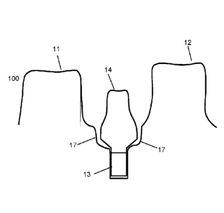
Fig. 1 shows a schematic of a conflict between of volumes of a virtual model
of a restoration and a gingival part of a virtual model of a set of teeth.
In procedures relating to a dental implant and a corresponding dental
restoration, such as the illustrated abutment, the dental technician often
produces a physical model of the set of teeth with an implant analog
positioned in the gingival part of the physical model. In some cases, the
restoration collides with the gingival part of the physical model of the set
of
teeth such that the restoration cannot be inserted in the physical model of
the
set of teeth, i.e. the dental restoration cannot be positioned in the
anatomical
correct position in the physical model of the set of teeth.
A virtual representation of this situation is illustrated in Figure 1, where a
virtual model 10 of the set of teeth shows two teeth 11, 12 and an implant 13
which in the manufactured physical model is replaced by an implant analog
and a first surface 15 of which at least a part relates to the emergence
profile
CA 02827552 2013-08-16
WO 2012/113407 PCT/DK2012/050062
34
of the gingiva. A virtual model of the restoration 14 is inserted in the
virtual
model 10 of the set of teeth in its anatomical correct position. As seen in
the
figure, there is an overlap 16 between the volumes of the virtual model of the
restoration 14 and the gingival part of the virtual model 10 of the set of
teeth.
For a restoration and a physical model of the set of teeth the conflict
represented by the virtual overlap 16 prevents the insertion of the
restoration.
The virtual model of the set of teeth 10 may be generated from one or more
three dimensional representations of the set of teeth provided by e.g.
scanning an impression of the set of teeth or by direct intra-oral scanning
using a handheld scanner, such as the TRIOSTm intra oral-scanner.
The scanning may provide a three dimensional representations in the form of
a point cloud which can be converted to a virtual model of the set of teeth by
e.g. triangulation.
Fig. 2 shows an embodiment of a method according to the invention, wherein
the virtual model of the set of teeth 10 is modified by virtually removing a
portion of the gingival part, such that space for a softer material is
provided in
a physical model manufactured from the modified virtual model of the set of
teeth.
In Fig. 2a, the virtual model 10 of the set of teeth shows two teeth 11, 12
and
an implant analog 13. The virtual model of the restoration 14 is virtually
inserted such that it is arranged in its anatomical correct position. The
overlap 16 prevents the insertion of the restoration in a physical model
corresponding to the virtual model 10 of the set of teeth as described above
in relation to Fig. 1.
The boundaries of a section which is to be modified can be identified using a
first 3D spline 151 and a second 3D spline 152. The 3D splines can be
defined manually be an operator using e.g. a pointing tool, such as a
CA 02827552 2013-08-16
WO 2012/113407 PCT/DK2012/050062
computer mouse, and a computer screen onto which the virtual model 10 of
the set of teeth is visualized. The boundaries can also be derived
automatically using computer implemented algorithms configured for
determining e.g. a preparation line of an abutment.
5
In Fig. 2b, the gingival part of the virtual model 10 of the set of teeth has
been modified such that the section now is shaped according to a second
surface 17, where the second surface 17 is such that the overlap 16 seen in
Fig. 2a is avoided. This corresponds to having virtually removed a portion of
10 the gingival part of the virtual model 10 of the set of teeth, such that
it
changes from having a shape according to the first surface 15 to a shape
according to the second surface 17 in the modified virtual model 100 of the
set of teeth.
In the figure, the second surface 17 has a smooth transition from the first to
15 the second 3D spline. The virtually removed portion may be also defined
by
extending a cylinder to a horizontal plane spanned by the second 3D spline
152, where the cross section of the cylinder is shaped according to the first
3D spline 151.
20 Based on the modified virtual model 100 of the set of teeth, a third
surface 18
may be determined digitally e.g. by defining third and fourth 3D splines on
the
modified virtual model 100. The third and fourth 3D splines are then
connected to define the third surface 18. The 3D third and fourth splines may
be identical to the first and second 3D splines 151, 152 used for identifying
25 the boundaries of the portion which is virtually removed to make space
for
the second gingival region 19. I.e. the region 19 is defined defined by the
second surface 17 and the third surface 18 as illustrated in Fig. 2c. In a
physical model 101 manufactured from the modified virtual model 100 of the
set of teeth a relatively soft, compressible material may be provided in the
30 second gingiva region 19.The softer material portion can be manufactured
by
3D printing and then arranged at the physical model of the set of teeth 101.
CA 02827552 2013-08-16
WO 2012/113407 PCT/DK2012/050062
36
Fig. 2d shows an abutment 140 inserted into the physical model of the set of
teeth 101 with a soft, compressible material in the second gingival region
190.
When the second gingival region 19 is shaped according to the third surface
18, there is still an overlap between the volumes of the virtual model of the
dental restoration and the gingival part of the modified virtual model 100 of
the set of teeth (now having a surface according to the third surface 18),
However, when the second material is sufficiently soft and compressible, the
second gingival region 190 is deformed when the physical restoration 140 is
inserted in the physical model of the set of teeth 100 thus allowing it to be
arranged in its anatomically correct position as illustrated in Fig. 2d.
Fig. 3 shows an embodiment of a method according to the invention, wherein
the virtual model 10 of the set of teeth is modified by virtually removing a
portion of the gingival part.
In Fig. 3a, the virtual model of the set of teeth 10 shows two teeth 11, 12
and
an implant/implant analog 13. The virtual model of the restoration 14 is
virtually inserted in its anatomical correct position. The virtual overlap 16
prevents the insertion of the restoration into a physical model corresponding
to the virtual model 10 of the set of teeth as described above in relation to
Fig. 1.
In Fig. 3b, a portion of the gingiva in the region configured for insertion of
the
restoration has been virtually removed such that the gingiva in the region
now defines a second surface 17. The virtual overlap 16 seen in Fig. 3a is
now avoided and there is space for the virtual model of the restoration 14 at
its anatomically correct position. The second surface 17 can be defined by
offsetting the surface of the virtual model of the restoration 14, such as by
providing a uniform offset as seen in the figure.
CA 02827552 2013-08-16
WO 2012/113407 PCT/DK2012/050062
37
Due to the undercut shape of the second surface 17, it may however not be
possible to insert the restoration in a physical model manufactured from the
virtual model 10 of the set of teeth illustrated in Fig. 3b.
In Figs. 3c and 3d, the insertion direction 20 for the restoration is taken
into
account. The insertion direction can be based on the orientation and position
of the implant/implant analog 13 in the virtual model 10 of the set of teeth.
A
circumference line 21 is defined at the outer circumference of the second
surface 17 when this is viewed along the insertion direction 20. The
circumference line 21 can be determined using computer implemented
algorithms.
A cylinder enclosing an extrusion volume can then be defined by the surface
generated by translating the circumference line 21 along the insertion path 20
away from the implant analog 13. When the extrusion volume is subtracted
from the virtual model 10 of the set of teeth a corrected second surface 171
is provided. Above the circumference line, the corrected second surface 171
differs from the second surface 17 due to said correction.
In a physical model manufactured from the modified virtual model 100 of the
set of teeth, the correction with respect to the insertion direction provides
that
the physical model has no undercuts when viewed along the insertion
direction of the restoration, such that the restoration can be inserted into
this
physical model.
Fig. 4 shows an example of digitally repositioning the the gingiva around a
restoration.
Fig. 4a) shows the virtual model 401 of the set of teeth before a portion of
the
gingiva 425 has been digitally repositioned.
Fig. 4b) shows the virtual model 401 of the set of teeth after a portion of
the
gingiva 425 has been digitally repositioned. After the gingival part 425 has
been moved, the virtual model 401 of the set of teeth can be manufactured.
CA 02827552 2013-08-16
WO 2012/113407 PCT/DK2012/050062
38
When a tooth is prepared in the mouth of the patient, so much of the tooth
may be grinded away, that the soft, compliant gingival tissue around the
prepared tooth will adjoin or follow or collaps to follow the new reduced
shape of the prepared tooth instead of remaining in the original shape
following the original non-prepared tooth. When digitally repositioning,
removing, or relocating the gingival part 425 of the virtual model 401 of the
set of teeth around the restoration 405 then there is space for a restoration
426 and veneering.
The gingival part 425 of the virtual model 401 of the set of teeth is moved
outwards relative to the restoration 405, i.e. away from the restoration, and
it
is moved without changing the size of gingival part 425, only the shape of the
gingival part 425 is changed.
If the virtual model of the restoration 426 is designed using CAD, it can be
derived from the CAD program how much the gingival part 425 on the virtual
model 401 of the set of teeth should be moved in order to fit the modeled
virtual model of the restoration 426.
Fig. 5 shows an embodiment of a method according to the invention, wherein
collision between an extrusion volume and a teeth portion of the virtual model
of the set of teeth is avoided
In Fig 5, the insertion direction 20 for the restoration is taken into account
when modifying the gingival part of the virtual model of the set of teeth. A
circumference line 21 line is defined at a second surface 17 defined by an
offset of the virtual model of the restoration 14. The insertion direction 20
is
determined from the orientation and position of the implant analog 13 in the
virtual model of the set of teeth. In the example of Fig 5, the insertion
direction 20 is tilted relative to the longitudinal axis of the teeth 11, 12.
This
causes the extrusion volume (defined by the insertion direction 20 and the
circumference line 21) to collide with a tooth portion 22 of the virtual model
of
CA 02827552 2013-08-16
WO 2012/113407 PCT/DK2012/050062
39
the set of teeth. The restoration hence cannot be inserted along the insertion
direction 20 into a physical model manufactured from the virtual model of the
set of teeth illustrated in Fig 5a. However, at some distance from the implant
region, the restoration may follow a different path such that the collision
may
be avoided while the path still is aligned with the insertion direction 20 at
the
implant analog. Such an insertion path 23 is illustrated in Fig. 5b.
The insertion path 23 may be derived by combining a first extrusion volume
defined by the circumference line 21 and the insertion direction 20 at the
implant analog and a second extrusion volume defined by an upper
circumference line and an upper insertion direction, where the upper
circumference line may be defined with the corresponding part of the
restoration at the incisal edge of the neighboring teeth.
Fig. 6 describes how the virtual model of the set of teeth can be generated
and modified to provide that the virtual model of the restoration can be
virtually inserted with no overlap between the volume of the restoration and
the volume of the gingival part of the virtual model of the set of teeth.
The starting point of this part of the procedure is where an implant is placed
in the patient's jaw bone and an operator wishes to design a virtual model of
the set of teeth such that a physical model manufactured from the virtual
model of the set of teeth allows an abutment to be inserted. When the
abutment can be inserted in the physical modelt of the teeth a crown
designed for the patient can be arranged at the abutment and the aestical
and functional properties of the designed crown (and abutment) can be
evaluated.
A second three dimensional representation of the set of teeth is obtained by
intra-oral scanning in 601 and a second virtual model of the set of teeth is
generated. A sealing unit may be arranged in the implant during this
scanning, but this sealing unit does not cover the emergence profile of the
CA 02827552 2013-08-16
WO 2012/113407 PCT/DK2012/050062
gingiva in the region. A second virtual model of the set of teeth which
includes the emergence line of the gingival is then generated.
A scan body is then arranged in the implant and a first three dimensional
5 representation of the set of teeth is obtained in a first scanning 602.
The first
and the second scanning use a common reference system such that the data
of the first three dimensional representation can be directly transferred to
the
second virtual model of the set of teeth. Data relating to the parts of the
set of
teeth surrounding the region in which the implant is situated were already
10 obtained in the second scanning so in the first scanning only the region
of the
implant is scanned
With the data from the first scanning transferred to the second virtual model
of the set of teeth, this virtual model now comprises both data relating to
the
15 emergence profile and to the scan body.
A CAD model of the scan body is then aligned 603 with the scan body portion
of this second virtual model. Thereby the postion and orientation of the
implant can be derived and a virtual model with the implant postion and
20 orientation and with the emergence profile is generated 604.
The order at which these two scans are obtained is not important, such that
the first scanning can be performed before the second. If the scan with the
scan body is made initially, the scan body is removed from the implant before
25 the scanning without the scan body is made. The emergence profile can
then
be extracted from the second virtual model (or directly from the second three
dimensional representation of the set of teeth) and transferred to the first
virtual model, such that a virtual model of the set of teeth is generated.
30 In both cases, the generated virtual model of the set of teeth comprises
a
gingival part of the set of teeth, said gingival part comprising a region
CA 02827552 2013-08-16
WO 2012/113407 PCT/DK2012/050062
41
configured for insertion of a restoration with both the emergence profile of
the
gingival and the implant position and orientation.
The first and second scanning can also be of impressions of the patient's set
of teeth using a scan flag to indicate the position and orientation of the
implant.
The antagonist may also be scanned such that the occlusion of the
restoration can be evaluated and taken into account when modelling e.g a
crown for the implant.
The insertion direction of the restoration is determined in step 605.
A virtual model of the restoration for the set of teeth, such as a virtual
model
of an abutment, is obtained in step 606 and virtually aligned with the
generated virtual model of the set of teeth. The virtual model of the
restoration may be designed to the patient's set of teeth by e.g. defining the
margin line using a 3D spline.
When a final design of the restoration is obtained, it is subtracted from the
virtual model of the set of teeth, or a volume corresponding to an offset
surface of the virtual model of the restoration is subtracted from the virtual
model of the set of teeth. An extrusion volume can then be determined based
on a circumference line of the restoration and the insertion direction. The
extrusion volume is then subtracted from the virtual model of the set of teeth
such that the modified virtual model of the set of teeth is privoded 607.
The generated and modified virtual model of the set of teeth is then such that
the virtual model of the dental restoration can be virtually inserted in said
region with no overlap between the volume of the restoration and the volume
of the gingival part. A result of this procedure is that a restoration
CA 02827552 2013-08-16
WO 2012/113407 PCT/DK2012/050062
42
manufactured from the virtual model of the of the restoration, such as an
abutment, can be inserted into a physical model of the set of teeth
manufactured from said modified virtual model of the set of teeth.
Fig. 7 shows how a cover arranged in relation to a physcial model of the
teeth can be used when realizing a second gingival region made of a
relatively soft and compressible material.
The cover 200 comprises a top portion 201 having a model facing surface
202 and a through channel 203. Further the cover 200 has an implant
engaging portion 204, which is configured to fit into the implant analog 130
of
a physical model of a set of teeth 101. When the cover 200 is arranged in
relation to the implant analog 130, the model facing surface 202 encloses a
volume 205 in collaboration with an opposing surface 206 of the physical
model. The through channel 203 provides a liquid connection to the enclosed
volume 205, such that the enclosed volume can be filled with a second
gingival material via the though channel 203. When the enclosed volume 205
is filled with the second gingival material, the second gingival material is
shaped according to the model facing surface 202.
When the cover is removed the second gingival region 206 (corresponding to
the enclosed volume 205) filled with the second gingival material has been
formed at the physical model of the set of teeth 101 as illustrated in Fig.
7b.
The surface 207 of the second gingival region 206 is shaped according to the
model facing surface 202 of the cover.
The surface 208 of the physical model 101 of the set of teeth can be defined
manually by grinding.
If the second gingival material is sufficiently soft and compressible, the
dental
restoration, here examplified by the abutment 140, can be inserted into the
CA 02827552 2013-08-16
WO 2012/113407 PCT/DK2012/050062
43
implant analog 130 be appliying a pressure which deforms the second
gingival material enough to allow the insertion as illustrated in Fig. 7c. The
second material can for example be a dental silicone.
In some embodiments, model facing surface of the cover is shaped
according to the surface of a known abutment. I.e. different covers can be
manufactured based on the surfaces of known abutments, such that the
second gingival section formed by one cover is shaped according to a
corresponding abutment. The requirements to the compressability of the
second gingival material is then less strict and a larger variety of materials
can be used.
Fig. 8 show schematic presentations of the inventive covers according to the
present invention.
Fig 8a. shows an example of a cover 200 with an implant engaging portion
204, and a top portion with a model facing surface 202 and a through
passage. When the cover is arranged relative to a physical model of a set of
teeth, the implant engaging portion 204 is placed in the implant analog and
the lower part 209 of the top portion rests on a gingical part of the physical
model. The length of the implant engaging portion 204 is adapted to provide
that its positoin in the implant analog can be adjusted such that the lower
part
209 encloses a volume in collaboration with the physical model.
Fig 8b. shows an example of a cover 200 where the model facing surface
202 is shaped according to the surface of a known abutment 14. The portion
of the model facing surface 202 nearest to the implant engaging portion 204
is shaped according to the corresponding surface of the abutment 14 or by a
well defined offset of the corresponding surface. When the second gingival
region is formed using this cover, the corresponding abutment will fitt into
the
CA 02827552 2013-08-16
WO 2012/113407 PCT/DK2012/050062
44
physical model even if the second gingival material is relatively
incompressible.
Fig. 9 shows an example of an implant analog configured to be arranged in
the gingival part of a physical model of a set of teeth.
The figure shows a side view of an implant analog 501 arranged in a hole
defined in the gingival part 503 of the physical model. A central 502 volume
entends along the longitudinal diretction of the implant analog from a crown
end towards its distal end 505. At the distal end 505, a stop surface 506 is
configured to have a reduced cross sectional area such that room is left for
rounded corners 507 of the wall of the hole and potentially for excess
material that was not removed from the hole. The rounded corners may be
generated when the drill used to defined the hole has a rounded tip. The
cross sectional shape 504 of the implant analog illustrates one way to ensure
that the implant analog can only be arranged with its correct orientation
relative to the gingival part of the physical model.
A height inspection groove 508 is defined in the implant analog 501 to allow
for visual or a contact based inspection of whether the implant analog 501 is
arranged in the correct postion in the gingival part of the physical model. A
window or a through hole (not illustatrated in the Figure) may be provided in
the ginival part of the model to allow visual and/or physical contact to the
inplant analog from the outside of the model.
The window or through hole may be provided in the virtual modelling of the
gingival part of a set of teeth such that it is formed directly with the
manufature of the physical model or after the manufacturing of the physical
model.
Fig. 10 show a screen shot from an implementation of the inventoin
A virtual model 1080 of the set of teeth with the implant 1081 position and
orientation has been generated using the method of the invention. Also seen
CA 02827552 2013-08-16
WO 2012/113407 PCT/DK2012/050062
in the screen shot is the restoration 1082 (an abutment) which is to be
designed based on the set of teeth.
Figure 6 described some embodiments of the metod for generation such a
the virtual model of the set of teeth.
5
Fig. 11 show screen shots from an implementation of the invention.
The virtual model of the set of teeth 1180 has now been equiped with a base
and connectors for e.g. arranging the manufactured physical model in an
10 articulator. In Fig lla the emergence profile 1190 is seen in the region
configured for the restoration.ln Fig. 11b, the virtual model of the set of
teeth
has been modified to provide that there is no overlap between the virtual
model of the restoration (the abutment) and the virtual of the set of teeth,
such that the manufactured restoration can be inserted in a physical model of
15 the set of teeth manufactured from said generted and modified virtual
model
of the set of teeth.
20 Although some embodiments have been described and shown in detail, the
invention is not restricted to them, but may also be embodied in other ways
within the scope of the subject matter defined in the following claims. In
particular, it is to be understood that other embodiments may be utilised and
structural and functional modifications may be made without departing from
25 the scope of the present invention.
In device claims enumerating several means, several of these means can be
embodied by one and the same item of hardware. The mere fact that certain
measures are recited in mutually different dependent claims or described in
30 different embodiments does not indicate that a combination of these
measures cannot be used to advantage.
CA 02827552 2013-08-16
WO 2012/113407 PCT/DK2012/050062
46
It should be emphasized that the term "comprises/comprising" when used in
this specification is taken to specify the presence of stated features,
integers,
steps or components but does not preclude the presence or addition of one
or more other features, integers, steps, components or groups thereof.
It should be emphasized that the term "according to any of the preceding
claims" may be interpreted as meaning "according to any one or more of the
preceding claims", such that the limitations of one or several dependent
claims may be read into an independent claim.
The features of the method described above and in the following may be
implemented in software and carried out on a data processing system or
other processing means caused by the execution of computer-executable
instructions. The instructions may be program code means loaded in a
memory, such as a RAM, from a storage medium or from another computer
via a computer network. Alternatively, the described features may be
implemented by hardwired circuitry instead of software or in combination with
software.
Items:
1. A method of generating and modifying a virtual model of a set of teeth,
said set of teeth comprising a region configured for insertion of a
restoration,
the region being located in a gingival part of the set of teeth, where the
method comprises:
- obtaining at least one three dimensional representation of the set of teeth;
CA 02827552 2013-08-16
WO 2012/113407 PCT/DK2012/050062
47
- generating a virtual model of the set of teeth from said three
dimensional
representation, where the virtual model of the set of teeth comprises a
gingival part, said gingival part comprising a virtual region corresponding to
said region configured for insertion of a restoration and at least part of the
gingiva surrounding said region;
- obtaining a virtual model of said restoration; and
- modifying the gingival part of the virtual model of the set of teeth such
that
the virtual model of the restoration can be virtually inserted into said
virtual
region with no overlap between the volume of said virtual model of the
restoration and the volume of said gingival part of the virtual model of the
set
of teeth.
2. The method according to item 1, wherein a first three dimensional
representation of the set of teeth is obtained by scanning the patient's set
of
teeth with a scan body arranged in said implant region.
3. The method according to item 1 or 2, where said virtual model of the set of
teeth is generated at least in part from said first three dimensional
representation.
4. The method according to any of the preceding items, wherein a second
three dimensional representation of the set of teeth is obtained by scanning
the patient's set of teeth with the emergence profile at said implant region
being visible.
5. The method according to any of the preceding items, where said virtual
model of the set of teeth is generated at least in part from said second three
dimensional representation.
CA 02827552 2013-08-16
WO 2012/113407 PCT/DK2012/050062
48
6. The method according to any of the preceding items, wherein one of said
first or second three dimensional representation of the set of teeth is
obtained
by scanning a relatively larger section of the patient's set of teeth, and the
other of said first or second three dimensional representation then is
obtained
by scanning a relatively smaller section around the implant region.
7. The method according to any of the preceding items, wherein the method
comprises generating a first virtual model of the set of teeth from said first
three dimensional representation of the set of teeth.
8. The method according to any of the preceding items, wherein the method
comprises generating a second virtual model of the set of teeth from said
second three dimensional representation of the set of teeth.
9. The method according to any of the preceding items, wherein the method
comprises combining the first and second virtual models of the set of teeth to
generate said virtual model of the set of teeth.
10. The method according to any of the preceding items, wherein a virtual
model of the scan body is provided and virtually aligned with the first
virtual
model of the set of teeth to determine the orientation and position of the
implant.
11. The method according to any of the preceding items, where the
restoration is designed based on the virtual model of the set of teeth.
12. The method according to any of the preceding items, where the
restoration is a pre-manufactured restoration such as a pre-manufactured
abutment.
CA 02827552 2013-08-16
WO 2012/113407 PCT/DK2012/050062
49
13. The method according to any of the preceding items, where modified
virtual model of the set of teeth is for manufacturing a physical model of the
set of teeth.
14. The method according to any of the preceding items, where at least a
sub-gingival part of the virtual model of the restoration is configured to
have
the shape of an anatomically correct restoration.
15. The method according to any of the preceding items, where the gingival
part of the generated virtual model of the set of teeth directly provides that
the volume of the restoration and the volume of the gingival part do not
overlap.
16. The method according to any of the preceding items, where the virtual
model of the set of teeth is generated in one step and where the gingival part
of the virtual model subsequently is modified to provide that the volume of
the
restoration and the volume of the gingival part do not overlap.
17. The method according to any of the preceding items, wherein the gingival
part of the virtual model of the set of teeth is modified to provide that the
adjoining surfaces of the virtual model of the restoration and the gingival
part
of the virtual model of the set of teeth follows each other.
18. The method according to any of the preceding items, wherein an offset is
provided between the adjoining surfaces of the virtual model of the
restoration and the gingival part of the virtual model of the set of teeth.
19. The method according to any of the preceding items, wherein the method
comprises determining in insertion path for the restoration.
CA 02827552 2013-08-16
WO 2012/113407 PCT/DK2012/050062
20. The method according to any of the preceding items, wherein the
insertion path is according to the insertion direction at the implant.
21. The method according to any of the preceding items, wherein the method
5 comprises identifying a circumference line for the restoration.
22. The method according to any of the preceding items, wherein the
circumference line is defined as the outer circumference of the restoration
when the virtual model of the restoration is viewed along the insertion
10 direction.
23. The method according to any of the preceding items, wherein the
circumference line is defined as the outer circumference of the offset surface
when the offset surface is viewed along the insertion direction.
24. The method according to any of the preceding items, wherein an
extrusion volume is defined by the insertion direction and the circumference
line.
25. The method according to any of the preceding items, wherein an
extrusion volume is defined by the insertion path and the circumference line.
26. The method according to any of the preceding items, wherein modifying
the gingival part of the virtual model of the set of teeth comprises digitally
cutting a portion of the gingiva away such that the volume of the restoration
and the volume of the gingiva do not overlap.
27. A method of generating and modifying a virtual model of a set of teeth for
manufacturing a physical model of the set of teeth, where the method
comprises:
CA 02827552 2013-08-16
WO 2012/113407 PCT/DK2012/050062
51
- obtaining a three dimensional representation of the set of teeth;
- generating a virtual model of the set of teeth from said three
dimensional
representation, the virtual model of the set of teeth comprising a gingival
part
comprising a gingiva; and
- modifying the gingival part to enable an insertion of a restoration in a
region
of the virtual model configured for insertion of a restoration.
28. The method according to any of the preceding items, wherein modifying
the gingival part comprises modifying the gingival part of the virtual model
generated from the three dimensional representation.
29. The method according to any of the preceding items, wherein modifying
the gingival part comprises configuring the material of the gingival part at
the
restoration to be sufficiently soft such that a restoration may deform the
gingival part.
30. The method according to any of the preceding items, wherein modifying
the gingival part comprises virtually removing a portion of said gingival part
in
the region configured for insertion of a restoration.
31. The method according to any of the previous items, wherein the method
comprises virtually adding material to the gingival part of the virtual model
in
the region configured for insertion of a restoration.
32. The method according to any of the preceding items, wherein the method
comprises digitally repositioning the gingival part of the model around the
restoration, before manufacturing the model.
CA 02827552 2013-08-16
WO 2012/113407 PCT/DK2012/050062
52
33. The method according to any of the preceding items, wherein digitally
repositioning the gingival part of the virtual model of the set of teeth
comprises digitally moving gingiva away from the virtual model of the
restoration.
34. The method according to any of the previous items, wherein the gingival
part of the virtual model of the set of teeth defines a first surface at the
region
configured for insertion of a restoration.
35. The method according to any of the preceding items, wherein the first
surface follows at least a section of said emergence profile.
36. The method according to any of the preceding items, wherein modifying
the virtual model of the set of teeth comprises replacing said first surface
by a
second surface, where said second surface is shaped such that the virtual
model of said restoration can be virtually arranged in said virtual implant
region with no overlap with the modified virtual model of the set of teeth.
37. The method according to any of the previous items, wherein the gingival
part of the virtual model of the set of teeth after the virtual removal of a
portion of the gingiva defines the second surface at the region configured for
insertion of a restoration.
38. The method according to any of the preceding items, wherein at least a
section of said second surface is defined by offsetting part of the surface of
the virtual model of the restoration.
39. The method according to any of the preceding items, wherein the method
comprises subtracting the virtual model of the restoration or the volume
enclosed by the offset surface from the virtual model of the set of teeth.
CA 02827552 2013-08-16
WO 2012/113407 PCT/DK2012/050062
53
40. The method according to any of the previous items, wherein the gingival
part of the virtual model of the set of teeth after virtually adding material
to
the gingiva defines a third surface at the region configured for insertion of
a
restoration.
41. The method according to any of the previous items, wherein the gingival
part of the virtual model of the set of teeth is divided into a first and a
second
gingival region by the second surface
42. The method according to any of the previous items, where said second
gingival region is arranged between the second surface and the third surface,
the second surface forming an interface between the first and the second
gingival region.
43. The method according to any of the previous items, wherein said third
surface is substantially identical to said first surface.
44. The method according to any of the previous items, wherein said first
gingival region is configured to be manufactured in a first material in a
physical model manufactured from the virtual model.
45. The method according to any of the previous items, wherein said second
gingival region is configured to be manufactured in a second material in a
physical model manufactured from the virtual model.
46. The method according to any of the preceding items, wherein said
second material is configured to be softer than said first material at ambient
conditions.
47. The method according to any of the preceding items, wherein the virtual
representation of the set of teeth is provided by scanning the set of teeth,
CA 02827552 2013-08-16
WO 2012/113407 PCT/DK2012/050062
54
such as by scanning the set of teeth by means of an intra-oral scanner or by
scanning an impression of the set of teeth.
48. The method according to any of the preceding items, wherein the
restoration comprises a full restoration or a part of a restoration, such as
an
abutment or a crown arranged on said abutment, an implant bar, or in
principle any other indication used in relation to dental restorations.
49. The method according to any of the preceding items, wherein modifying
the gingival part of the virtual model of the set of teeth comprises digitally
cutting a portion of the gingiva away such that the volume of the restoration
and the volume of the gingival part do not overlap.
50. The method according to any of the preceding items, wherein said
second material is configured to be comprised in a removable unit in a
physical model manufactured from the virtual model.
51. The method according to any of the preceding itemsõ wherein the
addition of material to the gingival part of the model comprises generating a
gingiva mask.
52. The method according to any of the preceding items, wherein the gingival
part after being configured to enable the correct positioning of a restoration
is
configured to follow the adjoining surface of the restoration.
53. The method according to any of the preceding items, wherein a void is
provided between the adjoining surfaces of the restoration and the gingival
part of the model.
54. The method according to any of items 51 to 53, wherein said gingiva
mask comprises a first retention structure configured to mate with a second
CA 02827552 2013-08-16
WO 2012/113407 PCT/DK2012/050062
retention structure arranged on the gingival part of the model, such that the
gingiva mask is arranged anatomically correct when said first and second
retention structures mate.
5 55. The method according to any of the preceding items, wherein the
gingival
part of the model comprises an undercut region, in which said second
gingival region is partly confined.
56. The method according to any of items 28 to 33, wherein the method
10 comprises configuring the gingiva mask to comprise an opening, where the
opening is configured to allow a restoration to access the gingival part
arranged below the gingiva mask.
57. The method according to any of the preceding items, wherein a virtual
15 hole is provided in said gingival part of the virtual model of the set
of teeth,
where said virtual hole is such that a corresponding hole in the physical
model of the set of teeth is configured to mate with a part of said
restoration
configured to fit into the gingival part of the physical model of the set of
teeth.
20 58: The method according to item 57, wherein said virtual hole is
configured
to allow an implant analog to be inserted manually in the corresponding hole
of the physical model of the set of teeth.
59. The method according to item 57 or 58, wherein said virtual hole and/or
25 said implant analog is configured such that said the implant analog can
be
inserted only in the correct anatomical position and orientation in the
gingival
part of the model.
60. The method according to any of items 57 to 59, wherein said implant
30 analog is configured to have a shape with reduced cross sectional
rotation
CA 02827552 2013-08-16
WO 2012/113407 PCT/DK2012/050062
56
symmetry, such as an N-fold symmetry, wherein N is an integer number
below 25
61. The method according to any of the preceding items, wherein several
steps in method are computer implemented.
62. The method according to any of the preceding items, wherein the gingival
part of the virtual model of the set of teeth, is configured to provide that a
corresponding ejection hole in the physical model of the set of teeth is in
fluid
connection with said hole such that a restoration or an implant analog can be
accessed through said ejection hole to be ejected from the gingival part of
the physical model of the set of teeth.
63. The method according to any of the preceding items, wherein the implant
analog is configured to comprise a stop section with a smaller cross sectional
area at its distal end, said stop section preferably being arranged centrally
around the longitudinal axis of the implant analog.
64. The method according to any of the preceding items, wherein the virtual
hole defined in the gingival part of the virtual model of the set of teeth is
configured to provide that the corresponding hole in the physical model of the
set of teeth has rounded edges at its distal end.
65. The method according to any of the preceding items, wherein the method
comprises providing a cover which in cooperation with the first gingival
region
is configured to enclose the second gingival region.
66. The method according to item 65, wherein the cover comprises an
opening configured to allow the injection of said second material into said
second gingival region in a physical model manufactured from the virtual
model of the teeth.
CA 02827552 2013-08-16
WO 2012/113407 PCT/DK2012/050062
57
67. The method according to item 65 or 66, wherein a surface of the cover
facing said second gingival region is shaped as said third surface.
68. The method according to any of the preceding items, wherein the method
comprises designing and configuring the model to be manufactured by
means of a specific manufacturing process.
69. A method of generating a virtual model of a set of teeth for manufacturing
a physical model of the set of teeth, where the method comprises:
- obtaining a virtual model of the set of teeth, the model comprising a
gingival
part comprising a gingiva; and
- obtaining a virtual model of a restoration configured to be arranged in its
anatomical correct position relative to said gingival part of the model;
where the surface of the gingiva defines a first surface at said restoration;
and
- modifying the gingiva at said restoration such that the surface of the
modifying gingiva defines a second surface at said restoration, wherein the
second surface is configured to avoid an overlap between the volume of the
restoration and the volume of the gingival part of the model.
70. A method of adjusting a virtual model of a set of teeth, where the virtual
model of the set of teeth is for manufacturing a physical model of the set of
teeth, where the method comprises:
- obtaining a pre-adjustment configuration of a virtual model of the set of
teeth, the virtual model of the set of teeth comprising a gingival part; and
- obtaining a virtual model of a restoration configured to be arranged in
its
anatomical correct position relative to said gingival part of the model,
CA 02827552 2013-08-16
WO 2012/113407 PCT/DK2012/050062
58
where the volume of the gingival part of the virtual model of the set of teeth
and the volume of the virtual model of the restoration overlaps when the
restoration is arranged in the anatomical correct position; and
- adjusting a portion of the gingival part of the virtual model of the set
of teeth
arranged at said restoration providing a post-adjustment configuration of the
virtual model of the set of teeth, in which post-adjustment configuration the
gingival part of the model is configured to avoid overlap between the volume
of the virtual model of the restoration and the volume of the gingival part of
the virtual model of the set of teeth.
71. The method according to item 70, wherein adjusting of the gingival part
comprises configuring the shape of the gingival part such that the overlap
between the volumes is avoided.
72. The method according to item 71 or 72, wherein the method comprises
configuring the material of the gingiva at the restoration to be sufficiently
soft
such that a restoration may deform the gingival part.
73. A method of generating a physical model of a set of teeth, where the
method comprises:
- obtaining at least one three dimensional representation of the set of
teeth;
- generating and modifying a virtual model of the set of teeth from said at
least one three dimensional representation, the virtual model of the set of
teeth comprising a gingival part; and
- modifying the gingival part to enable insertion of a restoration in a
region of
the virtual model of the set of teeth configured for insertion of a
restoration.
CA 02827552 2013-08-16
WO 2012/113407 PCT/DK2012/050062
59
- manufacturing said physical model from said virtual model of the set of
teeth.
74. The method according to any of the preceding items, wherein the method
comprises manufacturing the physical model by means of three dimensional
printing or milling.
75. The method according to item 73 or 74, wherein the physical model may
be manufactured using a casting mold for casting an at least partly soft
mould as part of the physical model of the set of teeth.
76. The method according to item 75, wherein a casting mold CAD model is
generated as an impression of at least a part of the virtual model, said
casting mold CAD model thereby comprising the negative geometry of the
set of teeth.
77. The method according to any of items 73 to 76, wherein said second
material is softer and more compressible than said first material at ambient
conditions.
78. A physical model of a set of teeth, wherein the physical model is
manufactured from a virtual model of the set of teeth generated and modified
by the method according to any of items 1-77.
79. An ejection tool for ejecting a restoration according to any of the
preceding items, when the restoration is arranged in a physical model of a
set of teeth according to any of the preceding items.
CA 02827552 2013-08-16
WO 2012/113407 PCT/DK2012/050062
80. The ejection tool according to item 79 comprising an elongated
component which is adapted to fit into a through hole in the gingival part of
the model.
5 81. The ejection tool according to item 79 or 80, comprising an elongated
component which is adapted to fit into a blind hole and/or a through hole in
the restoration.