Note: Descriptions are shown in the official language in which they were submitted.
SHROUDED SENSOR CLIP ASSEMBLY AND BLOOD CHAMBER FOR AN
OPTICAL BLOOD MONITORING SYSTEM
RELATED APPLICATIONS
[0001] This application claims priority to U.S. Provisional Application No.
61/553,078, filed October 28, 2011. This application also claims priority to
U.S.
Application No. 13/034,788, filed February 25, 2011. '
TECHNICAL FIELD
[0002] The disclosure generally relates to optical blood monitoring systems
used to
monitor extracorporeal patient blood flow and take real-time measurement of
hematocrit,
oxygen saturation levels and/or other blood constituents. The disclosure more
particularly
is directed to improving the noise immunity of such systems.
BACKGROUND
[0003] Patients with kidney failure or partial kidney failure typically
undergo
hemodialysis treatment in order to remove toxins and excess fluids from their
blood. To
do this, blood is taken from a patient through an intake needle or catheter
which draws
blood from an artery or vein located in a specifically accepted access
location - - e.g., a
shunt surgically placed in an arm, thigh, subclavian and the like. The needle
or catheter is
connected to extracorporeal tubing that is fed to a peristaltic pump and then
to a dialyzer
that cleans the blood and removes excess fluid. The cleaned blood is then
returned to the
patient through additional extracorporeal tubing and another needle or
catheter.
Sometimes, a heparin drip is located in the hemodialysis loop to prevent the
blood from
coagulating.
[0004] As the drawn blood passes through the dialyzer, it travels in straw-
like tubes
within the dialyzer that serve as semi-permeable passageways for the unclean
blood. Fresh
dialysate solution enters the dialyzer at its downstream end. The dialysate
surrounds the
straw-like tubes and flows through the dialyzer in the opposite direction of
the blood
flowing through the tubes. Fresh dialysate collects toxins passing through the
straw-like
tubes by diffusion and excess fluids in the blood by ultra filtration.
Dialysate containing
the removed toxins and excess fluids is disposed of as waste. The red cells
remain in the
straw-like tubes and their volume count is unaffected by the process.
CA 2828293 2017-07-26
CA 02828293 2013-08-23
WO 2012/116336
PCT/US2012/026637
2
[0005] A blood monitoring system is often used during hemodialysis
treatment or
other treatments involving extracorporeal blood flow. One example is the CRIT-
LINE
monitoring system sold by Fresenius USA Manufacturing, Inc. of Waltham, MA.
The
CRIT-LINE blood monitoring system uses optical techniques to non-invasively
measure
in real-time the hematocrit and the oxygen saturation level of blood flowing
through the
hemodialysis system. The blood monitoring system measures the blood at a
sterile blood
chamber attached in-line to the extracorporeal tubing, typically on the
arterial side of the
dialyzer.
[0006] In general, blood chambers along with the tube set and dialyzer are
replaced for
each patient. The blood chamber is intended for a single use. The blood
chamber defines
an internal blood flow cavity comprising a substantially flat viewing region
and two
opposing viewing lenses. LED emitters and photodeteetors for the optical blood
monitor
are clipped into place onto the blood chamber over the lenses. Multiple
wavelengths of
light may be directed through the blood chamber and the patient's blood
flowing through
the chamber with a photodetector detecting the resulting intensity of each
wavelength.
[0007] The preferred wavelengths to measure hematocrit are about 810 nm,
which is
substantially isobestic for red blood cells, and about 1300 nm, which is
substantially
isobestic for water. A ratiometric technique implemented in the CRIT-LINE
controller,
substantially as disclosed in U.S. Patent No. 5,372,136 entitled "System and
Method for
Non-Invasive Hematocrit Monitoring," which issued on December 13, 1999 and is
assigned to the assignee of the present application, uses this light intensity
information to
calculate the patient's hematocrit value in real-time. The hematocrit value,
as is widely
used in the art, is a percentage determined by the ratio between (1) the
volume of the red
blood cells in a given whole blood sample and (2) the overall volume of the
blood sample.
[0008] In a clinical setting, the actual percentage change in blood volume
occurring
during hemodialysis can be determined, in real-time, from the change in the
measured
hematocrit. Thus, an optical blood monitor is able to non-invasively monitor
not only the
patient's hematocrit level but also the change in the patient's blood volume
in real-time
during a hemodialysis treatment session. The ability to monitor real-time
change in blood
volume helps facilitate safe, effective hemodialysis.
[0009] To monitor blood in real time, light emitting diodes (LEDs) and
photodetectors
for them are mounted on two opposing heads of a sensor clip assembly that fit
over the
CA 02828293 2013-08-23
WO 2012/116336
PCT/US2012/026637
3
blood chamber. For accuracy of the system, it is important that the LEDs and
the
photodetectors be located in a predetermined position and orientation each
time the sensor
clip assembly is clipped into place over the blood chamber. The predetermined
position
and orientation ensures that light traveling from the LEDs to the
photodetectors travels
through a lens of the blood chamber.
[0010] The optical monitor is calibrated for the specific dimensions of the
blood
chamber and the specific position and orientation of the sensor clip assembly
with respect
to the blood chamber. For this purpose, the heads of the sensor clips are
designed to mate
to the blood chamber so that the LEDs and the photodetectors are at a known
position and
orientation. In the CRIT-LINE monitoring system, the head of the sensor clips
and the
blood chamber have complementary D-shaped configurations.
[0011] Under certain conditions, unwanted light can mix with the light
traveling
directly from the LEDs, through the blood in the chamber and into the
photodetectors,
causing inaccuracies in the measured hematocrit and/or oxygen saturation
levels. Signal
processing techniques remedy most of the issues pertaining to ambient light
under most
conditions. In addition, the blood chambers may include a "moat" around the
lens area,
which allows blood to flow around the area of the lens as well as through it.
This moat
fills with blood and under most conditions effectively blocks unwanted light
from reaching
the photodetectors on the sensor clip assembly. The effectiveness of the moat
depends on
many factors including the condition of the patient's blood and the wavelength
spectrum of
the unwanted light. For example, when a patient displays very low hematocrit
values, the
ability of the moat to effectively block unwanted light is compromised,
allowing greater
amounts of noise to enter into the signal measured by the photodetectors.
SUMMARY
[0012] A primary objective of the disclosure is to facilitate the accurate
measurement
of hematocrit and oxygen saturation levels over the expected full dynamic
range of their
values, and to particularly maintain highly accurate values when the blood
being measured
is characterized by low hematocrit levels.
[0013] In one aspect of the disclosure, a body of a blood chamber is at
least partially
made of material opaque to a wavelength region of a light beam associated with
the blood
chamber to calculate oxygen saturation levels of blood flowing through the
blood
chamber, thereby attenuating unwanted light (noise) and effectively preventing
it from
CA 02828293 2013-08-23
WO 2012/116336
PCT/US2012/026637
4
mixing with the measurement light beam. Attenuating unwanted light (e.g.,
light ducting
through the chamber body) increases the range of accurate measurement of
oxygen
saturation levels and particularly improves the accuracy of the measurements
at low levels
of oxygen saturation that commonly are present when patients have low
hematocrit values.
[0014] To further protect against the injection of noise into the
measurement light
beam, another aspect of the disclosure shrouds photoemitters and
photodetectors that
create and detect the light beam in order to prevent ambient light from
strongly mixing
with the measurement beam. The shroud is an integral part of either the blood
chamber
and/or a sensor clip assembly that generates and detects the light beam. In
the illustrated
embodiment, the shroud is part of the sensor clip assembly and a free end of
each shroud is
received by an annular receptacle in the blood chamber. But the shroud may
alternatively
be part of the blood chamber and the receptacle part of the clip. Either way,
each shroud
encircles and extends between a window of the blood chamber and a lens of the
photoemitters or the photodetectors. By extending each shroud beyond the lens
in the head
of the sensor clip assembly, the shroud effectively isolates optical noise
sourcing from
both ambient light and indirect light from the photoemitters resulting from
ducting of the
photoemitters' light by areas of the chamber outside of the window.
[0015] Another aspect of the disclosure enables the shrouds to fasten the
sensor clip
assembly to the blood chamber, thereby providing an optically robust and
mechanically
secure interface between the chamber and clip. To fasten the clip and blood
chamber, the
illustrated embodiment includes a spring in the clip so the blood chamber is
gripped
between the two heads of the clip. When mated with the shrouds, annular
receptacles in
the blood chamber align the photoemitters and the photodetectors with the
windows of the
blood chamber. To prevent relative rotation between them, the interface
between the clip
and the chamber includes a mechanical stop. In the illustrated embodiment, the
blood
chamber includes one or more anti-rotation tabs that are received by
complementary anti-
rotation slots in the housing to prevent relative rotation. The anti-rotation
tabs and mating,
complementary slots may take on any reasonable geometric shape. Furthermore,
other
types of fasteners or rotational stops may work equally well and are
contemplated. For
example, either the blood chamber or the clip can include alignment posts that
guide the
blood chamber and clip into proper engagement to both register the LEDs and
photodetectors with the lenses and prevent rotation.
CA 02828293 2013-08-23
WO 2012/116336
PCT/US2012/026637
100161 Other advantages and features of the disclosure may be apparent to
those
skilled in the art upon reviewing the drawings and the following description
thereof.
BRIEF DESCRIPTION OF THE DRAWINGS
[0017] Fig. 1 is a perspective view of a patient undergoing hemodialysis
treatment
with a non-invasive, optical blood monitor monitoring the patients blood in
real-time as it
passes through extracorporeal tubing in the hemodialysis system utilizing a
conventional
blood chamber and sensor clip assembly.
[0018] Fig. 2A is a perspective view of the sensor clip assembly and blood
chamber of
Fig. 1.
[0019] Fig. 2B is the perspective view of Fig. 2A with the a blood chamber
removed
from the clip assembly in order to better view the blood chamber;
[0020] Fig. 3A is an isolated view of the blood chamber shown in Figs. 1
and 2A.
[0021] Fig. 3B is an isolated view of the sensor clip assembly shown in
Figs. 1 and
2A.
[0022] Fig. 4A is a longitudinal, cross-sectional view of the blood chamber
and sensor
clip assembly taken along line 4-4 in Fig. 2A.
[0023] Fig. 4B is the cross-sectional view of the blood chamber and sensor
clip
assembly in Fig. 4A with the clip assembly exploded away from the blood
chamber.
[0024] Fig. 5A is a schematic drawing illustrating the detection of light
and infrared
light at various wavelengths through the blood chamber in order to monitor the
hematocrit
and oxygen saturation of the blood passing through the blood chamber.
[0025] Fig. 5B is a schematic drawing similar to Fig. 5A further
illustrating the effect
of ambient or ducted light that does not pass through the direct path through
the blood in
the blood flow chamber.
[0026] Fig. 6 is a front elevational view of the controller for the optical
blood monitor
illustrating data including real-time hematocrit (HCT), change in blood volume
(BVA),
hemoglobin (HBG), and oxygen saturation (SAT) levels, as well as the amount of
time
into the hemodialysis treatment session and a graphical representation of the
change in
blood volume during the course of the hemodialysis treatment session.
[0027] Fig. 7 is a perspective view of a blood chamber constructed in
accordance with
the first embodiment.
CA 02828293 2013-08-23
WO 2012/116336
PCT/US2012/026637
6
[0028] Fig. 8 is a view similar to Fig. 7 showing a window body exploded
away from a
chamber body.
[0029] Fig. 9 is a perspective view of the back side of the blood chamber
shown in
Figs. 7 and 8.
[0030] Fig. 10 is a longitudinal sectional view taken along line 10-10 in
Fig. 7.
[0031] Fig. 11 is a sectional view taken along line 11-11 in Fig. 10.
[0032] Fig. 12 is a view similar to Fig. 8 illustrating a second
embodiment.
[0033] Fig. 13 is a longitudinal sectional view of the second embodiment.
[0034] Fig. 14 is a sectional view taken along line 14-14 in Fig. 13.
[0035] Fig. 15 is a perspective view of one side of a blood chamber
constructed in
accordance with a third embodiment.
[0036] Fig. 16 is a perspective view of the other side of the blood chamber
shown in
Fig. 15.
[0037] Fig. 17 is a front elevation view of the blood chamber shown in Fig.
15.
[0038] Fig. 18 is a longitudinal, cross-sectional view taken along line 18-
18 in Fig. 15.
[0039] Fig. 19 is a perspective view of an alternative embodiment of the
sensor clip
assembly for mating to the third embodiment of the blood chamber.
[0040] Fig. 20A is the perspective view of the sensor clip assembly of Fig.
19 fastened
to the blood chamber of Figs. 15-18.
[0041] Fig. 20B is the perspective view of the sensor clip assembly and the
blood
chamber in Fig. 20A with the sensor clip assembly exploded away from the blood
chamber
to enable a better view of the surfaces of the assembly and chamber that
engage when the
two are fastened together.
[0042] Fig. 21A is a longitudinal, cross-sectional view of the sensor clip
assembly
fastened to the blood chamber taken along the line 21-21 in Fig. 20A.
[0043] Fig. 21B is the longitudinal sectional view of Fig. 21A with the
sensor clip
assembly exploded away from the blood chamber.
[0044] Fig. 22 illustrates an alternative arrangement for fastening the
blood chamber to
the sensor clip assembly.
[0045] Fig. 23 is a perspective view of the blood chamber in Fig. 22
showing one of
the faces of the chamber that mates to an arm of the sensor clip assembly.
CA 02828293 2013-08-23
WO 2012/116336
PCT/US2012/026637
7
[0046] Fig. 24 is a perspective view of the blood chamber in Fig. 22
showing the other
face of the chamber that mates with the other arm of the sensor clip assembly.
[0047] Fig. 25A illustrates a perspective view of a still further
alternative embodiment
of the blood chamber.
[0048] Fig. 25B is a top elevation view of the blood chamber of Fig. 25A.
[0049] Fig. 25C is a side elevation view of the blood chamber of Fig. 25A.
[0050] Fig. 25D is a longitudinal cross-sectional view of the blood chamber
of Fig.
25A.
[0051] Fig. 25E is an exploded perspective view of the blood chamber of
Fig. 25A.
[0052] Fig. 26A is a perspective view of the main body portion of the blood
chamber
of Figs. 25A-E.
[0053] Fig. 26B is a top elevation view of the main body portion of the
blood chamber
of Fig. 26A.
[0054] Fig. 27A is a perspective view of one of the two opposing lenses of
the blood
chamber of Figs. 25A-E.
[0055] Fig. 27B is a bottom elevation view of the lens of Fig. 27A.
[0056] Fig. 28A is a side elevation view of the connector of the blood
chamber of Figs.
25A-E.
[0057] Fig. 28B is a top elevation view of the connector of Fig. 28A.
[0058] Fig. 29A is an exploded perspective view of the sensor clip assembly
and the
blood chamber of Figs. 25A-E.
[0059] Fig. 29B is an elevation view of the shroud-mating surface of one
arm of the
sensor clip assembly of Fig. 29A.
[0060] Fig. 29C is a perspective view of the sensor clip assembly attached
to the blood
chamber of Figs. 25A-E.
[0061] Fig. 29D is a longitudinal cross-sectional view of the blood chamber
of Figs.
25A-E when attached to the sensor clip assembly of Fig. 29A.
[0062] Fig. 29E is a transverse cross-sectional view of the blood chamber
of Figs.
25A-E when attached to the sensor clip assembly of Fig. 29A.
CA 02828293 2013-08-23
WO 2012/116336
PCT/US2012/026637
8
DETAILED DESCRIPTION
[0063] Fig. 1 illustrates a patient 10 undergoing hemodialysis treatment
with a
conventional hemodialysis system 12, and also illustrates a non-invasive,
optical blood
monitor 14. A typical hemodialysis clinic will have several hemodialysis
systems 12 for
treating patients.
[0064] An input needle or catheter 16 is inserted into an access site of
the patient 10,
such as shunt in the arm, and is connected to extracorporeal tubing 18 that
leads to a
peristaltic pump 20 and then to a dialyzer or blood filter 22. The dialyzer 22
removes
toxins and excess fluid from the patient's blood. The dialysized blood is
returned from the
dialyzer 22 to the patient through extracorporeal tubing 24 and a return
needle or catheter
26. The extracorporeal blood flow in the United States generally receives a
heparin drip to
prevent clotting although that is not shown in Fig. 1. Excess fluids and
toxins are removed
by clean dialysatc liquid which is supplied to the dialyzer 22 via tube 28 and
removed for
disposal via tube 30. A typical hemodialysis treatment session in the United
States takes
about 3 to 5 hours. In a typical hemodialysis treatment as described in Fig.
1, the access
site draws arterial blood from the patient. If no arterial access is available
then a venous
catheter may be used to access the patient's blood. As mentioned, other
dialysis
applications such as low flow Continuous Renal Replacement Therapy (CRRT)
sometimes
used in the Intensive Care Unit and high-flow perfusion measurements during
cardiac
surgery also measure blood from the patient. Current art indicates that oxygen
saturation
levels in venous blood correlate to the cardiac output for the patient. The
typical blood
monitor 14 shown in Fig. 1 can be used in these other applications as well.
[0065] The optical blood monitor 14 includes a blood chamber 32, a sensor
clip
assembly 34, and a controller 35. The blood chamber 32 is typically located in
line with
the extracorporeal tubing 18 upstream of the dialyzer 22. Blood from the
peristaltic pump
20 flows through the tubing 18 into the blood chamber 32. The preferred sensor
assembly
34 includes LED photoemitters that emit light at substantially 810 nin, which
is isobestic
for red blood cells, substantially 1300 nm, which is isobestic for water, and
at substantially
660 nm, which is sensitive for oxygenated hemoglobin. The blood chamber 32
includes
windows so that the sensor emitters and detector(s) can view the blood flowing
through
the blood chamber 32, and determine the patient's real-time hematocrit value
and oxygen
saturation value using known ratiometric techniques.
CA 02828293 2013-08-23
WO 2012/116336
PCT/US2012/026637
9
[0066] Referring now to Figs. 2A-4B, the body of a conventional blood chamber
32 is
made of molded, medical grade, clear polycarbonate. It includes a raised,
stepped rim 33
having a double-D configuration. It also includes two viewing windows 36, 38
(see Fig.
4A). In the illustrated embodiments described hereinafter, the inlet 40 and
outlet 42 of the
blood chamber are designed to be compatible with standard medical industry
connecting
devices, conventionally known as luer lock connectors. Alternatively, one or
both of the
inlet 40 and outlet 42 may be configured to include an opening that accepts
the outer
circumference of the tubing 30.
[0067] In the blood chamber 32 shown in Figs. 2A-4B, the inlet 40 is
integrally
molded with the blood chamber 32, whereas the outlet 42 consists of a suitable
off-the-
shelf connection adapter bonded to the body of the blood chamber 32. The
adapter is
preferably a non-latex and non-phtalate material such as AM287T tri-octyl-
trimellithate.
Bonding of the adapter to provide the outlet 42 is accomplished using a mixed
solvent
cocktail of 50% methyl ethyl ketone and 50% Cyclohexanone. Bonding the adapter
to the
body of the blood chamber must be done carefully to avoid getting the solvent
in the cavity
of the blood chamber. Exposure of the internal cavity of the blood chamber to
the solvent
will crack the body of the chamber.
[0068] The sensor assembly 34 includes emitter and detector subassemblies
44 and 46,
respectively. Referring to Figs. 3B and 4A, each of the emitter and detector
subassemblies
44 and 46 has a head portion 47 and 45, respectively. Referring to the cross
sectional view
in Fig. 4A, each side of the heads 45, 47 provides an opening into which the
molded
diffusion lenses 50, 54 are mounted. The sensor assembly 34 is a spring-loaded
clip
assembly adapted to be removably mounted to the blood chamber 32, as shown in
Fig. 2A.
[0069] Each of the heads 47 and 45 has a double-D configuration
complementing the
double-D configuration of the blood chamber 32 as best seen in Fig. 3A. The
interlocking
double-D configuration fixes the sensor clip 34 in a predetermined position
both laterally
and rotationally when it is fastened to the blood chamber 32 as illustrated in
Fig. 2A.
Fixing the sensor clip 34 in a predetermined position significantly reduces
system noise
induced by an otherwise freely rotating sensor on the blood chamber. As will
be
appreciated from the several embodiments illustrated hereinafter, various
mechanical
arrangements are contemplated for fixing the sensor clip assembly 34 onto the
blood
CA 02828293 2013-08-23
WO 2012/116336
PCT/US2012/026637
to
chamber so as to correctly position the photoemitters and photodetectors with
respect to
viewing windows of the blood chamber.
[0070] One side of the blood chamber 32 is molded to have a double-D
configuration
that complements and receives the double-D configuration of head 45 of the
sensor
assembly 34 in order to fix the assembly in a predetermined position with
respect to the
blood chamber 32 when the assembly and blood chamber are fastened together. As
mentioned, blood chamber 32 is a single-use clear polycarbonate component.
Between
patient treatments, the blood chamber 32 is replaced along with the
extracorporeal tubing
18, 24, and blood filter 22.
[0071] As best shown in Figs. 4A and 4B, an emitter circuit board 48
containing LEDs
emitting light at substantially 660 nm, 810 nm and 1300 nm is mounted within
the housing
for the sensor subassembly 46. The photoemitters on the LED circuit board 48
emit
visible and infrared light through a molded lens 50 mounted in the head 45.
They direct
visible and infrared light through the viewing window 36 for the blood chamber
32. The
controller 35 (Fig. 1), controls the operation of the respective LED emitters
and detector(s)
in order to multiplex the independent wavelength measurements so that the
emitter and
respective detector measurements remain correlated.
[0072] Another circuit board 52 contains photodetectors, at least one made
of silicon
to detect light intensity at substantially 810 nm and 660 nm, and at least one
made of
InGaAs to detect light intensity at 1300 nm. The detector circuit board 52 is
mounted
within the housing for the detector subassembly 44. A molded lens 54 is
mounted in the
head 47 of the subassembly 44. The controller 35 includes data acquisition
hardware and
software which receives signals proportional to the intensities detected by
the InGaAs and
Si detector diodes.
[0073] The viewing window 38 in the blood chamber 32 facilitates
transmission of
visible and infrared light at the respective wavelengths to the detectors on
the circuit board
52 of the detector subassembly 44. Note that the viewing window 38 is molded
into a
separate insert 58 (referred to as the lens body 58) that is sonically welded
to the body of
the blood chamber 32. Blood flows from the inlet 40 through the passageway 60
to a
central viewing region 62, also referred to herein as an internal blood flow
cavity 62. The
internal blood flow cavity provides a substantially flat, thin (e.g., less
than 0.1 inches)
viewing region for the blood flowing through the blood chamber 32. The
multiplexed
CA 02828293 2013-08-23
WO 2012/116336
PCT/US2012/026637
11
visible or infrared light at the three selected wavelengths, namely about 810
nm, 1300 nm
and 660 nm, are transmitted through the blood flowing through the flat viewing
region
provided by internal blood flow cavity 62, as well as through the viewing
windows 36, 38
in the chamber 32. A moat 64 surrounds the flat viewing region 62. The moat 64
is
somewhat deeper than the flat viewing region 62. The moat helps distribute non-
laminar
flow evenly and steadily through the viewing region and provides a region of
higher
volume blood which under most operating conditions optically isolates the
detectors from
detecting ambient or ducted light that does not pass through the direct path
through the
blood in the blood flow chamber. One or more turbulence posts 66 are located
immediately upstream of the viewing region 62 to create steady eddy currents
in the flow
across the viewing region 62.
[0074] Fig. 5A is
a schematic illustration of a blood chamber 32 with a patient's blood
82 flowing through the chamber 32. As described above, the blood 82 enters the
blood
chamber through an inlet 40 and then flows into a moat 64 surrounding the flat
viewing
area 62. The distance across the viewing area 62 is given by the arrow labeled
db, which
signifies the thickness of the blood flowing through the flat viewing area 62.
After the
blood leaves the flat viewing area 62, it flows into the moat 64 located on
the other side of
the viewing area 62 and out of the chamber through the outlet 42. Fig. 5A
shows three
LED emitters 84, 86 and 88. LED 84 emits infrared light at substantially 1300
nm, LED
86 emits infrared light at substantially 810 nm, and LED 88 emits red light at
substantially
660 nm. As mentioned, each of the LEDs 84, 86, 88 emits light at a fixed
average
intensity. The LEDs are pulsed on for a time period such that any given LED is
on at a
time when the other LEDs are not on (i.e., timed-based multiplexing), although
other
methods of multiplexing are possible. As shown in Fig. 5A, light from each LED
emitter
84, 86, and 88 is first transmitted through the clear polycarbonate
transmission window 90
in the blood chamber 32, then through the blood flowing through the flat
viewing region
62, and finally transmitted through the clear polycarbonate receiving window
92 on the
other side of the blood chamber 32. An indium gallium arsenide detector 93
detects the
intensity of the 1300 nm light wave that is transmitted through the walls of
the blood
chamber 32 and the blood flowing through the flat viewing region 92. A silicon
detector
95 detects the intensity of the light at 810 nm and at 660 nm transmitted
through the walls
of the blood chamber 32 and the blood flowing through the flat viewing region
62.
CA 02828293 2013-08-23
WO 2012/116336
PCT/US2012/026637
12
[0075] The mathematical ratiometric model for determining the hematocrit
(HCT)
value can be represented by the following equation:
lnI 22 \
HCT = f (10 22 )
Eq. (1)
ln 121
/0 - 21
where iv is the infrared light intensity detected by the photoreceiver at
about 810 nm, im is
the infrared intensity detected at 1300 nm and IO2 and I0.2/ are constants
representing the
infrared light intensity incident on the blood accounting for losses through
the blood
chamber. The function f is a mathematical function which has been determined
based on
experimental data to yield the hematocrit value. Preferably, the function f in
the above
Equation (1) is a relatively simply polynomial, e.g., a second order
polynomial. The above
Equation (1) holds true only if the distance traveled by the infrared light
radiation from the
LED emitters to the photodetectors at both wavelengths are constant distances
and preferably
the same distance.
[0076] The preferred wavelengths to measure oxygen saturation level are
about 810
nm and about 660 nm. The mathematical ratiometric model for determining oxygen
saturation level (SAT) can be represented by the following equation:
(
123
ln _____________________________
/0- A3
SAT = g ________________________________ Eq. (2)
121
in
_ /0- ,u) _
where ix3 is the light intensity of the photoreceiver at 660 nm, IA/ is the
detected intensity at
810 nm, and 10_23 and Io_Ai are constants representing the intensity incident
on the blood
accounting for losses through the blood chamber. The function g is a
mathematical
function determined based on experimental data to yield the oxygen saturation
level, again
preferably a second order polynomial. Also, like Equation (1) for the
hematocrit
calculation, Equation (2) for the oxygen saturation level calculation holds
true only if the
distance traveled by the visible and infrared light from the respective LED
emitter to the
respective detector at both the 660 run and 810 nm wavelengths are constant
distances and
preferably the same distance.
[0077] The intensity of the light at each of the various wavelengths is
reduced by
attenuation and scattering from the fixed intensity of the light emitted from
each of the
CA 02828293 2013-08-23
WO 2012/116336
PCT/US2012/026637
13
LEDs 84, 86, 88. Beers Law, for each wavelength of light, describes
attenuation and
scattering as follows:
= /0-t2 ¨c
)(4, e¨ebxbdb e¨sPxp4 Eq. (3)
where iõ = received light intensity at wavelength n after attenuation and
scattering; Io_n is
the transmitted light intensity at wavelength n incident to the measured
medium; e is the
natural log exponential term; is the extinction coefficient for the measured
medium (p =
polycarbonate, b = blood); X is the molar concentration of the measured medium
(p =
polycarbonate, b = blood); and d is the distance through the measured medium
(pt ¨
transmitting polycarbonate, b = blood, pr = receiving polycarbonate).
[0078] Since the properties of the polycarbonate blood chamber do not
change, the first
and third exponential terms in the above Equation (3) are normally assumed to
be constants
for each wavelength. Mathematically, these constant terms are multiplicative
with the initial
constant term Io_n which represents the fixed intensity of the radiation
transmitted from the
respective LED emitter 84, 86, and 88. For simplification purposes, Equation
(3) if often
rewritten in the following fomi using bulk extinction coefficients and a
modified initial
constant fo-, as follows:
,--abdb
Eq. (4)
where in is the received light intensity at wavelength n after attenuation and
scattering as
though the detector were at the blood boundary; a is the bulk extinction
coefficient for
blood; ab = c b X b; and I'o_n is the equivalent transmitted radiation
intensity at wavelength n
boundary accounting for losses through the blood chamber walls.
[0079] Using the approach defined in Equation (4) above, the 810 nm
wavelength,
which is isobestic for red blood cells, and the 1300 nm wavelength, which is
isobestic for
water, can be used to determine the patient's hematocrit. The ratio of the
normalized
amplitudes of the measured intensity at these two wavelengths produces the
ratio of the
composite extinction values a for the red blood cells and the water
constituents in the blood
chamber, respectively. Therefore, the following mathematical function defines
the measured
HCT value:
_
r
ln 1810
HCT = f I0-810)Eq. (5)
/1300
in
- 0 - 1300
CA 02828293 2013-08-23
WO 2012/116336
PCT/US2012/026637
14
where i810 is the detected infrared intensity of the photoreceiver 95 (Fig.
5A) at 810 nm,
j1300 is the detected infrared intensity of the photodetector 93 (Fig. 5A) at
1300 nm, and 10-
8io and 10-1300 are constants representing the infrared light intensity
incident on the blood
accounting for losses through the blood chamber at 810 nm and 1300 nm,
respectively.
The above equation holds true assuming that the flow of blood through the
blood chamber
32 is in steady state, i.e., the viewing area 62 is completely full of the
blood under test.
The preferred function f is a second order polynomial having the following
form:
r -2 - =
/810 /810 /810
In ______________________ in ___ I ln(
\ /0 - 810 10-81o)
IICT = f = A \Io ¨ 810
+1J ____________________________________________ , +C.
= ( = \
/1300 /1300 /1300 Eq. (6)
ln ______________________ in ________ ln( ___
_ /0 -1300 ) \, /0 -1300 j_ /0 -1300 )
[0080] A second
order polynomial is normally adequate as long as the infrared radiation
incident at the first and second wavelengths is substantially isobestic.
[0081] The oxygen
saturation level, or the oxygenated hemoglobin level, is determined
using a ratiometric equation for the intensity of red light at 660 nm detected
by detector 95
(see Fig. 5A) and the intensity of infrared light at 810 nm detected by
detector 95(see Fig.
5A). The form of the ratiometric model for determining oxygen saturation level
is as
follows:
( ; =\-
1660
In ______________________________
/0- 660 )
SAT = g ( Eq. (7)
1810
ln
_ /o - 810 ) _
where i660 is the detected intensity of the photoreceiver at 660 nm, i810 is
the detected
intensity of the photodetector at 810 nm and 10-660 and I0,810 are constants
representing the
light intensity incident on the blood accounting for losses through the blood
chamber. The
function g is a mathematical function based on experimental data to yield the
oxygen
saturation level, again preferably a second order polynomial:
;r ,\ -2 - \ -
/660 /660 /660
In _____________________ In ________ in __
\ /0- 660 , /0 - 660 ) \µ. /0 - 660 )
SAT =g = A _________ +B +C.
/810 Eq. (8)
In _____________________ ln( islo
ln i810
/0 - 810 _ - -810 J._ /0 - 810
CA 02828293 2013-08-23
WO 2012/116336
PCT/US2012/026637
[0082] Fig. 5B is a schematic drawing similar to Fig. 5A further
illustrating the effect of
ambient ducted light that does not pass through a direct path through the
blood in the blood
flow chamber. In this regard, ray 96 is illustrative of ambient or ducted
light. If ambient or
ducted light is sensed by the detectors 93, 95, measurement inaccuracies can
occur if not
appropriately accounted for by signal processing. It has been found desirable
to physically
eliminate the effect of ambient light that might otherwise be detected by the
photodetectors
93, 95. As mentioned, this is done by providing shrouds on the sensor clip
assembly and
providing a single-use blood chamber with a mating configuration. It is also
desirable to
construct the chamber body of an opaque material to further attenuate ambient
light.
[0083] A signal ray 96 of ducted light radiation is shown in Fig. 5B, and
in particular, a
single ray of red light having a wavelength of about 660 nm that is sensed by
the
photodetector 95. Light piping occurs when the incident angle of the light
from the LED
(e.g., 660 nm light wave from the LED 88) at the boundary of the chamber 32
and the blood
82 is smaller than the critical angle defined by Snell's Law. In this
circumstance, the light
reflects into the blood chamber material 32 rather than passing through the
blood 82 directly
to the photodetector 95. Due to the geometry of the blood chamber 32 and the
ability of its
clear polycarbonate material to transmit light via reflection/refraction,
ducted light can take
many unique paths prior to being refracted towards the detector 95. In
actuality, the resulting
signal at the photodetector 95 is the summation of all direct and all piped
rays that arrive at
that location. Because the wavelength of the light is comparatively small,
virtually any
change in the manufacturing tolerance from blood chamber to blood chamber will
negate any
ability to fully and predictably characterize a transfer function for the
piped or ducted light.
Ducting is a function (but not limited to) the material of the blood chamber
32, the blood
chamber dimensions, the number of reflections/refractions from the LED emitter
to the
photodetector, the wavelength of the light or infrared radiation, and the
total path of distance
traveled. For simplicity and analysis, the intensity of piped light at the
detector (ip) is a
function of several variables:
= p(v1,v2,v3 = = = =vn)Io Eq. (9)
where:
Io = the impressed intensity from the LED photoemitter at the wavelength of
interest;
CA 02828293 2013-08-23
WO 2012/116336
PCT/US2012/026637
16
ip = the received intensity from the direct piping path at the photodetector;
p= the piping function of several variables: vi, v2, V3,= VII
[0084] The total intensity received at the photodetector 95 will be the
resultant sum of
the individual light signals arriving at the photodetector 95. Because light
exhibits both
particle and wave characteristics, it is reasonable to conclude that this
summation will be in
vector form comprised of the vector sums of the amplitude at the respective
phases of each
respective light component. In general:
I = is + ip Eq. (10)
where:
i = the total intensity signal summed and integrated into a current at the
photodetector
is = the component of light arriving from the LED 88 along the signal path db
ip = the component of light arriving from the LED 88 through light piping
paths.
With ducting present, Equation (8) must be modified by ip added to each
ratiometric term is.
Since ip and the ratiometric term is do not change proportionally, the
polynomial g has no
solution and cannot be determined if the value of ip is significant compared
to the ratiometric
term is.
[0085] Considering that the total intensity signal i includes both the
component 15 for the
direct signal path db and the components of ducted light ii,, it becomes
difficult if not
impossible to determine an adequately reliable function g for the above
Equation (7) over the
full dynamic range necessary to measure oxygen saturation levels when the
patient's
hematocrit is low so that the light piping signals 96 are not attenuated by
blood in moat 64 of
the blood chamber 32. Efforts to mathematically account for light piping
errors have to date
been difficult to achieve. It has been found that the preferred method is to
eliminate the
intensity of piped light detected by the photodetector 95. This is done by
adding materials or
tinting to the blood chamber body that attenuate the light at the appropriate
wavelengths as it
travels through the blood chamber body, thereby eliminating light piping terms
from the
necessary mathematics for the ratiometric model.
[0086] Fig. 6 is a front elevational view of an exemplary controller 35 for
the optical
blood monitor 14. The controller 35 includes a display 68 that provides real-
time blood
monitoring data for the patient undergoing hemodialysis. The display 68 in
Fig. 6 illustrates
17
the amount of time 70 that the patient 10 has been undergoing hemodialysis for
the current
treatment session. The time 70 displayed on the screen 68 in Fig. 6 is 2 hours
and 53
minutes. The display 68 also illustrates real-time values for the optically
monitored
hematocrit (HCT) 72 and oxygen saturation (SAT) level 74, as well as the
calculated values
for hemoglobin (HGB) 76 and change in blood volume (BVA) during the treatment
session.
The graph 80 on the display 68 illustrates the change in the patient's blood
volume over the
course of the 2 hour and 53 minute treatment session. This data is displayed,
as shown in
Fig. 1, in a location that is located within the vicinity of the patient 10.
FIRST EMBODIMENT
[0087] Figs. 7 through 11 illustrate a blood chamber 100 in accordance with
the first
illustrated embodiment. Structurally, the blood chamber 100 is similar in many
respects to
the conventional blood chamber 32 shown in Figs. 3A and 4A; however, the blood
chamber
100 has a chamber body 102 that includes a blue-tinted portion 108 in order to
attenuate light
ducting at the 660 nna wavelength. Referring in particular to Fig. 10, the
window 106 on the
chamber body 102 is preferably made of clear, medical grade polycarbonate
material which
is molded with a polished finish in order to facilitate reliable light
transmission, e.g., Bayer
TM
Makrolon FCR2458-55115 (no regrind allow), which is blood contact approved,
USP XX 11 ,
Class VI. It is expected that the material be certified as to grade number,
lot number, and
date of manufacture.
[0088] No mold release should be used, and any lubrications should be food
grade and
not silicon-based. The molded parts should be produced with no loose foreign
material
greater than 0.1 mm2 and no embedded foreign material greater than 0.2 mm2.
The mold
finish is preferably SPIA3 (scale) except along the surfaces for the viewing
windows in
which the finish is preferably at least SPIAl. Moreover, the viewing windows
should
contain no splay, bubbles or marks when looking through the display window
viewed from
12 inches with the normal eye. Parts should be cleaned and free of dirt, oils
and other
foreign matters before use. The clear window 106 is preferably molded prior to
overmolding
the remaining blue-tinted portion of the chamber body 102. More specifically,
the clear
window 106 is placed in the mold, while the blue-tinted portion 108 of the
chamber body is
overmolded. The material of the blue-tinted portion 108 should be compatible
with the
material of the clear window 106, and preferably is the same material (medical
grade
polyearbonate) except for the tinting. Compatibility of the materials is
important because it
CA 2828293 2017-07-26
CA 02828293 2013-08-23
WO 2012/116336
PCT/US2012/026637
18
is unacceptable for leaking to occur at the seam between the clear window 106
and the
remaining blue-tinted portion 108.
[0089] The blue-tinted portion 108 is preferably tinted in a dark blue
which is opaque
and not transparent to red light in general, and in particular red light
having a wavelength of
about 660 nm. A suitable blue-tint for the polycarbonate material for this
purpose is Pantone
PMS 2935.
[0090] It should be noted that the blood chamber 100 in Figs. 7-11 does not
include a
moat surrounding the viewing area within the blood flow cavity 120. As
mentioned, it may
be desirable to remove the moat from the blood chamber if the system is able
to eliminate the
effects of ambient light, for example through the use of appropriate signal
processing.
[0091] The window body 104 is preferably made entirely of clear, medical
grade
polycarbonate, and is sonically welded into place on the chamber body 102. The
overmolded
window 106 in the chamber body 102 includes a substantially flat internal wall
110 which
forms part of the internal blood flow cavity 120. The window body 104 includes
a
substantially flat internal wall 112.
[0092] The chamber body includes a substantially flat internal wall 110
which forms part
of the internal blood flow cavity 120. When the window 104 is attached to the
chamber
body 102, the flat internal wall 112 on the window body is substantially
parallel to the flat
internal wall 110 on the chamber body 102. The flat internal wall 112 on the
window 104 is
separated from the flat internal wall 110 on the chamber body 102 by a
predetermined fixed
distance. The clear portions 106 on the overmolded chamber body 102 and the
window 104
commensurate with at least a portion of the flat internal walls 110, 112 serve
as viewing
windows for blood flowing through the internal blood flow cavity 120. The
blood flow
cavity 120 is defined by the flat internal walls 110, 112 as well as a
peripheral wall 114 (Fig.
8) on the chamber body 102 that extends between the periphery of the flat
internal walls 110,
112 when the window 104 is welded into place. The chamber body 102 includes a
first port
122 and a channel 124 which are in fluid communication through a first opening
126 in the
peripheral wall 114 with the internal blood flow cavity 120. The chamber body
102 also
includes a second port 128 and channel 130 which are in fluid communication
through a
second opening 132 in the peripheral wall 114 with the internal blood flow
cavity 120. In the
embodiment shown on Figs. 7 through 11, the second port 128 and channel 130
are in axial
alignment with the first port 122 and channel 124 along an axis that spans
across the middle
CA 02828293 2013-08-23
WO 2012/116336 PCT/US2012/026637
19
of the internal blood flow cavity 120. As mentioned, the internal flow cavity
120 in the
embodiment shown in Figs. 7-11 does not include a moat around the periphery of
the
viewing area.
[0093] The chamber body 102 also includes a pair of turbulence posts 118
which ensure
robust, non-laminar flow through the viewing area in the internal blood flow
cavity 120. By
disrupting laminar flow through the chamber, the posts 118 ensure the cavity
of the chamber
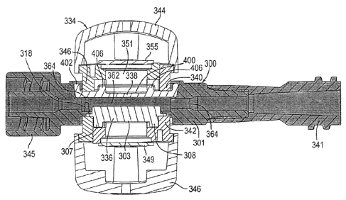
fills with blood. The flow resistance provided by the posts 118 create eddy
currents in the
cavity that also help mix the blood to make it more homogeneous when measured.
In order
to provide the function of filling the cavity of the blood chamber and mixing
the blood, the
posts 118 must be on the inlet side of the chamber 100. In the illustrated
embodiment, the
first port 122 is the inlet port. However, the second port 128 could
alternatively be the inlet
port. In that case, referring to Fig. 10, the posts 118 would be located on
the right hand side
of the cavity so that blood is intercepted by the posts upon entry into the
cavity of the blood
chamber. The posts 118 need to be on the upstream side of the cavity to be
effective.
[0094] The blood chamber 100 fastens to the clip assembly in substantially
the same
manner as illustrated and described in connection with the introductory
embodiment
illustrated in Figs. 1-6. As best illustrated in Fig. 7, the blood chamber 100
includes an
annular rim whose inner circumference defines a double-D configuration much
like that of
the blood chamber 32 in Fig. 3A. The interlocking double-D configuration fixes
the sensor
clip 34 in a predetermined position both laterally and rotationally when it is
fastened to the
blood chamber 32 as illustrated in Fig. 2A.
SECOND EMBODIMENT
[0095] Figs. 12 through 14 illustrate a blood chamber 200 constructed in
accordance
with a second illustrated embodiment. Blood chamber 200 includes a moat 264
surrounding
the internal blood flow cavity 220 as in the conventional blood flow chamber
32 illustrated in
Figs. 3A and 4A. In fact, the structure and dimensions of the blood chamber
200 shown in
Figs. 12 through 14 are substantially the same as those shown in the blood
chamber 32, with
the primary difference being that portion 208 of the blood chamber body 202 is
made of a
blue-tinted material, such as the dark blue tinted polyearbonate of the first
embodiment, in
order to attenuate ducted red light particularly at 660 nm if the LED emitter
88 emits red
light at 660 nm. Because of the presence of the moat 264, ducting of the
infrared radiation
through the chamber body 202 (or ambient light) is even less likely to cause
errors in the
CA 02828293 2013-08-23
WO 2012/116336
PCT/US2012/026637
mathematics pertaining to the ratiometric models for determining the real-time
oxygen
saturation and hematocrit levels.
[0096] As with the blood chamber 100 shown in Figs. 7 through 11, the
viewing window
206 on the chamber body 202 is preferably made of clear, polished
polycarbonate material,
and the remaining portion 208 of the chamber body 202 is overmolded to the
window 206.
As mentioned previously, the opaque (blue-tinted) portion 208 of the chamber
body 202 is
preferably made of the same material as the clear lens portion 206, but tinted
blue in order to
block the transmission of red light occurring at the relevant wavelengths,
e.g., about 660 nm.
As in the previous embodiments, the lens body 204 is made of clear material,
e.g., clear
polycarbonate, which is sonically welded to the chamber body 202. Also as in
the previous
embodiment, the blood chamber 200 includes a pair of turbulence posts 218 that
ensure
robust, non-laminar flow through the viewing area in the internal blood flow
cavity 220. The
posts are positioned on the upstream side of the cavity as explained in
connection with the
similar posts in the first embodiment.
[0097] The blood chamber 200 fastens to the sensor clip assembly 34 of Fig.
3B in
substantially the same manner as illustrated and described above for the blood
chambers 32
and 100. Specifically, the blood chamber 200 includes an annular rim whose
inner
circumference defines a double-D configuration much like that of the blood
chambers 32 and
100 in Figs. 3B and 7, respectively. The interlocking double-D configuration
fixes the
sensor clip 34 in a predetermined position both laterally and rotationally
when it is fastened
to the blood chamber 200.
THIRD EMBODIMENT
[0098] Figs. 15 through 18 illustrate a blood chamber 300 constructed in
accordance
with a third illustrated embodiment. This embodiment includes a blue-tinted
portion in
keeping with the first and second illustrated embodiments and is configured to
mate with a
sensor clip assembly that includes a shroud for blocking unwanted light from
the window of
the blood chamber. The blood chamber 300 includes first and second exterior
sides as
illustrated in Figs. 15 and 16. Each side has a viewing window and a separate,
distinct
shroud mating surface located circumferentially around the viewing window.
Preferably, on
one exterior surface of the blood chamber (Fig. 16), the viewing window is
raised above the
circumferential shroud mating surface such that a sunken annular well is
formed around the
raised viewing window. The shape of the floor of the sunken annular well
complements the
CA 02828293 2013-08-23
WO 2012/116336
PCT/US2012/026637
21
shape of the shroud mating surface on that side of the blood chamber.
Preferably, when
mounted on the blood chamber the shroud on the clip assembly substantially
fills the area of
the floor of the sunken annular well, thereby maximizing the amount of
unwanted light
blocked by the shroud. An upstanding wall on the other exterior surface of the
blood
chamber (Fig. 15) surrounds the second viewing window and separates it from
the shroud
mating surface on that side of the blood chamber. In this way, an annular well
is formed
around the second viewing window, although this annular well is at
substantially the same
depth as the viewing window on that side of the blood chamber. Again, the
shape of the
floor of the annular well complements the shape of the shroud mating surface
on the exterior
side of the blood chamber, and has dimensions substantially the same as the
dimensions of
the floor of the sunken annular well on the other side of the blood chamber so
that the shroud
fills the surface area of the floor of the well.
[0099] Fig. 16 illustrates a first exterior side of the blood chamber 300.
The blood
chamber 300 is constructed from a molded chamber body 301 which includes an
inlet and an
outlet as well as a first viewing window 303. The chamber body 301 may be
molded entirely
of clear, medical grade polycarbonate material or other suitable material.
Alternatively, it
may be desirable to use a window insert 302 made of entirely clear, medical
grade
polycarbonate, and overmold the remaining parts of the chamber body 301 with
an opaque
material such as the blue-tinted medical grade polycarbonate of the previous
embodiments.
In either case, the preferred chamber body 301 includes a circular viewing
window 303 and a
separate, distinct shroud mating surface 304 located circumferentially around
the viewing
window 303. The shroud mating surface 304 is sunken with respect to the
surface of the
viewing window 303, and is adapted to receive a shroud on a sensor clip
assembly as will be
discussed in more detail below. Fig. 16 also illustrates two anti-rotation
tabs 307, 308
formed on the exterior surface of the blood chamber 300. The anti-rotation
tabs 307, 308 are
raised above the surface of the window 303.
[0100] Fig. 15 illustrates the other exterior side of the blood chamber
300. This side
of the blood chamber 300 includes a second circular viewing window 400. The
region
between the second viewing window 400 in Fig. 15 and the first viewing window
303 in
Fig. 16 consists of material such as clear, medical grade polycarbonate and
the blood
flowing through the internal blood flow cavity within the blood chamber 300.
The
windows 303, 400 thus provide an optically neutral view for the sensor clip
assembly to
CA 02828293 2013-08-23
WO 2012/116336
PCT/US2012/026637
22
monitor the blood flowing through the blood chamber 300. Referring still to
Fig. 15, an
upstanding annular wall 406 surrounds the second viewing window 400. An
annular well
404 is formed between the upstanding, annular wall 406 and a peripheral wall
410 on the
blood chamber 300. The floor of this annular well 404 is another shroud mating
surface
which again is separate and distinct from the viewing window 400. In
accordance with the
presently preferred embodiment, a window body 402 containing the viewing
window 400,
the upstanding wall 406, and the surrounding annular well 404, is molded of a
clear
polycarbonate material and is attached via sonic welding or other means to the
chamber
body 301 during the manufacturing process.
[0101] Fig. 18 shows the cross section of the blood chamber 300. The
chamber body
301 includes a substantially flat internal wall 310 that forms part of the
internal blood flow
cavity 320. The window body 402 attached to the chamber body 301 also includes
a
substantially flat internal wall 312 that is substantially parallel to the
substantially flat
internal wall 310 on the chamber body 301. The flat internal wall 312 on the
window
body 402 is separated from the flat internal wall 310 on the chamber body 301
by a
predetermined fixed distance. The first viewing window 303 on the chamber body
301
and the second viewing window 400 on the window body 402 serve as viewing
windows
336 and 338 (Fig. 21) for blood flowing through the internal blood flow cavity
320. The
chamber body 301 (Fig. 18) includes a first port 322 and a channel 324 (inlet)
that are in
fluid communication through a first opening 326 in the internal blood flow
cavity 320.
The chamber body 301 also includes a second port 328 and channel 330 (outlet)
that are in
fluid communication through a second opening 332 to the internal blood flow
cavity 320.
[0102] As best seen in Fig. 18, a pair of turbulence posts 318 is
positioned at the
entrance of the cavity 320. As with the posts of the previous embodiments, the
posts 318
provide resistance to suppress any tendency of the blood flow to be a laminar
flow, which
may result in the blood not filling the cavity 320. The posts also create an
eddy current of
the blood in the cavity 320, which tends to mix the blood to a more homogenous
consistency that provides for better measurements. Because each of the first
and second
ports 322 and 328 can serve as either an outlet or an inlet, the posts 318 can
be on either
end of the cavity 320 as explained above in connection with the first and
second
embodiments. The posts must be positioned at the flow's entrance into the
cavity 320 to
CA 02828293 2013-08-23
WO 2012/116336
PCT/US2012/026637
23
ensure the posts properly provide the resistance to break up a laminar flow
and to create
eddy currents to mix the blood before it is measured.
[0103] Fig. 19 illustrates a sensor clip assembly 334 configured in
accordance with a
presently preferred embodiment. The sensor clip assembly 334 is used to
monitor the
patients blood flowing through the blood chamber 300. As depicted in the
embodiment
illustrated in Fig. 20B, the LED emitter arm 344 and the photodetector arm 346
are affixed
into place around a blood chamber 300 in order to monitor the hematocrit,
hemoglobin,
change in blood volume and oxygen saturation level, and/or other blood
constituents of
blood flowing through the blood chamber 300. Accordingly, the sensor clip
assembly 334
preferably includes a spring biased bridge 348 or equivalent structure to
attach a sensor
clip assembly 334 to a blood chamber 300.
[0104] The sensor clip assembly 334 includes an LED emitter arm 344 and a
photodetector arm 346, which are connected via a spring biased bridge 348. The
LED
emitter arm 344 contains an emitter subassembly with at least two LED
emitters, one
emitting infrared light or radiation at a first wavelength (xi) of about 1300
nm and another
emitting infrared light or radiation at a second wavelength (X2) of about 810
nm. The LED
emitter preferably also includes a third LED emitter for emitting visible
light or radiation
at a third wavelength (73) of about 660 nm. Other wavelengths could be
substituted or
added to measure additional blood constituents or properties of other fluids.
The detector
aim i 346 contains preferably two types of photodetectors: a silicon
photodetector to detect
the approximate 660 and 810 nm wavelengths, and an indium gallium arsenide
photodetector to detect the approximate 1300 nm wavelength. As configured in
the
embodiment depicted in Figs. 19-21, the sensor clip assembly 334 emits
infrared light or
radiation through the viewing lenses 303 and 400 and through the viewing
windows 336
and 338 and through the blood flowing through the flat viewing region 362 of
the blood
chamber 300 (see Figs. 21A and 21B).
[0105] In contrast to the sensor assembly 34 of Figs. 2A, 2B, 3B, 4A and
4B, the
sensor clip assembly 334 of Figs. 19, 20A, 20B, 21A and 21B includes two
shrouds
extending from the heads of the arms 344 and 346 of the assembly. One shroud
340 is on
the inner housing piece of the emitter arm 344 subassembly and prevents
ambient light
from entering the blood chamber through the viewing windows. A second shroud
342 is
CA 02828293 2013-08-23
WO 2012/116336
PCT/US2012/026637
24
on the inner housing piece of the detector arm 346 subassembly and also
prevents ambient
light from entering the blood chamber through the viewing windows.
[0106] The shrouds 340 and 342 are preferably mirror images of one another.
The
description of shroud 340 on the emitter arm 344 therefore is representative
and applies
equally to the description of the shroud 342 on the detector arm 346.
Referring in
particular to Fig. 19, it can be seen that shroud 342 contains an outer
annular ledge or step
surface 350 and an inner annular ledge or step surface 352. The difference in
the heights
of the step surfaces 350, 352 corresponds to the height of the annular wall
406 on the
second exterior side of the blood chamber 300 (see Fig. 15), and also to the
height at
which the window surface 303 is raised above the sunken well 304 on the first
side of the
blood chamber 300 (see Fig. 16). Preferably, the shape and surface area of the
outer
annular step surface 350 is substantially equal to the shape and surface area
of the
respective shroud mating surfaces 304, 404 on the blood chamber 300, see Figs.
20A, 20B
and 21A, in order to maximize the blocking of ambient light.
[0107] Still referring to Fig. 19, the shroud 342 illustrated in Fig. 19
includes slots
354, 356 that are adapted to receive the anti-rotation tabs 307, 308 on the
blood chamber
300 (see Fig. 16). The shroud 340 on the emitter arm 344 includes identical
slots so that
the sensor clip assembly 334 may be clipped on to the blood chamber 300 in
either
direction. In either direction, however, the sensor clip assembly is fixed in
a
predetermined position and rotational orientation that assists in eliminating
noise that
would otherwise likely result from motion artifacts during the factory
calibration for the
optical monitoring system. This fixed position can be established and
maintained in
several ways. For example, the shape of the anti-rotation tabs 307, 308 and
the
corresponding slots 354, 356 may take on any reasonable shape. Also, placing
anti-
rotation tabs on the shrouds and including mating detents or slots on the
blood chamber
may be a suitable alternative.
[0108] One skilled in the art will appreciate that any anti-rotational
arrangement for
fastening the sensor clip assembly and the blood chamber may be suitable so
long as the
clip and chamber mate so as to correctly position the LEDs and sensors of the
sensor clip
assembly with the window of the blood chamber. For example, in co-pending U.S.
application No. 12/876,798, filed September 7, 2010, which is assigned to the
same
assignee as the present application, the described blood chamber mates with a
sensor clip
CA 02828293 2013-08-23
WO 2012/116336
PCT/US2012/026637
assembly similar to the clip assembly 34 and 334 illustrated herein. Figs. 1A,
2A and 4A
from the '798 application are reproduced herein as Figs. 22, 23 and 24,
respectively, to
illustrate an exemplary alternative anti-rotation arrangement. Fig. 22 shows
the sensor clip
assembly 411 fastened to the blood chamber 412 with the photodetectors on the
left hand
arm 416A and the photoemitters on the right hand arm 416B. The dimensional
characteristics of the left side arm 416A and the right side arm 416B of the
sensor
assembly 411 are normally congruent, however the blood flow chamber 412 is
designed to
be used with the photodetectors on either the right hand arm or the left hand
arm with the
photoemitters being on the opposite side.
[0109] Referring in particular to Figs. 23 and 24, the blood chamber 412
includes
upstanding pedestals 462 axially disposed along the body 424 of the blood
chamber. The
pedestals 462 extend outwardly from a sensor receiving wall 464 of the chamber
412. The
sensor receiving wall 464 is substantially parallel to a circular lens 438 for
the viewing
area, and provides an opening for the lens 438 to be exposed to the
photoemitters in the
head 418A of the sensor clip assembly 411. The pedestals 462 guide the mating
arm 416B
of the sensor clip assembly 411 into proper rotational alignment when the clip
is fastened
to the blood chamber 412. This configuration results in the face of the clip
arm 418B
seating to the blood chamber 412 in proper parallel and rotational orientation
with respect
to the viewing area for the viewing lens 438.
[0110] Referring now in particular to Fig. 24, the other side of the
chamber body 424
includes detented receiving ledges 470 surrounding the circular viewing area
440. The
chamber body 424 also includes upstanding fingers 466 and guide walls 468 that
guide the
photodetectors on arm 416B of the sensor clip assembly 411 into proper
alignment when
the clip sensor assembly is fastened to the blood chamber 412.
[0111] Returning to Figs. 21A and 21B, the shrouds at the heads of the arms
344 and
346 of sensor clip assembly 334 are shown in cross section clipped to the
blood chamber
300 (Fig. 21A) and exploded away (Fig. 21B) from the chamber. Referring
specifically to
the blood chamber 300 as shown in Figs. 21A and 21B, the blood chamber 300
includes
two viewing windows 336 and 338. Surface 303 of the first viewing window 336
is
exposed on the first exterior side of the blood chamber 300 (see Fig. 16). The
exterior
surface of the other viewing window 338 is exposed on the other exterior side
of the blood
chamber 300 (see Fig. 15). The blood chamber 300 includes an inlet 345 and
outlet 341
CA 02828293 2013-08-23
WO 2012/116336
PCT/US2012/026637
26
that are designed to be compatible with standard medical industry connecting
devices
conventionally known as luer lock connectors. In the blood chamber 300 shown
in Figs.
21A and 21B, the inlet 345 is integrally molded with the blood chamber 300,
whereas the
outlet 341 consists of a suitable off-the-shelf connector adapter bonded to
the body of the
blood chamber 300. Alternatively, tubing can be attached directly to the body
of the blood
chamber 300 in place of the connector 341. The LED emitter subassembly 344 as
shown
in Figs. 21A and 21B contains an emitter circuit board 355 containing LEDs
emitting light
at substantially 660 nm, 810 nm and 1300 nm. The LEDs radiate light through
the molded
diffusing lens 351. As shown in Figs. 21A and 21B, the shroud 340 on the
emitter sub-
housing 344 is spaced apart from the molded diffusing lens 351. In addition,
the shroud
340 extends towards the detector subassembly 346 beyond diffusing lens 351.
[0112] The photodetector subassembly 346 includes a circuit board 349 to
which the
silicon photodetector, which can detect radiation at 810 nm and 660 nm, and
the indium
gallium arsenide photodetector, which can detect radiation at 1300 nm, are
mounted. The
photodetectors are mounted to receive light energy through a molded diffusing
lens 353.
Figs. 21A and 21B show that the shroud 342 is spaced apart from the diffusing
lens 353
and also that the shroud 342 extends beyond the diffusing lens 353 toward the
emitter
subassembly 344. In Figs. 21A and 21B, the anti-rotation tabs 307, 308 are
shown in the
cross section taken along line 21-21 in Fig. 20A.
[0113] The viewing window 336 of the blood chamber 300 in Figs. 21A and 21B
is
either part of a separate insert which is then overmolded to the remainder of
the chamber
body 301 if an opaque body is desired or the window can be molded as part of
the chamber
body 301 as one piece. The viewing window 338 on the other side of the blood
chamber 300
is part of a separately molded insert, which is sonically welded or otherwise
adhered to the
chamber body. While the windows 303 and 400 should be made of clear material,
it is
desirable to tint the remaining portions of the chamber body in keeping with
the first and
second embodiments as described above in order to provide additional
protection from
unwanted light. Specifically, the blue-tinted polycarbonate material may be
used for the
remaining portions of the chamber body.
[0114] Blood flows from the inlet into the central viewing region of the
blood chamber
300 in Figs. 21A and 21B, which has been referred to previously as the
internal blood flow
cavity 362. The internal blood flow cavity 362 provides a substantially flat,
thin (e.g., less
27
than 0.1 inches) viewing area for the blood flowing through the blood chamber
300. The
multiplexed visible or infrared light at the selected wavelengths is
transmitted through the
blood flowing through the flat viewing region as well as through the viewing
windows 336
and 338. A moat 364 surrounds the flat viewing region 362 and provides yet
additional
protection from unwanted light. The moat 364 is somewhat deeper than the flat
viewing
region 362, and serves in part to distribute non-laminar flow evenly and
steadily through the
viewing region. The moat 364 also provides a thicker region of blood which
under most
operating conditions optically isolates the detectors from unwanted (e.g.,
ducted or ambient)
light that does not pass through the direct path from the photoemitters,
through the blood and
to the photodetectors.
[0115] The viewing windows 303 and 400 are preferably made of clear,
medical grade
polycarbonate material which is molded with a polished finish in order to
facilitate reliable
TM
light transmission, e.g., Bayer Malcrolon FCR 2458-5515 (no re-grind allowed),
which is
blood contact approved, USPXX11 class VI. It is expected that the material be
certified as to
grade number, lot number and date of manufacture. Moreover, the viewing
windows should
contain no splay, bubbles or marks when looking through the display window
viewed from
twelve inches with the normal eye. The molded parts should be produced with no
lose
foreign material greater than 0.1 mm2 and no embedded foreign material greater
than 0.2
MM2. No mold release should be used and any lubrications should be food-grade
and not
silicon-based. The mold finish is preferably SPIA3 (scale) except along the
surfaces for the
viewing windows, which should preferably be at least SPIA 1. Parts should be
cleaned and
free and dirt, oils and other foreign matter before use.
FOURTH EMBODIMENT
[0116] Figs. 25A-29E illustrate a still further embodiment of the blood
chamber. The
primary difference between the third and the fourth embodiments is the
construction of the
blood chamber. In this embodiment, the two halves of the main body portion of
the blood
chamber are made to be mirror images of one another. Likewise, the two
opposing lenses
welded to the main body portion are constructed to be mirror images of one
another. In
addition to these features that ease manufacturing of the blood chamber, the
lenses are
welded to the main body portion of the blood chamber and held in place without
the addition
of overmolding on the main body portion. The connector affixed to the main
body portion is
CA 2828293 2017-07-26
CA 02828293 2013-08-23
WO 2012/116336
PCT/US2012/026637
28
substantially similar to the connector described in association with the
previous
embodiments.
[0117] Fig. 25A shows a perspective view of blood chamber 500 of the fourth
embodiment. Blood chamber 500 includes main body portion 501, lenses 502, 503,
and
connector 504. Main body portion 501 is constructed by molding together two
mirror
image halves. Each half of main body portion 501 has a recess into which
lenses 502, 503
can be molded in place. When molded into main body portion 501, lenses 502,
503 are
separated by a predetermined distance, and along with main body portion 501
create a
blood flow cavity within blood chamber 500.
[0118] Lenses 502, 503 may be made entirely of clear, medical-grade
polycarbonate
material. When lenses 502, 503 are assembled together with main body portion
501,
lenses 502, 503 provide each side of the blood chamber 500 with a viewing
window 505,
506 into the blood flow cavity. Each lens 502, 503 has a shroud mating surface
507, 508
located circumferentially around viewing windows 505, 506. The shroud mating
surfaces
507, 508 are configured to receive the shrouds of the sensor clip assembly.
Preferably,
when mounted on blood chamber 500, the shrouds on the sensor clip assembly
substantially fill shroud mating surfaces 507, 508, thereby maximizing the
amount of
unwanted light blocked by the shrouds.
[0119] Viewing windows 505, 506 are surrounded by upstanding annular walls
509,
510 which separate shroud mating surfaces 507, 508 from viewing windows 505,
506. In
this manner, annular wells 511, 512 are formed around viewing windows 505, 506
that are
at substantially the same depth as the viewing window. Annular wells 511, 512
are
congruent to shroud mating surfaces 507, 508 on the exterior sides of blood
chamber 500.
[0120] Fig. 25B illustrates one exterior side of blood chamber 500. Because
the sides
of blood chamber 500 are mirror images of each other, only one side of blood
chamber 500
will be discussed in detail. Blood chamber 500 is constructed from a main body
portion
501 which includes an inlet and an outlet. Main body portion 501 may be molded
entirely
of clear, medical-grade polycarbonate material or other suitable material.
Alternatively, it
may be desirable to mold main body portion 501 with an opaque material such as
the blue-
tinted medical-grade polycarbonate material of the previous embodiments.
[0121] In either case, main body portion 501 includes lenses 502, 503, each
of which
has a circular viewing window 505, 506. In operation, blood flows through the
internal
CA 02828293 2013-08-23
WO 2012/116336
PCT/US2012/026637
29
blood flow cavity defined by lenses 502, 503 and main body portion 501 within
blood
chamber 500. Lenses 502, 503, including their circular viewing window portions
505,
506, are made of a material such as clear, medical-grade polycarbonate.
Viewing windows
505, 506 thus provide an optically neutral view for the sensor clip assembly
to monitor the
blood flowing through blood chamber 500. As shown in Fig. 29B, upstanding
annular
wall 509 surrounds viewing window 505. Lenses 502, 503 each also have
peripheral walls
513, 514 that are concentric with upstanding annular walls 509, 510, and
positioned at the
periphery of lenses 502, 503. In this manner, annular wells 511, 512 are
defined by
annular walls 509, 510 and peripheral walls 513, 514. The floor of annular
well 511 is
another shroud mating surface 507 which is separate and distinct from viewing
window
505. In accordance with the presently preferred embodiment, lens 502
containing viewing
window 505, upstanding annular wall 509, peripheral wall 513, and annular well
511, is
molded from a clear polycarbonate material and is attached via sonic welding
or other
means to main body portion 501 during the manufacturing process. Lens 503,
with its
associated components, is manufactured from the same material and attached to
main body
portion 501 in the same fashion as lens 502.
101221 Fig. 25C illustrates a side elevation of blood chamber 500. Main
body portion
501 is connected to connector 504. Connector 504 may be a luer lock connector,
such as
the connectors previously described herein. Main body portion 501 is fitted
with lenses
502, 503 on opposing sides of main body portion 501. Peripheral walls 513, 514
of lenses
502, 503 are visible from the side elevation view of blood chamber 500. The
internal
blood flow cavity is defined by the predetermined distance between lenses 502,
503 when
they are molded to main body portion 501, and is bounded around its
circumference by
main body portion 501.
[0123] Fig. 25D shows the cross section of blood chamber 500. Lenses 502,
503 are
attached to main body portion 501, and include substantially flat internal
walls 515, 516
that form part of blood flow cavity 517. Internal wall 515 is substantially
parallel to
internal wall 516, and separated therefrom by a predetermined fixed distance.
Viewing
windows 505, 506 allow observance of the blood flowing through internal blood
flow
cavity 517.
101241 Main body portion 501 includes a first port 518 and a second port
519. Blood
flow cavity 517 includes a first opening 520 and a second opening 521. First
port 518 and
CA 02828293 2013-08-23
WO 2012/116336
PCT/US2012/026637
first opening 520 are in fluid communication through first channel 522. Second
port 519
and second opening 521 are in fluid communication through second channel 523.
Blood
chamber 500 also includes inlet 524 and outlet 525. In a preferred embodiment,
blood
flows into inlet 524, through first channel 522, and into blood flow cavity
517 where its
characteristics are sensed by the sensor clip assembly. After passing through
blood flow
cavity 517, the blood passes through second channel 523, and exits blood
chamber 500
from outlet 525.
[0125] As best seen in Fig. 25D, lenses 502, 503 each include a pair of
turbulence
posts. Pairs of turbulence posts 526, 527 are integral to lenses 502, 503.
Lenses 502, 503
are molded to main body portion 501 such that pairs of turbulence posts 526,
527 are
positioned near first opening 520 of blood flow cavity 517. As with the posts
of previous
embodiments, pairs of turbulence posts 526, 527 provide resistance to suppress
any
tendency of the blood to flow a purely laminar fashion, which if not
suppressed, may result
in the blood not filling blood flow cavity 517. Pairs of posts 526, 527 also
create eddy
currents within the blood in blood flow cavity 517, which tend to mix the
blood to a more
homogenous consistency that provides for better measurements by the sensor
clip
assembly. Because either first port 518 or second port 519 could serve as an
inlet port, as
explained in connection with the first and second embodiments, pairs of posts
526, 527
can be on either end of blood flow cavity 517 depending on the molding
orientation of
lenses 502, 503 with respect to main body portion 501. Turbulence posts
positioned at the
blood flow's entrance into blood flow cavity 517 ensure that the posts
properly provide
resistance to break up a laminar flow and to create eddy currents to mix the
blood before
its characteristics are sensed.
[0126] Fig. 25E shows an exploded view of the components of the fourth
embodiment
of blood chamber 500. During the manufacturing process, lenses 502, 503 are
received in
and attached to main body portion 501, creating internal blood flow cavity 517
of blood
chamber 500. Lenses 502, 503 are mirror images of each other. When lenses 502,
503 are
attached to main body portion 501, they are positioned such that they are
substantially
parallel and opposite each other. Connector 504 may be attached to main body
portion
501 at the second port 519 of main body portion 501. In this manner, blood
flowing from
internal blood flow cavity 517 passes out of second port 519 through second
channel 523,
CA 02828293 2013-08-23
WO 2012/116336
PCT/US2012/026637
31
through connector 504, and out of outlet 525. After the blood flows from
outlet 525, it
may flow to a dialyzer or blood filter, such as dialyzer or blood filter 22
shown in Fig. I.
[0127] Figs. 26A-B illustrate main body portion 501 of blood chamber 500 in
its
simplest form, where the two molded halves of the body are mated, but the
connector and
lenses have not yet been added. As shown in Fig. 26A, sidewall 528 extends in
a
substantially circular manner around an inner periphery of main body portion
501.
Extending substantially orthogonally from sidewall 528 are receiving ledges
529, 530.
Receiving ledges 529, 530 are also substantially circular. During assembly,
lenses 502,
503 are received in main body portion 501 on receiving ledges 529, 530. To
properly
orient lenses 502, 503 with respect to main body portion 501, main body
portion 501
includes protrusions 531, 532 located on receiving ledges 529, 530, adjacent
sidewall 528.
In the illustrated embodiment, protrusions 531, 532 are located on opposite
sides of
sidewall 528, and positioned in line with second opening 521. Lenses 502, 503
are
positioned with respect to main body portion 501 so that protrusions 531, 532
are received
in complementary recesses on lenses 502, 503. When paired in this manner,
lenses 502,
503 become seated on receiving ledges 529, 530, and internal walls 515, 516 of
lenses
502, 503, together with sidewall 528, define blood flow cavity 517.
Specifically, sidewall
528 extends between flat internal walls 515, 516 when lenses 502, 503 are
mated to main
body portion 501. Although the illustrated embodiment uses generally cubical
protrusions
and recesses, the protrusions and recesses may take on any reasonable
geometric shape.
Furthermore, other types of fasteners or rotational stops may work equally
well and are
contemplated.
[0128] Main body portion 501 includes inlet 524, which houses first port
518. First
channel 522 passes through first port 518. First port 518 and first channel
522 are in fluid
communication with internal blood flow cavity 517 through first opening 520 in
sidewall
528. Main body portion 501 also includes second port 519. Second channel 523
passes
through second port 519. Second port 519 and second channel 523 are in fluid
communication with internal blood flow cavity 517 through second opening 521
in
sidewall 528. In the embodiment shown in Figs. 26A-B, second port 519 and
second
channel 523 are in axial alignment with first port 518 and first channel 522
along an axis
that spans across the middle of internal blood flow cavity 517.
CA 02828293 2013-08-23
WO 2012/116336
PCT/US2012/026637
32
[0129] Fig. 26B shows a top elevation view of main body portion 501. During
assembly, lens 502 (not shown) is received on receiving ledge 529 and the
complementary
recess on lens 502 is aligned with protrusion 531, forming one boundary of
blood flow
cavity 517. In the illustrated embodiment, inlet 524 is opposite second port
519 on main
body portion 501. Inlet 524 is also in axial alignment with second port 519
along an axis
that spans across the middle of internal blood flow cavity 517.
[0130] Figs. 27A-B show perspective and bottom elevation views of one of
the lenses
of blood chamber 500 by itself. Because lenses 502, 503 are mirror images of
each other,
only lens 502 will be discussed in detail. Fig. 27A shows lens 502 with
viewing window
505 at its center. Lens 502 includes two concentric rings of ridges. The inner
ring is
upstanding annular wall 509, and it surrounds viewing window 505. The outer
ring is
peripheral wall 513 and it is situated at the periphery of lens 502 near where
the lens mates
to main body portion 501. The area between annular wall 509 and peripheral
wall 513
defines annular well 511. The floor of annular well 511 is shroud mating
surface 507,
which is separate and distinct from viewing window 505 and configured to
receive the
shroud of one arm of the sensor clip assembly.
[0131] When mated, annular well 511 and the spring bias of the clip
assembly hold the
clip and blood chamber 500 together. To prevent relative rotation of the clip
and blood
chamber 500, lens 502 includes anti-rotation tab 533 that extends radially
inward from
peripheral wall 513. Lens 503 has a corresponding anti-rotation tab 534. Anti-
rotation
tabs 533, 534 are positioned on the sides of lenses 502, 503 that are opposite
internal walls
515, 516. That is, when lenses 502, 503 are received in main body portion 501,
anti-
rotation tabs 533, 534 face outward from the exterior sides of blood chamber
500. In this
orientation, anti-rotation tabs 533, 534 mate to receiving slots in the
shrouds of the sensor
clip assembly, rotationally locking the mated clip assembly and blood chamber
500.
[0132] The anti-rotation tabs 533, 534 of lenses 502, 503 and the
complementary
receiving slots on the shrouds of the sensor clip assembly may take on any
reasonable
geometric shape. Furthermore, other types of fasteners or rotational stops may
work
equally well and are contemplated. For example, either the lenses or the clip
can include
alignment posts that guide the blood chamber and clip into proper engagement
to both
register the LEDs and photodetectors with the lenses and to prevent rotation.
The
CA 02828293 2013-08-23
WO 2012/116336
PCT/US2012/026637
33
interaction between anti-rotation tabs 533, 534 and the receiving slots on the
shrouds of
the sensor clip assembly will be described further below.
10133] Fig. 27B illustrates pair of turbulence posts 526 on lens 502 that
extend into
blood flow cavity 517 and introduce turbulence into the blood flow. Without
posts 526,
the blood flow through blood flow cavity 517 of blood chamber 500 is more
susceptible to
a laminar flow that reduces the quality of the measurements by the detectors
in the clip
assembly. Also shown in Fig. 27B is complementary recess 535 of lens 502.
Complementary recess 535 is configured to mate with protrusion 531 that sits
on receiving
ledge 529 of main body portion 501 (see Figs. 26A-B). The protrusion-recess
interaction
helps ensure that lenses 502, 503 are properly oriented with respect to main
body portion
501 during assembly of the blood chamber 500. When complementary recess 535 is
positioned substantially in line with and opposite turbulence posts 526, as
shown on lens
502, turbulence posts 526 are in near proximity to first opening 520 of main
body portion
501. Locating turbulence posts 526 near first opening 520 and the entrance of
blood flow
cavity 517 causes turbulence posts 526 to disturb the laminar flow of blood
entering the
blood flow cavity of blood chamber 500.
[0134] Figs. 28A-B are views of connector 504 in isolation. In the blood
chamber of
the fourth embodiment, inlet 524 (see Fig. 25D) and outlet 525 of connector
504 are
designed to be compatible with standard medical industry connecting devices,
conventionally known as luer lock connectors.
[0135] Figs. 29A-E illustrate a sensor clip assembly configured in
accordance with a
presently preferred embodiment. The blood chamber of this embodiment fits with
the
sensor clip assembly in substantially the same way as described above in
connection with
the third illustrated embodiment. When the sensor clip assembly is mated to
the blood
chamber, the shrouds of the sensor clip assembly block unwanted light from the
viewing
windows of the blood chamber.
10136] Fig. 29A shows sensor clip assembly 537 exploded away from blood
chamber
500. Shrouds 538 and 539 are mirror images of one another, and attached to
both arms of
sensor clip assembly 537 at the heads of the arms. Shrouds 538, 539 each
include an outer
annular ledge 540, 541, and an inner annular ledge 542, 543. The difference in
the heights of
the outer and inner annular ledges corresponds to the height of annular walls
509, 510 on
lenses 502, 503. Preferably, the shape and surface area of outer annular
ledges 540, 541 are
CA 02828293 2013-08-23
WO 2012/116336
PCT/US2012/026637
34
substantially similar to the shape and surface area of the respective shroud
mating surfaces
507, 508 on lenses 502, 503 in order to maximize the blocking of ambient
light.
[0137] As shown in Fig. 29A, and more particularly in the front elevation
view of one
arm of the sensor clip assembly in Fig. 29B, shrouds 538, 539 each include a
pair of
receiving slots. In particular, shroud 538 includes receiving slots 544, 545,
and shroud 539
includes receiving slots 546, 547. In the illustrated embodiment, and on each
shroud, the two
receiving slots are diametrically opposed from each other and are positioned
on outer annular
ledges 540, 541. Receiving slots 544, 545 and 546, 547 are adapted to receive
anti-rotation
tabs 533, 534 that are integral to lenses 502, 503 of blood chamber 500.
Depending on the
orientation of blood chamber 500 with respect to sensor clip assembly 537,
anti-rotation tab
533 may be received in receiving slot 544 or in receiving slot 545, and anti-
rotation tab 534
may be received in receiving slot 546 or in receiving slot 547.
[0138] When the shrouds and their receiving slots arc configured in this
manner, sensor
clip assembly 537 may be fixed in a predetermined positional and rotational
orientation with
respect to blood chamber 500 that assists in eliminating noise that would
otherwise likely
result from motion artifacts during the factory calibration of the optical
monitoring system.
The fixed position can be established and maintained in other ways, including
those
previously described in connection with other embodiments. For example, the
shapes of
anti-rotation tabs 533, 534 and corresponding receiving slots 544, 545 and
546, 547 may take
on any reasonable configuration. Also, placing anti-rotation tabs on the
shrouds and
including mating detents or receiving slots on the blood chamber may be a
suitable
alternative.
[0139] Fig. 29C shows sensor clip assembly 537 attached to blood chamber
500. In the
illustrated embodiment, sensor clip assembly 537 is oriented at a
substantially right angle
with respect to blood chamber 500 due to the relative positions of anti-
rotation tabs 533, 534
on lenses 502, 503 and receiving slots 544, 545 and 546, 547 on shrouds 538,
539. It is
contemplated that the relative position of the anti-rotation tabs and
receiving slots could be
different than that shown, producing a different relative angle between sensor
clip assembly
537 and blood chamber 500.
[0140] Figs. 29D and 29E show, respectively, longitudinal and transverse
cross-
sectional views of blood chamber 500 when attached to sensor clip assembly
537. As
shown, blood chamber 500 includes viewing windows 505, 506, which are bounded
by
CA 02828293 2013-08-23
WO 2012/116336
PCT/US2012/026637
annular walls 509, 510 of lenses 502, 503. While viewing windows 505, 506
should be
made of clear material, it is desirable to tint main body portion 501 of blood
chamber 500 in
keeping with the first, second, and third embodiments described above in order
to provide
additional protection from unwanted light. Specifically, blue-tinted
polycarbonate material
may be used to manufacture main body portion 501.
[0141] When not attached to sensor clip assembly 537, the exterior sides of
viewing
windows 505, 506 are exposed on the exterior sides of blood chamber 500 (see
Figs. 25A-
D). Blood chamber 500 includes inlet 524 and outlet 525 that are designed to
be
compatible with standard medical industry connecting devices conventionally
known as
luer lock connectors. In the illustrated embodiment, inlet 524 is integrally
molded with
main body portion 501, whereas outlet 525 comprises a suitable off-the-shelf
connector
504 bonded to main body portion 501 of blood chamber 500. Alternatively,
tubing can be
attached directly to main body portion 501 of blood chamber 500 in place of
connector
504.
[0142] In operation, blood flows from inlet 524 into blood flow cavity 517
of blood
chamber 500. Blood flow cavity 517 provides a substantially flat, thin (e.g.,
less than 0.1
inches) viewing area for the blood flowing through blood chamber 500. While in
blood flow
cavity 517, multiplexed visible or infrared light at various wavelengths is
transmitted through
the blood flowing through the flat viewing area, and also through viewing
windows 505,
506.
[0143] Attached to blood chamber 500 is sensor clip assembly 537. Sensor
clip
assembly 537 includes two arms, one comprising LED emitter subassembly 548,
and the
other comprising photodetector subassembly 549. LED emitter subassembly 548
contains
emitter circuit board 550, which includes LEDs configured to emit radiation at
the desired
wavelengths, such as substantially 660 nm, 810 nm, and 1300 nm. The emitted
radiation
passes through molded diffusing lens 551. Photodetector subassembly 549
includes
detector circuit board 553 that is configured to detect the radiation emitted
by LED emitter
subassembly 548. Detector circuit board 553 is mounted to receive the
radiation emitted
by LED emitter subassembly 548 through molded diffusing lens 552.
[0144] As shown in Fig. 29D, shroud 538 on LED emitter subassembly 548
extends
beyond molded diffusing lens 551 and toward photodetector subassembly 549.
Conversely, shroud 539 on photodetector subassembly 549 extends beyond molded
36
diffusing lens 552 and toward LED emitter subassembly 548. Molding diffusing
lenses
551, 552 are spaced apart from viewing windows 505, 506. When sensor clip
assembly
537 is mated to blood chamber 500, shrouds 538, 539 are received on shroud
mating
surfaces 507, 508 of blood chamber 500 such that outer annular ledges 540, 541
of shrouds
538, 539 are substantially flush with shroud mating surfaces 507, 508.
Furthermore,
annular walls 509, 510, that bound viewing windows 505, 506, come into contact
with
inner annular ledges 542, 543 of shrouds 538, 539. In this manner, shrouds
538, 539 help
to optically isolate the light emitted by LED emitter subassembly 548 and
detected by
photodetector subassembly 549.
[0145] The cross-sectional view of Fig. 29E shows anti-rotation tabs 533,
534, as well as
receiving slots 544, 545 and 546, 547 of shrouds 538, 539. When the anti-
rotation tabs of
blood chamber 500 are received in the receiving slots of shrouds 538, 539, as
shown in Fig.
29E, sensor clip assembly 537 is in a fixed predetermined positional and
rotational
orientation with respect to blood chamber 500.
[0146] The use of the terms "a" and "an" and "the" and similar referents in
the context
of describing the invention (especially in the context of the following
claims) are to be
construed to cover both the singular and the plural, unless otherwise
indicated herein or
clearly contradicted by context. The terms "comprising," "having,"
"including," and
"containing" are to be construed as open-ended terms (i.e., meaning
"including, but not
limited to,") unless otherwise noted. Reference to the alignment of the
photoemitters and
the photodetectors concerns only an approximate relative physical alignment
that enables
light from the emitters to be received by the detectors. Except as indicated
otherwise
either expressly or in its context, references to "light" include
electromagnetic radiation of
any frequency and should not be construed as limited to radiation visible to
the human eye.
Unwanted light reaching the photodetectors is ambient light from sources other
than the
photoemitters and/or light from the photoemitters reaching the photodetectors
indirectly
such as through the light piping effect described above. Fastening and
connecting as used
herein is intended to include both mechanical engagements and pressure
engagement.
CA 2828293 2017-07-26
37
Recitation of ranges of values herein are merely intended to serve as a
shorthand method
of referring individually to each separate value falling within the range,
unless otherwise
indicated herein, and each separate value is incorporated into the
specification as if it were
individually recited herein. All methods described herein can be performed in
any suitable
order unless otherwise indicated herein or otherwise clearly contradicted by
context. The
use of any and all examples, or exemplary language (e.g., "such as") provided
herein, is
intended merely to better illuminate the invention and does not pose a
limitation on the
scope of the invention unless otherwise claimed. No language in the
specification should
be construed as indicating any non-claimed element as essential to the
practice of the
invention.
[0147] Preferred embodiments of this invention are described herein,
including the
best mode known to the inventors for carrying out the invention. Variations of
those
preferred embodiments may become apparent to those of ordinary skill in the
art upon
reading the foregoing description. The inventors expect skilled artisans to
employ such
variations as appropriate, and the inventors intend for the invention to be
practiced
otherwise than as specifically described herein. Accordingly, this invention
includes all
modifications and equivalents of the subject matter recited in the claims
appended hereto
as permitted by applicable law. Moreover, any combination of the above-
described
elements in all possible variations thereof is encompassed by the invention
unless
otherwise indicated herein or otherwise clearly contradicted by context.
CA 2828293 2017-07-26