Note: Descriptions are shown in the official language in which they were submitted.
CA 02828568 2013-08-28
WO 2012/118941
PCMJS2012/027230
1
INTERBODY DEVICE AND PLATE FOR SPINAL STABILIZATION AND
INSTRUMENTS FOR POSITIONING SAME
BACKGROUND
The present application relates generally to spinal stabilization involving an
interbody implant device and related support plate, and to instruments and
methods for
inserting and positioning the device and the plate together relative to the
spinal column.
Several techniques and systems have been developed for correcting and
stabilizing
the spine and for facilitating fusion at various levels of the spine. Some of
these include
positioning one or more interbody implants in a spinal disc space between
adjacent
vertebrae. When an implant is placed into a disc space, the channel or path
that the
implant took to enter the disc space provides a path for retrograde movement
of the
implant from the disc space. In some forms, a plate can be used to prevent
retrograde
movement of the implant and/or to provide additional stability to the adjacent
vertebrae. If
used, the plate is often positioned into engagement with the adjacent
vertebrae in a
separate surgical step that follows implantation of the implant. The implant
can also be
attached to the plate prior to implantation, although such attachment can
limit adjustability
of the implant and plate relative to one another to accommodate for various
aspects of the
spinal anatomy of the vertebrae and/or increase the length and complexity of
the surgical
procedure.
Thus, there remains a need for further improvements in spinal stabilization
involving an interbody implant device and related support plate, and in the
instruments
and methods for inserting and positioning the same.
SUMMARY
Interbody implants and related support plates for spinal stabilization, as
well as
instruments and techniques for inserting and positioning an implant and plate
together
relative to the spinal column, are provided. More particularly, in one form a
system
includes an implant configured to be positioned in a disc space between the
first and
second vertebrae and a freestanding plate for engagement with the first and
second
vertebrae. The system also includes an insertion instrument with an engaging
portion
configured to releasably engage with the implant and the plate such that the
implant and
plate can be positioned together relative to the first and second vertebrae in
a single
81773521
2
surgical step. In one aspect, an angular orientation of the implant relative
to the plate is
adjustable when the implant and the plate are engaged by the instrument. In
this or another
aspect, the implant and plate are held in a contiguous relationship when
engaged by the
instrument. However, different forms and applications are also envisioned.
According to one aspect of the present invention, there is provided a system
for
providing spinal stabilization, comprising: an implant including a body
extending from a
leading end to an opposite trailing end, said body including a superior bone
engaging surface
and an opposite inferior bone engaging surface, said superior and inferior
bone engaging
surfaces engaging respective endplates of upper and lower vertebrae when said
implant is
positioned in a spinal disc space between the upper and lower vertebrae; a
plate for
engagement with the upper and lower vertebrae, said plate including a plate
body extending
between an upper end and an opposite lower end, said plate body including a
top surface and
an opposite bottom surface facing the upper and lower vertebrae when said
plate is engaged
therewith; and an insertion instrument including an engaging portion
configured to releasably
engage with said implant and said plate, wherein an angular orientation of
said implant
relative to said plate is adjustable when said implant and said plate are
engaged by said
instrument, wherein rotation of said plate relative to said instrument is
limited when said
implant and said plate are engaged by said insertion instrument and wherein
engagement of
said implant and said plate with said instrument prevents displacement of said
implant relative
to said plate and upon disengagement of said implant and said plate from said
instrument said
implant is freely displaceable relative to said plate.
In another embodiment, a system for providing spiral stabilization includes an
implant
including a body extending from a leading end to an opposite trailing end. The
body further
includes a superior bone engaging surface and an opposite inferior bone
engaging surface,
with the superior and inferior bond engaging surfaces engaging respective
endplates of upper
and lower vertebrae when the implant is positioned in a spinal disc space
between the upper
and lower vertebrae. The system also includes a plate for engagement with the
upper and
lower vertebrae and including a body extending between an upper end and an
opposite lower
CA 2828568 2018-04-30
81773521
2a
end. The plate body further includes a proximal surface, an opposite distal
surface, and a
distal facing intermediate portion configured to cooperate with the trailing
end of the implant.
An insertion instrument includes an engaging portion configured to releasably
engage with
the implant and the plate such that the implant and the plate are held in a
contiguous
relationship when engaged by the instrument and the implant is displaceable
from the plate
upon disengagement of the instrument.
In still another embodiment, a method for providing spinal stabilization
between first
and second vertebrae includes providing an implant including a body extending
from
CA 2828568 2018-04-30
CA 02828568 2013-08-28
WO 2012/118941
PCT/US2012/027230
3
a leading end to an opposite trailing end, with the body also including a
superior bone
engaging surface and an opposite inferior bone engaging surface. The method
also
includes providing a plate for engagement with the first and second vertebrae.
The plate
includes a body extending between an upper end and an opposite lower end.
Further steps
of the method include engaging an insertion instrument with the implant and
the plate,
which includes retaining the implant and the plate in a contiguous, uncoupled
arrangement; and inserting the leading end of the implant in a spinal disc
space between
the first and second vertebrae with the insertion instrument and advancing the
implant into
the disc space until a bottom surface of the plate contacts extradiscal
surfaces of the first
and second vertebrae. A further aspect of this embodiment includes rotating
the implant
relative to the plate when the insertion instrument is engaged with the
implant and the
plate and the implant and the plate are retained in the contiguous, uncoupled
arrangement.
Still, another aspect of this embodiment includes guiding at least one
fastener along a
corresponding guide hole through the insertion instrument and the plate into
engagement
with one of the vertebrae.
Other embodiments include unique methods, techniques, systems, devices, kits,
assemblies, equipment, and/or apparatus for use in connection with the
stabilization and
support of first and second vertebrae. However, in other embodiments,
different forms
and applications are also envisioned.
Further embodiments, forms, features, aspects, benefits, objects and
advantages of
the present application will become apparent from the detailed description and
figures
provided herewith.
CA 02828568 2013-08-28
WO 2012/118941
PCT/1JS2012/027230
4
BRIEF DESCRIPTION OF THE FIGURES
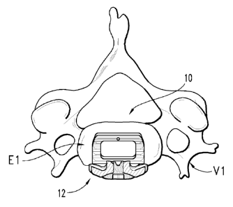
FIG. 1 is a diagrammatic plan view, with some features being shown in section,
looking toward the axial plane of an endplate of a vertebral body of a spinal
column with
an interbody implant and plate positioned relative thereto.
FIG. 2 is a diagrammatic elevation view looking toward the sagittal plane at a
vertebral level of the spinal column including the vertebral body, interbody
implant and
plate of FIG. 1.
FIG. 3 is atop, plan view of the interbody implant illustrated in FIG. I.
FIG. 4 is a side, plan view of the interbody implant illustrated in FIG. 1.
FIGS. 4A and 4B are side, plan views of alternative embodiment interbody
implants.
FIG. 5 is a perspective view of the interbody implant illustrated in FIG. 1.
FIG. 6 is a front, plan view of the plate illustrated in FIG. 1.
FIGS. 7 and 8 are opposite, side plan views of the plate illustrated in FIG.
1.
FIG. 9 is a plan view of one embodiment insertion instrument configured to
insert
and position the interbody implant and plate of FIG. 1 relative to the spinal
column.
FIG. 10 is a plan view of the insertion instrument illustrated in FIG. 9
rotated
ninety degrees about its longitudinal axis.
FIG. 11 is a plan view of the distal end of the instrument illustrated in FIG.
9
rotated one hundred and eighty degrees about its longitudinal axis.
FIG. 12 is section view of the proximal end of the instrument illustrated in
FIG. 9
taken along view line 12-12.
FIG. 13 is a plan view of an inner member of the instrument illustrated in
FIG. 9.
FIG. 14 is a section view of the inner member illustrated in FIG. 13 taken
along
view line 14-14.
FIG. 15 is a perspective view of the distal end of the inner member
illustrated in
FIG. 13.
FIG. 16 is a plan view of an outer member of the instrument illustrated in
FIG. 9.
FIG. 17 is a section view of the outer member illustrated in FIG. 16 taken
along
view line 17-17.
FIG. 18 is a plan view of the instrument illustrated in FIG. 9 engaged with
the
interbody implant and plate of FIG. 1.
CA 02828568 2013-08-28
WO 2012/118941
PCT/US2012/027230
FIG. 19 is a plan view of an alternative embodiment insertion instrument
configured to insert and position the interbody implant and plate of FIG. 1
relative to the
spinal column.
FIG. 20 is a plan view of the insertion instrument illustrated in FIG. 19
rotated
5 ninety degrees about its longitudinal axis and with some features being
shown in section
along view line 20-20.
DESCRIPTION OF THE ILLUSTRATED EMBODIMENTS
For the purposes of promoting an understanding of the principles of the
invention,
reference will now be made to the embodiments illustrated in the drawings and
specific
language will be used to describe the same. It will nevertheless be understood
that no
limitation of the scope of the invention is thereby intended. Any such
alterations and
further modifications in the illustrated devices and described methods, and
any such
further applications of the principles of the invention as illustrated herein
are contemplated
as would normally occur to one skilled in the art to which the invention
relates.
Methods, techniques, instrumentation, devices and implants are provided to
restore
and/or maintain a collapsed, partially collapsed, damaged, diseased, or
otherwise impaired
spinal disc space at a desired disc space height and adjacent endplate
orientation. The
instruments and implants may be used in techniques employing minimally
invasive
instruments and technology to access the disc space, although access in non-
minimally
invasive procedures is also contemplated. Access to the collapsed disc space
can be uni-
portal, bi-portal, or multi-portal. The instruments and implants may also be
employed in a
direct anterior approach to the spinal disc space, although other approaches
are also
contemplated, including lateral, antero-lateral, postero-lateral, oblique, and
posterior
approaches. Also, the surgical methods, techniques, instruments and implants
may find
application at all vertebral segments of the spine, including the lumbar,
thoracic and
cervical spinal regions.
In one aspect, interbody implants and related support plates for spinal
stabilization,
as well as instruments and techniques for inserting and positioning an implant
and plate
together relative to the spinal column, are provided. More particularly, in
one form a
system includes an implant configured to be positioned in a disc space between
the first
and second vertebrae and a freestanding plate for engagement with the first
and second
vertebrae. The system also includes an insertion instrument with an engaging
portion
CA 02828568 2013-08-28
WO 2012/118941
PCT/US2012/027230
6
configured to releasably engage with the implant and the plate such that the
implant and
plate can be positioned together relative to the first and second vertebrae in
a single
surgical step. In one aspect, an angular orientation of the implant relative
to the plate is
adjustable when the implant and the plate are engaged by the instrument. In
this or
another aspect, the implant and plate are held in a contiguous relationship
when engaged
by the instrument. However, different forms and applications are also
envisioned.
Referring now generally to FIG. 1, it illustrates a plan view, with some
features
being shown in section, looking caudally toward the axial plane of a vertebral
body Vi.
As illustrated in FIGS. 1 and 2, spinal interbody implant 10 is positioned on
the vertebral
endplate El intradiscally between vertebral bodies V1, V2, and a plate 12 is
secured
substantially extradiscally, or outside the disc space, to vertebral bodies
V1, V2 with a
plurality of bone engaging fasteners, two of which are shown in the form of
bone screws
14, 16. In the illustrated form, a portion of plate 12 also extends between
vertebral bodies
VI, V2, although forms in which plate 12 is positioned entirely extradiscally
with no
portion of it extending between vertebral bodies VI, V2 are also contemplated.
Vertebral
body V1 along with vertebral body V2 and spinal disc space D comprise a level
of spinal
column segment SC in the cervical region, although implantation of implant 10
and plate
12 in the thoracic and lumbar regions is also possible and contemplated, as
indicated
above. Implant 10 is positioned in disc space D between vertebral bodies VI
and V2 so
that when it is in its implanted orientation it contacts endplates El and E2.
In the
illustrated form, plate 12 is positioned so that it lies along the anterior
facing surfaces of
vertebral bodies V1, V2, although positioning of plate 12 along alternatively
facing
surfaces of vertebral bodies V1, V2 depending on the orientation of implant 10
to vertebral
bodies V1, V2 is also contemplated. Similarly, in the illustrated form
vertebral bodies V1,
V2 are accessed from an anterior approach, although lateral, antero-lateral,
postero-lateral,
oblique, and posterior approaches are also possible. Further, as illustrated,
implant 10 and
plate 12 are generally positioned adjacent to and in abutting engagement with
one another,
although it should be appreciated that movement of implant 10 away from plate
12 is
possible since implant 10 and plate 12 are not physically attached or
otherwise coupled to
one another as will be discussed in greater detail below.
Referring now generally to FIGS. 3-5, implant 10 includes a body 18 sized to
fit
within the disc space D between adjacent vertebral bodies V1, V2. Body 18
extends from
a leading end 20 to an opposite trailing end 22. In the illustrated form,
leading end 20
CA 02828568 2013-08-28
WO 2012/118941
PCT/US2012/027230
7
generally includes a planar surface 24 positioned between angled surfaces 26,
28 which
can facilitate insertion of implant 10 into disc space D and/or distraction of
vertebral
bodies V1, V2. In other non-illustrated forms, leading end 20 can include a
convexly
rounded nose to facilitate insertion into disc space D and distraction of
vertebral bodies
V1, V2. As illustrated in FIG. 3 for example, body 18 also includes a
receptacle 23 in
which a radiographic marker can be positioned to facilitate image-guided
placement of
implant 10 between vertebral bodies V1, V2.
Body 18 also includes superior and inferior bone engaging surfaces 30, 32 with
ridges 34, 36 (only a few of which are referenced to preserve clarity) to
enhance
engagement with the vertebral end plates El, E2. In other forms, superior and
inferior
bone engaging surfaces 30, 32 can be provided with threads, grooves, teeth
knurling or
other surface roughening, just to provide a few possibilities, to enhance
engagement with
vertebral endplates El, E2. In the illustrated form, bone engaging surface 30
includes a
generally convex configuration between leading end 20 and trailing end 22,
while bone
engaging surface 32 includes a generally planar or straight configuration
between leading
end 20 and trailing end 22. In other forms, it should be appreciated that bone
engaging
surface 30 could also be planar and that bone engaging surface 32 could also
be convexly
curved. Still, other variations in the configurations of bone engaging
surfaces 30, 32
between leading end 20 and trailing end 22 are possible. Further, bone
engaging surfaces
30, 32 are generally configured such that implant 10 is received between and
in contact
with at least a portion of endplates El, E2 along at least a portion of body
18. Body 18
also includes opposite side walls 38, 40 extending from leading 20 to trailing
end 22, and
also extending from bone engaging surface 30 to bone engaging surface 32. Side
walls 38,
40 can be parallel to one another, or tapered relative to one another to
converge or diverge
toward the leading end 20. Side walls 38, 40 can be planar, concave or convex
from
leading end 20 to trailing end 22, concave or convex from bone engaging
surface 30 to
bone engaging surface 32, or combinations thereof.
Body 18 also includes a cavity 42 that opens through bone engaging surfaces
30,
32 to facilitate bone growth through body 18, although forms where cavity 42
is not
present are also possible. In other non-illustrated forms, it is contemplated
that body 18
could also include one or more openings extending through side walls 38, 40
and/or
leading and trailing ends 20, 22 and into communication with cavity 42. In
addition, while
not illustrated, it should be appreciated that one or more biocompatible
materials which,
CA 02828568 2013-08-28
WO 2012/118941
PCT/US2012/027230
8
for example, provide a therapeutic effect or enhance bone growth through
implant 10 can
be positioned in cavity 42. Examples of such biocompatible materials may
include
calcium phosphate, hyrdroxyapatite-tricalcium phosphate (HA-TCP) compounds,
bioactive glasses, calcium sulfate bone void fillers, collagen, fibrin,
albumin, karatin, silk,
clastin, demineralized bone matrix, particulate bone, mysenchymal stem cells,
hormones,
growth factors such as transforming growth factor beta (TGFb) proteins, bone
morphogenic proteins (including BMP and BMP2), or platelet derived growth
factors, just
to provide a few possibilities. In one aspect, the biocompatible material(s)
may, when
included, extend slightly above and below bone engaging surfaces 30, 32,
respectively, to
facilitate compressive loading by the adjacent vertebral bodies onto and
through the
biocompatible material(s).
As illustrated in FIG. 4 for example, trailing end 22 of implant 10 is
generally
convexly curved between bone engaging surfaces 30, 32. In addition, trailing
end 22 also
includes an elongate slot 44 that is positioned between bone engaging surfaces
30, 32 and
extends between side walls 38, 40, although in other forms it should be
appreciated that
trailing end 22 can be provided without elongate slot 44. Body 18 also
includes a first
receptacle 46 formed in side wall 38 and generally including a circular
arrangement
configured to receive a correspondingly configured portion of an insertion
instrument,
further details of which will be provided below. Body 18 also includes a notch
or groove
47 formed in side wall 38. Groove 47 includes upper and lower surfaces 48, 50
and lateral
facing surfaces 52, 54, and extends through trailing end 22 into communication
with first
receptacle 46. In the illustrated form, upper and lower surfaces 48, 50 are
generally
arranged in an oblique orientation relative to one another, although other
forms are
contemplated. Body 18 also includes a second receptacle 56 formed in side wall
40 and
generally including a circular arrangement configured to receive a
correspondingly
configured portion of an insertion instrument, further details of which will
be provided
below. Body 18 also includes a notch or groove 57 formed in side wall 40.
Groove 57
includes upper and lower surfaces 58, 60 and lateral facing surfaces 62, 64,
and extends
through trailing end 22 into communication with second receptacle 56. In the
illustrated
form, upper and lower surfaces 58, 60 are generally arranged in an oblique
orientation
relative to one another, although other forms are contemplated.
While not previously discussed, it should be appreciated that the generally
circular
arrangement of receptacles 46, 56 which allows receipt of a correspondingly
configured
CA 02828568 2013-08-28
WO 2012/118941
PCT/US2012/027230
9
portion of the insertion instrument allows an angular orientation of implant
10 relative to
plate 12 to be adjusted when implant 10 and plate 12 are engaged by the
insertion
instrument, further details of which will be provided below. However, it
should be
appreciated that other configurations of implant 10 are possible for allowing
the angular
orientation of implant 10 relative to plate 12 to be adjusted when implant 10
and plate 12
are engaged by the insertion instrument. For example, with reference to FIG.
4A, elongate
slot 44 and grooves 47, 57 have been omitted from alternative embodiment
implant 10a.
In addition, receptacle 46a is generally configured as an elongated slot
configured to
receive a round feature of the insertion instrument in order to hold implant
10a with the
instrument while also allowing adjustment of the angular orientation of
implant 10a
relative to plate 12 and the insertion instrument when implant 10a and plate
12 are
engaged by the insertion instrument. As another example, FIG. 4B illustrates
another
alternative embodiment implant 10b from which elongate slot 44 and grooves 47,
57 have
been omitted. Implant 10b includes a receptacle 46b in the form of an
arcuately shaped
slot configured to receive a round feature of the insertion instrument in
order to hold
implant 10b with the instrument while also allowing adjustment of the angular
orientation
of implant 10b relative to plate 12 and the insertion instrument when implant
10b and plate
12 are engaged by the insertion instrument. While not shown in FIGS. 4A and
4B, it
should be appreciated that the receptacles positioned opposite of receptacles
46a, 46b are
configured the same as receptacles 46a, 46b. In addition, it should also be
appreciated that
other than the differences described above, implants 10a, 10b will generally
be configured
the same as implant 10.
Further details regarding plate 12 are shown in FIGS. 6-8. Plate 12 includes a
body 66 that extends along a central axis 68 that is oriented to extend
generally along the
central axis of the spinal column SC and from vertebral body Vito vertebral
body V2
when plate 12 is implanted. In the illustrated form, body 66 generally
includes a
substantially square configuration, although forms in which body 66 is
elongated along
central axis 68 and includes a rectangular, oval or elliptical shape, just to
provide a few
examples, are also contemplated. Body 66 includes an upper or cephalad end 70
and an
opposite lower or caudal end 72, and opposite side surfaces 74, 76 that extend
between
ends 70, 72. Body 66 also includes superior bone screw holes 78, 80 adjacent
upper end
70 and inferior bone screw holes 82, 84 adjacent lower end 72. Bone screw
holes 78, 80
and 82, 84 extend through and open at top surface 86 and bottom surface 88 of
body 66,
CA 02828568 2013-08-28
WO 2012/118941
PCT/US2012/027230
and are generally arranged to allow bone screws to extend obliquely through
and away
from body 66. More particularly, bone screw holes 78, 80 are generally
arranged to allow
bone screws extending therethrough to extend obliquely to plate 12 in a
lateral, cephalad
direction, while bone screw boles 82, 84 are generally arranged to allow bone
screws
5 extending therethrough to extend obliquely to plate 12 in a lateral,
caudal direction.
Among other things, the orientation of bone screw holes 78, 80 and 82, 84 in
this
arrangement allows the use of relatively longer bone screws, resulting in
better
engagement and purchase with the adjacent vertebral bodies. Further, in this
arrangement,
the trajectories of bone screw holes 78, 80 and 82, 84 extend toward a common
location
10 above plate 12 such that the operating space necessary for inserting
screws through plate
12 is reduced, thereby minimizing the impact to the surrounding patient
anatomy. In other
non-illustrated forms, it should be appreciated that plate 12 can be provided
with one bone
screw hole or more than two bone screw holes adjacent each of upper end 70 and
lower
end 72.
Body 66 also includes a retaining element 86 which can be secured to body 66
with
a threaded shaft, clip or other configuration that allows retaining element 86
to rotate
while attached to body 66. Retaining element 86 includes a cross-like
configuration
including ends 86a-d and a central driving tool receptacle 88. For the sake of
clarity, it
should be appreciated that retaining element 86 has been omitted from body 66
in FIGS.
7-8. The retaining element 86 also includes concavely curved sidevvall
portions 90a-b that
can be aligned simultaneously with the respective adjacent bone screw hole 78,
80, 82, 84
to allow insertion of a bone screw and its proximal head into the adjacent
bone screw hole
78, 80, 82, 84. When the bone screw heads are seated in bone screw holes 78,
80, 82, 84,
retaining element 86 can be rotated so that ends 86a-d overlap the respective
bone screw
hole 78, 80, 82, 84 and block or contact the bone screw head to prevent bone
screw back-
out from bone screw holes 78, 80, 82, 84. It should also be appreciated that
other shapes
and designs of retaining element 86 are possible for preventing bone screw
back-out from
screw holes 78, 80, 82, 84. For example, in one non-illustrated form,
retaining element 86
can be in the form of a threaded fastener which is engaged with plate 12 after
it is attached
to vertebral bodies V1, V2 such that at least a portion of an enlarged head of
the threaded
fastener extends over screw holes 78, 80, 82, 84.
Body 66 of plate 12 also includes grooves 92, 94 that extend into side
surfaces 74,
76 and from top surface 86 to bottom surface 88. As illustrated in FIG. 7,
groove 92
CA 02828568 2013-08-28
WO 2012/118941
PCT/US2012/027230
11
includes a receptacle 96 that generally has a racetrack shaped configuration.
More
particularly, receptacle 96 includes parallel sides between which extend
arcuate or
rounded end portions. As illustrated in FIG. 8, groove 94 includes a
receptacle 98 that
also generally has a racetrack shaped configuration. In other non-illustrated
forms, it
should be appreciated that other configurations, including oval or polygonal
to provide a
few possibilities, are also contemplated. Receptacles 96, 98 are configured to
receive
correspondingly configured portions of an insertion instrument, further
details of which
will be provided below.
In addition, body 66 also includes an intermediate portion 100 that includes a
concavely shaped surface 102 facing away from top surface 86. Surface 102 is
generally
configured to cooperate with trailing end 22 of implant 10 when implant 10 and
plate 12
are positioned adjacent to one another. Intermediate portion 100 extends away
from top
surface 86 such that surface 102 is offset away from top surface 86 relative
to upper and
lower portions 104, 106 of bottom surface 88. Similarly, as illustrated in
FIG. 2 for
example, this arrangement results in surface 102 and at least a portion of
intermediate
portion 100 being positioned in disc space D between vertebral bodies V1, V2
when upper
and lower portions 104, 106 of bottom surface 88 contact vertebral bodies V1,
V2 and
plate 12 is engaged with vertebral bodies VI, V2. In other non-illustrated
forms however,
it should be appreciated that surface 102 can be aligned with upper and lower
portions
104, 106 of bottom surface 88 or offset toward top surface 86 relative to
upper and lower
portions 104, 106 of bottom surface 88 such that no portion of plate 12
extends into disc
space D when it is engaged with vertebral bodies V1, V2. In other non-
illustrated forms,
surface 102 can be flat or include a convex shape that is configured to
cooperate with
implant 10 having a concave trailing end 22.
Referring now generally to FIGS. 9-17, further details regarding an insertion
instrument 120 configured to engage with implant 10 and plate 12 and position
implant 10
and plate 12 relative to vertebral bodies VI, V2 will be provided. Instrument
120 extends
along longitudinal axis L from proximal end 122 to distal end 124 and includes
an inner
member 126, outer member 128 and a drive member 130. Inner member 126 extends
between a threaded proximal portion 132 and a distal engaging portion 134.
Distal
engaging portion 134 is bifurcated into portions 148, 150 which surround a
hollow interior
160 and from which tines 136, 138 extend. Portions 148, 150 also include
tapered
surfaces 162, 164 adjacent the proximal ends of tines 136, 138, and are
pivotable about
CA 02828568 2013-08-28
WO 2012/118941
PCT/US2012/027230
12
passage 158 such that tines 136, 138 can be moved relative to one another to
facilitate
engagement and disengagement of instrument 120 with implant 10 and plate 12.
While
not illustrated, it should be appreciated that a spring or other resiliently
elastic material,
such as a rubber plug, can be positioned in passage 158 such that tines 136,
138 are
normally biased away from one another. Tine 136 includes a distal, generally
circular
shaped projection 140 configured to be positioned in receptacle 46 of implant
10. Tine
136 also includes a generally racetrack shaped projection 142 proximally
spaced from
projection 140 and configured to be positioned in receptacle 96 of plate 12.
Tine 136 is
further configured to be positioned in groove 47 of implant 10 and groove 92
of plate 12
when instrument 120 is engaged with implant 10 and plate 12. Tine 138 includes
a distal,
generally circular shaped projection 144 configured to be positioned in
receptacle 56 of
implant 10. Tine 138 also includes a generally racetrack shaped projection 146
proximally
spaced from projection 144 and configured to be positioned in receptacle 98 of
plate 12.
Tine 138 is further configured to be positioned in groove 57 of implant 10 and
groove 94
of plate 12 when instrument 120 is engaged with implant 10 and plate 12. Inner
member
126 also includes an opening 152 that extends obliquely to longitudinal axis L
and into
communication with hollow interior 160. Another set of openings 154, 156 are
positioned
opposite of opening 152 and extend obliquely to longitudinal axis L and into
communication with hollow interior 160.
Outer member 128 extends between proximal end 170 and distal end 172 and
includes a hollow interior 174 which receives inner member 126. Outer member
128 also
includes an opening 176 that extends obliquely to longitudinal axis L and into
communication with hollow interior 174. A ridge 177 extends along a portion of
opening
176 and defines opposite portions 176a, 176b of opening 176. Another set of
openings
178, 180 (FIG. 11) are positioned opposite of opening 176 and extend obliquely
to
longitudinal axis L and into communication with hollow interior 174. When
inner
member 126 is positioned in outer member 128 and instrument 120 engages with
implant
10 and plate 12, opening 176 of outer member 128 generally aligns with opening
152 of
inner member 126 and openings 178, 180 of outer member 128 generally align
with
openings 154, 156 of inner member 126. Similarly, in this arrangement,
cooperation of
openings 152, 176 allows placement of bone screws through instrument 120 into
and
through bone screw holes 82, 84 of plate 12. More particularly, portion 176a
of opening
176 and opening 152 are arranged such that ridge 177 guides a bone screw to
bone screw
CA 02828568 2013-08-28
WO 2012/118941
PCT/US2012/027230
13
opening 82 of plate 12, while portion 176b of opening 176 and opening 152 are
arranged
such that ridge 177 guides a bone screw to bone screw opening 84 of plate 12.
Further,
cooperation of openings 154, 156 and openings 178, 180 allows placement of
bone screws
through instrument 120 into and through bone screw holes 78, 80 of plate 12.
More
particularly, openings 154 and 178 are generally arranged relative to
instrument 120 to
guide a bone screw to bone screw opening 80 of plate 12, while openings 156
and 180 are
generally arranged relative to instrument 120 to guide a bone screw to bone
screw opening
78 of plate 12. In addition, while not previously discussed, it should be
appreciated that
cooperation of openings 152, 176 may also facilitate engagement of receptacle
88 of
retaining element 86 to facilitate rotation of retaining element 86 following
placement of
the bone screws, although engagement of retaining element 86 by inserting an
instrument
along the length of instrument 120 through hollow interior 160 is also
contemplated. In
addition, while not previously discussed, it should also be appreciated that
the cooperation
of openings 152, 176, openings 154, 178 and openings 156, 180 may also
facilitate access
to vertebral bodies V1, V2 with one or more instruments such as awls, drills
or taps, just to
provide a few possibilities, to prepare vertebral bodies V1, V2 for the bone
screws.
Proximal end 170 of outer member 128 also includes an annular groove 182
within
which is positioned a retaining ring 184 in order to couple outer member 128
with drive
member 130 such that drive member 130 is independently rotatable relative to
outer
member 128. Drive member 130 includes internal threading configured to engage
with
threaded proximal portion 132 of inner member 126. Similarly, rotation of
drive member
130 results in axial displacement of inner member 126 along longitudinal axis
L relative to
outer member 128. A pin 186 extends from outer member 128 into a slot 190 on
inner
member 126 to prevent rotation of inner member 126 relative to outer member
128.
Further, a locking member 188 extends through drive member 130 and is
selectively
engageable with inner member 126 to prevent rotation of drive member 130
relative to
inner member 126 once a desired relationship between inner member 126 and
outer
member 128 has been obtained. While not previously discussed, it should be
appreciated
that axial movement of inner member 126 along longitudinal axis L in a
proximal
direction relative to outer member 128 results in engagement of distal end 172
of outer
member 128 with tapered surfaces 162, 164 of distal engaging portion 134 of
inner
member 126. As distal end 172 engages with tapered surfaces 162, 164, portions
148, 150
and tines 136, 138 are forced toward one another. Moreover, axial movement of
inner
CA 02828568 2013-08-28
WO 2012/118941
PCT/US2012/027230
14
member 126 along longitudinal axis L in a distal direction relative to outer
member 128
disengages distal end 172 of outer member 128 from tapered surfaces 162, 164
to allow
portions 148, 150 and tines 136, 138 to be moved away from one another.
As indicated above, implant 10 and plate 12 are not coupled or otherwise
attached
with one another. However, implant 10 and plate 12 can be positioned adjacent
one
another with trailing end 22 of implant 10 cooperating with surface 102 of
plate 12. When
implant 10 and plate 12 are positioned in this arrangement, they may each be
engaged by
instrument 120 as illustrated in FIG. 18 for example. More particularly, tine
136 can be
positioned in groove 94 of plate 12 and in groove 57 of implant 10 with
projection 140
positioned in receptacle 56 of implant 10 and projection 142 positioned in
receptacle 98 of
plate 10. Similarly, tine 138 can be positioned in groove 92 of plate 12 and
in groove 47
of implant 10 with projection 144 positioned in receptacle 46 of implant 10
and projection
146 positioned in receptacle 96 of plate 10. Once tines 136, 183 are engaged
with implant
10 and plate 12, inner member 126 can be moved proximally relative to outer
member 128
to clamp implant 10 and plate 12 between tines 136, 138. Engagement of implant
10 and
plate 12 with instrument 120 generally holds implant 10 and plate 12 in a
contiguous
relationship. More particularly, engagement of projections 140, 144 with
receptacles 46,
56 of implant 10 and engagement of projections 142, 146 with receptacles 96,
98 of plate
10 prevents displacement of implant 10 from plate 12. However, once disengaged
by
instrument 120, implant 10 may be displaced from plate 12.
While not previously discussed, it should be appreciated that the circular
configuration of receptacles 46, 56 and projections 140, 144, as well as the
convex shape
of trailing end 22 of implant 10 and the corresponding concave shape of
surface 102 of
plate 12, allow implant 10 to rotate relative to instrument 120 and plate 12
when it is
engaged by instrument 120. Further, the racetrack shaped configuration of
grooves 96, 98
and projections 142, 146 prevents rotation of plate 12 relative to instrument
120 when it
engages plate 12. In the illustrated form, rotation of implant 10 relative to
plate 12 and
instrument 120 will be limited in a first direction by contact of tine 136
with upper surface
58 of groove 57 and of tine 138 with upper surface 48 of groove 47, and in a
second
direction by contact of tine 136 with lower surface 60 of groove 57 and of
tine 138 with
lower surface 50 of groove 47. Similarly, it should be appreciated that the
orientation of
upper and lower surfaces 48, 50 relative to one another and of upper and lower
surfaces
58, 60 relative to one another can be modified to facilitate differing degrees
of rotation of
CA 02828568 2013-08-28
WO 2012/118941
PCT/US2012/027230
implant 10 relative to plate 12 when they are engaged by instrument 120. In
other forms
however, it is contemplated that implant 10 can be configured such that its
rotation relative
to plate 12 is not limited.
When engaged by instrument 120, implant 10 and plate 12 can be positioned
5 relative to vertebral bodies V1, V2 together in a single surgical step.
More particularly,
leading end 20 of implant 12 can be positioned in disc space D between
vertebral bodies
VI, V2 and advanced into disc space D until bottom surface 88 of plate 12
contacts
vertebral bodies V1, V2. As implant 10 is inserted and advanced into disc
space D, it can
rotate relative to plate 12 as necessary to accommodate for the orientation of
vertebral
10 bodies V1, V2 relative to disc space D. For example, when implant 10 and
plate 12 are
used in a curved or lordotic portion of the spinal column Sc, implant 10 may
extend
obliquely as illustrated in FIG. 18, rather than orthogonally, to plate 12
once it is inserted
in disc space D. Once implant 10 is properly positioned in disc space D and
plate 12 is
positioned against vertebral bodies VI, V2, bone screws can be inserted
through
15 instrument 120 to attach plate 12 to vertebral bodies V1, V2, and
retaining element 86 can
be rotated to position ends 86a-d over the bone screws to prevent screw back-
out. While
only two bone screws have bone illustrated in FIG. 18, it should be
appreciated that plate
12 may be attached to vertebral bodies VI, V2 with an upper pair of screws and
a lower
pair of screws. After the screws have been inserted and covered by retaining
element 86,
instrument 120 may be disengaged from implant 10 and plate 12 and removed from
the
surgical site.
An alternative embodiment insertion instrument 200 configured to engage with
implant 10 and plate 12 and position implant 10 and plate 12 relative to
vertebral bodies
VI, V2 is illustrated in FIGS. 19-20. Instrument 200 extends along
longitudinal axis LL
from proximal end 202 to distal end 204 and includes an inner member 206,
outer member
208 and a drive member 210. Inner member 206 extends between a threaded
proximal
portion 212 and a distal engaging portion 214. Distal engaging portion 214 is
bifurcated
into portions 216, 218 that form tines 220, 222. Portions 216, 218 also
include tapered
surfaces positioned proximally of tines 220, 222 and are laterally
displaceable relative to
one another such that tines 220, 222 can be moved to facilitate engagement and
disengagement of instrument 200 with implant 10 and plate 12. Tine 220
includes a distal,
generally circular shaped projection 224 configured to be positioned in
receptacle 46 of
implant 10. Tine 220 also includes a generally racetrack shaped projection 226
proximally
CA 02828568 2013-08-28
WO 2012/118941
PCT/US2012/027230
16
spaced from projection 224 and configured to be positioned in receptacle 96 of
plate 12.
Tine 220 is further configured to be positioned in groove 47 of implant 10 and
groove 92
of plate 12 when instrument 200 is engaged with implant 10 and plate 12. Tine
222
includes a distal, generally circular shaped projection 228 configured to be
positioned in
receptacle 56 of implant 10. Tine 222 also includes a generally racetrack
shaped
projection 230 proximally spaced from projection 228 and configured to be
positioned in
receptacle 98 of plate 12. Tine 222 is further configured to be positioned in
groove 57 of
implant 10 and groove 94 of plate 12 when instrument 200 is engaged with
implant 10 and
plate 12.
Outer member 208 extends between proximal end 232 and distal end 234 and
includes a hollow interior within which inner member 206 is received. Distal
end 234 also
includes opposing tines 236, 238 which are configured to extend along and
engage with
lateral surfaces of tines 220, 222. Outer member 208 also includes tapered
surfaces
positioned proximally of tines 236, 238 and configured to engage with the
tapered surfaces
of inner member 206. Proximal end 232 of outer member 208 also includes an
annular
groove within which is positioned a retaining ring 240 in order to couple
outer member
208 with drive member 210 such that drive member 210 is independently
rotatable relative
to outer member 208. Drive member 210 includes internal threading configured
to engage
with threaded proximal portion 212 of inner member 206. Similarly, rotation of
drive
member 210 results in axial displacement of inner member 206 along
longitudinal axis L
relative to outer member 208. A pin 242 extends from outer member 208 into a
slot on
inner member 206 to prevent rotation of inner member 206 relative to outer
member 208.
Further, a locking member 244 extends through drive member 210 and is
selectively
engageable with inner member 206 to prevent rotation of drive member 210
relative to
inner member 206 once a desired relationship between inner member 206 and
outer
member 208 has been obtained. While not previously discussed, it should be
appreciated
that axial movement of inner member 206 along longitudinal axis L in a
proximal
direction relative to outer member 208 results in engagement of the tapered
surfaces of
outer member 208 with the tapered surfaces of inner member 206 which forces
tines 220,
222 toward one another. Moreover, axial movement of inner member 206 along
longitudinal axis L in a distal direction relative to outer member 208
disengages the
tapered surfaces of inner and outer members 206, 208 to allow portions 216,
218 and tines
220, 222 to be moved away from one another.
CA 02828568 2013-08-28
WO 2012/118941
PCT/US2012/027230
17
When implant 10 and plate 12 are positioned adjacent one another as discussed
above, they may each be engaged by instrument 200. More particularly, tine 220
can be
positioned in groove 94 of plate 12 and in groove 57 of implant 10 with
projection 224
positioned in receptacle 56 of implant 10 and projection 226 positioned in
receptacle 98 of
plate 10. Similarly, tine 222 can be positioned in groove 92 of plate 12 and
in groove 47
of implant 10 with projection 228 positioned in receptacle 46 of implant 10
and projection
230 positioned in receptacle 96 of plate 10. Once tines 220, 222 are engaged
with implant
and plate 12, inner member 206 can be moved proximally relative to outer
member 208
to clamp implant 10 and plate 12 between tines 220, 222. Engagement of implant
10 and
10 plate 12 with instrument 200 generally holds implant 10 and plate 12 in
a contiguous
relationship. More particularly, engagement of projections 224, 228 with
receptacles 46,
56 of implant 10 and engagement of projections 226, 230 with receptacles 96,
98 of plate
10 prevents displacement of implant 10 from plate 12. However, once disengaged
by
instrument 200, implant 10 is freely displaceable from plate 12.
While not previously discussed, it should be appreciated that the circular
configuration of receptacles 46, 56 and projections 224, 228, as well as the
convex shape
of trailing end 22 of implant 10 and the corresponding concave shape of
surface 102 of
plate 12, allows implant 10 to rotate relative to instrument 200 and plate 12
when it is
engaged by instrument 200. Further, the racetrack shaped configuration of
grooves 96, 98
and projections 226, 230 prevents rotation of plate 12 relative to instrument
200 when it
engages plate 12. Tn the illustrated form, rotation of implant 10 relative to
plate 12 and
instrument 200 will be limited in a first direction by contact of tine 220
with upper surface
58 of groove 57 and of tine 222 with upper surface 48 of groove 47, and in a
second
direction by contact of tine 220 with lower surface 60 of groove 57 and of
tine 222 with
lower surface 50 of groove 47. Similarly, it should be appreciated that the
orientation of
upper and lower surfaces 48, 50 relative to one another and of upper and lower
surfaces
58, 60 relative to one another can be modified to facilitate differing degrees
of rotation of
implant 10 relative to plate 12 when they are engaged by instrument 200. In
other forms
however, it is contemplated that implant 10 can be configured such that its
rotation relative
to plate 12 is not limited. When engaged by instrument 200, implant 10 and
plate 12 can
be positioned relative to vertebral bodies VI, V2 together in a single
surgical step, as
discussed above in connection with instrument 120. Once implant 10 and plate
12 are
positioned relative to vertebral bodies V1, V2, one or more instruments for
preparing
CA 02828568 2013-08-28
WO 2012/118941
PCT/US2012/027230
18
vertebral bodies VI, V2 to receive bone screws can be positioned between tines
220, 222
and through the bone screw holes 78, 80, 82, 84 of plate 12, followed by
insertion of the
bone screws through plate 12 from between tines 220, 222.
As discussed above, instruments 120, 200 can be used to engage and insert
implant
10 and plate 12 which is freestanding from implant 10; i.e., plate 12 is not
mechanically
attached or otherwise coupled to implant 10. In this form, implant 10 and
plate 12 are held
adjacent to one another in a contiguous relationship by instruments 120, 200,
but are
otherwise freely displaceable to one another when not engaged by instruments
120, 200.
Further, engagement of implant 10 and plate 12 with instruments 120, 200
allows implant
10 to be pivoted relative to plate 12, which is held stationary by instruments
120, 200, and
to instruments 120, 200 so that the orientation of implant 10 relative to
plate 12 can be
adjusted during implantation of implant 10 and plate 12. In other non-
illustrated forms, it
should be appreciated that the configurations of implant 10 and plate 12 can
be reversed
such that plate 12 can be pivoted relative to implant 10, which is held
stationary by
instruments 120, 200, and to instruments 120, 200 so that the orientation of
plate 12
relative to implant 10 can be adjusted during implantation of implant 10 and
plate 12. In
other forms, it is also contemplated that instruments 120, 200 can be used to
engage and
insert an implant which is coupled to a plate. Moreover, while specific
designs of implant
10 and plate 12 have been illustrated and described, it should be appreciated
that other
designs of implant 10 and plate 12 also fall within the scope of this
disclosure.
In addition, while not previously discussed, it should be appreciated that
implant
10 is generally centered on plate 12 when implant 10 and plate 12 are engaged
by
instruments 120, 200. Similarly, in this arrangement, plate 12 will generally
be centered
relative to implant 10 and the corresponding disc space into which implant 10
is inserted
following positioning of implant 10 and plate 12 with instruments 120, 200
without any
further manipulation or adjusting of plate 12. Amongst other things, the
centering of plate
12 relative to implant 10 by this arrangement results in bone screw holes 78,
80 and 82, 84
being appropriately positioned relative to the endplates of the vertebrae
positioned on
opposite sides of the disc space to facilitate insertion of bone screws
therethrough and into
engagement with the vertebrae. Similarly, in certain aspects, given the proper
placement
of bone screw holes 78, 80 and 82, 84 relative to the adjacent vertebrae due
to the
centering effect of plate 12 relative to implant 10 provided by instruments
120, 200, plate
CA 02828568 2013-08-28
WO 2012/118941
PCT/US2012/027230
19
12 can be provided with a relatively smaller length. However, in other
aspects, it is
contemplated that the length of plate 12 is not adjusted due to this
arrangement.
In one embodiment, a system for providing stabilization to first and second
vertebrae includes an implant configured to be positioned between the
vertebrae and a
plate configured to be positioned against and engaged with an exterior surface
of each
vertebra. The implant and plate can each be engaged by a single surgical
instrument in an
arrangement that facilitates adjustment of the orientation of the implant and
plate relative
to one another during implantation of the implant and plate. Further,
engagement of the
implant and plate by the instrument facilitates implantation of the implant
and plate
together in a single surgical step without eliminating adjustability of
implant relative to the
plate. In one aspect, the implant and plate are freestanding relative to each
other (i.e., the
implant and plate are not coupled to one another) and the instrument holds the
implant and
plate in a contiguous relationship when it is engaged therewith.
While not previously discussed, it should be appreciated that, unless
otherwise
described, the implants, devices, and instruments described herein may be made
from any
suitable biocompatible material, including but not limited to titanium,
titanium alloy,
stainless steel, metallic alloys, polyaryletherketone (PAEK),
polyetheretherketone
(PEEK), carbon-reinforced PEEK, polyetherketoneketone (PEKK), polysulfone,
polyetherimide, polyimide, ultra-high molecular weight polyethylene (UHMWPE),
and
plastics, just to name a few possibilities. The implants and plates can be
made from the
same material, or of different material. Of course, it is understood that the
relative size of
the components can be modified for the particular vertebra(e) to be
instrumented and for
the particular location or structure of the vertebrae relative to which the
implant and plate
will be positioned.
Further, it should also be appreciated that the implants, instruments,
devices,
systems, techniques and methods described herein may also be used in surgical
procedures
involving animals, or in demonstrations for training, education, marketing,
sales and/or
advertising purposes. Furthermore, the implants, instruments, devices,
systems,
techniques and methods described herein may also be used on or in connection
with a non-
living subject such as a cadaver, training aid or model, or in connection with
testing of
surgical systems, surgical procedures, orthopedic devices and/or apparatus.
Any theory, mechanism of operation, proof, or finding stated herein is meant
to
further enhance understanding of the present application and is not intended
to make the
CA 02828568 2013-08-28
WO 2012/118941
PCT/US2012/027230
present application in any way dependent upon such theory, mechanism of
operation,
proof, or finding. It should be understood that while the use of the word
preferable,
preferably or preferred in the description above indicates that the feature so
described may
be more desirable, it nonetheless may not be necessary and embodiments lacking
the same
5 may be contemplated as within the scope of the application, that scope
being defined by
the claims that follow. In reading the claims it is intended that when
words/phrases such
as "a", "an", "at least one", and/or "at least a portion" are used, there is
no intention to
limit the claim to only one item unless specifically stated to the contrary in
the claim.
Further, when the language "at least a portion" and/or "a portion" is used,
the item may
10 include a portion and/or the entire item unless specifically stated to
the contrary.
While the application has been illustrated and described in detail in the
drawings
and foregoing description, the same is to be considered as illustrative and
not restrictive in
character, it being understood that only the selected embodiments have been
shown and
described and that all changes, modifications and equivalents that come within
the spirit of
15 the application as defined herein or by any of the following claims are
desired to be
protected.