Note: Descriptions are shown in the official language in which they were submitted.
CA 02828910 2013-09-03
WO 2012/129146
PCT/US2012/029600
- 1 -
METHODS AND APPARATUS FOR A MANUAL RADIAL ARTERY
COMPRESSION DEVICE
FIELD OF THE INVENTION
The present disclosure relates to a radial artery compression device. In
particular, this invention relates to a self-contained manual vascular
compression device
and a method for controlling bleeding and facilitating closure of the radial
artery. More
specifically, the present disclosure relates to a radial artery compression
device
configured to be releasably secured to the wrist of a patient and to provide
an adjustable
level of compression pressure on the radial artery to achieve hemostasis at,
or in the area
of, a vascular access site.
BACKGROUND OF THE INVENTION
Current estimates put the number of cardiac catheritization procedures and
interventions at over three million per year. Historically, such procedures
were
performed via the femoral artery. Since 1989, however, the number of cardiac
procedures performed through the radial artery has increased significantly.
The benefit
of radial access lies in the potentially lower direct costs, patient
preference, lower
incidence of vascular complications (and their subsequent costs), as well as
earlier
ambulation.patients, the radial artery branches off of the brachial artery
just below the
level of the elbow crease. At this point, it passes on the lateral margin of
the forearm
until it reaches the level of the wrist. There are a significant number of
patients
(reported to be up to 12%) that may have an anatomic variant. The most common
involves the radial artery originating just superior to the elbow, although in
a few
patients it may originate much higher in the arm.
In a typical cardiac intervention procedure through the radial artery, a
sheath
having a haemostatic valve is utilized to access a peripheral artery utilizing
the
administration of a local anesthetic at the vascular access site. A pre-shaped
catheter is
then introduced into the patient's vasculature through the sheath. The
catheter can then
be advanced to the ostium of the relevant coronary artery or to another
desired location
within the patient. The catheter enables delivery of medical instruments,
medicines or
fluids such as radiography contrast medium, angioplasty wires, balloons, and
stents.
CA 02828910 2013-09-03
WO 2012/129146
PCMJS2012/029600
- 2 -
During or after completion of the procedure, the sheath and catheter are
removed and
hemostasis can be achieved by manual compression, suturing the access site, or
by
utilizing another direct repair procedure.
The relatively superficial position of the distal radial artery enables
relatively
direct application of compression to the artery to achieve and maintain
hemostasis
during a procedure. Additionally the radial artery allows quick and direct
closure at the
catheter access site as soon as the arterial catheter has been removed at the
end of the
procedure.
As with any arterial puncture, achieving hemostasis during and/or after a
procedure can be challenging. Typically the access site, or opening, in the
artery is
created utilizing a micropuncture apparatus, dilator or can even be formed
utilizing a
single straight incision to form a slit in the artery. The arterial walls
include a layer of
smooth muscle cells that expand and contract in conjunction with the rhythm of
the heart
to complement the pumping of the heart and to facilitate movement of blood
throughout
the body. The expanding and contracting of the radial artery may present
challenges to
achieving hemostasis at the access site. As a result of this and other
factors, during the
course of the procedure, blood may leak through the access site and around the
outside
diameter of the sheath or catheter. Existing radial artery compression devices
are not
adapted to provide desired and/or adjustable compression to the radial artery
at the
vascular access site during the course of a procedure.
When the procedure has been completed, typically the catheter is removed and
the practitioner or medical professional will apply pressure at the vascular
access site to
achieve hemostasis and effectuate closure of the vascular access site. One
technique for
achieving hemostasis is to apply pressure at, or at a point slightly upstream,
of the
vascular access site. Typically, continuous pressure is necessary to stop
bleeding and
achieve hemostasis at the access site. While the applied pressure should
remain
relatively constant, there are advantages to applying a higher level of
compression
pressure at the beginning of the compression period and then reducing the
level of
compression pressure after a determined amount of time has elapsed. By
gradually
reducing the compression pressurization during the compression period, while
continually maintaining at least a threshold level of compression, blood can
begin to
flow through the artery at a reduced pressure, providing nutrient rich blood
to the tissue
CA 02828910 2013-09-03
WO 2012/129146
PCMJS2012/029600
- 3 -
downstream from the access site. Blood flowing through the artery can then
hasten
clotting to enable hemostasis without application of ongoing compression. Not
only can
this provide improved closure, but also can improve the relative comfort of
the patient.
Compression is typically applied to an access site by a nurse or other
practitioner
by manually holding a dressing at the access site. Although employing a
practitioner to
provide compression permits the gradual reduction of pressurization at the
access site, it
can also be a costly use of practitioner time. Alternative existing radial
artery
compression techniques which do not require the ongoing manual application of
pressure by the practitioner may employ tape or a compression bandage at the
vascular
access site. These devices and techniques, while allowing the practitioner to
attend to
other matters, can render it difficult or impractical to adjust the
compression pressure
while maintaining continuous pressure. As a result, the tape or compression
bandages
may end up being positioned around the access site without being loosened or
adjusted
until they are removed.
Various types of automated manual solutions have been developed to, in part,
address these issues. One example of an automated solution is shown by
Petersen in
U.S. Patent No. 5,554,168. Petersen describes a free standing apparatus which
may be
attached to the bottom frame of a hospital bed. A pressure applying head is
mounted on
a swing arm attached to the vertical shaft of the base and can be positioned
directly
above the wound. Pressure is developed by either compressed air or an electric
motor.
Two pressure shoes can be positioned to provide both vertical and horizontal
pressure.
Another automated solution is described by Lee in U.S. Patent No. 5,133,734.
Lee discloses a pneumatically operated femoral artery compressor applying
calibrated
and calibrateable external pressure on the puncture site of the femoral artery
with the
plunger end of a mounted pressurized assembly.
Breen et. al describes another type of partly automated solution, which also
uses
pneumatic pressure, in U.S. Patent No. 5,792,173. Breen describes a wound
closure
device that includes an inflatable balloon with an inflation and deflation
outlet. The
balloon is coupled to patch, having an aperture for receiving the
inflation/deflation
outlet. The assembly is coupled to the placement patch and is held via a belt
strap at
either the wound site or on a bleeding vessel.
- 4 -
McNeese et al (US Pub. No. 2009/0281565) describes an even more complicated
solution comprising a rotatable knob coupled to a threaded shaft and a pad.
The screw
can be tightened to provide pressure on the radial artery.
These automated compression devices are far from ideal, however. They tend to
be expensive, difficult to maintain in good working order, consume a great
deal of space
and are difficult to keep sterile.
A number of manual compression devices have been described as well. Roth, in
U.S. Patent No. 5,263,965, describes a device that is used to apply direct
pressure to
arterial and venous incisions to promote hemostasis. It consists of a round
flat disk with
a user manipulable member used for applying downward pressure. In the
preferred
embodiment of the invention, the user manipulable member consists of a peg
over which
a cylindrical weight is pivotally mounted. A stretchable bandage is used to
secure the
weight in place.
Another type of manual compression device is described by Toiler in U.S.
Patent
No. 5,342,388. This manual compression aid is comprised of a cylindrically
shaped
handle above a sterile disposable disk. The disk is placed above the catheter
insertion
point with the catheter inside the notch of the disk. As the catheter is
removed, pressure
is applied to the handle to force the disk to compress the artery and thereby
control
bleeding - ultimately achieving hemostasis. This type of device has a number
of
disadvantages including: the cost of the apparatus; the difficulty associated
in ensuring a
minimal level of cleanliness; and the time associated in connecting the
disposable disk to
the assembly prior to its use on a patient.
Benz et. al describe another form of manual compression device in Pub No. US
2003/0028214. This manual vascular compression device also includes a handle
an
elongated shaft and a pad or disk. In this device the pad or disk is integral
to the
assembly and the entire apparatus is disposable. Like the pad of Toiler, the
pad is flat
and contains a notched or equivalent area for locating the catheter.
These, as well as currently commercially available hemostatic control devices
such as the
RadistopTM (RADI, Uppsala, Sweden), and the TR BandTM (Terumo, Japan) have
been moderately
effective in helping to achieve hemostasis in radial artery interventions and
have established the
standard of care at between 2-6 hours post-procedure to achieve hemostasis, as
well as having
significant potential for re-bleeding. These relatively long
CA 2828910 2018-09-21
CA 02828910 2013-09-03
WO 2012/129146
PCMJS2012/029600
- 5 -
latencies in achieving result in increased patient discomfort as well
significant healthcare
(e.g., nursing and monitoring) resources being devoted to patients. What is
therefore
needed is a more efficient system for achieving radial artery hemostasis more
quickly
and efficiently and with a reduced potential for re-bleeding.
SUMMARY OF THE INVENTION
The present invention relates to a radial artery compression system.
In a first aspect, the radial artery compression system is comprised of a
radial
artery compression device. The radial artery compression device of the
invention is
configured to be releasably secured by a strap or band to the underside of a
wrist of a
patient to provide continuous and adjustable compression in the area of a
radial artery
access site. The radial artery access site can be an opening foimed utilizing
a
micropuncture apparatus, a dilator, an incision, or other percutaneous access
device or
procedure which allows insertion of a sheath and/or a catheter into the radial
artery. The
radial artery compression device can be configured to provide compression
pressure in
the area of the radial artery access site to achieve hemostasis. The radial
artery
compression device of the present invention is effective for achieving
hemostasis at the
access site during and after a medical procedure such as a vascular delivery
procedure.
According to one embodiment, the radial artery compression device includes a
body having a pump, a pressure control device and a pressure bladder. As the
pump is
engaged the pressure bladder is filled with fluid, and the pressure bladder
applies
pressure through the skin of a subject onto the radial artery. In one
embodiment, the
pump comprises a fluid containing bladder, which when depressed or otherwise
compressed moves fluid from the pump to the pressure bladder, thereby
increasing the
pressure on the radial artery. Ina preferred embodiment the fluid is air. The
pressure
control device regulates the pressure in the pressure bladder. In one
embodiment, the
pressure control device can be actuated to release fluid from the pressure
bladder thereby
reducing the pressure in the pressure bladder. In another embodiment, the
pressure
control device is bidirectional and serves to allow fluid, preferably air,
into the pump,
which the pump then transmits to the pressure bladder. In a preferred
embodiment, the
pressure control device is a valve. In a most preferred embodiment the pump
and the
pressure bladder are formed as a unitary piece. In another aspect the pump,
the pressure
CA 02828910 2013-09-03
WO 2012/129146
PCMJS2012/029600
- 6 -
bladder and the pressure control device are disposed on a single plane and
form a unit.
In a most preferred embodiment the body of the radial artery compression
device which
comprises the pump, the pressure bladder and the pressure control device is
such that the
entire unit may be disposed on a patient's arm or wrist and no portion of the
device
extends past the boundaries of the patients limb.
The radial artery compression device can further comprise a band coupled to
the
body and configured to secure the body to the underside of a wrist of a
patient in the
area of the radial artery.
The radial artery compression device can further comprise a covering or
sheathing that covers and/or encloses portions of the pump and the pressure
bladder. In
one embodiment, the covering is disposed on at least a portion of the top
surface of the
pressure bladder (i.e. the surface not in contact with the patient) and serves
to restrain
the pressure bladder from expanding thereby directing the force of the
pressure bladder
in the direction of the radial artery.
In a further embodiment the radial artery compression system comprises (a) the
radial artery compression device including the various embodiments described
above;
and (b) a brace for immobilizing the wrist of the arm to which the radial
artery
compression device is attached. In one aspect, the brace is comprised of (a)
an
elongated rigid member having a proximal and distal end; and (b) a plurality
of fasting
members disposed at the proximal and distal ends of said member and capable of
securing said elongated rigid member to the wearer's forearm. In one preferred
embodiment, the rigid member is curved throughout its length and about its
longitudinal
axis. The brace is positionable on the dorsal aspect of the forearm wrist and
hand to
support and immobilize the portion of the hand proximal to the wrist, the
wrist and the
portion of the forearm proximal to the wrist and among other functions
prevents
rotational movement of the hand around the wrist joint.
In a further embodiment the radial artery compression system of the invention
comprises (a) the radial artery compression device including the various
embodiments
described above; optionally (b) a brace for immobilizing the wrist of the arm
to which
the radial artery compression device is attached; and (c) a compression pad
disposed to
be in direct contact with the wound site. In one aspect the compression pad is
comprised
of a hemostatic agent. In a preferred embodiment the hemostatic agent is poly-
N-Acetyl
7
Glucosamine.
The invention also contemplates a method for compressing a radial artery at an
access site of a
radial artery of a subject, the method comprising:(a) providing a radial
artery compression device
comprising, a pump to be actuated by a user by application of pressure to said
pump, a pressure bladder
capable by being inflated by the pump and pressure regulation means for
regulating the pressure exerted
by the pressure bladder; (b) positioning the device such that the pressure
bladder is in contact with the
underside of the patient's wrist near an access site, the access site
providing access to the radial artery; (c)
attaching the device to the wrist of a patient, with the pressure bladder; and
(d) manually actuating the
pump and inflating the pressure bladder to a desired pressure.
In one aspect of the method the invention, the method further comprises
providing a brace, as
disclosed above, configured to secure the patient's wrist from rotating or
moving; affixing the brace to the
dorsal (back) side of patient's wrist.
In yet another aspect of the method of the invention, the method further
comprises providing a
compression pad to be disposed between the pressure bladder of the compression
device of the invention
and the underside of the patient's wrist.
In one embodiment the method of the invention further comprises a device being
inserted into the
radial artery via the access site. In a preferred embodiment the pressure
provided by the pressure bladder
is sufficient to cause hemostasis.
In a preferred aspect of the invention, the use of the system of the invention
will result in
hemostasis in one hour or less, preferably in 30 minutes or less and roost
preferably in 15 minutes or less.
In one aspect hemostasis may be achieves within 10 minutes.
In another preferred aspect, the incidence of any complication associated with
use of the system is
less than 10%, preferably less than 5% and most preferably less than 1%.
Accordingly, in one aspect, the present invention resides in a radial artery
compression system
comprising a radial artery compression device, the radial artery compression
device adapted to allow a
user to provide varying degrees of pressurization against a patient's radial
artery to maintain a desired
degree of hemostasis at a percutaneous access site, the radial artery
compression device comprising: a
body, the body comprising: a pump having a check valve, a pressure bladder,
and a pressure control
mechanism that can be activated to release fluid from the pressure bladder,
thereby reducing a pressure in
the pressure bladder, wherein the pump, the pressure bladder and the pressure
control mechanism are
CA 2828910 2018-09-21
7a
disposed on a single plane and form a unit, and securing means to secure the
body to the underside of a
wrist of a patient in the area of the radial artery, such that the pressure
bladder can be positioned adjacent
the wrist of the patient; wherein the user activates the pump to inflate the
pressure bladder between a first
position and at least a second position to provide varying degrees of
pressurization to the wrist of a patient
in a manner that prevents blood from flowing out through an opening in the
patient's radial artery to
thereby achieve hemostasis at the access site; wherein when the pressure
bladder is in the first position,
the pressure bladder applies a first amount of pressurization against the
wrist of the patient and when the
pressure bladder is inflated to the second position by actuation of the pump,
the pressure bladder provides
a second greater amount of pressurization against the patient's wrist than the
first amount, and when the
pressure bladder is deflated to a third position by activation of the pressure
control mechanism, the
pressure bladder provides a third amount of pressurization against the
patient's wrist that is greater than
the first amount of pressurization and less than the second amount of
pressurization.
BRIEF DESCRIPTION OF THE DRAWINGS
FIG. I is a front perspective view of the radial artery compression device of
the invention.
CA 2828910 2018-09-21
CA 02828910 2013-09-03
WO 2012/129146
PCMJS2012/029600
- 8 -
FIG. 2 is a rear perspective view of the radial artery compression device of
the
invention shown in FIG. 1.
FIG. 3 is a top view schematic of the central portion of the radial artery
compression device of the invention shown in FIG. 1.
FIG. 4 is a top view of the radial artery compression device of the invention
shown in FIG. 1.
FIG. 5 is a partially exploded perspective view of the radial artery
compression
device shown in FIG. 1.
FIG. 6 is a perspective view of an exemplar brace of the invention.
FIG. 7A is a top perspective view of the exemplar brace of the invention.
FIG. 7B is a top view of the elongated rigid member of an exemplar brace of
the
invention.
FIGs. 8A and 8B are a top view and a bottom view respectively of the elongated
rigid member of an exemplar brace of the invention.
DETAILED DESCRIPTION OF THE INVENTION
To more clearly set forth the invention, reference will be made to the
embodiments illustrated in the drawings and specific language will be used.
Nevertheless, it should be understood that the invention should not be deemed
limited to
particular embodiments, descriptions or drawings contained herein.
The vascular compression apparatus of the invention is used on a patient to
apply
pressure on an area near or at a wound site, such as a blood vessel puncture,
most often
after a cannulated procedure such as angioplasty, for the purpose of
controlling the
patient's bleeding and, further, of achieving hemostasis. Specifically, the
device may be
CA 02828910 2013-09-03
WO 2012/129146
PCMJS2012/029600
- 9 -
used to provide pressure and control hemostasis of the radial artery.
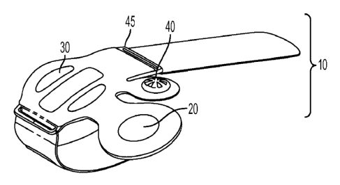
FIG. 1 shows the manual vascular compression device of the invention 1. The
device has a body 10 having pump 20, a pressure bladder 30 and a pressure
control
mechanism 40. The pump is in direct fluid connection with the pressure bladder
30. In
turn, the pressure bladder 30 is in direct fluid communication with the
pressure control
mechanism 40. As shown in FIG. 1 the pressure control mechanism 40 and the
pump 20
are not in direct fluid communication with each other and are otherwise only
connected
through the pressure bladder 30.
The pressure bladder 30 may take a number of different forms but is generally
made out of any flexible and/or pliable material. The device is placed on a
patient's
body on or near the area that requires hemostasis or occlusion. The bladder is
inflated
by means of the pump 20 using a fluid. Preferable the fluid is a gaseous
fluid, most
preferably the fluid is air.
It is preferable that the pressure bladder be inflated to a volume sufficient
to provide
hemostasis and/or occlude the vessel of interest, without occluding other
vessels. This is
particularly important in the ease of cardiac interventions through the radial
artery. In a
preferred embodiment the pressure bladder is capable of being inflated with
about 15-25
cc of fluid and in a preferred embodiment the pressure bladder is capable of
inflated to a
volume of no more than about 20 cc.
The device of FIG. 1 also includes securing means 50 for securing the body to
a
patient's body, for use in providing pressure for hemostasis. Preferably the
device 1 is
secured to a patient's for aid in providing pressure to the radial artery. The
securing
means may be a unitary piece or may be two separate pieces akin to a watch
band.
FIG. 2 is a rear prospective of an embodiment of the subject device and shows
further details of the securing means including optional attachment means 90
that can be
disposed on the securing means 50 to secure the device 1 to the patient. In
one
embodiment the attachment means may be a portion of hook and loop fastener
such as
VELCRO . Alternatively the securing means may comprise adhesives including
adhesive table or a system of holes and tines similar to a conventional watch
band.
FIG. 3 is a schematic of the device body 10 and further shows the
relationships
of the various components to one another. FIG. 3 further shows optional
coupling
means for coupling device body 10 to the securing means 50. In the
illustrative
CA 02828910 2013-09-03
WO 2012/129146
PCMJS2012/029600
- 10 -
embodiment the coupling means comprises openings 45 and 46 in the device body
which allow the securing means 50, which is preferably a flexible band to
pass, through
and secure and couple the securing means 50 to the device body. In a preferred
embodiment the securing means is unitary in nature (i.e. a single continues
piece) and is
disposed through the openings 45 and 46 and over the device body 10. FIG. 4 is
a
schematic of just such a securing means.
FIG. 5 shows a partial exploded view of the embodiment of FIG. 1. The view
shows an optional restrictor 80, which is disposes between the device body 10
and the
securing means 50. The restrictor is made of a rigid or semi-rigid material
and serves to
focus the force of the expanding pressure in a downward direction towards the
patient.
FIG. 5 also shows one embodiment of the pressure regulator 40 comprising a
valve 41
and a valve receptacle 42, the valve receptacle being integral to the device
body.
In one embodiment, the pump 20 is a commercially available configuration
which has been refined for efficient actuation between the thumb and side of
pointer
finger. The pump may optionally include an integral check valve to allow flow
into the
bladder and a return element to restore the pump to the starting position. The
return
element may take a number of different form including a spring, elastic or
other type of
device that is capable of providing sufficient force to return the pump to its
starting
position including but not limited to spring(s), elastics or other devices. In
one
embodiment the return element is made of foam, and provides a force to the
pump when
the pump is compressed.
In one aspect, where the fluid used to operate the device is air, a hole or
other
opening in the surface of the pump is introduced to allow the pump to refill
with air on
the return stroke. In a preferred embodiment, the hole is closed by the thumb
during
pumping to create pressure and hence flow through the check valve into the
bladder.
The bladder 30 is a generally spherical inflatable chamber which applies
pressure
between the bridge and the patient's wrist. The size of the bladder was
developed to
allow sufficient stroke to fill the space under the bridge and transfer the
internal pressure
to the patient incision site. The spherical form allows focusing the applied
force at the
point of contact at the center of the footprint. The pressure capacity,
volume, and
reliability requirements of the bladder have not been deteimined.
CA 02828910 2013-09-03
WO 2012/129146
PCMJS2012/029600
- 11 -
The pressure control mechanism 40 can take a number of different forms. As
discussed above, in one aspect, the pressure control mechanism 40 is comprised
of a
valve 41 for exhausting the fluid and a valve receptacle 42. In one
embodiment, the
exhaust valve is a normally closed valve seated by a spring and the closure
force is
increased when the bladder is pressurized. When actuated via pressing a button
on the
valve, the valve opens allowing flow which exhausts the pressure in the
bladder.
In one aspect of the invention the pressure bladder 30 is connected to the
pressure control mechanism 40 through an exhaust path. The exhaust path may
optionally contain a flow restrictor in the channel between the bladder and
the exhaust
valve. The flow restrictor may be used to control the exhaust flow rate so the
user can
reduce the pressure in a gradual and controlled manner. In a preferred
embodiment, the
flow restrictor is 0.006" in internal diameter and 0.25" long.
The vascular compression device is generally molded of a mostly flexible
material. The only requirement is that the material is sturdy enough to
withstand the
application of downward pressure onto a human patient, sufficient to cause a
complete
occlusion of an artery. Generally, the device should be capable of promoting
hemostasis
at blood pressure of at least about 200mm or mercury or 3.9 PSI. In a
preferred
embodiment, the device should be capable of generating at least about 8 PSI or
greater
of internal pressure or in other words at least about 2 times the blood
pressure. The
device 1 may be packaged and sterilized as a sterile medical product so that
the user
needs not clean or wash it prior to its use. In a preferred embodiment the
material is
transparent so that the user can more easily align the device with the wound.
In a further embodiment, the radial artery compression system of the invention
comprises: (a) the radial artery compression device defined herein in all of
its aspects
and embodiments; and (b) a brace for restricting the movement and/or rotation
of the
subject's wrist. Applicants have found that restricting movement of the wrist
and
associated structures improves the performance of the system and ultimately
improves
patient outcomes.
FIG. 6 shows an exemplar embodiment of the brace of the invention In this
embodiment the brace 600 has a an elongated rigid member 610 having a proximal
end
612 and distal end 611 along which a subject's forearm and hand would be
disposed in a
"palm up" orientation; the palm being disposed across and along the distal end
611. The
- 12 -
brace also has a plurality of fastening members 620 to secure the brace to the
subject's
arm. In the pictured embodiment, at least one fastening member 631 attaches to
a
fastener 611 at the distal end of the brace, thereby securing the subject's
hand and a
second fastening member 622 attaches to a fastener 632, thereby securing the
subject's
forearm. In the pictured embodiment the fastening member 620 are permanently
attached on one side of the brace and are fastened to the fasteners 631 and
632 on the
opposite side of the brace. One of ordinary skill, however would realize that
the brace
could be attached using a plurality of fastening members in other ways.
In the illustrated embodiment the brace includes one or more side-walls 630
that
are disposed approximately perpendicularly to the elongated rigid member 610
of the
brace. The optional side walls provide for better placement of the brace as
well as
further enhance the ability of the brace to restrict movement of the wrist and
its
associated structures. The side wall of the brace may also have a securing
region 635 to
which the securing means 50 of the vascular compression device 1 may be
secured. In
one embodiment the securing means may be a hook and loop type of fastener such
as
VELCROTm.
In one preferred embodiment, the rigid member is curved throughout its length
and about its longitudinal axis. The brace is attached on the dorsal aspect of
the
forearm, wrist and hand to support and immobilize the portion of the hand
proximal to
the wrist, the wrist and the portion of the forearm proximal to the wrist and
among other
functions prevents rotational movement of the hand around the wrist joint.
FIGs. 7A
and 7B show an alternative fastening arrangement of the brace 610. FIG. 7A
shows a
pair of fastening members 721 and 722 that are detachable from the brace and
can be
fastened at various positions through a series of holes. FIG. 7B shows the
brace 610
having a plurality of fastener pairs (731A and731B) and (732A and 732B)
disposed at
the distal end 611 of the brace 610 to which a fastener 721 may be attached. A
second
fastener 722 would be attached to a second pair 733A and 733B of fasteners at
the
proximal end 612 of the brace 610.
FIG. 8 shows the embodiment of FIGs. 7A and 7B with the addition of a
plurality of moldable flat areas 810 and 820. These moldable flat areas, which
may be
incorporated in any embodiment of the invention provide for a location to
which
adhesive may be applied to better secure the brace to the dorsal side of the
forearm. IN
CA 2828910 2018-09-21
CA 02828910 2013-09-03
WO 2012/129146
PCMJS2012/029600
- 13 -
one embodiment the adhesive may be a double ¨sided adhesive tape, but any
suitable
adhesive would be appropriate.
This detailed description of the invention is for illustrative purposes only.
A
reading by those skilled in the art will bring to mind various changes without
departing
from the spirit and scope of the invention.
EXAMPLES
Prior Art Devices
The standard of post procedure care for achieving hemostasis following radial
artery diagnostic and interventional cardiac catheterization is typically 2 to
6 hours,
using a variety of compression techniques.
An analysis of the current standard of care of the two most used devices the
TR
Band which is a wrist band based compression device and the Radistop which is
an
immobilization based device produced the following results (Comparison of TR
Band ad
Radistop Hemostatic Compression Devices After Transradial Coronary
Intervention,
Catheterization and Cardiovascular Interventions 76:660-667 (2010))
Wrist band (TR Band, Terumo, Japan)
Time to Hemostasis: 5.3 2.3 hours
Lowest time to Hemostasis: 1 hour obtained in approximately 3% of the patients
Local complication
Ecchymosis 11.4%
Oozing 6.1%
Large hematoma 2.8%
Small hematoma 6.1%
CA 02828910 2013-09-03
WO 2012/129146
PCMJS2012/029600
- 14 -
Arm Immobilization (Radistop, RAM: Uppsala, Sweden)
Time to Hemostasis: 4.8 2.2 hours
Lowest time to Hemostasis: 2 hours obtained in approximately 10% of the
patients
Local complication
Ecchymosis 10.6%
Oozing 7.1%
Large hematoma 1.5%
Small hematoma 4.8%
The addition of a hemostatic patch also does not seem to greatly improve
results.
A recent study (Korn et al., A New Vascular Closure Device for the Transradial
Approach, Journal of Interventional Cardiology Vol. 21, No. 4, 2008)
Showed the following results:
Wrist Band with Thrombin Hemostatic Patch
Mean duration of compression 4.6 hours
Bleeding after removal of the system 18.6%
Hematoma >5 cm 4.4%
Other complications (paresthesia of the thumb) 0.9%
It appears from the literature that regardless of the system used the mean
time to achieve
hemostasis is approximately four hours. And that the lowest reported
hemostasis time
was obtained by Rathere et al at 1 hour. This was obtained in only 3% of the
patients
receiving an aim immobilization device.
- 15 -
Methods:
Based on these results a clinical trial was organized to test if the subject
invention could increase the proportion of patients achieving hemostasis in
one hour.
Fifty (50) patients undergoing diagnostic and interventional radial cardiac
catheterization were studied as follows:
Group A
In 15 patients hemostasis was attempted using application the wrist band
component
of the invention plus SyvekTM Patch. The SyvekTm patch is a hemostatic patch
comprising
poly-N-Acetyl-Glucosamine (p-G1cNAc) as the hemostatic agent. The 15 patients
were
randomly assigned to 10, 30, or 60 minute compression intervals and hemostasis
was assessed
at each of these intervals. Hemostasis is defined as the ceasing of bleeding
with no re-
bleeding within 1 hour of the initial hemostasis.
Group B
In 35 patients hemostasis was attempted using the invention (wrist band and
the
brace) plus a SyvekTM Patch. The 35 patients were randomly assigned to 10, 30,
or 60 minute
compression.
Plethysmography and oxymetry were recorded and a Barbeau classification was
determined for both radial and ulnar artery flow at baseline, immediately
after
compression release, and lhr, 4hr, and 1 day post hemostasis depending on the
length of
patient hospital stay.
Results:
Group A
15 patients hemostasis was attempted using the wrist band component of the
invention plus
SyvekTM Patch (p-G1cNAc):
CA 2828910 2018-09-21
- 16 -
Hemostasis was successful as follows:
minutes ¨ 3 successful, 2 failures
5 30 minutes ¨ 4 successful, 1 failure
60 minutes ¨ 5 successful, 0 failures
No local complications
Group B
35 patients hemostasis was achieved using the invention (wrist band and the
brace) plus
a SyvekTM Patch.
Hemostasis was successful as follows:
10 minutes ¨ 12 successful (92.3%), 1 failure
30 minutes ¨ 12 successful (100%), 0 failures
60 minutes ¨ 10 successful (100%), 0 failures
No local complications
Results:
Unexpectedly the subject invention not only increases the proportion of
patients
achieving hemostasis at one hour from 3% to 100% but also was able to
achieving
hemostasis at 30 minutes (100%) and at 10 minutes (92.3%). Remarkably, the
patients
treated with subject inventions had no local complications.
What is claimed is:
CA 2828910 2018-09-21