Note: Descriptions are shown in the official language in which they were submitted.
CA 02828918 2013-09-03
WO 2012/120127
PCT/EP2012/054148
1
DEVICE AND METHOD FOR CLOSURE OF A BODY LUMEN
Field of the Invention
This invention pertains in general to the field of medical devices and methods
for closure of
openings in body lumina, such as a vessel, in a patient. More particularly,
the invention relates in
some embodiments to the field of sealing apertures created by medical
procedures that pierce the
walls of blood vessels in living tissue.
Background of the Invention
During certain types of medical surgery or treatment, an introducer is used to
access the
vascular system of a patient. The introducer is inserted through the wall of a
blood vessel in order to
obtain access to the vascular system and may thereafter be used for guiding
medical instruments
such as catheters, guide wires and the like.
After completion of the medical procedure, there will be an incision or a
wound in the wall
of the blood vessel corresponding to the size of the introducer. The bleeding
from the wound, which
is a result of such a surgical operation, can be stopped by applying direct
pressure on the wound.
However, applying direct pressure on the wound will require assistance of
medical personnel and
may also restrict the flow of blood through the vessel. lschemia may occur and
can lead to serious
consequences.
In cases of puncturing the femoral arteries, the required time may be as long
as about 45
minutes or more and in some cases re-bleeding occurs if the patient is not in
rest. Bleeding from a
vessel puncture in a substantially sized blood vessel can be severe.
A variety of methods and devices have been suggested for replacing the
traditional method
disclosed above, some of which involve introducing chemical compounds which
act as homeostasis
2 5 catalysts or as adhering agents, whilst others aim at introducing
various forms of plugging members
into the puncture.
Sealing devices in form of sealing plugs in the cutaneous channel at the
puncture site
outside the vessel are known, e.g. from US patent application 2009/0054926 or
European patent EP
1349501. However, the blood pressure inside the vessel may press the plug out
of position before a
3 0 reliable sealing has occurred.
Other sealing devices in form of double button type fasteners that are affixed
to each other,
in the type of an outside member and inside member in relation to the lumen
and a crosspiece
arranged across the puncture opening, both inside and outside of the vessel
wall are known, e.g.
from US patent 7,488,340, or US patent 7,572,274. The outside member and
inside member are
35 brought in a locked configuration upon assembly and compress the vessel
wall tissue around the
puncture opening.
However, such button type sealing devices may not seal off the puncture
optimally.
For instance, such button type devices may restrict the lumen and blood flow
therein. The
inside member of the device protruding into the blood vessel often
substantially restricts the patency
40 of the lumen. This is in particular the case for small diameter lumen,
such as at peripheral vessels. A
protruding member may also lead to a turbulent flow, which might cause
secondary effects, such as
creation of thrombosis or embolies.
Furthermore, the devices may damage the vessel wall, in particular in the case
of
peripheral vessels, such as the arteria subclavia, the arteria axillaris,
which for instance are
45 accessed in the region of the clavicle, or the arteria radialis for
access in the arm, which all are brittle
vessels.
The aforementioned double sided tissue compression of the vessel wall causes a
pressure
onto the wall tissue, which brings about a number of issues.
CA 02828918 2013-09-03
WO 2012/120127
PCT/EP2012/054148
2
For instance, the vessel wall may be damaged when the applied compression or
pressure
is too high. Necrotic tissue may be built up. The vessel wall may be
structurally weakened. The
vessel wall may get damaged by the device. A rupture of the vessel wall may
occur. As a
consequence, a dissection may occur, i.e. a bleeding out of the vessel wall
into surrounding tissue of
the vessel.
For instance arteriosclerotic vessel are conventionally difficult to seal off.
Arteriosclerotic
vessels are brittle, conventional sealing devices have difficulties to find
hold or damage the brittle
vessel wall, the lumen diameter is already reduced and may be further reduced
by members of the
known sealing devices protruding into the lumen, etc.
1 0 When the applied compression or pressure is too low, i.e. the button
device is put in place
too loose, pressure damages are avoided. However, a leakage may then occur.
Leakage of blood from the puncture site is not desired, and should be avoided.
In particular repeated puncture, e.g. necessary during intensive treatment
periods, of such
anatomically sensitive vessels, may lead to damage of the vessel.
W02006/034114 discloses thin film devices implantable within a human subject
for
occlusion of an aneurysm or body vessel. The devices are movable from an
elongated, collapsed
configuration for delivery to a deployed configuration within the body. Such
an occlusion device
includes a thin film mesh attached to a carrying frame. The carrying frame is
moveable between a
collapsed configuration and an expanded configuration. The thin film mesh can
include a plurality of
slits, slots and/or pores that typically vary in degree of openness as the
carrying frame moves
between the collapsed and the expanded configurations. The occlusion device is
transluminally
positioned within the blood vessel so that the thin film mesh substantially
reduces or completely
blocks blood flow to a diseased portion of a blood vessel. However, a puncture
itself is needed to
deploy this device into the vessel as it is not suitable to be delivered
itself through an opening to be
closed,
US2009/0143815 discloses a device for sealing a puncture opening in a wall of
a blood
vessel that includes a base frame including a first bi-stable material having
a first stable state
corresponding to a delivery configuration of the base frame, in which the base
frame is retracted to
have a relatively smaller overall profile, and a second stable state
corresponding to a deployed
configuration of the base frame, in which the base frame is extended to have a
relatively larger
overall profile. The base frame is sized to engage an interior surface of the
blood vessel wall when in
the deployed configuration. A sealing section is coupled axially as a section
to the base frame and
includes a second bi-stable material having a first stable state corresponding
to an initial
configuration of the sealing section, in which the sealing section permits
fluid flow, and a second
3 5 stable state corresponding to a barrier configuration of the sealing
section, in which the sealing
section prevents fluid flow. The sealing section in the barrier configuration
is sized to block fluid flow
through the puncture opening when the base frame is in the deployed
configuration. However, this
device is difficult to deliver as deployment of the base frame is not
controllable. Thus, the base frame
may expand and engage the vessel wall before the sealing section is correctly
positioned. Moreover,
this device may migrate along the vessel with too low self expansion pressure
of the base frame.
The device may also migrate into the vessel wall and damage the latter at too
high self expansion
force of the base frame. Reliable sealing is thus difficult to achieve with
this device. Moreover, a
structure as disclosed in US2009/0143815 is traumatic in relation to the
vessel wall, in particular at
the tissue surrounding the puncture opening.
EP2292147 Al of the same applicant as the present application, which was not
published
at the priority date of the present application, and which is incorporated
herein by reference in its
entirety for all purposes, discloses a medical device and a method for closure
of a puncture in a
body lumen by a device delivered through the puncture. The device has an
aggregate of a support
structure and a substantially fluid tight patch member attached thereto at an
attachment unit. Upon
CA 02828918 2013-09-03
WO 2012/120127
PCT/EP2012/054148
3
delivery through the puncture, the aggregate is detached from a delivery
device and the puncture is
intraluminally closed in a leakage tight manner.
However, EP2292147 Al does neither disclose the transluminal delivery of the
device to
other treatment sites than punctures, nor the use of a fiducial marker, nor
that the distal end of the
elongate delivery unit is radially releasably attacheable to the aggregate at
a radial attachment
position of the support structure intermediate between ends of the patch
member only and
detachable therefrom upon deployment of the aggregate in the body lumen.
It is an object of the present invention to provide a novel and inventive
device for closure
and sealing of an opening, like a puncture or incision formed in a blood
vessel or in other body
organs. It is an object of the present invention to provide a novel and
inventive device for closure
and/or sealing of a structural weakening in a body lumen wall, such as at
aneurysms. A further object
of the invention is to provide a puncture closure method or re-inforcement
method, in embodiments
utilizing the sealing device. The medical arts would benefit from a device
that allows for the sealing
of blood vessel wall punctures that are created at the termination of a tissue
tract that passes
through intervening tissues between the vessel wall puncture and a puncture
through the skin. It
would be preferred if the device was self-securing and small in size so as to
be introduced without
the need to enlarge the tissue tract beyond the size needed to perform the
primary medical
procedure. Preferably, the device has a high ratio of expanded to compressed
state while providing
reliable sealing right from the outset upon delivery. Preferably, the device
is retrievable or at least
repositionable.
Hence, an improved medical device or methods for closure of a puncture in a
body lumen
would be advantageous, and in particular allowing for increased flexibility,
and/or patient-friendliness
would be advantageous. Advantageously the solution should be atraumatic in
relation to the vessel
wall, in particular at the tissue surrounding the puncture opening to provide
a reliable sealing.
Summary of the Invention
Accordingly, embodiments of the present invention preferably seek to mitigate,
alleviate or
eliminate one or more deficiencies, disadvantages or issues in the art, such
as the above-identified,
singly or in any combination by providing a device, a kit and a method
according to the appended
patent claims.
According to a first aspect of the invention, a medical device is provided.
The medical
device is adapted for closure of a puncture in a body lumen, such as a vessel,
in a patient. The
device comprises an aggregate of a) a support structure having a first shape,
which is a temporary
delivery shape, for delivery to an interior of the body lumen and to be
subsequently subjected to a
change of shape to a second shape, which is a tubular shape, when delivered in
the body lumen,
and b) a substantially fluid tight patch member attached to the support
structure, which patch
member is at least partly arranged radially outside of the tubular support
structure and at least partly
arranged towards an inner tissue wall of the body lumen at a site of the
puncture of the body lumen
when the support structure has the second, tubular shape, such that the
puncture is sealed off by the
aggregate.
More particularly, a medical device for closing a puncture in a body lumen
from the inside
thereof, such as a vessel, in a patient, is provided. The device comprises an
elongate delivery unit
having a distal end; and an aggregate of a support structure having a first
shape, which is a
temporary delivery shape, for delivery to an interior of the body lumen
through the puncture and to
be subsequently controllably subjected to a change of shape to a second shape,
which is a tubular
shape, when delivered in the body lumen, and a patch member attached to the
support structure at
an intermediate portion between two opposite ends thereof. The distal end of
the elongate delivery
unit is radially releasably attached to the aggregate at an attachment
position intermediate between
ends of the patch member and detachable therefrom upon deployment of the
aggregate in the body
CA 02828918 2013-09-03
WO 2012/120127
PCT/EP2012/054148
4
lumen. The attachment position is for instance of a delivery wire distal end.
The attachment position
is preferably in the center of the patch. The patch member is sized and shaped
for arranging it
towards an inner tissue wall of the body lumen at a site of the puncture of
the body lumen and
extending over a puncture opening. The delivery unit extends in a direction
radially from the
aggregate through the opening, such that the puncture is controllably sealed
off by the patch when
drawing the delivery device in a direction out of the puncture and tightening
the patch over the
opening before the change of shape of the support structure.
In embodiments the device comprises an elongate delivery unit and a detachment
unit
detachably, wherein the delivery unit is connected to the aggregate by means
of the detachment unit
for delivery thereof to the interior of the body lumen, and for detachment of
the aggregate upon the
delivery to the interior of the body lumen; wherein the elongate delivery unit
preferably comprises a
delivery catheter, and/or a delivery wire releasably attached to the
aggregate. A single point
attachment of the elongate delivery unit may be provided in some embodiments,
preferably as a
delivery wire.
According to another aspect of the invention, a medical procedure is provided
in form of a
method. The method is a method of closure of a puncture in a body lumen, such
as a vessel, in a
patient, by a medical device. The method comprises deploying an aggregate of a
support structure
and a patch member in the body lumen through a puncture opening in the body
lumen at the
puncture site, wherein the patch member is a substantially fluid tight patch
member attached to the
support structure, and wherein the deploying comprises delivering the support
structure in a
temporary delivery shape to an interior of the body lumen, subsequently
subjecting the support
structure to a change of shape to a second shape, which is a tubular shape, in
the body lumen, and
thus arranging the patch member at least partly radially outside of the
tubular support structure and
at least partly towards an inner tissue wall of the body lumen the site of the
puncture of the body
lumen when the support structure has the second, tubular shape, and thus
permanently sealing off
the puncture from inside the body lumen by the aggregate.
Alternatively, some embodiments of the device may be delivered transluminally
or
transvascularly. Both arterial or venous access may be chosen. In this case,
an attachment of
embodied closure devices to the delivery device may be omitted. Such devices
may for instance be
3 0 pushed out of a delivery catheter in a conventional way.
For transvascular delivery the aggregate is provided during delivery with a
rotational
orientation of the patch towards the opening to be occluded. This may be
facilitated by means of
fiducial markers comprised in some embodiments of components of the aggregate.
For instance the
patch may comprise radiopaque threads. The fiducial markers may be of Barium
nitrate. The fiducial
markers may be comprised in the support structure and/or the patch. Thus the
rotational orientation
of the patch segment is identifiable inside the body relative the opening to
be occluded. Preferably
the fiducial markers are visible in X-ray imaging. Other imaging modalities
may alternatively or in
addition be used: MR, CT, US. Thus for instance a delivery catheter sheath in
which the aggregate is
collapsed, is rotated to a desired rotational direction and then the aggregate
is released from the
sheath and eventually detached from the delivery device/wire.
According to another aspect, a method of closure of a puncture in a body
lumen, such as a
vessel, in a patient, by a medical device, is provided. The "closure" may
comprise reinforcing of a
structurally weakened lumen wall section, such as at an aneurysm. The method
comprises providing
a patch member arranged on an outside of a tubular support structure and
attached to the support
structure at an intermediate portion between ends thereof; and an elongate
delivery unit having a
distal end, wherein the distal end thereof is radially releasably attached to
the support structure or
the patch member at an intermediate position between ends of the patch member
and detachable
therefrom upon delivery; wherein the patch member is arranged radially outside
of the tubular
support structure only at a partial radial section and axial section thereof
and arrangeable towards an
CA 02828918 2013-09-03
WO 2012/120127
PCT/EP2012/054148
inner tissue wall of the body lumen at a site of the puncture of the body
lumen when the support
structure has the second, tubular shape, such that the puncture is sealed off
by the patch of the
device hold by the support structure in the body lumen at the site.
According to another aspect, a method of closure of a puncture in a body
lumen, such as
5 a vessel, in a patient, by a medical device, is provided. The method
comprises providing a medical
device for closing the puncture in the body lumen from the inside thereof, the
method comprising
providing an elongate delivery unit having a distal end; and an aggregate of a
support structure
having a first shape, which is a temporary delivery shape, for delivery to an
interior of the body lumen
through the puncture and to be subsequently controllably subjected to a change
of shape to a
1 0 second shape, which is a tubular shape, when delivered in the body
lumen, and a patch member
attached to the support structure at an intermediate portion between two
opposite ends thereof;
wherein the distal end of the elongate delivery unit is radially releasably
attached to the aggregate at
an attachment position intermediate between ends of the patch member and
detachable therefrom
upon deployment of the aggregate in the body lumen; arranging the patch member
arranging
towards an inner tissue wall of the body lumen at a site of the puncture of
the body lumen and
extending over a puncture opening, and drawing the delivery device in a
direction out of the puncture
and tightening the patch thereby controllably sealing off the puncture by the
patch; and initiating the
change of shape of the support structure for anchoring the aggregate at the
puncture site in the
vessel; and releasing the delivery device from the aggregate.
In embodiments, the method comprises releasably attaching an elongate delivery
unit to
the aggregate for delivery thereof to the interior of the body lumen, and
detaching the aggregate
upon the delivery to the interior of the body lumen; wherein the elongate
delivery unit preferably
comprises a delivery catheter, and/or a delivery wire releasably attached to
the aggregate.
Further embodiments of the invention are defined in the dependent claims,
wherein
features for the second and subsequent aspects of the invention are as for the
first aspect mutatis
mutandis.
Some embodiments of the invention provide for safe anchoring of a puncture
closing or
sealing device with minimal risk of migration in a body lumen. The device is
anchored against the
vessel wall from the inside thereof. The device does thus not migrate or get
washed away from the
puncture site. The anchoring may be enhanced, e.g. by anchoring units or
members, such as barbs,
hooks, protrusions, or other means, such as tissue glue. Also, a protrusion
into the puncture
opening, e.g. an attachment or detachment device, arranged in the puncture
opening remaining
there after delivery, avoids a longitudinal migration or wash away. In
addition, barbs extending
through the patch, even at the puncture opening i.e. without engaging tissue
of the body lumen
surrounding the puncture opening, provide for a reliable fixation of the patch
member to the support
structure.
Hence, some embodiments provide for transpuncture delivery of a sealing
device. A patch
member of the device is atraumatic at delivery from the punctured lumen wall.
The patch is soft and
conformable. The patch is positioned against the luminal structure of the body
lumen,
A stent like support structure digs traumatically into the body lumen wall
around the patch.
The patch is arranged between the lumen wall and the stent and supported by
the stent. In this
manner, a weakening or puncture/opening in the lumen wall is covered
atraumatically, but reliably
kept in place by the traumatic engagement of the stent structure into the
lumen wall. The patch
covers circumferentially less than 360 degrees of the stent and is oriented
towards the
opening/weakening.
A desired radial orientation of the patch towards the opening of the puncture
and/or wall
weakening may be provided in several ways. For instance, the rotational
orientation of the stent and
attached patch may be controlled based on radiopaque or fiducial markers. The
stent may comprises
such markers, such as for instance of gold. Additionally or alternatively, the
patch may comprise
CA 02828918 2013-09-03
WO 2012/120127
PCT/EP2012/054148
6
radiopaque sections. These may be provided by radiopaque threads woven into
the patch fabric.
Radiopaque threads may be used for attaching the patch to the support
structure. This allows for a
folded over delivery compressed configuration that is very space efficient in
terms of cross section,
as described below.
An improved compression ratio is provideable as the patch is only arranged
over a portion
of the circumference of the support structure and further only attached to it
at a limited portion of the
circumference, e.g. at a single point or along a longitudinal line along the
length of the tubular
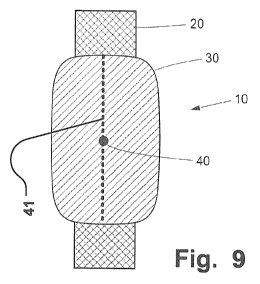
support structure (see 40 in the Figures or 41 in Fig. 9).
The fiducial marker may be a suture thread for attaching a fabric patch to the
stent like
1 0 tubular support structure (see Fig. 9).
The aggregate is preferably repositionable.
The aggregate is preferably retrievable.
The aggregate is positioned at the opening (transpuncture or transluminal
delivery). The
patch is rotated such that it is oriented towards the opening. This can for
instance be made by
rotating a delivery catheter, or a specific balloon inflated at a rotational
off-center position only. An
inflatable balloon may be arranged in opposite radial direction as the patch
to provide a directed
expansion of the patch in a desired direction. When delivering the aggregate
transpunctually, the
orientation may be provided by the radial position of the delivery wire out of
the puncture channel,
away from the lumen, and the radial attachment point of the wire at/through
the patch to the support
structure.
A proximal part of the aggregate of support structure/patch may only be
released from a
delivery catheter. In this manner, positioning may be checked. If desired, the
aggregate may be
retracted into the catheter sheath. Retrieval or re-positioning may then be
made for improved
delivery to a desired lumen site with an opening. Rotational re-positioning
may be done before a new
2 5 release attempt.
Upon complete expansion of the support structure, the latter anchors into the
lumen wall.
The support structure may be self-expandable. Alternatively, the support
structure may not
be self-expandable and then be expanded by an expansion unit, such as an
inflatable balloon.
However, a balloon will use more volume during delivery.
Start of expansion of the support structure is preferably controlled. This is
for instance
provided by a controllable lockable unit, which for instance may be tether
based. The lockable unit
may also be magnetically activated, or electrically activated. Alternatively,
or in addition, expansion
may be triggered by breakable connection points. The breakable connection
points may be activated
to break upon contact with body fluid, or at body temperature. Activation may
be time delayed, such
as for instance a pre-defined time after contact with body fluids or at body
temperature.
The wings are then folded over (rolled without creating edges, plies or
creases in the
patch) around the compressed tubular support structure, like a carpet. Thus
put into a delivery
catheter sheath, the aggregate is restricted. In this manner a very compact,
low cross section,
delivery configuration of the aggregate is provided. Delivery through small
vessels is thus facilitated,
reaching treatment sites longer into the vasculature that could not be treated
previously. When
provided with fiducial markers, such as radiopaque markers, as described
above, rotational
orientation upon delivery, i.e. before, during and after expansion of the
support structure is provided.
For instance upon being released from a delivery catheter, the folded over
wings of the patch will
unfold, e.g. turbulently supported by blood flow in the lumen. The patch is
then positioned against the
opening. Thereafter, the support structure is expanded. This expansion may be
triggered, as
described above. Upon fully expansion, the support structure will support the
patch over the opening
in the correct rotational orientation of the aggregate. The support structure
digs into the lumen tissue
where the patch is not arranged in-between, reliably anchoring the aggregate
at the opening.
Migration is avoided. Sealing of the opening is provided reliably and secure
by the atraumatic patch
CA 02828918 2013-09-03
WO 2012/120127
PCT/EP2012/054148
7
pushed against the orifice and surrounding tissue of the opening, or over the
lumen wall at the tissue
of the puncture channel end. Endoleakage out of the lumen is reliably avoided.
Embodiments of self-expanding support structures and aggregated patch provide
for very
compact delivery configurations. The collapsible and self-expanding tubular
support structure, like a
stent frame, is compressible to a very narrow diameter. The patch, attached at
a radial position
thereof, and not over the entire circumference, extends tangentially
outwardly, like wings.
The wing-like structure (before final delivery, see Fig. 2) is only in contact
at the radial
position of the attachment point in certain embodiments. It may be
additionally fixed at adjacent
radial positions, but always allowing for the radial orientation towards the
opening to be occluded
while not being attached to the support structure at its peripheral edges.
The patch may be made of a non-woven fabric, like a felted fabric. In
preferred
embodiments, the patch is made of a woven fabric.
The patch is preferably made of a natural material, like cotton. However, it
may
advantageously be made of a synthetic fabric, e.g. made of PTFE (GoreTexO).
Further treatment indications or areas of application of the aggregate and
related methods
comprise closure of openings. Such openings may comprise aneurysm neck
openings in certain
examples. Other examples comprise dissections or other perforations. Other
embodiments comprise
closure of side vessels originating from a main vessel. The side vessel may be
occluded by delivery
of the aggregate through the side vessel. Alternatively, the aggregate may be
delivered through the
main vessel. Positioning and delivery is reliably provided. Migration and
endoleakage is efficiently
avoided.
Some embodiments of the invention provide for self removing devices that leave
a vessel
wall with no remaining device after a certain period of time. This is for
instance achieved by means of
bioabsorbable or bioresorbable material. Such material may be applied both in
the support structure
2 5 and/or the patch member.
Some embodiments of the invention provide for a reduced risk of narrowing of
the body
lumen at the closed or radially sealed off puncture opening. The devices of
embodiments have a low
profile in radial direction. Some embodiments of the invention thus provide
for avoidance of a
turbulent flow in the body lumen downstream the puncture site. Thus secondary
effects are avoided,
3 0 such as creation of thrombosis or embolies.
Some embodiments of the invention provide for minimized or eliminated
shortcomings of
known sealing devices, such a minimized or eliminated risk for damaging
brittle lumen tissue, such
as vessel tissue, e.g. of the arteria subclavia, or the arteria axillaris. The
device and method of
embodiments may be applied to body lumen, which conventionally could not be
sealed off with
3 5 known devices.
Some embodiments of the invention avoid a manipulation of the vessel wall. For
instance
arteriosclerotic vessel walls are advantageously sealed off at puncture sites.
Some embodiments of the invention provide for intraluminal devices, which
apply a radially
outwardly oriented force, thus minimizing the risk of arterial vessel wall
manipulation.
40 Some embodiments of the invention avoid a squeezing of the vessel wall
tissue, thus
providing for reduced risk of tissue damage of the vessel wall.
Some embodiments provide for devices that are not substantially extending into
lumen.
The devices are flat, i.e. have a substantially lesser profile than the
diameter of the body lumen in
which they are anchored. The devices have a low profile in radial direction.
This provides for
45 avoiding turbulent flow at the puncture site when it is sealed off.
Some embodiments of the invention provide for a quick procedure for sealing a
puncture
site. The devices of embodiments are applied in a short time. There is no need
to wait for hardening
of chemical sealing agents. Conventional techniques, including the Seldinger
technique, may be a
basis for introducing embodiments of the device into the body lumen.
CA 02828918 2013-09-03
WO 2012/120127
PCT/EP2012/054148
8
Some embodiments of the invention provide for biocompatible devices reducing
potential
irritation or other implications of the puncture site.
Embodiments of the invention provide for an effective avoidance of bleeding
when the
puncture site is sealed off by means of devices according to embodiments.
Some embodiments of the invention also provide for devices that apply no
double sided
compression of a body lumen for sealing a puncture opening.
Embodiments of the invention provide for an effective atraumatic sealing of
such puncture.
This is for instance provided by a patch contacting tissue wall surrounding
the puncture opening. The
patch provides at the same time for a reliable sealing of the puncture as it
extends over the opening
from inside the vessel.
Embodiments provide for a reliable controllability of the sealing process as
the patch is
arranged in the device to be positioned against the tissue wall surrounding
the puncture opening and
across the opening before it is anchored in the vessel by the support
structure and released from a
delivery device. The patch prevents that blood leaves the vessel at the
puncture site through he
puncture opening and a percutaneous puncture channel. The puncture channel is
a tissue tract
communicating with the blood vessel.
Embodiments provide for the use of an introducer for the positioning and
deployment of the
puncture sealing device. An introducer already positioned in the patient for a
surgical procedure is
left in place for delivery of the puncture sealing device. This is in
particular advantageous from a
clinical perspective, providing for user acceptance of the device, and cost
efficiency as the introducer
is useable even during closing of the puncture site.
Embodiments provide for a device that is self-securing and small in size, and
to be
introduced without the need to enlarge the tissue tract (herein also called
puncture channel) beyond
the size needed to perform a primary medical procedure.
It should be emphasized that the term "comprises/comprising" when used in this
specification is taken to specify the presence of stated features, integers,
steps or components but
does not preclude the presence or addition of one or more other features,
integers, steps,
components or groups thereof.
Brief Description of the Drawings
These and other aspects, features and advantages of which embodiments of the
invention
are capable of will be apparent and elucidated from the following description
of embodiments of the
present invention, reference being made to the accompanying drawings, in which
Fig. 1 is a view from above showing a schematic illustration of an embodiment
of an
3 5 aggregate for sealing a puncture;
Fig. 2 is a frontal view showing a schematic illustration of the aggregate of
Fig. 1 attached
to a delivery wire;
Fig. 3A is a lateral view of the aggregate of Fig. 2;
Fig. 3B is a lateral cross sectional view of the aggregate of Fig. 2 in a
delivery catheter;
Fig. 4 is a view from above showing a schematic illustration of anther
embodiment of an
aggregate for sealing a puncture in a first shape;
Fig. 5 is a lateral view showing a schematic illustration of the embodiment of
Fig. 4 in a
second shape;
Fig. 6 is a cross sectional view of the aggregate of Fig. 4 in its first shape
attached to a
delivery device;
Figs. 7A-7J are schematic views illustrating a method of sealing a puncture
site by means
of a sealing device of the type shown in Figs. 1 and 2;
Figs. 8A-8D are schematic views illustrating a method of sealing a puncture
site by means
of a sealing device of the type shown in Figs. 4 and 5; and
CA 02828918 2013-09-03
WO 2012/120127
PCT/EP2012/054148
9
Fig. 9 illustrates an aggregate having a longitudinal fixation of a patch.
Description of embodiments
Specific embodiments of the invention will now be described with reference to
the
accompanying drawings. This invention may, however, be embodied in many
different forms and
should not be construed as limited to the embodiments set forth herein;
rather, these embodiments
are provided so that this disclosure will be thorough and complete, and will
fully convey the scope of
the invention to those skilled in the art. The terminology used in the
detailed description of the
embodiments illustrated in the accompanying drawings is not intended to be
limiting of the invention.
1 0 In the drawings, like numbers refer to like elements.
The following description focuses on embodiments of the present invention
applicable to a
blood vessel and in particular to a peripheral blood vessel. However, it will
be appreciated that the
invention is not limited to this application but may be applied to many other
punctured blood vessels
or body lumen, including for example those of the urinary tract, or the
gastrointestinal tract, including
bile ducts or liver vessels or ducts, or of kidney vessels or ducts, or the
central nervous system, or
side lumina thereof etc. However, embodiments do not include devices for
treatment of defects in
intra cardiac structures, such as atrial appendices, atrial or ventricular
septal defects, as these are
not body lumina within the meaning of this application.
Now turning to the Figures 1-3 an embodiment of the invention is described in
more detail.
Fig. 1 is a view from above showing a schematic illustration of an embodiment
of an aggregate 10 of
a medical device for closure of a puncture in a body lumen, such as a vessel,
in a patient. Fig. 2 is a
frontal view showing a schematic illustration of the aggregate 10 of Fig. 1
attached to a delivery wire.
Fig. 3A is a lateral view of the aggregate 10 of Fig. 2. Fig. 3B is a lateral
cross sectional view of the
aggregate 10 of Fig. 2 in a delivery catheter 60.
The medical sealing device is adapted for delivery through the puncture site
itself, into to
the interior of the body lumen, for deployment therein.
The medical device for closure or sealing of the puncture in the body lumen
comprises the
aggregate 10, which comprises a support structure 20 and a patch member 30.
The support structure 20 has a first shape, which is a temporary delivery
shape, for
delivery to an interior of the body lumen. Here, the first shape is a radially
compressed shape and
the support structure 20 is a collapsible tubular structure.
The tubular support structure 20 is expandable from the first shape, when
subsequently
subjected to a change of shape, to a second shape. The second shape is a
tubular shape. The
aggregate is adapted to change shape from the first shape to the second shape
when delivered in
the body lumen. The aggregate is deployed in the body lumen, and engages the
lumen wall of the
body lumen for a secure anchoring or fixation therein, avoiding a migration in
longitudinal direction of
the device along the lumen.
The tubular shape of the support structure 20 may be a net-like shape formed
of closed
loops, or a mesh shape of a braided, woven or knitted fabric. The support
structure may be produced
by suitably laser cutting a solid tube to provide a strut structure. The
support structure may be
provided in form of a stent like tubular member. The support structure may be
self-expandable.
Alternatively, or in addition, the support structure may be expandable by
expanding units, such as an
inflatable balloon. When self-expandable, expansion may be controllable as
described below.
Expansion by expanding units renders the change of shape controllable as such.
The anchoring may be enhanced, e.g. by anchoring members, such as barbs,
hooks,
protrusions, or other means, such as tissue glue comprised in the aggregate
10. Either, or both, the
support structure 20 and the patch member 30 may comprise such radially
outwardly arranged
anchoring members. The anchoring members engage with the wall tissue of the
body lumen, and
may protrude into the surrounding tissue. Anchoring members may also protrude
from the support
CA 02828918 2013-09-03
WO 2012/120127
PCT/EP2012/054148
structure through the patch member at the puncture opening, thus keeping it
reliably in place in
addition to the radially outwardly oriented anchoring force thereof.
In the embodiment, the support structure 20 is made of a resilient material
and is self-
expanding, and wherein the first shape is tubular of a smaller diameter than
the second, tubular
5 shape. A restriction unit may be provided for restricting resiliency
based expansion until the patch is
positioned over the puncture opening.
Alternatively, or in addition, the support structure 20 may be made of shape
memory
material, such as a shape memory polymer, or a shape memory alloy, such as
NiTinol. A restriction
unit may be provided for restricting shape memory based expansion until the
patch is positioned over
1 0 the puncture opening.
The patch member 30 is substantially fluid tight. This may be implemented by
providing the
patch member 30 of a suitable fabric. Alternatively, the patch member 30 may
be provided in form of
a solid membrane material.
The patch member is made of a tissue friendly material. The patch member is
not
necessarily 100% fluid tight, depending on the application. For instance,
blood coagulation may
occur upon deployment in the patch member providing for a sufficient sealing
to stop a bleeding out
of the puncture.
The patch member is semi rigid. The patch member is thus adapted to get into
apposition
with the tissue wall of the body lumen and conform to the structure thereof.
This provides for easy
2 0 deployment and a reliable sealing, e.g. upon retracting the delivery
wire.
The patch member may even be stretched or partly drawn into the puncture
channel, like a
thick paper tissue.
The patch member is atraumatically held into position over the puncture
opening by an
elongate delivery device attached to the sealing device within the surface
covered by the patch
member. Thus the puncture member is fixatable over the puncture opening by
drawing the delivery
device in a direction out of the patient.
The support structure may then be fully deployed to a tubular shape in the
body lumen,
Such release of the support structure to the tubular shape may be initiated by
active user operated
and controlled means, e.g. a tether, electrically, etc Alternatively, the
release of the support structure
to the tubular shape may be initiated automatically, e.g. after a certain time
in contact with body
fluids. The time is suitably chosen such that reliable positioning of the
patch member is provideable
for sealing the puncture before the change of shape is initiated.
Alternatively, or in addition, the
change of shape may be provided partly upon release in the body vessel, and
then finalized to the
fully tubular shape upon user operation or automatically after a suitable
time.
Upon final deployment of the support structure, the device is released from
the delivery
device and left in-situ.
The patch member 30 is attached to the support structure 20. Attachment is
made on at
least one defined point of the support structure 20, as illustrated at
attachment point 40 for a delivery
device in the Figures.
The patch member 30 is arranged radially outside of the tubular support
structure along a
portion of the tubular structure, when the support structure has its second
shape. The patch member
is adapted to fit over the puncture opening 110, thus being supported by the
support structure
providing a fluid tight sealing of a puncture site 100. The patch member is
thus at least partly
arranged towards an inner tissue wall of the body lumen at a site of the
puncture of the body lumen
when the support structure has the second, tubular shape, such that the
puncture is sealed off by the
aggregate 10.
The patch member itself is non-tubular and has a longitudinal extension
shorter than a
length of the support structure in the expanded diameter. Further, the patch
member has an
extension shorter than a circumference of the support structure in the
expanded diameter at the
CA 02828918 2013-09-03
WO 2012/120127
PCT/EP2012/054148
11
puncture site. The patch member is thus arrangeable radially outside of the
tubular support structure
only at a partial radial section and axial section thereof. This has the
advantage that migration along
the body vessel is prevented, as anchoring is provided by the support
structure outside of the patch
member when in contact with wall tissue, even in an axial portion along its
length at the puncture
opening.
In embodiments, the patch is not a so called "thin film" (only several microns
thick). A thin
film would not be suitable for attachment of a delivery unit due to lacking
structural strength.
The patch member has in some implementations for instance a thickness of 0,1mm
to
lmm, depending on the application site of the device. The patch thickness
should not substantially
reduce the channel cross section of the body lumen when the device is
implanted therein.
As can be seen in Fig. 1 and Fig. 2, the patch member may be attached to the
support
structure at a single location only. Preferably, this is a central location
where also the attachment unit
40 is located.
The periphery of the patch member is thus not attached to the support member.
In this
manner, an expansion of the support structure is not hindered by the attached
patch member.
The conformable patch member thus conforms to the inner of the tissue wall of
the body
vessel. Upon the change of shape it is anchored in that position from the
inside of the vessel by the
support structure.
As shown in Fig. 1, the elongate delivery unit is a delivery wire 50
releasably attached to
the device 10, at a position between two opposite ends of the device, at a
distal end of the delivery
wire. 50. The delivery wire 50 is attached to the device 10 at an area covered
by the patch member
30. Preferably, the delivery wire is attached to the device centrally at the
patch member. This
provides for a symmetrical arrangement. Alternatively, asymmetrical attachment
arrangements may
be provided, e.g. depending on requirements of the puncture site to be sealed
off.
The delivery wire is sufficiently rigid to push the sealing devices through a
catheter and/or
an introducer to the body vessel through the puncture channel.
As shown in Fig. 1, the delivery wire 50 is going through the patch member 30
and is
releasably attached to the support structure. Alternatively, or in addition,
the delivery wire may be
attached to the patch member, which then in turn is attached to the support
member.
The delivery device is retractable through the channel of the puncture out of
the patient.
This retraction is done after detachment when the aggregate of patch and
support structure is
deployed and seals the puncture from inside the body lumen.
The elongate delivery unit may further comprise a separate delivery catheter
insertable
through an introducer positioned in the puncture. The catheter is not attached
to the aggregate, but
merely facilitates delivery thereof.
In embodiments, the device's support structure 20 has a diameter in the
second, tubular
shape that is, at least slightly, larger than a diameter of the body lumen at
the puncture site. In this
manner the support structure 20 is devised to anchor the aggregate 10 in an
interior of a body lumen
210 at the puncture site 100. The patch member 30 is arranged to extend over
the puncture opening
110 in the body lumen 210 at the puncture site 100 for the closure of the
puncture. Also, in this
manner, the expanded shape of the aggregate 10, because it has a diameter
larger than that of the
lumen, will somewhat expand the wall of the body lumen 210 radially outwards.
In this manner, the
aggregate is radially outwardly oriented in relation to the natural diameter
of the inner body lumen ¨ it
is "recessed", pushing the lumen wall outside. This improves anchoring on the
one hand, but also
provides a large opening of the body lumen at the puncture site upon sealing
with the aggregate 10.
As shown in Fig. 2, the device further comprises an elongate delivery unit and
an
attachment unit 40 for temporary attaching the delivery unit to the aggregate
10. The delivery unit
may comprise a delivery wire 50 and a delivery catheter sheath 60, as
described above with
reference to Fig. 1. The delivery unit is elongate. At its distal end, the
delivery unit is connected to
CA 02828918 2013-09-03
WO 2012/120127
PCT/EP2012/054148
12
the aggregate 10 by means of the attachment unit 40, which might comprise a
detachment unit for
controlled release. When assembled, the aggregate is ready for delivery to the
interior of the body
lumen. Detachment of the aggregate upon the delivery to the interior of the
body lumen may be
made in various ways, such as by releasing a threaded attachment, activating a
detachment means,
such as a thermally, electrically, chemically initiated detachment, etc.
In another example, the proximal portion of the delivery wire 50 may be cut
off. This is
preferably made as close to the distal attachment position as possible.
In another examples, the delivery device may include a gripper or forceps like
tool at the
end of the delivery device. The attachment unit may then be shaped matingly to
allow for a reliable
engagement with the tool. The attachment unit may be spherically shaped,
allowing for the pivoted
movement during delivery. When the tool is locked, e.g. by a sleeve put over
the forceps or gripper,
a flexible deployment is provided with a reliable delivery without the risk of
unintentionally losing the
device into the body lumen. Detachment may be controlled from the outside of
the patient, e.g. by
withdrawing the locking sleeve and then opening the gripper or forceps.
In some embodiments, the delivery wire 50 is connected to the aggregate 10 at
the
attachment unit 40 by releasably threaded attachment. The attachment unit is a
threaded unit such
that the aggregate is detachable from the delivery unit by unscrewing the
delivery wire 50 from the
aggregate 10. This may leave a protruding attachment unit 40 in the puncture
opening, as will be
seen below, which advantageously contributes to the anchoring of the aggregate
10 at the puncture
site 100 for a reliable sealing. As shown in the figures, the thread is
arranged in a radial direction
outward from the support structure. The radial direction is substantially
perpendicular to a
longitudinal axis of the sealing device. As e.g. shown in Fig. 7H, the
attachment unit may extend
radially from the support structure 20 for being received in the puncture
opening, and also in the
puncture channel. In some embodiments, the attachment unit 40 is not extending
radially from the
support structure, but instead extending axially from one of the ends of the
support structure 20, so
that the support structure 20 can be pulled, transluminally, through a body
lumen, such as the body
lumen 210.
In some embodiments, the device or the aggregate 10 is provided with at least
one fixation
point or attachment unit 40 for attachment to a delivery unit or a delivery
wire 50. Such a fixation
point may be threaded or have a screw with windings for attaching the
aggregate 10 to the delivery
unit or the delivery wire 50. A threaded fixation point may be provided with
an external thread or with
an internal thread. Thus, the delivery unit or the delivery wire 50 should be
provided with a matching
threading, i.e. an internal threading or an external threading. If the
fixation point is threaded, the
device or the aggregate may be detached from the delivery unit or the delivery
wire 50 by
unscrewing the connection. In some embodiments, the at least one fixation
point may not be
provided with thread. Instead, the at least one fixation point is provided
with a non-threaded surface.
The delivery unit or the delivery wire 50 may then be provided with a gripper,
pincers, plier-like tool
or a forceps-like tool for gripping the fixation point of the device or the
aggregate 10. Thus, in order to
attach the delivery unit or the delivery wire 50 to the device or the
aggregate 10, the gripper will grip
the fixation point and in order to detach the delivery unit or the delivery
wire 50 from the device or the
aggregate 10, the gripper will release the fixation point.
In embodiments, the support structure 20 and/or the patch member 30 are made
of a
biodegradable and/or bioresorbable material.
The support structure 20 is for instance made of a polymer material, or
stainless steel, a
titanium alloy or a magnesium alloy. The support structure 20 may be provided
in form of a wire
structure.
The patch member 30 is for instance made of a biopolymer, or a metal alloy
like the
aforementioned. The patch member 30 is provided as a fabric. In other
examples, the patch member
30 may be provided additionally or alternatively as a solid membrane. The
patch member is semi
CA 02828918 2013-09-03
WO 2012/120127
PCT/EP2012/054148
13
rigid. The patch member is thus adapted to get into apposition with the tissue
wall of the body lumen
and to conform to the structure thereof.
The material has a suitable degradation rate under physiological conditions in
order to
make the aggregate become degraded or absorbed when the puncture has healed
completely.
Suitable biocompatible polymer materials are e.g. described in published US
patent
application US 2008/0095823, or PCT application PCT/EP2006/062400, which are
incorporated by
reference herein in their entirety for all purposes. Biocompatible polymer
materials comprise polymer
compositions with controlled degradation rates, such as polyhydroxyalkanoate.
In some embodiments, the support structure 20 and/or the patch member 30
comprise a
1 0 pharmaceutical agent.
The pharmaceutical agent is for instance adapted to prohibit a thickening of a
wall of the
body lumen, such as any one in the group of cyclosporine, taxiferol, rapamycin
and tacrolimus. Thus,
a reduction of the lumen diameter is further prevented and a flow through the
lumen maintained,
even during a healing phase of the puncture.
The pharmaceutical agent may comprise an anti-coagulation agent, such as
Heparin or an
anti-thrombotic agent. Thus, the passage of the lumen is effectively kept
open, and thrombosis at or
downstream the puncture site is prevented.
The patch member 30 may comprise a fibrosis promoting agent. This provides for
an
accelerated healing process for finalizing the final, biological sealing of
the puncture quicker. The
patch member 30 may comprise a scar reducing agent. In this manner, scars at
the puncture site
100 are effectively reduced, which is of interest for cosmetic treatment. A
fibrosis promoting agent or
scar reducing agent is preferably arranged at the patch member 30 such that it
is oriented towards
the vessel wall, more preferably towards the puncture opening. This may be
implemented by having
the agent as a coating or a surface layer on a side of the patch member 30
oriented in this manner
when the aggregate 10 is deployed.
The pharmaceutical agent may include any one in the group of an endothelia
growth
promoting agent, such as Endothelium Growth Factor. This provides for an
improved growth of a
thin layer of endothelia over the aggregate in the inner of the body lumen.
This layer of endothelia
further supports sealing of the puncture opening. Once a layer of endothelia
has built up,
biodegradation of the aggregate may be initiated, e.g. controlled by a delayed
biodegradation after
deployment.
The pharmaceutical agent may comprise an anti-pathogenic agent, or an anti-
infectious
agent, such as Nitric Oxide. This provides for a more reliable healing of the
puncture.
Any of the aforementioned agents may be present in an arbitrary suitable
combination at
the aggregate 10.
The sealing device of embodiments comprises at least one element to activate
or de-
activate the change of shape, such as a connection element of the support
structure that is arranged
such that a connection formed by the connection element between a first and
second part of the
support element. The connection element is configured to break when the
connection element is
subjected to a specific external influence, such as stress, temperature,
moisture, biodegration, or
absorption. Such connection elements are in detail describe in
PCT/EP2006/062403, which is
incorporated by reference herein in its entirety for all purposes. Thus, the
change of shape and
engagement with the tissue structure is controllable.
The sealing device of certain embodiments has a support structure 20 that is
bistable
between a first state of minimum energy and a second state of minimum energy,
whereby the
change of shape, in use, is obtained as a movement between the first state of
minimum energy and
the second state of minimum energy. Bistable devices, however for different
application than sealing
devices, are for instance disclosed in US patent application US 2002/0142119
or US patent
CA 02828918 2013-09-03
WO 2012/120127
PCT/EP2012/054148
14
application US 2004/0193247, which are incorporated by reference herein in
their entirety for all
purposes.
The body lumen is in specific embodiments a peripheral blood vessel, and the
puncture is
a percutaneous puncture of the blood vessel. More particularly, the blood
vessel is an arterial, high
blood pressure, blood vessel to be sealed off at a puncture site, preferably
after a finished surgical
procedure involving the use of intra body access through the puncture, e.g.
via an introducer unit.
The device is thus in specific embodiments an intravascular closure device.
More particularly, the
puncture is a blood vessel wall puncture at the termination of a tissue tract
that passes through
intervening tissues between the vessel wall puncture and a puncture through
the skin.
1 0 As for instance can be seen in Fig. 7H, the attachment unit 40 or
detachment unit is
arranged at the aggregate 10 such that the tubular support structure 20 is
arranged symmetrically in
longitudinal direction in the body lumen 210 in relation to the puncture site.
A kit comprises the afore described sealing device and an introducer sheath
90.
Figs. 7A-7J are schematic views illustrating a method of sealing a puncture
site by means
of a sealing device of the type shown in Figs. 1 and 2.
In the method an aggregate 10 of a support structure 20 and a patch member 30
are
deployed in the body lumen 210 through a puncture opening 110 in the body
lumen 210 at the
puncture site 100.
An introducer 90 having a port 94 at an exterior cap at a proximal end 92 is
shown inserted
in a puncture in Fig. 7A. The distal end of the introducer is in communication
with the proximal end
92, and inserted through the skin surface 151, the surrounding tissue 45, and
the body lumen wall
tissue 200.
To gain access to the body lumen, the Seldinger technique is employed. This
involves
placing a small gauge hollow needle through the skin at about a 30 degree
angle to intersect the
desired lumen. The needle is known to have punctured a blood vessel wall when
blood exits the
needle at the proximal end. A guidewire is inserted through the needle into
the vessel and the needle
is removed. A dilator with a lumen sized to fit the guidewire has a leading
tapered end and an
outside diameter sized to fit closely in an introducer sheath 90 placed over
it. The introducer sheath
size 90 is selected to accommodate the catheters anticipated to be used in the
procedure. The
introducer sheath 90 and tapered dilator are advanced together over the
guidewire through the skin
and into the vessel. The dilator and guidewire are then removed, since the
vascular pathway from
outside the body through the sheath and into the vessel have been established.
A self sealing
stretchable valve may be provided at the proximal end 92 of the introducer
sheath 90, which
minimizes blood loss from the introducer sheath during the procedure.
When a procedure performed via this port in the patient's body is finished,
the puncture
has to be sealed.
According to embodiments of the sealing method, the support structure 20 is
delivered in a
temporary delivery shape to the interior of the body lumen 210 through the
introducer 90, as
illustrated in Fig. 7B.
In Fig. 70, the aggregate 10 is advanced through the introducer 90, together
with the
catheter 60 and the delivery wire 50. 18. The aggregate 10 is attached to the
delivery unit by
screwing the delivery wire 50 to connect it to the aggregate 10 by releasably
threaded attachment.
Later on, the aggregate 10 is detached from the delivery unit by unscrewing
the delivery wire 50 from
the aggregate 10, as described further below.
Then, the delivery catheter 60 is partly withdrawn, releasing the aggregate
into the body
lumen, while still attached to the delivery wire 50 at the attachment unit 40,
as illustrated in Fig. 7D.
Further, the introducer and the delivery catheter are further withdrawn, such
as illustrated in Fig. 7E.
The delivery wire 50 is withdrawn, such that the attachment point 40 is drawn
to the puncture site
110. The aggregate 10 is thus suitably rotated and pivoted in relation to the
delivery wire 40, as
CA 02828918 2013-09-03
WO 2012/120127
PCT/EP2012/054148
illustrated in Figs. 7E and 7F. The patch member is automatically aligned
centrally in relation to the
puncture opening 110. The correct pivotal movement may be ensured by a
rotation of the guide wire
50.
The elongate delivery unit is thus radially releasably attached to the
aggregate. Attachment
5 may be made via a hinge, swivel or pivoting means at the attachment point
to the aggregate.
In this manner, the tightness of the body fluid leaking out of the puncture is
controlled by
the patch drawn against the opening.
At the same time, the support structure 20 is controllably subjected to a
change of shape to
a second shape, which is a tubular shape, in the body lumen. This change of
shape may for instance
10 be provided by resiliently expanding the support structure 20 when it is
released out a protective
sheath, restricting the support structure 20 from expanding during delivery.
Alternatively, or in
addition, the support structure 20 may change its shape based on a shape
memory effect, e.g.
initiated by the body temperature of the fluid in the body lumen 210. The
change of shape is
illustrated in Figs. 7D-7H.
15 The change of shape may be activated or de-activated by means of at
least one
connection element of the support structure that is arranged such that a
connection formed by the
connection element between a first and second part of the support element is
configured to break
when the connection element is subjected to a specific external influence,
such as stress,
temperature, moisture, biodegradation, or absorption.
The change of shape may be obtained by transforming the support structure from
a
bistable first state of minimum energy in the first shape to a second state of
minimum energy in the
second shape, by a movement between the first state of minimum energy and the
second state of
minimum energy.
The illustrated method comprises expanding the support structure to a diameter
in the
second, tubular shape that is larger than a diameter of the body lumen at the
puncture site. Further,
the support structure is anchored in the interior of the body lumen at the
puncture site, whereby the
patch member is arranged to extend over the puncture opening in the body lumen
at the puncture
site for the closure of the puncture. The anchoring may be enhanced, e.g. by
anchoring members,
such as barbs, hooks, protrusions, or other means, such as tissue glue.
The method may comprise self expanding the support structure 20 in the body
lumen 210
upon delivery therein. Preferably this is done when the patch is suitably
positioned and sealing
tightness is achieved.
Thus, the support structure upon the change of shape is holding the aggregate
in the body
lumen at the puncture site extending over the patch member radially between
the tissue wall and the
3 5 support structure in the second, tubular shape. The patch member is
overlappingly contacting the
inner tissue wall of the body lumen and is extending over the puncture
opening.
The patch member may be made to partly extend into the puncture channel at the
puncture
opening. This may provide for a particular quick and reliable sealing of the
puncture.
Hence, the puncture opening is initially closed with the patch member before
the support
member changes shape to the tubular shape.
The method comprises retracting the delivery device upon detaching from the
device
through a channel of the puncture out of the patient.
Hence, the support structure upon the change of shape is holding the aggregate
in the
body lumen at the puncture site extending over the patch member radially
between the tissue wall
and the support structure in the second, tubular shape.
The patch member is overlappingly contacting the inner tissue wall of the body
lumen and
is extending over the puncture opening.
The method may comprise drawing the patch member partly into the tissue tract
from the
puncture opening.
CA 02828918 2013-09-03
WO 2012/120127
PCT/EP2012/054148
16
In certain medical procedures, the puncture channel may additionally be closed
by injecting
or inserting a clotting induction agent such as collagen that encourages
clotting in the puncture
channel.
The method may comprise initially closing the puncture opening with the patch
member
before the support member changes shape to the tubular shape.
The method may comprise retracting the delivery device upon detaching from the
device
through a channel of the puncture out of the patient.
The patch member is thus arranged radially outside of the tubular support
structure,
towards an inner of tissue wall 200 of the body lumen 210 at the puncture site
100 of the body lumen
when the support structure has the second, tubular shape, and thus the
aggregate 10 is permanently
sealing off the puncture from inside the body lumen by the aggregate.
The sealing effect of the aggregate 10 is supporting or enhanced by a
physiological
pressure of a body fluid in the body lumen onto the patch member, pressing it
against the tissue wall
200 of the body lumen 210.
Thus, an intra-luminal leakage tight sealing of the puncture is obtained.
The method may comprise delivering a pharmaceutical agent, as those described
above,
from the aggregate 10 at the puncture site to the body lumen 210.
The elongate delivery unit releasably attached to the aggregate 10 for
delivery thereof to
the interior of the body lumen 210 is then detached from the aggregate,
leaving the aggregate
2 0 securely in place, as illustrated in Fig. 7H.
The puncture channel through the vessel wall 200 and the surrounding tissue,
as well as
the outer skin will heal, as illustrated in Fig. 7H.
The method may further comprise biodegrading the aggregate 10 when deployed in
the
body lumen at a degradation rate under physiological conditions. When the
aggregate 10 is made of
a biodegradable or bioresorbable material, the puncture site will be reliably
sealed, without any
remainders of the aggregate 10 at the previous puncture site, as shown in Fig.
7J. The aggregate 10
may be provided to start to biodegrade when endothelium has covered the
aggregate installed in the
body lumen.
The method and device facilitate re-puncturing the body-lumen 210 at the
puncture site
100 by re-enforcing the lumen wall 210 of the body lumen 210 and supporting a
patency of body
lumen.
Both the support structure 20 and the patch member 30 are penetratable by a
needle tip
when a new puncture of the body lumen 210 is desired after sealing of the
puncture site 100.
When the aggregate 10 is absorbed or degraded, the previous puncture site is
also
3 5 available for a new puncture.
In embodiments, the body lumen is an arterial blood vessel. In particular, the
body lumen is
a peripheral blood vessel, and the puncture is a percutaneous puncture of the
body vessel, wherein
the device is an intravascular closure or sealing device for a vascular
puncture.
The peripheral blood vessel is in particular an arteria subclavia, or an
arteria axillaris of the
patient. The puncture site is in particular in a region of a clavicle of the
patient.
In embodiments, the body lumen is the patient's aorta, including the ascendant
or
descendent aorta, or branch vessel of the aorta.
A further embodiment of the device and method of the invention is illustrated
in Figs. 4-6
and 8A-8D. Fig. 4 is a view from above showing a schematic illustration of
another embodiment of an
aggregate 10 for sealing a puncture, in a first shape. The first shape is in
this embodiment an
elongate shape, which is substantially straight.
The support structure is made of a resilient material and/or a shape memory
material, such
as a shape memory polymer or a shape memory metal or alloy thereof. The
second, tubular shape is
CA 02828918 2013-09-03
WO 2012/120127
PCT/EP2012/054148
17
a helically coiled shape of the support structure 20. Fig. 5 is a lateral view
showing a schematic
illustration of the embodiment of Fig. 4 in the second shape.
Fig. 6 is a cross sectional view of the aggregate 10 of Fig. 4 in its first
shape attached to a
delivery wire 50 in its first shape. Like the above described embodiments, the
aggregate 10 may be
restricted in this first shape by a catheter sheath.
The patch member 30 is an elongate strip of fluid tight material. The patch
member is
attached to the support structure 30 along a portion of its length. The length
of the patch member is
at the most equal to the entire length of the support structure 30.
The patch member 30 may be put like a sock over the elongate support structure
20.
The patch member extends like a collar from the support structure.
The patch member may have a plurality of patch sub units (not shown) arranged
along the
length of the elongate support member. The patch units, e.g. of fabric, are
arranged at a distance
from each other such that they are arranged in the same radial direction at
the attachment point of
the delivery device distal end at the tubular support structure in the second,
tubular coiled shape.
When the support structure is in its second shape, the patch member is
arranged
overlapping itself to provide a fluid tight structure. The patch member may
overlap on the inside or
the outside of the tubular structure. In this manner, a fluid tight structure
of overlapping strips 31 at
each turn or winding of the helical coil making up the tubular structure, is
provided, as illustrated in
Fig. 5.
The minimum length of patch member is such that it extends along so many
windings that
longitudinally extend over the puncture opening and a bit further, in order to
provide a reliable sealing
thereof.
The width of the patch member 30 is determined by the pitch of the helical
coil, and is
larger than the distance between two windings of the latter. Thus the distance
between the windings
is bridged, and by the overlapping sections of adjacent windings, a continuous
fluid tight structure is
provided along the tubular support structure.
The width may also vary along the length of the patch member 30. For instance
the end
section may narrow down. The middle section may have a larger width to provide
a larger overlap.
The longitudinal width variation may be chosen suitably depending on various
parameters, such as
the anatomical structure of the body lumen at the puncture site, the pressure
of a body fluid at the
puncture site, etc.
Figs. 8A-8D are schematic views illustrating a method of sealing a puncture
site by means
of a sealing device of the type shown in Figs. 4 to 6.
Introduction of the delivery assembly of Fig. 6 into the body lumen at the
puncture site is
made similar as described above with reference to Figs. 7A-7E and is not
repeated to avoid
redundancy.
The method comprises transforming the support structure 20 of Fig. 4-6 from
the first
elongate delivery shape, to the second, tubular shape, which is a helically
coiled shape of the
support structure, wherein the transforming is based on an elasticity or shape
memory effect of the
4 0 support structure 20.
Fig. 8A shows the aggregate 10 attached to the delivery wire when deployed at
the
puncture site 100 and during transformation from the first to the second
shape. The delivery wire 50
is withdrawn, thus arranging the attachment unit 40 in the puncture opening
and centering the device
longitudinally in relation to the opening. This is illustrated in Fig. 8B,
where the strips 31 are shown in
full overlap and the guide wire detached from the aggregate 10.
When the coiled tubular member has section that are not covered by overlapping
strips 31
at its en section or end sections, this provides an enhanced anchoring of the
aggregate in the tissue
wall 200 of the body lumen as the support member may at least partly migrate
into the tissue wall.
CA 02828918 2013-09-03
WO 2012/120127
PCT/EP2012/054148
18
In Fig. 80 and 8D, like in Figs. 71 and 7J, the puncture site is shown healed
and the device
resorbed, respectively.
Alternatively, some embodiments of the device may be delivered transluminally
or
transvascularly. Both arterial or venous access may be chosen. In this case,
an attachment of
embodied closure devices to the delivery device may be omitted. Such devices
may for instance be
pushed out of a delivery catheter in a conventional way.
For transvascular delivery the aggregate is provided during delivery with a
rotational
orientation of the patch towards the opening to be occluded. This may be
facilitated by means of
fiducial markers comprised in some embodiments of components of the aggregate.
For instance the
patch may comprise radiopaque threads. The fiducial markers may be of Barium
nitrate. The fiducial
markers may be comprised in the support structure and/or the patch. Thus the
rotational orientation
of the patch segment is identifiable inside the body relative the opening to
be occluded. Preferably
the fiducial markers are visible in X-ray imaging. Other imaging modalities
may alternatively or in
addition be used: MR, CT, US. Thus for instance a delivery catheter sheath in
which the aggregate is
collapsed, is rotated to a desired rotational direction and then the aggregate
is released from the
sheath and eventually detached from the delivery device/wire.
Hence, some embodiments provide for transpuncture delivery of a sealing
device. A patch
member of the device is atraumatic at delivery from the punctured lumen wall.
The patch is soft and
conformable. The patch is positioned against the luminal structure of the body
lumen,
A stent like support structure digs traumatically into the body lumen wall
around the patch.
The patch is arranged between the lumen wall and the stent and supported by
the stent. In this
manner, a weakening or puncture/opening in the lumen wall is covered
atraumatically, but reliably
kept in place by the traumatic engagement of the stent structure into the
lumen wall. The patch
covers circumferentially less than 360 degrees of the stent and is oriented
towards the
opening/weakening.
A desired radial orientation of the patch towards the opening of the puncture
and/or wall
weakening may be provided in several ways. For instance, the rotational
orientation of the stent and
attached patch may be controlled based on radiopaque or fiducial markers. The
stent may comprises
such markers, such as for instance of gold. Additionally or alternatively, the
patch may comprise
radiopaque sections. These may be provided by radiopaque threads woven into
the patch fabric.
Radiopaque threads may be used for attaching the patch to the support
structure. This allows for a
folded over delivery compressed configuration that is very space efficient in
terms of cross section,
as described below.
An improved compression ratio is provideable as the patch is only arranged
over a portion
of the circumference of the support structure and further only attached to it
at a limited portion of the
circumference, e.g. at a single point or along a longitudinal line along the
length of the tubular
support structure (see 40 in the Figures or 41 in Fig. 9).
The fiducial marker may be a suture thread for attaching a fabric patch to the
stent like
tubular support structure (see Fig. 9).
The aggregate is preferably repositionable.
The aggregate is preferably retrievable.
The aggregate is positioned at the opening (transpuncture or transluminal
delivery). The
patch is rotated such that it is oriented towards the opening. This can for
instance be made by
rotating a delivery catheter, or a specific balloon inflated at a rotational
off-center position only. An
inflatable balloon may be arranged in opposite radial direction as the patch
to provide a directed
expansion of the patch in a desired direction. When delivering the aggregate
transpunctually, the
orientation may be provided by the radial position of the delivery wire out of
the puncture channel,
away from the lumen, and the radial attachment point of the wire at/through
the patch to the support
structure.
CA 02828918 2013-09-03
WO 2012/120127
PCT/EP2012/054148
19
A proximal part of the aggregate of support structure/patch may only be
released from a
delivery catheter. In this manner, positioning may be checked. If desired, the
aggregate may be
retracted into the catheter sheath. Retrieval or re-positioning may then be
made for improved
delivery to a desired lumen site with an opening. Rotational re-positioning
may be done before a new
release attempt.
Upon complete expansion of the support structure, the latter anchors into the
lumen wall.
The support structure may be self-expandable. Alternatively, the support
structure may not
be self-expandable and then be expanded by an expansion unit, such as an
inflatable balloon.
However, a balloon will use more volume during delivery.
Start of expansion of the support structure is preferably controlled. This is
for instance
provided by a controllable lockable unit, which for instance may be tether
based. The lockable unit
may also be magnetically activated, or electrically activated. Alternatively,
or in addition, expansion
may be triggered by breakable connection points. The breakable connection
points may be activated
to break upon contact with body fluid, or at body temperature. Activation may
be time delayed, such
as for instance a pre-defined time after contact with body fluids or at body
temperature.
The wings are then folded over (rolled without creating edges, plies or
creases in the
patch) around the compressed tubular support structure, like a carpet. Thus
put into a delivery
catheter sheath, the aggregate is restricted. In this manner a very compact,
low cross section,
delivery configuration of the aggregate is provided. Delivery through small
vessels is thus facilitated,
reaching treatment sites longer into the vasculature that could not be treated
previously. When
provided with fiducial markers, such as radiopaque markers, as described
above, rotational
orientation upon delivery, i.e. before, during and after expansion of the
support structure is provided.
For instance upon being released from a delivery catheter, the folded over
wings of the patch will
unfold, e.g. turbulently supported by blood flow in the lumen. The patch is
then positioned against the
opening. Thereafter, the support structure is expanded. This expansion may be
triggered, as
described above. Upon fully expansion, the support structure will support the
patch over the opening
in the correct rotational orientation of the aggregate. The support structure
digs into the lumen tissue
where the patch is not arranged in-between, reliably anchoring the aggregate
at the opening.
Migration is avoided. Sealing of the opening is provided reliably and secure
by the atraumatic patch
pushed against the orifice and surrounding tissue of the opening, or over the
lumen wall at the tissue
of the puncture channel end. Endoleakage out of the lumen is reliably avoided.
Embodiments of self-expanding support structures and aggregated patch provide
for very
compact delivery configurations. The collapsible and self-expanding tubular
support structure, like a
stent frame, is compressible to a very narrow diameter. The patch, attached at
a radial position
3 5 thereof, and not over the entire circumference, extends tangentially
outwardly, like wings.
The wing-like structure (before final delivery, see Fig. 2) is only in contact
at the radial
position of the attachment point in certain embodiments. It may be
additionally fixed at adjacent
radial positions, but always allowing for the radial orientation towards the
opening to be occluded
while not being attached to the support structure at its peripheral edges.
The patch may be made of a non-woven fabric, like a felted fabric. In
preferred
embodiments, the patch is made of a woven fabric.
The patch is preferably made of a natural material, like cotton. However, it
may
advantageously be made of a synthetic fabric, e.g. made of PTFE (GoreTexO).
Further treatment indications or areas of application of the aggregate and
related methods
comprise closure of openings. Such openings may comprise aneurysm neck
openings in certain
examples. Other examples comprise dissections or other perforations. Other
embodiments comprise
closure of side vessels originating from a main vessel. The side vessel may be
occluded by delivery
of the aggregate through the side vessel. Alternatively, the aggregate may be
delivered through the
CA 02828918 2013-09-03
WO 2012/120127
PCT/EP2012/054148
main vessel. Positioning and delivery is reliably provided. Migration and
endoleakage is efficiently
avoided.
While several embodiments of the present invention have been described and
illustrated
herein, those of ordinary skill in the art will readily envision a variety of
other means and/or structures
5 for performing the functions and/or obtaining the results and/or one or
more of the advantages
described herein, and each of such variations and/or modifications is deemed
to be within the scope
of the present invention as defined by the patent claims.
For instance the attachment unit 40 may be integrated in the patch member 30,
e.g. in form
of a loop, ring, etc. The attachment unit may be non-protruding. The
attachment unit may be part of
1 0 the delivery unit and removed therewith upon closure of the puncture.
Alternatively to the expansion of the aggregate as described above and
illustrated in the
Figs., the aggregate may be first drawn towards the puncture opening 110, such
that the patch
member 30 abuts around the opening. The expansion of the support member may
then be
controllably initiated. For instance the tubular expansion may be suitably
triggered, e.g. by breaking
15 connection points by a suitable external influence.
Different method steps than those described above, even merely in a different
order. The
different features and steps of the invention may be combined in other
combinations than those
described. The scope of the invention is only limited by the appended patent
claims.