Note: Descriptions are shown in the official language in which they were submitted.
CA 02829097 2015-01-09
MEDICAL DEVICE FOR USE WITH A STOMA
FIELD OF INVENTION
This invention relates to a device for use in percutaneous applications by
patients who have undergone surgery as a result of which an opening or stoma
has been left in the wall of a hollow body cavity, such as an intestine,
and/or in the
abdominal wall.
BACKGROUND OF THE INVENTION
For patients .having intestinal surgery or other operations to repair or
remove a section of intestine, it is frequently necessary to perform a
colostomy
operation or an ileostomy operation. With a colostomy, the large intestine is
brought through the abdominal wall, and with an ileostomy, the small intestine
is
brought through the abdominal wall. In each case, an opening called a stoma is
created to provide a conduit for allowing elimination of waste material from
the
patients body. Drainage or discharge from the digestive system of the patient
takes place through the opening or stoma in the abdominal wall. The body duct
protruding from the abdominal wall is typically sutured or otherwise adhered
to the
skin surrounding the opening. A flexible bag or other receiving means is
typically
attached to the stoma to collect and retain liquid, solid, and gaseous waste
material eliminated through the stoma.
An exemplary such procedure is illustrated in Figures 1A and1B, showing a
loop ileostomy 20. The stoma 24 is created by cutting the loop of the
intestine
protruding from the abdomen. Upstream section 23 of the intestine empties the
intestinal contents through the stoma. A mucous fistula 21 is formed on the
downstream end 22 of the intestine, typically blocking that section from
receiving
1
CA 02829097 2015-01-09
intestinal contents while the stoma is in place. A shunt 5 is sometimes used
between the skin and the loop of intestine.
Externalizing the intestine to form a stoma has disadvantages. It is
sometimes difficult to. control the flow of intestinal contents and there
arises a
consequential risk of infection and skin irritation. Attachment of ostomy
appliances for collection of the intestinal matter can also be difficult.
Stenosis and
prolapse of the intestine are additional risks with this type of procedure.
A similar procedure might be undertaken to connect two hollow biddy
cavities or organs within the body, thereby allowing one organ to drain into
another. For instance, a stoma, may be created in a hollow body cavity within
the
body in order to allow the cavity to drain into the GI tract.
SUMMARY OF THE INVENTION
Applicant has addressed the many disadvantages associated with
conventional stomas by providing a device that can be utilized with a stoma
and,
for example, eliminates ,the need to externalize an intestine through the
abdominal
wall. In an exemplary embodiment, the invention provides a device including a
proximal portion adapted for placement intermediately within an intestine, or
other
hollow body cavity or organ, to capture and divert contents; the proximal
portion
being expandable, optionally by using a self-expandable nitinol stent, from an
initial state with an initial diameter smeller than a diameter of the
intestine for
insertion of the proximal portion into the intestine, for example, into an
expanded
state with ,a diameter greater than the initial diameter for engaging the
proximal
portion with an inner well ofthe intestine; and a distal portion, connected to
the
proximal portion, adapted to extend through the abdominal wall, or
alternatively
into another hollow body cavity or organ, to conduct the contents out of the
proximal portion in an alternative embodiment, the device also inclUdes a
valve
connected to the distal portion to provide continence, allowing contents to be
selectively, discharged from the distal portion. The device optionally
includes a
transitional portion connecting the proximal portion to the distal portion.
The
2
CA 02829097 2013-09-04
WO 2012/122220
PCT/US2012/027984
proximal portion is optionally compressible from the expanded state for
removal of
the proximal portion from the intestine or other hollow body cavity or organ.
In alternative embodiments, the distal portion has an adjustable length,
either through compression, or by removing portions of the device in a
controlled
manner. The distal portion is also optionally corrugated. The device may be
flexible, crush resistant, and kink resistant.
In another aspect, a method of draining hollow body cavity contents is
provided comprising the steps of (a) making an incision in the GI tract; (b)
making
an incision into the hollow body cavity wall; (c) inserting through said
incisions a
device according to the present invention; (d) positioning said proximal
portion
within the hollow body cavity; (e) deploying said proximal portion to capture
and
divert hollow body cavity contents; and (f) positioning said distal portion
within the
GI tract to drain the hollow body cavity contents out of the proximal portion
and
into the GI tract,
In yet another aspect, the invention provides a method of diverting intestinal
contents from an intestine without bringing the intestine through an abdomen
comprising the steps of (a) making an incision through the abdominal wall; (b)
making an incision into the intestine without severing an entire diameter of
the
intestine; (c) percutaneously inserting through the incisions a device of the
present
invention; (d) positioning the proximal portion within the intestine; (e)
deploying the
proximal portion to capture and divert intestinal contents; and (f)
positioning the
distal portion to extend through the abdominal wall to conduct the intestinal
contents out of the proximal portion. Optionally, the invention includes the
step of
attaching a valve to the device to provide continence allowing intestinal
contents
to be selectively discharged from the device. Further optional steps include
attaching the intestine to an inner wall of the abdomen to seal the intestine,
and
adjusting the length of the distal portion to account for the thickness of the
abdominal wall. The invention also alternatively includes the step of removing
said device from the intestine,
3
CA 02829097 2013-09-04
WO 2012/122220
PCT/US2012/027984
DESCRIPTION OF THE DRAWINGS
Fig. 1A is a side view of a loop ileostomy according to the prior art.
Fig. 1B is a schematic side view of a loop ileasotmy according to the prior
art,
Fig. 2 is a side view of an exemplary embodiment of the present invention.
Fig. 3 is a side view of another exemplary embodiment of the present
invention.
Fig, 4 is a side view of another exemplary embodiment of the present
invention
Fig. 5 is a side view of another exemplary embodiment of the present
invention.
Fig. 6 is a side view of another aspect of the present invention.
Fig. 7 is a side view of another aspect of the present invention,
Fig. 8 is a side view of another exemplary embodiment of the present
invention.
Fig. 9 is a side view of another aspect of the present invention,
Fig. 10 is a side view of another embodiment of the present invention.
Fig. 11 is a side view of the embodiment of Fig. 10 commencing inversion,
Fig. 12 is a perspective view of the embodiment of Fig. 10 partially inverted.
Fig. 13 is a side view of one embodiment of the device of the present
invention preferentially bent into a C-shape orientation.
Fig. 14 is a perspective view of the device of Fig. 13 in use.
Fig, 15 is a perspective view of another embodiment of the present
invention.
DETAILED DESCRIPTION OF THE INVENTION
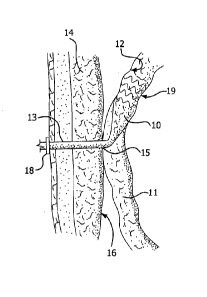
Figure 2 illustrates an exemplary embodiment of a device according to the
present invention. A proximal portion 10 of the device is disposed
intermediately
within an intestine 11. "Intermediately" as used herein means within the
length of
4
CA 02829097 2013-09-04
WO 2012/122220
PCT/US2012/027984
the intestine, as opposed to at a surgically severed end thereof. Proximal
portion
is adapted to capture and divert intestinal contents from within intestine 11.
The material of construction for proximal portion 10 may be durable
biocompatible
barrier material that prevents leaks and allows intestinal contents to pass
along
5 the internal length of proximal portion 10 without sticking to it. These
attributes
may be achieved by the material itself, or by combining the material with a
suitable
coating. Preferably, proximal portion 10 is made of a multilayer construction
of
fiuoropolymer, such as expanded polytetrafluoroethylene (ePTFE). Alternative
materials for proximal portion 10 include other fluoropolymers (such as FEP),
10 polyethylene, polypropylene, polyolefins, polyimides, polyesters,
silicone,
fluorosilicone, and bioabsorbable materials such as polymers and copolymers of
PGA, TMC, PLA and any combination of any of these materials. In certain
embodiments, the barrier material of the proximal portion may comprise at
least
one aperture therein.
Proximal portion 10 includes support structure 19. Support structure 19 is
preferably a self-expanding material, such as nitinol. Alternatively, support
structure 19 is stainless steel or other biocompatible metal or polymer which
is
expandable by the application of an external force, such as balloon-expandable
materials. Also alternatively, support structure 19 may be formed of a
polymeric
material. Support structure 19 may be bioabsorbable or nonbioabsorbable.
Support structure 19 may be disposed on the inside or the outside of the
perimeter of distal portion 10; that is, support structure 19 may be around
the
outside of the ePTFE (for example) used for the proximal portion 10, or it may
be
disposed inside the ePTFE used for proximal portion 10 It could alternatively
be
sandwiched between layers or coatings of the material used for proximal
portion
10. In any case, it is attached to the ePTFE (for example) and is used to
exert an
outward force that engages the inner wall of intestine 11 and secures proximal
portion 10 in place therein, allowing intestinal contents to be substantially
fully
diverted from intestine 11.
5
CA 02829097 2013-09-04
WO 2012/122220
PCT/US2012/027984
Support structure 19 enables proximal portion 10 to be expandable, from
an initial state with an initial diameter smaller than the diameter of
intestine 11 for
insertion of the proximal portion into intestine 11, into an expanded state
with a
diameter greater than said initial diameter, for engaging the proximal portion
10
with an inner wall 12 of intestine 11. Proximal portion 10 is also
compressible
from its expanded state for removal of proximal portion from intestinal 11.
Figure 3 illustrates an alternative embodiment of the present invention.
Specifically, in Fig. 3, support structure 19 extends the length of proximal
portion
and beyond, extending into intestine 11 below proximal portion 10. This
10 structure provides for added reinforcement, and therefore patency, of
intestine 11
at the stoma site. It limits twisting or kinking of intestine 11 near the
stoma site,
providing the benefit of preventing narrowing (such as by occlusion or
obstruction)
of intestine 11 leading to an undesirable slowdown of intestinal flow.
Figure 4 illustrates an embodiment of the invention in which support
structure 19 is included in distal portion 13. The support structure can be of
the
same alternative constructions as described above in connection with proximal
portion 10.
Figure 15 depicts yet another alternative support structure 19 in which
portions along the length of the device are unsupported while regions of both
the
distal portion 13 and the proximal portion 10 are supported.
As shown in Figures 2-4, the device also includes a distal portion 13,
connected to proximal portion 10. Distal portion 13 may be adapted to extend
through abdominal wall 14 to conduct the intestinal contents out of proximal
portion 10. At least distal portion 13 may be kink resistant to prevent
twisting or
kinking thereof. This may be done by constructing distal portion 13 of any
biocompatibie material that can be made into a tube. Preferably, distal
portion 13
is made of ePTFE, reinforced by a support structure similar to that described
above in connection with support structure 19 Figure 4 illustrates a preferred
embodiment wherein the support structure for distal portion 13 is a series of
nitinol
6
CA 02829097 2013-09-04
WO 2012/122220
PCT/US2012/027984
rings Alternatively, the reinforcement can be FEP. In certain embodiments, the
material of the distal portion may comprise at least one aperture therein.
Distal portion 13 optionally has an adjustable length to accommodate
different width of abdominal wall 14. The adjustable length may be provided by
selection of material that is cut to size, or by use of corrugated or
telescoping
construction to facilitate compressibility or extension.
Devices of the present invention may further comprise a funnel structure
(not shown) at the distal end of the device which could assist in preventing
migration or movement of the device and potentially avoid pull through of the
device through the stoma.
The device of the present invention optionally includes a transitional portion
connecting proximal portion 10 to distal portion 13 for attaching intestine 11
to
an inner wall 16 of the abdominal wall 14. A flange or other securing means 18
is
optionally also included at the opening to connect and seal distal portion 13
to the
15 patient's skin.
In alternative embodiments, the device of the present invention includes a
valve incorporated at any point along the device, preferably the valve could
be
connected to either said proximal portion 11 or said distal portion 13 for
providing
continence to the patient, thereby allowing intestinal contents to be
selectively
discharged from distal portion 13. A valve located in proximal portion 11 may
provide an advantage in that the larger diameter of the valve opening could
allow
for easier passage of material and potentially reduce the risk of blockage. In
another embodiment, a valve may be located in a proximal portion of the device
but controlled from the distal portion,
Figure 5 depicts an embodiment of the present invention including retention
means 51, which can be barbs or scales or the like, on proximal portion 52 for
retaining device 50 in place within the intestine. Support structure 53 in
this
embodiment comprises nitinol stent rings that extend the entire length from
proximal portion 52 through and including distal portion 54. In this
embodiment,
device 50 also includes a retention collar 55 and an iris valve 56
pneumatically
7
CA 02829097 2013-09-04
WO 2012/122220
PCT/US2012/027984
actuated, Retention collar 55 is designed to be on the inside wall of the
abdomen
to prevent movement or migration of device 50 out of the patient. Iris valve
56 is
intended to allow a patient to have control over the external release of
intestinal
contents and is designed to be disposed outside the body,
Figure 6 illustrates another aspect of the invention. Figure 6 shows a dual
disc fistula collar 60. Collar 60 is preferably made from a bioresorbable
material
that is designed to last as long as the device is intended to be in place. A
more
permanent device may be used, and for example, an ePTFE scaffold can be used
with the bioresorbable material. The purpose of fistula collar 60 is to anchor
the
inside of the intestine to the abdominal wall. This provides support for the
device
that passes through the middle of collar 60 via lumen 63. This facilitates
sealing
of the intestine so that intestinal contents do not leak in the abdominal
cavity. A
retention collar (55, Figure 5) on device 50 keeps device 50 from being
withdrawn
into the abdominal cavity. End 61 of collar 60 is designed to be placed inside
the
intestine, while end 62 is designed to be placed against the abdominal wall.
Compression cords 64 are pulled after placement to allow accordion effect of
central lumen 63 to clamp down on device 50 and draw the intestine towards the
abdominal wall.
Another aspect of the invention is illustrated in Figure 7, Figure 7
illustrates
an intestinal plug 70 which is used to seal the natural fistula channel that
remains
after removal of the device. Intestinal plug 70, as with dual disc fistula
collar 60,
can be made from a bioresorbable material, alone or with a scaffold made, for
example, of ePTFE to provide strength and longer life. The dual disc fistula
collar
60 is left behind in vivo after removal of the device. The design of
intestinal plug
70 is similar to dual disc fistula collar 60 but without the central lumen 63.
End 71
of intestinal plug 70 is designed to be placed in the intestine, and end 72 is
designed to be placed against the abdominal wall. Compression cords 74 pull
the
two discs 71 and 72 together.
Figure 8 illustrates an embodiment of the invention in which the in-dwelling
device is shown post-placement and before removal. Proximal portion 81 diverts
8
CA 02829097 2013-09-04
WO 2012/122220
PCT/US2012/027984
intestinal contents. Dual disc fistula collar 82 anchors the device in place.
Retention collar 83 prevents the device from retracting into the intestine,
Iris valve
84 allows patient to control release of intestinal contents. Compression cords
85
seal and pull intestine toward abdominal wall. Support structure 86 in this
embodiment is a nitinol frame which comprises a spine that creates a
preferential
bend in the device that helps hold it in place within the intestine but is
flexible
enough to allow removal of the device with a removal sheath. Note that the
bottom of the device bend is held in place by the dual disc fistula collar 82.
Figure 9 illustrates removal of the device according to the present
invention. A removal sheath 90 is used to compress the device so that it can
be
removed percutaneously, ideally in an outpatient setting. Removal sheath 90 is
a
hollow polymeric tube sized to allow the withdrawal of the device 91, in the
illustrated embodiment, into the central lumen of the removal sheath 90.
Removal
sheath 90 is rigid enough to prevent collapse or accordioning when removing
device 91. Tensioning members 92 are attached to the end of device 91 (after
removal of the valve) to pull the stent graft into the sheath. The sheath is
advanced as the tensioning members are pulled to completely capture device 91
then removed from the patient. Figure 9 is an illustration of device 91 in the
process of being retracted into sheath 90.
An alternative method of removal is demonstrated in Figures 10 -12
wherein the proximal end of the device can be inverted into the main channel
of
the device for ease of removal. Figure 10 shows an embodiment of the present
invention comprising a radial component 101 that reduces the diameter of the
proximal portion 10, or at least the proximal end 103 of the device 100. In
communication with the radial component 101 is a tensioning member 102, which
may be in the form of, for example, a tensioning cord or retrieval line. The
tensioning cord or retrieval line may be a separate member from the radial
component or may be an extended end portion of the radial component. Upon
force being applied to the tensioning member, as in Fig. 11, tension is
applied to
the radial component 101 which reduces the diameter of at least the proximal
end
9
CA 02829097 2013-09-04
WO 2012/122220
PCT/US2012/027984
of the device and anchor fins 111, positioned circumferentially on the
proximal
portion and/or proximal end of the device are disengaged from the surrounding
tissues. Once the proximal end is so reduced, additional force applied to the
tensioning member 102 serves to pull the proximal end of the device into a
main
channel of the device and begin the inversion process as shown in Figure 12.
The
larger diameter of the proximal portion is thereby reduced.
In yet another embodiment, the device of the present invention can be
pulled apart in a controlled manner in order to ease removal from the
intestine or
other hollow body cavity or organ. In one embodiment, to facilitate the pull
apart
method the device may comprise a retrieval line that is attached directly to
the
proximal end of the support frame, such as a nitinol, one piece, wire frame.
Pulling on the retrieval line would pull the proximal end of the support out
of the
graft material. Continued tension on the retrieval line would continue to pull
the
nitinol wire free of the graft material in a continuous length. In one
embodiment,
when enough of the wire has been pulled out that the supported proximal region
has a diameter similar to that of the stoma, the device can be removed.
Alternatively, where a device comprises a one piece nitinol wire support frame
but
no retrieval line is present, one could begin pulling on the distal end of the
nitinol
wire. This method would require the user to unravel most of the device prior
to
removal as the largest diameter portion of the device would be unraveled last.
As described above, the devices of the present invention may further
comprise a preferential bending mechanism which imparts a radius of curvature
to
a region of the device, preferentially in one direction, upon expansion of the
device. The region of the device capable of achieving a radius of curvature
may
be located in the proximal portion of the device, the distal portion of the
device or
any transitional portion therebetvveen, The preferential bending mechanism may
be in the form of a spine along one side of a region of the device, Where the
device comprises a support frame, an asymmetrical support frame on opposing
sides could provide a suitable spine to achieve preferential bending of the
device
upon expansion. As shown in Figure 11, where the device comprises a support
CA 02829097 2013-09-04
WO 2012/122220
PCT/US2012/027984
frame 53, longer apical distances 112 between apices 113 on adjacent stent
rings
along at least a region of one side of the device could provide a spine for
the
device and would be a suitable preferential bending mechanism. Alternatively,
the
preferential bending mechanism may be in the form of bridging members along a
length of device connecting adjacent stent rings on one side of the device
thereby
creating a spine. Alternatively, a spine could comprise an area of denser
barrier
material along one side of a region of the device or any other longitudinal
stiffening member. Figure 13 shows the device preferentially bent along the
spine
150 of the device 100 in order to render the device into a c-shape or j-shape.
In
Figure 15 the spine 150 of the device comprises a longitudinal support
structure
along one side of an otherwise unsupported region of the device which provides
a
suitable preferential bending mechanism.
The preferential bending mechanism may further comprise a locking
feature that allows the device to remain in the bent position, until such time
as the
lock is removed or opened. One advantage to locking the device into a
preferentially bent orientation is that the device itself can operate as a
clamp,
thereby clamping surrounding tissues and eliminating the need for supplemental
anchoring means to keep the anchor in place and prevent migration. Generally,
the proximal portion of the device would be located within a first hollow body
cavity
or organ and the distal end of the device could be located in a second hollow
body
cavity, suitable for receiving drainage from the first, or, alternatively,
through the
abdominal wall, However, the devices of the present invention may further be
held in place by adherence to the surrounding tissue, for example, by suturing
or
other means. Figure 14 shows a locked and preferentially bent device 100 in
use
and connecting two hollow body cavities or organs.
Any number of active agents, such as antimicrobials, may also be included
as fillers or coatings in conjunction with any of the embodiments described
herein,
While particular embodiments of the present invention have been illustrated
and described herein, the present invention should not be limited to such
illustrations and descriptions. It should be apparent that changes and
11
CA 02829097 2013-09-04
WO 2012/122220
PCT/US2012/027984
modifications may be incorporated and embodied as part of the present
invention
within the scope of the following claims
12