Note: Descriptions are shown in the official language in which they were submitted.
CA 02829565 2013-09-09
WO 2012/121726 PCT/US2011/027849
AWL SCREW FIXATION MEMBERS AND RELATED SYSTEMS
BACKGROUND
[0001] Bone screws are commonly used to fix adjacent bones or bone fragments
with
respect to each other, or to attach structure to bone. For example, bone
screws are commonly
used to help repair fractures in bone, to attach bone plates to bone, to fix
adjacent vertebral
bodies, and the like.
[0002] Existing bone screws and conventional methods of bone screw insertion
can,
however, introduce undesirable complications in such procedures. For example,
conventional
methods of bone screw insertion can lead to, inter alia, small and/or mobile
bone fragments
dislocating from the bone or bone segment due to axial pressure and insertion
torque
transmission during screw insertion; screw loss during operation (including
transporting the
screw from its storage place to final fixation location in the patient); shear
off and cam out of the
screw head during screw insertion and/or removal; slipping between the screw
driver interface
and the screw driver; stripping of the screw driver interface; bone milling
during rotational
insertion of self drilling and/or self tapping screws; misalignment of the pre-
drilled holes in
adjacent bone fragments and/or bone plates which can lead to secondary
dislocation and
inaccurate positioning of the bone fragments and/or bone plate; suboptimal
screw fixation due to
angular misalignment of a pre-drilled pilot hole's axis and the desirable
screw insertion axis; and
post operative back-out of screws.
[0003] In some cases, when conventional bone screws are used to attach small
bone
segments that have little structural support, the axial and rotational force
required to start a screw
into such small fragments can be such that the fragment becomes dislocated.
Additionally, when
it is desirable to use a long bone screw, driving the screw into bone can be
laborious and time
consuming.
[0004] Existing bone screw fixation systems also, in some cases, require the
user to
form a pilot hole in the bone so as to provide a hole with which the screw
threads can engage.
Forming this pilot hole, however, is labor-intensive and time-consuming, and
can complicate the
fixation procedure.
- 1 -
CA 02829565 2013-09-09
WO 2012/121726 PCT/US2011/027849
SUMMARY
[0005] In accordance with one embodiment, the present disclosure provides bone
implant assemblies, the assemblies including an implant that includes opposed
bone-engaging
surfaces and further defines at least one aperture extending therethrough; and
a bone anchor
configured to extend through the at least one aperture an into a bone so as to
fix the implant to
the bone, the bone anchor including: a proximal end, a distal end opposite the
proximal end, and
an intermediate portion extending between the proximal and distal ends,
wherein the distal end
defines a tip configured to cut into the bone, at least a portion of the
intermediate portion being
unthreaded, and the proximal end of the bone anchor defines an exterior thread
configured to
engage a complementary thread of implant in the aperture.
[0006] The present disclosure also provides bone anchors, the anchors
including a body
comprising a tip, a shaft that extends proximally from the tip, and an
externally threaded head
extending proximally from the shaft, the tip configured to penetrate into a
bone, wherein at least
a portion of the shaft is unthreaded and extends proximally from the tip; and
an engagement
feature configured to engage a complementary feature of a driving instrument
that is configured
to apply a torsional force to the bone anchor so as to drive the tip into the
bone.
DESCRIPTION OF THE DRAWINGS
[0007] The foregoing summary, as well as the following detailed description of
the
preferred embodiments of the application, will be better understood when read
in conjunction
with the appended drawings. For the purposes of illustrating the present
disclosure, there are
shown in the drawings preferred embodiments. It should be understood, however,
that the
instant application is not limited to the precise arrangements and/or
instrumentalities illustrated
in the drawings, in which:
[0008] Fig. 1A is a perspective view of a bone anchor constructed in
accordance with
one embodiment;
[0009] Fig. 1B is a side elevation view of the bone anchor illustrated in Fig.
1A;
[0010] Fig. 1C is a front elevation view of the bone anchor illustrated in
Fig. 1A;
[0011] Fig. 2A is a perspective view of a bone anchor constructed in
accordance with
another embodiment;
[0012] Fig. 2B is a side elevation view of the bone anchor illustrated in Fig.
2A;
[0013] Fig. 2C is a front elevation view of the bone anchor illustrated in
Fig. 2A;
[0014] Fig. 3A is a perspective view of a bone anchor constructed in
accordance with
another embodiment;
- 2 -
CA 02829565 2013-09-09
WO 2012/121726 PCT/US2011/027849
[0015] Fig. 3B is a side elevation view of the bone anchor illustrated in Fig.
3A;
[0016] Fig. 3C is a front elevation view of the bone anchor illustrated in
Fig. 3A;
[0017] Fig. 4A is a perspective view of a bone anchor constructed in
accordance with
another embodiment;
[0018] Fig. 4B is a side elevation view of the bone anchor illustrated in Fig.
4A;
[0019] Fig. 4C is a front elevation view of the bone anchor illustrated in
Fig. 4A;
[0020] Fig. 5A is a perspective view of a bone anchor constructed in
accordance with
another embodiment;
[0021] Fig. 5B is a side elevation view of the bone anchor illustrated in Fig.
5A;
[0022] Fig. 5C is a front elevation view of the bone anchor illustrated in
Fig. 5A;
[0023] Fig. 6A is a perspective view of a bone anchor constructed in
accordance with
another embodiment;
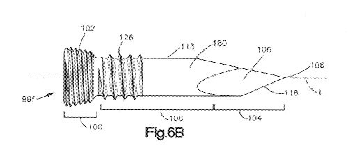
[0024] Fig. 6B is a side elevation view of the bone anchor illustrated in Fig.
6A;
[0025] Fig. 6C is a front elevation view of the bone anchor illustrated in
Fig. 6A;
[0026] Fig. 6D is a side elevation view of the bone anchor similar to Fig. 6B,
but
including threads constructed in accordance with another embodiment;
[0027] Fig. 7 is a perspective view of a bone implant assembly in accordance
with one
embodiment, including an implant and a plurality of bone anchors;
[0028] Fig. 8 is an exploded perspective view of the bone implant assembly
illustrated
in Fig. 7;
[0029] Fig. 9 is a side elevation of the bone implant assembly illustrated in
of Fig. 7;
[0030] Fig. 10 is an exploded view of a bone implant assembly constructed in
accordance with another embodiment;
[0031] Fig. 11 is an exploded view of the bone implant assembly illustrated in
Fig. 10;
[0032] Fig. 12 is an exploded view of a portion of the bone implant assembly
illustrated
in Fig. 10;
[0033] Fig. 13A is a top plan view of a fixation plate of the implant
illustrated in Fig.
10;
[0034] Fig. 13B is a front elevation view of the fixation plate illustrated in
Fig. 10A;
[0035] Fig. 13C is a top elevation view of fixation plate similar to the
fixation plate
illustrated in Fig. 13A, but constructed in accordance with another
embodiment;
[0036] Fig. 13D is a front elevation view of the fixation plate illustrated in
Fig. 10C;
[0037] Fig. 14A is a side elevation view of a bone anchor constructed in
accordance
with another embodiment;
- 3 -
CA 02829565 2013-09-09
WO 2012/121726 PCT/US2011/027849
[0038] Fig. 14B is a side elevation view of a bone implant assembly including
an
implant and a bone anchor, showing the bone anchor inserted through an implant
and partially
inserted into a pilot hole of an underlying bone;
[0039] Fig. 14C is a side elevation view of the bone implant assembly
illustrated in Fig.
14B, showing the bone anchor further driven into the pilot hole such that
threads of the bone
anchor engage the underlying bone;
[0040] Fig. 14D is a side elevation view of the bone implant assembly
illustrated in Fig.
14C, showing the bone anchor further driven into the bone and seated against
the implant;
[0041] Fig. 15A is an exploded view of an exemplary bone implant assembly;
[0042] Fig. 15B is a side elevation view of the bone implant assembly of Fig.
15A, with
the fixation plate and spacer assembled together;
[0043] Fig. 15C is a perspective view of the bone implant assembly of Fig.
15A, with
the fixation plate and spacer assembled together;
[0044] Fig. 15D is a top elevation view of the bone implant assembly of Fig.
15C; and
[0045] Fig. 15E is a rear elevation view of the bone implant assembly of Fig.
15D.
DETAILED DESCRIPTION
[0046] The present disclosure may be understood more readily by reference to
the
following detailed description taken in connection with the accompanying Figs.
and examples,
which form a part of this disclosure. It is to be understood that this
disclosure is not limited to
the specific devices, methods, applications, conditions or parameters
described and/or shown
herein, and that the terminology used herein is for the purpose of describing
particular
embodiments by way of example only and is not intended to be limiting of the
present
disclosure.
[0047] Also, as used in the specification including the appended claims, the
singular
forms "a," "an," and "the" include the plural, and reference to a particular
numerical value
includes at least that particular value, unless the context clearly dictates
otherwise. The term
"plurality", as used herein, means more than one. When a range of values is
expressed, another
embodiment includes from the one particular value and/or to the other
particular value.
Similarly, when values are expressed as approximations, by use of the
antecedent "about," it will
be understood that the particular value forms another embodiment. All ranges
are inclusive and
combinable.
[0048] It is to be appreciated that certain features of various embodiments
set forth in
the present disclosure which are, for clarity, described herein in the context
of separate
- 4 -
CA 02829565 2013-09-09
WO 2012/121726 PCT/US2011/027849
embodiments, may also be provided in combination in a single embodiment.
Conversely,
various features of the present disclosure that are, for brevity, described in
the context of a single
embodiment, may also be provided separately or in any subcombination. Further,
reference to
values stated in ranges includes each and every value within that range.
[0049] Certain terminology is used in the following description for
convenience only
and is not limiting. The words "right", "left", "top" and "bottom" designate
directions in the
drawings to which reference is made. The words "inwardly" and "outwardly"
refer to directions
toward and away from, respectively, the geometric center of the device and
designated parts
thereof. The words, "anterior", "posterior", "superior", "inferior",
"lateral", "medial", "sagittal",
"axial", "coronal," "cranial," "caudal" and related words and/or phrases
designate preferred
positions and orientations in the human body to which reference is made and
are not meant to be
limiting. The terms "anchor" and "fixation member" may be used
interchangeably.
[0050] The disclosed components will now be described by way of reference to
the
appended figures.
[0051] Referring now to Figs. 1A-1C, a bone fixation member, such as bone
anchor
99a, includes a body 101 that is elongate along a central longitudinal axis L,
and defines a
proximal end 100, a distal end 104 spaced from the proximal end 100 along the
longitudinal axis
L, and disposed opposite the proximal end 100, and an intermediate portion 108
disposed
between the proximal end 100 and the distal end 104.
[0052] The bone anchor 99a defines a tip 116 at the distal end 104 that is
capable of
penetrating or cutting a vertebral body (e.g., bone) or other structure. The
proximal end 100 of
the bone anchor 99a defines a head 103 that is suitably configured so as to
engage at least one
complementary engagement feature 111 of a driving instrument, such as a
screwdriver or other
driver device, that applies a force that biases the tip of bone anchor 99a
into an underlying bone,
such as a vertebral body.
[0053] The bone anchor 99a can further include a shaft 113 that can have a
substantially constant diameter and extends between the head 103 and the tip
116. The shaft can
also increase in diameter along a direction from the tip 116 toward the head
103, however the
slope of the outer surface of the shaft 113 can be different from that of the
tip 116.
[0054] The shaft 113 may have a diameter that remains essentially constant
over its
length. Alternatively, for instance as shown in Fig. 5A, the diameter of the
shaft 113 may vary,
for instance, along a direction from the distal end to the proximal end by any
amount as desired.
For instance, the outer surface of the shaft 113 can define an angle of from
about 3 degrees to
about 15 degrees. Accordingly, the diameter of the distal end of the shaft can
be reduced, in
- 5 -
CA 02829565 2013-09-09
WO 2012/121726
PCT/US2011/027849
some embodiments, relative to the diameter of the proximal end of the shaft
113 by from about
99.65% to about 99.8%. The shaft 113 may be of essentially constant diameter
along a portion
of its length, and include a region of varying diameter.
[0055] The distal end 104 can have a longitudinal length relative to that of
the shaft 113
as desired. For instance, the length of the taper on distal end 104 to the
total length of shaft
below the screw head (108+104)) can be from about 1:2 to about 1:5.
[0056] The length of the unthreaded portion of the shaft 113 may be from about
10 mm
to about 25 mm, or from about 12 mm to about 20 mm. The ratio of the length of
the threaded
portion of the shaft 113 to the length of the anchor below the head (i.e., 104
+ 108) is suitably in
the range of from about 1:1 to 1:10, or from 1:2 to about 1:5. The radial
height of the threads
126 on the threaded portion of the shaft can be from about 0.1 mm to about 0.5
mm, or even
from about 0.2 mm to about 0.3 mm. Adjacent threads 126 may be spaced from one
another by
from about 0.8 mm to about 2 mm, or even by from about 0.9 mm to about 1.8 mm.
[0057] In
this regard, it should be appreciated that when the shaft 113 defines a
substantially constant diameter, the outer surface of shaft 113 also defines a
slope different from
that of the tip 116. It should be further appreciated that the slope of the
outer surface of the shaft
113 can be substantially equal to that of the tip 116 (see Figs. 5A-C). The
engagement member
111 of the head 103 can be provided as a recess 112 that is star-shaped as
illustrated, but can be
cross-shaped, pyramidal, hexagonal, helical, or other configurations known in
the art that
facilitate robust engagement between the anchor and the driving instrument.
The StarDriveTM
system from Synthes (www.synthes.com) is considered a suitable system for
driving the anchors
described herein. In some embodiments (not shown), the bone anchor 99a may
define a tip 116
and shaft 113 that extends proximally from the tip 116. The shaft may have a
proximal end (at a
distance from the tip 116), which end is adapted to engage with a driving
instrument. For
example, the proximal end may include a recess as described above. In some
variations, such
anchors may be characterized as being free of a head 103. In such embodiments,
the shaft is
directly driven.
[0058] The engagement member 111 can alternatively be configured as a
protrusion as
desired that is configured so as to engage with a driver device that applies a
distal biasing force
to the bone anchor 99a so as to implant the bone anchor 99a into the
underlying bone. Such a
protrusion may have a cross-section that is triangular, square, pentagonal,
hexagonal, or
otherwise shaped as desired. The protrusion can, for instance, be received by
a socket or other
grip of the installation device.
- 6 -
CA 02829565 2013-09-09
WO 2012/121726 PCT/US2011/027849
[0059] The bone anchor 99a can be configured as a bone screw, whereby the
intermediate section 108 of the bone anchor body 101 defines an exterior
feature, such as a
thread 110 that extends about the shaft 113. The exterior feature can be
configured as a bone
thread that is adapted to securably engage with the underlying bone into which
the bone anchor
is implanted. The thread 110 may be helical in configuration (e.g., helical
thread 126 in Fig.
6B), or may be a stepped, helical thread in configuration, as illustrated in
Figs. 1A-B. The thread
110 may be pyramidal in cross-section and have a sharp distal end.
[0060] The thread 110 may alternatively define a flat distal end, a rounded
distal end, or
any alternatively sized or shaped distal end as desired. The thread 110 may
span the entire
length of the intermediate region 108, or can alternatively span only a
portion of the intermediate
region 108, as shown by the thread 126 in Fig. 6B. The user can drive the bone
anchor 99a into
the underlying bone until the thread 110 contacts the bone, at which point the
user may then
apply a torsional force to the bone anchor 99a so as to screw the bone anchor
99a into the bone
for final seating.
[0061] The thread 110 may have the cross-section of an obtuse or scalene
triangle,
which cross-section allows the installed fixation body to resist a pull-out
force. Thread 110
allow bone anchor to be installed by applying of a torsional force to the bone
anchor 99a so as to
advance the thread 110 into the underlying bone. The thread 110 defines a
height that extends
out from the shaft 113 along a direction angularly offset from the
longitudinal axis L of the bone
anchor 99a, such as substantially perpendicular to the longitudinal axis L.
The height can be
substantially constant, or can vary along the length (e.g., along longitudinal
axis L) of the shaft
113. For example, the thread 110 may have a height that is larger (i.e., is
taller) closer to the
proximal end 100 of the anchor 99a and that is smaller (i.e., shorter) closer
to the distal end 104
of the anchor 99a. In one exemplary embodiment, the thread 110 at the distal
end of the anchor
99a may have a height of x, and the thread 110 at the proximal end of the
anchor may have a
height of 1.3x. Alternatively, the thread 110 may have a height that is
constant along the length
of the bone anchor. In one such embodiment, the thread 110 may have a height
of x at all thread
locations. In yet another embodiment (not shown), the intermediate region 108
of the anchor 99a
tapers from the proximal to the distal ends of the anchor 99a, but the thread
100 varies in height
along the longitudinal axis L such that that the diameter of the anchor 99a is
constant.
[0062] The bone anchor 99a can further define an external thread 102 located
at the
proximal end 100 of the body 101, for instance at the head 103. The external
thread 102 is
suitably configured to engage a complementary thread in another component,
such as an implant,
so as to provide locking fixation between the implant and the underlying bone.
For example, the
- 7 -
CA 02829565 2013-09-09
WO 2012/121726 PCT/US2011/027849
external thread 102 may engage an internal thread of an aperture in a fixation
plate or other
device into which the anchor is installed. The SynfixTM system from Synthes
(www.synthes.com) is one example of such a locking system.
[0063] In some embodiments, the aperture may be an enclosed channel extending
through a portion of the implant. Such an embodiment is shown by figure 15a,
in which aperture
228 extends through the fixation plate 216. Channels that are circular in
cross-section are
considered suitable apertures. In some embodiments, the channel is fully
enclosed within the
implant. The channel, however, need not be fully enclosed within the implant;
in some
embodiments, the aperture may be a channel or slot that is at least partially
open to the
environment exterior to the implant.
[0064] The exterior thread 102 of the bone anchor may be a dual lead thread;
such dual
leads enable the user to more quickly implant the bone anchor into a component
that bears a
complementary thread. The external thread 102 may be an external helical
thread. In some
embodiments, the proximal end of the bone anchor 99a includes one or more
splines that engage
with complementary structures in a fixation body or other component.
[0065] The tip 116 of the bone anchor 99a can be configured so as to penetrate
or cut
vertebral bone so as to enable secure insertion of the bone anchor into the
vertebral body. As
shown in Fig. 1, the distal end 104 is adapted to penetrate bone. The distal
end 104 includes a
sharp tip 116, and can further include cutting facets 114 that can extend
helically about the tip
116. By reference to Fig. 2, the distal end 104 can be configured as a trocar
tip. Such tips allow
the user to implant the bone anchor (at least partially) into the vertebral
body by hammering or
otherwise forcing the tip into the vertebral body; a pilot hole is not always
needed. Trocar tips
may be pyramidal or multi-faceted; the distal end 104 shown in Figure 2B is
pyramidal in
configuration.
[0066] An awling motion or other back-and-forth reciprocating motion may be
used to
effect penetration of the tip 116 into the underlying bone, which awling
motion in turn biases the
tip 116 against the bone and effects cutting or penetration. The awling may be
a
twisting/torquing back-and-forth motion while the anchor 99a is biased into
the underlying bone.
This may be contrasted with a screwing-type motion in which the anchor 99a is
rotated in a
single direction while being biased or otherwise driven into the underlying
bone.
[0067] In one non-limiting example, the user may engage a screwdriver or
similar
implement into recess 112 of the anchor 99a, and then apply an awling motion
to the anchor 99a
so as to install the anchor into underlying bone. In some embodiments, the
user may form a pilot
hole in the underlying bone. Such pilot holes, however, are not necessary, and
the anchor 99a
- 8 -
CA 02829565 2013-09-09
WO 2012/121726 PCT/US2011/027849
may be configured so as to permit installation into underlying bone without
the use of a pilot
hole. In other cases, the tip 116 is driven distally into the underlying bone
and penetrates into
the bone in a nail-like manner.
[0068] In other embodiments, the tip 116 is driven into the underlying bone,
and the
anchor 99a is further inserted into the bone by way of the described awling
motion. In other
embodiments, the tip 116 is driven (e.g., via hammering) into the underlying
bone, and the
anchor 99a is itself then further driven into the bone by way of a hammering
or nailing force.
Anchors 99a may thus be installed by a nailing or hammering force, an awling,
or some
combination. The anchor 99a may also be configured ¨ e.g., with a helical
thread ¨ so as to be
installed by application of a screwing force. It is to be understood that the
above-described
techniques are applicable to any of the anchors 99a, 99b, 99c, 99d, 99e, 99f,
and 99g disclosed in
Figures 1-6, 14, and elsewhere herein, and that the various described anchors
do not limit the
scope of this disclosure.
[0069] The anchor may be constructed such that the tip 116, the distal end
104, or both,
may be installed by a nailing, hammering, or awling motion, and the remainder
of the anchor 99a
is then installed by a screwing motion. The tip 116 of the bone anchor 99a can
be configured as
a trocar tip that can include multiple facets 106, that are separated from one
another by sharp
edges 118, and thus configured to drive into the underlying bone. The tip 116
may have a helical
or screw-like configuration, as shown by the distal end 104 in Fig. 1B. The
anchor 99a can thus
increase the speed of implantation as compared to existing bone screws that
receive a continuous
torsional driving force. The bone anchor 99a can be implanted into a pre-
formed pilot hole that
extends into the underlying bone (see, for instance Figs. 14B-D).
[0070] With continuing reference to Figs. 1A-C, the bone anchor 99a can
further
include an external thread 102 at the proximal end 100, for instance at the
head 103, the thread
102 configured to engage with a complementary thread of a bone implant, such
as a bone
fixation plate. The head 103 can further be conical in shape, and is
configured to be used in
conjunction with a receiving aperture in another component, such as an
implant, which receiving
aperture is cylindrical or even conical in configuration. Accordingly, the
depth of penetration of
the bone anchor 99a can be controlled, as the head 103 cannot be advanced
beyond the point at
which the conical head has fully engaged with the conical receiving aperture
(see e.g., Figs. 7-8).
[0071] Referring now to Figs. 4A-C, the exterior feature of the intermediate
section 108
can include ridges 122. The ridges 122 may all be of the same height or of
different heights.
The ridges 122 may be of the same height along the entirety of the
intermediate section 108, or
of differing heights along the entirety of the intermediate section. In one
embodiment, the ridges
- 9 -
CA 02829565 2013-09-09
WO 2012/121726 PCT/US2011/027849
122 define a greater height at the proximal end of the anchor than at the
distal end of the anchor,
as shown in, e.g., Figure 14A. In accordance with the illustrated embodiment,
the ridges 122
encircle at least a portion of the intermediate portion 108. The ridges 122
may be configured so
as to resist proximally-directed pull-out forces when the bone anchor 99a has
been installed into
a body, such as underlying bone.
[0072] The ridges 122 also allow the bone anchor to be implanted by
application of a
distal driving force, such as a hammering applied to the head 103. This force
may be applied by
way of a mallet or by mechanical means, such as a sonic hammer or other
driver. A pinion drive
may also be used to drive the anchor into the vertebral body. The ridges may,
as described
elsewhere herein, be characterized as being a right triangle in cross-section.
The ridges may also
be scalene, equilateral, or obtuse triangles in cross-section. The ridges may
all be of the same
height; some of the ridges may be of different heights from one another.
Furthermore, the tip
116 can be pointed, and thus devoid of cutting facets 114 illustrated in Figs.
1A-1C.
[0073] Ridges 122 are suitably triangular in cross-section so as to resist
pull-out when
the anchor has been inserted into vertebral bone or other material. While the
exemplary
embodiment shown in Fig. 4 includes a plurality of ridges 122 that can be
concentric, and can be
arranged along essentially the entire length of the intermediate region 108,
the ridges 122 of any
of the bone anchors described above can extend along all or a portion of the
length of the
intermediate region 108, such as the shaft 113. For example, the ridges may be
present only at
the part of the intermediate region that is immediate adjacent to the proximal
end 100.
Alternatively, the ridges may be present at the part of the intermediate
region that is adjacent to
the distal end 104 of the anchor. The anchor may bear one, two, three, or more
ridges. The
ridges may configured such that the intermediate region includes one or more
"ridge-less"
regions that are smooth and free of ridges. One such region is shown by
unthreaded region 180
in Figure 2A.
[0074] The distal end 104 of the bone anchor embodiment 99d of Figs. 4A-C
features a
pyramidal tip 116 that defines a plurality of facets 106 separated from one
another by
longitudinally extending edges 118. In this particular embodiment, the ridges
122 are present on
portions of the distal end 104 that are not facets of the tip.
[0075] An alternative variant is shown in Figs. 2A-C, showing that the
intermediate
region 108 of the bone anchor 99b may be substantially smooth and thus free of
ridges, screw
threads, and the like. The intermediate region 108 may be of essentially
constant cross-section
(as shown), but can also taper inwardly toward the longitudinal axis L along a
direction from the
proximal end 100 toward the distal end 104 of the bone anchor. The bone anchor
99b may
- 10 -
CA 02829565 2013-09-09
WO 2012/121726 PCT/US2011/027849
include a neck 120 or other transition region disposed between the head 103
and the shaft 113 of
the bone anchor 99b.
[0076] The neck 120 may act to prevent over-insertion of the anchor into an
implant
device, so as to control the depth to which the anchor is inserted. As one
example, when the
anchor shown in Figs. 2A-C is inserted into a bone implant (e.g., a bone
fixation plate), the
thread 102 of the proximal end 100 engages with a complementary thread of the
implant. Once
the exterior thread 102 of the anchor is fully engaged with the complementary
thread of the
implant, the collar 120 of the anchor can contact a flange of the implant. The
flange can be sized
so as to prevent passage of the collar 120. In this way, the inventive anchors
and systems may be
configured to control the depth of anchor penetration.
[0077] In the case of the variant bone anchor 99b shown in Fig. 2A-C, the
anchor may
be installed by biasing (e.g., by hammering) the tip of the anchor into the
bone material of
interest. The user may then further insert the anchor into the bone by
application of additional
force (by hammering or by applying a constant pushing force, such as force
applied by a pinion
or screw drive). Once the anchor has been sufficiently inserted into the
subject that the locking
means (e.g., the external thread 102 of the proximal end 100) engages with a
complementary
thread on another component of an implant, such as a fixation plate.
[0078] The user may then apply a twisting force via the recess 112 in the
anchor so as
to fully engage the thread 102 of the anchor and to lock the anchor into
place. Such anchors
enable robust, rapid insertion into subjects, as twisting force need be
applied only at the end of
the procedure in order to lock the anchor into place. Fig. 2C illustrates a
front elevation of the
anchor. In an alternative embodiment (not shown), the anchor 99b may include a
ridge or other
structure disposed at the proximal end 100 that securably engages with a
complementary feature
on another component of an implant, such as a fixation plate. In one non-
limiting embodiment,
the aforedescribed ridge engages with a complementary ring within a socket of
another
component of an implant, the complementary ring being sized such that some
force is required to
advance the ridge beyond the ring. Once so advanced, the ridge and anchor are
held in place by
the ring.
[0079] Referring to Figs. 3A-3C, the bone anchor 99c is constructed
substantially as
illustrated in Figs. 2A-C, but is devoid of a collar or other transition
region. The anchor of Figs.
3A-3C is thus locked into place by engagement of the conical head 100 and
associated thread
102 with a complementarily-shaped conical socket and thread of the implant
component into
which the anchor is inserted. The other numbered elements of Figs. 3A-3C are
explained by
reference to the like-numbered elements of Figs. 2A-2C. As shown by exemplary,
non-limiting
-11-
CA 02829565 2013-09-09
WO 2012/121726 PCT/US2011/027849
figure 3A, the anchor 99c may include an unthreaded region 180, which region
may be smooth
(as shown), or may include ridges, spikes, or teeth (not shown).
[0080] Referring now to Figs. 5A-C, the illustrative bone anchor 99e the
intermediate
section 108 is tapered inwardly along a longitudinal direction (illustrated by
longitudinal axis L)
from the proximal end 100 toward tip 116. As shown by the side elevation of
Fig. 5B, the
anchor's proximal end 100 includes an external thread 102, which thread is
suitably configured
to engage with a complementary thread on an implant component so as to lock
the inserted
anchor into place. The anchor includes a recess 112 for engaging a delivery
device, such as a
screwdriver or mechanized device. The anchor may alternatively include a
protrusion extending
from the proximal end of the anchor, which protrusion may engage with a
delivery device so as
to allow a user to apply a bias to the anchor. As one example, the protrusion
may be hexagonal
in cross-section so as to mate with a complementary hexagonal recess of a
delivery device.
[0081] The bone anchor further includes a thread 124 that extends along the
intermediate region and distal end 108 and 104. In accordance with the
illustrated embodimentõ
the thread 124 is of variable height (or even of variable pitch), and runs
from a lower (shorter)
height at the distal end 104 to a higher (taller) height closer to the
proximal end 100 of the bone
anchor 99e. This configuration enables the user to seat the bone anchor 99e
into an existing pilot
hole formed in bone (e.g., by awling or by operation of a drill or other
suitable instrument). By
applying a twisting force to the bone anchor 99e seated in the pilot hole, the
user can seat all of
the threads of the anchor within the bone by using fewer turns than would be
needed to seat
every thread of the anchor if the anchor had to penetrate the bone starting
with its tip. In an
alternative embodiment (not shown), the anchor 99e shown in Figure 5A includes
ridges (now
shown) in place of thread 124. Such a ridged anchor may then be driven (via,
e.g., hammering or
awling) into underlying bone.
[0082] Referring to Figs. 6A-6D, the illustrative bone anchor 99f can include
an
external thread 126 that extends along a portion of the intermediate region
108 (as shown in Fig.
6B), or may extend for the entirety of the intermediate region 108 as
described above. The bone
thread 126 may be triangular in cross-section, or may alternatively have a
flattened or rounded
crown. Furthermore, as described above, distal end 104 can include a trocar
tip composed of
facets 106 separated by edges 118. The distal end 104 may include a sharpened
tip 116.
[0083] The proximal end 100 is configured such that a distal biasing force
applied to
the anchor 99f that is positioned such that the tip 116 is adjacent an
underlying bone will cause
the tip 116 to cut or penetrate the underlying bone. The intermediate region
108 of the bone
anchor 99f, and thus the shaft 113, may include an unthreaded portion 180 that
is devoid of
- 12 -
CA 02829565 2013-09-09
WO 2012/121726 PCT/US2011/027849
threads. It should be appreciated that the unthreaded portion 180 can extend
along the shaft 113
from the tip 116 to any location along the shaft, up to the head 103. Thus,
the shaft 113 can
include a thread 126 extending distally from any location distal of the head
103 that terminates at
location proximal of the tip 116, such that the unthreaded portion extends
from the thread 126 to
the tip 116. It should be further appreciated that the unthreaded portion 180
can include
alternative fastening structure, such as ridges such as ridges 122, teeth,
spikes, or the like. Thus,
the user can drive the anchor 99f into the underlying bone so as to insert at
least a portion of the
anchor (e.g., the tip 116 and the non-threaded portion of the shaft 113
intermediate region 108)
longitudinally in the bone without applying any twisting or torquing force
about the longitudinal
axis L to the bone anchor 99f. Once the thread 126 has reached the underlying
bone, the user
can then apply a torquing (i.e., screwing) force about the longitudinal axis L
to the bone anchor
99f so as to seat the bone thread 128 in the bone and to then engage the
locking thread 102 with a
complementary thread on an implant component (not shown) into which the anchor
has been
inserted, as described above.
[0084] The bone thread 126 may, as shown by Fig. 6B, be tapered and rounded in
cross-section. As illustrated in Fig. 6D, the bone thread 126 may also be
triangular in cross
section. Such a conformation may enable the anchor to more efficiently seat in
bone and to resist
pull-out forces once the anchor has been situated in the bone. The anchor 99f
may include an
unthreaded region 180. The unthreaded region 180 may lack threads, but may
include ridges,
teeth, spikes, and the like. As shown, the anchor 99f may include a shaft 113
that includes a
threaded region 126 and an unthreaded region 180.
[0085] The helical thread region 126 or a stepped thread region or even a
ridged region
may occupy less than the entire length of the intermediate portion, as shown
in Fig. 6B. The
thread or ridges may be disposed such that a region of the intermediate region
adjacent to the tip
of the bone anchor is smooth, and free of thread or ridges. This in turn
enables the user to at
least partially install the bone anchor by driving the member into the
vertebral (or other) body
without also having to apply a torquing force to advance the bone anchor into
the body.
[0086] The thread may be of constant or varying pitch. The thread may also
vary in
height along its length. For example, the thread closer to the tip of the
anchor may have a
comparatively low height, and then transition to a taller thread closer to the
proximal end of the
anchor.
[0087] The distal end 104 of the anchor is suitably configured as a trocar
tip. Such a tip
includes facets 106 that are separated by edges 118. The end 104 suitably has
a tip 116 that is
sharpened so as to be capable of penetrating or cutting bone when a force is
applied to bias the
- 13 -
CA 02829565 2013-09-09
WO 2012/121726 PCT/US2011/027849
anchor against the bone. The tip may be slightly blunted or flattened so as to
achieve a particular
penetration profile.
[0088] Referring to Figs. 7-9, a bone implant assembly 215 includes a bone
implant
200, such as an intervertebral implant configured to be implanted in an
intervertebral disc space
between a pair of adjacent vertebrae, and a plurality of bone anchors 99 of
the type described
above. It is to be understood that the anchor 99 shown in described implant
systems is
exemplary, and that the systems may include any of the disclosed anchors 99a,
99b, 99c, 99d,
99e, 99f, 99g, and any variations thereof
[0089] The bone implant 200 includes a spacer 208 and a fixation plate 216
configured
to attach to the spacer 208. The implant 200 may further include a blocking
plate 232 and a
locking screw 238 as illustrated. The head of the bone anchor can be
configured to lock the
anchors 99 into the fixation plate 216 in the manner described above.
[0090] One or more bone anchors 99 may be utilized to securely anchor an
assembled
configuration of the intervertebral implant 200 within an intervertebral space
between adjacent
vertebral bodies. Unless otherwise indicated, the intervertebral implant 200
and its components
can be manufactured from any suitable biocompatible material known in the art
including but not
limited to titanium, titanium alloy such as TAN, stainless steel, reinforced
plastics, allograft
bone, and the like.
[0091] The spacer 208 defines a posterior side 208a, an anterior side 208b
opposite the
posterior side, lateral sides 208c, an upper surface 208d, and a lower surface
208e opposite the
upper surface. In one example embodiment, a portion of the posterior side 208a
between the
lateral sides 208c may be curved inwardly in the direction of the anterior
side 208b, defining a
rounded, generally rectangular kidney-like footprint. The posterior side 208a
can have a height
(as measured from the tops of teeth or ridges present on the upper or lower
surfaces of the
spacer) in the range of from about 5 to about 20 mm, or from about 8 to about
18 mm, or even
from about 10 to about 15 mm. The height (measured from the tops of teeth or
ridges present on
the spacer) of the anterior side 208b can be in the range of from about 8 mm
to about 25 mm, or
from about 10 mm to about 20 mm, or even from about 12 mm to about 15 mm.
Furthermore,
the height of the anterior side can be greater than that of the posterior
side.
[0092] In an alternative embodiment, a portion of the posterior side 208a
between the
lateral sides 208c may be curved outwardly in a direction away from the
anterior side 208b. In
yet another alternative embodiment, the posterior side 208a may be
substantially straight
between the lateral sides 208c, defining a rounded, generally rectangular
footprint.
- 14 -
CA 02829565 2013-09-09
WO 2012/121726 PCT/US2011/027849
[0093] The spacer 208 may have a central bore 210 formed therethrough, the
shape of
which substantially conforms to the footprint of the spacer 208 (e.g., a
rounded, generally
rectangular kidney-like footprint, or a rounded, generally rectangular
footprint, depending upon
the geometry of the posterior side 208a). The central bore 210 can be filled
with bone growth
inducing substances to allow bony ingrowth and to assist in fusion between the
spacer 208 and
adjacent vertebral bodies.
[0094] In an example embodiment of the spacer 208, the opposed upper and lower
surfaces 208d and 208e define bone-engaging surfaces that may have gripping
features 208h
such as teeth, spikes, or other similar structures, formed thereon and
configured to facilitate
gripping engagement between the upper and lower surfaces 208d and 208e and the
end plates of
adjacent vertebral bodies. The teeth 214 may be pyramidal, saw toothed or
other similar shapes.
In alternative embodiments of the spacer 208, portions of and/or the entirety
of the upper and
lower surfaces 208d and 208e may be substantially smooth and devoid of any
protrusions.
[0095] Upper and lower edges 208f and 208g, defined where the upper and lower
surfaces 208d and 208e intersect with the posterior, anterior, and lateral
sides 208a, 208b, and
208c respectively around the outer perimeter of the spacer 208, may be
rounded.
[0096] In one example embodiment, the upper and lower edges 208f and 208g may
be
rounded using a uniform radius of curvature around the perimeter of the
implant. In an
alternative embodiment, the upper and lower edges 208f and 208g may be rounded
using a non-
uniform radius of curvature around the perimeter of the implant. In another
alternative
embodiment, the upper and lower edges 208f and 208g along the anterior side
208b may be
rounded with a greater radius than the remainder of the upper and lower edges
208f and 208g,
such that a bull nose outer surface is created on the anterior side 208b of
the implant. Rounding
upper and lower edges 208f and 208g may facilitate easier insertion of the
spacer 208, for
example by minimizing distraction of the end plates of adjacent vertebral
bodies.
[0097] In an example embodiment, the spacer 208 has a generally wedge-shaped
side-
view profile. This wedge shape is suitably defined by a gradual decrease in
the height of the
spacer 208 (as measured between the upper and lower surfaces 208d and 208e)
extending
between the posterior side 208a in the direction of the anterior side 208b.
The spacer 208 has a
generally constant height between lateral sides 208c. In alternative
embodiments, the spacer 208
may have a gradual increase in height followed by a gradual decrease in height
extending from
one lateral side 208c to the other, and/or may have a generally constant
height between the
posterior and anterior sides 208a and 208b, or may have convex and/or concave
upper and lower
surfaces 208d and 208e, thereby defining a gradual increase in height followed
by a gradual
- 15 -
CA 02829565 2013-09-09
WO 2012/121726 PCT/US2011/027849
decrease in height extending from the posterior side 208a to the anterior side
208b and from one
lateral side 208c to the other.
[0098] A plurality of grooves or indentations 212 may be formed within the
spacer 208
where the upper and lower surfaces 208d and 208e intersect with the anterior
side 208b. The
grooves 212 may be concave and may be configured to align with apertures 228
that extend
through an anterior side 218a of the fixation plate 216 when the spacer 208
and the fixation plate
216 are in an assembled configuration. In an example embodiment, the grooves
212 may be
substantially smooth and free of protrusions. Retaining grooves 214 may be
formed within the
lateral sides 208c of the spacer 208 between the upper and lower surfaces 208d
and 208e. The
retaining grooves 214 may be configured to engage complementary engaging ribs
220 of the
fixation plate 216.
[0099] The fixation plate 216 is suitably defined by a generally C-shaped,
channel-like
body 218 that includes an anterior side 218a with upper and lower sides 218b
and 218c opposite
each other, and lateral sides (which may be termed "arms") 218d extending from
opposite sides
of the anterior side 218a in a generally perpendicular direction from the
anterior side 218a. The
anterior, upper, lower, and lateral sides 218a, 218b, 218c, and 218d may form
a generally
channel-like structure (in essence, a cradle) which may be configured to
receive the anterior side
208b and at least a portion of the lateral sides 208c in partial nested
engagement. As such, the
upper and lower sides 208b and 208c may define gradual increases and/or
decreases in height in
a posterior direction from the anterior side 218a and/or between the lateral
sides 208d, in order to
generally conform the fixation plate 216 to the geometry of the spacer 208.
The lateral sides
218d may have engaging ribs 220 formed thereon at the ends opposite the
anterior side 218a, the
engaging ribs 220 configured to be releasably received within the retaining
grooves 214 of the
spacer 208.
[0100] The anterior side 218a of the fixation plate 216 may have apertures 222
formed
therethrough configured to receive grasping features of a delivery instrument.
As shown, a bone
anchor 99 suitably has a length greater than the length of an aperture. In an
example
embodiment, the apertures 222 may be substantially D-shaped. Any other
aperture shape may,
however, be defined as appropriate. The apertures 222 may have a retaining rib
224 formed
therein configured to engage with a complimentary grasping rib of a delivery
instrument. The
anterior side 218a of the fixation plate 216 may also have a central bore 226
formed therethrough
having an inner surface 226a with threads configured to engage complimentary
threads of a
locking screw 238. The anterior side 218a of the fixation plate 216 may also
have a concave
- 16-
CA 02829565 2013-09-09
WO 2012/121726 PCT/US2011/027849
recess 230 formed therein configured to receive a complimentary convex surface
234d of the
blocking plate 232. The recess may matably engage with the blocking plate 232.
[0101] The anterior side 218a of the fixation plate 216 may also have a
plurality of
apertures 228 formed therethrough configured to receive the bone anchors 99
and to define an
insertion trajectory for the bone anchors. In an example embodiment, the
apertures 228 may
have a generally uniform cross sectional geometry configured to closely
conform to the cross
sectional geometry of the bone anchor 99. The apertures 228 may also include
an interior thread
that engages with an external disposed on the proximal end (head) of the bone
anchor.
[0102] The apertures 228 may be dimensioned such that the proximal end of the
bone
anchor is flush with the surface 230 (or 218a) of the fixation plate when the
anchor is fully
installed, although this flush orientation is not necessary. The aperture 228
may also be
configured such that the proximal end of the anchor is sunken below the
surface of the fixation
plate when the anchor is fully installed; the end of the anchor may also
protrude from the fixation
plate.
[0103] The apertures 228 may be disposed about the optional central bore 226
in any
desired configuration and may define any insertion trajectories as
appropriate. In the example
embodiment depicted in Figs. 7-9, the apertures 228 are formed in opposing
quadrants around
the central bore 226, with two apertures 228 located near the upper side 218b
and defining two
generally cranial insertion trajectories, and two apertures 228 located near
the lower side 218c
and defining two generally caudal insertion trajectories. This configuration
of aperture 228
locations and bone anchor 99 insertion trajectories is merely an example, and
the scope of the
instant disclosure should not be limited thereto.
[0104] An optional blocking plate 232 is shown; such plates are not a
requirement, as
the anchors 99 may be capable of securing the implant structure to the
underlying bone without
the assistance of a blocking or other structure. The plate 232 is defined by a
generally disc-
shaped body 234 with planar upper and lower surfaces 234a and 234b, an
anterior surface 234c,
and a posterior surface 234d. The upper and lower surfaces 234a and 234b and
the height of the
body 234 (as measured between the upper and lower surfaces 234a and 234b) may
be defined to
match the height (as measured between the upper and lower surfaces 218b and
218c) of the
anterior side 218a of the fixation plate 216 when the blocking plate 232 is in
a fully assembled
configuration. The anterior surface 234c of the body 234 may be generally
planar, or may be
defined to match the outer surface of the anterior side 218a of the fixation
plate 216 when the
blocking plate 232 is in a fully assembled configuration.
- 17 -
CA 02829565 2013-09-09
WO 2012/121726 PCT/US2011/027849
[0105] The posterior surface 234d may be defined as a convex surface
configured to
engage with the concave recess 230 formed in the anterior side 218a of the
fixation plate 216
when the blocking plate 232 is in a fully assembled configuration. The body
234 may have an
aperture 236 formed therethrough. In an example embodiment, the diameter of
the aperture may
be slightly larger than the diameter of the central bore 226 of the fixation
plate 216, such that a
locking screw 238 may be inserted into the aperture with no interference
therebetween. In
another embodiment, the diameter of the aperture 236 may be substantially the
same as that of
the central bore 226, and the inner surface of the aperture may have threads
formed thereon, the
threads configured to engage complimentary threads of the locking screw 238.
The aperture 236
may further be defined by a concave recess 236a formed within the anterior
surface 234c, the
concave recess 236a configured to receive the convex head 242 of the locking
screw 238.
[0106] The optional locking screw 238 includes a shaft 240 that defines
longitudinally
opposing proximal and distal ends 240a and 240b, respectively, and a head 242
coupled to the
proximal end 240a of the shaft 240, either directly or indirectly via an
unthreaded neck 244 that
is coupled between the proximal end 240a of the shaft 240 and the head 242.
The head 242 can
define a generally convex shape between the interface of the head 242 and the
neck 244 that
extends outward towards a proximal end 242a of the head 242. The convex shape
of the head
may be configured to engage the concave recess 236a of the blocking plate 232.
The head 242
can assume any other suitable alternative shape as appropriate. Helical
threads 246 extend
radially out from the shaft 240 at locations at and between the proximal and
distal ends 240a and
240b that are configured to engage complementary threads on the inner surface
226a of the
central bore 226 of the fixation plate 216. Thus, a substantial entirety of
the shaft 240 between
the proximal and distal ends 240a and 240b may be threaded. The distal end
242a of the head
242 may have driving features 242b defined therein, designed to engage with
complementary
driving features of a delivery instrument.
[0107] During operation, the spacer 208 is seated within the fixation plate
216 such that
the retaining ribs engage with the retaining grooves on the lateral sides of
the spacer 208. Four
bone anchors 99 are inserted through corresponding grooves within the fixation
plate 216, and
have been driven to an essentially fully inserted position. In this
embodiment, the heads of the
bone anchors 99 may be flush with the surface of the fixation plate 216. The
fixation plate may
include an aperture that is configured to releasably engage with a delivery
instrument, which
instrument may include an armature or other extension that engages with the
fixation plate. The
heads of the anchors may, as described elsewhere herein, include a thread that
engages with a
complementary thread of
- 18 -
CA 02829565 2013-09-09
WO 2012/121726 PCT/US2011/027849
[0108] Fig. 9 depicts an example embodiment of the intervertebral implant 200
partially
assembled inside of an intervertebral space between adjacent vertebral bodies
V6 and V7 (the
blocking plate and locking screw have been omitted for simplicity). As an
initial step, the spacer
208 has been prepared for insertion, for example by being packed with bone
growth inducing
substance and or/having its outer surfaces properly prepared. The spacer 208
has also been
seated within the fixation plate 216 such the retaining ribs are seated with
the retaining grooves
on the lateral sides of the spacer 208. The spacer 208 is then inserted into
the intervertebral
space between the adjacent vertebral bodies V6 and V7 using a delivery
instrument. An
instrument is then used to deliver the four bone anchors 99 into the grooves
in the fixation plate
and drive them into an almost fully inserted position.
[0109] If a blocking plate and locking screw are used, an instrument is used
to drive the
bone anchors 99 into their fully inserted position in the manner described
above, and the
blocking plate is received within the concave recess in the anterior side of
the fixation plate, and
the locking screw would be driven into the central bore of the fixation plate
and finally tightened,
thereby blocking the bone anchors 99 from backing out of the assembled
intervertebral implant
200.
[0110] It should be appreciated that the intervertebral implant 200 can be
alternatively
constructed as desired. For instance, referring now to Fig. 10-13D, the
fixation plate 256 is
defined by a generally rectangular body 258 that includes an anterior side
258a and lateral sides
258b extending therefrom, the lateral sides 258b configured to engage with the
retaining grooves
252 of the spacer 248. In an example embodiment, the lateral sides 258b are
generally J-shaped,
extending initially from opposite sides of the anterior side 258a in a
direction perpendicular to
and away from the anterior side 258a, and through curved sections 258c before
returning in a
direction perpendicular to and towards the anterior side 258a and terminating
in distal ends 258d.
It should be noted that this configuration for lateral sides 258b is merely an
example, and any
other geometry may be used as appropriate.
[0111] Upper and lower edges of the anterior side 258a, defined where upper
and lower
surfaces 258e and 258f of the anterior side intersect with an anterior surface
258g of the anterior
side, may be rounded. In an example embodiment, the upper and lower edges 258e
and 258f
may be rounded using a uniform radius of curvature. In an alternative
embodiment, the upper
and lower edges 258e and 258f may be rounded using a non-uniform radius of
curvature.
Rounding upper and lower edges 258e and 258f may facilitate easier insertion
of the fixation
plate 256, for example by minimizing distraction of the end plates of adjacent
vertebral bodies.
- 19 -
CA 02829565 2013-09-09
WO 2012/121726 PCT/US2011/027849
[0112] The lateral sides 258b may have retaining ribs 260 formed thereon at
the distal
ends 258d, the retaining ribs 260 configured to be releasably received within
the retaining
grooves 252 of the intervertebral implant 258. Access grooves 262 and 264 may
be formed
within the retaining ribs 260 and the lateral sides 258b, in the area where
the lateral sides 258b
interface with the anterior side 258a, respectively. The access grooves 262
and 264 may be
configured to align with complimentary access grooves 254 of the spacer 248,
thereby defining
an access cavity 268 for receiving an engaging feature of a delivery
instrument when the
spacer248 and the fixation plate 256 are in an assembled configuration. The
access grooves 264
may have a retaining shelf 266 formed therein configured to engage with an
engaging feature of
a delivery instrument, for example the raised ribs 258d formed on the
insertion rods 258 of the
delivery instrument 278, described in greater detail below. The lateral sides
258b may also have
bores 278 formed within the curved sections 258c, the apertures configured to
receive, for
example the distal engagement tips 258c of the insertion rods of 258 of the
delivery instrument
278.
[0113] The anterior side 258a of the fixation plate 256 may have gripping
grooves 268
formed within the upper and lower surfaces 258e and 258f of the anterior side
258a, the gripping
grooves 268 configured to receive grasping arms of a delivery instrument. The
gripping grooves
268 may have a gripping ridge 270 formed therein, the gripping ridge
configured to be engaged
by the complimentary grasping features formed at the ends of the grasping arms
of the delivery
instrument. The anterior side 258a of the fixation plate 256 may also have a
recess 272 formed
therein configured to receive additional components of the intervertebral
implant 200, for
example a ratchet blade 288, a blocking plate 280, or the like. The anterior
side 258a may also
have a central bore 274 formed therethrough having an inner surface 274a with
threads
configured to engage complimentary threads of a locking screw 238. In an
example
embodiment, the central bore 274 may be formed within the recess 272.
[0114] The anterior side 258a of the fixation plate 256 may also have a
plurality of
apertures 276 formed therethrough configured to slidably receive the bone
anchors 99 and to
define an insertion trajectory for each of the bone anchors 99; as shown in
Fig. 11, the grooves
may include interior threads adapted to engage complementary threads on the
anchors.
Alternatively, a bone anchor may also include splines or flanges that engage
with the fixation
plate so as to maintain the bone anchor in position. The bone anchor is
suitably installed by way
of a driving instrument that applies a force to bias the tip of the at least
one bone anchor into a
vertebral body.
- 20 -
CA 02829565 2013-09-09
WO 2012/121726 PCT/US2011/027849
[0115] In an example embodiment, the apertures 276 may have a generally
uniform
cross sectional geometry configured to closely conform to the cross sectional
geometry of the
body of the bone anchor 99 between the head and the distal end.
[0116] When a bone anchor 99 is in a fully inserted position within a
respective
aperture 276, the surface of the head of the fixation device may be flush with
the outer surface of
the anterior side 258a of the fixation plate 256. The head may also protrude,
in some
embodiments, from the surface of the fixation plate.
[0117] The apertures 276 may be disposed about the central bore 274 in any
desired
configuration and may define any insertion trajectories as appropriate.
[0118] The blocking plate 280 is defined by a generally rectangular body 282
with an
anterior surface 282a, and a plurality of angled posterior surfaces 282b
generally opposite the
anterior surface 282a. The body 282 may have an aperture 286 formed
therethrough In an
example embodiment, the diameter of the aperture may be slightly larger than
the diameter of the
central bore 274 of the fixation plate 256, such that a locking screw 238 may
be inserted into the
aperture with no interference therebetween.
[0119] In another embodiment, the diameter of the aperture 286 may be
substantially
the same as that of the central bore 274, and the inner surface of the
aperture may have threads
formed thereon, the threads configured to engage complimentary threads of the
locking screw
238. The aperture 286 may further be defined by a concave recess 286a formed
within the
anterior surface 282a, the concave recess 286a configured to receive the
convex head 242 of the
locking screw 238.
[0120] The height, width, and depth of the body 282 may be proportioned so
that the
blocking plate 280 will be received within the recess 272 of the fixation
plate 256, such that the
anterior surface 282a of the body 282 is substantially flush with the anterior
surface 258g of the
anterior side 258a of the fixation plate 256 when the fixation plate 256 and
the blocking plate
280 are in an assembled configuration. The anterior surface 282a of the body
282 may be
generally planar, or may be defined to match the outer surface of the anterior
side 258a of the
fixation plate 256 when the blocking plate 280 and the fixation plate 256 are
in a fully assembled
configuration.
[0121] In an example embodiment wherein the blocking plate 280 and locking
screw
238 are installed after the spacer 248 and fixation plate 256 have been
inserted into an
intervertebral space and the bone anchors 99 driven into their fully inserted
positions, the angled
posterior surfaces 282b and chamfered corners 284 of the blocking plate 280
may be configured
to engage the heads of the bone anchors 99 within the recess 272 of the
fixation plate 256 when
-21 -
CA 02829565 2013-09-09
WO 2012/121726 PCT/US2011/027849
the blocking plate 280 is installed followed by the locking screw 238. When
final tightening of
the locking screw 238 is performed, the blocking plate 280 may rigidly fix the
bone anchors in
position, and additionally prevent pullout of the members.
[0122] In another exemplary, non-limiting embodiment wherein the spacer 248,
the
fixation plate 256, the blocking plate 280, and the locking screw 238 are pre-
assembled, but not
finally tightened, and then inserted into an intervertebral space before the
bone anchors 12C are
inserted and driven into position, the angled posterior surfaces 282b and
chamfered corners 284
of the blocking plate 280 may be configured to allow the bone anchors to be
inserted and driven
into position with the blocking plate 280 and the locking screw 238 in place.
[0123] In this embodiment, the angled posterior surfaces 282b may have wedge
features formed thereon that are configured to interfere between the heads of
the bone anchors
and the surrounding structure of the fixation plate 256, for example by
applying outward force
laterally upward and downward on the bone anchors 99 to lock them in place
when final
tightening is applied to the locking screw, and additionally to prevent
pullout of the bone
anchors.
[0124] Referring now to Fig. 10, an example embodiment of the intervertebral
implant
200 in a completely assembled configuration outside of an intervertebral space
is shown. The
fixation plate 256 has been engaged with the spacer 248 such that the
retaining ribs of the
fixation plate 256 are seated with the retaining grooves of the spacer 248.
Four bone anchors 99
have been inserted through corresponding grooves within the fixation plate
256, and have been
driven to a fully inserted position. The blocking plate 280 and the locking
screw 238 have been
installed and finally tightened.
[0125] Referring to Fig. 12, a fixation plate 256 includes a plurality of
apertures 276
that extend therethrough, the grooves being configured to accept bone anchors
99 inserted
therethrough. If desired, the system also can also include a ratchet plate
288, which plate is
assembled into the fixation plate 256 using a locking screw 238. The bone
anchor 99 suitably
includes a locking thread disposed on the outside of the head of the anchor,
which thread is
adapted to engage with a thread disposed on the interior of aperture 276 so as
to lock the anchor
into place.
[0126] Another embodiment is shown in Figures 15a-15e. As shown in Figure 15a,
an
implant assembly 200 may include a spacer 208 and a fixation plate 216. The
fixation plate is
suitably made from titanium, although other metals, polymers, or composite
materials are
suitable. The spacer is suitably made from PEEK or other polymers.
- 22 -
CA 02829565 2013-09-09
WO 2012/121726 PCT/US2011/027849
[0127] The fixation plate 216 suitably includes a bore hole 216, which hole
may be
adapted to receive or otherwise engage an installation instrument, an aiming
device, or both. The
apertures 228 in the fixation plate 216 suitably include internal threads
(shown), which threads
are adapted to engage complementary threads on an anchor 99.
[0128] The spacer 208 suitably defines a posterior side 208a, an anterior side
208b
opposite the posterior side, lateral sides 208c, an upper surface 208d, and a
lower surface 208e
opposite the upper surface. In one example embodiment, a portion of the
posterior side 208a
between the lateral sides 208c may be curved inwardly in the direction of the
anterior side 208b,
defining a rounded, generally rectangular kidney-like footprint. The implant
may include a
somewhat curved or J-shaped arm 252, which arm is configured to engage with a
complementary
feature 220 on the fixation plate.
[0129] The spacer 208 may optionally include a side channel 210a, which side
channel
may be packed with a bone growth inducing substance or other material. The
spacer may
include one, two, or more side channels. The implant 200 may optionally
include a side aperture
210b, which channel allows material to pass into or out of channel 210a. The
implant 200 also
suitably includes a central channel 210, which central channel may be packed
with bone growth
material, if desired. A central aperture 226b may be present, which central
aperture allows
material to pass into or out of the central channel 210. The central channel
and aperture are
optional. The spacer 208 may include cutouts 212 that align with the apertures
228 of the
fixation plate 216 when the fixation plate is installed with the spacer 208.
[0130] The implant 200 may also include strips 290. These strips suitably
extend from
the surface of the implant 200, and act to promote effective engagement
between the implant 200
and the fixation plate 216. As shown in Figure 15a, the strips 290 may have
sloped or wedged
edges so as to facilitate slidably engaging the implant 200 and the fixation
plate 216. Once the
fixation plate and implant are engaged, the strips 290 act as shims or braces
so as to more tightly
engage the fixation plate with the implant; as shown in Figure 15d, the strips
may wedge
between the fixation plate 216 and the spacer 208.
[0131] Figure 15b is a side-on view of spacer 208. This view shows side
aperture
210a, teeth 208h, and lateral side 208c.
[0132] Figure 15c illustrates an alternative view of the implant 200, in which
figure the
fixation plate and spacer are in an assembled configuration. As shown in this
figure, the cutouts
212 align with the apertures 228 of the fixation plate. The arms 252 of the
spacer 208 are
configured to slidably engage with the complementary features of the fixation
plate so as to
guide and maintain the fixation plate 216 in position..
- 23 -
CA 02829565 2013-09-09
WO 2012/121726 PCT/US2011/027849
[0133] Figure 15d is a top view of the implant 200 in its assembled
configuration. The
side channels 210a and central channel 210 are shown; as described elsewhere
herein, the
channels can be packed with bone growth materials. Marker 291 is suitably an x-
ray-visible
material disposed within the spacer 208. This may be accomplished by forming a
hole or recess
in the spacer and then inserting the marker material (e.g., titanium) into the
hole or recess.
Figure 15d also illustrates the strips 290 acting as shims to maintain
fixation plate 216 in
position.
[0134] Figure 15e is a posterior view of the implant 200, showing the
posterior side
208a, the lateral side 208c, the upper surface 208d, and teeth 208h.
[0135] In some embodiments, the implant 200 is a single body. Such unitary
bodies
may be made of titanium, PEEK or other materials. Figure 15c illustrates the
configuration of
such a single body; such a single body would integrate fixation plate 216 and
spacer 208 into a
single body; i.e., the fixation plate and spacer 208 are integrated into a
single body and are not
separate from one another.
[0136] It should be noted that although the description and accompanying
Figures.
illustrating the intervertebral implant included herein depict example
embodiments of the
intervertebral implant that include four bone anchors, with two of the four
bone anchors having a
generally cranial insertion trajectory and the remaining two bone anchors
having a generally
caudal insertion trajectory (e.g., Fig. 7). Other configurations of the
intervertebral implant using
more or fewer bone anchors and/or varying insertion trajectories are possible
and intended to be
included within the scope of the instant disclosure. For example, in an
alternative embodiment
of the intervertebral implant, the fixation plate may have three grooves
formed therein having
any desirable placement and/or insertion trajectory with respect to the
central bore (e.g., with two
of the three bone anchors having a generally caudal insertion trajectory and
the remaining bone
anchor having a generally cranial insertion trajectory, or with two of the
three bone anchors
having a generally cranial insertion trajectory and the remaining bone anchor
having a generally
caudal insertion trajectory).
[0137] Referring to Figs. 13A-D the spacer 248 has a generally C-shaped
footprint
defined by a posterior side 248a, lateral sides 248b terminating in distal
ends 248c opposite the
posterior side 248a, an upper surface 248d, and a lower surface 248e opposite
the upper surface.
In an example embodiment, a portion of the posterior side 248a between the
lateral sides 248b
may be curved inwardly in a direction toward the distal ends 248c, as depicted
in Figs. 13C and
13D. In an alternative embodiment, a portion of the posterior side 248a
between the lateral sides
248b may be curved outwardly in a direction away from the distal ends 248c. In
another
- 24 -
CA 02829565 2013-09-09
WO 2012/121726 PCT/US2011/027849
alternative embodiment, the posterior side 248a may be substantially straight
between the lateral
sides 248b, as depicted in Figs. 13A and 13B. The posterior side 248a and
lateral sides 248b
define an open central bore 250, the shape of which substantially conforms to
the footprint of the
spacer 248. The central bore 250 can be filled with bone growth inducing
substances to allow
bony ingrowth and to assist in fusion between the spacer 248 and adjacent
vertebral bodies.
[0138] In an example embodiment of the spacer 248, the upper and lower
surfaces 248d
and 248e may have gripping features such as teeth, spikes, or similar
structures formed thereon
and configured to facilitate gripping engagement between the upper and lower
surfaces 248d and
248e and the end plates of adjacent vertebral bodies. The teeth may be
pyramidal, saw toothed
or other similar shapes. In alternative embodiments of the spacer 248,
portions of and/or the
entirety of the upper and lower surfaces 248d and 248e may be substantially
smooth and devoid
of any protrusions. Upper and lower edges 248f and 248g, defined where the
upper and lower
surfaces 248d and 248e intersect with the posterior and lateral sides 248a and
248b respectively
around the perimeter of the spacer 248, may be rounded. In an example
embodiment, the upper
and lower edges 248f and 248g may be rounded using a uniform radius of
curvature around the
perimeter of the implant. In an alternative embodiment, the upper and lower
edges 248f and
248g may be rounded using a non-uniform radius of curvature around the
perimeter of the
implant. Rounding upper and lower edges 248f and 248g may facilitate easier
insertion of the
spacer 248, for example by minimizing distraction of the end plates of
adjacent vertebral bodies.
[0139] In an example embodiment, the spacer 248 has a generally wedge-shaped
side-
view profile. This wedge shape is defined by a gradual increase in the height
of the spacer 248
(as measured between the upper and lower surfaces 248d and 248e) extending
outwardly in a
direction away the posterior side 248a in the direction of the distal ends
248c. The spacer 248
has a generally constant height between lateral sides 248b. In alternative
embodiments, the
spacer 248 may have a gradual increase in height followed by a gradual
decrease in height
extending from one lateral side 248b to the other, and/or may have a generally
constant height
between the posterior sides 248a and the distal ends 248c, or may have convex
and/or concave
upper and lower surfaces 248d and 248e, thereby defining a gradual increase in
height followed
by a gradual decrease in height extending from the posterior side 248a to the
distal ends 248c
and from one lateral side 248b to the other.
[0140] Retaining grooves 252 may be formed within the distal ends 248c of the
spacer
248, for example in a vertical direction substantially perpendicular to a
horizontal midplane
defined between the upper and lower surfaces 248d and 248e. The retaining
grooves 252 may be
configured to releasably engage complementary retaining ribs 260 of the
fixation plate 256. The
- 25 -
CA 02829565 2013-09-09
WO 2012/121726 PCT/US2011/027849
distal ends 248c may also have access grooves 254 formed therein between the
upper and lower
surfaces 248d and 248e. The access grooves 254 may be configured to align with
complimentary access grooves of the fixation plate 256.
[0141] The intervertebral implant may of course have matching grooves formed
therein. Such alternative embodiments with two bone anchors having one of a
generally cranial
or caudal trajectory and a third bone anchor having the opposite general
trajectory may allow for
the stacking of two or more assembled configurations of the intervertebral
implant in place of
adjacent vertebral bodies removed from an intervertebral space. Additionally,
while the bone
anchors illustrated in the various Figures herein generally have divergent
insertion trajectories
with respect to each other, fixation plates may also be configured so that one
or more of the bone
anchors will have convergent insertion trajectories with respect to each
other, or similar insertion
trajectories (e.g., laterally towards a common side).
[0142] For example, bone anchors with generally cranial insertion trajectories
may
converge toward one another, may diverge away from one another, or may both
follow similar
insertion trajectories, while bone anchors with generally caudal insertion
trajectories may
converge toward one another, may diverge away from one another, or may both
follow similar
insertion trajectories. Any combination of the above insertion trajectory
configurations may be
used as appropriate.
[0143] One exemplary method of installing the claimed anchors is now
described, with
reference to Fig. 2 and Figs. 10-13D; these figures are illustrative only and
the disclosed
installation technique is suitable for other disclosed anchors. The spacer 248
is suitably
assembled with the fixation plate 256 and is installed into the appropriate
location in the patient's
spine. One suitable technique for performing this installation is described in
literature associated
with the Synthes SynfixTM system (www.synthes.com), which technique is
incorporated herein
by reference for all purposes. Once the assembly of the implant and fixation
plate are installed in
the patient, the user inserts a bone anchor 99 into groove or aperture 276,
which is suitably
configured to define an insertion trajectory for the bone anchor 99. The
aperture 276 is suitably
frustoconical in configuration and includes an interior thread complementary
to the thread 102
disposed on the head of the bone anchor 99.
[0144] Insertion of the anchor into the aperture 276 may be accomplished
manually or
with mechanical assistance. In some embodiments, an aiming component is used
to assist with
alignment and insertion of the anchor. The aiming component may include a bore
or groove to
maintain the anchor in proper orientation or alignment relative to the
aperture 276 into which the
anchor is inserted.
- 26 -
CA 02829565 2013-09-09
WO 2012/121726 PCT/US2011/027849
[0145] The aiming component may be configured so as to engage with the
fixation
plate so as to provide a stable platform for anchor insertion. For example,
the aiming component
may include a protrusion or tab that engages with a complementary recess,
aperture, or slot on
the fixation plate so as to maintain the aiming component's position. The
aiming component
may also relasably or rotatably mount to the fixation plate. The aiming
component may be
configured to support insertion of a single anchor at a time, or even two or
more anchors.
[0146] In one exemplary embodiment, the user inserts the anchor into the
aperture 276
until the tip of the anchor contacts one of the vertebrae that is the subject
of the implantation
process. Once the anchor tip contacts the bone, the user may effect an awling
motion on the
anchor so as to bias the tip into the vertebral bone and to drive the anchor
more deeply into the
bone. The awling motion may be a twisting back-and-forth motion (e.g.,
torquing) accompanied
by applying a force in the direction of the tip of the anchor so as to bias
the tip into the bone
during awling.
[0147] In another embodiment, the user forms, with an awl or other implement,
a hole
(which may be termed a pilot hole) in the vertebral body. The user then
inserts the anchor into
this hole. As described elsewhere herein, the anchor may (e.g., Fig. 5)
include a thread of
variable height (or even variable pitch), that runs from a lower height at the
distal end to a higher
(taller) height closer to the proximal end of the anchor.
[0148] The awl is usually of the same or a similar configuration as the minor
diameter
of the tapered anchor. In this way, when the user inserts the anchor into the
pilot hole, at least a
portion of the anchor is present within the hole when the tip of the anchor
makes contact with the
end of the hole. By then applying a twisting force to the anchor, the user can
seat all of the
threads of the anchor within the bone by using fewer turns than would be
needed to seat every
thread of the anchor if the anchor had to penetrate the bone starting with its
tip.
[0149] For instance, as illustrated in Figs. 14A-B, the user may form a pilot
hole into
the underlying bone for use with assembly system 215. By reference to Figs.
14A and 14B, the
user may in some embodiments form a pilot hole 117 into underlying bone 119,
such that the
pilot hole 117 conforms to or is similar to the minor diameter of the bone
anchor 99g shown in
Fig. 14A. The pilot hole may be formed with an awl, drill, or other similar
implement. The pilot
hole is not necessary in all embodiments, as the anchor 99 may be configured
to pierce and
engage with bone without the assistance of a pilot hole.
[0150] The user may then insert an anchor 99g through the fixation plate (not
labeled)
into the pilot hole 117. The pilot hole 117 is suitably sized such that the
threads of the anchor
99g contact the interior of the underlying bone 119 that defines the pilot
hole 117 while the
-27 -
CA 02829565 2013-09-09
WO 2012/121726 PCT/US2011/027849
anchor 99g is partially inserted in the hole 117, thereby preventing the
anchor 99g from being
further inserted into the pilot hole 117 without application of a force to
seat the threads 122 in the
underlying bone 119.
[0151] The user may then applying a screwing or torquing motion to the bone
anchor
99g, so as to engage additional threads 122 in the bone 119, as shown in Fig.
14B. Further
torquing may then be applied to seat additional threads in the bone material,
as shown in Fig.
14C. Fig. 14D illustrates the fully-seated anchor 99g. As shown by the
progression of Figs.
14B-14D, the anchor 99g enables a user to seat the anchor and engage all of
the anchor's threads
with fewer turns than would be needed to seat every thread of the anchor if
the anchor had to
penetrate the bone starting with its tip. This in turn can speed the
installation of the anchor.
[0152] In other embodiments, the assembly 115 includes the bone anchor 99g
seated in
the pilot hole 117 may include ridges along the length of the anchor, such as
the shaft 103. In
some embodiments, the ridges are of varying height, going from a lower height
at the tip to a
taller height at the proximal end. The ridges may also be of the same height
along the length of
the anchor. With this configuration, the user can seat all ridges of an anchor
in bone without
having to drive the entire length (i.e., seat every ridge) of the anchor into
the bone. This, too,
can speed installation of the anchor.
[0153] The user may, in some embodiments, drive the anchor into the subject
bone by
application of an impact force, such as tapping or even hammering. The
modulation of such
force will depend on the user's needs; such force is suitably modulated in
accordance so as to
avoid damaging any anatomical structures that are in the vicinity of the
anchor insertion.
[0154] The user suitably applies sufficient force for a sufficient time that
the proximal
end and external thread 102 of the anchor 99g contacts the complementarily-
shaped entry of the
groove. Once the threads contact one another, the user may apply a torquing
force to the anchor
so as to engage the external thread of the anchor with the internal thread of
the aperture 276. The
user then tightens the thread of the anchor so as to secure the anchor into
place.
[0155] In an alternative embodiment, the anchor includes splines at the
proximal end of
the anchor 99. The splines may then engage with complementary splines disposed
within the
aperture 276. The spline-spline engagement between the anchor and aperture
acts to prevent the
installed anchor from rotating. The installed anchors may then be secured into
place using a
locking bolt and blocking plate, as described elsewhere herein. While anchor
99g is used in the
cited figures for illustrative purposes, it is to be appreciated that other
anchors (e.g., anchors 99a-
99f) may be used in the disclosed systems.
-28-
CA 02829565 2013-09-09
WO 2012/121726 PCT/US2011/027849
[0156] In one procedure for installing the disclosed devices, the user
performs a
discectomy, which procedure may include the removal of cartilaginous
endplates. A suitably
sized implant assembly (e.g., elements 216 and 200 in figure 15a) is selected
and inserted into
the patient; this insertion may be accomplished with the use of the Synthes
Quick
Inserter/Distractor (SQUIDTM) device; www.synthes.com.
[0157] An aiming device (e.g., an aiming device associated with the Synthes
SynFixTM
system; www.synthes.com) may be inserted into a guide hole (e.g., element 226
on figure 15a) to
assist the user with aligning the anchors 99 for installation into the implant
assembly. The user
may then use a driving instrument (e.g., a screwdriver, or U-joint driver) to
install the first
anchor 99. The awl tip 104 of the anchors 99 enables the user to insert a
first anchor into the
spinal bone by effecting an awling, twisting, or other back-and-forth motion
while biasing the
anchor into the bone. Once the anchor is inserted and the threads 102 on the
head of the anchor
contact the complementary threads of the apertures (e.g., element 228 in
Figure 15a) of the
fixation plate (e.g., element 216 in Figure 15a), the user may then use a
screwdriver or other
instrument to engage and lock the threaded anchors to the fixation plate.
Other anchors are
installed in a similar fashion.
[0158] In some embodiments, the anterior edge of the installed implant is
flush to about
3 mm-recessed relative to the anterior aspect of the adjacent vertebrae. The
user may assess the
positioning of the implant assembly by using an x-ray or other imaging device
to determine the
location of the implant relative to the vertebral bodies. The fixation plate
(suitably fashioned
from titanium) may be visible on an x-ray. The implant may also include an x-
ray marker (e.g.,
element 291 on Figure 15d), which is suitably titanium or other x-ray opaque
material, which
marker enables the user to assess the position and orientation of the implant.
(The x-ray marker
may be about 2 mm from the posterior edge of the implant.) The fixation plate
and x-ray marker
thus allow for intraoperative radiographic assessment of the installed
implant.
[0159] It should be appreciated that a variety of kits can be provided that
include one or
more components of the fixation system or the intervertebral implant. A kit
may contain
multiple, identical components (e.g., multiple anchors that are the same size
and configuration)
or may contain different components (e.g., multiple anchors of different
sizes.) In this way, a
single kit may include a range of components, resulting in the kit being
suitable for use with a
range of patients having need for differently-sized or configured implants.
[0160] For example, within a single kit, bone anchors may be provided that
have
varying lengths, differing head, shaft, or tip configurations, differing cross
sectional geometries,
- 29 -
CA 02829565 2013-09-09
WO 2012/121726 PCT/US2011/027849
and so on, depending, for example, on the type of procedure being performed by
a surgeon, or on
the particular anatomies of individual patients.
[0161] The kits may also be configured differently with respect to which
components of
the individual systems are included in the kits. For example, a kit may
include bone anchors
with varying configurations and/or features, and may or may not include a
device to hold the
assembled implant for insertion into the subject. The kit may also include
fixation rods and the
like.
[0162] In another example, a kit for the intervertebral implant may include
bone
anchors of varying lengths and features, and may also include one or more
intervertebral
implants, one or more fixation plates, one or more blocking plates, one or
more ratchet screws, or
one or more locking screws. Example kits may also include an instrument for
delivering the
system into a subject, and may also include a mallet, a pinion drive, a sonic
hammer, a
screwdriver, or the like for biasing an anchor into a vertebral body and/or
for locking the anchor
into place.
[0163] Although bone anchors and the other components of the fixation and
implants
have been described herein with reference to preferred embodiments or
preferred methods, it
should be understood that the words which have been used herein are words of
description and
illustration, rather than words of limitation. For example, it should be
appreciated that the
structures and/or features of components of the fixation system may be
combined with or
otherwise integrated with the structures and/or features of the intervertebral
implant. Although
the fixation system and the intervertebral implant have been described herein
with reference to
particular structures, methods, and/or embodiments, the scope of the instant
disclosure is not
intended to be limited to those particulars, but rather is meant to extend to
all structures,
methods, and/or uses of the fixation system and the intervertebral implant.
Those skilled in the
relevant art, having the benefit of the teachings of this specification, may
effect numerous
modifications to the fixation system and/or the intervertebral implant as
described herein, and
changes may be made without departing from the scope and spirit of the instant
disclosure, for
instance as recited in the appended claims.
- 30 -