Note: Descriptions are shown in the official language in which they were submitted.
1
A THIENOPYRIMIDINE PHOSPHOINOSITIDE 3-KINASE INHIBITOR WITH
A ZINC BINDING MOIETY
BACKGROUND OF THE INVENTION
Phosphoinositides (Pis), which are phosphorylated derivatives of
phosphatidylinositol, arc essential in eukaryotic cells, regulating nuclear
processes,
cytoskeletal dynamics, signalling and membrane trafficking. Among the enzymes
involved in PI metabolism, P13-kinases (PI3K) have attracted special attention
because of
their oneogenic properties and potential as dn.ig targets. P13-kinases
phosphorylatc
phosphatidylinositols or PIs at the 3-position of the inositol ring. (Lindmo
et. al. Journal
of Cell Science 119, 605-614, 2006). The 3-phosphorylated phospholipids
generated by
PI3K activity bind to the pleckstrin homology (PH) domain of protein kinase B
(PKB),
causing translocation of PKB to the cell membrane and subsequent
phosphorylation of
PKB. Phosphorylated PKB inhibits apoptosis-inducing proteins such as FKHR,
Bad, and
caspases, and is thought to play an important role in cancer progression. The
PI3Ks are
divided into classes 1-111, and class I is further subclassified into classes
Ia and Ib. Among
these isoforms, class la enzymes are thought to play the most important role
in cell
proliferation in response to growth factor-tyrosine kinase pathway activation
(Hayakawa
et al., Bioorganic & Medicinal Chemistry /4 6847-6858, 2006). Three frequent
mutations
in cancer constitutively activate PI3Ka and, when expressed in cells, they
drive the
oncogenic transformation and chronic activation of downstream signalling by
molecules
such as PKB, S6K and 4E bpi that is commonly seen in cancer cells. (Stephens
et al.,
Current Opinion in Pharmacology, 5(4) 357-365, 2005). As such, P13-kinascs arc
attractive targets for the treatment of proliferative diseases.
There are several known P13-kinase inhibitors including Wortmannin and
LY294002. Although Wortmannin is a potent PI3K inhibitor with a low nanomolar
1050
value, it has low in vivo anti-tumor activity. (Hayakawa et al., Bioorg. Med.
Chein.
14(20), 6847-6858 (2006)). Recently, a group of morpholine substituted
quinazoline,
pyridopyrimidine and thienopyrimidine compounds have been reported to be
effective in
inhibiting Pl3kinase p1 10a. (Hayakawa, 6847-6858). Oral dosage of a
morpholine
CA 2830822 2017-09-15
CA 02830822 2013-09-19
WO 2012/135571
PCT/US2012/031361
2
substituted thienopyrimidine compound (GDC-0941) has shown tumor suppression
in
glioblastoma xenografts in vivo. (Folkes et al., Journal of Medicinal
Chemistry, 51, 5522-
5532, 2008). The following publications disclose a series of thienopyrimidine,
pyridopyrimidine and quinazoline based P13-Kinase inhibitors: WO 2008/073785;
WO
2008/070740; WO 2007/127183; U.S. Patent Publication 20080242665.
,yo r
0 0
Me0 0,õ
¨N
0 =
NH
0 1\1 0 = /¨N
\iv¨? =
40 0 , 0
\ O,
-0
LY294002 Wortmannin GDC-0941
Histone acetylation is a reversible modification, with deacetylation being
catalyzed
by a family of enzymes termed histone deacetylases (HDACs). HDAC's are
represented
by 18 genes in humans and are divided into four distinct classes (J Mol Biol,
2004, 338:1,
17-31). In mammalians class I HDAC's (HDAC1-3, and HDAC8) are related to yeast
RPD3 HDAC, class 2 (HDAC4-7, HDAC9 and HDAC10) related to yeast HDA1, class 4
(HDAC11), and class 3 (a distinct class encompassing the sirtuins which are
related to
yeast Sir2).
Csordas, Biochem. 1, 1990, 286: 23-38 teaches that histones are subject to
post-
translational acetylation of the E-amino groups of N-terminal lysine residues,
a reaction
that is catalyzed by histone acetyl transferase (HAT1). Acetylation
neutralizes the positive
charge of the lysine side chain, and is thought to impact chromatin structure.
Indeed,
access of transcription factors to chromatin templates is enhanced by histone
hyperacetylation, and enrichment in underacetylated histone H4 has been found
in
transcriptionally silent regions of the genome (Taunton et al., Science, 1996,
272:408-
411). In the case of tumor suppressor genes, transcriptional silencing due to
histone
modification can lead to oncogenic transformation and cancer.
Several classes of HDAC inhibitors currently are being evaluated by clinical
investigators. Examples include hydroxamic acid derivatives, Suberoylanilide
hydroxamic acid (SAHA), PXD101 and LAQ824, are currently in the clinical
development. In the benzamide class of HDAC inhibitors, MS-275, MGCD0103 and
CI-
994 have reached clinical trials. Mourne et al. (Abstract #4725, AACR 2005),
CA 02830822 2013-09-19
WO 2012/135571
PCT/US2012/031361
3
demonstrate that thiophenyl modification of benzamides significantly enhance
HDAC
inhibitory activity against HDAC 1.
Certain cancers have been effectively treated with such a combinatorial
approach;
however, treatment regimes using a cocktail of cytotoxic drugs often arc
limited by dose
limiting toxicities and drug-drug interactions. More recent advances with
molecularly
targeted drugs have provided new approaches to combination treatment for
cancer,
allowing multiple targeted agents to be used simultaneously, or combining
these new
therapies with standard chemotherapeutics or radiation to improve outcome
without
reaching dose limiting toxicities. However, the ability to use such
combinations currently
is limited to drugs that show compatible pharmacologic and pharmacodynamic
properties.
In addition, the regulatory requirements to demonstrate safety and efficacy of
combination
therapies can be more costly and lengthy than corresponding single agent
trials. Once
approved, combination strategies may also be associated with increased costs
to patients,
as well as decreased patient compliance owing to the more intricate dosing
paradigms
required.
SUMMARY OF THE INVENTION
The present invention relates to a compound of Formula I:
RO-NH -N
N
(1)
and pharmaceutically acceptable salts thereof, where R is hydrogen or an acyl
group. The
acyl group is preferably R1C(0)-, where R1 is substituted or unsubstituted
preferably CI-Cm-alkyl, and more preferably Ci-C6-alkyl; substituted or
unsubstituted C2-
C24-alkenyl, preferably C2-Cio-alkenyl, and more preferably C2-C6-alkenyl;
substituted or
unsubstituted C2-C24-alkynyl, preferably C2-Cio-alkynyl, and more preferably
C2-C6-
alkynyl; substituted or unsubstituted aryl, preferably substituted or
unsubstituted phenyl;
or substituted or unsubstituted heteroaryl.
CA 02830822 2013-09-19
WO 2012/135571
PCT/US2012/031361
4
The invention also relates to pharmaceutical compositions comprising a
compound
of Formula I, or a pharmaceutically acceptable salt thereof, in combination
with a
pharmaceutically acceptable excipient or carrier.
The compounds of Formula I and, in particular, Compound 1, have advantageous
properties for use as therapeutic agents, such as for the treatment of cancers
and other
diseases and disorders associated with PI3 kinase actitivy and/or HDAC
activity.
Compound 1, for example, has potent inhibitory activity against the molecular
targets
PI3K and HDAC and potent antiproliferative activity against a variety of
cancer cell lines
in vitro. Compound 1 has significant oral bioavailability as observed in
animal models.
Upon either oral or intravenous dosing in xenograft tumor bearing mice, the
compound
shows significant uptake by the tumor tissue and pharmacodynamic activity in
tumor
tissue. Compound 1 also shows substantial antitumor activity in mouse
xenograft tumor
models following either oral or intravenous administration. The compound also
has a
favorable safety profile, as shown, for example, by genotoxicity testing using
the Ames
test.
The invention further relates to the use of the compounds of the invention in
the
treatment of PI3K related diseases and disorders such as cancer. These
compounds further
act as an HDAC inhibitor by virtue of its ability to bind zinc ions. The
compounds are
active at multiple therapeutic targets and are effective for treating a
variety of diseases.
Moreover, in some cases it has been found that these compounds have enhanced
activity
when compared to the activities of combinations of separate molecules
individually having
P13-Kinase inhibitory activity and HDAC inhibitory activity. In other words,
the
combination of P13-kinase and HDAC inhibitory activity in a single molecule
may provide
a synergistic effect as compared to the PI3-kinase and HDAC inhibitors
separately.
Moreover, the efficacy of single-agent PI3K pathway inhibitors is limited by
the
presence of primary/acquired genetic alterations and activation of multiple
pro-survival
and growth pathways (Engelman (2009) Nature Reviews Cancer, 9: 550-562).
Inhibition
of PI3K by single-agent PI3K pathway inhibitors can actually upregulate
signaling of the
RAF-MEK-ERK pathway by the release of negative feedback loops. The compounds
of
the invention, by virtue of their integrated PI3K/HDAC inhibitory activities,
provide the
potential to overcome the limitations in the treatment of cancers with single-
target PI3K
inhibitors. The compounds of the invention disrupt cancer networks in in vivo
and in vitro
experiments, resulting from durable inhibition of the PI3K-AKT-mTOR pathway,
the
inhibition of the RAF-MEK-ERK pathway, and the downregulation of receptor
tyrosine
CA 02830822 2013-09-19
WO 2012/135571
PCT/US2012/031361
kinase (RTK) protein levels. In addition, the compounds of the invention
induce cell cycle
arrest and apoptosis resulting from the upregulation of tumor suppressors p53
and p21 in
tumor cell lines in vitro. Accordingly, compounds of the invention have the
potential to
overcome primary and acquired drug resistance and may be more efficacious than
mono-
5 treatment with single-agent P13K pathway inhibitors in clinical
applications.
Another aspect of the invention provides methods of inhibiting PI3 kinase
activity,
by contacting a PI3 kinase with an effective inhibitory amount of a compound
of Formula
1, or a pharmaceutically acceptable salt thereof.
BRIEF DESCRIPTION OF THE DRAWINGS
Figure 1 is a graph of concentration of Compound 1 versus time in plasma and
tumor tissue following oral administration to H2122 xenograft tumor-bearing
nude mice.
Figure 2A is a graph of Compound 1 plasma concentration versus time in Daudi
xenograft tumor-bearing Scid mice following oral dosing at 25, 50 and 100
mg/kg.
Figure 2B is a graph of Compound 1 tumor concentration versus time in Daudi
xenograft tumor-bearing Scid mice following oral dosing at 25, 50 and 100
mg/kg.
Figure 2C is a graph of Compound 1 concentration versus time in plasma and
tumor tissue in Daudi xenograft tumor-bearing Scid mice following oral dosing
at 100
mg,/kg.
Figure 3 presents Western blots of tumor tissue extracts from control and
Compound 1 treated (25, 50 and 100 mg/Kg) Scid mice bearing Daudi tumor
xenografts.
Figure 4 is a graph of Compound 1 plasma concentration versus time in beagle
dogs following oral or intravenous dosing.
Figure 5A is a graph of tumor growth versus time in H2122 xenograft tumor-
bearing nude mice treated with Compound 1 or vehicle.
Figure 5B is a graph of tumor growth versus time in Daudi xcnograft tumor-
bearing nude mice treated with Compound 1 or vehicle.
Figure 5C is a graph of tumor growth versus time in OPM2 xenograft tumor-
bearing nude mice treated with Compound 1 or vehicle.
Figure 6 is a graph showing circulating blood levels of T and B lymphocytes
following treatment with Compound 1 or vehicle.
Figures 7A to 7G present Western blots of extracts from control and Compound 1
treated H460 (Kras, PI3K) cells. GDC is GDC-0941; LBH is LBH-589.
CA 02830822 2013-09-19
WO 2012/135571 PCT/US2012/031361
6
Figures 8A to 8C present Western blots of extracts from control and Compound 1
treated H1975 (EGFR, PI3K), BT474 (HER2, PI3K), H1975 (EGFR, PI3K), A375 (B-
Raf) and RPMI-822 (p53_) cells.
Figure 9 is a graph of tumor growth versus time in Daudi xenograft tumor-
bearing
Scid mice treated orally with Compound 1 or vehicle.
Figure 10 is a graph of tumor growth versus time in Daudi xenograft tumor-
bearing
Scid mice treated with vehicle, Compound 1, SAH, GDC-0941 or a combination of
SAHA
and GDC-0941.
Figure 11 is a graph of tumor growth versus time in SU-DHL4 xenograft tumor-
bearing nude mice treated orally with Compound 1 or vehicle.
Figure 12 is a graph of tumor growth versus time in OPM2 xenograft tumor-
bearing nude mice treated with Compound 1 or vehicle.
Figure 13 is a graph of tumor growth versus time in MM1S xenograft tumor-
bearing SCID mice treated with Compound 1 or vehicle.
Figure 14 is a graph of tumor growth versus time in MM1R xenograft tumor-
bearing SCID mice treated with Compound 1 or vehicle.
Figure 15 presents Western blots of tumor extracts from Compound 1 treated
SCID
mice bearing Daudi, SuDHL-4, HS-Sultan, DOHH-2, OPM-2, MM1R or MM1S
xenograft tumors.
Figure 16 is a graph of tumor growth versus time in Daudi tumor-bearing SCID
mice treated with Compound 1, CAL-101 or vehicle.
Figure 17 is a graph of tumor growth versus time in Daudi tumor-bearing SCID
mice treated with Compound 1, cyclophosphamide, combination of Compound 1 and
cyclophosphamide or vehicle.
Figure 18 is a graph of tumor growth versus time in MM1S tumor-bearing SCID
mice treated with Compound 1, lenalidomide, combination of Compound 1 and
lenalidomide or vehicle.
CA 02830822 2013-09-19
WO 2012/135571 PCT/US2012/031361
7
DETAILED DESCRIPTION OF THE INVENTION
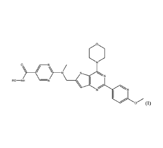
In a preferred embodiment, the compound of Formula I is set forth below:
0
N)
HO-NH -N
NN
(hereinafter "Compound 1", also referred to as N-hydroxy-2-(((2-(6-
methoxypyridin-3-
y1)-4-morpholinothieno[3,2-d]pyrimidin-6-yOmethyl)(methyDamino)pyrimidine-5-
carboxamide or a pharmaceutically acceptable salt thereof.
The invention further provides methods for the prevention or treatment of
diseases
or conditions involving aberrant proliferation, differentiation or survival of
cells. In one
embodiment, the invention further provides for the use of one or more
compounds of the
invention in the manufacture of a medicament for halting or decreasing
diseases involving
aberrant proliferation, differentiation, or survival of cells. In a preferred
embodiment, the
disease is cancer. In one embodiment, the invention relates to a method of
treating cancer
in a subject in need of treatment comprising administering to said subject a
therapeutically
effective amount of a compound of the invention.
The term "cancer" refers to any cancer caused by the proliferation of
malignant
neoplastic cells, such as tumors, neoplasms, carcinomas, sarcomas, leukemias,
lymphomas
and the like. For example, cancers include, but are not limited to,
mesothelioma,
leukemias and lymphomas such as cutaneous T-cell lymphomas (CTCL),
noncutaneous
peripheral T-cell lymphomas, lymphomas associated with human T-cell
lymphotrophic
virus (HTLV) such as adult T-cell leukemia/lymphoma (ATLL), B-cell lymphoma,
acute
nonlymphocytic leukemias, chronic lymphocytic leukemia, chronic myelogenous
leukemia, acute myelogenous leukemia, lymphomas, and multiple myeloma, non-
Hodgkin
lymphoma, acute lymphatic leukemia (ALL), chronic lymphatic leukemia (CLL),
Hodgkin's lymphoma, Burkitt lymphoma, adult T-cell leukemia lymphoma, acute-
myeloid
leukemia (AML), chronic myeloid leukemia (CML), or hepatocellular carcinoma.
Further
examples include myelodisplastic syndrome, childhood solid tumors such as
brain tumors,
neuroblastoma, retinoblastoma, Wilms' tumor, bone tumors, and soft-tissue
sarcomas,
common solid tumors of adults such as head and neck cancers (e.g., oral,
laryngeal,
CA 02830822 2013-09-19
WO 2012/135571 PCT/US2012/031361
8
nasopharyngeal and esophageal), genitourinary cancers (e.g., prostate,
bladder, renal,
uterine, ovarian, testicular), lung cancer (e.g., small-cell and non small
cell), breast cancer,
pancreatic cancer, melanoma and other skin cancers, stomach cancer, brain
tumors, tumors
related to Gorlin's syndrome (e.g., mcdulloblastoma, mcningioma, etc.), and
liver cancer.
Additional exemplary forms of cancer which may be treated by the subject
compounds
include, but are not limited to, cancer of skeletal or smooth muscle, stomach
cancer,
cancer of the small intestine, rectum carcinoma, cancer of the salivary gland,
endometrial
cancer, adrenal cancer, anal cancer, rectal cancer, parathyroid cancer, and
pituitary cancer.
Additional cancers that the compounds described herein may be useful in
preventing, treating and studying are, for example, colon carcinoma, familiary
adenomatous polyposis carcinoma and hereditary non-polyposis colorectal
cancer, or
melanoma. Further, cancers include, but are not limited to, labial carcinoma,
larynx
carcinoma, hypopharynx carcinoma, tongue carcinoma, salivary gland carcinoma,
gastric
carcinoma, adenocarcinoma, thyroid cancer (medullary and papillary thyroid
carcinoma),
renal carcinoma, kidney parenchyma carcinoma, cervix carcinoma, uterine corpus
carcinoma, endometrium carcinoma, chorion carcinoma, testis carcinoma, urinary
carcinoma, melanoma, brain tumors such as glioblastoma, astrocytoma,
meningioma,
medulloblastoma and peripheral neuroectodermal tumors, gall bladder carcinoma,
bronchial carcinoma, multiple myeloma, basalioma, teratoma, retinoblastoma,
choroidea
melanoma, seminoma, rhabdomyosarcoma, craniopharyngeoma, osteosarcoma,
chondrosarcoma, myosarcoma, liposarcoma, fibrosarcoma, Ewing sarcoma, and
plasmocytoma. In one aspect of the invention, the present invention provides
for the use
of one or more compounds of the invention in the manufacture of a medicament
for the
treatment of cancer.
In one embodiment, the compounds of the invention are used to treat a
hematological cancer or hematological precancerous condition. Hematological
cancers
include leukemias, lymphomas and multiple mycloma. Examples include
lymphocytic
leukemias, such as acute lymphocytic leukemia, including precursor B acute
lymphoblastic leukemia, precursor T acute lymphoblastic leukemia, Burkitt's
leukemia,
and acute biphenotypic leukemia; and chronic lymphocytic leukemia, including B-
cell
prolymphocytic leukemia; and myologenous leukemias, such as acute myologenous
leukemia, including acute promyelocytic leukemia, acute myeloblastic leukemia,
and
acute megakaryoblastic leukemia; and chronic myologenous leukemia, including
chronic
monocytic leukemia; acute monocytic leukemia. Other leukemias include hairy
cell
CA 02830822 2013-09-19
WO 2012/135571 PCT/US2012/031361
9
leukemia; T-cell prolymphocytic leukemia; large granular lymphocytic leukemia;
and
Adult T-cell leukemia. Lymphomas include Hodgkin's lymphoma and Non-Hodgkin's
lymphoma, including B-cell lymphomas, T-cell lymphomas, such as cutaneous T-
cell
lymphoma, and NK cell lymphomas. Hematological precancerous conditions include
myelodysplastic syndrome and myeloproliferative disorders, such as primary
myelofibrosis, polycythemia vera, and essential thrombocythemia.
Compounds of the invention have been shown to induce reversible lymphopenia
and are therefore of use for removing or decreasing the circulating levels of
cancer cells of
lymphocytic lineage. Such compounds are also of use for treating autoimmune
disorders
or for modulating an immune response.
In one embodiment, the invention provides a method for reducing the
circulating
lymphocyte count in a subject, comprising administering to the subject an
effective
amount of a compound of the invention. In a preferred embodiment, the reduced
circulating lymphocyte count is reversible, that is, the circulating
lymphocyte count
returns to the normal range after dosing with the compound of the invention is
stopped. In
one embodiment, the reduced circulating lymphocyte count is below the normal
range and
the subject is lymphopenic. Preferably, the subject derives a therapeutic or
prophylactic
benefit from the reduced circulating lymphocyte count. Such subjects include
those
suffering from a hematologic disease, such as a hematologic cancer, those
suffering from
an autoimmune disorder, and those requiring modulation of an immune response
such as
patients suffering from diabetes or organ transplant recipients. In a human
subject, the
circulating lymphocyte count, for example, B-lymphocytes, T-lymphocytes or
both, can
drop from a normal range to a lymphopenic range. In certain diseases the
circulating
lymphocyte count is abnormally high. In such diseases, the circulating
lymphocyte count
can be reduced to the normal range or to a lymphopenic state.
In one embodiment, the present invention includes the use of one or more
compounds of the invention in the manufacture of a medicament that prevents
further
aberrant proliferation, differentiation, or survival of cells. For example,
compounds of the
invention may be useful in preventing tumors from increasing in size or from
reaching a
metastatic state. The subject compounds may be administered to halt the
progression or
advancement of cancer or to induce tumor apoptosis or to inhibit tumor
angiogenesis. In
addition, the instant invention includes use of the subject compounds to
prevent a
recurrence of cancer.
CA 02830822 2013-09-19
WO 2012/135571 PCT/US2012/031361
This invention further embraces the treatment or prevention of cell
proliferative
disorders such as hyperplasias, dysplasias and pre-cancerous lesions.
Dysplasia is the
earliest form of pre-cancerous lesion recognizable in a biopsy by a
pathologist. The
subject compounds may be administered for the purpose of preventing said
hyperplasias,
5 dysplasias or pre-cancerous lesions from continuing to expand or from
becoming
cancerous. Examples of pre-cancerous lesions may occur in skin, esophageal
tissue, breast
and cervical intra-epithelial tissue.
"Combination therapy" includes the administration of the subject compounds in
further combination with other biologically active ingredients (such as, but
not limited to,
10 a second and different antineoplastic agent) and non-drug therapies
(such as, but not
limited to, surgery or radiation treatment). For instance, the compounds of
the invention
can be used in combination with other pharmaceutically active compounds,
preferably
compounds that are able to enhance the effect of the compounds of the
invention. The
compounds of the invention can be administered simultaneously (as a single
preparation or
separate preparation) or sequentially to the other drug therapy. In general, a
combination
therapy envisions administration of two or more drugs during a single cycle or
course of
therapy.
In one aspect of the invention, the subject compounds may be administered in
combination with one or more separate agents that modulate protein kinases
involved in
various disease states. Examples of such kinases may include, but are not
limited to:
serine/threonine specific kinases, receptor tyrosine specific kinases and non-
receptor
tyrosine specific kinases. Serine/threonine kinases include mitogen activated
protein
kinases (MAPK), meiosis specific kinase (MEK), RAF and aurora kinase. Examples
of
receptor kinase families include epidermal growth factor receptor (EGFR)
(e.g.,
HER2/neu, HER3, HER4, ErbB, ErbB2, ErbB3, ErbB4, Xmrk, DER, Let23); fibroblast
growth factor (FGF) receptor (e.g., FGF-R1,GFF-R2/BEK/CEK3, FGF-R3/CEK2, FGF-
R4/TKF, KGF-R); hepatocyte growth/scatter factor receptor (HGFR) (e.g., MET,
RON,
SEA, SEX); insulin receptor (e.g. 1GFI-R); Eph (e.g., CEK5, CEK8, EBK, ECK,
EEK,
EHK-1, EHK-2, ELK, EPH, ERK, HEK, MDK2, MDK5, SEK); Axl (e.g., Mer/Nyk, Rse);
RET; and platelet-derived growth factor receptor (PDGFR) (e.g., PDGFa-R, PDGI3-
R,
CSF1-R/FMS, SCF-R/C-KIT, VEGF-R/FLT, NEK/FLK1, FLT3/FLK21STK-1). Non-
receptor tyrosine kinase families include, but are not limited to, BCR-ABL
(e.g., p43abl,
ARG); BTK (e.g., ITK/EMT, TEC); CSK, FAK, FPS, JAK, SRC, BMX, FER, CDK and
SYK.
CA 02830822 2013-09-19
WO 2012/135571 PCT/US2012/031361
11
In another aspect of the invention, the subject compounds may be administered
in
combination with one or more separate agents that modulate non-kinase
biological targets
or processes. Such targets include histone deacetylases (HDAC), DNA
methyltransferase
(DNMT), heat shock proteins (e.g., HSP90), hedgehog pathway-related proteins
(e.g.,
sonic hedgehog, patched, smoothened) and proteosomes.
In a preferred embodiment, subject compounds may be combined with
antineoplastic agents (e.g., small molecules, monoclonal antibodies, antisense
RNA, and
fusion proteins) that inhibit one or more biological targets such as Zolinza,
Tarceva,
Iressa, Tykerb, Gleevec, Sutent, Sprycel, Nexavar, Sorafinib, CNF2024, RG108,
BMS387032, Affinitak, Avastin, Herceptin, Erbitux, AG24322, PD325901, ZD6474,
PD184322, Obatodax, ABT737, GDC-0449, IPI-926, BMS833923, LDE225, PF-
04449913 and AEE788. Such combinations may enhance therapeutic efficacy over
efficacy achieved by any of the agents alone and may prevent or delay the
appearance of
resistant mutational variants.
In certain preferred embodiments, the compounds of the invention are
administered
in combination with a chemotherapeutic agent. Chemotherapeutic agents
encompass a
wide range of therapeutic treatments in the field of oncology. These agents
are
administered at various stages of the disease for the purposes of shrinking
tumors,
destroying remaining cancer cells left over after surgery, inducing remission,
maintaining
remission and/or alleviating symptoms relating to the cancer or its treatment.
Examples of
such agents include, but are not limited to, alkylating agents such as mustard
gas
derivatives (Mechlorethamine, cyclophosphamide, chlorambucil, melphalan,
ifosfamide),
ethylenimines (thiotepa, hexamethylmelanine), Alkylsulfonates (Busulfan),
Hydrazines
and Triazines (Altretamine, Procarbazine, Dacarbazine and Temozolomide),
Nitrosoureas
(Carmustine, Lomustine and Streptozocin), Ifosfamide and metal salts
(Carboplatin,
Cisplatin, and Oxaliplatin); plant alkaloids such as Podophyllotoxins
(Etoposidc and
Tenisopide), Taxancs (Paclitaxcl and Docetaxel), Vinca alkaloids (Vincristinc,
Vinblastine, Vindesine and Vinorelbine), and Camptothecan analogs (lrinotecan
and
Topotecan); anti-tumor antibiotics such as Chromomycins (Dactinomycin and
Plicamycin), Anthracyclines (Doxorubicin, Daunorubicin, Epirubicin,
Mitoxantrone,
Valrubicin and Idarubicin), and miscellaneous antibiotics such as Mitomycin,
Actinomycin and Bleomycin; anti-metabolites such as folic acid antagonists
(Methotrexate, Pemetrexed, Raltitrexed, Aminopterin), pyrimidine antagonists
(5-
Fluorouracil, Floxuridine, Cytarabine, Capecitabine, and Gemcitabine), purine
antagonists
CA 02830822 2013-09-19
WO 2012/135571
PCT/US2012/031361
12
(6-Mercaptopurine and 6-Thioguanine) and adenosine deaminase inhibitors
(Cladribine,
Fludarabine, Mercaptopurine, Clofarabine, Thioguanine, Nelarabine and
Pentostatin);
topoisomerase inhibitors such as topoisomerase I inhibitors (Ironotecan,
topotecan) and
topoisomerasc II inhibitors (Amsacrinc, etoposide, etoposide phosphate,
teniposide);
monoclonal antibodies (Alemtuzumab, Gemtuzumab ozogamicin, Rituximab,
Trastuzumab, Ibritumomab Tioxetan, Cetuximab, Panitumumab, Tositumomab,
Bevacizumab); and miscellaneous anti-neoplastics such as ribonucleotide
reductase
inhibitors (Hydroxyurea); adrenocortical steroid inhibitor (Mitotane); enzymes
(Asparaginase and Pegaspargase); anti-microtubule agents (Estramustine);
retinoids
(Bexarotene, Isotretinoin, Tretinoin (ATRA), and Lenalidomide.
In certain preferred embodiments, the compounds of the invention are
administered
in combination with a chemoprotective agent. Chemoprotective agents act to
protect the
body or minimize the side effects of chemotherapy. Examples of such agents
include, but
are not limited to, amfostine, mesna, and dexrazoxane.
In one aspect of the invention, the subject compounds are administered in
combination with radiation therapy. Radiation is commonly delivered internally
(implantation of radioactive material near cancer site) or externally from a
machine that
employs photon (x-ray or gamma-ray) or particle radiation. Where the
combination
therapy further comprises radiation treatment, the radiation treatment may be
conducted at
any suitable time so long as a beneficial effect from the co-action of the
combination of
the therapeutic agents and radiation treatment is achieved. For example, in
appropriate
cases, the beneficial effect is still achieved when the radiation treatment is
temporally
removed from the administration of the therapeutic agents, perhaps by days or
even weeks.
It will be appreciated that compounds of the invention can be used in
combination
with an immunotherapeutic agent. One form of immunotherapy is the generation
of an
active systemic tumor-specific immune response of host origin by administering
a vaccine
composition at a site distant from the tumor. Various types of vaccines have
been
proposed, including isolated tumor-antigen vaccines and anti-idiotype
vaccines. Another
approach is to use tumor cells from the subject to be treated, or a derivative
of such cells
______________________________________________________________ (reviewed by
Schirt macher et al., (1995) 1 Cancer Res. Clin. neat. 12 1:487). In U.S.
Pat. No. 5,484,596, Hanna Jr., et al. claim a method for treating a resectable
carcinoma to
prevent recurrence or metastases, comprising surgically removing the tumor,
dispersing
the cells with collagenase, irradiating the cells, and vaccinating the patient
with at least
three consecutive doses of about 107 cells.
CA 02830822 2013-09-19
WO 2012/135571 PCT/US2012/031361
13
It will be appreciated that the compounds of the invention may advantageously
be
used in conjunction with one or more adjunctive therapeutic agents. Examples
of suitable
agents for adjunctive therapy include a 5HT1 agonist, such as a triptan (e.g.,
sumatriptan or
naratriptan); an adenosine Al agonist; an EP ligand; an NMDA modulator, such
as a
glycine antagonist; a sodium channel blocker (e.g., lamotrigine); a substance
P antagonist
(e.g., an NKi antagonist); a cannabinoid; acetaminophen or phenacetin; a 5-
lipoxygenase
inhibitor; a leukotriene receptor antagonist; a DMARD (e.g., methotrexate);
gabapentin
and related compounds; a tricyclic antidepressant (e.g., amitryptilline); a
neuron stabilising
antiepileptic drug; a mono-aminergic uptake inhibitor (e.g., venlafaxine); a
matrix
metalloproteinase inhibitor; a nitric oxide synthase (NOS) inhibitor, such as
an iNOS or an
nNOS inhibitor; an inhibitor of the release, or action, of tumour necrosis
factor .alpha.; an
antibody therapy, such as a monoclonal antibody therapy; an antiviral agent,
such as a
nucleoside inhibitor (e.g., lamivudine) or an immune system modulator (e.g.,
interferon);
an opioid analgesic; a local anaesthetic; a stimulant, including caffeine; an
H2-antagonist
(e.g., ranitidine); a proton pump inhibitor (e.g., omeprazole); an antacid
(e.g. aluminium or
magnesium hydroxide); an antiflatulent (e.g., simethicone); a decongestant
(e.g.,
phenylephrine, phenylpropanolamine, pseudoephedrine, oxymetazoline,
epinephrine,
naphazoline, xylometazoline, propylhexedrine, or levo-desoxyephedrine); an
antitussive
(e.g., codeine, hydrocodone, carmiphen, carbetapentane, or dextramethorphan);
a diuretic;
or a sedating or non-sedating antihistamine.
The compounds may also be used in the treatment of a disorder involving,
relating
to or, associated with dysregulation of histone deacetylase (HDAC). There are
a number
of disorders that have been implicated by or known to be mediated at least in
part by
HDAC activity, where HDAC activity is known to play a role in triggering
disease onset,
or whose symptoms are known or have been shown to be alleviated by HDAC
inhibitors.
Disorders of this type that would be expected to be amenable to treatment with
the
compounds of the invention include the following but not limited to: Anti-
proliferative
disorders (e.g., cancers); Neurodegenerative diseases including Huntington's
Disease,
Polyglutamine disease, Parkinson's Disease, Alzheimer's Disease, Seizures,
Striatonigral
degeneration, Progressive supranucl ear palsy, Torsion dystonia, Spasmodic
torticollis and
dyskinesis, Familial tremor, Gilles de la Tourette syndrome, Diffuse Lewy body
disease,
Progressive supranuclear palsy, Pick's disease, intracerebral hemorrhage,
Primary lateral
sclerosis, Spinal muscular atrophy, Amyotrophic lateral sclerosis,
Hypertrophic interstitial
polyneuropathy, Retinitis pigmentosa, Hereditary optic atrophy, Hereditary
spastic
CA 02830822 2013-09-19
WO 2012/135571
PCT/US2012/031361
14
paraplegia, Progressive ataxia and Shy-Drager syndrome; Metabolic diseases
including
Type 2 diabetes; Degenerative Diseases of the Eye including Glaucoma, Age-
related
macular degeneration, Rubeotic glaucoma; Inflammatory diseases and/or Immune
system
disorders including Rheumatoid Arthritis (RA), Osteoarthritis, Juvenile
chronic arthritis,
Graft versus Host disease, Psoriasis, Asthma, Spondyloarthropathy, Crohn's
Disease,
inflammatory bowel disease Colitis Ulcerosa, Alcoholic hepatitis, Diabetes,
Sjoegrens's
syndrome, Multiple Sclerosis, Ankylosing spondylitis, Membranous
glomerulopathy,
Discogenic pain, Systemic Lupus Erythematosus; Disease involving angiogenesis
including cancer, psoriasis, rheumatoid arthritis; Psychological disorders
including bipolar
disease, schizophrenia, mania, depression and dementia; Cardiovascular
Diseases
including the prevention and treatment of ischemia-related or reperfusion-
related vascular
and myocardial tissue damage, heart failure, restenosis and arteriosclerosis;
Fibrotic
diseases including liver fibrosis, cystic fibrosis and angiofibroma;
Infectious diseases
including Fungal infections, such as candidiasis or Candida Albicans,
Bacterial infections,
Viral infections, such as Herpes Simplex, poliovirus, rhinovirus and
coxsackievirus,
Protozoal infections, such as Malaria, Leishmania infection, Trypanosoma
brucei
infection, Toxoplasmosis and coccidlosis and Haematopoietic disorders
including
thalassemia, anemia and sickle cell anemia.
The compounds of the invention can also be used in the treatment of a disorder
involving, relating to or, associated with dysregulation of PI3 kinase. PI3
kinase activity
has been implicated in or shown to be involved in a variety of disorders. In
certain cases,
PI3 kinase activity is involved in triggering disease onset, while in others,
symptoms are
known or have been shown to be alleviated by inhibitors of PI3 kinase
activity. Disorders
of this type that would be expected to be amenable to treatment with the
compounds of the
invention include but are not limited to cancers, including leukemia, skin
cancer, bladder
cancer, breast cancer, uterine cancer, ovarian cancer, prostate cancer, lung
cancer, colon
cancer, pancreatic cancer, renal cancer, gastric cancer and brain cancer;
restenosis,
atherosclerosis, bone disorders, arthritis, diabetic retinopathy, psoriasis,
benign prostatic
hypertrophy, atherosclerosis, inflammation, angiogenesis, immunological
disorders,
pancreatitis and kidney disease.
In one embodiment, compounds of the invention can be used to induce or inhibit
apoptosis, a physiological cell death process critical for normal development
and
homeostasis. Alterations of apoptotic pathways contribute to the pathogenesis
of a variety
of human diseases. Compounds of the invention, as modulators of apoptosis,
will be
CA 02830822 2013-09-19
WO 2012/135571 PCT/US2012/031361
useful in the treatment of a variety of human diseases with aberrations in
apoptosis
including cancer (particularly, but not limited to, follicular lymphomas,
carcinomas with
p53 mutations, hormone dependent tumors of the breast, prostate and ovary, and
precanccrous lesions such as familial adenomatous polyposis), viral infections
(including,
5 but not limited to, herpes virus, poxvirus, Epstein-Barr virus, Sindbis
virus and
adenovirus), autoimmune diseases (including, but not limited to, systemic
lupus,
erythematosus, immune mediated glomerulonephritis, rheumatoid arthritis,
psoriasis,
inflammatory bowel diseases, and autoimmune diabetes mellitus),
neurodegenerative
disorders (including, but not limited to, Alzheimer's disease, AIDS-related
dementia,
10 Parkinson's disease, amyotrophic lateral sclerosis, retinitis
pigmentosa, spinal muscular
atrophy and cerebellar degeneration), AIDS, myelodysplastic syndromes,
aplastic anemia,
ischemic injury associated myocardial infarctions, stroke and reperfusion
injury,
arrhythmia, atherosclerosis, toxin-induced or alcohol induced liver diseases,
hematological
diseases (including, but not limited to, chronic anemia and aplastic anemia),
degenerative
15 diseases of the musculoskeletal system (including, but not limited to,
osteoporosis and
arthritis), aspirin-sensitive rhinosinusitis, cystic fibrosis, multiple
sclerosis, kidney
diseases, and cancer pain.
In one aspect, the invention provides the use of compounds of the invention
for the
treatment and/or prevention of immune response or immune-mediated responses
and
diseases, such as the prevention or treatment of rejection following
transplantation of
synthetic or organic grafting materials, cells, organs or tissue to replace
all or part of the
function of tissues, such as heart, kidney, liver, bone marrow, skin, cornea,
vessels, lung,
pancreas, intestine, limb, muscle, nerve tissue, duodenum, small-bowel,
pancreatic-islet-
cell, including xeno-transplants, etc; to treat or prevent graft-versus-host
disease,
autoimmune diseases, such as rheumatoid arthritis, systemic lupus
erythematosus,
thyroiditis, Hashimoto's thyroiditis, multiple sclerosis, myasthenia gravis,
type I diabetes
uvcitis, juvenile-onset or recent-onset diabetes mellitus, uvcitis, Graves
disease, psoriasis,
atopic dermatitis, Crohn's disease, ulcerative colitis, vasculitis, auto-
antibody mediated
diseases, aplastic anemia, Evan's syndrome, autoimmune hemolytic anemia, and
the like;
and further to treat infectious diseases causing aberrant immune response
and/or
activation, such as traumatic or pathogen induced immune disregulation,
including for
example, that which are caused by hepatitis B and C infections, HIV,
staphylococcus
aureus infection, viral encephalitis, sepsis, parasitic diseases wherein
damage is induced
by an inflammatory response (e.g., leprosy); and to prevent or treat
circulatory diseases,
CA 02830822 2013-09-19
WO 2012/135571 PCT/US2012/031361
16
such as arteriosclerosis, atherosclerosis, vasculitis, polyarteritis nodosa
and myocarditis.
In addition, the present invention may be used to prevent/suppress an immune
response
associated with a gene therapy treatment, such as the introduction of foreign
genes into
autologous cells and expression of the encoded product. Thus in one
embodiment, the
invention relates to a method of treating an immune response disease or
disorder or an
immune-mediated response or disorder in a subject in need of treatment
comprising
administering to said subject a therapeutically effective amount of a compound
of the
invention.
In one aspect, the invention provides the use of compounds of the invention in
the
treatment of a variety of neurodegenerative diseases, a non-exhaustive list of
which
includes: I. Disorders characterized by progressive dementia in the absence of
other
prominent neurologic signs, such as Alzheimer's disease; Senile dementia of
the
Alzheimer type; and Pick's disease (lobar atrophy); II. Syndromes combining
progressive
dementia with other prominent neurologic abnormalities such as: A) syndromes
appearing
mainly in adults (e.g., Huntington's disease, Multiple system atrophy
combining dementia
with ataxia and/or manifestations of Parkinson's disease, Progressive
supranuclear palsy
(Steel-Richardson-Olszewski), diffuse Lewy body disease, and
corticodentatonigral
degeneration; and B) syndromes appearing mainly in children or young adults
(e.g.,
Hallervorden-Spatz disease and progressive familial myoclonic epilepsy); III.
Syndromes
of gradually developing abnormalities of posture and movement such as
paralysis agitans
(Parkinson's disease), striatonigral degeneration, progressive supranuclear
palsy, torsion
dystonia (torsion spasm; dystonia musculorum deformans), spasmodic torticollis
and other
dyskinesis, familial tremor, and Gilles de la Tourette syndrome; IV. Syndromes
of
progressive ataxia such as cerebellar degenerations (e.g., cerebellar cortical
degeneration
and olivopontocerebellar atrophy (OPCA)); and spinocerebellar degeneration
(Friedreich's
atazia and related disorders); V. Syndrome of central autonomic nervous system
failure
(Shy-Drager syndrome); VI. Syndromes of muscular weakness and wasting without
sensory changes (motorneuron disease such as amyotrophic lateral sclerosis,
spinal
muscular atrophy (e.g., infantile spinal muscular atrophy (Werdnig-Hoffman),
juvenile
spinal muscular atrophy (Wohl fart-Kugelberg-Wel ander) and other forms of
familial
spinal muscular atrophy), primary lateral sclerosis, and hereditary spastic
paraplegia; VII.
Syndromes combining muscular weakness and wasting with sensory changes
(progressive
neural muscular atrophy; chronic familial polyneuropathies) such as peroneal
muscular
atrophy (Charcot-Marie-Tooth), hypertrophic interstitial polyneuropathy
(Dejerine-Sottas),
CA 02830822 2013-09-19
WO 2012/135571 PCT/US2012/031361
17
and miscellaneous forms of chronic progressive neuropathy; VIII. Syndromes of
progressive visual loss such as pigmentary degeneration of the retina
(retinitis
pigmentosa), and hereditary optic atrophy (Leber's disease). Furthermore,
compounds of
the invention can be implicated in chromatin remodeling.
The invention encompasses pharmaceutical compositions comprising
pharmaceutically acceptable salts of the compounds of the invention as
described above.
The invention also encompasses solvates of the compounds of the invention and
pharmaceutical compositions comprising such solvates, such as hydrates,
methanolates or
ethanolates. The term "solvate" refers to a solid, preferably crystalline,
form of a
compound which includes the presence of solvent molecules within the crystal
lattice. A
solvate of a compound comprising a given solvent is typically prepared by
crystallization
of the compound from that solvent. Solvates can include a variety of solvents,
including
water, methanol and ethanol. The term "hydrate" refers to a solvate in which
the solvent is
water, and includes, but is not limited to, hemihydrate, monohydrate,
dihydrate, trihydrate
and the like. The invention further encompasses pharmaceutical compositions
comprising
any solid or liquid physical form of the compound of the invention, including
crystalline
and crystalline solvate forms. For example, the compounds can be in a
crystalline form, in
an amorphous form, and have any particle size. The particles may be
micronized, or may
be agglomerated, particulate granules, powders, oils, oily suspensions or any
other form of
solid or liquid physical form.
The compounds of the invention, and derivatives, fragments, analogs, homologs,
pharmaceutically acceptable salts or solvates thereof can be incorporated into
pharmaceutical compositions suitable for administration, together with a
pharmaceutically
acceptable carrier or excipient. Such compositions typically comprise a
therapeutically
effective amount of any of the compounds above, and a pharmaceutically
acceptable
carrier. Preferably, the effective amount when treating cancer is an amount
effective to
selectively induce terminal differentiation of suitable neoplastic cells and
less than an
amount which causes toxicity in a patient.
Compounds of the invention may be administered by any suitable means,
including, without limitation, parenteral, intravenous, intramuscular,
subcutaneous,
implantation, oral, sublingual, buccal, nasal, pulmonary, transdermal,
topical, vaginal,
rectal, and transmucosal administrations or the like. Topical administration
can also
involve the use of transdermal administration such as transdermal patches or
iontophoresis
devices. Pharmaceutical preparations include a solid, semisolid or liquid
preparation
CA 02830822 2013-09-19
WO 2012/135571 PCT/US2012/031361
18
(tablet, pellet, troche, capsule, suppository, cream, ointment, aerosol,
powder, liquid,
emulsion, suspension, syrup, injection, etc.) containing a compound of the
invention as an
active ingredient, which is suitable for selected mode of administration. In
one
embodiment, the pharmaceutical compositions arc administered orally, and arc
thus
formulated in a form suitable for oral administration, i.e., as a solid or a
liquid preparation.
Suitable solid oral formulations include tablets, capsules, pills, granules,
pellets, sachets
and effervescent, powders, and the like. Suitable liquid oral formulations
include
solutions, suspensions, dispersions, emulsions, oils and the like. In one
embodiment of the
present invention, the composition is formulated in a capsule. In accordance
with this
embodiment, the compositions of the present invention comprise in addition to
the active
compound and the inert carrier or diluent, a hard gelatin capsule.
Any inert excipient that is commonly used as a carrier or diluent may be used
in
the formulations of the present invention, such as for example, a gum, a
starch, a sugar, a
cellulosic material, an acrylate, or mixtures thereof A preferred diluent is
microcrystalline cellulose. The compositions may further comprise a
disintegrating agent
(e.g., croscarmellose sodium) and a lubricant (e.g., magnesium stearate), and
may
additionally comprise one or more additives selected from a binder, a buffer,
a protease
inhibitor, a surfactant, a solubilizing agent, a plasticizer, an emulsifier, a
stabilizing agent,
a viscosity increasing agent, a sweetener, a film forming agent, or any
combination
thereof. Furthermore, the compositions of the present invention may be in the
form of
controlled release or immediate release formulations.
For liquid formulations, pharmaceutically acceptable carriers may be aqueous
or
non-aqueous solutions, suspensions, emulsions or oils. Examples of non-aqueous
solvents
are propylene glycol, polyethylene glycol, and injectable organic esters such
as ethyl
oleate. Aqueous carriers include water, alcoholic/aqueous solutions, emulsions
or
suspensions, including saline and buffered media. Examples of oils arc those
of
petroleum, animal, vegetable, or synthetic origin, for example, peanut oil,
soybean oil,
mineral oil, olive oil, sunflower oil, and fish-liver oil. Solutions or
suspensions can also
include the following components: a sterile diluent such as water for
injection, saline
solution, fixed oils, polyethylene glycols, glycerine, propylene glycol or
other synthetic
solvents; antibacterial agents such as benzyl alcohol or methyl parabens;
antioxidants such
as ascorbic acid or sodium bisulfite; chelating agents such as
ethylenediaminetetraacetic
acid (EDTA); buffers such as acetates, citrates or phosphates, and agents for
the
CA 02830822 2013-09-19
WO 2012/135571 PCT/US2012/031361
19
adjustment of tonicity such as sodium chloride or dextrose. The pH can be
adjusted with
acids or bases, such as hydrochloric acid or sodium hydroxide.
In addition, the compositions may further comprise binders (e.g., acacia,
cornstarch, gelatin, carbomer, ethyl cellulose, guar gum, hydroxypropyl
cellulose,
hydroxypropyl methyl cellulose, povidone), disintegrating agents (e.g.,
cornstarch, potato
starch, alginic acid, silicon dioxide, croscarmellose sodium, crospovidone,
guar gum,
sodium starch glycolate, Primogel), buffers (e.g., tris-HCI., acetate,
phosphate) of various
pH and ionic strength, additives such as albumin or gelatin to prevent
absorption to
surfaces, detergents (e.g., Tween 20, Tween 80, Pluronic F68, bile acid
salts), protease
inhibitors, surfactants (e.g., sodium lauryl sulfate), permeation enhancers,
solubilizing
agents (e.g., glycerol, polyethylene glycerol, polyethylene glycol), a glidant
(e.g., colloidal
silicon dioxide), anti-oxidants (e.g., ascorbic acid, sodium metabisulfite,
butylated
hydroxyanisole), stabilizers (e.g., hydroxypropyl cellulose,
hydroxypropylmethyl
cellulose), viscosity increasing agents (e.g., carbomer, colloidal silicon
dioxide, ethyl
cellulose, guar gum), sweeteners (e.g., sucrose, aspartame, citric acid),
flavoring agents
(e.g., peppermint, methyl salicylate, or orange flavoring), preservatives
(e.g., Thimerosal,
benzyl alcohol, parabens), lubricants (e.g., stearic acid, magnesium stearate,
polyethylene
glycol, sodium lauryl sulfate), flow-aids (e.g., colloidal silicon dioxide),
plasticizers (e.g.,
diethyl phthalate, triethyl citrate), emulsifiers (e.g., carbomer,
hydroxypropyl cellulose,
sodium lauryl sulfate), polymer coatings (e.g., poloxamers or poloxamines),
coating and
film forming agents (e.g., ethyl cellulose, acrylates, polymethacrylates)
and/or adjuvants.
In one embodiment, the active compounds are prepared with carriers that will
protect the compound against rapid elimination from the body, such as a
controlled release
formulation, including implants and microencapsulated delivery systems.
Biodegradable,
biocompatible polymers can be used, such as ethylene vinyl acetate,
polyanhydrides,
polyglycolic acid, collagen, polyorthoesters, and polylactic acid. Methods for
preparation
of such formulations will be apparent to those skilled in the art. The
materials can also be
obtained commercially from Alza Corporation and Nova Pharmaceuticals, Inc.
Liposomal
suspensions (including liposomes targeted to infected cells with monoclonal
antibodies to
viral antigens) can also be used as pharmaceutically acceptable carriers.
These can be
prepared according to methods known to those skilled in the art, for example,
as described
in U.S. Pat. No. 4,522,811.
It is especially advantageous to formulate oral compositions in dosage unit
form
for ease of administration and uniformity of dosage. Dosage unit form as used
herein
CA 02830822 2013-09-19
WO 2012/135571 PCT/US2012/031361
refers to physically discrete units suited as unitary dosages for the subject
to be treated;
each unit containing a predetermined quantity of active compound calculated to
produce
the desired therapeutic effect in association with the required pharmaceutical
carrier. The
specification for the dosage unit forms of the invention arc dictated by and
directly
5 dependent on the unique characteristics of the active compound and the
particular
therapeutic effect to be achieved, and the limitations inherent in the art of
compounding
such an active compound for the treatment of individuals.
Formulations of the invention intended for oral administration can include one
or
more permeation enhancers, including long chain fatty acids or salts thereof,
such as
10 decanoic acid and sodium decanoate.
In one preferred embodiment, the compound can be formulated in an aqueous
solution for intravenous injection. In one embodiment, solubilizing agents can
be suitably
employed. A particularly preferred solubilizing agent includes cyclodextrins
and modified
cyclodextrins, such as sulfonic acid substituted P-cyclodextrin derivative or
salt thereof,
15 including sulfobutyl derivatized-P-cyclodextrin, such as sulfobutylether-
7-P-cyclodextrin
which is sold by CyDex, Inc. under the tradename CAPTISOLO.
The pharmaceutical compositions can be included in a container, pack, or
dispenser together with instructions for administration.
Daily administration may be repeated continuously for a period of several days
to
20 several years. Oral treatment may continue for between one week and the
life of the
patient. Preferably the administration may take place for five consecutive
days after
which time the patient can be evaluated to determine if further administration
is required.
The administration can be continuous or intermittent, e.g., treatment for a
number of
consecutive days followed by a rest period. The compounds of the present
invention may
be administered intravenously on the first day of treatment, with oral
administration on the
second day and all consecutive days thereafter.
The preparation of pharmaceutical compositions that contain an active
component
is well understood in the art, for example, by mixing, granulating, or tablet-
forming
processes. The active therapeutic ingredient is often mixed with excipients
that are
pharmaceutically acceptable and compatible with the active ingredient. For
oral
administration, the active agents are mixed with additives customary for this
purpose, such
as vehicles, stabilizers, or inert diluents, and converted by customary
methods into suitable
forms for administration, such as tablets, coated tablets, hard or soft
gelatin capsules,
aqueous, alcoholic or oily solutions and the like as detailed above.
CA 02830822 2013-09-19
WO 2012/135571 PCT/US2012/031361
21
The amount of the compound administered to the patient is less than an amount
that would cause toxicity in the patient. In certain embodiments, the amount
of the
compound that is administered to the patient is less than the amount that
causes a
concentration of the compound in the patient's plasma to equal or exceed the
toxic level of
the compound. Preferably, the concentration of the compound in the patient's
plasma is
maintained at about 10 nM. In one embodiment, the concentration of the
compound in the
patient's plasma is maintained at about 25 nM. In one embodiment, the
concentration of
the compound in the patient's plasma is maintained at about 50 nM. In one
embodiment,
the concentration of the compound in the patient's plasma is maintained at
about 100 nM.
In one embodiment, the concentration of the compound in the patient's plasma
is
maintained at about 500 nM. In one embodiment, the concentration of the
compound in
the patient's plasma is maintained at about 1000 nM. In one embodiment, the
concentration of the compound in the patient's plasma is maintained at about
2500 nM. In
one embodiment, the concentration of the compound in the patient's plasma is
maintained
at about 5000 nM. The optimal amount of the compound that should be
administered to
the patient in the practice of the present invention will depend on the
particular compound
used and the type of cancer being treated.
DEFINITIONS
Listed below are definitions of various terms used to describe this invention.
These
definitions apply to the terms as they are used throughout this specification
and claims,
unless otherwise limited in specific instances, either individually or as part
of a larger
group.
The term "acyl" refers to hydrogen, alkyl, partially saturated or fully
saturated
cycloalkyl, partially saturated or fully saturated heterocycle, aryl, and
heteroaryl
substituted carbonyl groups. For example, acyl includes groups such as (Ci-
C6)alkanoyl
(e.g., formyl, acetyl, propionyl, butyryl, valcryl, caproyl, t-butylacetyl,
etc.), (C3-
C6)cycloalkylcarbonyl (e.g., cyclopropylcarbonyl, cyclobutylcarbonyl,
cyclopentylcarbonyl, cyclohexylcarbonyl, etc.), heterocyclic carbonyl (e.g.,
pyrrolidinylcarbonyl, pyrrolid-2-one-5-carbonyl, piperi dinyl carbonyl,
piperazinyl carbonyl,
tetrahydrofuranyl carbonyl, etc.), aroyl (e.g., benzoyl) and heteroaroyl
(e.g., thiopheny1-2-
carbonyl, thiopheny1-3-carbonyl, furany1-2-carbonyl, furany1-3-carbonyl, 1H-
pyrroy1-2-
carbonyl, 1H-pyrroy1-3-carbonyl, benzo[b]thiopheny1-2-carbonyl, etc.). In
addition, the
alkyl, cycloalkyl, heterocycle, aryl and heteroaryl portion of the acyl group
may be any
CA 02830822 2013-09-19
WO 2012/135571 PCT/US2012/031361
22
one of the groups described in the respective definitions. When indicated as
being
"optionally substituted", the acyl group may be unsubstituted or optionally
substituted
with one or more substituents (typically, one to three substituents)
independently selected
from the group of substituents listed below in the definition for
"substituted" or the alkyl,
cycloalkyl, heterocycle, aryl and heteroaryl portion of the acyl group may be
substituted as
described above in the preferred and more preferred list of substituents,
respectively.
The term "alkyl" embraces linear or branched radicals having one to about
twenty
carbon atoms or, preferably, one to about twelve carbon atoms. More preferred
alkyl
radicals are "lower alkyl" radicals having one to about ten carbon atoms. Most
preferred
are lower alkyl radicals having one to about eight carbon atoms. Examples of
such radicals
include methyl, ethyl, n-propyl, isopropyl, n-butyl, isobutyl, sec-butyl, tert-
butyl, pentyl,
iso-amyl, hexyl and the like.
The term "alkenyl" embraces linear or branched radicals having at least one
carbon-carbon double bond of two to about twenty carbon atoms or, preferably,
two to
about twelve carbon atoms. More preferred alkenyl radicals are "lower alkenyl"
radicals
having two to about ten carbon atoms and more preferably about two to about
eight carbon
atoms. Examples of alkenyl radicals include ethenyl, allyl, propenyl, butenyl
and 4-
methylbutenyl. The terms "alkenyl", and "lower alkenyl", embrace radicals
having "cis"
and "trans" orientations, or alternatively, "E" and "Z" orientations.
The term "alkynyl" embraces linear or branched radicals having at least one
carbon-carbon triple bond of two to about twenty carbon atoms or, preferably,
two to
about twelve carbon atoms. More preferred alkynyl radicals are "lower alkynyl"
radicals
having two to about ten carbon atoms and more preferably about two to about
eight carbon
atoms. Examples of alkynyl radicals include propargyl, 1-propynyl, 2-propynyl,
1-butyne,
2-butynyl and 1-pentynyl.
The term "aryl", alone or in combination, means a carbocyclic aromatic system
containing one, two or three rings wherein such rings may be attached together
in a
pendent manner or may be fused. The term "aryl" embraces aromatic radicals
such as
phenyl, naphthyl, tetrahydronaphthyl, indane and biphenyl.
The terms "heterocyclyl", "heterocycle", "heterocyclic" or "heterocyclo"
embrace
saturated, partially unsaturated and unsaturated heteroatom-containing ring-
shaped
radicals, which can also be called "heterocyclyl", "heterocycloalkenyl" and
"heteroaryl"
correspondingly, where the heteroatoms may be selected from nitrogen, sulfur
and
oxygen. Examples of saturated heterocyclyl radicals include saturated 3 to 6-
membered
CA 02830822 2013-09-19
WO 2012/135571 PCT/US2012/031361
23
heteromonocyclic group containing 1 to 4 nitrogen atoms (e.g. pyrrolidinyl,
imidazolidinyl, piperidino, piperazinyl, etc.); saturated 3 to 6-membered
heteromonocyclic
group containing 1 to 2 oxygen atoms and 1 to 3 nitrogen atoms (e.g.
morpholinyl, etc.);
saturated 3 to 6-membered heteromonocyclic group containing 1 to 2 sulfur
atoms and 1 to
3 nitrogen atoms (e.g., thiazolidinyl, etc.). Examples of partially
unsaturated heterocyclyl
radicals include dihydrothiophene, dihydropyran, dihydrofuran and
dihydrothiazole.
Heterocyclyl radicals may include a pentavalent nitrogen, such as in
tetrazolium and
pyridinium radicals. The term "heterocycle" also embraces radicals where
heterocyclyl
radicals are fused with aryl or cycloalkyl radicals. Examples of such fused
bicyclic
radicals include benzofuran, benzothiophene, and the like.
The term "heteroaryl" embraces unsaturated heterocyclyl radicals. Examples of
heteroaryl radicals include unsaturated 3 to 6-membered, preferably 5 or 6-
membered,
heteromonocyclic group containing 1 to 4 nitrogen atoms, for example,
pyrrolyl,
pyrrolinyl, imidazolyl, pyrazolyl, pyridyl, pyrimidyl, pyrazinyl, pyridazinyl,
triazolyl (e.g.,
4H-1,2,4-triazolyl, 1H-1,2,3-triazolyl, 2H-1,2,3-triazolyl, etc.) tetrazolyl
(e.g. 1H-
tetrazolyl, 2H-tetrazolyl, etc.), etc.; unsaturated condensed heterocyclyl
group containing 1
to 5 nitrogen atoms, for example, indolyl, isoindolyl, indolizinyl,
benzimidazolyl,
quinolyl, isoquinolyl, indazolyl, benzotriazolyl, tetrazolopyridazinyl (e.g.,
tetrazolo[1,5-
blpyridazinyl, etc.), etc.; unsaturated 3 to 6-membered, preferably 5- or 6-
membered,
heteromonocyclic group containing an oxygen atom, for example, pyranyl, furyl,
etc.;
unsaturated 3 to 6-membered heteromonocyclic group containing a sulfur atom,
for
example, thienyl, etc.; unsaturated 3 to 6-membered, preferably 5- or 6-
membered,
heteromonocyclic group containing 1 to 2 oxygen atoms and 1 to 3 nitrogen
atoms, for
example, oxazolyl, isoxazolyl, oxadiazolyl (e.g., 1,2,4-oxadiazolyl, 1,3,4-
oxadiazolyl,
1,2,5-oxadiazolyl, etc.) etc.; unsaturated condensed heterocyclyl group
containing 1 to 2
oxygen atoms and 1 to 3 nitrogen atoms (e.g. benzoxazolyl, benzoxadiazolyl,
etc.);
unsaturated 3 to 6-membered, preferably 5- or 6-membered, heteromonocyclic
group
containing 1 to 2 sulfur atoms and 1 to 3 nitrogen atoms, for example,
thiazolyl,
thiadiazolyl (e.g., 1,2,4- thiadiazolyl, 1,3,4-thiadiazolyl, 1,2,5-
thiadiazolyl, etc.) etc.;
unsaturated condensed heterocyclyl group containing 1 to 2 sulfur atoms and 1
to 3
nitrogen atoms (e.g., benzothiazolyl, benzothiadiazolyl, etc.) and the like.
The term "heterocycloalkyl" embraces heterocyclo-substituted alkyl radicals.
More
preferred heterocycloalkyl radicals are "lower heterocycloalkyl" radicals
having one to six
carbon atoms in the heterocyclo radicals.
CA 02830822 2013-09-19
WO 2012/135571 PCT/US2012/031361
24
The term "substituted" refers to the replacement of one or more hydrogen
radicals
in a given structure with the radical of a specified substituent including,
but not limited to:
halo, alkyl, alkenyl, alkynyl, aryl, heterocyclyl, thiol, alkylthio, arylthio,
alkylthioalkyl,
arylthioalkyl, alkylsulfonyl, alkylsulfonylalkyl, arylsulfonylalkyl, alkoxy,
aryloxy,
aralkoxy, aminocarbonyl, alkylaminocarbonyl, arylaminocarbonyl,
alkoxycarbonyl,
aryloxycarbonyl, haloalkyl, amino, trifluoromethyl, cyano, nitro, alkylamino,
arylamino,
alkylaminoalkyl, arylaminoalkyl, aminoalkylamino, hydroxy, alkoxyalkyl,
carboxyalkyl,
alkoxycarbonylalkyl, aminocarbonylalkyl, acyl, aralkoxycarbonyl, carboxylic
acid,
sulfonic acid, sulfonyl, phosphonic acid, aryl, heteroaryl, heterocyclic, and
aliphatic. It is
understood that the substituent may be further substituted.
For simplicity, chemical moieties are defined and referred to throughout can
be
univalent chemical moieties (e.g., alkyl, aryl, etc.) or multivalent moieties
under the
appropriate structural circumstances clear to those skilled in the art. For
example, an
"alkyl" moiety can be referred to a monovalent radical (e.g., CH3-CH2-), or in
other
instances, a bivalent linking moiety can be "alkyl," in which case those
skilled in the art
will understand the alkyl to be a divalent radical (e.g., -CH2-CH2-), which is
equivalent to
the term "alkylene." Similarly, in circumstances in which divalent moieties
are required
and are stated as being "alkoxy", "alkylamino", "aryloxy", "alkylthio",
"aryl",
"heteroaryl", "heterocyclic", "alkyl" "alkenyl", "alkynyl", "aliphatic", or
"cycloalkyl",
those skilled in the art will understand that the terms alkoxy", "alkylamino",
"aryloxy",
"alkylthio", "aryl", "heteroaryl", "heterocyclic", "alkyl", "alkenyl",
"alkynyl",
"aliphatic", or "cycloalkyl" refer to the corresponding divalent moiety.
The terms "halogen" or "halo" as used herein, refers to an atom selected from
fluorine, chlorine, bromine and iodine.
As used herein, the term "aberrant proliferation" refers to abnormal cell
growth.
The phrase "adjunctive therapy" encompasses treatment of a subject with agents
that reduce or avoid side effects associated with the combination therapy of
the present
invention, including, but not limited to, those agents, for example, that
reduce the toxic
effect of anticancer drugs, e.g., bone resorption inhibitors, cardioprotective
agents; prevent
or reduce the incidence of nausea and vomiting associated with chemotherapy,
radiotherapy or operation; or reduce the incidence of infection associated
with the
administration of myelosuppressive anticancer drugs.
The term "angiogenesis," as used herein, refers to the formation of blood
vessels.
Specifically, angiogenesis is a multi-step process in which endothelial cells
focally
CA 02830822 2013-09-19
WO 2012/135571 PCT/US2012/031361
degrade and invade through their own basement membrane, migrate through
interstitial
stroma toward an angiogenic stimulus, proliferate proximal to the migrating
tip, organize
into blood vessels, and reattach to newly synthesized basement membrane (see
Folkman et
al., Adv. Cancer Res., Vol. 43, pp. 175-203 (1985)). Anti-angiogcnic agents
interfere with
5 this process. Examples of agents that interfere with several of these
steps include
thrombospondin-1, angiostatin, endostatin, interferon alpha and compounds such
as matrix
metalloproteinase (MMP) inhibitors that block the actions of enzymes that
clear and create
paths for newly forming blood vessels to follow; compounds, such as
.alpha.v.beta.3
inhibitors, that interfere with molecules that blood vessel cells use to
bridge between a
10 parent blood vessel and a tumor; agents, such as specific COX-2
inhibitors, that prevent
the growth of cells that form new blood vessels; and protein-based compounds
that
simultaneously interfere with several of these targets.
The term "apoptosis" as used herein refers to programmed cell death as
signaled
by the nuclei in normally functioning human and animal cells when age or state
of cell
15 health and condition dictates. An "apoptosis inducing agent" triggers
the process of
programmed cell death.
The term "cancer" as used herein denotes a class of diseases or disorders
characterized by uncontrolled division of cells and the ability of these cells
to invade other
tissues, either by direct growth into adjacent tissue through invasion or by
implantation
20 into distant sites by metastasis.
The terms "compound" and "compound of the invention", as used herein, refer to
compounds of Formula I and pharmaceutically acceptable salts thereof. The
compounds
of the invention can be obtained in different forms, including crystalline and
amorphous
forms. The compounds can also occur as solvates, for example, hydrates, or
solvates of an
25 organic solvent, preferably a pharmaceutically acceptable solvent. The
compounds can
also occur in multiple crystalline, or polymorphic, forms. The compounds of
the invention
further include pharmaceutically acceptable prodrugs and esters of the
compounds of
Formula I.
The term "device" refers to any appliance, usually mechanical or electrical,
designed to perform a particular function.
As used herein, the term "dysplasia" refers to abnormal cell growth, and
typically
refers to the earliest form of pre-cancerous lesion recognizable in a biopsy
by a
pathologist.
CA 02830822 2013-09-19
WO 2012/135571 PCT/US2012/031361
26
As used herein, the term "effective amount of the subject compounds," with
respect to the subject method of treatment, refers to an amount of the subject
compound
which, when delivered as part of desired dose regimen, brings about, e.g., a
change in the
rate of cell proliferation and/or state of differentiation and/or rate of
survival of a cell to
clinically acceptable standards. This amount may further relieve to some
extent one or
more of the symptoms of a neoplasia disorder, including, but is not limited
to: I) reduction
in the number of cancer cells; 2) reduction in tumor size; 3) inhibition
(i.e., slowing to
some extent, preferably stopping) of cancer cell infiltration into peripheral
organs; 4)
inhibition (i.e., slowing to some extent, preferably stopping) of tumor
metastasis; 5)
inhibition, to some extent, of tumor growth; 6) relieving or reducing to some
extent one or
more of the symptoms associated with the disorder; and/or 7) relieving or
reducing the
side effects associated with the administration of anticancer agents.
The term "hyperplasia," as used herein, refers to excessive cell division or
growth.
The phrase an "immunotherapeutic agent" refers to agents used to transfer the
immunity of an immune donor, e.g., another person or an animal, to a host by
inoculation.
The term embraces the use of serum or gamma globulin containing performed
antibodies
produced by another individual or an animal; nonspecific systemic stimulation;
adjuvants;
active specific immunotherapy; and adoptive immunotherapy. Adoptive
immunotherapy
refers to the treatment of a disease by therapy or agents that include host
inoculation of
sensitized lymphocytes, transfer factor, immune RNA, or antibodies in serum or
gamma
globulin.
The term "inhibition," in the context of neoplasia, tumor growth or tumor cell
growth, may be assessed by delayed appearance of primary or secondary tumors,
slowed
development of primary or secondary tumors, decreased occurrence of primary or
secondary tumors, slowed or decreased severity of secondary effects of
disease, arrested
tumor growth and regression of tumors, among others. In the extreme, complete
inhibition,
is referred to herein as prevention or chemoprevention.
The term "metastasis," as used herein, refers to the migration of cancer cells
from
the original tumor site through the blood and lymph vessels to produce cancers
in other
tissues. Metastasis also is the term used for a secondary cancer growing at a
distant site.
The term "neoplasm," as used herein, refers to an abnormal mass of tissue that
results from excessive cell division. Neoplasms may be benign (not cancerous),
or
malignant (cancerous) and may also be called a tumor. The term "neoplasia" is
the
pathological process that results in tumor formation.
CA 02830822 2013-09-19
WO 2012/135571 PCT/US2012/031361
27
As used herein, the term "pre-cancerous" refers to a condition that is not
malignant, but is likely to become malignant if left untreated.
The term "proliferation" refers to cells undergoing mitosis.
The phrase "P13 kinasc related disease or disorder" refers to a disease or
disorder
characterized by inappropriate phosphoinositide-3-kinase activity or over-
activity of the
phosphoinositide-3-kinase. Inappropriate activity refers to either: (i) PI3
kinase expression
in cells which normally do not express PI3 kinase; (ii) increased PI3 kinase
expression
leading to unwanted cell proliferation, differentiation and/or growth; or,
(iii) decreased P13
kinase expression leading to unwanted reductions in cell proliferation,
differentiation
and/or growth. Over-activity of P13 kinase refers to either amplification of
the gene
encoding a particular P13 kinase or production of a level of PI3 kinase
activity which can
correlate with a cell proliferation, differentiation and/or growth disorder
(that is, as the
level of the PI3 kinase increases, the severity of one or more of the symptoms
of the
cellular disorder increases).
The phrase a "radio therapeutic agent" refers to the use of electromagnetic or
particulate radiation in the treatment of neoplasia.
The term "recurrence" as used herein refers to the return of cancer after a
period of
remission. This may be due to incomplete removal of cells from the initial
cancer and
may occur locally (the same site of initial cancer), regionally (in vicinity
of initial cancer,
possibly in the lymph nodes or tissue), and/or distally as a result of
metastasis.
The term "treatment" refers to any process, action, application, therapy, or
the like,
wherein a mammal, including a human being, is subject to medical aid with the
object of
improving the mammal's condition, directly or indirectly.
The term "vaccine" includes agents that induce the patient's immune system to
mount an immune response against the tumor by attacking cells that express
tumor
associated antigens (Teas).
As used herein, the term "pharmaceutically acceptable salt" refers to those
salts
which are, within the scope of sound medical judgment, suitable for use in
contact with
the tissues of humans and lower animals without undue toxicity, irritation,
allergic
response and the like, and are commensurate with a reasonable benefit/risk
ratio.
Pharmaceutically acceptable salts are well known in the art. For example, S.
M. Berge, et
al. describes pharmaceutically acceptable salts in detail in J. Pharmaceutical
Sciences, 66:
1-19 (1977). The salts can be prepared in situ during the final isolation and
purification of
the compounds of the invention, or separately by reacting the free base
function with a
CA 02830822 2013-09-19
WO 2012/135571 PCT/US2012/031361
28
suitable organic acid or inorganic acid. Examples of pharmaceutically
acceptable
nontoxic acid addition salts include, but are not limited to, salts of an
amino group formed
with inorganic acids such as hydrochloric acid, hydrobromic acid, phosphoric
acid,
sulfuric acid and perchloric acid or with organic acids such as acetic acid,
malcic acid,
tartaric acid, citric acid, succinic acid lactobionic acid or malonic acid or
by using other
methods used in the art such as ion exchange. Other pharmaceutically
acceptable salts
include, but are not limited to, adipate, alginate, ascorbate, aspartate,
benzenesulfonate,
benzoate, bisulfate, borate, butyrate, camphorate, camphorsulfonate, citrate,
cyclopentanepropionate, digluconate, dodecylsulfate, ethanesulfonate, formate,
fumarate,
glucoheptonate, glycerophosphate, gluconate, hemisulfate, heptanoate,
hexanoate,
hydroiodide, 2-hydroxy-ethanesulfonate, lactobionate, lactate, laurate, lauryl
sulfate,
malate, maleate, malonate, methanesulfonate, 2-naphthalenesulfonate,
nicotinate, nitrate,
oleate, oxalate, palmitate, pamoate, pectinate, persulfate, 3-
phenylpropionate, phosphate,
picrate, pivalate, propionate, stearate, succinate, sulfate, tartrate,
thiocyanate, p-
toluenesulfonate, undecanoate, valerate salts, and the like. Representative
alkali or
alkaline earth metal salts include sodium, lithium, potassium, calcium,
magnesium, and
the like. Further pharmaceutically acceptable salts include, when appropriate,
nontoxic
ammonium, quaternary ammonium, and amine cations formed using counterions such
as
halide, hydroxide, carboxylate, sulfate, phosphate, nitrate, alkyl sulfonate
having from 1 to
6 carbon atoms, sulfonate and aryl sulfonate. Certain salts such as the
sodium, potassium
and choline base salts as well as acidic salts such as sulfate and
methanesulfonate salts
have been found to improve the solubility of compounds of Formula I in
pharmaceutically
acceptable aqueous media. In one embodiment, the pharmaceutically acceptable
salt of
Compound 1 is the choline salt. Preferred salts of Compound 1 include the
sodium salt and
the potassium salt. Other preferred salts include the sulfate and
methanesulfonate salts.
As used herein, the term "pharmaceutically acceptable ester" refers to esters
which
hydrolyze in vivo and include those that break down readily in the human body
to leave
the parent compound or a salt thereof. Suitable ester groups include, for
example, those
derived from pharmaceutically acceptable aliphatic carboxylic acids,
particularly alkanoic,
alkenoic, cycloalkanoic and alkanedioic acids, in which each alkyl or alkenyl
moiety
advantageously has not more than 6 carbon atoms. Examples of particular esters
include,
but are not limited to, formates, acetates, propionates, butyrates, acrylates
and
ethylsuccinates.
CA 02830822 2013-09-19
WO 2012/135571 PCT/US2012/031361
29
The term "pharmaceutically acceptable prodrugs" as used herein refers to those
prodrugs of the compounds of the present invention which are, within the scope
of sound
medical judgment, suitable for use in contact with the tissues of humans and
lower animals
with undue toxicity, irritation, allergic response, and the like, commensurate
with a
reasonable benefit/risk ratio, and effective for their intended use, as well
as the
zwitterionic forms, where possible, of the compounds of the present invention.
"Prodrug",
as used herein means a compound which is convertible in vivo by metabolic
means (e.g.
by hydrolysis) to a compound of the invention. Various forms of prodrugs are
known in
the art, for example, as discussed in Bundgaard, (ed.), Design of Prodrugs,
Elsevier
(1985); Widder, et al. (ed.), Methods in Enzymology, Vol. 4, Academic Press
(1985);
Krogsgaard-Larsen, et al., (ed). "Design and Application of Prodrugs, Textbook
of Drug
Design and Development, Chapter 5, 113-191 (1991); Bundgaard, et al., Journal
of Drug
Deliver Reviews, 8:1-38(1992); Bundgaard, J. of Pharmaceutical Sciences, 77
:285 et seq.
(1988); Higuchi and Stella (eds.) Prodrugs as Novel Drug Delivery Systems,
American
Chemical Society (1975); and Bernard Testa & Joachim Mayer, "Hydrolysis In
Drug And
Prodrug Metabolism: Chemistry, Biochemistry And Enzymology, John Wiley and
Sons,
Ltd. (2002).
As used herein, "pharmaceutically acceptable carrier" is intended to include
any
and all solvents, dispersion media, coatings, antibacterial and antifungal
agents, isotonic
and absorption delaying agents, and the like, compatible with pharmaceutical
administration, such as sterile pyrogen-free water. Suitable carriers are
described in the
most recent edition of Remington's Pharmaceutical Sciences, a standard
reference text in
the field, which is incorporated herein by reference. Preferred examples of
such carriers
or diluents include, but are not limited to, water, saline, Ringer's
solutions, dextrose
solution, and 5% human serum albumin. Liposomes and non-aqueous vehicles such
as
fixed oils may also be used. The use of such media and agents for
pharmaceutically active
substances is well known in the art. Except insofar as any conventional media
or agent is
incompatible with the active compound, use thereof in the compositions is
contemplated.
Supplementary active compounds can also be incorporated into the compositions.
As used herein, the term "pre-cancerous" refers to a condition that is not
malignant, but is likely to become malignant if left untreated.
The term "subject" as used herein refers to an animal. Preferably the animal
is a
mammal. More preferably the mammal is a human. A subject also refers to, for
example,
dogs, cats, horses, cows, pigs, guinea pigs, fish, birds and the like.
CA 02830822 2013-09-19
WO 2012/135571 PCT/US2012/031361
The compounds of this invention may be modified by appending appropriate
functionalities to enhance selective biological properties. Such modifications
are known in
the art and may include those which increase biological penetration into a
given biological
system (e.g., blood, lymphatic system, central nervous system), increase oral
availability,
5 increase solubility to allow administration by injection, alter
metabolism and alter rate of
excretion.
The synthesized compounds can be separated from a reaction mixture and further
purified by a method such as column chromatography, high pressure liquid
chromatography, or recrystallization. As can be appreciated by the skilled
artisan, further
10 methods of synthesizing the compounds of the formulae herein will be
evident to those of
ordinary skill in the art. Additionally, the various synthetic steps may be
performed in an
alternate sequence or order to give the desired compounds. Synthetic chemistry
transformations and protecting group methodologies (protection and
deprotection) useful
in synthesizing the compounds described herein are known in the art and
include, for
15 example, those such as described in R. Larock, Comprehensive Organic
Transformations,
VCH Publishers (1989); T.W. Greene and P.G.M. Wuts, Protective Groups in
Organic
Synthesis, 2d. Ed., John Wiley and Sons (1991); L. Fieser and M. Fieser,
Fieser and
Fieser's Reagents for Organic Synthesis, John Wiley and Sons (1994); and L.
Paquette,
ed., Encyclopedia of Reagents for Organic Synthesis, John Wiley and Sons
(1995), and
20 subsequent editions thereof.
Pharmaceutical Compositions
The pharmaceutical compositions of the present invention comprise a
therapeutically effective amount of a compound of the invention formulated
together with
25 one or more pharmaceutically acceptable carriers or excipients.
Preferably, the pharmaceutically acceptable carrier or excipient is a non-
toxic, inert
solid, semi-solid or liquid filler, diluent, encapsulating material or
formulation auxiliary of
any type. Some examples of materials which can serve as pharmaceutically
acceptable
carriers are sugars such as lactose, glucose and sucrose; cyclodextrins such
as alpha- (a),
30 beta- (p) and gamma- (y) cyclodextrins; starches such as corn starch and
potato starch;
cellulose and its derivatives such as sodium carboxymethyl cellulose, ethyl
cellulose and
cellulose acetate; powdered tragacanth; malt; gelatin; talc; excipients such
as cocoa butter
and suppository waxes; oils such as peanut oil, cottonseed oil, safflower oil,
sesame oil,
olive oil, corn oil and soybean oil; glycols such as propylene glycol; esters
such as ethyl
CA 02830822 2013-09-19
WO 2012/135571
PCT/US2012/031361
31
oleate and ethyl laurate; agar; buffering agents such as magnesium hydroxide
and
aluminum hydroxide; alginic acid; pyrogen-free water; isotonic saline;
Ringer's solution;
ethyl alcohol, and phosphate buffer solutions, as well as other non-toxic
compatible
lubricants such as sodium lauryl sulfate and magnesium stearate, as well as
coloring
agents, releasing agents, coating agents, sweetening, flavoring and perfuming
agents,
preservatives and antioxidants can also be present in the composition,
according to the
judgment of the formulator.
The pharmaceutical compositions of this invention may be administered orally,
parenterally, by inhalation spray, topically, rectally, nasally, buccally,
vaginally or via an
implanted reservoir, preferably by oral administration or administration by
injection. The
pharmaceutical compositions of this invention may contain any conventional non-
toxic
pharmaceutically-acceptable carriers, adjuvants or vehicles. In some cases,
the pH of the
formulation may be adjusted with pharmaceutically acceptable acids, bases or
buffers to
enhance the stability of the formulated compound or its delivery form. The
term parenteral
as used herein includes subcutaneous, intracutaneous, intravenous,
intramuscular,
intraarticular, intraarterial, intrasynovial, intrasternal, intrathecal,
intralesional and
intracranial injection or infusion techniques.
Liquid dosage forms for oral administration include pharmaceutically
acceptable
emulsions, microemulsions, solutions, suspensions, syrups and elixirs. In
addition to the
active compounds, the liquid dosage forms may contain inert diluents commonly
used in
the art such as, for example, water or other solvents, solubilizing agents and
emulsifiers
such as ethyl alcohol, isopropyl alcohol, ethyl carbonate, ethyl acetate,
benzyl alcohol,
benzyl benzoate, propylene glycol, 1,3-butylene glycol, dimethylformamide,
oils (in
particular, cottonseed, groundnut, corn, germ, olive, castor, and sesame
oils), glycerol,
tetrahydrofurfuryl alcohol, polyethylene glycols and fatty acid esters of
sorbitan, and
mixtures thereof. Besides inert diluents, the oral compositions can also
include adjuvants
such as wetting agents, emulsifying and suspending agents, sweetening,
flavoring, and
perfuming agents.
Injectable preparations, for example, sterile injectable aqueous or oleaginous
suspensions, may be formulated according to the known art using suitable
dispersing or
wetting agents and suspending agents. The sterile injectable preparation may
also be a
sterile injectable solution, suspension or emulsion in a nontoxic parenterally
acceptable
diluent or solvent, for example, as a solution in 1,3-butanediol. Among the
acceptable
vehicles and solvents that may be employed are water, Ringer's solution,
U.S.P. and
CA 02830822 2013-09-19
WO 2012/135571
PCT/US2012/031361
32
isotonic sodium chloride solution. In addition, sterile, fixed oils are
conventionally
employed as a solvent or suspending medium. For this purpose any bland fixed
oil can be
employed including synthetic mono- or diglycerides. In addition, fatty acids
such as oleic
acid arc used in the preparation of injectables.
The injectable formulations can be sterilized, for example, by filtration
through a
bacterial-retaining filter, or by incorporating sterilizing agents in the form
of sterile solid
compositions which can be dissolved or dispersed in sterile water or other
sterile injectable
medium prior to use.
In order to prolong the effect of a drug, it is often desirable to slow the
absorption
of the drug from subcutaneous or intramuscular injection. This may be
accomplished by
the use of a liquid suspension of crystalline or amorphous material with poor
water
solubility. The rate of absorption of the drug then depends upon its rate of
dissolution,
which, in turn, may depend upon crystal size and crystalline form.
Alternatively, delayed
absorption of a parenterally administered drug form is accomplished by
dissolving or
suspending the drug in an oil vehicle. Injectable depot forms are made by
forming
microencapsule matrices of the drug in biodegradable polymers such as
polylactide-
polyglycolide. Depending upon the ratio of drug to polymer and the nature of
the
particular polymer employed, the rate of drug release can be controlled.
Examples of
other biodegradable polymers include poly(orthoesters) and poly(anhydrides).
Depot
injectable formulations are also prepared by entrapping the drug in liposomes
or
microemulsions that are compatible with body tissues.
Compositions for rectal or vaginal administration are preferably suppositories
which can be prepared by mixing the compounds of this invention with suitable
non-
irritating excipients or carriers such as cocoa butter, polyethylene glycol or
a suppository
wax which are solid at ambient temperature but liquid at body temperature and
therefore
melt in the rectum or vaginal cavity and release the active compound.
Solid dosage forms for oral administration include capsules, tablets, pills,
powders,
and granules. In such solid dosage forms, the active compound is mixed with at
least one
inert, pharmaceutically acceptable excipient or carrier such as sodium citrate
or dicalcium
phosphate and/or: a) fillers or extenders such as starches, lactose, sucrose,
glucose,
mannitol, and silicic acid; b) binders such as, for example,
carboxymethylcellulose,
alginates, gelatin, polyvinylpyrrolidinone, sucrose, and acacia; c) humectants
such as
glycerol; d) disintegrating agents such as agar-agar, calcium carbonate,
potato or tapioca
starch, alginic acid, certain silicates, and sodium carbonate; e) solution
retarding agents
CA 02830822 2013-09-19
WO 2012/135571
PCT/US2012/031361
33
such as paraffin; 0 absorption accelerators such as quaternary ammonium
compounds; g)
wetting agents such as, for example, cetyl alcohol and glycerol monostearate;
h)
absorbents such as kaolin and bentonite clay; and i) lubricants such as talc,
calcium
stearate, magnesium stearate, solid polyethylene glycols, sodium lauryl
sulfate, and
mixtures thereof. In the case of capsules, tablets and pills, the dosage form
may also
comprise buffering agents.
Solid compositions of a similar type may also be employed as fillers in soft
and
hard-filled gelatin capsules using such excipients as lactose or milk sugar as
well as high
molecular weight polyethylene glycols and the like.
The solid dosage forms of tablets, dragees, capsules, pills, and granules can
be
prepared with coatings and shells such as enteric coatings and other coatings
well known
in the pharmaceutical formulating art. They may optionally contain opacifying
agents and
can also be of a composition that they release the active ingredient(s) only,
or
preferentially, in a certain part of the intestinal tract, optionally, in a
delayed manner.
Examples of embedding compositions that can be used include polymeric
substances and
waxes.
Dosage forms for topical or transdermal administration of a compound of this
invention include ointments, pastes, creams, lotions, gels, powders,
solutions, sprays,
inhalants or patches. The active component is admixed under sterile conditions
with a
pharmaceutically acceptable carrier and any needed preservatives or buffers as
may be
required. Ophthalmic formulation, ear drops, eye ointments, powders and
solutions are
also contemplated as being within the scope of this invention.
The ointments, pastes, creams and gels may contain, in addition to an active
compound of this invention, excipients such as animal and vegetable fats,
oils, waxes,
paraffins, starch, tragacanth, cellulose derivatives, polyethylene glycols,
silicones,
bentonites, silicic acid, talc and zinc oxide, or mixtures thereof.
Powders and sprays can contain, in addition to the compounds of this
invention,
excipients such as lactose, talc, silicic acid, aluminum hydroxide, calcium
silicates and
polyamide powder, or mixtures of these substances. Sprays can additionally
contain
customary propellants such as chlorofluorohydrocarbons.
Transdermal patches have the added advantage of providing controlled delivery
of
a compound to the body. Such dosage forms can be made by dissolving or
dispensing the
compound in the proper medium. Absorption enhancers can also be used to
increase the
=
34
flux of the compound across the skin. The rate can be controlled by either
providing a rate
controlling membrane or by dispersing the compound in a polymer matrix or gel.
For pulmonary delivery, a therapeutic composition of the invention is
formulated
and administered to the patient in solid or liquid particulate form by direct
administration
(e.g., inhalation into the respiratory system). Solid or liquid particulate
forms of the active
compound prepared for practicing the present invention include particles of
respirable
size: that is, particles of a size sufficiently small to pass through the
mouth and larynx
upon inhalation and into the bronchi and alveoli of the lungs. Delivery of
aerosolized
therapeutics, particularly aerosolized antibiotics, is known in the art (see,
for example U.S.
Pat. No. 5,767,068 to VanDevanter et al., U.S. Pat. No. 5,508,269 to Smith et
al., and WO
98/43650 by Montgomery. A
discussion of pulmonary delivery of antibiotics is also found in U.S. Pat. No.
6,014,969.
By a "therapeutically effective amount" of a compound of the invention is
meant
an amount of thc compound which confers a therapeutic effect on the treated
subject, at a
reasonable benefit/risk ratio applicable to any medical treatment. The
therapeutic effect
may be objective (i.e., measurable by some test or marker) or subjective
(i.e., subject gives
an indication of or feels an effect). An effective amount of the compound
described above
may range from about 0.1 mg/Kg to about 500 mg/Kg, preferably from about 1 to
about
50 mg/Kg. Effective doses will also vary depending on route of administration,
as well as
the possibility of co-usage with other agents. It will be understood, however,
that the total
daily usage of the compounds and compositions of the present invention will be
decided
by the attending physician within the scope of sound medical judgment. The
specific
therapeutically effective dose level for any particular patient will depend
upon a variety of
factors including the disorder being treated and the severity of the disorder;
the activity of
the specific compound employed; the specific composition employed; the age,
body
weight, general health, scx and diet of the patient; the time of
administration, route of
administration, and rate of excretion of the specific compound employed; the
duration of
thc treatment; drugs uscd in combination or contemporaneously with the
specific
compound employed; and like factors well known in the medical arts.
The total daily dose of the compounds of this invention administered to a
human or
other animal in single or in divided doses can be in amounts, for example,
from 0.01 to 50
mg/kg body weight or more usually from 0.1 to 25 mg/kg body weight. Single
dose
compositions may contain such amounts or submultiplcs thereof to make up thc
daily
CA 2830822 2017-09-15
CA 02830822 2013-09-19
WO 2012/135571 PCT/US2012/031361
dose. In general, treatment regimens according to the present invention
comprise
administration to a patient in need of such treatment from about 10 mg to
about 1000 mg
of the compound(s) of this invention per day in single or multiple doses.
The compounds of the formulae described herein can, for example, be
5 administered by injection, intravenously, intraarterially, subdermally,
intraperitoneally,
intramuscularly, or subcutaneously; or orally, buccally, nasally,
transmucosally, topically,
in an ophthalmic preparation, or by inhalation, with a dosage ranging from
about 0.1 to
about 500 mg/kg of body weight, alternatively dosages between 1 mg and 1000
mg/dose,
every 4 to 120 hours, or according to the requirements of the particular drug.
The methods
10 herein contemplate administration of an effective amount of compound or
compound
composition to achieve the desired or stated effect. Typically, the
pharmaceutical
compositions of this invention will be administered from about 1 to about 6
times per day
or alternatively, as a continuous infusion. Such administration can be used as
a chronic or
acute therapy. The amount of active ingredient that may be combined with
15 pharmaceutically excipients or carriers to produce a single dosage form
will vary
depending upon the host treated and the particular mode of administration. A
typical
preparation will contain from about 5% to about 95% active compound (w/w).
Alternatively, such preparations may contain from about 20% to about 80%
active
compound.
20 Lower or higher doses than those recited above may be required. Specific
dosage
and treatment regimens for any particular patient will depend upon a variety
of factors,
including the activity of the specific compound employed, the age, body
weight, general
health status, sex, diet, time of administration, rate of excretion, drug
combination, the
severity and course of the disease, condition or symptoms, the patient's
disposition to the
25 disease, condition or symptoms, and the judgment of the treating
physician.
Upon improvement of a patient's condition, a maintenance dose of a compound,
composition or combination of this invention may be administered, if
necessary.
Subsequently, the dosage or frequency of administration, or both, may be
reduced, as a
function of the symptoms, to a level at which the improved condition is
retained when the
30 symptoms have been alleviated to the desired level. Patients may,
however, require
intermittent treatment on a long-term basis upon any recurrence of disease
symptoms.
EXAMPLES
CA 02 83 0822 201 3-0 9-1 9
WO 2012/135571 PCT/US2012/031361
36
The compounds and processes of the present invention will be better understood
in
connection with the following examples, which are intended as an illustration
only and not
limiting of the scope of the invention. Various changes and modifications to
the disclosed
embodiments will be apparent to those skilled in the art and such changes and
modifications including, without limitation, those relating to the chemical
structures,
substituents, derivatives, formulations and/or methods of the invention may be
made
without departing from the spirit of the invention and the scope of the
appended claims.
The synthesis of Compound 1 and the methanesulfonate, sodium, potassium and
choline salts thereof is illustrated in the schemes below.
O
N e , 0H3OH
a
P0CI3 lk= H o--- _,,..
µ J..,,N0
or KOCN N 01
NH2 H
101 102 103
0 0 0
C DN ) C )
n-BuLl, THF, DMF C N CH3NH2/CH3OH N
&N
..........S"---1 1
// NaBH4
______________________________ \ I ,1,,
0 N CI N -NH CI N CI
104 105 106
OH
o 0
C _13., ,,..
0
,I R HO - N
N
R-2-1 (R=Et) or R-2-2 (R=Me) _________________________________ ...
___________________ ' R: ) __ e-)-N
N CI
' `-=N \
-----T
Or
107-1 (R=Et)
107-2 (R=Me)
0
R-3-2
0 0
C D C D
N N
NH2OH
_____________________________________ ..- S----A'N
0 , N U 0 _______
n/ u --
____________ N/ _N
R ( N 1 N
'0 -N \ N't"11.,I HO-NH `=N1 \
CY-
108-1 (R=Et) 1
108-2 (R=Me)
CA 02830822 2013-09-19
WO 2012/135571
PCT/US2012/031361
37
0
0
C ) C D
N
N
N 1_4\SIL. N CH3S031-1 S I ' il
HON N N
- \
0 0
2 Nntau 3
CH3S03H 0
( ) o
N
Ot_r ,,µ>._ N \ 1 Neti
HO-NH \=N \ I
KO-t-BuCholine hydroxide
or KOH 1
0
(0 (
) I ,
N HO
I H2SO4
0
0cN
\ N \ I o.... N
0 0-NH N \ I
..,' 0.,
C )
N 5
4
H2S0
(:)r'___L
Nh)_N/-
HO-NH \=N
6
The intermediate 107-1 or 107-2 can be prepared by reacting 106 with either R-
2-1 or R-2-
2, respectively. The synthetic schemes for the synthesis of R-2-1 and R-2-2
are illustrated
below:
0
M
/C)e
HNyjLO''..-''
1) Na/Et0H, HCOOEt
Eta=,./-.I Urea/HCI ,k I
_________________________ . 0' N
CO2Et Et0H H
2) Me2SO4
201 202 203
0 0
Br2/AcOHN .'4.).L0.-- POCI3
0 H HBr CI N
204 R-2-1
CA 02830822 2013-09-19
WO 2012/135571 PCT/US2012/031361
38
Or by an alternative method:
,,ONa0
,0 0, NaH DME I-12N HI-12 HCI
0,nr-O,R NIIOR
0 0 0
0 0 I-12N N
205 206 207
INaNO2, HCI
0 0
ZnCl2, NaNO2
N- POCI35N.N-)LO N
HCI, CH2Cl2
CI N HO 'N
R-2-1: R = CH2C1-13 208
R-2-2: R = CH3
Intermediate 108-1 and 108-2 can be prepared by the coupling reaction of 107-1
or 107-2
with either R-3-1 or R-3-2, where R-3-1 and R-3-2 can be prepared according to
the
following scheme:
91-1
HO
BN
NBS, CH3CN
0
301R-3-1
Brt\I
BrN
303 -B
NaOCH3 c'os,B¨Bis 04 0 N
PdC12(dP11) R-3-2
302 KOAG
EXAMPLE 1: Preparation of N-hydroxy-2-(02-(6-methoxypyridin-3-y1)-4-
morpholinothieno [3,2-d] pyrimidin-6-yl)m ethyl)(methyl)amin o)pyrimidin e-5-
carboxamide (Compound 1)
Step a: (Z)-Ethyl-2-(ethoxymethyl)-3-methoxyacrylate (Compound 202)
Sodium (40.9 g, 1.78 mol) was added to ethanol (750 mL) in portions carefully
and
the solution was concentrated to give Na0Et powder after all sodium metal
disappeared.
Under stirring, hexane (1.0 L) was added and the mixture was cooled with ice-
water bath.
A mixture of 201 (130 g, 0.89 mol) and ethyl formate (131 g, 1.78 mol) was
added
dropwise at 0-5 C. The reaction mixture was stirred at room temperature
overnight.
Dimethyl sulfate (224 g, 1.78 mol) was added dropwise with cooling of ice-
water bath.
The resulting mixture was heated at 50 C for 2 h. To the mixture was added
CA 02830822 2013-09-19
WO 2012/135571
PCT/US2012/031361
39
triethylammonium chloride (122 g) and sodium hydroxide (20 g). The mixture was
then
stirred at room temperature for 4 h and filtered. The filtrate was washed with
water and
dried over Na2SO4. It was concentrated to afford the titled compound (140 g,
37%) as a
colorless oil which was used in the next step without further purification.
Step b: Ethyl 2-oxo-1,2,3,4-tetrahydropyrimidine-5-carboxylate (Compound 203)
A mixture of compound 202 (140 g, 0.745 mol), urea (40.0 g, 0.697 mol) and
concentrated hydrochloric acid (34 mL) in ethanol (500 mL) was heated at
reflux
overnight. After evaporating -50% of volume of reaction, the resulting
suspension was
filtered, washed with small amount of ethanol, and dried to give compound 203
(47 g,
37%) as a white solid. LCMS: 171 [M+1]+. 1H NMR (400 MHz, CDC13): 6 1.19 (t, J
= 7.2
Hz , 3H), 3.92 (s, 2H), 4.08 (q, J= 7.2 Hz, 2H), 7.0 (s, 1H), 7.08 (d, J = 6.0
Hz, 1H), 8.83
(d, br, J= 4.8 Hz, 1H).
Step c: Ethyl 2-oxo-1,2-dihydropyrimidine-5-carboxylate (Compound 204)
To a solution of compound 203 (47 g, 280 mmol) in acetic acid (500 mL) was
added bromine (49.0 g, 307 mmol). The mixture was heated at reflux for 2 h,
cooled to
room temperature, further cooled at 0-5 C and filtered to give the title
compound 204 as a
yellow solid (38 g, 54%). LCMS: 169 [M+11+. 1H NMR (400 MHz, D20): 6 1.28 (t,
J=
7.2 Hz , 3H), 4.32 (q, J= 7.2 Hz , 2H), 9.00 (br, s, 2H).
Step d: Ethyl 2-chloropyrimidine-5-carboxylate (Compound R-2-1)
A mixture of compound 204 (38.0 g, 153 mmol) and phosphoryl trichloride (300
mL) and N, N-dimethylaniline (3 mL) was heated at reflux for 2 h, cooled to
room
temperature and concentrated. The residue was quenched carefully with ice-
water,
adjusted pH to 7-8 with sodium carbonate and extracted with Et0Ac. The
combined
organics were washed with ice-water and brine, dried over Na2SO4, evaporated,
and
purified by column chromatography (eluted with Et0Ac/Hexanes, 10%) to afford
compound R-2-1 (15 g, 52%) as a white solid. LCMS: 187 [M+l] 1H NMR (400 MHz,
CDC13): 6 1.36 (t, J= 7.5 Hz, 3H), 4.39 (q, J = 7.5 Hz, 2H), 9.08 (s, 2H).
Step e: Sodium (Z)-2-(dimethoxymethyl)-3-methoxy-3-oxoprop-1-en-1-olate
(Compound
206)
A mixture of NaH (27 g, 60% in mineral oil, 0.675mo1) in anhydrous 1,2-
dimethoxyethane (300 mL) was heated to 40-50 C and methyl 3,3-dimethoxy
propionate
(205) (100 g, 0.675 mol) was added dropwise. The resulting mixture was stirred
for 0.5 h
and anhydrous methyl formate (81 g, 1.35mol) was added dropwise at 40-50 C.
The
resulting mixture was stirred at 40-50 'V (inner temperature) for 2 h before
it was cooled
CA 02830822 2013-09-19
WO 2012/135571 PCT/US2012/031361
to 0 C. The reaction mixture was allowed to warm to 25 C slowly and stirred
overnight.
Et20 (150 mL) was added and stirred for 30 min. The resulting suspension was
filtered.
The solid was washed with Et20 (100mL), collected and dried to afford the
title compound
206 (82 g, 61%) as an off-white solid. LCMS (m/z): 130.8 [M+1]1. 1HNMR (400
MHz,
5 CD30D): 6 3.36 (s, 6H), 3.60 (s, 3H), 5.34 (s, 1H), 8.92 (s, 1H).
Step f: 2-Amino-pyrimidine-5-carboxylic acid methyl ester (Compound 207)
To a mixture of guanidine hydrochloride (42.2 g, 0.44 mol) in DMF (300 mL) was
added compound 206 (80 g, 0.40 mol). The resulting mixture was heated at 100
C for 1 h.
The reaction mixture was filtered before cooled. The filter cake was washed
with 50 mL
10 of DMF and the combined filtrate was concentrated to leave a residue
which was
suspended in cold Et0H and washed with cold Et0H (50 mL) to afford the
compound 207
(38 g, 61.5%) as a yellow solid. LCMS (mlz): 154.2 [M+1]+, 195.1[M+42]+.
1f1NMR
(400 MHz, CD30D): 6 3.88 (s, 3H), 8.77 (s, 2H).
Step g: Methyl 2-chloropyrimidine-5-carboxylate (Compound R-2-2)
15 Compound 207 (7 g, 0.046 mol) was added to a mixture of concentrated
hydrochloric acid (15.2 mL) and CH2C12(60 mL). After cooling, ZnC12 (18.6 g,
0.138
mol) was added at 15-20 C. The mixture was stirred at 15-20 C for 0.5 h and
cooled to 5-
10 C. NaNO2 (9.5 g, 0.138 mol) was added portion wise while keeping the
internal
temperature 5-10 C. The reaction was continued for - 2 h. The reaction
mixture was
20 poured into ice-water (50 mL). The organic layer was separated and the
aqueous phase
was extracted with CH2C12(30 mL*2). The combined organic extracts were
concentrated
to afford crude product (4.2 g). The crude compound was suspended in hexane
(20 mL),
heated at 60 C for 30 minutes and filtered. The filtrate was concentrated to
afford the title
compound R-2-2 (3.5 g, 44.4 %) as an off-white solid. LCMS (m/z): 214.1[M+42]-
1.
25 11-1NMR (400 MHz, CDC13): 6 4.00 (s, 3H), 9.15 (s, 2H).
Step h: 5-Bromo-2-methoxypyridine (Compound 303)
A solution of 2-methoxy-pyridine (100 g, 0.92 mole), NBS (180 g, 1.0 mole) in
acetonitrile (1.0L) was stirred at reflux for 21 h. TLC showed that the
reaction was
complete. The reaction mixture was cooled to room temperature and
concentrated. -900m1
30 solvent was collected. The resulting suspension was filtered and washed
with n-hexane
(-400mL). The filtrate was concentrated again to afford crude product. The
crude product
was distilled at reduced pressure (30 C1-0.3mmHg) to afford the title compound
as a clear
oil (146 g, 84%). LCMS (m/z): 190.0 [M+1]+. 1H NMR (400 MHz, CDC13): 6 3.90
(s,
3H), 6.65 (d, J= 8.8 Hz, 1H), 7.62 (dd, J= 8.8 Hz, 2.4Hz, 1H), 8.19 (s, 1H).
CA 02830822 2013-09-19
WO 2012/135571
PCT/US2012/031361
41
Step i: 6-Methoxypyridin-3-ylboronic acid (R-3-1):
To a solution of compound 303 (20 g, 0.11 mole) in anhydrous THF (180 ml) was
added dropwise n-BuLi (59 mL, 2M in THF) at -78 C, the resulting mixture was
stirred
for 1 h. Triisopropyl borate (37mL) was added at -78 C and the reaction
mixture was
warmed to room temperature and continued to stir overnight. TLC (hexanes/ethyl
acetate
=5:1) showed reaction complete. The mixture was adjusted pH to 3-4 with 4N HC1
(90
m1). The precipitate was collected by filtration to afford crude compound R-3-
1 (21 g,
128%). The crude compound R-3-1 (21g) was dissolved in water (200 ml) and the
solution
was adjusted pH to 8-9 with concentrated ammonia solution, the precipitate was
collected
by filtration to afford the pure title compound R-3-1 as a white solid.(11 g,
67%). LCMS
(m/z): 154.1 [M+1]1. 1H NMR (400 MHz, DMSO-d6): 6 3.86 (s, 3H), 6.76 (d, J=
8.4 Hz,
1H), 7.99 (dd, J= 8.4 Hz, 2.0 Hz, 1H), 8.05 (br, 2H), 8.52 (d, J= 2.0 Hz, 1H).
Step j: 2-methoxy-5-(4,4,5,5,-tetramethy1-1,3,2-dioxaborolan-2-yOpyridine
(Compound
R-3-2)
A mixture of compound 303 (55 g, 0.29 mol), 4,4,4',4',5,5,5',5'-octamethyl -
2,2'-
bi(1,3,2-dioxaborolanc) (90 g, 0.35 mol), potassium acetate (57 g, 0.58 mol)
and
bis(triphenylphosphine)palladium(11) chloride (2.2 g, 3 mmol) in anhydrous
dioxane (500
mL) was heated at 108 C under N2 atmosphere overnight. The reaction mixture
was
concentrated and purified by column chromatography eluted with hexanes/ethyl
acetate to
afford title compound R-3-2 (58 g, 84 %). 1H NMR (400 MHz, DMSO-d6): 6 1.30
(s,
12H), 3.88 (s, 3H), 6.81 (d, J= 8.0 Hz, 1H), 7.88 (dd, J= 8.0 Hz, 2.0Hz, 1H),
8.41 (d, J=
2.0Hz, 1H).
Step k: Thieno[3,2-d]pyrimidine-2,4(1H,3H)-dione (Compound 102)
Urea method: A mixture of methyl 3-aminothiophene-2-carboxylate (101) (90.0
g, 573 mmol, 1.0 eq) and urea (277.6 g, 4.6 mol, 8.0 eq) was heated at 190 C
for 3-4 h and
cooled to room temperature. To the reaction mixture was added aq. NaOH (10%,
800 mL).
After stirring at ambient temperature for 1 h, the solid was removed by
filtration. The
filtrate was acidified with HC1 to pH 3-4, the precipitated solid was
collected by filtration,
washed with water and dried in vacuo to give the desired product compound 102
as an off-
white solid (87 g, 89%). m.p.:280-285 C. LCMS (m/z): 169.0 [M+1] . 1H NMR (400
MHz, DMS0- d6): 6 6.92 (d, ./= 5.2 Hz, 1H), 8.05 (d, = 5.2 Hz, 1H), 11.0-11.5
(br, 2H).
KOCN method: To a mixture of 3-aminothiophene-2-carboxylate (101) (100.0 g,
636.9 mmol, 1.0 eq), acetic acid (705 mL) and water (600 mL) was added a
solution of
CA 02830822 2013-09-19
WO 2012/135571
PCT/US2012/031361
42
potassium cyanate (154.8 g, 1.91 mol, 3.0 eq) in water (326 mL) slowly over a
period of 1
h. The resulting mixture was stirred at room temperature for 20 h, filtered
and rinsed with
water (500 mL). The cake was charged to a suitably sized reactor and 2 M
aqueous sodium
hydroxide solution (1.65 L) was added, the slurry was stirred for 2 h and LCMS
confirmed
the formation of the desired product. The mixture was cooled to 10 C and 3 M
aqueous
hydrochloric acid (1.29 L) was added until the pH = 5.0 - 6Ø The slurry was
filtered,
rinsed with water (700 mL), and dried in vacuum oven at 50 C for 24 h to
afford
compound 102 (100 g, 94%) as an off-white solid. LCMS (m/z): 169.1 [M+1]+. 1H
NMR
(400 MHz, DMSO-d6): 6 6.92 (d, J= 5.2 Hz, 1H), 8.04 (d, J= 5.2 Hz, 1H), 11.14
(s, 1H),
11.51 (s, 1H).
Step 1: 2,4-Dichlorothieno[3,2-d]pyrimidine (Compound 103)
Phosphorous oxychloride (152 mL, 1.67 mol, 7.0 eq) was added slowly to cold
solution of compound 102 (40 g, 238 mmol, 1.0 eq) and N,N-dimethylaniline
(22.5 mL,
179 mmol, 0.75 eq) in acetonitrile (250 mL) while maintaining the temperature
below
20 C. The mixture was then heated to 85 C and stirred for 24 h. The reaction
mixture was
cooled to 15 C, and then poured slowly onto a mixture of ice and cold water
(360 mL).
The resulting slurry was filtered, rinsed with cold water (200 mL). The cake
was dried in
vacuum oven at 40 C for 24 h to afford compound 103 (40.5 g, 83%) as an off-
white
solid. M.p.:245-250 C. LCMS (m/z): 205.0 [M+1] . 1H NMR (400 MHz, DMSO-d6): 6
7.75 (d, ./= 5.2 Hz, 1H), 8.71 (d, = 5.2 Hz, 1H).
Step m: 2-Chloro-4-morpholinothieno[3,2-d]pyrimidine (Compound 104)
To a mixture of compound 103 (34.2 g, 167 mmol, 1.0 eq) and methanol (500 mL)
was added morpholine (31.2 mL, 367 mmol, 2.2 eq) slowly. The reaction mixture
was
stirred at room temperature overnight. The precipitate was collected by
filtration, washed
with methanol and dried in vacuo to give the desired product compound 104 as a
light-
yellow solid (39 g, 91%). M.p.: 250-255 C. LCMS (m/z): 256.0 [M+1]-. 1H NMR
(400
MHz, DMSO-d6): 6 3.76 (t, Js 5.2 Hz, 4H), 3.92 (t, J = 5.2 Hz, 4H), 7.42 (d, J
= 5.2 Hz,
1H), 8.32 (d, J = 5.2 Hz, 1H).
Step n: 2-Chloro-4-morpholinothieno[3,2-d]pyrimidine-6-carbaldehyde (Compound
105)
To a suspension of compound 104 (20 g, 78.4 mmol, 1.0 eq) in THF (anhydrous,
320 mL) at -78 C was added n-BuLi (in hexanes, 2.4 M, 40.8 mL, 102 mmol, 1.3
eq)
slowly under nitrogen. The resulting slurry was allowed to warm up to -60 C to
turn into a
clear brown solution. The reaction mixture was then cooled to -78 C again and
DMF
CA 02830822 2013-09-19
WO 2012/135571 PCT/US2012/031361
43
(anhydrous, 9.1 mL, 118 mmol, 1.5 eq) was added slowly. The resulting solution
was
stirred at -78 C for 0.5 h, warmed up to 0 C over 1 h and was poured slowly to
a mixture
of aq HO (0.25 M, 660 mL) and ice water (320 mL). The resulting slurry was
stirred at 0-
C for 0.5 h, filtered, washed with cold water and dried in vacuo to afford
compound
5 105 as a yellow solid (22 g, 98%). M.p.:260-265 C. LCMS (m/z): 284.0
[M+1]-' 1H NMR
(400 MHz, DMSO-d6): 6 3.77 (t, .I= 5.2 Hz, 4H), 3.96 (t, J= 5.2 Hz, 4H), 8.30
(s, 1H),
10.21 (s, 1H).
Step o: (2-Chloro-4-morpholin-4-yl-thieno[3,2-d]pyrimidin-6-ylmethyl)- methyl-
amine
(Compound 106)
10 To a solution of compound 105 (20.0 g, 70.4 mmol, 1.0 eq) in methanol
(125 mL)
was added methylamine solution in methanol (27% v/v, 75 mL, 563.2 mmol) under
nitrogen atmosphere. The reaction mixture was stirred at room temperature
overnight and
the solvent was removed in vacuo to give a crude solid product, which was
dissolved in
methanol (550 mL) and THF (220 mL) under nitrogen. Sodium borohydride (8g,
211.2
mmol) was added in portions and reaction mixture was stirred at room
temperature
overnight. The reaction mixture was evaporated in vacuo and water (300 mL) was
added.
The aqueous mixture was extracted with methylene chloride and the combined
extracts
were dried over Na2SO4 and concentrated. The residue was dissolved in 6M HC1
(230 mL)
and stirred for 30 min. The aqueous solution was washed with methylene
chloride for
several times, and adjusted to pH 9-10 with NaOH (4N). The precipitated solid
was
collected by filtration and dried (60 C, 6h) to give a light yellow solid (18
g, 85%). M.p.:
240-245 C. LCMS (m/z): 299 [M+1]+. 1H NMR (400 MHz, DMSO-d6): 6 2.32 (s, 3H),
3.74 (t, J= 5.2 Hz, 4H), 3.88 (t, J= 5.2 Hz, 4H), 3.96 (s, 2H), 7.24 (s, 1H).
Step p(a): 2-[(2-Chloro-4-morpholin-4-yl-thieno[3,2-d]pyrimidin-6-ylmethyl)-
methyl-
amino]-pyrimidine-5-carboxylic acid ethyl ester (Compound 107-1)
To a mixture of 106 (10 g, 33.6 mmol) and R-2-1 (6.8 g, 36.4 mmol) in CH3CN
(400 mL) at room temperature was added diisopropylethylamine (220 mL, 1.26
mol). The
resulting mixture was stirred at room temperature overnight. The mixture was
then
evaporated and followed by the addition of methylene chloride (300 mL). The
organic
phase was washed with water, dried over Na2SO4 and concentrated in vacuo to
leave a
residue. To the residue was added ethyl acetate and the resulting mixture was
stirred at
ice/water bath temperature for 50 min. The resulting solid was collected by
filtration to
give the titled product 107-1 as a white solid (10.6 g, 70%). LCMS: 449 [WHI].
11-1
CA 02830822 2013-09-19
WO 2012/135571
PCT/US2012/031361
44
NMR (400 MHz, DMSO-d6): 6 1.30 (t, Js 7.2 Hz, 3H), 3.25 (s, 3H), 3.71 (t, J=
5.2 Hz,
4H), 3.83 (t, J= 4.8 Hz, 4H), 4.29 (m, 2H), 5.21 (s, 2H), 7.39 (s, 1H), 8.87
(s, 2H).
Step p(b): 2-[(2-Chloro-4-morpholin-4-yl-thieno[3,2-d]pyrimidin-6-ylmethyl)-
methyl-
amino]-pyrimidine-5-carboxylic acid methyl ester (Compound 107-2)
A mixture of compound 106 (25 g, 84 mmol), CH3CN (500 mL) and R-2-2 (16 g,
92 mmol) was stirred at room temperature. Diisopropylethylamine (DIPEA) (500
mL, 2.9
mol) was added. The solution was stirred overnight and evaporated. After
methylene
chloride (500 mL) was added, the organic phase was washed with water, dried
with
Na2SO4 and concentrated in vacuo. To the residue was added ethyl acetate (200
mL) and
the mixture was stirred in ice/water bath for 50 min. The title product was
collected as a
white solid (29.4 g, 81%). LCMS (m/z): 435.2 [M+1]'. 11-1NMR (400 MHz, DMSO-
d6):
3.25 (s, 3H), 3.71 (t, J= 5.2 Hz, 4H), 3.82-3.84 (m, 7H), 5.21 (s, 2H), 7.39
(s, 1H), 8.87
(s, 2H).
Step q(a): Ethyl-2-(((2-(6-methoxypyridin-3-y1)-4-morpholinothieno [3,2-
d]pyrimidin- 6-
yl) methyl) (methyl)amino)pyrimidine-5-carboxylate (Compound 108-1)
Method A: A mixture of compound 107-1 (12 g, 26.7 mmol), R-3-1 (4.9 g, 32
mmol), NaHCO3 (6.7 g, 80.1 mmol) and bis(triphenylphosphine)palladium(II)
chloride
(188 mg, 0.267 mmol) in a mixed solvents of toluene (80 ml), ethanol (50 ml)
and water
(10 ml) was heated at 108 C for 4.5 h under N2 atmosphere. TLC showed reaction
was
complete. The reaction mixture was then cooled to room temperature and water
(20 ml)
was added. The resulting solid was collected by filtration and was then
suspended in
ethanol (100 mL). The suspension was stirred at room temperature for 30
minutes and
filtered. The collected solid was washed with ethanol and dried in vacuo to
afford titled
compound 108-1 as a white solid (10g, 72%).
Method B: A mixture of compound 107-1 (1.5 g, 3.34 mmol), R-3-2 (1.6 g, 6.68
mmol), NaHCO3 (0.84 g,10.0 mmol) and bis(triphenylphosphine)palladium(II)
chloride
(118 mg, 0.167 mmol) in a mixed solvents of toluene (24 ml), ethanol (15
ml),and water
(3 ml) was heated at 108 C under N2 atmosphere overnight. The reaction mixture
was
partitioned between dichloromethane and water. The organic layer was separated
and was
washed with brine, dried over Na2SO4, filtered and evaporated in vacuo to give
a residue
which was purified by column chromatography eluted with hexanes/ethyl acetate
to afford
compound 108-1 as a white solid (1.7 g, 98 %).
CA 02830822 2013-09-19
WO 2012/135571 PCT/US2012/031361
m.p.198-202 C. LCMS: 522.30 [M-Fl]. 1H NMR (400 MHz, DMSO-d6): 6 1.31
(t, J= 7.2 Hz, 3H), 3.28 (s, 3H), 3.76 (t, J= 4.4 Hz, 4H), 3.93 (t, J= 4.4 Hz,
4H), 3.94 (s,
3H), 4.30 (q, J= 7.2 Hz, 2H), 5.24 (s, 2H), 6.92 (d, J= 8.8 Hz, 1H), 7.47 (s,
1H), 8.57 (dd,
J = 8.8 Hz, 2.0Hz, 1H), 8.88 (s, 2H), 9.15 (d, J= 2.0 Hz, 1H).
5 Step q(b): Methyl-24((2-(6-methoxypyridin-3-y1)-4-morpholinothieno
[3 ,2-d]pyrimidin-
6-yl) methyl) (methyl)amino)pyrimidine-5-carboxylate (Compound 108-2)
To a mixture of compound 107-2 (20 g, 46.0 mmol), B-3-1 (9.2 g, 60.2 mmol, 1.3
eq.) in dioxane (540 mL) at room temperature was added solid NaHCO3 (11.6 g,
138.1
mmol, 3 eq.) followed by the addition of water (40 mL). The resulting mixture
was
10 degassed by passing N2 through surface of solution.
Bis(triphenylphosphine)
palladium(II) chloride (323 mg, 0.46 mmol, 0.01eq.) was then added and the
resulting
mixture was heated at 108 C for 15h. TLC and LCMS showed reaction complete.
The
reaction mixture was filtered through Celite while it was still hot (>90 C)
and washed with
dioxane (70 mL). The filtrate was cooled gradually to room temperature and
white fine
15 crystals formed during cooling period. The suspension was filtered and
washed with
dioxanc (80 mL) to afford the titled compound 108-2 as a white solid (18g,
78%). LCMS
(m/z): 508.3 [M+1]'. 1H NMR (400 MHz, DMSO-d6): 6 3.28 (s, 3H), 3.76 (t, J =
4.8 Hz,
4H), 3.82 (s, 3H); 3.92 (m, 4H), 3.93 (s, 3H), 5.20 (s, 2H), 6.91 (d, = 8.8Hz,
1H), 7.47 (s,
1H), 8.57 (dd, J= 8.8Hz, 2.4Hz, 1H), 8.88 (s, 2H), 9.15 (d, = 2.0Hz, 1H).
20 Step r: N-Hydroxy-2-4(2-(6-metboxypyridin-3-y1)-4-morpholinothieno[3,2-
d] pyrimidin-
6-yl)methyl)(methyl)amino)pyrimidine-5-carboxamide (Compound 1)
Preparation of hydroxylamine methanol solution
A mixture of NH2OH.HC1 (80 g, 1.12 mol) in Me0H (400 mL) was heated at 60-
65 C for lh to form a clear solution. It was then cooled in an ice-water
bath. To the cold
25 mixture was added a solution of KOH (96 g, 1.68 mol) in Me0H (240 mL)
dropwise
while maintaining the reaction temperature at 0-10 C. The resulting mixture
was stirred
at 0 C for 30 minutes and then filtered through a constant pressure funnel
filled with
anhydrous Na2SO4 (700 g). The filtrate was collected under an ice-bath and
stored in
refrigerator for future use.
30 Preparation of Compound 1 from compound 108-1
Compound 108-1 (10 g, 19 mmol) was suspended in the above freshly prepared
hydroxylamine methanol solution (1.79M, 350 ml). To this mixture was added
dichloromethane (100 mL). The reaction flask was sealed and the mixture was
stirred at
CA 02830822 2013-09-19
WO 2012/135571 PCT/US2012/031361
46
room temperature for 5 h before it turned into clear solution. Reaction was
stirred for
additional 9 h. and was filtered to remove any insoluble solid. The filtrate
was adjusted to
pH 6-7 with the addition of acetic acid to form solid precipitate. The solid
was collected
by filtration and washed with water and minimum amount of methanol, dried in
vacuo at
60 C for 5h to afford compound 1 as a white solid (9.2g, 96%). m.p. 177-180 C.
LCMS:
509.3 [M+1] . 1H NMR (400 MHz, DMSO-d6): 6 3.24 (s, 3H), 3.76 (t, J= 5 Hz,
4H),
3.92 (t, J= 5 Hz, 4H), 3.92 (s, 3H), 5.20 (s, 2H), 6.90 (d, = 8.8 Hz, 1H),
7.44 (s, 1H),
8.57 (dd, J= 8.8 Hz, 2.4Hz, 1H), 8.75 (s, 2H), 9.01 (s, 1H), 9.14 (d, J= 2.0
Hz, 1H), 11.08
(s,1H).
Preparation of Compound 1 from compound 108-2
To a suspension of compound 108-2 (31 g, 61.1 mmol) in dichloromethane (310
mL) at room temperature was added above freshly prepared hydroxylamine
methanol
solution (1.79M, 744 m1). The reaction flask was sealed and the reaction
mixture was
stirred at room temperature for 5 h. The reaction mixture turned into a clear
solution. The
reaction solution was filtered to remove any insoluble solid. To the filtrate
was then added
water (310 mL) and there was no solid formed during the addition. Acetic acid
(18.5 mL)
was added to adjust pH to 10.20 (continuously monitored by pH meter) while
stirring.
There was no internal temperature change during acetic acid addition. The
resulting
reaction mixture was continued to stir for another 4 h. White solid gradually
formed. The
suspension was filtered and washed with minimum amount of methanol (100mL x
3). The
collected white solid was re-suspended in methanol (620mL) and water (124mL)
to form a
suspension. To the above suspension was added additional acetic acid (11g) to
adjust the
pH to 5-6. The change of the solid form was observed. The suspension was
continued to
stir for another 2 h and filtered through filter paper and washed with minimum
amount of
methanol (100 mL x 3). The collected white solid was dried in oven (50 C) for
12 h to
afford the title Compound 1 as a white solid (23.6g, 76.0%). m. p.: 255-259 C.
LCMS
(m/z): 509.3 [M+l] 1H NMR (400 MHz, DMSO-d6): 6 3.24 (s, 3H), 3.76 (t, J = 5.2
Hz,
4H), 3.92 (t, J = 5.2Hz, 4H), 3.92 (s, 3H), 5.20 (s, 2H), 6.91 (d, J = 8.4Hz,
1H), 7.45 (s,
1H), 8.57 (dd, J= 8.4Hz, 2.4Hz, 1H), 8.75 (s, 2H), 9.07 (s, 1H), 9.14 (d, J=
2.4Hz, 1H),
11.14 (s,1H).
CA 02830822 2013-09-19
WO 2012/135571 PCT/US2012/031361
47
EXAMPLE 2: Preparation of N-hydroxy-2-(02-(6-methoxypyridin-3-y1)-4-
morpholinothieno [3,2-d] pyrimidin-6-yl)methyl)(methyl)amino)pyrimidine-5-
carboxamide methanesulfonate (Compound 2)
Method A: To a mixture of Compound 1 (300 mg, 0.59 mmol) and McOH/E120
(3/1, 40 mL) was added a solution of methanesulfonic acid (114 mg, 1.18 mmol)
in Me0H
(3 mL) at 0 C. The resulting mixture was stirred at 0 C for 3 h. The
precipitate was
collected by filtration and washed with Et20 to afford Compound 2 as a white
solid (260
mg, 73%).
Method B: To a suspension of Compound 1 (1.5 g, 2.95 mmol) in
dichloromethane; Me0H (40 mL / 10 mL) was added methanesulfonic acid (341 mg,
3.55
mmol) in 2 mL Me0H at room temperature (15 C) to form a clear solution. The
reaction
mixture was stirred at room temperature overnight. The reaction mixture was
still clear.
Ethyl acetate (40mL) was added to the mixture and continued to stir for 3 h at
room
temperature. The resulting precipitate was collected by filtration to afford
Compound 2 as
a white solid (1.45g, 83%).
m.p.: 179-185 C. LCMS: 509.3 [M+1] 1H NMR (400 MHz, DMS046): 6 2.35
(s, 3H), 3.26 (s, 3H), 3.78 (t, J = 9.6 Hz, 4H), 3.95 (s, 3H), 4.03 (t, J= 9.2
Hz, 4H), 5.24
(s, 2H), 6.99 (d, = 8.8 Hz, 1H), 7.50 (s, 1H), 8.54 (dd, = 8.8 Hz, 2.4 Hz,
1H), 8.76 (s,
2H), 9.12 (d, = 2.4 Hz, 1H), 11.11 (br, 1H).
EXAMPLE 3: Preparation of N-hydroxy-2-(02-(6-methoxypyridin-3-y1)-4-
morpholinothieno [3,2-d] pyrimidin-6-yl)methyl)(methyl)amino)pyrimidine-5-
carboxamide sodium salt (Compound 3)
To a suspension of Compound 1 (300 mg, 0.59 mmol) in methanol (30 mL) at 0 C
was added slowly t-BuONa (85 mg, 0.88 mmol). The resulting mixture was warmed
to
room temperature and continued to stir for 2 h. The reaction was concentrated
and the
residue was triturated and washed with ethanol followed by filtration to
afford Compound
3 as a white solid (230 mg, 73%). m.p.: 178-183 C. LCMS: 509.3 [M+l] IHNMR
(400
MHz, DMSO-d6): 6 3.17 (s, 3H), 3.75 (s, 4H), 3.92 (s, 7H), 5.16 (s, 2H), 6.90
(d, J= 8.4
Hz, 1H), 7.42 (s, 1H), 8.57 (d, J= 8.0 Hz, 1H), 8.65 (s, 2H), 9.14 (s, 1H).
CA 02830822 2013-09-19
WO 2012/135571 PCT/US2012/031361
48
EXAMPLE 4: Preparation of N-hydroxy-2-(02-(6-methoxypyridin-3-y1)-4-
morpholinothieno [3,2-d] pyrimidin-6-yl)methyl)(methyl)amino)pyrimidine-5-
carboxamide potassium salt (Compound 4)
To a mixture of Compound 1 (400 mg, 0.78 mmol) in methanol (50 mL) was
added t-BuOK (132 mg, 1.17 mmol) at 0 C under N2. The mixture was stirred at 0
C for
lh and continued to stir at room temperature for 1.5h. The insoluble solid was
removed by
filtration and the filtrate was cooled to -20 C. Et20 (100 mL) was added to
the filtrate. The
resulting mixture was stirred at -20 C for lh. Hexanes (70 mL) was added and
the
mixture was continued to stir at -20 C for 2h. The solid was collected by
filtration and
dried in vacuo to afford Compound 4 as a white solid (150 mg, 35%). m.p.: 174-
179 C.
LCMS: 509.3[M+1]. 1H NMR (400 MHz, DMSO-d6): 6 3.16 (s, 3H), 3.74-3.76 (m,
4H),
3.90-3.93 (m, 7H), 5.15 (s, 2H), 6.90 (d, J= 8.4Hz, 1H), 7.43 (s, 1H), 8.39
(br, 1H), 8.58
(d, J= 8.8Hz, 1H), 8.62 (s, 2H), 9.15 (s, 1H).
EXAMPLE 5: Preparation of N-hydroxy-2-(02-(6-methoxypyridin-3-y1)-4-
morpholinothieno [3,2-d] pyrimidin-6-yl)methyl)(methyl)amino)pyrimidine-5-
carboxamide choline salt (Compound 5)
To a solution of Compound 1 (200 mg, 0.39 mmol) in DCM/Me0H (60 mL/12
mL) was added choline hydroxide (106 mg, 0.39 mmol, 45% in Me0H). The mixture
was
stirred at room temperature for 2 h and was then concentrated to remove ¨ 30
mL of the
solvent. Ethyl acetate (60 mL) was added and the mixture was stirred at room
temperature
for 2 h. After a small amount of precipitation occurred, the mixture was
concentrated to
remove ¨ 40 mL of the solvent and additional ethyl acetate (60 mL) was added.
The
mixture was stirred at room temperature for 2 h and filtered to afford
Compound 5 as a
white solid (180 mg, 76%). m.p.: 181-185 C. LCMS: 509.3[M+1]. 1H NMR (400MHz,
DMSO-d6): 6 3.11 (s, 9H), 3.17 (s, 3H), 3.40 (t, J= 4.8Hz, 2H), 3.75 (t, J=
4.8Hz, 4H),
3.84 (br, 2H), 3.90-3.93 (m, 7H), 5.15 (s, 2H), 6.89 (d, J= 8.8Hz, 1H), 7.41
(s, 1H), 8.57
(dd, J= 8.8Hz, 2.4Hz, 1H), 8.64 (s, 2H), 9.14 (d, J= 2.0Hz, 1H).
CA 02830822 2013-09-19
WO 2012/135571 PCT/US2012/031361
49
EXAMPLE 6: Preparation of N-hydroxy-2-(02-(6-methoxypyridin-3-y1)-4-
morpholinothieno [3,2-d] pyrimidin-6-yl)methyl)(methyl)amino)pyrimidine-5-
carboxamide sulfate (Compound 6)
To a suspension of Compound 1 (200 mg, 0.39 mmol) in DCM/Me0H (30 mL/7.5
mL) was added sulfuric acid (77 mg, 0.79 mmol, in 1 mL Me0H) to form a clear
solution.
The reaction mixture was stirred at room temperature overnight. The
precipitation
occurred and tert-butyl methyl ether (60 mL) was then added. The resulting
mixture was
continued to stir for 1 h at room temperature. The solid was collected by
filtration to afford
Compound 6 as a white solid (180 mg, 76%). M.p.: 243-246 C. LCMS: 509.3
[M+1]+. 1H
NMR (400 MHz, DMSO-d6): 6 3.26 (s , 3H), 3.78 (t, J = 4.8 Hz, 4H), 3.96 (s,
3H), 4.03 (t,
J = 4.4 Hz, 4H), 5.24 (s, 3H), 6.98 (d, J = 8.4 Hz, 1H), 7.50 (s, 1H), 8.54
(dd, J = 8.8 Hz,
2.4 Hz, 1H), 8.76 (s, 2H), 9.12 (d, J = 2.0 Hz, 1H) , 11.06 (br, , 1H).
Example 7: P13 Kinase Activity Assay
The following assays were used to determine the ability of Compound 1 to
inhibit
various isoforms and mutants of PI3K.
PI3Ka
PI3Ka activity was measured using ADP-Glo luminescent kinase assay. P13Ka, a
complex of N-terminal GST-tagged recombinant full-length human p1 1O and
untagged
recombinant full length human p85a were coexpressed in a Baculovirus infected
Sf9 cell
expression system. (GenBank Accession No. for p110a, U79143; for p85a,
XM_043865).
The proteins were purified by one-step affinity chromatography using
glutathione-agarose.
A competition assay was performed to measure the amount of ADP generated from
ATP
in the presence of purified recombinant PI3Ka (p110a/p85a) and PIP2. PI3Ka was
incubated with 20 M PIP2 substrate in the reaction buffer (50 mM HEPES, pH
7.4, 150
mM NaC1, 5 mM MgC12, 3 uM Naorthovanadatc, 1 mM DTT, 101..tM ultra pure ATP
and
0.5% DMSO) for 30 minutes at 30 C. The ADP generated in the reaction was then
measured by the ADP-Glo Assay. The assay was performed in two steps; first an
equal
volume of ADPGLOTM Reagent (Promega) was added to terminate the kinase
reaction
and deplete the remaining ATP. In the second step, the Kinase Detection
Reagent was
added, which simultaneously converts ADP to ATP. The newly synthesized ATP was
measured using coupled luciferase/luciferin reaction. The IC50 determined for
Compound
1 in this assay was less than 100 nM.
CA 02830822 2013-09-19
WO 2012/135571 PCT/US2012/031361
The ability of Compound 1 to inhibit the PI3Ka mutants H1047R and E545K was
also determined using the general procdure described above. The IC50
determined for both
mutants was less than 100 nm.
5 PI3K13
Activity of PI3K13 was measured using time-resolved fluorescence resonance
energy transfer (TR-FRET) assay utilizing homogenous time resolved
fluorescence
(HTRF) technology. P131(13, a complex of N-terminal histidine-tagged
recombinant full-
length human p11013 and untagged recombinant full length human p85a were
coexpressed
10 in a Baculovirus infected Sf21 cell expression system. (GenBank
Accession No. for
p11013, NM_006219; for p85a, XM_043865). The proteins are purified by one-step
affinity chromatography using glutathione-agarose. A competition assay was
performed
to measure the amount of PIP3 generated from PIP2 in the presence of purified
recombinant PI3Kbeta (p11013/p85a). PI3K13 was incubated with 10 [tM PIP2
substrate in
15 the reaction buffer (20 mM HEPES, pH 7.5, 10 mM NaC1, 4 mM MgC12, 2 mM
DTT, 10
LM ATP and 1% DMSO) for 30 minutes at 30 C. The reaction product was then
mixed
with a PIP3 detector protein, europium-labeled antibody, biotin-labeled PIP3
probe and
allophycocyanin-labeled Streptavidin. A sensor complex is formed to generate a
stable
TR-FRET signal in the reaction mixture. This signal intensity decrease as
biotin-labeled
20 probe binding to the PIP3 detector is displaced by PIP3 produced by
enzymatic activity
and the amount of unbound biotin-labeled PIP3 probe in the mixture increases.
TR-FRET
signal was determined using microplate reader with background subtraction.
The IC50 determined for Compound 1 in this assay was between 100 and 1000 nM.
25 P131(6
Activity of PI31(6 was measured using fluorescence polarization assay. P131(6,
a
complex of N-terminal histidine-tagged recombinant full-length human p1106 and
untagged recombinant full length human p85a were coexpressed in a Baculovirus
infected
Sf9 cell expression system. (GenBank Accession No. for p1106, NM_005026). The
30 proteins are purified by one-step affinity chromatography using
glutathione-agarose. A
competition assay was performed to measure the amount of PIP3 generated from
PIP2 in
the presence of purified recombinant P131(.3 (p1106/p85 a) . P131(6 was
incubated with 10
1..(M PIP2 substrate in the reaction buffer (20 mM HEPES (pH 7.5), 10 mM NaC1,
4 mM
CA 02830822 2013-09-19
WO 2012/135571
PCT/US2012/031361
51
MgC12, 2 mM DTT, 101.M ATP and 1% DMSO) for 1 hour at 30 C. The reaction
product
was then mixed with a PIP3 detector protein and the fluorescent PIP3 probe.
Polarization
(mP) values decrease as fluorescent probe binding to the PIP3 detector is
displaced by
PIP3 produced by enzymatic activity and the amount of unbound fluorescent
probe in the
mixture increases. Polarization degrees (mP) value was determined using
microplate
reader with background subtraction.
The IC50 determined for Compound 1 in this assay was less than 100 nM.
PI3Ky
Activity of PI3Ky was measured using time-resolved fluorescence resonance
energy transfer (TR-FRET) assay utilizing homogenous time resolved
fluorescence
(HTRF) technology. N-terminal histidine tagged human P13K8 was expressed in a
Baculovirus infected Sf9 cell expression system. (GenBank Accession AF327656).
The
proteins are purified by one-step affinity chromatography using glutathione-
agarose. A
competition assay was performed to measure the amount of PIP3 generated from
PIP2 in
the presence of purified recombinant PI3Ky (p120y). PI3Ky (2 nM) was incubated
with
10 ttM PIP2 substrate in the reaction buffer (20 mM HEPES, pH 7.5, 10 mM NaC1,
4 mM
MgC12, 2 mM DTT, 10 1..tM ATP and 1% DMSO) for 30 minutes at 30 C. The
reaction
product was then mixed with a PIP3 detector protein, europium-labeled
antibody, biotin-
labeled PIP3 probe and allophycocyanin-labeled Streptavidin. A sensor complex
is
formed to generate a stable TR-FRET signal in the reaction mixture. This
signal intensity
decrease as biotin-labeled probe binding to the PIP3 detector is displaced by
PIP3
produced by enzymatic activity and the amount of unbound biotin-labeled PIP3
probe in
the mixture increases. TR-FRET signal was determined using microplate reader
with
background subtraction.
The IC50 determined for Compound 1 in this assay was between 100 and 1000 nM.
Example 8: HDAC Activity Assay
HDAC inhibitory activity was assessed using the Biomol Color de Lys system
(AK-500, Biomol, Plymouth Meeting, PA). Briefly, HeLa cell nuclear extracts
were used
as a source of HDACs. Different concentrations of test compounds were serially
diluted in
dimethylsulfoxide (DMSO) and added to HeLa cell nuclear extracts in the
presence of a
colorimetric artificial substrate. Final assay condition contained 50 mM
Tris/C1, pH 8.0,
137 mM NaC1, 2.7 mM KC1 and 1 mM MgC12. Reactions were carried in room
CA 02830822 2013-09-19
WO 2012/135571 PCT/US2012/031361
52
temperature (25 C) for 1 hour before addition of developer for termination.
Relative
enzyme activity was measured in the WALLAC Victor II 1420 microplate reader as
fluorescence intensity (excitation: 350- 380 nm; emission: 440-460 nm). Data
were
analyzed using GraphPad Prism (v4.0a) with a sigmoidal dose response curve
fitting for
IC50 calculation. The IC50 determined for Compound 1 in this assay was less
than 100
nM.
The activities of Compound 1 against HDAC isotypes were also determined.
HDAC specificity assays were performed at BPS Bioscience (San Diego, CA),
following
their standard operating procedure. Briefly, purified flag- (human HDAC-1),
NCOR2-
(human HDAC3), GST- (human HDAC4, 6, 7, 10 and 11) or His- (human HDAC 2, 5, 8
and 9) tagged enzymes were expressed in Sf9 insect cells and purified before
use. The
substrate used for HDAC1, 2, 3, 6, 7, 8, 9 and 11 was HDAC Substrate 3
developed by
BPS Bioscience. For other HDAC enzymes, HDAC Class 2a substrate was used. All
enzymatic reactions were conducted in duplicate at 37 C for 30 minutes, except
HDAC11
enzyme assay, which was conducted at room temperature for 3 hours.
The table below sets forth the results for each of HDACs 1-11, with IC50
values
provided as follows: I> 1000 nM; 100 nM < II < 1000 nM; 10 nM < III < 100 nM;
IV <
10 nM.
HDAC 1 2 3 8 4 5 6 7 9 10 11
IC50 IV IV IV II II II III II II IV IV
Example 9: Cell Proliferation Assay
Human cancer cell lines were purchased from American Type Culture Collection
(Manassas, VA) and plated at 5,000 to 10,000 per well in 96-well flat-bottomed
plates
with culture medium, as suggested by the provider. The cells were then
incubated with
compounds at various concentrations for 72 hours in culture medium
supplemented with
0.5% (v/v) fetal bovine serum (FBS). Growth inhibition was accessed by
adenosine
triphosphate (ATP) content assay using Promega CellTiter-Glo kit. Promega
CellTiter-Glo
kit is an ATP monitoring system based on firefly luciferase. Briefly, 16 1 of
mammalian
cell lysis and substrate solution was added to 84 1 of culture medium per well
to lyse the
cells and stabilize the ATP. The mixture was shaken and incubated for 30
minutes and
CA 02830822 2013-09-19
WO 2012/135571 PCT/US2012/031361
53
subsequently the luminescence was measured. ICso values were calculated using
PRISM
software (GraphPad Software) with sigmoidal dose-response curve fitting.
Table 1 shows the antiproliferative activity in these cell-based assays of
Compound
1 and reference compounds SAHA, GDC-0941 and the combination of SAHA and GDC-
0941. In these assays, the following grading was used: 1> 10,000 nM, 10,000 nM
> 11 >
1000 nM, 1000 nM > III > 100 nM, 100 nM > IV > 10 nM, and V < 10 nm for IC50.
Table 1
Cancer Type GDC- SAHA/
SAHA Cmpd 1
Cell Line 0941 GDC-0941
WiDr II I III IV
HCT116 II II III V
5W403 II I II V
Colon
SW620 11 1 111 V
SWT-116 II 1 II V
T-84 II II III IV
H358 II II II V
H292 II II III V
H2122 II II III V
NSCLC
H460 1 1 11 IV
A549 II II II IV
Calu6 II I II IV
MiaPaca2 II I II IV
CaPan2 II I III IV
Pancreas CFPAC-1 II I II IV
PANC-1 11 11 11 IV
SW1990 II 1 II V
Breast HCC1500 II I II V
HCC1806 II I II Iv
MDA-MB-231 II I II IV
SKBr3 II I II Iv
BT474 11 111 111 V
MDA-MB-361 11 111 IV V
CA 02830822 2013-09-19
WO 2012/135571
PCT/US2012/031361
54
UACC-893 II II III IV
MDA-MB-453 111 111 111 v
MCF-7 II IIT III v
T47D II I III Iv
ZR-75-1 II III II IV
MDA-MB-468 II III II IV
MOLT-4 III III III v
ALL
SUP-B15 111 11 111 v
HL-60 TTT IIT III v
U937 III II III v
AML
THP-1 I II III IV
MV-4-11 III II III v
Pfeiffer II III III v
Raji 11 1 11 IV
RL TIT II III v
B-Cell
DOHH2 III IV IV v
Lymphoma
Granta 519 II I III v
Su-DHL4 II III III v
Daudi II I III IV
HH 111 111 111 v
T-Cell
MJ TIT 1 III v
Lymphoma
HuT78 IV III IV v
K562 II II III IV
CML
MEG-01 II I II v
RPMI-8226 II I III v
Multiple
OPM-2 111 IV 111 v
Myeloma
ARH77 II 1 III v
Example 10: Formulations of Compound 1
a. Compound 1 in 30% Captisol (10 mg/mL):
To a vial containing compound 1 (10 mg) was added 30% Captisol (0.937 m1). The
mixture was sonicated for 2 min. To the mixture was added sodium hydroxide (1
N, 39.3
CA 02830822 2013-09-19
WO 2012/135571 PCT/US2012/031361
2 eq.) and sonicated/vortexed to give a clear solution (pH =12). The solution
was then
adjusted to pH = 10 with hydrochloric acid (1 N, 23.6 j.il, 1.2 eq.).
b. Compound 1 in 30% Captisol (7.5 mg/mL):
5 To a vial containing compound 1 (7.5 mg) was added 30% Captisol (0.941
m1).
The mixture was sonicated for 2 min. To the mixture was added sodium hydroxide
(1 N,
29.5 1, 2 eq.) and sonicated/vortexed to give a clear solution (pH =12). The
solution was
then adjusted to pH = 5 with hydrochloric acid (1 N, 29.5 1, 2 eq.).
10 c. Compound 1 in C1O/PEG1450/PEG400 (5 mg/mL):
To a vial containing compound 1 (5 mg), sodium decanoate (20 mg), PEG400 (40
p.1), and PEG1450 (40 mg) was added H20 (0.88 ml) and NaOH (1 N, 24.6 jil, 2.5
eq.).
The mixture was sonicated and vortexed to give a clear solution which was then
adjusted
to pH = 10 with HC1 (1 N, 7.4 j.tl, 0.75 eq.).
Example 11: Pharmacokinetics and Pharmacodynamics Studies in Tumor-Bearing
Mice
Nude mice bearing H2122 tumors
Nude mice bearing H2122 (human non-small cell lung cancer cell line) xenograft
tumors were used for pharmacokinetics studies. Compound 1 was formulated in
water
with sodium decanoate and PEG400 (5 mg/ml) and was administered orally (PO)
via
gav-age to each animal at a dose of 50 mg/kg. At various time points following
compound
administration, three mice per time point were euthanized with CO2, and blood
and tumor
tissues were collected. Blood was collected into tubes containing sodium
heparin. The
plasma was separated via centrifugation. Plasma and tissues were stored at ¨80
C for later
analysis. A PE Sciex API-3000 LC-MS/MS system (Applied Biosystems, Inc.,
Foster
City, CA) was used to analyze compound concentrations in plasma and tumor
tissues.
The results of this study are summarized in Figure 1 and Table 2, below.
Figure 1
is a graph of Compound 1 concentration in plasma and tumor tissue versus time
following
oral administration. The results show that Compound 1 preferentially
accumulates in
tumor tissue. This is supported by the results set forth in Table 3, which
show a
CA 02830822 2013-09-19
WO 2012/135571
PCT/US2012/031361
56
significantly longer half-life of Compound 1 in tumor tissue than in plasma as
well as
significantly greater exposure of tumor tissue to Compound 1 (AUC).
Table 2
Parameter Plasma Tumor
Half-life (Hours) 5.9 10.1
C. (ng/mL) 186 154
Area under the Curve 478 2126
(ng,/mL*hr)
Bioavailability (%) 7.8 14.8
SCID Mice bearing Daudi Tumors
Daudi (non-Hodgkin's lymphoma cell line) cells were implanted into female Scid
(severe complex immune-deficient) mice. Following establishment of tumors,
animals
were dosed by oral gavage with 25, 50 or 100 mg/kg Compound 1, formulated in
30%
Captisol, pH 10, at a concentration of 1.875, 3.75 or 7.5 mg/mL, respectively.
At various time points following compound administration, three mice per time
point were euthanized with CO2, and blood and tumor tissues were collected.
Blood was
collected into tubes containing sodium heparin. The plasma was separated via
centrifugation. Plasma and tissues were stored at ¨80 C for later analysis. A
PE Sciex
API-3000 LC-MS/MS system (Applied Biosystems, Inc., Foster City, CA) was used
to
analyze compound concentrations in plasma.
The results of this study are summarized in Figures 2A, 2B and 2C and Table 3,
below. Figure 2A is a graph of Compound 1 concentration in plasma versus time
following oral administration and shows a dose-dependent exposure to the
compound.
Figure 2B is a graph of Compound 1 concentration in tumor tissue versus time
following
oral administration. The results show that Compound 1 preferentially
accumulates in
tumor tissue in a dose dependent manner. Plasma and tumor concentrations
following the
100 mg/kg dose are compared in Figure 2C, which shows that tumor tissue
preferentially
takes up Compound 1. This is supported by the results set forth in Table 3,
which show a
significantly longer half-life of Compound 1 in tumor tissue than in plasma as
well as
significantly greater exposure of tumor tissue to Compound 1 (AUC).
CA 02830822 2013-09-19
WO 2012/135571
PCT/US2012/031361
57
Table 3
Parameter Plasma Tumor
Half-life (Hours) 7.73 12.62
C. (ng/mL) 2285.39 1044.7
Area under the Curve 1899.26 3973.56
(ng/mL*hr)
T. (hr) 0.24 0.10
Pharmacodynamics
Tumors were collected for PD evaluation following treatment with a single dose
of Compound 1 at 25 mg/Kg, 50 mg/kg and 100mg/kg. Protein was extracted from
tumor tissues using a Tissuelyser (Qiagen, Valencia, CA) according to the
manufacturer's instructions. 3Oug of protein was routinely used for WB
analysis as
described above. Cell lysates were resolved on NuPAGE Novex 4-12% Bis-Tris
gels
(Invitrogen) and transferred to nitrocellulose membranes (Bio-Rad
Laboratories,
Hercules, CA). The blots were probed with various primary antibodies overnight
at 4 C.
GAPDH (glyceraldehyde 3-phosphate dehydrogenase, 1:30,000, Abeam, Cambridge,
MA) was used as an internal control for each assay. Membranes were then
incubated
with infrared labeled secondary antibodies (1:10000) conjugated-IR Dye-800
(Rockland
Immunochemicals, Inc. Gilbertsville, PA) or conjugated-Alexa 680 (Invitrogen).
Membranes were imaged with the Odyssey Infrared Imaging System (Li-Cor
Biotechnology, Lincoln, NE).
The results of this study arc set forth in Figure 3, which presents Western
blots of
tumor tissue extracts from the three dose groups. These results show that
Compound 1
inhibits the PI3K-AKT-mTOR pathway, supresses the RAF-MEK-ERK pathways,
downregulates RTK protein levels and up-regulates tumor suppressor p53 and p21
levels.
Example 12: Pharmacokinetic Study in Dogs
A pharmacokinetic study of Compound 1 in beagle dogs was also conducted using
iv administration at 5 mg/kg in water with sodium decanoate/PEG400 (5 mg/ml)
and oral
administration at 5 mg/kg with sodium decanoate/PEG4000/PEG1450 (pH 10) in
enteric
capsules. Plasma was collected at various time points and analyzed for
Compound 1
CA 02830822 2013-09-19
WO 2012/135571 PCT/US2012/031361
58
concentration by LC-MS/MS. The results of the study are shown in Figure 4 and
Table 4,
below. Figure 4 is a graph of plasma concentration versus time for both oral
and iv
dosing. Significant plasma levels of Compound 1 are achieved via oral dosing.
Table 4
Parameter IV PO Capsule
Half-life (Hours) 1.85 4.88
Cinax (ng/mL) 6156.16 312.1
Area under the Curve 2977.47 450.4
(ng/mL*hr)
Bioavailability (%) 15.1
Example 13: Pharmacokinetic Study in Rats
The purpose of this study was to determine the plasma pharmacokinetics of
Compound 1 in male Sprague-Dawley rats following oral administration of
Compound 1.
Compound 1 was dissolved in 30% Captisol in water to yield a nominal
concentration of 10 mg/mL (pH=10) for oral administration. The resulting clear
yellow
solution was stored at room temperature until picked up for dosing.
Three male Sprague-Dawley rats from Charles River Laboratories were used in
this study. High fat diet (VHFD, D12492i) from Research Diets Inc. were
provided ad
libitum throughout the in-life portion of the study. Compound 1 was
administered via a
single oral (PO) gavage dose at 20 mg,/kg.
Blood samples (approximate volume 150 L) were collected tail vein at 0.25,
0.5,
1, 3, 6, and 24 hours postdose. Blood samples were placed into tubes
containing sodium
heparin and centrifuged at 8000 rpm for 6 minutes at 4 C to separate plasma
from the
samples. Following centrifugation, the resulting plasma was transferred to
clean tubes and
stored frozen at -80 C pending bioanalysis.
The concentrations of Compound 1 and its primary metabolite in the plasma
samples were determined using a PE Sciex API-3000 LC-MS/MS system (PE-Sciex.,
Foster City, CA).
The pharmacokinetic parameters were determined from mean concentration-time
data in the test subjects. A compartmental modeling of WINNONLIN Professional
5.2.
was used to calculate parameters. Any concentrations that were below the limit
of
CA 02830822 2013-09-19
WO 2012/135571 PCT/US2012/031361
59
quantitation (lower limit of quantitation = 1 ng/mL) were omitted from the
calculation of
parameters in individual animals.
Following oral administration of Compound 1, the mean values of C. and T.
for Compound 1 were 39.5 [tg/L and 0.1 hr, respectively. The mean value of
AUC(0_.)
was 163.6 [ig/L*hr. The value of half-life (LA) was 11.7 hr.
Example 14: Evaluation of Compound 1 in Xenograft Tumor Models
A. SU-DHL4, H2122, Daudi and OPM2 Xenograft Tumor Models
SU-DHL4 (diffuse large B-cell lymphoma cell line), H2122 (human NSCLC cell
line), Daudi (non-Hodgkin's lymphoma cell line), and OPM2 (multiple myeloma
tumor
cell line) cells were implanted into either nude or Scid (severe complex
immune-deficient)
mice. Following establishment of tumors, animals with sufficient tumor size
were
randomly assigned into active (Compound 1) and control (vehicle) groups.
Compound 1
was formulated for oral administration as in Example 7(b), and delivered by
oral gav-age
based on the body weight of each individual animal. The control groups were
treated with
vehicle using the same dosing schedule as the corresponding active group.
The H2122 tumor group (nude mice) received Compound 1 at doses of 75 mg/kg
twice a day initially and then 50 mg/Kg twice a day from Day-11 for five days
per week
due to body weight loss at 75 mg/Kg. In one study, the Daudi tumor group (Scid
mice)
received Compound 1 at doses of 25, 50 or 100 mg/Kg five day per week. In
another
study, the Daudi tumor group was dosed at 50 mg/Kg twice a day for five days
per week.
In another study, the efficacy of orally administered Compound 1 in the Daudi
tumor
model was compared to oral GDC-0941 and oral vorinostat, both individually and
in
combination. The OPM2 tumor group received Compound 1 at doses of 50 mg/kg
twice a
day for five days per week. The SU-DHL4 tumor group was dosed at 100 mg/Kg
orally or
50 mg/Kg intravenously.
Tumors were measured during the study period with an electronic caliper, and
body weights were measured twice a week. The following formula was used to
calculate
the tumor volume:
Tumor volume = (length X width2)/2
Percentage of tumor volume change was used to describe compound activity over
the
treatment period.
CA 02830822 2013-09-19
WO 2012/135571 PCT/US2012/031361
The results of these studies are summarized in Figures 5A to 5C and 9 to 12,
which
show tumor size versus time for active and control groups for each of the
tumor types.
Figures 5A, 5B, 5C and 12 show that Compound 1 is efficacious in the H2122,
Daudi and
OPM2 tumor models. As set forth in Figure 9, Compound 1 inhibited Daudi tumor
5 growth in a dose-dependent manner. Figure 10 compares the antitumor
activity of
Compound 1 at 100 mg/Kg in the Daudi model with either GDC-0941 or vorinostat
alone
or in combination. The indicated doses are the maximum tolerated dose (MTD) of
each
treatment, and the pretreatment tumor size was 157 65 mm3 (mean SE). The
data
indicate that Compound 1 is more efficacious than vorinostat, GDC-0941, or a
10 combination of both. Finally, Compound 1 strongly inhibited tumor growth
in the SU-
DHL4 diffuse large B-cell lymphoma xenograft model following intravenous (IV)
administration at 50 mg/kg or orally (PO) at 100 mg/kg (Figure 11). The
pretreatment
tumor size was 147 21 mm3.
15 MM1S Xenograft Model
Female SCID/Beige mice at age 4 weeks were housed in ventilated micro-isolator
cages (INNOCAGEOIVC, Innovive Inc., San Diego, CA) in a controlled climate,
fed with
sterile high-fat diet (Problab-RMH 2000) ad libitum and provided with
sterilized water.
All housing and supplies for SCID/Beige mice were sterilized by autoclaving
before use.
20 Mice were inspected daily including weekends/holidays by trained animal
facility
personnel and investigators. All animal procedures were performed under
sterile
conditions within a biosafety cabinet (for injections) or laminar flow hood
(for animal
husbandry and non-invasive procedures).
MM1S human MM cells (Goicimart-teikin RE, et al., J Lab Clin Invest.
25 1980;113:33.5- -.345) were originally obtained from peripheral blood of
a multiple myeloma
patient. Cryopreserved cells were thawed in a 37 C water bath and cultured in
RPMI
medium plus 10% Fetal Bovine Scrum (FBS) in a tissue culture incubator at 5%
CO2.
Cells were sent to outside vendors for contaminants and rodent pathogen
screening
intended to rule out contamination by mycoplasma (by PCR) and/or virus (by MAP
test,
30 Mouse Antibody Production). When the cells in culture were enough for
implantation,
they washed with serum free Hank's balanced salt solution (HBSS). Finally the
cells were
diluted in HBSS for implantation. Only single-cell suspensions of greater than
90%
viability (by trypan blue exclusion) were used for injection and 20 million
cells per animal
suspended in 0.2 ml HBSS were injected subcutaneously in the right hind flank
region of
CA 02830822 2013-09-19
WO 2012/135571 PCT/US2012/031361
61
the mouse after a minimum 7 day acclimation period, using a 1CC syringe with a
26G
hypodermic needle, taking care to avoid blood vessels. Successful implantation
was
indicated by the formation of a round, raised mass under the skin. The
implanted mice
were monitored for general health and tumor development daily.
Tumors were detectable about two weeks following implantation. Tumor size was
measured with a caliper. The following formula was used to calculate the tumor
volume:
Tumor volume = (length X width2)/2
Three weeks after tumor implantation, tumors reached an average of 194.6
37.9
mm3. Animals with acceptable tumor size and shape were randomly assigned into
two
groups of eight animals each, using sorting software, one vehicle control and
one
treatment group.
Compound 1 was formulated and dosed as follows: 7.5 mg/ml was dissolved in
30% Captisol with 2 molar equivalents of NaOH and HO each and dosed by oral
gavage
everyday five times per week based on body weight of each mouse. The control
group was
dosed with vehicle (30% Captisol) using same dosing paradigm.
During each animal study, tumors were measured with calipers, tumor size
determined using the above mentioned formula, and tumor size changes in
percentage
calculated. Mouse body weights were measured with a scale twice per week.
Studies were
continued until either: a) the predetermined end date indicated in the study
design; or b)
the onset of health problems, whichever occurred first. In addition, the
following tumor-
related parameters warranted provision of euthanasia: (1) tumor burden
exceeding 2500
mm3 and/or (2) loss of-20()/0 of starting body weight. In addition to the
determination of
tumor size changes, the last tumor measurement was used to generate the tumor
weight
change ratio (T/C value), a standard metric developed by the National Cancer
Institute
(NCI) for xenograft tumor evaluation T/C values were calculated using the
following
formula: % T/C = 100 x AT/AC if AT > O. In cases where tumor regression
occurred,
however, the following formula was used: % T/To = 100 x AT/TO if AT < O.
The treatment period was 15 days. Tumor sizes and body weights were measured
again on the last day of the study.
As shown in Figure 13, Compound 1 single agent inhibited tumor growth in the
MM1S subcutaneous tumor model. The T/C values are calculated to be 27.37%
CA 02830822 2013-09-19
WO 2012/135571 PCT/US2012/031361
62
(p(0.0001, ANOVA) based on day 14. No body weight loss or other side effects
were
observed for the Compound 1 single agent treatment group.
MM1R Xcnograft Model
Female SC1D/Beige mice at age 4 weeks were housed in ventilated micro-isolator
cages (INNOCAGERAVC, Innovive Inc., San Diego, CA) in a controlled climate,
fed with
sterile high-fat diet (Problab-RMH 2000) ad libitum and provided with
sterilized water.
All housing and supplies for SCTD/Beige mice were sterilized by autoclaving
before use.
Mice were inspected daily including weekends/holidays by trained animal
facility
personnel and investigators. All animal procedures were performed under
sterile
conditions within a biosafety cabinet (for injections) or laminar flow hood
(for animal
husbandry and non-invasive procedures).
MM1R human MM cells were originally obtained from peripheral blood of a
multiple myeloma patient (Gokinian-Leik in RE, et ai., JJ.ah (lin Invest.
1980, 113:3:35--
345). Cryopreserved cells were thawed in a 37 C water bath and cultured in
RPMI
medium plus 10% Fetal Bovine Serum (FBS) in a tissue culture incubator at 5%
CO2.
Cells were sent to outside vendors for contaminants and rodent pathogen
screening
intended to rule out contamination by mycoplasma (by PCR) and/or virus (by MAP
test,
Mouse Antibody Production). When the cells in culture were enough for
implantation,
they washed with serum free Hank's balanced salt solution (HBSS). Finally the
cells were
diluted in HBSS for implantation. Only single-cell suspensions of greater than
90%
viability (by trypan blue exclusion) were used for injection and 15 million
cells per animal
suspended in 0.1 ml HBSS were injected subcutaneously in the right hind flank
region of
the mouse after a minimum 7 day acclimation period, using a 1CC syringe with a
26G
hypodermic needle, taking care to avoid blood vessels. Successful implantation
was
indicated by the formation of a round, raised mass under the skin. The
implanted mice
were monitored for general health and tumor development daily.
Tumors were detectable about two weeks following implantation. Tumor size was
measured with a caliper. The following formula was used to calculate the tumor
volume:
Tumor volume = (length X width2)/2
Three weeks after tumor implantation, tumor reached an average of 131.7 28.7
mm3. Animals with acceptable tumor size and shape were randomly assigned into
two
CA 02830822 2013-09-19
WO 2012/135571 PCT/US2012/031361
63
groups of eight animals each, using sorting software, one vehicle control and
one
treatment group.
Compound 1 was formulated and dosed as follows: 7.5 mg/ml was dissolved in
30% Captisol with 2 molar equivalents of NaOH and HC1 each and dosed by oral
gavage
everyday five times per week based on body weight of each mouse. The control
group was
dosed with vehicle (30% Captisol) using same dosing paradigm.
During each animal study, tumors were measured with calipers, tumor size
determined using the above mentioned formula, and tumor size changes in
percentage
calculated. Mouse body weights were measured with a scale twice per week.
Studies were
continued until either: a) the predetermined end date indicated in the study
design; or b)
the onset of health problems, whichever occurred first. In addition, the
following tumor-
related parameters warranted provision of euthanasia: (1) tumor burden
exceeding 2500
mm' and/or (2) loss of 20% of starting body weight. In addition to the
determination of
tumor size changes, the last tumor measurement was used to generate the tumor
weight
change ratio (T/C value), a standard metric developed by the National Cancer
Institute
(NCI) for xenograft tumor evaluation. T/C values were calculated using the
following
formula: % T/C = 100 x AT/AC if AT > O. In cases where tumor regression
occurred,
however, the following formula was used: % T/To = 100 x AT/TO if AT < O.
The treatment period was 18 days. Tumor sizes and body weights were measured
again on the last day of the study.
As shown in Figure 14, Compound 1 single agent inhibited tumor growth in the
MM1R subcutaneous tumor model. The T/C values are calculated to be 21.15%
(p<0.0001, ANOVA) based on day-17. No body weight loss or other side effects
were
observed for the Compound 1 single agent treatment group.
Example 15: Effect of Compound 1 on Circulating Lymphocytes
A study examining the effect of Compound 1 on circulating T and B lymphocytes
was conducted in CD1 wild type mice. Five mice were treated with Compound 1
formulated as in Example 8(b) (5 mg/mL) at 100 mg/kg orally for five
consecutive days.
Another 5 mice were treated with vehicle. Blood was collected at various time
points
(including pre-dosing, during dosing and post-dosing) from the mandibular
vein. Blood
was analyzed with a flow cytometer for T and B cell quantification.
CA 02830822 2013-09-19
WO 2012/135571
PCT/US2012/031361
64
The effect of Compound 1 on T and B lymphocyte levels in lymphoid organs,
spleen and lymph nodes, was also evaluated. Mice were treated with Compound 1
orally
at 100 mg/kg for five consecutive days. The animals were sacrificed, and the
lymphoid
organs were collected. Cells were physically dissociated from the tisucs and
analyzed
with a flow cytometer. Anti-CD3 and -CD19 antibodies were used to stain T and
B cells,
respectively.
The results of these studies are shown in Figure 6, a graph showing blood
lymphocyte levels over time. Compound 1 shows a significant reversible
reduction in the
blood levels of both T and B lymphocytes compared to control. A similar effect
is seen in
lymphocyte levels in the spleen and lymph nodes. Both of these organs show a
significant
reduction in both T and B lymphocytes following dosing with Compound 1
compared to
controls.
Example 16: Effect of Compound 1 on Hematopoietic Cells in Bone Marrow
Bone marrow was also removed from the mice sacrificed in Example 12. Bone
marrow content were collected from the mice long bones and analyzed with flow
cytometer. Various markers for progenitor or mature lymphocytes were used. The
results
showed that treatment with Compound 1, while causing a decrease in peripheral
T and B
lymphocyte counts, induced a compensatory increase in marrow lymphocyte
progenitor
cells compared to controls.
Example 17: Mini-Saimonella/Mammalian-Microsome Reverse Mutation Assay
This study was conducted to evaluate the ability of Compound 1 to induce
reverse
mutations either in the presence or absence of mammalian microsomal enzyme (S9-
mix)
at the histidine locus in the genome of 2 strains of Salmonella typhimurium
(TA98 and
TA100).
The tester strains used in the mutagcnicity assay were Salmonella typhimurium
tester strains TA98 (for detecting frame-shift reverse mutation) and TA100
(for detecting
point reverse mutation). The assay was conducted in both the presence and
absence of S9
mixture along with concurrent vehicle (DMSO, 20 ul/well) and positive controls
in
duplicate using 6-well plates. Five concentrations with 2X succeeding
dilutions ranging
from 1000 to 62.5 ug/well (equivalent to 5000 to 312.5 ug/plate in standard
Ames assay)
were tested for each of the compounds. After incubation at 37 C for 48-72
hours, plates
CA 02830822 2013-09-19
WO 2012/135571
PCT/US2012/031361
were observed for compound insolubility and cytotoxicity, and scanned to count
revertants
colonies. A reproducible two-fold increase (>2x of vehicle control) of
revertant colonies
over the daily average control value is considered a positive response of gene
mutation for
each strain.
5 Compound 1
was dissolved in dimethyl sulfoxide (DMSO), which also served as
the negative (vehicle) control. 2-nitrofluorene and sodium azide served as the
positive
controls in the absence of S9 for TA98 and TA100 respectively. 2-
Aminoanthracene
served as the positive controls in the presence of S9 for TA98 and TA100.
10 Results
Compound 1 formed a maroon solution when dissolved in DMSO at a
concentration of 50 mg/ml, which was the most concentrated stock solution. The
test
article remained a light maroon to colorless solution in all 2X succeeding
dilutions down
to 3.125 mg/ml. The precipitation of the test article was observed when the
test article and
15 soft agar
were mixed together at the concentration of 25Oug/well and above. After 48-72
hours incubation, test article precipitation was seen slightly under
dissecting microscopy at
250 ug/well with TA98 and TA100 in the absence of S9 mix only, test article
precipitation
and minor reduction of background lawn were seen slightly to moderately at 500
and 1000
[ig/well with TA98 and TA100 in the presence and absence of S9 mix. There was
no
20 evidence of
a significant increase in the mean number of revertant colonies compared to
the average control when tested in the presence and absence of S9 mix with
strains TA98
and TA100 (Table 5).
Results from the current study showed that Compound 1 did not induce a
positive
mutagenic response with strains TA98 and TA100 in presence and absence of
microsomal
25 enzymes when the test articles were tested up to the maximum
concentration of 1000
,ug/well (equivalent to 5000 g/plate in standard Ames assay).
CA 02830822 2013-09-19
WO 2012/135571
PCT/US2012/031361
66
Table 5: Mutagenicity Assay Results of Compound 1
REVERTANTS PER WELL
Conc. Background
TA98 TA100
[tg/well Lawn c
1 2 Mean 1 2 Mean TA98/
TA100
MICROSOMES: NONE (-S9)
DMSO - 5 6 6 26 32 29 4/4
Compound 1 62.5 4 4 4 34 30 32 4/4
Compound 1 125 4 6 5 32 34 33 4/4
Compound 1 250 6 5 6 34 29 32 4,sp/4,sp
Compound 1 500 5 3 4 33 31 32 3,sp/3,sp
Compound 1 1000 4 5 5 28 26 27 3,mp/3,mp
POSITIVE CONTROL a 55 63 59* >300 >300 >300* 4/4
MICROSOMES (+S9)
DMSO - 6 6 6 34 33 34 4/4
Compound 1 62.5 4 6 5 34 30 32 4/4
Compound 1 125 6 3 5 28 30 29 4/4
Compound 1 250 8 6 7 30 28 29 4/4
Compound 1 500 6 6 6 32 37 35 3,sp/3,sp
Compound 1 1000 6 7 7 33 34 34 3,mp/3,mp
POSITIVE CONTROL b >300 >300 >300* >300 >300 >300* 4/4
CA 02830822 2013-09-19
WO 2012/135571
PCT/US2012/031361
67
a TA98: 2-nitrofluorene, 0.4ng/we11; TA100: Sodium azide, 2.0 ng/well
b TA98 and TA100: 2-aminoanthracene , 0.8n/well
Background Lawn Evaluation Codes:
5. enhanced growth compared to the solvent controls,
4. similar as vehicle control (normal, no toxicity),
3. less than 25% reduction (less than 25% cytotoxicity),
2. more than 25% but less than 50% reduction (less than 50% cytotoxicity),
1. morc than 50% reduction (more than 50% cytotoxicity),
O. no growth (100% cytotoxicity).
sp=slight precipitate mp=moderate precipitate hp=heavy precipitate
*: positive increase
Example 18: Pharmacodynamic Study in Tumor Cell Lines
Tumor cell lines H460 (Kras, PI3K), BT474 (HER2, PI3K), A375 (B-Raf) and
H1975 (EGFR, PI3K) were cultured and treated with DMSO alone (vehicle control)
or 0.1
iumol/L Compound 1 or reference compound for 16 hours. Cell extracts were
prepared in
the presence of SDS and 2-mercaptoethanol and resolved in polyacrylamide gels.
Proteins
were transferred to nitrocellulose filter and blotting was done using standard
procedures
with blocking solutions (Li-Cor Bioscience) containing the indicated primary
antibody.
Primary antibodies against p-EGFR, EGFR, p-HER2, HER2, p-HER3, HER3, p-MET,
MET, p-bRaf, p-cRaf, pMEK, MEK, p-ERK, ERK and tubulin were purchased from
Cell
Signaling Technology. Secondary antibody conjugated with IRdye 680, 800CW were
used and the signal was detected with Li-Cor Odyssey Imager.
Immunocytochemistry was performed on cells grown in monolayer culture that
were treated as indicated in the figure legends and then fixed in 4% (w/v)
paraformaldehyde. After washing in lx PBS, immunostaining was performed in Li-
Cor
blocking solution containing the indicated primary antibodies and IRDye 680-
or 800CW-
conjugated secondary antibodies. For in-cell-western, a Li-Cor Odyssey
infrared imager
was used for detection and quantification of results.
For histological examination of pharmacodynamic markers, tumor xenografts were
harvested and embedded in paraffin, and then 4-5-mm sections were prepared.
The
sections were mounted on slides and reacted with primary antibodies followed
by
horseradish peroxidase-conjugated secondary antibody (Envision polymer-HRP,
Dako,
CA 02830822 2013-09-19
WO 2012/135571 PCT/US2012/031361
68
Glostrup, Denmark). The color reaction was then performed using
diaminobenzidine
(DAB) as recommended by the supplier. Counterstaining of the sections was done
with
hematoxylin.
The results of this study arc summarized in Figures 7A-7g and 8A-8C. Compound
1 inhibits HDAC activity and PI3K pathway signaling in KRAS- and PI3KCA-mutant
H460 non-small cell lung cancer (NSCLC) cells. Cells were treated with DMSO
alone
(vehicle control) or containing test compounds for 1 h before Western blot or
in-cell-
western was performed. Figure 7A shows that Compound 1 at 1 iimol/L increases
the
levels of acetylated histone 3 (Ac-H3), tubulin (Ac-Tub), and p53 (Ac-p53).
The
compound also upregulates total p53 and p21 content. The data set forth in
Figures 7B-7E
show that Compound 1 increases levels of acetylated-tubulin (Figure 7B),
acetylated
histone 3 (Figure 7C), acetylated p53 (Figure 7D), and acetylated p21 (Figure
7E) in a
dose-dependent manner. Resulting IC50 values suggest that Compound 1 has
comparable
HDAC-inhibitory potency to LBH 589 in the cancer cells examined. At 1 mon,
Compound 1 inhibits the activation of AKT and the downstream signaling
proteins 4EBP-
1 and p70S6 (Figure 7F). Compound 1 also persistently and potently inhibits
phosphorylation of Akt in a dose-dependent manner (Figure 7G).
One major limitation of PI3K inhibitors in the treatment of cancers is the
activation
of the RAF-MEK-ERK pathway. HDAC inhibitors are able to inhibit kinase levels
in this
signaling pathway in cancer cells via epigenetic modification. In tumors cells
with various
mutations, such as the KRAS and PI3K mutations in H460 cells, the B-Raf
mutation in
A375 cells, HER2 and PI3K mutations in BT-474 cells, and EGFR mutations in
H1975
cells, 100 nM Compound 1 suppressed activation of Raf, MEK, and ERK. The
potent
HDAC inhibitor LBH 589 showed similar activities in some of these Western blot
assays
(Figure 8A).
In addition to inhibition of thc PI3K and MEK pathways, treattmcnt of RPMI-
8226
mycloma cells with 1 iuM Compound 1 for 16 h inhibited p-STAT3 (Y-705) and p-
Src
(Figure 8B).
In EGFR-L858R-T790M double-mutant H1975 NSCLC cells and HER2-
overexpressing BT-474 breast cancer cells, Compound 1 was shown to reduce the
levels
of phosphorylated and total receptor tyrosine kinases EGFR, HER2, HER3, and
MET after
incubation for 16 h. Similar downregulation of the same kinases was observed
after
treatment of these cells with LBH 589 (Figure 8C).
CA 02830822 2013-09-19
WO 2012/135571 PCT/US2012/031361
69
Example 19. Expression of PI3K110 a, 13, y and ò in Hematological Xenograft
Tumor
Models
Female immuno-deficient mice (Beige/SCID) at age 6-8 weeks were housed in
ventilated micro-isolator cages in a controlled climate, fed with sterile high-
fat diet
(Problab-RMH 2000) ad libitum and provided sterilized water. All housing and
supplies
for SCID beige mice were disposable, and purchased irradiated from Innovive
prior to use.
Mice were inspected daily including weekends/holidays by trained animal
facility
personnel and investigators. All animal procedures were performed under
sterile
conditions within a biosafety cabinet (for injections) or laminar flow hood
(for animal
husbandry and non-invasive procedures).
Human hematological cancer cell lines were originally obtained from human
cancer patients. Cryopreserved cells were thawed in a 37 C water bath and
cultured in
RPMI medium plus 10-15% Fetal Bovine Serum (FBS) in a tissue culture incubator
at 5%
CO2. Cells were sent to outside vendors for contaminants and rodent pathogen
screening
intended to rule out contamination by mycoplasma (by PCR) and/or virus (by MAP
test,
Mouse Antibody Production).
When the cells in culture reached desired number, they were harvested and
washed
with serum free Dulbecco's phosphate buffered saline (DPBS). Finally the cells
were
diluted in DPBS for implantation. Only single-cell suspensions of greater than
90%
viability (by trypan blue exclusion) were used for injection. After a seven
day acclimation
period, 10 to 20 million cells per animal suspended in 0.1m1 DPBS were
injected
subcutaneously (SC) in the right hind flank region of the animal using a 0.5CC
syringe
with a 26G hypodermic needle, taking care to avoid blood vessels. Successful
implantation was indicated by the formation of a round, raised mass under the
skin. The
implanted mice were monitored for general health and tumor development daily.
Tumors were detectable about two and half vveeks following implantation. Tumor
size was measured with a caliper. The following formula was used to calculate
the tumor
volume:
Tumor volume = (length X width2)/2
When tumor sizes reached about 150-300 mm3, mice were separated into four
groups including three treatment groups (25 mg/kg, 50 mg/kg and 100 mg,/kg)
and one
control group. Following dosing with Compound 1, tumors were collected at 15
minutes,
CA 02830822 2013-09-19
WO 2012/135571 PCT/US2012/031361
1, 3, 6, 24 hours (3 mice for each time point). Tumors were collected
according to the
time points listed above after mice were euthanized with CO2. Samples were
placed in the
dry ice until transferred to a -80 C freezer for Western blot analysis.
Protein was extracted from the tumor tissues using a homogenizer (Tissuelyser,
5 Qiagen, Valencia, CA) according to the manufacturer's instructions. The
adapters for
holding the tissue tubes were frozen at -20 C, and the lysis buffer and beads
were chilled
at 4 C before use.
100-200 Itg tissue was homogenized in 300 1T-PER Mammalian Tissue Protein
Extraction reagent (Pierce, Rockford, IL) supplemented with phosphatase
inhibitors (1:100
10 viv, Tyr & Ser/Thr phosphatase inhibitor cocktails, Upstate). The
specimen were checked
visually after each cycle (time: 0.15 minutes; frequency: 30Hz) until tissues
were fully
homogenized. Approximately four cycles were needed in most cases. The tissue
lysates
were centrifuged at 14,000 rpm at 4 C for 10 minutes. 200 1 supernatant was
collected
and kept at -80 C. Protein concentration was measured using the BCA Protein
Assay kit
15 (Pierce, Rockford, IL) according to the manufacturer's instructions.
30 ug of total protein extract was resolved on NuPAGE Novex 4-12% Bis-Tris
gels (Invitrogen) and transferred to nitrocellulose membranes (Bio-Rad) using
a Bio-Rad
Semi-Dry Transfer Machine. The blots were incubated with 10 ml Blocking Buffer
(Odyssey Infrared Imaging System) for 1 hour and then probed with the primary
antibody
20 overnight at 4 C on a shaker. The blots were probed with the primary
antibody overnight
at 4 C. Primary antibodies included PI3 Kinase p110a (#4249, 1:1000, Cell
Signaling),
PI3 Kinase p1100 (#3011, 1:1000, Cell Signaling), PI3 Kinase p110 y (#5405,
1:1000,
Cell Signaling), PI3 Kinase p110 6 (SC-7176 (1:1000, Santa Cruz Biotechnology,
Santa
Cruz, CA). GAPDH (glyceraldehyde 3-phosphate dehydrogenase, 1/30,000, Abcam,
25 Cambridge, MA) was used as an internal control for each assay.
The membrane was rinsed four times with Tris-buffercd saline Tween-20
(TBST;DAKO) and incubated for 1 hour at room temperature with the infrared
conjugated
secondary antibodies (1:10000): anti-Rabbit conjugated-IR Dye 800(Rockland),
or anti-
Mouse conjugated-Alexa 680(Molecular Probes). The membrane was washed with
TBST
30 and then placed in the Odyssey Infrared Imaging System for imaging and
analysis.
The results are set forth in Figure 15, which shows Western blots of PT3K p110
isoforms, AKT and pAKT from several Non-Hodgkin's Lymphoma and multiple
myeloma xenografts. The results show that activation of AKT is driven by
multiple PI3K
P110 isoforms.
CA 02830822 2013-09-19
WO 2012/135571 PCT/US2012/031361
71
Example 20: Comparison of Compound 1 and CAL-101 in the Daudi Xenograft
Tumor Model
Female SCID beige mice (CD-1 Beige SCID) at 6-8 weeks of age were housed in
ventilated micro-isolator cages in a controlled climate, fed with sterile high-
fat diet
(Problab-RMH 2000 ad libitum and provided with sterilized water. All housing
and
supplies for SCID beige mice are disposable, and purchased irradiated from
Innovive prior
to use. Mice were inspected daily including weekends/holidays by trained
animal facility
personnel and investigators. All animal procedures were performed under
sterile
conditions in a biosafety cabinet (for injections) or laminar flow hood (for
animal
husbandry and non-invasive procedures).
Daudi human Burkitt's lymphoma cells were originally obtained from a human
Burkitt's lymphoma patient. Cryopreserved cells were thawed in a 37 C water
bath and
cultured in RPM1-1640 medium plus 15% Fetal Bovine Serum (FBS), 1% Penstrep,
and
1% Glutamax in a tissue culture incubator at 5% CO2. Cells were sent to
outside vendors
for pathogen screening intended to rule out contamination by mycoplasma (by
PCR)
and/or virus (by MAP test, Mouse Antibody Production). When the cells in
culture
reached desired numbers, they were harvested by centrifuging. After
collection, the cells
were washed with serum-free Dulbecco's phosphate buffered saline (DPBS).
Finally the
cells were diluted in DPBS for implantation. Only single-cell suspensions of
greater than
90% viability (by trypan blue exclusion) were used for injection and 20
million cells per
animal suspended in 0.1 ml DPBS were injected subcutaneously in the right hind
flank
region of the mouse after a minimum 7 day acclimation period, using a 0.5CC
syringe
with a 26G hypodermic needle, taking care to avoid blood vessels. Successful
implantation was indicated by the formation of a round, raised mass under the
skin. The
implanted mice were monitored for general health and tumor development daily.
Tumors were detectable about two weeks following implantation. Tumor size was
measured with a caliper. The following formula was used to calculate the tumor
volume:
Tumor volume = (length X width2)/2
Four weeks after tumor implantation, tumors reached an average size of 300
126
mm3. Animals with acceptable tumor size and shape were randomly assigned into
three
CA 02830822 2013-09-19
WO 2012/135571 PCT/US2012/031361
72
groups of seven animals each, using sorting software, one vehicle control and
two
treatment groups.
Groups Number of mice Compounds Dose ( mg/kg) Schedule
1 7 30% Captisol 0 Qd* (Mon-Fri),
PO**
2 7 CAL-101 30 BID*** (Mon-
Fri)
3 7 Compound 1 100 Qd* (Mon-Fri),
PO**
*Qd = Once daily dosing, **PO = Oral Gavage dosing, ***BID, twice daily
Compound 1 was formulated and dosed as follows: Compound 1 (7.5 mg/ml) was
dissolved in 30% Captisol with 2 molar equivalents of NaOH, balanced with 2
molar
equivalents of HCl, and dosed via oral gavage daily Monday through Friday. The
control
group was dosed with vehicle (30% Captisol) using the same dosing paradigm as
the
100mg/kg volume (6.67 ul/g).
During each animal study, tumors were measured with calipers, tumor size
determined using the above mentioned formula, and tumor size changes in
percentage
calculated. Mouse body weights were measured with a scale twice per week.
Studies
were continued until either: a) the predetermined end date indicated in the
study design; or
b) the onset of health problems, whichever occurred first. In addition, the
following
tumor-related parameters warranted provision of euthanasia: tumor burden
exceeding 2500
mm3 and/or loss of 2.0% of starting body weight. In addition to the
determination of
tumor size changes, the last tumor measurement was used to generate the tumor
weight
change ratio (T/C value), a standard metric developed by the National Cancer
Institute
(NCI) for xenograft tumor evaluation. T/C values were calculated using the
following
formula: % TIC = 100 x AT/AC if AT > 0. In cases where tumor regression
occurred,
however, the following formula was used: % T/To = 100 x AT/TO if AT < 0.
The treatment period was 15 days for the vehicle and CAL-101 groups, which
required earlier termination due to tumor size exceeding 10% of body weight,
and 18 days
CA 02830822 2013-09-19
WO 2012/135571 PCT/US2012/031361
73
for the Compound 1 group. Tumor sizes and body weights were measured again on
the
last day of the study.
The results of the study are presented in Figure 16, which shows tumor growth
for
the active and control groups as a function of treatment time. The Compound 1
group
showed significantly reduced tumor growth compared to the CAL-101 and control
groups.
Example 21. Combination of Compound 1 and Cyclophosphamide in the Daudi
Xenograft Tumor Model
Female SCID beige mice (CD-1 Beige SCID) at 6-8 weeks of age were housed in
ventilated micro-isolator cages in a controlled climate, fed with sterile high-
fat diet
(Problab-RMH 2000 ad libitum and provided with sterilized water. All housing
and
supplies for SCID beige mice are disposable, and purchased irradiated from
Innovive prior
to use. Mice were inspected daily including weekends/holidays by trained
animal facility
personnel and investigators. All animal procedures were performed under
sterile
conditions in a biosafety cabinet (for injections) or laminar flow hood (for
animal
husbandry and non-invasive procedures).
Daudi human Burkitt's lymphoma cells were originally obtained from a human
Burkitt's lymphoma patient. Cryopreserved cells were thawed in a 37 C water
bath and
cultured in RPMI-1640 medium plus 15% Fetal Bovine Serum (FBS), 1% Penstrep,
and
1% Glutamax in a tissue culture incubator at 5% CO2. Cells were sent to
outside vendors
for pathogen screening intended to rule out contamination by mycoplasma (by
PCR)
and/or virus (by MAP test, Mouse Antibody Production). When the cells in
culture
reached desired numbers, they were harvested by centrifuging. After
collection, the cells
were washed with serum-free Dulbecco's phosphate buffered saline (DPBS).
Finally, the
cells were diluted in DPBS for implantation. Only single-cell suspensions of
greater than
90% viability (by trypan blue exclusion) were used for injection and 20
million cells per
animal suspended in 0.1 ml DPBS were injected subcutaneously into the right
hind flank
region of the mouse after a minimum 7 day acclimation period, using a 0.5 cc
syringe with
a 26G hypodermic needle, taking care to avoid blood vessels. Successful
implantation
was indicated by the formation of a round, raised mass under the skin. The
implanted
mice were monitored for general health and tumor development daily.
Tumors were detectable about two weeks following implantation. Tumor size was
measured with a caliper. The following formula was used to calculate the tumor
volume:
CA 02830822 2013-09-19
WO 2012/135571 PCT/US2012/031361
74
Tumor volume = (length X width2)/2
Four weeks after tumor implantation, tumors reached an average size of 189 +
47
mm3. Animals with acceptable tumor size and shape were randomly assigned into
four
groups of eight animals each, using sorting software, one vehicle control and
three
treatment groups.
Groups Number of mice Compounds Dose Schedule
(mg/kg)
1 8 30% Captisol 0 Qd* (Mon-Fri),
0.9%NS PO**
2 8 Compound 1 75 Qd* (Mon-Fri),
PO**
3 8 CTX 50 Day-0, iv
4 8 Compound 1 75 Qd* (Mon-Fri),
+CTX 50 PO**
Day-0, iv
*Qd = Once daily dosing, **PO = Oral Gavage dosing, ***BID, twice daily
Compound 1 was formulated and dosed as follows: Compound 1 (7.5 mg/ml) was
dissolved in 30% Captisol with 2 molar equivalents of NaOH, balanced with 2
molar
equivalents of HC1, and dosed via oral gavage daily Monday through Friday at
75mg/kg.
Cyclophosphamide ("CTX") was dissolved in 0.9% NS at 5mg/ml, and dosed iv
(tail vein
injection) to animals at 50mg/kg on Day-0. The combination group was dosed
with both
Compound 1 and CTX using same dosing schedule. The control group was dosed
with
vehicle (30% Captisol) and 0.9% NS using the same paradigm as for the
combination.
During each animal study, tumors were measured with calipers, tumor size
determined using the above mentioned formula, and tumor size changes in
percentage
calculated. Mouse body weights were measured with a scale twice per week.
Studies
were continued until either: a) the predetermined end date indicated in the
study design; or
b) the onset of health problems, whichever occurred first. In addition, the
following
tumor-related parameters warranted provision of euthanasia: tumor burden
exceeding 2500
CA 02830822 2013-09-19
WO 2012/135571 PCT/US2012/031361
mm3 and/or loss of 2.0% of starting body weight. In addition to the
determination of
tumor size changes, the last tumor measurement was used to generate the tumor
weight
change ratio (T/C value), a standard metric developed by the National Cancer
Institute
(NCI) for xenograft tumor evaluation. T/C values were calculated using the
following
5 formula: % T/C = 100 x AT/AC if AT > O. In cases where tumor regression
occurred,
however, the following formula was used: % T/To = 100 x AT/TO if AT < O.
The treatment period was 2 weeks. Tumor sizes and body weights were measured
again on the last day of the study.
The results of this study are set forth in Figure 17, which shows tumor growth
as a
10 function of treatment time for the control and treatment groups. As
single agents,
Compound 1 and cyclophosphamide ave similar activity in this model. The
combination
of Compound 1 and cyclophosphamide showed substantially greater effeicacy than
either
agen alone.
15 Example 22. Compound 1 in Combination with Lenalidomide in the MM1S
Xenograft Model
Female SCID/Beige mice at age 4 weeks were housed in ventilated micro-isolator
cages (INNOCAGE IVC, Innovive Inc., San Diego, CA) in a controlled climate,
fed with
sterile high-fat diet (Problab-RMH 2000) ad libitum and provided with
sterilized water.
20 All housing and supplies for SC1D/Beige mice were sterilized by
autoclaving before use.
Mice were inspected daily including weekends/holidays by trained animal
facility
personnel and investigators. All animal procedures were performed under
sterile
conditions within a biosafety cabinet (for injections) or laminar flow hood
(for animal
husbandry and non-invasive procedures).
25 Cryopreserved MM1S human MM cells were thawed in a 37 C water bath and
cultured in RPMI medium plus 10% Fetal Bovine Serum (FBS) in a tissue culture
incubator at 5% CO2. Cells were sent to outside vendors for contaminants and
rodent
pathogen screening intended to rule out contamination by mycoplasma (by PCR)
and/or
virus (by MAP test, Mouse Antibody Production). When the cells in culture were
enough
30 for implantation, they washed with serum free Hank's balanced salt
solution (HBSS).
Finally the cells were diluted in HBSS for implantation. Only single-cell
suspensions of
greater than 90% viability (by trypan blue exclusion) were used for injection
and 20
million cells per animal suspended in 0.2 ml HBSS were injected subcutaneously
in the
CA 02830822 2013-09-19
WO 2012/135571 PCT/US2012/031361
76
right hind flank region of the mouse after a minimum 7 day acclimation period,
using a
ICC syringe with a 26G hypodermic needle, taking care to avoid blood vessels.
Successful implantation was indicated by the formation of a round, raised mass
under the
skin. The implanted mice were monitored for general health and tumor
development
daily.
Tumors were detectable about two weeks following implantation. Tumor size was
measured with a caliper. The following formula was used to calculate the tumor
volume:
Tumor volume = (length X width2)/2
Three weeks after tumor implantation, tumor reached an average of 192 32
mm3.
Animals with acceptable tumor size and shape were randomly assigned into 6
groups of 7
animals each, using sorting software, one vehicle control and six treatment
groups.
Groups Number of mice Compounds Dose ( mg/kg) Schedule
1 7 30% Captisol 0 Qd* (Mon-Fri), PO**
MCT Qd* (Mon-Fri), PO**
2 7 Compound 1 75 Qd* (Mon-Fri), PO**
3 7 Lenalidomide 12.5 Qd* (Mon-Fri), PO**
4 7 Lenalidomide 12.5 Qd* (Mon-Fri), PO**
5 7 Compound 1 75 Qd* (Mon-Fri), PO**
+Lenalidomide 12.5 Qd* (Mon-Fri), PO**
6 7 Compound 1 75 Qd* (Mon-Fri), PO**
+Lenalidomide 25 Qd* (Mon-Fri), PO**
Compound I was formulated and dosed as follows: Compound 1 (7.5 mg/m1) was
dissolved in 30% Captisol with 2 molar equivalents of NaOH, balanced with 2
molar
equivalents of HC1, and dosed via oral gavage daily Monday through Friday at
75mg/kg.
Lenalidomide (Selleck, 2.5mg/m1) was formulated in MCT (0.5% methyl cellulose
and
0.2% Tween80), and dosed at 12.5 mg/kg or 25mg/kg. The two combinations groups
were
dosed with Compound 1 at 75 mg/kg plus one dose level of lenalidomide (either
12.5 or
mg/kg). The control group was dosed with vehicle (30% Captisol) and MCT using
the
same paradigm as for the combination.
=
77
During each animal study, tumors were measured with calipers, tumor size
determined using the above mentioned formula, and tumor size changes in
percentage
calculated. Mouse body weights were measured with a scale twice per week.
Studies
were continued until either: a) the predetermined end date indicated in the
study design; or
b) the onset of health problems, whichever occurred first. In addition, the
following
tumor-related parameters warranted provision of euthanasia: (1) tumor burden
exceeding
2500 mm' and/or (2) loss of .20% of starting body weight. In addition to the
determination of tumor size changes, the last tumor measurement was used to
generate the
tumor weight change ratio (T/C value), a standard metric developed by the
National
Cancer Institute (NCI) for xcnograft tumor evaluation. T/C values were
calculated using
the following formula: % T/C = 100 x AT/AC if AT > O. In cases where tumor
regression
occurred, however, the following formula was used: % T/To = 100 x AT/TO if AT
< O.
The treatment period was 17 days. Tumor sizes and body weights were measured
again on the last day of the study.
Thc results of this study arc presented in Figure 18, which shows tumor growth
as
a function of treatment time. The results show that Compound 1 at 75 mg/Kg PO
is morc
effective than Lenalidomide at either 12.5 or 25 mg/Kg PO as single agents.
The results
also show that the combination of Compound 1 and lenalidomide is significantly
more
effective than either compound alone.
The patent and scientific literature referred to herein establishes the
knowledge that
is available to those with skill in the art.
While this invention has been particularly shown and described with references
to
preferred embodiments thereof, it will be understood by those skilled in the
art that various
changes in form and details may be made therein without departing from the
scope of the
invention encompassed by thc appended claims.
CA 2830822 2017-09-15