Note: Descriptions are shown in the official language in which they were submitted.
CA 02830957 2015-09-10
,
1
LAPAROSCOPE SYSTEM
FIELD OF THE INVENTION
The present invention generally relates to endoscopy systems, and more
specifically to micro-laparoscopy systems and methods of deployments thereof.
BACKGROUND OF THE INVENTION
Laparoscopic or minimally invasive surgery includes the use of several
relatively small ports into the abdomen by which different types of
instrumentation
and accessories are introduced and used for different surgical interventions
(usually
performed under endoscopic vision). Although usually considered superior in
several
aspects to open surgery, the use of plurality of 5 to 15 mm ports still leads
to local
pain, scars, and possibly port related complications such as hernia in scars
and the
need for one or two assistants in addition to the surgeon. Laparoscopic
methods and
surgical device are described, for example, in US patents Nos. 5,980,493,
7,593,777
and 7,316,699.
In a relatively new laparoscopic approach commonly referred to as
"needlescopy", the laparoscopic ports are replaced with small incisions,
usually
between 2 to 3 mm in diameter. Narrow guide tubes are inserted into the small
incisions and tiny surgical instruments are provided and manipulated through
the
tubes. The small instruments have very slender tips which make dissection and
tissue maneuveration more difficult. Furthermore, the instrument tips may have
a
greater tendency to break and their removal may be cumbersome and difficult.
The
needlescopic surgery is performed under visualization made by a small
television
camera, replacing the traditional laparoscope which is substantially greater
in size
(commonly 5-10 mm in diameter) and contains illumination capabilities, that is
introduced via a relatively large trocar unit, usually via the umbilicus. The
small
television camera, usually 3 mm or less in diameter, may be seen inferior
considering its ability to capture and transfer high definition (HD) visual
data, with
CA 02830957 2016-09-19
2
respect to the traditional laparoscopes, due to its miniature size. A
miniature camera
is subjected to carry a smaller sized video sensor which inherently provides
smaller
resolution due to the decreased number of effective pixels. In order to
achieve HD
video resolution using approximately 5pm pixels size the minimal active sensor
surface should be about 8 mm the diameter, whereas in RGB format using
approximately 2.5pm pixels size, the minimal effective sensor area should be
at least
about 4 mm the diameter.
Due to the smaller effective size of the pixel, the amount or flux of the
captured illumination may also be seen inferior, hence further affecting video
quality.
Currently, the needlescopic approach applies a plurality of thin optical
fibers
transferring illumination into the body cavity from external illumination
source(s), due
to excessive reduction of the transmitted light, as with respect to
traditional
laparoscopes built-in illumination.
A miniature camera may also be suffering from a smaller field-of-view (FOV),
usually provided between 75 to 900 in standard laparoscopes, due to its use
of a
small diameter objective lens. Furthermore, a surgeon may also prefer a
greater
depth-of-field (DOF), which may be inherently compromised with a smaller lens,
so
that tissues and organs in background to the target location being in-focus
will not be
too blurred to identifying and monitoring.
SUMMARY OF THE INVENTION
According to one aspect of the present invention, there is provided a visual
device for laparoscopy comprising:
an elongated connector for conducting a signal, having a distal end and a
proximal end, said elongated connector is configured to be slidably arranged
at least
partially inside an insertion portion of a manipulation device while a
proximal end of
said insertion portion extends into a body cavity through a first opening and
while a
distal end of said insertion portion extends out said body cavity through a
second
opening;
CA 02830957 2016-09-19
2a
a vision head member comprising an image pickup device and an illumination
source;
wherein said head member is attached to said distal end of said elongated
connector and said elongated connector includes image pickup contacts at said
proximal end of said elongated connector for insertion into said distal end of
said
insertion opening to enable direct communication with an at least one contact
element of said manipulation device,
wherein said vision head member is at least 5 mm in diameter,
wherein said insertion portion of the manipulation device has an outer
diameter of 0.5 to 3 mm, and
wherein said vision head member is introduced into said body cavity through
said second opening.
According to another aspect of the present invention, there is also provided a
visual system for laparoscopy comprising:
a visual device including an elongated connector for conducting a
signal, having a distal end and a proximal end, said elongated connector is
configured to be slidably arranged partially inside an insertion portion of a
manipulation device, and including a vision head member comprising an image
pickup device and an illumination source, wherein said head member is attached
to
said distal end of said elongated connector and said elongated connector is
facilitating direct communication with an at least one contact element of said
manipulation device;
a manipulation device including an insertion portion having a distal end,
a proximal end and a lumen, wherein said lumen extending axially partially
about a
length of said insertion portion, said insertion portion is rigid, and
including a
handheld operation portion having a communication unit for communicating with
an
external device, and including a first contact element for facilitating direct
communication to a vision head member of a visual device, wherein said
handheld
operation portion is arranged at said proximal end of said insertion portion
and an
CA 02830957 2016-09-19
2b
opening is arranged at said distal end of said insertion portion for slidably
positioning
an elongated connector of said visual device inside said lumen of said
insertion
portion;
an external device comprising means to communicate with said vision
head member, via said first contact element, when said elongated connector is
mounted in said lumen of said insertion portion; and
wherein said insertion portion of said manipulation device is configured
to be extendable out from a body cavity through an airtight sleeve with an
airtight
passage extending from inside the body cavity to outside the body cavity,
whereby
said elongated connector is configured to be slidably mounted into said lumen
of
said insertion portion outside of said body cavity upon said insertion portion
is
withdrawn into said body cavity.
According to another aspect of the present invention, there is also provided a
use of a visual system comprising a manipulation device having an insertion
portion
and a first contact element to perform a procedure within a cavity, the
manipulation
device being connectable with a visual device having a vision head member, a
proximal end of said insertion portion being extendable into said cavity while
a distal
end of said insertion portion being extendable out of said cavity through an
airtight
passage such that said distal end extends from inside said cavity to outside
said
cavity, said airtight passage extending from inside the cavity to outside said
cavity
and comprising an internal diameter greater than a maximal diameter of said
vision
head member, said visual device being connectable to said manipulation device,
and
said visual device being configured to be withdrawn into said cavity through
said
airtight passage.
According to another aspect of the present invention, there is also provided a
manipulation device for laparoscopy, comprising:
an insertion portion having a distal end, a proximal end and a lumen,
said lumen extending axially at least partially along a length of said
insertion portion;
CA 02830957 2016-09-19
2c
an elongated connector for conducting a signal, having a distal end and
a proximal end, said distal end of said elongated connector including image
pickup
contacts;
a handheld operation portion having a communication unit for
communicating with an external device;
a contact element for direct connecting to a vision head member;
wherein said handheld operation portion is arranged at said proximal
end of said insertion portion and at said distal end of said insertion portion
the vision
head member is detachable to pass through said image pickup contacts of said
elongated connector; and
wherein an outer diameter of the vision head member is greater than
an outer diameter of said insertion portion.
According to another aspect of the present invention, there is also provided a
visual system for laparoscopy, said visual system has a proximal end and a
distal
end, said system comprising:
a handheld operation portion arranged at the proximal end of said
visual system;
a vision head member arrange at said distal end of said visual system;
a rigid elongated connector configured for conducting a digital signal
between said vision head member and said handheld operation portion, said
rigid
elongated connector including image pickup contacts;
an external device comprising means to communicate with said vision
head member, via a first contact element arranged in said visual system;
wherein
said visual system is mountable by extending a distal end of said visual
system out from a body cavity through an airtight sleeve with an airtight
passage
extending from inside the body cavity to outside the body cavity, whereby said
vision
head member is attached outside the body cavity to said first contact element
at an
distal end of said elongated connector, or
CA 02830957 2016-09-19
2d
by said elongated connecter being pre-connected to said vision head
member and a proximal end of said elongated connector is slidably connected to
said first contact element arranged apposition said handheld operation portion
via
said image pickup contacts, upon connection, a direct communication is enabled
between said visual head member and said external device, whereafter said
vision
head member is withdrawn into said cavity through said airtight passage.
Other aspects, objects, embodiments, variants and/or advantages of the
present invention, all being preferred and/or optional, are briefly summarized
hereinbelow.
For example, embodiments of the present invention preferably seek to
mitigate, alleviate or eliminate one or more deficiencies, disadvantages or
issues in
the art, such as the above-identified, singly or in any combination by
providing a
device, system, and a method, according to the one(s) described in the present
patent specification.
In an aspect of the invention a visual device for laparoscopy is provided. The
visual device is part of a visual system. The visual device includes an
elongated
connector for conducting a signal, such as a digital signal. The elongated
connector
has a distal end and a proximal end and is configured to be slidably arranged
at
least partially inside an insertion portion of a manipulation device. The
insertion
portion may be an elongated hollow needle. The vision device further includes
a
vision head member comprising an image pickup device and an illumination
source.
The visual head member is attached to the distal end of the elongated
connector and
the elongated connector is facilitating direct communication with an at least
one
contact element of the manipulation device.
In one embodiment of the visual device, the elongated connector is an
elongated
printed circuit board (PCB). The elongated connector may be at least 5 cm,
optionally at
_
CA 02830957 2013-09-23
WO 2012/126967 PCT/EP2012/055041
- 3 -
least 10 cm, optionally at least 15 cm, optionally at least 20 cm, optionally
between 15
cm to 35 cm, or higher, or lower or intermediate.
In one embodiment of the visual device, the proximal end of the elongated
connector has a second contact element. The second contact element may
comprise an
image pickup contact and/or illumination contacts and/or power contact. This
second
contact element is connected to the first contact element of the manipulator.
In one embodiment of the visual device, the image pick up device comprises an
image sensor and/or a lens. A lens may also mean a lens system including more
than
one lens element.
In one embodiment of the visual device, the image sensor may have an effective
area size equal or larger than the outer diameter of the elongated connector.
Further the
lens may have a diameter equal or larger than the outer diameter of the
elongated
connector. The sensor is preferably configured to provide high definition
image. The
larger senor and/or lens provides for example improved field-of-view and depth
of field
capabilities.
In one embodiment of the visual device, the illumination source is a LED. The
illumination source is provided directly at the vision head member to provide
improved
illumination capability. The LED may be white light LEDs or LEDs having narrow
spectra around a preferred wavelength.
In one embodiment of the visual device, the illumination source is positioned
at
a distance from an objective opening of the image pick up device. When
positioned
from an objective opening, the vision head member may further comprising means
for
collecting, reflecting and/or projecting at least a portion of the light
created by the
illumination source towards a target.
In one embodiment of the visual device, the means for collecting, reflecting
and/or projecting may be a reflector having a deployable formation.
In one embodiment of the visual device, the reflector is expandable and/or
contractible between a smaller diameter to a greater diameter. This may for
example be
an iris design comprising a plurality of rigid or semi-rigid members.
In one embodiment of the visual device, the illumination source is coupled to
a
plurality of fiber optics provided over and along a length of the vision head
member.
CA 02830957 2013-09-23
WO 2012/126967 PCT/EP2012/055041
- 4 -
In one embodiment of the visual device, the plurality of optical fibers may be
positioned over an expandable member thereby allowing projection of light in a
cone-
like form.
In one embodiment of the visual device, the elongated connector has a maximal
outer diameter of 3 mm.
In one embodiment of the visual device, the elongated connector has an outer
diameter of 0.1 to 0.3 mm smaller than an outer diameter of the insertion
portion.
In one embodiment of the visual device, the image pickup device provides a
field of view of 70 to 140 . The image pickup device may also be configured
to
provide a depth of field of lcm-30cm.
In one embodiment of the visual device, the elongated connector is non-rigid.
In one embodiment of the visual device, the vision head member is
substantially
greater in diameter relative to a diameter of the insertion portion.
In one embodiment of the visual device, the vision head member is at least 5
mm in diameter.
A second aspect of the invention provides a manipulation device for
laparoscopy. The manipulation device comprises an insertion portion having a
distal
end, a proximal end and a lumen. The lumen is extending axially at least
partially a
length of the insertion portion. The insertion portion is rigid. The insertion
portion may
be a hollow needle. When the insertion portion is housing an elongated
connector, the
insertion portion may provide a protection for and/or fortify the elongated
connector.
The manipulator device further includes a handheld operation portion having a
communication unit for communicating with an external device. The external
device
may be a power source, an electrical signal device, an image signal device, a
video
receiver, or others. The manipulation device also includes a contact element
for
facilitating direct communication to a vision head member of a visual device.
The
handheld operation portion is arranged at the proximal end of the insertion
portion and
an opening is arranged at the distal end of the insertion portion for slidably
positioning
an elongated connector of the visual device inside the lumen of the insertion
portion.
In one embodiment of the manipulation device, the contact element is arranged
inside said handheld operation portion.
CA 02830957 2013-09-23
WO 2012/126967 PCT/EP2012/055041
- 5 -
In one embodiment of the manipulation device, the communication unit may be
a cable or a contact for a cable. The communication unit may also additionally
and/or
alternatively provide wireless connection to an external device.
In one embodiment of the manipulation device, the insertion portion has an
outer
diameter of 0.5 to 3 mm. A sensor effective surface size and/or a lens
diameter, any of
which optionally provided in the vision head, may be greater than a maximal
outer
diameter of the manipulation device.
In one embodiment of the manipulation device, the lumen has an internal
diameter smaller than an outer diameter of said insertion portion by 0.1 to
0.3 mm.
In one embodiment of the manipulation device, the insertion portions comprise
a
sharp distal end capable of piercing through bodily tissues.
In one embodiment of the manipulation device, the insertion portion provides
support and rigidity to said elongated connector when it is housed therein.
In one embodiment of the manipulation device, the manipulation device is
configured to be lengthened enough for manipulation of the vision head to any
location/orientation in the cavity and be protruded out of the body via a
distant airtight
passage.
A further aspect of the invention provides a visual system for laparoscopy.
The
visual system comprises a visual device having a visual head member and an
elongated
connector; a manipulation device having a handheld operation portion, an
insertion
portion and a first contact element; and an external device comprising means
to
communicate with the vision head member, via the first contact element, when
the
elongated connector is mounted in a lumen of the insertion portion.
The insertion portion of the manipulation device is configured to be
extendable
out from a body cavity through an airtight passage whereby the elongated
connector is
configured to be slidably mounted into the lumen of the insertion portion
outside of the
body cavity upon the insertion portion is withdrawn into said body cavity.
A further aspect of the invention provides a method of assembling a visual
system comprising a manipulation device having an insertion portion and a
first contact
element and is connectable with a visual device having a vision head member.
The
method of assembling comprising extending the distal end of the insertion
portion out of
a cavity through an airtight passage. The airtight passage is extending from
inside the
body cavity to outside the body cavity. The airtight passage comprising an
internal
CA 02830957 2013-09-23
WO 2012/126967 PCT/EP2012/055041
- 6 -
diameter greater than a maximal diameter of the vision head member. The method
further comprising connecting the visual device to the manipulation device and
withdrawing the visual device into the cavity through the airtight passage.
In one embodiment of the method, the visual device is a rigid laparoscope or a
laparoscopic camera.
In one embodiment of the method, the vision head member comprises at least
one of lens, visual signal conductor, digital signal conductor, printed
circuit board
(PCB).
In one embodiment of the method, the insertion portion has a maximal diameter
1() equal or smaller than 3 mm.
In one embodiment of the method, the vision head member comprises at least
one of lens, image sensor and illumination source.
In one embodiment of the method, further comprising, passing telescopically an
airtight sleeve into the cavity through the airtight passage until adjacent
the insertion
portion's distal end, the sleeve comprising a minimal inner diameter equal or
greater
than a maximal diameter of the vision head member.
In one embodiment of the method, the extension of said insertion portion's
distal
end is through the airtight sleeve.
In one embodiment of the method, the vision head member is provided
connected to an elongated connector slidably mountable into a lumen of the
insertion
portion and comprising at least one PCB, and/or at least one second contact
element
disposed on a proximal end thereof.
In one embodiment of the method, the visual system further comprising a
control unit and/or a display device connectable to the insertion portion.
In one embodiment of the method, the connection of the visual device to the
manipulation device comprising slidably mounting a proximal end of the
elongated
connector into a lumen of the insertion portion and connecting the control
unit and/or
display device to the insertion portion and/or visual device to facilitate
direct
communication with the at least one second contact element.
One aspect of the invention provides for an alternative manipulation device
for
laparoscopy. The manipulation device comprises an elongated connector for
conducting
a signal. The manipulation device has a distal end and a proximal end. Further
the
manipulation device includes a handheld operation portion having a
communication
CA 02830957 2013-09-23
WO 2012/126967 PCT/EP2012/055041
- 7 -
unit for communicating with an external device and a contact element for
direct
connecting to a vision head. The handheld operation portion is arranged at the
proximal
end of the insertion portion and at the distal end of the insertion portion
the vision head
member is detachable.
Additionally the manipulator may include an additional rigid insertion portion
housing the elongated connector for supporting the elongated connector.
One aspect of the invention provides for an alternative visual system that has
a
proximal end and a distal end. The visual system comprises a handheld
operation
portion arranged at the proximal end of the visual system and a vision head
member
arrange at said distal end of said visual system. The system further includes
an
elongated connector configured for conducting a digital signal between the
vision head
member and the handheld operation portion. The system also includes an
external
device comprising means to communicate with the vision head member, via a
first
contact element arranged in the visual system.
The visual system is mountable by extending a distal end of the visual system
out from a body cavity through an airtight passage, whereby the visual head
member is
detached to the first contact element at a distal end of the elongated
connector.
Alternatively, the elongated connecter is pre-connected to the vision head
member and a
proximal end of the elongated connector is slidably connected to the first
contact
element arranged apposition the handheld operation portion.
In both cases, upon connection, a direct communication is facilitated between
the visual head member and the external device, whereafter the vision head
member is
withdrawn into the cavity through the airtight passage.
It should be emphasized that the term "comprises/comprising" when used in this
specification is taken to specify the presence of stated features, integers,
steps or
components but does not preclude the presence or addition of one or more other
features, integers, steps, components or groups thereof
BRIEF DESCRIPTION OF THE DRAWINGS
Some embodiments of the invention are herein described, by way of example
only, with reference to the accompanying drawings. With specific reference now
to the
drawings in detail, it is stressed that the particulars shown are by way of
example and
for purposes of illustrative discussion of embodiments of the invention. In
this regard,
CA 02830957 2013-09-23
WO 2012/126967 PCT/EP2012/055041
- 8 -
the description taken with the drawings makes apparent to those skilled in the
art how
embodiments of the invention may be practiced.
In the drawings:
Figs. 1A-D illustrate different deployment stages of a schematically
illustrated
conceptual visual system, in accordance with embodiments of the invention;
Fig. 2 schematically illustrates a first exemplary visual system, in
accordance
with embodiments of the invention;
Figs. 3A-C schematically illustrate perspective and cut views of an exemplary
laparoscopic insert unit, in accordance with embodiments of the invention;
Figs. 4A-D illustrate different deployment stages of the exemplary visual
system
of Fig. 2, in accordance with embodiments of the invention;
Fig. 5 schematically illustrates a second exemplary visual system, in
accordance
with embodiments of the invention;
Fig. 6 schematically illustrates a third exemplary visual system, in
accordance
with embodiments of the invention;
Fig. 7 schematically illustrates a fourth exemplary visual system, in
accordance
with embodiments of the invention;
Fig. 8 schematically illustrates a partial cut view of an exemplary
laparoscopic
insert unit comprising an illumination reflector, in accordance with
embodiments of the
invention;
Fig. 9 schematically illustrates a partial cut view of an exemplary
laparoscopic
insert unit comprising illumination fiber optics, in accordance with
embodiments of the
invention;
Fig. 10 A-B are illustrating an exemplary embodiment of a visual device having
an elongated connector a vision head member at one end and a male connector at
the
other end;
Fig. 11 is illustrating an exemplary embodiment of a manipulation device; and
Fig. 12 A-B are illustrating an exemplary embodiment of a system before visual
device is slidably connected to the manipulation device.
DETAILED DESCRIPTIONS OF EXEMPLARY EMBODIMENTS
It is understood that the terminology used herein is used for the purpose of
describing particular embodiments only, and is not intended to limit the scope
of the
CA 02830957 2013-09-23
WO 2012/126967 PCT/EP2012/055041
- 9 -
invention. It also is be noted that as used herein and in the appended claims,
the singular
forms "a," "an," and "the" include the plural reference unless the context
clearly dictates
otherwise. Thus, for example, a reference to "a lens" is a reference to one or
more
lenses and equivalents thereof known to those skilled in the art.
Unless defined otherwise, all technical and scientific terms used herein have
the
same meanings as commonly understood by one of ordinary skill in the art to
which the
invention pertains. The embodiments of the invention and the various features
and
advantageous details thereof are explained more fully with reference to the
non-limiting
embodiments and examples that are described and/or illustrated in the
accompanying
drawings and detailed in the following description. It should be noted that
the features
illustrated in the drawings are not necessarily drawn to scale, and features
of one
embodiment may be employed with other embodiments as the skilled artisan would
recognize, even if not explicitly stated herein. Descriptions of well-known
components
and processing techniques may be omitted so as to not unnecessarily obscure
the
embodiments of the invention. The examples used herein are intended merely to
facilitate an understanding of ways in which the invention may be practiced
and to
further enable those of skill in the art to practice the embodiments of the
invention.
Accordingly, the examples and embodiments herein should not be construed as
limiting
the scope of the invention, which is defined solely by the appended claims and
applicable law. Moreover, it is noted that like reference numerals reference
similar parts
throughout the several views of the drawings.
In some instances, preferred embodiments may be described in the context of
exemplary laparoscopic imaging systems for ease of description and
understanding.
However, the invention is not limited to the specifically described devices
and systems,
and may be adapted to various applications without departing from the overall
scope of
the invention.
In an aspect of some embodiments of the present invention, there is provided a
laparoscopic system capable of acquiring images in a patient's body cavity
during
laparoscopic surgeries, the laparoscopic system includes a microlaparoscopic
sized
elongated body, usually 3 mm or less in diameter, detachably connectable to a
regular
sized laparoscopic insert unit or camera head. The slender body and regular
sized
camera head are connectable within the body cavity after first penetrating
therein with
the slender body at a chosen entry point, leaving a minimal
penetration/incision mark
CA 02830957 2013-09-23
WO 2012/126967 PCT/EP2012/055041
- 10 -
and avoiding potential complications and/or hazards associated with regular
sized trocar
entries as in classical laparoscopic surgeries. Due to its small diameter, the
elongated
body can be introduced at more possible entry points across the anterior
abdominal
wall, that otherwise, such as in classic laparoscopic surgeries, would have
been avoided
mainly due to clinical and cosmetic considerations. Although in laparoscopic
surgeries
the laparoscope is introduced via a large trocar usually through an incision
at the
umbilicus, it is advantageous to position a laparoscopic system at different
positions.
For example, in gallbladder removal surgeries it may be advantageous to
position the
camera head at the left upper quadrant of the abdominal cavity, whereas in
colon
surgeries it may be advantageous to position the camera head close to the
dissection in
the upper abdomen. Other than the advantage of creating a small sized,
optionally
scarless entry, such a displaced laparoscope positioning vacates the main
umbilicus
trocar for inserting large sized instruments into the body cavity, such as
suction,
clippers, and staplers.
Optionally, two or more camera heads are introduced and assembled in a body
cavity at different positions and/or orientations, optionally at least one
camera head is
deployed in addition to a regular laparoscope deployed in the main trocar. The
use of
two or more camera heads and/or laparoscopes, provided a few centimeters
distant one
from the other, may be beneficiary to obtain high quality 3D images. In some
embodiments, at least two camera heads are deployed at a distance of 7 cm or
more,
known as the minimal focal length of the human eye.
The present invention, in some embodiments thereof, relates to a laparoscopic
system configured for assembly, optionally in and/or by traveling through a
body cavity,
where it should be then activated for monitoring a surgical procedure. After
ending of
the surgical procedure, the system may then be disassembled in/through the
body cavity
and its parts may be removed. In some embodiments, the system includes at
least two
parts differentiated by dimensions and/or shape that are delivered into the
body cavity
through different openings of the cavity, optionally surgically created
openings, either
by pre-made incisions or by actual front tip penetrations. In some
embodiments, the
laparoscopic system includes a slender elongated body detachably connectable
to a
regular sized camera head. In some embodiments, a first part or member of the
laparoscopic system (e.g., the elongated body) is introduced into the cavity
at an entry
point, then projected out of the cavity and/or out of patient body at a second
point, then
CA 02830957 2013-09-23
WO 2012/126967 PCT/EP2012/055041
- 11 -
attached with a second part or member (e.g., the camera head) and withdrawn
back into
the cavity. In some embodiments, the system parts delivery and/or assembly is
monitored using a second visual system and/or by an optional camera head of
the
laparoscopic system. The same camera head may be first introduced into
patient's body
cavity through a main port (e.g, at umbilicus positioned port/trocar) to
assist in choosing
an entry point for the camera head elongated body and to monitor its
penetration and
entry; and then be connected to the elongated body by first being pulled back
from the
body cavity and connected (in reversed position) at its back side, for example
outside
patient's body.
In some embodiments, the system includes a thin member that is entered through
a first smaller opening (optionally, 3 mm or less in diameter) and a second
wider
member that is entered through a second greater opening (optionally, 5 mm or
more in
diameter). In some embodiments the laparoscopic system comprises an elongated
hollow needle having an external diameter equal or less than 3 mm that is
optionally
configured for penetrating into the body cavity while passing tissue layers,
such as skin
tissue and/or connective tissues. In some embodiments, the hollow needle is
rigid or
semi-rigid.
In some embodiments, the laparoscopic system further includes a camera head
which comprises at least one of: an image sensor, a lens, and an illumination
source. In
embodiments of the invention, the camera head is sized to include at least one
medium
or large image sensor, optionally an HD image sensor, having pixel size of at
least
2.5um the pixel, optionally at least 4um the pixel. The camera may allow high
definition recording or real-time projecting on large screen or TV with a
chosen DOF
and contrast, thereby allowing high quality monitoring of the surgical
procedure by the
practicing team. In some embodiments, the camera head is 5 mm or more in
diameter,
optionally between 8 to 10 mm, or higher. In some embodiments, the camera head
includes a lens (e.g., an objective, optionally in combination with more
optical
elements), optionally allowing a field-of-view of about 75 or more,
optionally 90 or
more, or higher, or lower, or intermediate. In some embodiments the DOF is
chosen to
cover the abdomen cavity. Preferably the DOF may be 1 cm-30cm. In some
embodiments of the invention, the camera head further includes at least one
illumination
source, optionally a plurality of illumination sources, optionally LED type.
CA 02830957 2013-09-23
WO 2012/126967 PCT/EP2012/055041
- 12 -
In some embodiments, the camera head is capable of acquiring and/or recording
at least one of: visual images, ultrasound and/or infrared images (for
example, in order
to observe tumors or lumps in tissues, or to observe blood vessels), optical
coherent
tomography image, marked antibodies images, or others.
In some exemplary embodiments of the invention, the camera head is provided
connected, optionally at its back end, to an elongated connector having at
least one
contact disposed at a free end thereof. The elongated connector may be
designed to
directly electrically connect between the camera head, positional at any
chosen point in
the body cavity and/or remotely from body cavity wall, and a power source
provided
outside patient's body. The elongated connector may be equipped to also
connect at
least one illumination source provided with/in the camera head with the
external power
source. Optionally, the elongated connector is slidably mountable in the
hollow needle.
In some embodiments, the elongated connector, once mounted in the hollow
needle,
facilitates connectability with an external device such as a power source, an
electrical
signal device, an image signal device, a video receiver, or others.
Alternatively or
additionally, the camera head may or may not include a mountable elongated
connector,
but is wire or wirelessly connectable to an outside source or receiver.
In some embodiments, the system further includes and/or is connectable with a
camera control unit and/or a display device which comprises means to
communicate
with the at least one contact when the elongated connector is mounted in said
needle.
The present invention, in some embodiments thereof, also relates to a method
of
assembling and/or deploying a visual device which comprises a slender
elongated body
connectable with a wide visual head, in a sealed perforatable cavity,
optionally a body
cavity, the method comprising: passing a distal end of the elongated body into
the cavity
through a first miniature perforation; providing an airtight passage extending
through a
second perforation, the airtight passage comprising an internal diameter
greater than a
maximal diameter of the visual head; extending the elongated body distal end
out of the
cavity through the airtight passage; connecting the visual head to the
elongated body;
and withdrawing the visual device into the cavity.
In some embodiments, the visual head is provided connected to an elongated
connector which is slidably mountable to a passage, optionally a lumen, of the
elongated body and comprising at least one PCB, and/or at least one contact
disposed on
CA 02830957 2013-09-23
WO 2012/126967 PCT/EP2012/055041
- 13 -
a free end thereof. In some embodiments, the visual device further comprises a
control
unit and/or a display device connectable to the elongated body and/or visual
head.
Optionally, connecting the visual head to the elongated body comprises the
following steps: slidably mounting the free end of the elongated connector in
the
elongated body passage; and connecting the control unit and/or display device
to the
elongated body and/or visual head to facilitate direct communication with the
at least
one contact.
Referring now to the drawings, Figs. 1A-D illustrate different deployment
stages
of a conceptual visual system 1000, in accordance with embodiments of the
invention.
1() System 1000 is deployed prior to utilization in a body cavity, for
example in abdominal
cavity CAV. System 1000 may include any endoscopic or laparoscopic related
visual
device, for example a rigid lens-bar type endoscope, that may be connectable
to a video
recording camera located outside the body; or alternatively, to an intrusive
camera unit
adapted to record video images from within CAV.
System 1000 includes a slender body 1100 which incorporates means for image
pickup and/or transfer from an image source (e.g., an illuminated internal
organ) located
in body cavity CAV to an image receiver located outside the body (not shown)
which
may be a human eye, a solid-state sensor, a camera, a video display device, or
others. In
some embodiments, slender body 1100 is particularly thin in relation to
dimensions of
currently known laparoscopes in order to produce minimal harm to bodily
tissues when
penetrating and/or operating through a port or an incision in tissues
surrounding CAV.
Slender body 1100 may include a maximal diameter equal or less than 5 mm,
optionally
equal or less than 3 mm, optionally equal or less than 1.5 mm, or higher or
lower or
intermediate values.
In some embodiments, system 1000 is fully operable only when coupled with
head 1200 that is detachably connectable to a distal tip of slender body 1100.
Head
1200 may include any function or element necessary for the proper and/or
requested
operation of system 1000, for example a camera, a lens, an illumination source
or any
combination thereof In some embodiments, it is necessary to introduce a system
part,
such as system head 1200, which incorporates a dimension that is substantially
greater
than a correlated dimension of slender body 1100. In some exemplary
embodiments,
system head 1200 includes a minimal diameter that is equal or greater than 3
mm,
optionally equal or greater than 5 mm, optionally equal or greater than 10 mm,
or higher
CA 02830957 2013-09-23
WO 2012/126967 PCT/EP2012/055041
- 14 -
or lower, or intermediate value. In some embodiments, head 1200 is configured
to be
transferrable through a regularly sized laparoscopic trocar unit, such as
trocar 100
(shown in fig. 1C), having minimal internal diameter that is equal or greater
than 5 mm,
optionally equal or greater than 10 mm.
In fig. 1A, system slender body 1100 is positioned after insertion into
abdominal
cavity CAV and prior to attaching of system head 1200. Optionally, trocar 100
may be
housing a second visual unit such as an endoscope (not shown), which can be
operated
for monitoring at least part of the surgical procedure, or only deployment
stages of
visual system 1000 in CAV. In order to attach head 1200 to distal end of
slender body
1100, the surgeon needs to pass slender body 1100 through the lumen of trocar
100
from CAV to an outer body environment (as shown in fig. 1B) optionally by
aiming
towards the endoscope lens (or "towards his eye"). Before or during body 1100
travel
through lumen of trocar 100, the endoscope is withdrawn. Next, as shown in
Fig. 1C,
head 1200 is connected, optionally manually, to body 1100. Then, the complete
visual
system 1000 is pulled back into abdominal cavity CAV and the surgical
procedure may
begin. Alternatively, instead of an endoscope, system head 1200 may be used to
monitor and assist in choosing point-of-entry and slender body 1100 and/or
traveling to
and through trocar 100 to be connected thereto outside patient's body.
In some embodiments, trocar 100 includes an air-tight two-way valve or other
air-sealing mechanism (not shown) that can allow traveling of instrumentation
therethrough in both directions totally or significantly without derived loss
of air/gas
(usually but not necessarily - CO2) previously introduced to abdominal cavity
CAV.
Trocar 100 may be of any preferred size, and usually between 3 to 20 mm in
diameter,
optionally about 10 mm or 12 mm (e.g., similar in size to regular laparoscopic
port).
Trocar 100 may be sized (e.g., smallest cross section) to accommodate a
largest of a
surgical tool in a specific tool kit.
In some embodiments, slender body 1100 includes a distal tip. Slender body
1100 tip is optionally sharp and/or chamfered in order to allow at least one
of tissue
penetration and easier engagement with head 1200. Optionally, the tip is a
Veres needle
allowing penetration through skin and abdominal wall tissue while preventing
injury of
internal organs (e.g., bowels) when not "armed". Optionally, slender body 1100
includes interlocking means at its distal portion, e.g., threading or a groove
for snap-
CA 02830957 2013-09-23
WO 2012/126967 PCT/EP2012/055041
- 15 -
locking (not shown), for firmly connecting with head 1200, or alternatively by
any
means of friction, pressure or other means known to art.
At least part of the instruments are made from rigid or semi-rigid
biocompatible
materials as known to a person skilled in the art, and may include stainless
steel,
optionally hardened or reinforced by carbon coating or fibers, ceramic
materials,
plastic/polymeric materials (e.g., PEEK), composite materials (e.g., carbon-
epoxy), or
any combination thereof
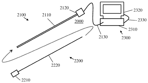
Reference is now made to Fig. 2 which schematically illustrates a first
exemplary visual system 2000, in accordance with embodiments of the invention.
In
some embodiments, system 2000 mainly comprises of a manipulation part 2100,
which
is similar in at least some aspects to previously presented slender body 1100;
a
laparoscopic insert unit 2200 which is detachably connectable with
manipulation part
2100; and an external visual unit 2300 that is connectable with manipulation
part 2100
and/or with laparoscopic insert unit 2200 with at least one wired or wireless
connection,
such as image pickup cable 2130.
In some embodiments, manipulation part 2100 comprises an insertion portion
2110 having length and outer diameter, optionally rigid or semi-rigid, which
facilitate
manual manipulation inside a body cavity, optionally allowing advancement to
and/or
orientation at any location in the relevant body cavity. The insertion portion
2110 is
connected to a handheld operation portion 2120. In some embodiments, insertion
portion 2110 includes a sharp and/or a chamfered distal tip. Alternatively or
additionally, insertion portion 2110 is configured for passing through a
laparoscopic
trocar. In some embodiments, insertion portion 2110 is configured for
enclosing and/or
facilitating strength to an image pickup device attachable thereto. In some
embodiments, insertion portion 2110 includes an inner lumen having a minimal
diameter and an opening at its distal end allowing insertion and enclosing of
longitudinal inserts having maximal outer diameter equal or smaller than the
inner
lumen minimal diameter.
In some embodiments, insertion portion 2110 largest cross section may be 0.5
to
5 mm in diameter, optionally 1 to 2.5 mm, optionally about 1 mm, about 1.5 mm
or
about 2 mm or higher or lower or intermediate. In some embodiments, insertion
portion
2110 includes a lumen having an internal diameter smaller than its outer
diameter by 0.1
to 0.3 mm. For example, insertion portion 2110 may include an outer diameter
of 2.2
CA 02830957 2013-09-23
WO 2012/126967 PCT/EP2012/055041
- 16 -
mm and internal diameter of 2.05 mm. Insertion portion 2110 length may be
between 15
to 50 cm, and optionally, a kit may include several length per patient size,
for example a
20 cm length for a baby, a 27 cm length for moderate size adult and 45 cm
length for
heavy sized adult.
In some embodiments, laparoscopic insert unit 2200 is a rigid lens bar type
laparoscope. Alternatively, laparoscopic insert unit 2200 is a video camera
type
laparoscope, optionally including a digital video camera. In some embodiments,
laparoscopic insert unit 2200 includes an image pickup device 2210 connected
(optionally, detachably connectable) to an elongated connector 2220. In some
ix)
embodiments, image pickup device 2210 includes at least one solid state
sensor, such as
CCD (charge coupled device) or CMOS (complementary metal oxide semiconductor),
and optionally further includes at least one lens and/or other optical
element, and/or at
least one illumination source or projector, such as LED (light-emitted diode)
illuminator. Elongated connector 2220 allows video and/or image signal and/or
digital
signal and/or electrical current and/or illumination transfer in at least one
direction
along its length. In some embodiments, elongated connector includes at least
one PCB
(printed circuit board) and/or optical fiber and/or communication cable.
Optionally,
elongated connector 2220 is non-rigid and gains mechanical endurance/strength
when
enclosed in insertion portion 2110. In some embodiments, elongated connector
2220
includes electrical conductive contacts in at least one end. In some
embodiments,
elongated connector 2220 includes a covering or sleeve (not shown) having
internal or
outer diameter between 0.5 to 2.5 mm, for example 1.8 mm in ID and 2 mm in OD
where it should enclose at least one elongated PCB and be mountable in a
insertion
portion 2110 having ID equal or slightly greater than 2 mm.
In some embodiments, laparoscopic insert unit 2200 is connectable to an
external visual unit 2300 when properly installed within manipulation part
2100,
optionally by image pickup cable 2130, thereby allowing control, display,
recording
and/or other functions from outside patient's body. External visual unit 2300
includes in
some embodiments a CCU (camera control unit) 2310 and a display device 2320,
optionally interconnectable with a communication cable 2330. In some
embodiments,
CCU 2310 includes a signal processing device with an image processing circuit.
CCU
2310 may be configured to generate a video signal based on transmitted image
signals
and to output the video signal to display device 2320.
CA 02830957 2013-09-23
WO 2012/126967 PCT/EP2012/055041
- 17 -
Reference is now made to Figs. 3A-C which schematically illustrate perspective
and cut views of an exemplary laparoscopic insert unit 3000, in accordance
with
embodiments of the invention. As shown in Fig. 3A, laparoscopic insert unit
3000,
similarly to previously described unit 2200, includes an exemplary image
pickup device
or camera head 3100 and an exemplary connector unit 3200. In embodiments, and
as
shown in Fig. 3B, which schematically illustrates a transverse cut view of a
distal
portion of laparoscopic insert unit 3000, exemplary camera head 3100 includes
a
housing 3110, a lens 3120 with at least one optical element, at least one LED
3130 (in
this example, two LEDs) and an image sensor circuit 3140 (including,
optionally, at
least one CCD or CMOS sensors). In some embodiments, lens 3120 allows an angle
or
a field of view between 70 to 140 , optionally 90 to 110 , whereas in a
provided kit,
different heads and/or lens couplings may be provided which are differentiated
by angle
of view. In some embodiments, camera head 3100 includes a distance of view
between
0.1 and 40 cm, optionally 1 to 20 cm. In some embodiments, camera head 3100
further
includes cooling means, passive or active, for LEDs 3130 (not shown).
In embodiments, and as shown in Fig. 3C, which schematically illustrates a
cross section cut view of connector unit 3200, and in Figs. 3A and 3C,
connector unit
3200 includes a sleeve 3210 enclosing longitudinal PCBs, in this example a
single
image pickup PCB 3220 and two LEDs PCBs 3230. Alternatively, at least one of
the
PCBs is substantially shorter whereas other means (such as wires) are used to
transmit
signals across connector unit 3200 length. Image pickup PCB 3220 is configured
to
transfer power and/or image signal and/or digital content from circuit 3140 to
an
external CCU (not shown) and/or power source and/or vice versa, when properly
in
contact with sensor-PCB contacts 3150 and image pickup contacts 3240.
Optionally, at
least ten sensor-PCB contacts 3150 are used, for example 14 contacts. LEDs
PCBs 3230
are configured to transfer power to LEDs 3130 from an external power source
(not
shown), when properly in contact with LEDs-PCB contact(s) 3160 and LEDs-power
contact(s) 3250. Alternatively, instead of LEDs PCBs 3230 and contacts 3160, a
power
line is connected (e.g., soldered) to LEDs 3130 and passed over and along
image pickup
PCB 3220 until its proximal end where optionally it is connected with short
PCB
connectors. In a third alternative, camera head 3100 includes a power source
(not
shown) for powering the LEDs 3130 and/or circuit 3140.
CA 02830957 2013-09-23
WO 2012/126967 PCT/EP2012/055041
- 18 -
In some embodiments, LEDs 3130 project illumination towards a target object
in a body cavity which is then reflected back and picked up by circuit 3140
through lens
3120 and captured as a digital image. Digital images are then transmitted to
an external
CCU (not shown) located outside the body cavity via image pickup PCB 3220.
Reference is now made to Figs. 4A-D which illustrate different deployment
stages of the exemplary visual system 2000, previously shown in Fig. 2, in
body cavity
CAV (optionally previously inflated), in accordance with embodiments of the
invention.
In some embodiments, manipulation part 2100 of system 2000 is slightly
introduced
into CAV in order to avoid any unnecessary harm to internal organs.
Manipulation part
2100 may penetrate into CAV through tissue layers or inserted through a
previously
performed incision or a dedicated trocar (not shown). Laparoscopic trocar 2400
is also
introduced into CAV, optionally through the umbilicus. In some embodiments,
trocar
2400 is configured to allow bi-directional travel therethrough from inside-out
and from
outside-in with minimal to no leak of inflating gas entrapped in CAV. Trocar
2400
includes or configured to allow passage of an internal telescopic sleeve 2450
which can
be extended, oriented and manipulated to a plurality of locations in CAV. A
laparoscope
2500 is inserted through trocar 2400 to allow visual for deploying visual
system 2000.
Laparoscope 2500 may be any type laparoscope, and optionally may include the
laparoscopic insert unit 2200 that is intended for later deployment with the
other parts
of system 2000.
As shown in Fig. 4A, laparoscope 2500 is used to scan CAV periphery for
manipulation part 2100 distal tip. In Fig. 4B, telescopic sleeve 2450,
optionally with
laparoscope 2500 enclosed within, is then extended towards until adjacent or
contacting
the protruding distal tip of manipulation part 2100. Alternatively, telescopic
sleeve 2450
is extended towards a chosen point on CAV periphery and pushes it outwardly
thereby
visually signaling an entry point for manipulation part 2100, to which it can
penetrate.
Inner diameter (e.g., lumen diameter) of telescopic sleeve 2450 may be about 3
to 15 mm, or optionally about 10 mm; and its outer diameter may be about 4 to
20mm.
In some embodiments, additionally or alternatively to using telescopic sleeve
2450,
other locating and/or guiding and/or grasping/connecting devices (not shown)
may be
used to locate and/or guide and/or grasp distal end of manipulation part 2100
in CAV
and assist or use in transferring it through trocar 2400 to outer body
environment.
CA 02830957 2013-09-23
WO 2012/126967 PCT/EP2012/055041
- 19 -
Once in direct contact, manipulation part 2100 can be pushed into and through
telescopic sleeve 2450 until projecting outside CAV and patient's body, as
shown in
Fig. 4C. While pushing manipulation part 2100, or before introduction into
telescopic
sleeve 2450, laparoscope 2500 is withdrawn. Next, laparoscopic inset unit 2200
is
introduced into manipulation part 2100 to assemble visual system 2000. A shown
in
Fig. 4D, system 2000 may then be pulled back into CAV and allowing an optional
use
of trocar 2400 for passage of elements therethrough and following surgical
intervention
under visual surveillance.
Reference is now made to Fig. 5 which schematically illustrates a second
1() exemplary visual system 4000, in accordance with embodiments of the
invention.
System 4000 includes a manipulation part 4100 comprising a rigid elongated
connector
4110, a handheld operation portion 4120 and an image pickup cable 4130
connectable
to an external visual unit (not shown). System 4000 further includes a camera
head 4200
detachably connectable to elongated connector 4110. In some embodiments,
camera
head 4200 is substantially greater in diameter relative to diameter of
elongated
connector 4110. In some embodiments, camera head 4200 is similar in design
and/or
operation to previously presented camera head 3100 although it may differ in
its
connection type and means with the elongated connector. Installment and/or
operation
of system 4000 may be similar to those of system 2000.
Alternatively, the elongated connector 4110 may be housed in a insertion
portion for protection and/or increasing the rigidity..
Reference is now made to Fig. 6 which schematically illustrates a third
exemplary visual system 5000, in accordance with embodiments of the invention.
System 5000 is a rigid rod lens type laparoscope which includes a thin rigid
laparoscope
unit 5100 comprising a rigid insertion portion 5110 optionally enclosing an
image guide
(e.g., including optic carrier and lenses); a handheld operation portion 5120
optionally
detachably connected to a video camera 5140; and an image pickup cable 5130
connectable to an external visual unit (not shown). System 5000 further
includes a
detachably connectable illumination sleeve 5200 having a substantially greater
diameter
in relation with the thin rigid laparoscope unit 5100. In some embodiments,
illumination
sleeve 5200 includes an internal lumen or bore having a diameter substantially
similar
to external diameter of a distal portion of insertion portion 5110, and is
configured to be
deployed thereupon. In some embodiments, illumination sleeve 5200 is self
powered or
CA 02830957 2013-09-23
WO 2012/126967 PCT/EP2012/055041
- 20 -
powered by an external power source connectable via rigid insertion portion
5110.
Installment and/or operation of system 5000 may be similar to those of system
2000.
Reference is now made to Fig. 7 which schematically illustrates a fourth
exemplary visual system 6000, in accordance with embodiments of the invention.
Similarly to system 5000, system 6000 is also a rigid rod-lens type
laparoscope, that
includes a slender rigid laparoscope unit 6100 comprising a rigid insertion
portion 6110
optionally enclosing a bundle of image guide and light guide (not shown); a
handheld
operation portion 6120 optionally detachably connected to a video camera 6150;
an
image pickup cable 6130 connectable to an external visual system (not shown);
and an
ix) illumination cable 6140 connectable to an external illumination source
(not shown).
System 6000 further includes a detachably connectable distal rod lens 6200
having a
substantially greater diameter in relation with the thin laparoscope unit
6100. In some
embodiments, distal rod lens 6200 allows greater view angle than can be
achieved in
smaller diameters rod lenses such as those enclosed in insertion portion 6110.
Installment and/or operation of system 6000 may be similar to those of system
2000.
In some embodiments of the present invention, a laparoscopic insert unit
and/or
a camera head may include at least one illumination source provided as an
integral part
or as a potential add-on component. In some embodiments, it may be preferable
to
project much more light to a target object, for example in order to improve
visualization
and/or video quality parameters, so that larger illumination sources (e.g.,
LEDs), and/or
in larger numbers, may be delivered with the laparoscopic insert unit.
Optionally,
alternatively or additionally, a need may arise to decrease/minimize to heat
created by
the illumination source(s) next to the lens/objective and/or any temperature-
sensitive
component. Optionally, alternatively or additionally, a need may arise to
decrease/minimize the diameter of the unit and, for example, make it only
slightly larger
than the cased lens/objective.
In some embodiments of the invention, according to any of the above
considerations, and/or to any other consideration, there may be provided a
laparoscopic
insert unit (or a camera head) comprising illumination source(s) located away
from,
optionally remotely behind, the lens/objective opening. In some variations of
these
embodiments, means may be provided to collect, reflect and/or project most or
all light
created in the illumination source(s) towards a certain target, optionally in-
front and/or
radially away from the object/lens.
CA 02830957 2013-09-23
WO 2012/126967 PCT/EP2012/055041
-21 -
Reference is now made to Fig. 8 which schematically illustrates a partial cut
view of an exemplary laparoscopic insert unit 7100 which comprises an
illumination
reflector 7130 (shown in transverse cut view), in accordance with embodiments
of the
invention. Laparoscopic insert unit 7100 includes a wide video camera head
body 7110
(shown in a non-cut side view), which may encase lens/objective, image sensor
and
electronics (not shown), that is provided connected with an elongated slender
connector
unit 7140 (shown in part). Camera head body 7110 may end with a smaller
diameter
portion 7112 that is connected or enclosing a proximal end of connector unit
7140. A
plurality of illumination sources (although one may suffice) 7120 are provided
on outer
periphery of smaller portion 7112 and optionally, though not necessarily, do
not emerge
over body 7110 largest diameter. The illumination sources may be set to point
radially
outward, in a reversed direction (towards connector unit 7140) or in any
angled fashion.
In an exemplary embodiment, the illumination sources are LED sources
electrically
connectable to a power source provided outside a patient's body (not shown)
via
connector unit 7140 and along its length. In some embodiments, reflector 7130
is
designed and shaped, at a deployed formation, to reflect most of the light
created by
illumination sources 7120. In some embodiments, reflector 7130 includes an
inner
surface 7132 made or coated with a reflecting material as known to art.
Reflector 7130
may be shaped to collect and/or focus scattered light originating from the
plurality of
illumination sources 7120 towards a chosen target area. Reflector 7130 may be
rigid,
semi-rigid or elastic; it may be formed of or assembled to a single piece or
comprise a
plurality of components (e.g., an iris design comprising a plurality of rigid
or semi-rigid
members; not shown). In some embodiments, reflector 7130 is expandable and/or
contractible between a smaller diameter to a greater diameter. The exemplary
smaller
diameter may be smaller, substantially the same or slightly greater than
diameter of
camera head body 7110 so that it may maintain a thinner introductory size and
later
expand, either selectively or predeterminedly, automatically or per demand,
when in
position inside a patient's body cavity.
Fig. 9 schematically illustrates a partial cut view of another exemplary
laparoscopic insert unit 7200 comprising illumination fiber optics 7230, in
accordance
with embodiments of the invention. Similarly to unit 7100, laparoscopic insert
unit 7200
includes a wide camera head body 7210 incorporating a smaller sized distal
portion
7212 and connected to a slender elongated connector unit 7240. A plurality of
CA 02830957 2013-09-23
WO 2012/126967 PCT/EP2012/055041
- 22 -
illumination sources 7220 are also similarly positioned over smaller portion
7212
periphery. Instead of reflecting means, the plurality of fiber optics 7230 may
be
provided over and along a length of camera head body 7210, thereby allowing
travel of
light from illumination sources 7220 distally and towards and in front of
camera head
body 7210. A plurality of optical fibers may be used to transfer light from a
single
illumination source. Optical fibers may be positioned over an expandable
member (not
shown) thereby allowing projection of light in a cone-like form.
Fig. 10A and B illustrate an exemplary one unit camera and male connector
8100 embodiment of a self-illuminating visual head 8130 connected to an
elongated
lo internal shaft 8120 being an elongated connector having a male connector
8110. The
visual head 8130 has a camera unit 8140 and to illuminating LEDs 8150. The
male
connector 8110 is in this embodiment a non-optical connector, such as
electrical
conductive, for powering, controlling and transmitting information. The use of
a non-
optical connector enables a small cross-section of the elongated internal
shaft 8120 and
may therefore be advantageous to be used for scarless laparoscopi procedures.
Fig. 11 illustrates an exemplary manipulation part 8200 where a rigid
insertion
portion is an outer shaft 8210, such as a needle. The rigid insertion portion
is configured
to provide a rigid support for the elongated connector. The manipulator part
8200
further comprises, a handheld operation portion, such as a handle 8220. Inside
the
handle 8220 is a female connector 8230 located to be connected to the male
connector
8110 of the camera and male connector 8100. Further, the handle 8200 has a
video
console cable connector 8240 for connecting the visual head 8130 with an
external
visual unit, such as a screen. Alternatively, instead of a video console cable
connector
8240, the handle 8220 may be equipped with a wireless communication unit for
transferring the signal to the external visual unit.
Fig. 12 A and B illustrates an exemplary assembly 8300 of a camera and male
connector 8100 and a manipulation part 8200. The internal shaft 8210 with the
male
connector 8110 is pushed into the outer shaft, such as a needle, 8210 until
the male
connector 8110 connects with the female connector 8230 inside the handle 8220.
When
connected, most of the internal shaft 8120 is housed inside the outer shaft
8210. The
mounting of the assembly 8300 is conducted by having the outer shaft 8210
extending
out of the body cavity through an airtight passage as previously described
herein.
CA 02830957 2015-09-10
23
As a non-limiting example, the exemplary embodiment illustrated in Fig. 10 to
12 may have a visual head 8130 having a maximum outer diameter of 10 mm and a
maximum length of 60 mm while the maximum outer diameter of the outer shaft
8210 is only 2.8 mm. The visual head 8130 is, apart from the two LEDs 8150,
fitted
with a camera unit 8140 being a state of the art high definition sensor. Each
part of
the assembly 8300 is designed to be cleaned and sterilized after each
procedure.
Any citation or identification of any reference in this application shall not
be
construed as an admission that such reference is available as prior art to the
present
invention. To the extent that section headings are used, they should not be
construed as necessarily limiting.
Although the invention has been described in conjunction with specific
embodiments thereof, it is evident that many alternatives, modifications and
variations will be apparent to those skilled in the art.
Finally, the scope of the claims should not be limited by the preferred
embodiments set forth in the examples, but should be given the broadest
interpretation consistent with the description as a whole.