Note: Descriptions are shown in the official language in which they were submitted.
CA 02831106 2012-24
IRRIGATED ABLATION CATHETER WITH DEFORMABLE HEAD
FIELD OF THE INVENTION
[0001]
The present invention relates generally to invasive
probes, and specifically to a probe with a deformable distal
end.
BACKGROUND
[0002]
Various therapeutic procedures such as cardiac ablation
use an invasive medical probe such as a catheter that is
inserted into a patient's body. During an ablation procedure
on a heart, there may be local overheating of the heart
surface being ablated, as well as of the heart tissue
underlying the surface.
The surface overheating may be
manifested as charring, and the overheating of the underlying
tissue may cause other damage to the tissue, even leading to
penetration of the tissue. To control the temperature of the
surface and the underlying tissue, the region being ablated
may be irrigated with an irrigation fluid, typically saline,
in order to prevent charring.
[0003]
In addition to the risk of charring, overheating of
blood in the region being ablated may cause the formation of
potentially dangerous blood clots, which can grow and
potentially cause a heart attack or a stroke.
While the
irrigation may slightly reduce blood clot formation by
cooling and diluting the blood, there is still a possibility
of clotting.
[0004]
A number of catheters with flexible tips have been
described in the patent literature. For example, U.S. Patent
1
CA 02831106 2012-24
5,720,719, whose disclosure is incorporated herein by
reference, describes a catheter having a probe end that
includes a malleable tube and a flexible tube.
These
elements are said to allow the probe end to conform to the
curvature of the cavity inside a patient's body.
[0005]
Documents incorporated by reference in the present
patent application are to be considered an integral part of
the application except that to the extent any terms are
defined in these incorporated documents in a manner that
conflicts with the definitions made explicitly or implicitly
in the present specification, only the definitions in the
present specification should be considered.
[0006]
The description above is presented as a general overview
of related art in this field and should not be construed as
an admission that any of the information it contains
constitutes prior art against the present patent application.
2
CA 02831106 2012-24
= =
SUMMARY OF THE INVENTION
[0007]
There is provided, in accordance with an embodiment of
the present invention a medical probe, including a flexible
insertion tube having a deformable distal end for insertion
into a body cavity of a patient, the deformable distal end
including a flexible and porous material configured to be
brought into contact with tissue in the body cavity.
The
medical probe also includes a means for inflating the
deformable distal end, and a channel contained within the
insertion tube and configured to convey a fluid that
irrigates the tissue through pores of the deformable distal
end.
The medical probe further includes an electrical
conductor passing through the flexible insertion tube,
terminating in the deformable distal end and configured to
convey radio frequency (RF) energy to the tissue via the
deformable distal end.
[0008]
In some embodiments, the flexible and porous material
may include a conductive material, the electrical conductor
can be coupled to the flexible and porous material so as to
convey the RF energy to the deformable distal end, and the RF
energy can be conveyed to the tissue by the deformable distal
end conveying the RF energy to the tissue.
[0009]
In embodiments where the flexible and porous material
may include a conductive material, the conductive material
may include a fabric woven from strands of Nitinol.
In
additional embodiments where the flexible and porous material
may include a conductive material, the conductive material
can be configured to transfer a current, and the medical
probe may further include a processor configured to determine
3
CA 02831106 2012-24
a position of the distal end in response to an impedance to
the current.
[0010]
In further embodiments, the fluid may include a saline
solution. In embodiments where the fluid includes a saline
solution, the electrical conductor can convey the RF energy
to the tissue by conveying the RF energy to the saline
solution, and the saline solution conveying the RF energy to
the tissue.
[0011]
In some embodiments, the medical probe may include an
intracardiac catheter, and the body cavity may include a
chamber of a heart.
In additional embodiments, the
deformable distal end can be configured to conform to the
tissue of the body cavity.
In further embodiments, the
insertion tube has a first diameter, and upon inflation of
the deformable distal end, the deformable distal end has a
second diameter greater than the first diameter.
In
supplementary embodiments, a contact area between the
deformable distal end and the tissue can increase upon
pressing the deformable distal end against the tissue.
[0012] In some embodiments, the means for inflating the
deformable distal end may include conveying the fluid so as
to generate a mechanical force sufficient to inflate the
deformable distal end.
In an alternative embodiment, the
means for inflating the deformable distal end may include a
wire frame protruding from a distal tip of the flexible
insertion tube and covered by the deformable distal end, and
a control wire, passing through the flexible insertion tube,
coupled to the wire frame and configured to resize the wire
frame.
In the alternative embodiment including the wire
4
CA 02831106 2013-10-24
, .
frame, the electrical conductor terminates in the wire frame,
and the wire frame is configured to convey the RF energy from
the electrical conductor to the deformable distal end.
[0013]
There is also provided, in accordance with an embodiment
of the present invention, a method, including inserting a
deformable distal end of a flexible insertion tube into a
body cavity of a patient, the deformable distal end including
a flexible and porous material configured to be brought into
contact with tissue in the body cavity.
The method also
includes inflating the deformable distal end, and conveying a
fluid through a channel contained within the flexible
insertion tube so as to irrigate the tissue through pores of
the deformable distal end.
The method further includes
conveying radio frequency (RF) energy to the tissue via the
deformable distal end.
CA 02831106 2012-24
, .
BRIEF DESCRIPTION OF THE DRAWINGS
[0014]
The disclosure is herein described, by way of example
only, with reference to the accompanying drawings, wherein:
[0015]
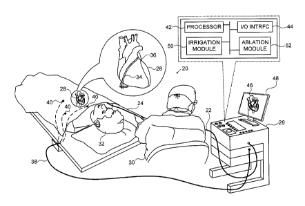
Figure 1 is a schematic, pictorial illustration of a
medical system that includes an invasive probe having a
deformable head, in accordance with an embodiment of the
present invention;
[0016]
Figure 2A is a schematic sectional view of the distal
end of the probe, in accordance with a first embodiment of
the present invention;
[0017]
Figures 2B and 2C are pictorial illustrations of a woven
fabric used to construct a deformable head of the probe, in
accordance with an embodiment of the present invention;
[0018]
Figure 2D is a schematic pictorial illustration of the
deformable head in contact with endocardial tissue of a
heart, in accordance with the first embodiment of the present
invention;
[0019]
Figure 3A is a schematic sectional view of the distal
end of the probe, in accordance with a second embodiment of
the present invention;
[0020]
Figure 3B is a schematic illustration of a wire frame
extending from the distal end of the probe, in accordance
with the second embodiment of the present invention;
[0021]
Figure 30 is a schematic pictorial illustration of the
deformable head incorporating the wire frame and in contact
with the endocardial tissue, in accordance with the second
embodiment of the present invention; and
[0022]
Figures 4A-4C show heat maps that illustrate areas of
6
CA 02831106 2013-10-24
. .
contact between the deformable catheter head and the
endocardial tissue, in accordance with an embodiment of the
present invention.
7
CA 02831106 2013-10-24
t .
DETAILED DESCRIPTION OF EMBODIMENTS
OVERVIEW
[0023]
Catheters used in invasive cardiac procedures, such as
intracardiac ablation for treatment of arrhythmias, typically
have a rigid tip. When the tip is brought into contact with
myocardial tissue at the proper angle and with sufficient
force, the tissue conforms to the tip, affording good
mechanical and electrical contact.
The area of contact is
typically limited by the tip size, however, and may be even
smaller, depending on the angle of contact and other
parameters.
[0024] Embodiments of the present invention provide an
irrigated ablation catheter that has a deformable head. The
deformable head increases the surface area that physically
interfaces with the tissue and may also be useful in ensuring
that the contact pressure between the catheter head and the
tissue is roughly constant over the entire area of contact.
[0025]
The catheter head can be fabricated from a porous cloth-
like material, which may itself be flexible and/or
conductive.
In some embodiments, during an ablation
procedure using the catheter, fluid, such as saline solution,
can be forced through the porous material, so as to irrigate
the area being ablated, as well as to irrigate the cloth-like
material.
(Irrigating the material ensures the integrity of
the material, by keeping the material cool.)
The fluid
provides a means for delivering sufficient mechanical force
to inflate the head, and to keep the head inflated during the
procedure. Because the same saline solution can be for both
8
CA 02831106 2012-24
irrigation and inflation, a separate inflating system is not
required.
[0026]
In alternative embodiments, the cloth-like material may
cover an expandable wire frame that protrudes from a distal
end of the catheter, and the size of the wire frame can be
managed via a control wire coupled to the frame.
For
example, an operator (e.g., a cardiologist) can expand the
wire frame which provides a means to "inflate" the head by
"pushing" the control wire toward the distal end. Likewise,
the operator can contract the wire frame (and thereby
"deflate" the head) by "pulling" the control wire away from
the distal end. While embodiments herein describe a catheter
head that can be resized via fluid pressure or a resizable
wire frame, other methods of expanding and contracting the
catheter head are considered to be within the spirit and
scope of the present invention.
[0027]
Once in contact with the tissue, the catheter head
conforms to the tissue, due to a mechanical response from the
tissue and the flexibility of the head. The conformation with
the tissue provides a larger interface surface compared with
a rigid catheter tip.
[0028]
During insertion, the catheter head need not be inflated
and a sheath of the catheter can thus have a small diameter.
After inflation the catheter head may be of a larger diameter
than the sheath, providing a larger surface for ablation.
Using this approach, a smaller-diameter ablation catheter can
be used to treat ablation areas that would otherwise require
a larger-diameter catheter for treatment.
Having a larger
ablation surface can support deeper ablation, such as for use
9
CA 02831106 2012-24
, .
in the left ventricle, or for use in forming scar tissue
where a specific target location may not be known.
[0029]
Although the disclosed embodiments relate specifically
to intracardiac ablation, the principles of this invention
may similarly be applied in other therapeutic and diagnostic
procedures, both in the heart and in other organs.
SYSTEM DESCRIPTION
[0030]
Figure 1 is a schematic, pictorial illustration of a
medical system 20 configured to perform an ablation
procedure, in accordance with an embodiment of the present
invention.
System 20 comprises a probe 22, in the present
example an intracardiac catheter comprising a flexible
insertion tube 24, and a control console 26.
Probe 22 is
typically connected by a suitable connector at its proximal
end to console 26.
[0031]
In the embodiment described hereinbelow, it is assumed
that probe 22 is used for diagnostic or therapeutic
treatment, such as mapping electrical potentials of a heart
28, or performing ablation of endocardial tissue of the
heart.
Alternatively, probe 22 may be used, mutatis
mutandis, for other therapeutic and/or diagnostic purposes in
the heart or in other body organs.
[0032]
An operator 30, such as a cardiologist, inserts probe 22
through the vascular system of a patient 32 so that a distal
end 34 of probe 22 enters a chamber of the patient's heart 28
(e.g., the left atrium).
Operator 30 advances probe 22 so
that a distal tip 36 of the probe engages body tissue at
desired locations.
As described in Figures 2A-3C
CA 02831106 2012-24
hereinbelow, distal tip 36 comprises a deformable head 62,
also referred to herein as a deformable distal end,
comprising a flexible and porous material that may be
conductive.
In some embodiments deformable head 62 is
conductive and can function as a position sensor, as
described hereinbelow.
In additional configurations,
deformable head 62 is configured to convey radio frequency
(RF) energy to intracardiac tissue of heart 28 during an
ablation procedure.
[0033]
In the example of Figure 1, console 26 is connected, via
a cable 38, to body surface electrodes, which typically
comprise adhesive skin patches 40 that are affixed to patient
32. Console 26 determines position coordinates of probe 22
inside heart 28 based on the impedance measured between the
deformable head, when it is conductive, and patches 40.
Although system 20 uses impedance-based sensing to measure a
location of deformable head 62, other position tracking
techniques may be used (e.g., magnetic-based sensors).
Magnetic position tracking techniques are described, for
example, in U.S. Patents 5,391,199, 5,443,489, 6,788,967,
6,690,963, 5,558,091, 6,172,499 6,177,792, whose disclosures
are incorporated herein by reference.
Impedance-based
position tracking techniques are described, for example, in
U.S. Patents 5,983,126, 6,456,864 and 5,944,022, whose
disclosures are incorporated herein by reference.
[0034]
Console 26 comprises a processor 42, which typically
comprises a general-purpose computer, with suitable front end
and interface circuits for receiving signals from probe 22
and controlling the other components of console 26.
An
11
CA 02831106 2013-10-24
k ,
input/output (I/0) communications interface 44 enables
console 26 to interact with probe 22 and patches 40. Based
on the signals received from probe 22 and from patches 40,
processor 42 produces and displays a map 46 showing the
position of distal tip 36 in the patient's body, the distance
and/or contact indication between the loop and the body
tissue, as well as status information and guidance regarding
the procedure that is in progress. Map 46 is presented to
operator 30 using a display 48. The position of probe 22 may
be superimposed on map 46 or on another image of heart 28.
[0035] Processor 42 typically comprises a general-purpose
computer, with suitable front end and interface circuits for
receiving signals from probe 22 and controlling the other
components of console 26. Processor 42 may be programmed in
software to carry out the functions that are described
herein.
The software may be downloaded to console 26 in
electronic form, over a network, for example, or it may be
provided on non-transitory tangible media, such as optical,
magnetic or electronic memory media. Alternatively, some or
all of the functions of processor 42 may be carried out by
dedicated or programmable digital hardware components.
[0036]
Console 26 also comprises an irrigation module 50 and an
RF ablation module 52. Processor 42 uses the ablation module
to monitor and control ablation parameters such as the level
of ablation power applied via deformable head 62. The
ablation module may also monitor and control the duration of
the ablation that is provided.
[0037]
Typically, during ablation, heat is generated in the
electrode (or electrodes) providing the ablation, as well as
12
CA 02831106 2012-24
,
in the surrounding region. In order to dissipate the heat
and to improve the efficiency of the ablation process, system
20 supplies an irrigation fluid to distal end 34 via a
channel (described hereinbelow).
System 20 uses irrigation
module 50 to monitor and control irrigation parameters, such
as the pressure, flow rate, and temperature of the irrigation
fluid.
CATHETER WITH A DEFORMABLE HEAD
[0038]
Figure 2A schematically illustrates distal end 34 of
probe 22, Figures 2B and 2C are pictorial illustrations of a
woven fabric used to construct deformable head 62, and Figure
2D is a schematic pictorial illustration of deformable head
62 in contact with endocardial tissue 70 of heart 28, in
accordance with a first embodiment of the present invention.
[0039]
Distal end 34 is covered by a flexible, insulating
sheath 60 having a distal tip 68, and deformable head 62 is
fixed to the distal end.
In operation, deformable head 62
can be inflated and irrigated by a saline solution, which
irrigation module 50 pumps through a channel 64, typically a
tube, within sheath 60. An electrical conductor 66 passes
within the catheter sheath to terminate at the deformable
head and to convey radio-frequency (RF) electrical energy to
deformable head 62.
[0040]
Deformable head 62 may be made out of any suitable
porous, flexible material.
For some applications, a
resilient, woven fabric may be advantageous.
For enhanced
mechanical strength and resilience, in one embodiment of the
present invention, the fabric may be woven at least partially
13
CA 02831106 2012-24
,
from elastic metal fibers, such as strands of Nitinol. This
sort of implementation is illustrated in Figures 2B and 2C,
which respectively show side and end views of deformable head
62.
As described hereinbelow, the use of a metal-based
fabric is also helpful in conducting electrical energy to the
intracardiac tissue.
[0041]
In the example shown in Figure 2D, irrigation module 50
conveys a saline solution 72 (or any other type of irrigation
fluid) through channel 64, thereby generating a mechanical
force sufficient to inflate deformable head 62 and to
irrigate the tissue via pores 69 (Fig. 2C) in the porous and
flexible material used to fabricate the deformable head.
While deformable head 62 is inflated and pressed against
endocardial tissue 70, the deformable head conforms to the
endocardial tissue, as shown in the Figure.
[0042]
In some embodiments, upon initially engaging endocardial
tissue 70, deformable head 62 has an initial contact area 74
that comprises a portion of the flexible material that is in
contact with the endocardial tissue. As deformable head 62
presses against endocardial tissue 70, contact area 74 can
increase (up to a maximum contact area) as the deformable
head deforms by spreading out.
[0043]
As described supra, during insertion of tube 24, the
deformable head need not be inflated. Therefore, sheath 60
may have a sheath diameter, and upon being inflated,
deformable head 62 may have a head diameter that is greater
than the sheath diameter. Typically, deformable head 62 has
a shape that forms a seal between the deformable head and
sheath 60. In some embodiments, deformable head 62 has a
14
CA 02831106 2013-10-24
"balloon"-like shape.
[0044]
When deformable head 62 is conductive, e.g., comprises
suitable metal strands or a conductive polymer, ablation
module 52 can convey RF energy to the deformable head via
electrical conductor 66, and the deformable head conducts the
energy to the tissue.
Alternatively or additionally,
electrical conductor 66 may apply the RF energy to saline
solution 72, in which case the saline solution may conduct
the RF energy through deformable head 62 to the endocardial
tissue.
[0045]
Figure 3A schematically illustrates distal end 34 of
probe 22 comprising a wire frame 80, Figure 3B is a schematic
pictorial illustration of the wire frame, and Figure 3C is a
schematic pictorial illustration of deformable head 62 in
contact with endocardial tissue 70 of heart 28, in accordance
with a second embodiment of the present invention.
In
Figures 3A and 3B, wire frame 80 is "mushroom"-shaped,
coupled to electrical conductor 66, and affixed to a distal
end of channel 64.
In Figure 3C, a wire frame 82 is
cylindrical, is coupled to electrical conductor 66, and is
affixed to a distal end of sheath 60.
[0046]
In embodiments of the present invention, deformable
distal end 62 can be expanded and contracted by resizing wire
frame 80.
In the configuration shown in Figure 3A, the
diameter of the mushroom-shaped wire frame can be resized via
a control wire 84 that passes within the catheter sheath and
is coupled to outer edges 86 of the wire frame. For example,
if the mushroom-shaped wire frame is flexible, and operator
30 pulls on control wire 84, then the control wire can
CA 02831106 2013-10-24
contract wire frame 80 by retracting outer edges 86 toward a
longitudinal axis 88 of sheath 60. Likewise, if operator 30
pushes on control wire 84, the control wire can expand wire
frame 80 by protracting outer edges 86 away from the
longitudinal axis of the sheath.
[0047]
While the examples in Figures 3A-3C show the wire frames
in mushroom and cylindrical shapes, other shapes are
considered to be within the spirit and scope of the present
invention.
Additionally, while the description hereinbelow
describes configuration and operation of wire frame 80, wire
frame 82 can be configured and operated in the same manner.
[0048]
In this second embodiment of the present invention,
probe 22 comprises wire frame 80 that protrudes from sheath
60 and is covered by deformable distal head 62. Wires of
frame 80 are flexible but resilient enough so that the frame
maintains its overall form under deformation, as shown in
Figure 3C.
Wire frame 80 thus provides some mechanical
stability to deformable head 62, allowing a more flexible
fabric to be used for the deformable head.
[0049]
As shown in Figure 3A, wire frame 80 extends from distal
sheath tip 68 of sheath 60 and is contained within deformable
head 62. Conductor 66 runs through sheath 60 and is coupled
to wire frame 80. During an ablation procedure, conductor 66
conveys RF energy from ablation module 52 to wire frame 80,
and the wire frame conveys the RF energy to endocardial
tissue 70 when the wire frame presses against deformable head
62 and the deformable head is in contact with the endocardial
tissue, as shown in Figure 3C. In an alternative embodiment,
wire frame 80 can convey the RF energy to saline solution 72
16
CA 02831106 2012-24
(i.e., within deformable head 62) without the frame
contacting head 62.
In this case the saline solution may
convey at least a portion of the RF energy to tissue 70.
[0050]
Figures 4A-4C show heat maps 90A-90C that illustrate one
of the benefits of the deformable catheter head in terms of
forming deeper and wider ablation lesions, by increasing an
area of contact between the catheter head and the tissue
(i.e., as deformable head 62 presses against endocardial
tissue 70, as shown in Figures 2D and 3C).
Figures 4A-4C
show heat maps 90 (i.e., a heat map 90A in Figure 4A, a heat
map 90B in Figure 4B and a heat map 90C in Figure 4C) that
indicate how energy (i.e., heat) diffuses into endocardial
tissue 70 during an ablation procedure using probe 22.
[0051]
In the heat maps, a Y-axis 96 indicates a depth of
endocardial tissue 70 relative to deformable head 62, and an
X-axis 98 indicates a transverse displacement from deformable
head 62 as the deformable head presses against the
endocardial tissue.
In Y-axis 96, a zero value indicates
that deformable head 62 is in contact with endocardial tissue
70 while causing a depression, of approximately 1 mm, in the
tissue.
[0052] The heat maps convey, using different visual patterns,
indicated in a legend 102, a calculated tissue temperature
during ablation as a function of depth (i.e., Y-axis 96) and
transverse displacement (i.e., X-axis 98) relative to a
center of the distal tip, which is located at the origin
(0,0) at the left side of each figure.
As shown in this
visual representation, the hottest temperatures are measured
at the left side of each figure below the catheter tip, and
17
CA 02831106 2012-24
the coolest temperatures are measured at areas distant from
the catheter tip, with intermediate temperatures measured in
regions between the hottest and the coolest temperatures.
[0053] While for purposes of simplicity, the heat maps show
regions 92 with uniform temperatures, in practice the
temperature changes are typically gradual between isothermal
lines 94, where each line style references a specific
temperature, as indicated in a legend 100.
[0054] In some embodiments of the present invention, the
contact area between distal tip 36 and endocardial tissue 70
can be controlled and increased as appropriate by suitably
inflating and deforming the deformable head against the
tissue. In the examples shown in Figures 4A-4C, the diameter
of the contact area is increased from about 3 mm in Figure
4A, to about 5 mm in Figure 4B, to about 10 mm in Figure 4C.
Additionally, the voltage on the catheter is increased from
25 V in Figure 4A, to 30 V in Figure 4B, to 35 V in Figure
4C, in order to keep the current density constant, and thus
to maintain a constant temperature at the hottest spot in the
tissue below the catheter. As shown in the heat maps, the
width of the resulting lesions increases linearly with the
distal tip contact area, while the depth increases, as well,
though less markedly.
[0055] It will be appreciated that the embodiments described
above are cited by way of example, and that the present
invention is not limited to what has been particularly shown
and described hereinabove. Rather, the scope of the present
invention includes both combinations and subcombinations of
the various features described hereinabove, as well as
18
CA 02831106 2013-10-24
variations and modifications thereof which would occur to
persons skilled in the art upon reading the foregoing
description and which are not disclosed in the prior art.
19