Note: Descriptions are shown in the official language in which they were submitted.
CA 02831867 2013-09-30
1
DESCRIPTION
THERAPEUTIC AGENT FOR CANCER, AND METHOD FOR DETERMINING
PROGNOSIS OF CANCER
TECHNICAL FIELD
[0001]
The present invention relates to a therapeutic agent for cancer, a method for
predicting the prognosis of cancer, and a method for detecting cancer.
BACKGROUND ART
[0002]
Esophagus cancer is a cancer with the eighth highest incidence rate and the
sixth highest number of deaths. In Japan, esophageal squamous cell carcinoma
(ESCC) accounts for not less than 90% of esophagus cancer. ESCC is a highly
malignant cancer that frequently causes distant metastasis and recurrence, and
its
1 5 prognosis is generally poor.
[0003]
On the other hand, it has been reported that ovarian cancer shows abnormal
expression of fibroblast growth factor receptor like-I (hereinafter referred
to as
"FGFRL I") (Non-patent Document 1). However, in this report, no statistical
2 0 analysis was carried out for the expression level of FGFRL1, and no
analysis on the
function of FGFRL1 was performed. Thus, this report never leads to inference
of
promotion of the cell growth by FGFRL1, utilization of its expression level
for
prediction of the prognosis, or its industrial applicability. Further,
although it has
been reported that microRNA (miRNA)-210 is involved in oncogenesis and that
one
2 5 of its target genes is FGFRL1 (Non-patent Document 2), this report does
not clearly
suggest utilization of FGFRL1 for prediction of the prognosis or for
therapeutic
agents. Further, the present inventors previously discovered that, in
esophageal
CA 02831867 2013-09-30
2
squamous cell carcinoma, microRNA-210 regulates the growth of cancer cells via
FGFRL1 (Non-patent Document 3). However, this report only elucidated that a
target gene of microRNA-210 is FGFRL1 and discussed about its downstream
pathway, and no suggestion was made about possible use of an anti-FGFRL1
antibody for a therapeutic agent for cancer or a tool for prediction of the
prognosis.
[0004]
On the other hand, although FGFRL1 has been named a "molecule like a
fibroblast growth factor receptor (FGFR)", it is clearly structurally
different from
other FGFRs since, unlike other FGFRs, FGFRL1 lacks the tyrosine kinase
domain,
the industrial applicability deduced therefrom.
[0005]
[Non-patent Document I] International Journal of Molecular Medicine 16, 1169-
1173, 2005
[Non-patent Document 2] Molecular Cell 35, 856-867, 2009.
[Non-patent Document 3] Journal of Biological Chemistry 286, 420-428, 2011
[Non-patent Document 4] Genomics 69, 275-279, 2000
[Non-patent Document 5] Journal of Biological Chemistry 278, 33857-33865, 2003
SUMMARY OF THE INVENTION
PROBLEMS TO BE SOLVED BY THE INVENTION
[0006]
An object of the present invention is to provide a novel therapeutic agent for
cancer such as esophageal squamous cell carcinoma. Another object of the
present
CA 02831867 2013-09-30
' 3
invention is to provide a method for predicting the prognosis of cancer such
as
esophageal squamous cell carcinoma. Still another object of the present
invention is
to provide a method for detecting, or predicting the prognosis of, cancer such
as
esophageal squamous cell carcinoma using a sample that can be collected less
invasively.
[0007]
As described specifically in the Examples below, the present inventors
collected esophageal squamous cell carcinoma tissues from a large number of
esophageal squamous cell carcinoma patients to investigate expression of
FGFRL1,
1 0 and conducted a follow-up study on association of the expression level
with the
prognosis. As a result, it was found that high expression of FGFRL1 is
associated
with poor prognosis. Further, the present inventors discovered that allowing
an
anti-FGFRL1 antibody to act on esophageal squamous cell carcinoma cells
enables
suppression of the growth of the cancer cells.
[0008]
That is, the present invention provides a therapeutic agent for cancer,
comprising as an effective component an antibody that undergoes antigen-
antibody
reaction with FGFRL I to suppress the growth of cancer cells, or an antigen-
binding
fragment thereof. Further, the present invention provides a therapeutic method
for
2 0 cancer, comprising administering to a cancer patient an effective
amount of an
antibody that undergoes antigen-antibody reaction with FGFRL1 to suppress the
growth of cancer cells, or an antigen-binding fragment thereof. Further, the
present
invention provides a method for predicting the prognosis of cancer, comprising
investigating the expression level of FGFRL1 in a cancer tissue separated from
a
2 5 living body, wherein a high expression level of FGFRL1 indicates poor
prognosis.
Further, the present invention provides a method for detecting cancer,
comprising
measuring FGFRL1 or a fragment thereof extracted from a body tissue, or FGFRL1
CA 02831867 2013-09-30
4
or a fragment thereof in blood separated from a living body, wherein a higher
concentration of FGFRL1 or the fragment thereof contained therein than the
concentration of FGFRL1 or the fragment thereof in the tissue or blood of a
healthy
individual indicates the presence of cancer. Further, the present invention
provides
a method for predicting the prognosis of cancer, comprising measuring FGFRL1
or a
fragment thereof in a tissue or blood separated from a cancer patient, wherein
a high
concentration of FGFRL1 or the fragment thereof contained therein indicates
poor
prognosis.
EFFECT OF THE INVENTION
[0009]
By the present invention, a novel therapeutic agent for cancer such as
esophageal squamous cell carcinoma was provided. Further, by the present
invention, a novel method for predicting the prognosis of cancer such as
esophageal
squamous cell carcinoma was provided. Since this method enables prediction of
the
1 5 prognosis of a highly malignant cancer such as esophageal squamous cell
carcinoma,
and hence enables appropriate selection of a therapeutic method, the method
contributes to treatment of cancer. Further, by the present invention, a
method for
detecting a cancer such as esophageal squamous cell carcinoma using a sample
that
can be collected less invasively was provided. Since this method is less
invasive,
2 0 the burden of the subject is light. Therefore, detection of cancer can
be easily
achieved also in medical examination and the like, and early detection and
early
treatment of cancer are possible.
BRIEF DESCRIPTION OF THE DRAWINGS
[0010]
2 5 Fig. 1 shows photographs for comparison of the results of
immunohistochemistry of esophageal squamous cell carcinoma carried out in the
Examples below with the results obtained for normal tissues.
CA 02831867 2013-09-30
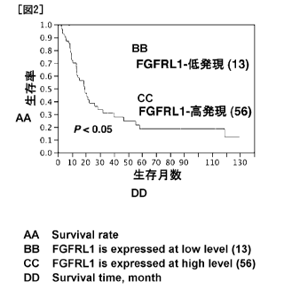
Fig. 2 is a diagram showing the relationship between the expression level of
FGFRL1 in the esophageal squamous cell carcinoma tissue measured in the
Examples below and the survival rate of patients at each month.
Fig. 3 is a diagram showing the results of measurement of the growth capacity
5 of esophageal squamous cell carcinoma cells observed after allowing an
anti-
FGFRL1 antibody to act on the cells in the Examples below, as compared to the
results obtained by treatment with a control antibody.
BEST MODE FOR CARRYING OUT THE INVENTION
[0011]
1 0 As described above, the therapeutic agent for cancer of the present
invention
comprises as an effective component an antibody that undergoes antigen-
antibody
reaction with FGFRL1 or an antigen-binding fragment thereof. Both the amino
acid
sequence and the gene base sequence are known for FGFRL1. A base sequence of
cDNA of FGFRL1 and the amino acid sequence encoded thereby are described, for
example, as NCBI Accession NO. NM_001004356.2. The base sequence of cDNA
of the FGFRL1 gene and the amino acid sequence encoded thereby are shown in
SEQ
ID NO:1, and the amino acid sequence alone is shown in SEQ ID NO:2. FGFRL1
is a single-transmembrane protein.
[0012]
2 0 The antibody used herein is an antibody that suppresses the growth of
cancer
cells, and the antibody undergoes antigen-antibody reaction with the N-
terminal
region of FGFRL1, that is, the epitope of the antibody is preferably present
in the N-
terminal region of FGFRL1 or in a region comprising the whole or a part of the
N-
terminal region (that is, a region that extends from the N-terminal region to
another
region). The N-terminal region herein means the extracellular region of
FGFRL1,
that is, the region between the N-terminus and the 378th amino acid in the
amino
acid sequence shown in SEQ ID NO:2. The antibody may be either a polyclonal
CA 02831867 2013-09-30
=
6
antibody or a monoclonal antibody. A polyclonal antibody whose epitope is the
N-
terminal region of FGFRL I is also commercially available, and such a
commercially
available product may also be preferably used. Further, an antigen-binding
fragment of the above-described antibody, such as the Fab fragment or the
F(ab')2
fragment, may also be used. Whether or not the antibody or fragment has an
effect
to suppress the growth of cancer cells can be investigated by, for example,
using the
well-known WST1 method as described in the Examples below.
[0013]
The antibody may be an antibody prepared by genetic engineering, or may be
1 0 a humanized antibody prepared by replacing the Fc region with that of a
human
antibody for suppression of rejection reaction in human. Further, in antibody
preparations, those prepared by binding a polyethylene glycol (PEG) chain or
the like
to an end of an antibody for making the antibody less likely to be degraded by
protease in the living body are widely used. Also in the therapeutic agent for
cancer
1 5 of the present invention, a stabilizing structure such as a PEG chain
may be attached
to an end of the above-described antibody or the antigen-binding fragment
thereof,
and the resultant may be contained in its entirety in the agent as an
effective
component. In cases where the antibody or an antigen-binding fragment thereof
is
stabilized by PEGylation, the size of the PEG is several thousand to 50,000,
20 preferably about 10,000 to 50,000. The method for binding PEG to an end
of a
polypeptide is well known. Such a product prepared by attaching a stabilizing
structure is also included in the "antibody or an antigen-binding fragment
thereof' in
the present invention.
[0014]
25 Further, the present antibody may also be utilized as a complex
antitumor
agent by chemically binding a low molecular weight antitumor agent or a
compound
having cytotoxicity against cancer thereto, or may be utilized as a navigator
in a drug
CA 02831867 2013-09-30
=
7
delivery system (DDS) to cancer cells
[0015]
In terms of the administration route of the therapeutic agent for cancer of
the
present invention, either parenteral administration or oral administration may
be
carried out. Parenteral administration such as injection to the cancer tissue,
intravenous injection, or intramuscular injection is preferred. The dose may
be
appropriately set depending on the clinical condition and the severity of the
disease to
be treated. For example, the therapeutic agent is administered at a dose of
0.1 to 20
mg per administration, preferably 1 to 10 mg per administration, per kg body
weight.
1 0 Further, the therapeutic agent for cancer of the present invention may
be formulated
by a well-known method into, for example, a solution in which the agent is
dissolved
in a physiological buffer. Further, a known additive(s) may be added to the
solution.
[0016]
Examples of the cancer to be treated with the therapeutic agent for cancer of
the present invention include, but are not limited to, epithelial solid
cancers.
Esophageal squamous cell carcinoma is especially preferred.
[0017]
The present inventors discovered, as specifically described in the Examples
below, that the expression level of FGFRL1 in a cancer tissue can be used as
an
2 0 index for predicting the prognosis of the cancer, that is, the survival
rate after
initiation of cancer treatment. Thus, the present invention also provides a
method
for predicting the prognosis of cancer, which method comprises investigating
the
expression level of FGFRL1 in a cancer tissue separated from a living body,
wherein
a high expression level of FGFRL1 indicates poor prognosis. The expression
level
2 5 of FGFRL1 can be measured by an immunoassay such as
immunohistochemistry.
For the immunoassay, the above-described anti-FGFRL1 antibody or an antigen-
binding fragment thereof may be used, and a polyclonal antibody or monoclonal
CA 02831867 2013-09-30
8
antibody whose epitope is the extracellular region of FGFRL1 may be preferably
used. Since, as described above, such a polyclonal antibody is also
commercially
available, it is also possible to use a commercially available product. Since
FGFRL1 is expressed in a state where it is penetrating the membrane, the
immunohistochemical staining as described in the Examples below is preferably
carried out as the immunoassay, but the immunoassay is not limited thereto.
[0018]
The higher the expression level of FGFRL I, the poorer the prognosis may be.
Thus, by preliminarily investigating the expression level of FGFRL1 and the
prognosis in a large number of patients with the same kind of cancer, it is
possible to
predict the prognosis based on how high the expression level of FGFRL1 is. For
example, as specifically described in the Examples below, in cases where the
expression level is investigated by immunohistochemical staining, the
prediction may
be made based on evaluation of the stained area per cancer tissue (0%: 1-50%:
+,
1 5 51-100%: +-1-), and the positivity per cell wherein strong positivity
is evaluated as
(+++) and negativity is evaluated as (-). In consideration of the extent of
expression
of FGFRL1 in a normal tissue, a total value of not less than (++) can be
judged as
positive (that is, poor prognosis).
[0019]
2 0 As described above, FGFRLI is a single-transmembrane protein, and has a
structure that undergoes the action of protease in the extracellular region.
Therefore,
it is thought that a tissue fluid extracted from a tissue, or blood, may
contain free
FGFRL1 or a free fragment of FGFRL1. The present inventors inferred that, by
quantifying free FGFRL1 or a free fragment of FGFRL1 in a tissue fluid
extracted
2 5 from a tissue, or blood, cancer can be detected. The present invention
also provides
a method for detecting cancer, which method comprises measuring FGFRL1 or a
fragment thereof extracted from a body tissue, or FGFRL 1 or a fragment
thereof in
CA 02831867 2013-09-30
= 9 =
blood separated from a living body, wherein a higher concentration of FGFRL1
or
the fragment thereof contained therein than the concentration of FGFRL I or
the
fragment thereof in the tissue or blood of a healthy individual indicates the
presence
of cancer. In such cases, the FGFRL1 or a fragment thereof in the tissue
extract or
blood can be quantified by an immunoassay using an antibody that undergoes
antigen-antibody reaction with the extracellular region of FGFRL1. The
immunoassay in such cases may be carried out by a well-known method such as
ELISA, which is widely used for quantification of various proteins in body
fluid; the
sandwich method, in which a fluorescent label or chemiluminescent label is
used; or
1 0 the immunoagglutination method, in which sensitized particles prepared
by
immobilizing an antibody on latex particles are used. The cut-off value in
such
cases may be a value significantly different from the mean value in healthy
individuals. The cut-off value may be, for example, 1.0 unit/mL, and the unit
value
in such cases is determined using as a standard the concentration in the
tissue or
1 5 blood of a healthy individual, although the unit value may vary
depending on
differential diagnosis from similar diseases and on background factors of the
patient.
[0020]
Further, based on the abundance of FGFRL1 or a fragment thereof in a tissue
fluid extracted from a tissue, or blood, prediction of the prognosis of cancer
can be
2 0 carried out similarly to the above-described cases where the prediction
is carried out
based on the expression level of FGFRL1 in the cancer tissue. In such a case,
the
criteria for evaluation of the prognosis may vary depending on whether the
survival
rate or the recurrence rate is to be evaluated, and for what disease the
evaluation is to
be done.
25 [0021]
The present invention is described below more specifically by way of
Examples. However, the present invention is not limited to the Examples below.
CA 02831867 2013-09-30
=10
EXAMPLES
[0022]
Example 1
Immunohistochemical Staining
Tissue sections were prepared from esophageal squamous cell carcinoma
tissues collected from 69 esophageal squamous cell carcinoma patients, and
subjected to deparaffinization (3 times of 5 minutes of immersion in xylene, 2
times
of 3 minutes of immersion in 100% ethanol, 3 minutes of immersion in 95%
ethanol,
3 minutes of immersion in 90% ethanol, 3 minutes of immersion in 85% ethanol,
5
minutes of washing with running water, and then 5 minutes of immersion in
distilled
water) and then antigen retrieval by heat treatment (treatment in 1 mM Tris
buffer
(pH 9.0) supplemented with 0.1 mM EDTA at 95 C for 40 minutes, followed by
allowing the resultant to cool at room temperature for 20 minutes, washing
with
running water and then immersion in distilled water). Subsequently, endogenous
1 5 peroxidase was blocked with 3% hydrogen peroxide solution (at room
temperature
for 10 minutes), and the sections were then washed with distilled water 3
times,
followed by immersion in 5 mM Tris buffer (pH 7.2) supplemented with 0.005%
Tween 20 and 15 mM NaC1 at room temperature for 5 minutes for achieving
equilibration. Anti-FGFRL1 antibody 11-300 (Santa cruse) was 50-fold diluted
with
2 0 Dako REAL antibody diluent (Dako), and treatment was carried out with
the
resulting dilution for 30 minutes. After washing the sections with 5 mM Tris
buffer
(pH 7.2) supplemented with 0.005% Tween 20 (trade name) and 15 mM NaC1 3
times, coloring with DAB (diaminobenzidine) was performed using Dako ChemMate
ENVISION kit (Dako), followed by washing with distilled water and then
performing
2 5 counter staining with Dako REAL Hematoxylin (prepared by 4-fold
dilution with
distilled water and then addition of Tween 20 (trade name) to adjust the Tween
20
concentration to 0.01%) at room temperature for 3 minutes. Thereafter, washing
CA 02831867 2013-09-30
=
11
with water, dehydration, clearing and embedding were carried out (5 minutes of
washing with water, 5 minutes of immersion in 80% ethanol, 5 minutes of
immersion
in 90% ethanol, 5 minutes of immersion in 95% ethanol, 2 times of 5 minutes of
immersion in 100% ethanol, and 3 times of 5 minutes of immersion in xylene,
followed by embedding with Leica CV5030). The stained area per cancer tissue
(0%: 0, 1-50%: 1, 51-100%: 2) and the staining intensity (no signal: 0, weak:
1,
moderate: 2, marked: 3) were scored, and a total score of not less than 4 was
defined
as high expression of the FGFRL1 protein, and a total score of less than 4 was
defined as low expression of the FGFRL1 protein. The survival rate was
compared
between both groups of patients by the Kaplan-Meier method and the log-rank
test.
[0023]
The results of immunohistochemical staining are shown in Fig. 1, and the
relationship between the expression level of FGFRL1 and the survival rate of
patients
at each month is shown in Fig. 2. As is evident from Fig. 2, the prognosis was
poor
in the cases where the expression level of FGFRL1 was high, and the survival
rate at
Month 60 in these cases was a little more than one third of the survival rate
observed
in the cases where the expression level of FGFRL1 was low.
[0024]
Example 2
Pharmacological Effect
An esophageal squamous cell carcinoma-derived cell line KYSE-170 was
plated in Ham F12 (Nissui)/RPMI1640 (Gibco) medium (pH 6.8) supplemented with
fetal bovine serum (5%, Equitech-Bio) filtered through a 0.22-1.1m PVDF
membrane
filter (Millipore), penicillin (100 unit/ml, Meiji), gentacin (4.44 mg/1,
Schering) and
sodium hydrogen carbonate (0.2%) on a 96-well dish (5x10³ cells/100 A/well),
and
cultured under the conditions of 5% CO2, a humidity of 100% and a temperature
of
37 C. Twenty four hours later, the cells were treated with anti-FGFRL1
antibody
CA 02831867 2013-09-30
12
H-300 (recognition site: N-terminus/extracellular region) or C-20 (recognition
site:
C-terminus/intracellular region) (Santa cruse) and a control IgG (Santa cruse)
of the
animal from which it was derived, which were diluted with the above-described
Ham
F12/RPMI1640 medium (final concentration, 20 p.g/m1). After 24 hours of
culture,
the cell growth was evaluated by the well-known WST1 method using a
commercially available reagent.
[0025]
The results are shown in Fig. 3. As shown in Fig. 3, in the case where the
monoclonal antibody whose epitope is the N-terminal region, that is, the
extracellular
region, of FGFRL1 was used, the growth of esophageal squamous cell carcinoma
cells was significantly suppressed as compared to the case where the treatment
was
carried out with the control antibody. Thus, such an antibody is useful as a
therapeutic agent for esophageal squamous cell carcinoma.
[SEQUENCE LISTING]