Note: Descriptions are shown in the official language in which they were submitted.
CA 02831969 2013-10-01
WO 2012/168003 PCT/EP2012/057897
1
Selective lysis of cells by ionic surfactants
FIELD OF THE INVENTION
The present invention relates to the lysis of eukaryotic cells, in particular
to
the lysis of animal cells, such as blood cells. The present invention further
relates to the
detection of a small number of micro-organisms such as bacteria or fungi in
samples
containing a large number of other cells.
BACKGROUND OF THE INVENTION
Molecular diagnostics aims at the rapid detection of minute amounts of
pathogens (typically bacteria) in samples such as blood. However, blood is a
complex matrix
and comprises white blood cells (leukocytes) for the adaptive immune system,
red blood cells
(erythrocytes) for oxygen transport, and platelets (thrombocytes) for wound
healing. This
composition complicates the direct detection of pathogens in samples such as
whole blood,
which contain a high amount of cellular material.
Classical detection methods comprise the growth of bacteria on selective
media and/or media containing indicators. Typically such assays require a
cultivation step of
at least 1 or 2 days before identification of the bacteria can take place.
For PCR based methods the amount of bacteria in a fresh blood sample is
theoretically high enough to be detected without further cultivation of the
bacteria present
within such sample. However, to allow an early detection of minute amounts of
bacteria,
large volumes of blood are required. The high amount of DNA in especially
white blood cells
dramatically increases the background in DNA based detection methods. Also the
presence
of heme from hemoglobin strongly decreases the activity of DNA polymerase. A
microliter
of human blood contains about 4,000 to 11,000 white blood cells and about
150,000 to
400,000 platelets. The concentration of DNA in blood is between 30 and 60
g/ml. It is
extremely challenging to detect the presence of about 10 to 100,000 of a
bacterial species in a
volume of 10 ml of whole blood.
The high amounts of DNA of the white blood cells may give rise to non
relevant PCR products, or may scavenge the primers designed for the detection
of bacterial
CA 02831969 2013-10-01
WO 2012/168003 PCT/EP2012/057897
2
DNA. This necessitates a thorough DNA purification and separation of
eukaryotic DNA
before the bacterial DNA can be detected via PCR or other methods.
Apart from interfering with the PCR reaction itself, the amount of mammalian
DNA increases the viscosity of a sample. In addition, proteins and membranes
from the lysed
mammalian cells form complexes which prevent the filtration of a sample. This
is
particularly a problem for miniaturized devices. Further dilution of the large
sample volume
results in unacceptable long manipulation steps.
For the above reasons, methods to remove human DNA from a blood sample
are accordingly required.
Methods to specifically assay bacterial DNA in the presence of mammalian
DNA are known. LooxtersTM from the company SIRSLab uses a method to enrich
methylated
DNA from a sample. As bacterial DNA is strongly methylated, this approach
results in an
enrichment of bacterial DNA. MolysisTM from the company Molzym, uses
chaotropic agents
and detergents to lyse selectively mammalian cells. This lysis step is
followed by a digest
with a DNAse which is not affected by this chaotropic agent/detergent.
Alternative
approaches such as commercialized by Roche (SeptifastTM) rely on PCR primer
pairs which
are specifically designed to prevent aspecific binding to human DNA and
amplification of
human DNA.
US 6,803,208 describes a method wherein a highly diluted suspension of
blood platelets doped with bacteria is lysed at 37 C for 15 minutes,
whereafter it is possible
to filter a small amount of the lysed sample over a 0.4 lAm filter for visual
inspection of the
bacteria which are retained on the filter. This method however does not allow
to process large
volumes of sample at ambient temperatures.
The non-published international patent application PCTAB2010/055628 by
Koninklijke Philips Electronics N.V. discloses a method for selective lysis of
eukaryotic cells
within a sample containing or suspected to contain micro-organisms, wherein a
non-ionic
detergent such as Triton X-100 (polyethylene glycol p-(1,1,3,3-
tetramethylbuty1)-phenyl
ether) and a buffer is added to a sample comprising eukaryotic cells to obtain
a solution
having a pH value of at least 9.5, and incubating said solution for a time
period sufficiently
long enough to lyse the eukaryotic cells. This method permits processing of
blood samples
having a volume of 5 ml by lysing the white and red blood cells in the sample,
degrading the
blood cell DNA while pathogenic micro-organisms remain intact, and can
subsequently be
enriched by centrifugation or filtration.
CA 02831969 2013-10-01
WO 2012/168003 PCT/EP2012/057897
3
In his article "Interactions of surfactants with lipid membranes" (Quarterly
Reviews of Biophysics 41 (2008), pages 205-264), H. Heerklotz discusses the
hypothetical
molecular mechanism of selective lysis of mammalian cells, and hypothesizes
that said
selective lysis depends on different steps. First, the surfactant ensures
lysis of the white and
red blood cells. In order to achieve this, the surfactant needs to be inserted
in the outer layer
of the cell membrane. In a second step, the surfactant will perform a so-
called flip-flop and is
transferred to the inner layer of the cell membrane. Once a sufficient amount
of surfactant is
present in the inner cell membrane and the outer cell membrane, the cell will
be lysed. Non-
ionic surfactants such as Triton X-100 were found to be well suited for cell
lysis as they
perform above-mentioned steps within a time frame of several hundred
milliseconds. In
contrast, SDS requires 10 to 30 s for its insertion into PC vesicles. In
addition, it is reported
that surfactants with larger or charged head groups may require hours or days
to cross the
membrane, as was shown for SDS at room temperature.
This hypothesis may explain why ionic surfactants are not suitable for
obtaining fast lysis of mammalian cells as has been described in scientific
literature (see
Heerklotz, H.). Surfactants comprising a large, bulky or charged hydrophilic
group such as
Tween0, ionic surfactants and Tritons having a long PEG chain are slow at the
flip-flop
movement and thus not suitable to obtain rapid cell lysis. In addition,
surfactants having a
very hydrophobic character such as Brij 35 or Triton X-45 will encounter
difficulties in
their initial insertion into the cell membrane. Ionic surfactants are
considered not suitable for
obtaining fast lysis of mammalian cells, because their charged hydrophilic
group cannot
perform the flip-flop transfer easily due to the presence of the charged
hydrophilic group
which has to pass the lipophilic membrane.
In contrast to the scientific knowledge, it has surprisingly been found that
an
ionic surfactant can be utilized for selective lysis of white and red blood
cells while keeping
microbial pathogens intact when said ionic surfactant is used in combination
with high pH.
Thus, in a first aspect, the present invention provides a method for selective
lysis of
eukaryotic cells within a sample containing or suspected to contain micro-
organisms. In a
second aspect, the present invention provides a kit-of-parts for performing
the method for
selective lysis of eukaryotic cells within a sample containing or suspected to
contain micro-
organisms. In a further aspect, the present invention provides a device for
detecting micro-
organisms in a sample containing eukaryotic cells.
SUMMARY OF THE INVENTION
CA 02831969 2013-10-01
WO 2012/168003 PCT/EP2012/057897
4
Particular and preferred aspects of the invention are set out in the
accompanying independent and dependent claims. Features from the dependent
claims may
be combined with features of the independent claims and with features of other
dependent
claims as appropriate and not merely as explicitly set out in the claims.
One aspect of the invention relates to a method for the selective lysis of
eukaryotic cells, in particular animal cells, within a sample containing or
suspected to contain
micro-organisms. This method comprises the steps of providing a sample with
eukaryotic
cells, in particular animal cells, containing or suspected to contain a micro-
organism, adding
a buffer having a pH of about 9.0 or more, preferably a pH of about 9.5 or
more and an ionic
surfactant to the sample to obtain a solution having a pH of about 9.0 or
more, preferably a
pH of about 9.5 or more, and incubating the solution for a time period
sufficiently long
enough to lyse the eukaryotic cells, in particular animal cells.
In particular embodiments, the sample is a blood sample, such as for example
whole blood. Preferably, the sample is a sample of vertebrate, more preferably
of a mammal,
in particular a domestic animal working animal or farm animal, and most
preferably a sample
of a human being.
In other particular embodiments the micro-organisms are bacteria and/or
unicellular fungi. The method of the present invention may also be suitable
for detecting
unicellular eukaryotic pathogens such as flagellated protozoan or apicomplexan
parasites..
According to particular embodiments, the ratio between the volume of added
surfactant and added buffer and the volume of sample is between 2/1 and 1/10.
In particular embodiments, the alkaline buffer as used herein has a
pKa value of above 9. Examples hereof are borate, carbonate, CAPS (N-
cyclohexy1-3-
aminopropanesulfonic), CAPSO (3-(cyclohexylamino)-2-hydroxy-1-propanesulfonic
acid),
CHES (2-(N-cyclohexylamino)ethane sulfonic acid), pyrophosphate and
ethanolamine. A
particular example is sodium carbonate.
In particular embodiments, the method further comprises the step of filtering
the incubated solution on a filter with a pore size which retains micro-
organisms on the filter,
such as a filter with a pore size of less than 0.5 p.m.
In particular embodiments, the method further comprises the step of adding
after the selective lysis an acid or acidic buffer to obtain a pH between
about 7 and 8 in the
lysed solution, "a neutralization step".
In particular embodiments, the methods as described above are followed by
lysis of the micro-organisms present in or suspected to be present in the
sample.
CA 02831969 2013-10-01
WO 2012/168003 PCT/EP2012/057897
Another aspect of the present invention relates to a kit-of-parts for
performing
the method described herein above. The kit comprises at least the alkaline
buffer and the
ionic surfactant. In preferred embodiments, the kit comprises a mixture of the
alkaline buffer
and the ionic surfactant in a fixed ratio. In an alternative embodiment, the
kit comprises
The kit-of-part may further comprise an acid or an acidic buffer in a
particular
embodiment for adjusting the pH of the solution to value of between about 7
and 8 after the
embodiment, the kit may comprise a lysis buffer for lysing the microorganisms
after they
have been enriched, and for releasing the microorganism's DNA.
In particular embodiments, the kit-of-parts comprises at least one means for
obtaining, processing and/or storing a sample or any solution that is
generated or obtained
In a further or additional embodiment, the kit-of-parts comprises at least one
means for filtering the incubated solution. Said means may for example be a
filter having a
pore-size which retains the micro-organisms on the filter. The filter may have
a pore size of
less than 0.5 tim. The filter may be present in or part of a cartridge.
20 Another aspect of the present invention relates to a device (1)
for the detection
of micro-organisms in sample, comprising: a lysis chamber (2) for accepting a
sample fluid
with a volume between 1 and 20 ml, a reservoir (3) comprising an alkaline
buffer with a pH
of about 9.0 or more, preferably a pH of about 9.5 or more, and comprising an
ionic
surfactant, or a reservoir comprising an alkaline buffer (31) with a pH of
about 9.0 or more,
Herein the alkaline buffer has typically a pKa above 9.0 and the ionic
Methods according to the present invention allow a selective lysis of white
and
red blood cells in a sample while bacteria and fungi remain intact (either
dead or alive).
Methods according to the present invention make it possible to process a
sample without substantially diluting such sample, and consequently allow to
process larger
CA 02831969 2013-10-01
WO 2012/168003 PCT/EP2012/057897
6
volumes of sample. In addition, there is no need for enzymatic degradation of
DNA by e.g.
DNase, making this method less complex compared to methods known in the prior
art.
Methods as described in the present invention result in lysed samples with a
low viscosity and a minimum of aggregates, which makes it possible to filter
the lysed
The above and other characteristics, features and advantages of the present
Fig. 1 shows a schematic overview of an embodiment of a device for
performing a selective lysis as described in embodiments of the present
invention.
Fig. 2 shows an example of an integrated device comprising a selective lysis
unit as described in embodiments of the present invention
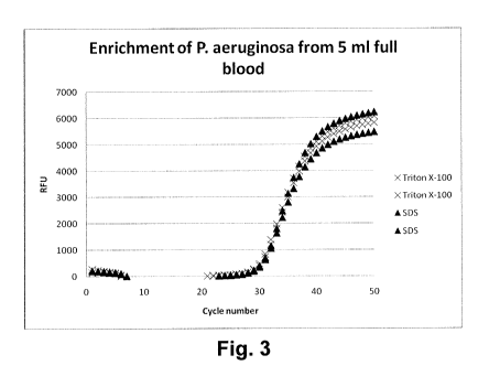
20 Fig. 3 shows the result of a quantitative RT-PCR for P. aeroginosa
enriched
from whole blood samples.
Fig. 4 shows the result of a quantitative RT-PCR for C. albicans enriched from
whole blood samples.
The present invention will be described with respect to particular
Furthermore, the terms first, second, third and the like in the description
and in
the claims, are used for distinguishing between similar elements and not
necessarily for
CA 02831969 2013-10-01
WO 2012/168003 PCT/EP2012/057897
7
describing a sequential or chronological order. It is to be understood that
the terms so used
are interchangeable under appropriate circumstances and that the embodiments
of the
invention described herein are capable of operation in other sequences than
described or
illustrated herein.
The following terms or definitions are provided solely to aid in the
understanding of the invention. These definitions should not be construed to
have a scope
less than understood by a person of ordinary skill in the art.
DETAILED DESCRIPTION OF THE EMBODIMENTS
"Blood cells" in the context of the present invention relates to mammalian
cells present in blood and includes red blood cells (erythrocytes), white
blood cells
(leukocytes) and blood platelets (thrombocytes).
"Whole blood" in the context of the present invention relates to unprocessed
blood comprising blood plasma and cells, potentially treated with an anti-
coagulant.
"Sample" relates to an aqueous suspension comprising cellular material and
comprises body fluids such as lymph, cerebrospinal fluid, blood (whole blood
and plasma),
saliva, but also comprises e.g. the aqueous fraction of homogenized
suspensions such as e.g.
muscles, brain, liver, or other tissues.
"Eukaryotic" in the present invention relates to any type of eukaryotic
organism, such as animals, in particular animals containing blood, and
comprises invertebrate
animals such as crustaceans and vertebrates. Vertebrates comprise both cold-
blooded (fish,
reptiles, amphibians) and warm blooded animal (birds and mammals). Mammals
comprise in
particular primates and more particularly humans. The term "eukaryotic" in the
present
invention does not comprise eukaryotic unicellular organisms such as
pathogenic or
opportunistic unicellular fungi and protozoa.
"Selective lysis" as used in the present invention is obtained when in a
sample
(such as blood) the percentage of micro-organism cells (such as bacterial
cells) in that sample
that remain intact is significantly higher (e.g. 2, 5, 10, 20, 50, 100, 250,
500, or 1,000 times
more) than the percentage of the eukaryotic cells from the organism from which
the sample is
collected that remain intact.
"Micro-organism" as used in the present invention relates to bacteria (gram
positive and gram negative bacteria, as well as bacterial spores) and
unicellular fungi such as
yeast and molds, which are present in the organism from which a sample has
been collected,
typically as a pathogen.
CA 02831969 2013-10-01
WO 2012/168003 PCT/EP2012/057897
8
A first aspect of the present invention relates to a method for the
selective lysis of eukaryotic cells, in particular animal cells, within a
sample, which contains
or is suspected to contain micro-organisms such as bacteria. The aim of the
method is to
increase the sensitivity of a test for the detection of minute amounts of
micro-organisms in a
sample (i.e. less than 10,000, 1,000, 100 or even less micro-organisms per ml
of sample). As
explained in the background of the invention, DNA from eukaryotic cells, in
particular from
animal cells, in a sample interferes with PCR based detection methods and this
DNA,
together with proteins and membranes form aggregates which increases viscosity
after lysis
and which has a dramatic impact on the filtration of a lysed sample. To solve
this problem,
the eukaryotic cells, in particular animal cells, are selectively lysed
whereby a substantial part
(i.e. more than 20%, 40%, 60%, 80%, 90% or even more that 95%) of the micro-
organisms
remains alive, or if killed by the treatment, still comprise the bacterial DNA
within the cell
wall. In methods as described in the present invention the above mentioned
problems are
addressed.
Methods as described in the present invention are applicable to any type of
sample wherein the detection of DNA from micro-organisms, particularly from
bacteria, is
impaired by the presence of other cells comprising DNA, in particular cells
from a host
wherein the micro-organism is present as a pathogen.
Methods as described in the present invention are now further illustrated for
embodiments wherein the presence of minute amounts of bacteria or fungi cells
in a
mammalian blood sample is investigated.
The blood sample can be stored as whole blood or a processed fraction such as
plasma or a platelet preparation. Typically, methods as described in the
present invention are
performed on freshly isolated whole blood. Such samples are generally treated
with e.g.
heparin, EDTA or citrate to avoid coagulation.
Alternatively the method is performed on fresh blood by collecting the blood
from a blood vessel such as an artery or vein directly in a tube with
detergent and buffer.
Accordingly, a fresh blood sample or a preserved sample is supplemented with
a buffer and an ionic surfactant. The selection of the buffer and its
concentration are chosen
in order to compensate the buffering capacity of the blood sample provided and
to obtain a
pH of about 9.0 or more, preferably a pH of about 9.5 or more, wherein pH
values above 11.5
are particularly suitable for gram positive bacteria and fungi. In a
particular embodiment, the
buffer has a pH of 9.0 or more. In a preferred embodiment, the buffer has a pH
of between
about 9.5 and about 11.5, more preferably a pH between about 9.5 and
about10.5. In a
CA 02831969 2013-10-01
WO 2012/168003 PCT/EP2012/057897
9
particular embodiment, the pH to obtain in the solution comprising the sample
is between
about 9.5 and about 11.5, even more particular between about 9.5 and about
10.5. Equally the
buffer is sufficiently concentrated such that at most a buffer volume of 200%,
150%, 100%,
50%, 20% or 10 % of the sample volume is added to the sample to obtain the
required change
in pH.
Suitable buffers in the context of the present invention typically have a pKa
above 9, above 9.5 or even above 10 and include borate, carbonate, CAPS,
CAPSO, CHES,
pyrophosphate, ethanolamine, and other commonly used buffers with an optimal
buffering
capacity in the above mentioned pH ranges.
Suitable surfactants are ionic surfactants, which at the one hand have a lytic
effect on the eukaryotic cells, in particular animal cells, only and on the
other hand have a
solubilising effect on DNA and proteins. The ionic surfactant may either be an
anionic
surfactant or a cationic surfactant, i.e. a surfactant molecule having a
positive ionic group.
Anionic surfactants have a negative ionic group, either based on a permanent
anion such as sulfate, sulfonate or phosphate, or on a pH-dependent anion such
as
carboxylate. The anionic surfactant may be selected from the group consisting
of alkyl
sulfates, alkyl ether sulfates, docusates, sulfonate fluorosurfactants, alkyl
benzene sulfonates,
alkyl aryl ether phosphates, alkyl ether phosphates, alkyl carboxylates, and
carbocxylate
fluorosurfactants. Examples of anionic surfactants are ammonium lauryl
sulfate, sodium
dodecyl sulfate (SDS), sodium deoxycholate, sodium-n-dodecylbenzenesulfonate,
sodium
lauryl ether sulfate (SLES), sodium myreth sulfate, dioctyl sodium
sulfosuccinate,
perfluorooctanesulfonate (PFOS), perfluorobutanesulfonate, sodium stearate,
sodium lauroyl
sarcosinate, perfluorononanoate, and perfluorooctanate (PFOA or PFO).
Cationic surfactants comprise a positive ionic group and pH-dependent
cationic surfactants are based on primary, secondary or tertiary amines,
whereas permanently
charged cationic surfactants are based on quaternary ammonium cation. Examples
of cationic
surfactants are cetyl trimethylammonium bromide (CTAB), cetyl
trimethylammonium
chloride (CTAC), cetylpyridinium chloride (CPC), Polyethoxylated tallow amine
(POEA),
benzalkonium chloride (BAC), benzthonium chloride (BZT), 5-bromo-5-nitro-1,3-
dioxane,
dimethyldioctadecylammonium chloride and dioctadecyldimethylammonium bromide
(DODAB).
The most effective concentration of surfactant depends from surfactant to
surfactant, but typically is within the range of between 0.1 and 5%, more
particularly
CA 02831969 2013-10-01
WO 2012/168003 PCT/EP2012/057897
between 0.1 and 1%. Depending from the detergent (solid or liquid) % refers to
respectively
w/v % or v/v %.
The incubation of a blood sample in the presence of buffer and detergent is
performed within 10 minutes, preferably between 30 seconds and 10 minutes and
more
5 preferably between about 1 to 3minutes, between about 1 to 5 minutes,
between about 1 to 8
minutes, or between about 1 to 10 minutes, at temperatures between 10 C and
30 C.
Methods according to the present invention have the advantage that a selective
lysis is
obtained within 0.5 to 3 minutes, at temperatures below 30 C. Accordingly,
the methods can
be generally performed at ambient temperatures without the need to heat the
sample.
10 Optionally, after the lysis the pH of the lysed sample is brought
to a neutral
value (i.e. between 7 and 8) by the addition of an acid or acidic buffer. It
was found that a
lysed sample at neutral pH could be stored for a prolonged time (up to 1, 2,
6, 12 or even 24
hours) without further lysis of bacterial cells and without dramatic changes
in the fluidic
properties of the lysed sample.
Another parameter investigated in the methods of the present invention is the
evaluation of the fluidic properties of the blood sample after lysis This can
be determined by
verifying which volume of lysed blood can be filtered through a 0.22 pm filter
with a
diameter of 2.5 cm. Methods in accordance with the present invention allow the
filtration of
at least 2, 5, 7.5 or even 10 ml of whole blood which was diluted by addition
of 1 volumes of
buffer/detergent solution to 1 volume of sample.
Generally, methods in accordance with the present invention comprise a step
wherein the intact micro-organisms are separated from the sample, typically
performed by
centrifugation or filtration. In particular embodiments intact micro-organisms
are separated
from the sample by passage of the lysed sample through a filter, with a pore
size below 1 [tin,
to retain micro-organisms which have typically a size between 0.5 and 101.im,
such as
commercially available filters with a pore size of 0.4 or 0.22 pm. For the
filtration of
samples, a wide variety of commercially available devices exists, such as
filters adapted to fit
on a syringe such that after lysis within in syringe, the fluid can be passed
over the filter by
manual pressure on the plunger of the syringe. These devices may be part of
the kit-of¨parts
of the present invention.
Hereafter the presence of micro-organisms on the filter can be investigated.
In
particular embodiments the presence of micro-organisms is investigated by PCR.
For this
purpose, the micro-organisms can be washed away from the filter and further
treated for PCR
CA 02831969 2013-10-01
WO 2012/168003 PCT/EP2012/057897
11
amplification. Alternatively the filter is rinsed with a lysis buffer to
release the DNA from the
micro-organisms, which is further used in a PCR reaction.
The lysis of the sample, filtration and detection of micro-organisms can be
performed within one device (schematically depicted in Fig. 1). Accordingly,
one aspect of
the present invention relates to a device (1), comprising a lysis chamber (2)
for accepting a
sample fluid with a volume between 1 and 10 ml, a reservoir (3) comprising an
alkaline
buffer with surfactants as described above, or a reservoir comprising an
alkaline buffer (31)
as described above and a reservoir comprising surfactants (32) as described
above, the
reservoirs connected to the lysis chamber (2). Within the device, the lysis
chamber is
connected to a filter (4) for filtering the sample after lysis whereby micro-
organisms are
retained on the filter. The device further comprises channels to remove the
micro-organisms
from the filter and lyse them in a separate chamber. Alternatively, the device
further
comprises means for lysing micro-organisms on the filter, and channels to
transfer DNA from
lysed bacterial or fungal cells from the filter to a separate chamber. The
device can further
contain a DNA purification and detection chamber (5) for assaying the presence
of DNA.
Typically the detection chamber is a PCR module.
An example of a device wherein selective lysis and subsequent DNA
purification and identification takes place is depicted in Fig. 2.
Other arrangements of the systems and methods embodying the invention will
be obvious for those skilled in the art.
It is to be understood that although preferred embodiments, specific
constructions and configurations, as well as materials, have been discussed
herein for devices
according to the present invention, various changes or modifications in form
and detail may
be made without departing from the scope and spirit of this invention.
EXAMPLE 1
Recovery of micro-organisms from blood samples
About 1.000 colony forming units (cfu) of either Pseudomonas aeruginosa or
Candida albicans were spiked in each 5 ml sample of human whole blood. An
equal volume
of lysis buffer (500 mM Na carbonate (pH 10.0) and either 1.0 % Triton X-100
(final
concentration) or 1 % sodium dodecyl sulfate (final concentration) was added
and the
mixture was incubated for 3 minutes at room temperature (about 23 C).
After the incubation the lysed sample was neutralized with a 1 M Tris solution
to restore the pH. The samples were centrifuged and washed with 1 ml phosphate-
buffered
CA 02831969 2013-10-01
WO 2012/168003 PCT/EP2012/057897
12
saline (PBS). Hereafter the micro-organisms were lysed by adding pre-heated
200 mM
NaOH, 1% SDS and their DNA was purified using standard silica spin columns
after the
eluates were neutralized with 1 M citric acid.
EXAMPLE 2
Detection of microbial DNA
For elution from the filter and for alkaline lysis of the micro-organisms, the
microbial cells were resuspended in 100 of a lysis solution containing 50 mM
NaOH and
0.25% SDS. Subsequently the samples were incubated for 10 min at 70 C, cooled
quickly to
room temperature and neutralized by addition of 30 p1 500 mM Tris-HC1, pH 7.0
(yielding a
final concentration of 150 mM Tris, i.e. 3 times the NaOH concentration).
For crude lysate PCR, unlysed cells and debris were removed from the sample
by centrifugation (5 min, 14,000 g). 1 111 of supernatant was added to a 25 p1
PCR reaction.
Detection by PCR was based on a Taqman PCR assay targeting the rRNA gene
(Apollo). The
PCR reaction was conducted in Taqman Universal mastermix (Applied Biosystems),
using
500 nM forward primer and 300 nM reverse primer and FAM-BHQ1 labeled probe
(all
oligonucleotides custom synthesized by Biolegio BV). The PCR reaction was
performed in a
Biorad CFX real-time PCR system. After an initial heating step of 10 min at 95
C to activate
the hot-start polymerase, 50 cycles of 15 sec at 95 C and 1 min at 60 C were
used for
amplification. Fluorescence signals were detected in each cycle during the 60
C step. Data
analysis was performed with the Biorad CFX software.
The Ct (cycle threshold) is defined as the number of cycles required for the
fluorescent signal to cross the threshold (i.e. exceeds background level). Ct
levels are
inversely proportional to the amount of target nucleic acid in the sample
(i.e. the lower the Ct
level the greater the amount of target nucleic acid in the sample). Ct values
<29 are strong
positive reactions indicative of abundant target nucleic acid in the sample.
Ct values of 30-37
are positive reactions indicative of moderate amounts of target nucleic acid.
Ct values of 38-
40 are weak reactions indicative of minimal amounts of target nucleic acid
which could
represent environmental contamination.
Figures 3 and 4 illustrate the results of the Taqman PCR assay for the
differential lysis and enrichment of micro-organisms from whole blood. Figure
3 displays the
relative fluorescence obtained during the PCR amplification of P. aeruginosa
DNA from
initially 1,000 cfu in 5 ml blood, wherein the lysis of the white and red
blood cells were
performed with either Triton X-100 or sodium dodecyl sulfate. Figure 4
displays the relative
CA 02831969 2013-10-01
WO 2012/168003 PCT/EP2012/057897
13
fluorescence obtained during the PCR amplification of C. albicans DNA from
initially 1,000
cfu in 5 ml blood, wherein the lysis of the white and red blood cells were
performed with
either Triton X-100 or sodium dodecyl sulfate. Both, figure 3 and figure 4
show that the non-
ionic surfactant and the ionic surfactant resulted in similar fluorescence
yields. Hence, it can