Note: Descriptions are shown in the official language in which they were submitted.
CA 02832194 2013-11-01
SCALABLE PRIMATE PLURIPOTENT STEM CELL AGGREGATE
SUSPENSION CULTURE AND DIFFERENTIATION THEREOF
STATEMENT REGARDING FEDERALLY SPONSORED RESEARCH OR
DEVELOPMENT
[0001] Part of the work performed during development of this invention
utilized U.S. Government
funds from National Institutes of Health Grant No. 5 R24 RR021313-05. The U.S.
Government has
certain rights in this invention.
RELATED APPLICATIONS
[0002] This application is a Continuation-in-Part under 35 U.S.C. 120 of U.S.
Patent Application
Serial No. 13/220,590, filed August 29, 2011, which is a Continuation of U.S.
Patent Application
Serial No.12/264,760, filed November 4, 2008, now U.S. Patent No. 8,008,075,
issued August 30,
2011, the disclosures of which are incorporated herein by reference in the
entireties.
FIELD OF THE INVENTION
[0003] The present invention relates to suspension cell aggregate compositions
that are essentially
serum and feeder-free and methods for differentiating the cell aggregate
suspensions.
BACKGROUND OF THE INVENTION
[0004] To date, there is no efficient system providing for a large-scale
manufacturing process
("scale-up") for mammalian pluripotent cells such as human embryonic stem
cells (hESC) as
described herein. To maintain hESC in an undifferentiated state in vitro, the
hESC are maintained
on mouse embryonic fibroblast (MEF) feeders and passaged by manual mechanical
dissociation
(e.g., micro-dissection) and transferring individual colony pieces. These
methods are sufficient for
research studies that do not require large-scale production of
undifferentiated hESC or
differentiated hESC, gene targeting, drug discovery, in vitro toxicology,
future clinical applications
require improved methods for the stable large-scale expansion of hESC,
including enzymatic
passaging.
[0005] Enzymatic expansion of hESC can be performed but these methods have
technical
disadvantages because hESC depend on cell-cell interactions as well as para-
and autocrine signals
CA 02832194 2013-11-01
for survival. Hence, hESC prefer this cellular microenvironment as compared to
existing as single
cells. Also, there are reports that enzymatic dissociation of hESC may lead to
abnormal karyotypes
and result in genetic and epigenetic changes. Thus, providing a highly
supportive culture
environment while at the same time allowing for robust large-scale expansion
(i.e., a manufacturing
process) of undifferentiated hES or differentiated hESC without compromising
the pluripotency,
multipotency or genetic stability over extended culture periods is essential.
[0006] Human pluripotent cells offer unique opportunities for investigating
early stages of human
development as well as for therapeutic intervention in several disease states,
such as diabetes
mellitus and Parkinson's disease. For example, the use of insulin-producing 13-
cells derived from
hESC would offer a vast improvement over current cell therapy procedures that
utilize cells from
donor pancreases. Currently cell therapy treatments for diabetes mellitus,
which utilize cells from
donor pancreases, are limited by the scarcity of high quality islet cells
needed for transplant. Cell
therapy for a single Type I diabetic patient requires a transplant of
approximately 8 x 10⁸ pancreatic
islet cells (Shapiro et al. 2000, N Engl J Med 343:230-238; Shapiro et al.
2001a, Best Pract Res
Clin Endocrinol Metab 15:241-264; Shapiro etal. 2001, British Medical Journal
322:861). As
such, at least two healthy donor organs are required to obtain sufficient
islet cells for a successful
transplant.
[0007] hESC thus represent a powerful model system for the investigation of
mechanisms
underlying pluripotent cell biology and differentiation within the early
embryo, as well as providing
opportunities for genetic manipulation of mammals and resultant commercial,
medical and
agricultural applications. Furthermore, appropriate proliferation and
differentiation of hESC can
potentially be used to generate an unlimited source of cells suited to
transplantation for treatment of
diseases that result from cell damage or dysfunction. Other pluripotent cells
and cell lines including
early primitive ectoderm-like (EPL) cells as described in International Patent
Application WO
99/53021, in vivo or in vitro derived ICM/epiblast, in vivo or in vitro
derived primitive ectoderm,
primordial germ cells (EG cells), teratocarcinoma cells (EC cells), and
pluripotent cells derived by
dedifferentiation or by nuclear transfer will share some or all of these
properties and applications.
International Patent Application WO 97/32033 and U.S. Patent No. 5,453,357
describe pluripotent
cells including cells from species other than rodents. Human ES cells have
been described in
International Patent Application WO 00/27995, and in U.S. Patent No.
6,200,806, and human EG
cells have been described in International Patent Application WO 98/43679.
2
CA 02832194 2013-11-01
[0008] The biochemical mechanisms regulating ES cell pluripotency and
differentiation are very
poorly understood. However, the limited empirical data available (and much
anecdotal evidence)
suggests that the continued maintenance of pluripotent ES cells under in vitro
culture conditions is
dependent upon the presence of cytokines and growth factors present in the
extracellular milieu.
[0009] While human ESCs offer a source of starting material from which to
develop substantial
quantities of high quality differentiated cells for human cell therapies,
these cells must be obtained
and/or cultured in conditions that are compatible with the expected regulatory
guidelines governing
clinical safety and efficacy. Such guidelines likely will require the use of
media with all
components sourced with cGMP. The development of such chemically defined/GMP
standard
conditions is necessary to facilitate the use of hESCs and cells derived from
hESCs for therapeutic
purposes in humans.
[0010] In addition, the eventual application of hESC based cell replacement
therapies will require
the development of methods that enable large scale culture and differentiation
conditions that are
compliant with regulatory guidelines. While several groups have reported
simplified growth
conditions for hESCs, there are substantial limitations with these studies. To
date, however, the
successful isolation, long-term clonal maintenance, genetic manipulation and
germ line
transmission of pluripotent cells has generally been difficult.
[0011] Most of the cell culture conditions for stem cells still contain serum
replacer (KSR) in the
media (Xu et al. 2005 Stem Cells, 23:315-323; Xu et al. 2005 Nature Methods,
2:185-189; Beattie
etal. 2005 Stem Cells, 23:489-495; Amit etal. 2004 Biol. Reprod., 70:837-845;
James etal. 2005
Development, 132:1279-1282). KSR contains a crude fraction of bovine serum
albumin (BSA)
rather than a highly purified source. Others have only performed short-term
studies, and therefore
it is not clear if their conditions would enable the maintenance of
pluripotency over extended
periods (Sato etal. 2004, Nature Med. 10:55-63; U.S. Patent Publication Nos.
2006/0030042 and
2005/0233446). Others have shown long-term maintenance of pluripotency in a
chemically defined
media with FGF2, activin A, and insulin, but the cells were grown on plates
that were coated with
human serum, which was "washed off" before plating of cells (Vallier et al.
2005 J Cell Sci., 118(Pt
19):4495-509). While FGF2 has been a component of all these media, it is not
clear if it provides a
primary or secondary self-renewal signal (Bendall etal. 2007 Nature 448:1015-
1027); particularly
as in some formulations it is necessary to use it at a high concentration (up
to 100 ng/mL, Xu et al.
2005 Nature Methods, 2:185-189).
3
CA 02832194 2013-11-01
[0012] Furthermore, all of these groups have either included insulin in their
media at 1.ig/mL levels,
or have insulin present due to the use of KSR. Insulin is typically considered
to function in glucose
metabolism and "cell survival" signaling via binding to the insulin receptor.
At levels above
physiological concentrations, however, insulin can also bind to the IGF1
receptor with a lower
efficiency and confer classical growth factor activity through the PI3
Kinase/AKT pathway. The
presence/requirement for such high levels of insulin (i_ig/mL levels) in KSR
or these other media
conditions suggests that the major activity is elicited via binding to the
IGF1 receptor, which is
expressed by hESCs (Sperger etal. 2003 PNAS, 100(23):13350-13355). Others have
noted the
expression of a full complement of IGF IR and intracellular signaling pathway
members in hESCs,
which is likely to signify the functional activity of this pathway (Miura
etal. 2004 Aging Cell,
3:333-343). Insulin or IGF1 may elicit a major signal required for the self-
renewal of hESCs, as is
suggested by the fact that all conditions developed thus far for the culture
of hESC contain either
insulin, insulin provided by KSR, or IGF1 provided by serum. In support of
this concept, it has
been shown that if PI3 Kinase is inhibited in hESC cultures, the cells
differentiate (D'Amour et al.
2005, Nat. Biotechnol 23:1534-41; McLean etal. 2007 Stem Cells 25:29-38).
[0013] A recent publication outlines a humanized, defined media for hESCs
(Ludwig et al. Nature
Biotechnology, published online January 1, 2006, doi:10.1038/nbt1177). This
recent formulation,
however, includes several factors that are suggested to influence the
proliferation of hESCs,
including FGF2, TGFI3, LiC1, y-aminobutyric acid and pipecolic acid. It is
noted that this recently
defined cell culture medium also contains insulin.
[0014] A self-renewal signaling paradigm for hESC based on a combination of
insulin/IGF1,
heregulin, Activin A signaling was previously reported by Applicant. See Wang
et al. 2007 Blood
110:4111-4119. In this context we have found that an exogenous FGF2 signal is
redundant and not
required (Schulz & Robins 2009, supra) Schulz & Robins 2009, (In: Lakshmipathy
et al. eds.,
Emerging Technology Platforms for Stem Cells. John Wiley & Sons., Hoboken, NJ,
pp. 251-274);)
Heregulin is a member of the EGF growth factor family. There are at least 14
members, including,
but not limited to, EGF, TGFP, heparin binding-EGF (hb-EGF), neuregulin-13.
(also named
heregulin-f3 (HRG-j3), glial growth factor and others), HRG-a, amphiregulin,
betacellulin, and
epiregulin. All these growth factors contain an EGF domain and are typically
first expressed as
transmembrane proteins that are processed by metalloproteinase (specifically,
ADAM) proteins to
generate soluble ectodomain growth factors. EGF family members interact with
either homo- or
4
CA 02832194 2013-11-01
hetero-dimers of the ErbB 1, 2, 3 and 4 cell surface receptors with different
affinities (Jones et al.
FEBS Lett, 1999, 447:227-231). EGF, TGFa and hbEGF bind ErbB1/1 (EGFR)
homodimers and
ErbB1/2 heterodimers at high affinity (1-100 nM range), whereas HRG-P binds
ErbB3 and ErbB4
at very high affinity (<1 nM range). Activated ErbB receptors signal through
the PI3 Kinase/AKT
pathway and also the MAPK pathway. ErbB2 and ErbB3 are amongst the most highly
expressed
growth factor receptors in hESCs (Sperger etal. 2003, PNAS, 100:13350-13355)
and HRG-P has
been shown previously to support the expansion of mouse primordial germ cells
(Toyoda-Ohno et
al. 1999, Dev. Biol., 215:399-406). Furthermore, over expression and
subsequent inappropriate
activation of ErbB2 is associated with tumorigenesis (Neve etal. 2001 Ann.
Oncol, 12(Suppl 1):S9-
13; Zhou & Hung, 2003 Semin. Oncol. 30(5 Suppl 16):38-48; Yarden, 2001,
Oncology, 61 Suppl
2:1-13). Human ErbB2 (Chromosome 17q), and ErbB3 (Chromosome 12q) are present
on
chromosomes that have been observed to accumulate as trisomies in some hESCs
(Draper et al.
2004 Nat. Biotechnol. 22:53-4; Cowan et al. 2004 N Engl. J. Med. 350(13):1353-
6; Brimble etal.
2004 Stem Cells Dev., 13:585-97; Maitra etal. 2005 Nat. Genet. 37:1099-103;
Mitalipova et at.
2005 Nat. BiotechnoL 23: 19-20; Draper etal. 2004 Stem Cells Dev., 13:325-36;
Ludwig et al.
Nature Biotech, published online January 1, 2006, doi:10.1038/nbt1177).
[0015] ErbB2 and ErbB3 (Brown etal. 2004 Biol. Reprod., 71:2003-11; Salas-
Vidal & Lomeli,
2004 Dev Biol. 265:75-89) are expressed in the mouse blastocyst, although not
specifically
restricted to the inner cell mass (ICM), and ErbB 1, EGF and TGFP are
expressed in the human
blastocyst (Chia et at. 1995 Development, 1221(2):299-307). HB-EGF has
proliferative effects in
human IVF blastocyst culture (Martin etal. 1998 Hum. Reprod. 13:1645-52;
Sargent et at. 1998
Hum. Reprod. 13(Suppl 4):239-48), and modest additional effects on mouse ES
cells grown in 15%
serum (Heo etal. 2006 Am. I Phy. Cell Physiol. 290:C123-33, Epub 2005 Aug 17.
Pre- and early
post-implantation development does not appear to be affected in ErbB2-I-,
ErbB3-/-, Neuregulinl-/-
(Britsch et al. 1998 Genes Dev., 12:1825-36), ADAM17-/- (Peschon et al. 1998
Science, 282:
1281-1284) and ADAM19-/- (Horiuchi 2005 Dev. Biol. 283:459-71) null embryos.
Therefore, the
importance of signaling through the ErbB receptor family in hESCs is, up to
now, unclear.
[0016] Neuregulin-1 (NRG1) is a large gene that exhibits multiple splicing and
protein processing
variants. This generates a large number of protein isoforms, which are
referred to herein
collectively as neuregulin. Neuregulin is predominantly expressed as a cell
surface transmembrane
protein. The extracellular region contains an immunoglobulin-like domain, a
carbohydrate modified
CA 02832194 2013-11-01
region and the EGF domain. NRG1 expression isoforms have been reviewed
previously (Falls
2003 Exp. Cell Res. 284:14-30). The cell membrane metalloproteases ADAM17 and
ADAM19
have been shown to process the transmembrane form(s) of neuregulin-1 to
soluble
neuregulin/heregulin. HRG-a and -13 are the cleaved ectodomains of neuregulin,
containing the
EGF and other domains. As the EGF domain is responsible for binding and
activation of the ErbB
receptors, a recombinant molecule containing only this domain can exhibit
essentially all of the
soluble growth factor effects of this protein (Jones et al. 1999 FEBS Lett.
447:227-31). Also, there
are processed transmembrane isoforms of neuregulin that are thought to trigger
juxtacrine signaling
in adjacent cells via interaction of the EGF domain with ErbB receptors.
[0017] Still, an important development in the progression of hESC research
toward maintaining
pluripotency in culture will be the elucidation of media and cell culture
conditions that are
compatible with the expected regulatory guidelines governing clinical safety
and efficacy. While
the best outcome would be the availability of chemically defined media for
hESC, components that
are not chemically defined would be acceptable if they were produced to GMP
standard. There is a
need, therefore, to identify methods and compositions for the culture and
stabilization of a
population of pluripotent stem cells that are able to be used for therapeutic
purposes, wherein the
culture compositions are defined and/or produced to GMP standard.
[0018] The production of committed progenitor or differentiated cell types
that can function
following transplantation is a central promise of the potential of hESC-based
therapeutic research.
Using a step-wise protocol, in particular a 4-stage step-wise protocol
substantially similar to that
described herein and previously in Applicant's patent and non-patent
publications, also referred to
herein, primate pluripotent stem cells (pPSC) e.g., hESC or iPSC, are
differentiable cells that can be
directed to differentiate to a mixed population of pancreatic type cells by
the end of stage 4. The
mixture of cells contains at least cells commonly referred to as "pancreatic
progenitors", or
"pancreatic endoderm", or "pancreatic epithelium" both also referred to as
"PE", or "PDX1-positive
pancreatic endoderm", or "pancreatic endoderm cells" or "PEC" or equivalents
thereof.
[0019] The cellular composition of PEC has been fully characterized as
described in Applicant's
prior patent and non-patent applications, including but not limited to Kroon
et al. 2008 Nature
Biotechnology 26:443-52, and U.S. Patent Nos. 7,534,608; 7,695,965; and
7,993,920, entitled
METHODS FOR MAKING INSULIN IN VIVO, and 8,278,106, entitled ENCAPSULATION OF
PANCREATIC CELLS DERIVED FROM HUMAN PLURIPOTENT STEM CELLS, which are
6
CA 02832194 2013-11-01
herein incorporated by reference in their entireties. Using flow cytometry,
quantification of more
than 20 samples from more than 10 different development lots of PEC showed the
following types
of cells. About 50% (ranges from 33-60%) of the cell mixture consisted of
cells that express
NKX6-1 but not Chromogranin (CHGA). About 44% (range 33-62%) poly-hormonal
endocrine
cells express CHGA. CHGA positive cells have been shown to develop and mature
to glucagon
expressing cells following in vivo transplantation or implantation. About 7%
(range 1.3-13%)
express PDX1 while at the same time do not express CHGA or NKX6-1 (PDX1 only
population).
A very small group of cells, about 1% (range 0.27-6.9%) in the mixture or
population express none
of the above markers: neither PDX1, nor NKX6-1, nor CHGA (or triple negative
cells). Hence,
PEC or equivalents thereof refers to this population or mixture of cells. PEC
composition or
population is also described in more detail in Example 27 and Table 12. Kroon
et al. 2008, supra,
Schulz et al. 2012, supra, which disclosures are all incorporated herein by
reference in their
entireties.
[0020] Implanted PEC, encapsulated or un-encapsulated, gives rise to
functioning islet-like
structures in vivo through a mechanism that appears to primarily involve the
de novo commitment
of pancreatic progenitors to the endocrine lineages followed by further
maturation to glucose-
responsive 13-cells. Such grafts are therefore capable of sensing blood
glucose, responding with
metered release of processed human insulin, and protecting against
streptozotocin (STZ)-induced
hyperglycemia in mice. See Kroon et al. 2008, supra.
[0021] While other candidate pancreatic lineages have been derived from hESC,
none have
demonstrated substantial post-engraftment function in vivo, as defined by both
long-term glucose-
responsive human c-peptide secretion and protection against STZ-induced
hyperglycemia. Without
demonstrated function in animal models, it is difficult to gauge the
scalability, or clinical potential,
of these alternate protocols. See Cai J. etal. 2009 J Mol Cell Biol 2:50-60;
Johannesson et al. 2009
PLoS One 4:e4794; Mfopou etal. 2010 Gastroenterology 138: 2233-2245; Ungrin
etal. 2011
Biotechnol Bioeng. Dec 2. doi:10.1002/bit.24375; Clark et al. 2007 Biochem
Biophys Res Commun
356:587-593; Jiang etal. 2007 Cell Res 17: 333-344; and Shim etal. 2007
Diabetologia 50:1228-
1238, which are incorporated herein by reference in their entireties.
[0022] The invention described herein follows on Applicant's previous
demonstration that feeder-
free conditions using defined media can support single cell passaging and bulk
culture of hESC.
See Schulz & Robins 2009, supra; and U.S. Patent No. 8,278,106, entitled
ENCAPSULATION OF
7
CA 02832194 2013-11-01
PANCREATIC CELLS DERIVED FROM HUMAN PLURIPOTENT STEM CELLS, which are
herein incorporated by reference in their entireties. Critical for the
progression of hESC-based
technology to clinical trials is a demonstration of comparable scalability.
Improvements that
enhance expansion efficiencies will also save time and produce cost savings,
as well as minimize
the potential for population drift over time spent in culture. See Maitra et
al. 2005 Nat Genet
37:1099-1103, which is incorporated herein by reference in its entirety.
Importantly, scaling using
roller bottles as described herein, for example, along with cryopreservation
of hESC, provides a
defined and consistent material for product manufacture for near and long term
research and
development strategies.
SUMMARY OF THE INVENTION
[0023] The invention relates to compositions comprising a basal salt nutrient
solution and an ErbB3
ligand, with the compositions being essentially free of serum.
[0024] The invention also relates to compositions comprising a basal salt
nutrient solution and a
means for stimulating ErbB2-directed tyrosine kinase activity in
differentiable cells.
[0025] The invention relates to methods of culturing differentiable cells,
with the methods
comprising plating the differentiable cells on a cell culture surface,
providing a basal salt nutrient
solution to the differentiable cells and providing a ligand that specifically
binds ErbB3.
[0026] The invention relates to methods of culturing differentiable cells,
with the methods
comprising plating the differentiable cells on a cell culture surface and
providing a basal salt
nutrient solution to the differentiable cells and a means for stimulating
ErbB2-directed tyrosine
kinase activity in the differentiable cells.
[0027] The invention also relates to methods of culturing differentiable
cells, with the methods
comprising providing a digest solution to a layer of differentiable cells that
are contained in a
culture chamber prior to digestion, where the digestion breaks apart the layer
of cells into single
cells. After digestion, the single cells are placed into a new tissue culture
chamber with a
differentiable cell culture solution, wherein the differentiable cell culture
solution comprises a basal
salt nutrient solution and an ErbB3 ligand. Once cultured, the single
differentiable cells are placed
in conditions that permit growth and division of the single cells.
8
CA 02832194 2013-11-01
[0028] The invention relates to methods for generating a hES cell aggregate in
suspension from a
pluripotent hES adherent culture, by culturing a hES cell in an adherent
growth culture condition
which allows for expansion in an undifferentiated state; disassociating the
adherent hES cell culture
into a single cell suspension culture; contacting the single cell suspension
culture with a first
differentiating culture condition which allows for formation of hES-derived
cell aggregates in
suspension by agitating the single cell suspension culture until such a period
of time when the
single cell suspension culture forms a hES-derived cell aggregate in
suspension, and thereby
generating a hES-derived cell aggregate in suspension. In preferred
embodiments, agitation of the
single cell suspension culture is performed by rotation at about 80 rpm to 160
rpm
[0029] The invention also relates to methods for generating a hES-derived cell
aggregate in
suspension from a hES-derived single cell suspension, by culturing a hES cell
in an adherent
growth culture condition which allows for expansion in an undifferentiated
state; contacting the
undifferentiated hES cell with a first differentiating culturing condition
suitable for differentiating
the hES cell and resulting in an adherent hES-derived cell; disassociating the
adherent hES-derived
cell into a single cell suspension culture; contacting the single cell
suspension culture with a second
differentiating culture condition which allows for formation of hES-derived
cell aggregates in
suspension by agitating the single cell suspension culture until such a period
of time when the
single cell suspension culture forms a hES-derived cell aggregate in
suspension, and thereby
generating a hES-derived cell aggregate in suspension. In preferred
embodiments, agitation of the
single cell suspension culture is performed by rotation at about 80 rpm to 160
rpm.
[0030] The invention relates to a roller bottle containing primate pluripotent
stem cell (pPSC)
aggregates in suspension. In certain aspects of the invention, the pPSC
aggregates are cells selected
from the group consisting of human embryonic stem cells (hESC), induced
pluripotent stem cells
(iPSC) and/or other human pluripotent stem cells. In one embodiment, the
roller bottle is not
vented, but can be vented depending on the incubator or oven capabilities.
[0031] The invention also relates to methods for generating a roller bottle
containing pPSC
aggregates by contacting pPSCs with a pluripotent stem cell culture condition,
and agitating the
culture until pPSC aggregates form, thereby generating pPSC aggregates in the
roller bottle. In
certain embodiments, agitation of the pPSC culture is performed by rotation at
about 3 rpm, about 4
rpm, about 5 rpm, about 6 rpm, about 7 rpm, about 8 rpm, about 9 rpm, about 10
rpm, about 11
rpm, about 12 rpm, about 13 rpm, about 14 rpm, about 15 rpm, about 16 rpm,
about 17 rpm, about
9
CA 02832194 2013-11-01
18 rpm, about 19 rpm, about 20 rpm, about 21 rpm, about 22 rpm, about 23 rpm,
about 24 rpm,
about 25 rpm, about 26 rpm, about 27 rpm, about 28 rpm, about 29 rpm and about
30 rpm.
Typically, agitation of the pPSC culture is performed by rotation at about 5
rpm, about 6 rpm, about
7 rpm, about 8 rpm, about 9 rpm, about 10 rpm, about 11 rpm, and about 12 rpm.
[0032] Another aspect of the invention relates to methods for differentiating
pPSC aggregates in a
roller bottle by contacting differentiable or undifferentiated pPSC aggregates
with a culturing
condition that differentiates the pPSCs, and agitating the pPSC aggregate
culture until formation of
pPSC-derived aggregates, thereby generating pPSC-derived aggregates in
suspension in a roller
bottle. In certain embodiments, agitation of the pPSC-derived aggregates
suspension culture is
performed by rotation at about 3 rpm, about 4 rpm, about 5 rpm, about 6 rpm,
about 7 rpm, about 8
rpm, about 9 rpm, about 10 rpm, about 11 rpm, about 12 rpm, about 13 rpm,
about 14 rpm, about 15
rpm, about 16 rpm, about 17 rpm, about 18 rpm, about 19 rpm, about 20 rpm,
about 21 rpm, about
22 rpm, about 23 rpm, about 24 rpm, about 25 rpm, about 26 rpm, about 27 rpm,
about 28 rpm,
about 29 rpm and about 30 rpm. Typically, agitation of the pPSC culture is
performed by rotation
at about 5 rpm, about 6 rpm, about 7 rpm, about 8 rpm, about 9 rpm, about 10
rpm, about 11 rpm,
and about 12 rpm.
[0033] Still another embodiment of the invention relates to methods where
fluid flow within a
rolling bottle type of vessel involves rolling movement that does not require
rotation or rolling the
bottle. In one embodiment, the rolling type movement is substantially re-
created but without the
use of a rolling vessel. In another embodiment, a primate pluripotent stem
cell culture has imparted
fluid movement, for example, by pumping or flowing a fluid in a smooth,
orderly manner with little
or no turbulence. In such embodiments, any sub-current generally moves in
parallel with any other
nearby sub-current(s). This type of movement is also characterized as laminar
flow (commonly
used to move viscous fluids, especially those moving at low velocities) or
streamline flow (a steady
movement of fluid movement). In a yet another embodiment, the fluid movement
involves one or
more baffles, which distribute the fluid flow within a chamber to create a
continuous, uniform
suspension of cells. In a still further embodiment, the fluid movement
involves one or a
combination of deflector plates, distribution channels, and/or flow channels.
In each embodiment,
there is included at least one or more seals on the culture vessel to ensure
an aseptic environment
inside the vessel during cell aggregation, growth and differentiation.
CA 02832194 2013-11-01
[0034] The invention also relates to methods for enriching or varying the
composition of the
resulting cell culture and/or population of an hES-derived cell aggregate
suspension by optimizing
the cell density of the pluripotent cell cultures or varying the concentration
of various growth
factors, for example, FGF10, EGF, KGF, noggin and retinoic acid, apoptotic
inhibitors, Rho-kinase
inhibitors and the like.
BRIEF DESCRIPTION OF THE DRAWINGS
[0035] FIGURE 1 depicts real time RT-PCR expression analysis of ADAM19,
Neuregulinl, and
ErbB1-3 in BGOlv grown in defined conditions (8 ng/mL FGF2, 100 ng/mL LR-IGF1,
1 ng/mL
Activin A). GAPDH and OCT4 control reactions are indicated.
[0036] FIGURE 2 depicts the inhibition of proliferation of BGOlv cells using
AG879. BGOlv cells
were plated in 6-well trays and exposed to DMSO (A), 50 nM-20 tM AG1478 (B),
or 100 mM-20
j.tM AG879 (C) 24 hours after plating. After 5 days in culture, the cultures
were fixed and stained
for alkaline phosphatase activity. AG1478 did not appear to affect
proliferation at these
concentrations (20 ,M shown in B), but AG879 substantially slowed cell growth
at 5 [tM (C).
[0037] FIGURE 3 depicts the morphology of BGOlv cells cultured in DC-I-IAIF,
which is defined
culture media containing 10 ng/mL HRG-f3, 10 ng/mL Activin A, 200 ng/mL LR-
IGF1 and 8
ng/mL FGF2 (A and B), and in defined culture media (DC) containing 10 ng/mL
HRG-13, 10 ng/mL
Activin A, and 200 ng/mL LR-IGF1 (C and D).
[0038] FIGURE 4 depicts the expression of ADAM19, Neuregulinl, and ErbB1-4 by
RT-PCR in
mouse ES cells (A) and MEFs (B).
[0039] FIGURE 5 depicts the inhibition of ErbB1 and ErbB2 signaling in mouse
ES cells. 2x10⁵
Mouse R1 ES cells were plated on 1:1000 MATRIGELTm in 10% FBS, 10% KSR with
1000 U/mL
mouse LIF (ESGRO). The following day, DMSO (carrier control), 1-50 [I,M
AG1478, or 1-501.IM
AG879 was added with fresh medium. The cultures were fixed on day 8, and
stained for alkaline
phosphatase activity. DMSO (A) and 1-50 1.1M AG1478 (B and C) did not overtly
inhibit
proliferation. AG879 substantially inhibited cell growth at 50 [tM (compare D
and F) and may
have slowed proliferation at 201.IM (E).
[0040] FIGURE 6 depicts the inhibition of proliferation of BG02 cells grown in
conditioned media
(CM). (A) 50 [tM AG825 inhibited proliferation of BG02 hESCs growing in CM.
(B) AG825
11
CA 02832194 2013-11-01
inhibits ErbB2 Y1248 phosphorylation in hESCs. (C) Colony counting of serial
passaging of
CyT49 hESCs in different combinations of growth factors. (D) Cell counting
analysis of the role of
IGF1 and HRG in hESC proliferation using BG02 cells (left). (E) OCT4/DAPI
immunostaining of
a duplicate repeated experiment demonstrated that IGF1 and HRG significantly
increased the
proportion of OCT4+ cells compared to ActA/FGF2 conditions. (F) RTK blotting
analysis of BG01
DC-HAIF hESCs starved of growth factors overnight; starved, then pulsed with
DC-HAIF for 15
minutes; or steady-state cultures are shown (left). The mean and range of
normalized relative
intensity is plotted (right).
[0041] FIGURE 7 depicts mouse ES cells grown in defined conditions with
different growth factor
combinations. (A) shows the scoring of AP + colonies after 2x10⁵ cells were
grown in different
growth factor combinations for 8 days. (B-G) show 4x magnification images of
AP+ colonies
grown in different growth factor combinations.
[0042] FIGURE 8 depicts the characterization of human ES cells that are
maintained in DC-HAIF
medium. (A) Analysis of teratomas from BG02 DC-HAIF p25 cells demonstrated
pluripotent
differentiation potential to ectoderm, mesoderm and endoderm. (B)
Immunostaining of BG02 cells
cultured in 15% FCS/5% KSR that have differentiated. (C) Venn diagram of the
distribution of
transcripts detected using high density Illumina Sentrix Human-6 Expression
Beadchips containing
47,296 transcript probes in BG02 cells maintained in CM (64 passages) or DC-
HAIF (10 or 32
passages in defined media). (D) Scatterplot analysis demonstrating that the
transcriptional profile
of BG02 DC-HAIF p32 cells is highly similar to that of BG02 cells maintained
in CM (top), and
was not substantially altered in early and late passage cultures in DC-HAIF
(bottom). (E)
Hierarchical clustering dendrogram of relative gene expression in different
populations generated
using the Beadstudio software.
[0043] FIGURE 9 depicts the morphology of cells cultured on humanized
extracellular matrices
(ECMs) in the presence of DC-HAIF medium. (A) CyT49 cells (diluted 1:200)
growing on growth
factor-reduced MATRIGELTm (diluted 1:200). CyT49 cells could also grow on
tissue culture
dishes coated with (B) whole human serum, (C) human fibronectin, and (D)
VITROGROTm.
[0044] FIGURE 10 depicts the single-cell passaging of human ES cells. (A-D)
Staged imaging of
BG02 cells after passaging with ACCUTASETm and plating about 5x10⁵ cells in a
60 mm culture
dish. (A) 1.5 hours after initial plating, showing viable cells adhering to
the dish. (B) At 20 hours
12
CA 02832194 2013-11-01
post-plating, the large majority of cells have aggregated to form small
colonies. These colonies
expand by proliferation by day 4, post-plating (C), and over the course of 5-6
days to form an
epithelial-like monolayer covering the entire dish (D). (E) Normal male
karyotype demonstrated in
a BG02 culture passaged 19 times with ACCUTASETm in DC-HAIF.
[0045] FIGURE 11 depicts cell morphology after single cell passaging of human
ES cells using (A)
ACCUTASETm, (B) 0.25% Trypsin/EDTA, (C) TrypLE, or (D) Versene.
[0046] FIGURE 12 depicts the large-scale growth of human ES cells cultured in
DC-HAIF. (A)
Flow cytometric analysis of BG02 cells after expansion to >1010 cells. >85% of
cells expressed
OCT4, CD9, SSEA-4, TRA-1-81. (B) RT-PCR analysis of expression of markers of
pluripotency
OCT4, NANOG, REX1, SOX2, UTF1, CRIPTO, FOXD3, TERT and DPPA5. Markers of
differentiated lineages, a-fetoprotein (AFP), MSX1 and HAND1 were not
detected. (C)
Fluorescence in situ hybridization (FISH) using human chromosome-specific
repeats demonstrated
maintenance of normal copy numbers for hChr 12, 17, X and Y.
[0047] FIGURE 13 depicts the morphology (A) and normal karyotype (B) of hESC
BG02 cells
grown in defined media comprising HRG-I3 and IGF1, but in the absence of FGF2
for 7 passages,
or >2 months.
[0048] FIGURE 14 depicts a scatter plot analysis of transcripts from hESCs
(BG02) that are
maintained in DC-HAIF (32 passages) or DC-HAI (10 passages). A large
proportion of the
expressed transcripts were detected in both samples, and transcription was not
substantially altered
by culturing hESCs in the absence of exogenous FGF2. Correlation coefficients
(R2) were
generated using all detected transcripts with an expression level of >0 (all
dots), or with transcripts
exhibiting a detection confidence level of >0.99 (R2 select, dots indicated by
dashed oval). Angled
lines delineate the mean and limits of a 2-fold difference.
[0049] FIGURE 15 depicts a hierarchical clustering dendrogram of relative gene
expression in
different populations of early and late passage BG02 cells maintained in DC-
HAIF. Cells clustered
tightly (-0.0075) and retained a close similarity to BG02 and BG03 cells
maintained in conditioned
medium (CM) (-0.037). BG02 cells maintained in DC-HAI also clustered tightly
with the other
hESC populations examined. By way of explanation in FIGURE 15, CM is
Conditioned Medium;
13
CA 02832194 2013-11-01
DC is defined culture medium, DC-HAIF as defined above; ap is ACCUTASETm
single cell
passaging; DC-HAI is identical to DC-HAIF as defined herein, except without
FGF2.
[0050] FIGURE 16 depicts the morphology and alkaline phosphatase staining of
BG02 cells
cultured in DC-HAIF in 96-well and 384-well plates. (A) Phase contrast imaging
and (B) alkaline
phosphatase staining of BG02 cells (104 cells/well) growing in one well of a
96-well plate. (C)
Phase contrast imaging and (D) alkaline phosphatase staining of BG02 cells
(103 cells/well)
growing in one well of a 384-well plate.
[0051] FIGURE 17 depicts dark field images of BG02 grown in DC-HAIF in
suspension culture.
Day 2 and day 6 cultures are shown. The images were captured using 4x
magnification
[0052] FIGURE 18 depicts the growth rates in adherent and suspension cultures
in DC-HAIF.
1x10⁶ BG02 cells were plated into parallel wells in adherent and suspension
culture and cell counts
were performed on days 1-6.
[0053] FIGURE 19 depicts qPCR analysis of suspension and adherent hESCs. BG02
cells growing
in suspension (S. hESCs) and adherent (hESCs) culture exhibited comparable
levels of OCT4, and
lacked SOX17 expression. Adherent cells differentiated to definitive endoderm
(DE), and
suspension hESCs differentiated to definitive endoderm in suspension (S. DE
d3), both exhibited
the expected marked down regulation of OCT4 and up regulation of SOX17
expression
[0054] FIGURE 20 depicts the enhancement of hESC aggregation in the presence
of Y27632 in
suspension culture. 2x10⁶ BG02 cells were seeded in 3 mL DC-HAIF or DC-HAIF +
Y27632, in
6-well trays, in an incubator on a rotating platform at 100 rpm. Images of
aggregates were captured
on days 1 and 3.
[0055] FIGURE 21 depicts RT-PCR analysis of suspension aggregates in the
presence of Y27632.
RT-PCR was performed on the expanded cultures to assess expression of markers
of pluripotency.
Expression of OCT4, NANOG, REX1, SOX2, UTF1, CRIPTO, FOXD3, TERT AND DPPA5 was
detected, whereas markers of differentiated lineages AFP, MSX1 and HAND1 were
not detected.
[0056] FIGURES 22 A-N are bar charts showing the expression patterns of marker
genes OCT4
(panel A), BRACH (panel B), SOX17 (panel C), FOXA2 or FINF3beta (panel D),
HNFlbeta (panel
E), PDX1 (panel F) NI0(6.1 (panel G), NI0(2.2 (panel H), INS (panel I), GCG
(panel J), SST
14
CA 02832194 2013-11-01
(panel K), SOX7 (panel L), ZIC1 (panel M), AFP (panel N), HNF4A (panel 0) and
PTF1A (panel
P), which is not an exhaustive list but markers which can be used to identify
pluripotent human
embryonic stem (hES) cells (stage , d0), definitive endoderm cells (stage 1;
d2), PDX1-negative
foregut endoderm cells (stage2; d5), PDX1-postiive endoderm cells (stage3,
d8), pancreatic
endoderm cells (stage4; dll), pancreatic endocrine precursors and/or hormone
secreting cells
(stage5; d15).
[0057] FIGURE 23 is a graph showing the range of the diameters of the cell
aggregates in
suspension (microns) in relationship to the total volume (mL) of media in the
culture.
[0058] FIGURES 24 A-D are bar charts showing the expression patterns of marker
genes PDX1
(panel A) NKX6.1 (panel B), NGN3 (panel C) and NKX2.2 (panel D) in hES-derived
cells in
relationship to the cell density of the hES cell cultures from which they were
derived.
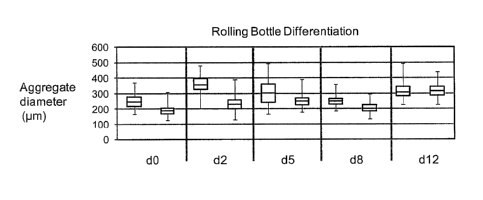
[0059] FIGURE 25 is a chart showing cell aggregate diameters of pluripotent
cells at day zero (d0)
and differentiating cell aggregates at days 2, 5, 8 and 12 (d2, d5, d8 and
d12, respectively). Cell
aggregate sizes were measured and plotted showing the minimum, maximum, 2nd
and 3rd quartile,
and median. Each day shows the plot for cell aggregates formed from 1 x 10⁶
cells/mL (left) and 2 x
106 cells/mL (right).
[0060] FIGURES 26A-D are bar charts showing the expression patterns of the
various indicated
marker genes in rolling bottle vessel format. The left sample of each chart
represents day zero (d0)
cell aggregates formed in 6-well trays (pluripotent cell marker control). The
samples marked by
bars represent (left to right): undifferentiated aggregates at day 0, and
differentiating aggregates at
days 2, 5, 8 and 12. Black bar, rolling bottles at 1 x 10⁶ cells / mL; black
dashed bar, rolling bottles
at 2 x 10⁶ cells/mL; grey bar, 6-well tray.
[0061] FIGURES 27A-D are bar charts showing the expression patterns of the
various indicated
marker genes in larger rolling bottle vessel formats as described in Table 11
and 12 in Example 27.
The left sample represents a 6-well tray hESC aggregation and differentiation
(control; FIG.27A);
Differentiation at days 0, 2, 5, 8 and 12 in vented (V) or not-vented (NV) 490
cm2 roller bottles
(about 1.2L capacity), are shown. Day 2 samples were not collected for the
last 490V sample (far
right column) due to loss of culture. The same dO control was used for each
roller bottle
differentiation (asterisk).
CA 02832194 2013-11-01
DETAILED DESCRIPTION OF THE INVENTION
[0062] In contrast to previously known methods of tissue engineering which are
based on seeding
individual cells into polymer scaffolds, matrices and/or gels, the methods
described herein use cell
aggregate suspensions formed from pluripotent hES single cell suspensions or
hES-derived
(differentiated) single cell suspensions as the building blocks of tissue
formation. Cell aggregates
are often comprised of hundreds to thousands of individual cells, connected
through junctional
adhesions and extracellular matrix that collectively contribute to the final
differentiated product. In
this regard, cell aggregates can be defined as a type of tissue that provides
a number of performance
advantages relative to more traditional engineered tissues.
[0063] In one embodiment of the invention, methods are provided for
producing hES cell
aggregate suspensions from a single cell suspension of pluripotent stem cell
cultures or hES-derived
cell cultures. The pluripotent stem cells can be initially cultured on
fibroblast feeders, or they can
be feeder-free. Methods of isolating hESC and culturing such on human feeder
cells was described
in U.S. Patent No. 7,432,104 entitled METHODS FOR THE CULTURE OF HUMAN
EMBRYONIC STEM CELLS ON HUMAN FEEDER CELLS, which is herein incorporated in
its
entirety by reference. Pluripotent ES cell aggregate suspension cultures made
directly or initiated
from hESCs cultured on feeders avoid the need for making hESC monolayers, for
example, as in
adherent cultures. These methods are described in detail in Examples 17 and
18.
[0064] Other embodiments of the invention provide for methods of producing
cell aggregate
suspensions directly into a differentiation media, e.g., a differentiating
media containing an agent,
preferably a TGF13 family member, which is capable of activating a TGFi3
family of receptor. Such
agents include but are not limited Activin A, Activin B, GDF-8, GDF-11, and
Nodal. Methods of
producing cell aggregate suspension in a differentiation media is
distinguished from other methods,
also described herein, which provide for production of cell aggregate
suspension cultures in a
pluripotent stem cell media, e.g., StemPro.
[0065] Still other embodiments of the invention provide for methods of
producing cell
aggregate suspensions formed from differentiated hES cell cultures (also
referred to as "hES-
derived cell cultures" or "hES-derived cell(s)"), e.g., cells from stages 1,
2, 3, 4 and 5 as described
in D'Amour et al. 2005, supra and D'Amour etal. 2006, Nature Biotech 26 2006:
1392-1401).
Hence, methods for making the cell aggregates described herein are not limited
to any one
16
CA 02832194 2013-11-01
pluripotent or multipotent stage of a hES or hES-derived cell, rather the
manner of use and need for
cell type optimization will dictate which methods are preferred. These methods
are described in
detail in Examples 19-22.
[0066] In another embodiment of the invention, methods are provided for
controlling the
resulting cell composition, e.g., controlling the percentage of pancreatic
endoderm cells, pancreatic
endocrine cells and/or PDX1-endoderm cells, by varying the concentration of
different growth
factors. These methods are described in detail in Example 21.
[0067] Unless otherwise noted, the terms used herein are to be understood
according to
conventional usage by those of ordinary skill in the relevant art. In addition
to the definitions of
terms provided below, definitions of common terms in molecular biology may
also be found in
Rieger et al. 1991 Glossary of genetics: classical and molecular, 5th Ed.,
Berlin: Springer-Verlag;
and in Current Protocols in Molecular Biology, F.M. Ausubel et al. Eds.,
Current Protocols, a joint
venture between Greene Publishing Associates, Inc. and John Wiley & Sons,
Inc., (1998
Supplement). It is to be understood that as used in the specification and in
the claims, "a" or "an"
can mean one or more, depending upon the context in which it is used. Thus,
for example,
reference to "a cell" can mean that at least one cell can be utilized.
[00681 Also, for the purposes of this specification and appended claims,
unless otherwise
indicated, all numbers expressing quantities of ingredients, percentages or
proportions of materials,
reaction conditions, and other numerical values used in the specification and
claims, are to be
understood as being modified in all instances by the term "about".
Accordingly, unless indicated to
the contrary, the numerical parameters set forth in the following
specification and attached claims
are approximations that may vary depending upon the desired properties sought
to be obtained by
the present invention. At the very least, and not as an attempt to limit the
application of the
doctrine of equivalents to the scope of the claims, each numerical parameter
should at least be
construed in light of the number of reported significant digits and by
applying ordinary rounding
techniques.
[0069] "About" as used herein means that a number referred to as "about"
comprises the
recited number plus or minus 1-10% of that recited number. For example,
"about" 100 cells can
mean 95-105 cells or as few as 99-101 cells depending on the situation.
Whenever it appears
herein, a numerical range such as "1 to 20" refers to each integer in the
given range; e.g., "1 to 20
17
CA 02832194 2013-11-01
cells" means 1 cell, 2 cells, 3 cells, etc., up to and including 20 cells.
Where about modifies a
range expressed in non-intergers, it means the recited number plus or minus 1-
10% to the same
=
degree of significant figures expressed. For example, about 1.50 to 2.50 mM
can mean as litte as
1.35 M or as much as 2.75M or any amount in between in increments of 0.01.
[0070] The present invention provides methods for production of
hES-derived cell aggregates
from hES-derived single cell suspensions. Because various mechanical and non-
physiological
factors effect movement and aggregation of cells in culture, the fluid
mechanical micro-
environment that correlates with optimal cell aggregate viability and
performance, as well as to
provide a normalizing variable that can be used for scale-up, it was necessary
to characterize the
movement of cells growing or differentiating in various culture vessels,
dishes, Erlenmeyer flasks,
bioreactors, bottles and the like and the effects, if any, of various media
conditions on the cells.
Some of these factors include but are not limited to, shear rate and shear
stress, cell density and
concentration of various growth factors in any cell medium.
[0071] Shear rate and shear stress are mechanical characteristics
that define the fluid shear
within a system. Shear rate is defined as the fluid velocity over a given
distance and is expressed as
sec* Shear rate is proportional to shear stress where shear rate (0) = shear
stress (t)/viscosity ( ).
Shear stress is defined as the fluid shear force acting tangentially to the
cell surface and is expressed
as force per unit area (dyne/cm2 or N/m2). Shear stress can be generated by
agitated liquid moving
past static cells, agitated cells moving through static liquid or by cells
moving within an agitated,
dynamic fluid environment. Fluid viscosity is typically measured in poise
where 1 poise=1 dyne
sec/cm2 = 100 centipoise (cp). The viscosity of water, one of the least
viscous fluids known, is 0.01
cp. The viscosity of a typical suspension of eukaryotic cells in media is
between 1.0 and 1.1 cp at a
temperature of 25 C. Both density and temperature can affect the viscosity of
a fluid.
[0072] Fluid velocity also dictates whether the flow will be
laminar or turbulent. Laminar flow
occurs when viscous forces dominate and is characterized by smooth, even
streamlines at low
velocities. In contrast, high velocity and inertial forces dominate during
turbulent flow, which is
characterized by the appearance of eddies, vortices and chaotic fluctuations
in the flow across space
and time. A dimensionless value known as the Reynold's number (Re) is
typically used to quantify
the presence of laminar or turbulent flow. The Reynold's number is the ratio
of inertial to viscous
forces and is quantitated as (density*velocity*length scale)/(viscosity).
Laminar flow dominates
with Re<2300 while turbulent flow dominates when Re>4000. Based on this
relationship with fluid
18
CA 02832194 2013-11-01
velocity, the Reynold's number and thus the degree to which fluid flow is
laminar or turbulent is
directly proportional to the shear rate and shear stress experienced by cells
in suspension.
However, high shear stress conditions can be generated in both laminar and
turbulent fluid
environments. Initially, there is a tendency for liquid to resist movement,
with the fluid closest to a
solid surface experiencing attractive forces that generate a boundary layer or
a region of no-flow
immediately adjacent to the surface. This creates a gradient in fluid velocity
from the surface to the
center of the fluid flow. The steepness of the velocity gradient is a function
of the speed at which
the liquid is moving and distance from the boundary layer to the region of
highest fluid velocity. As
the liquid flow rate through or around a container accelerates, the velocity
of the flow overcomes
the viscosity of the liquid and the smooth, laminar gradient breaks down
producing turbulent flow.
Thomas et at. showed that cell lysis under turbulent conditions occurs most
frequently in regions of
locally high shear stress and high energy dissipation rates. See Thomas etal.
(1994)
Cytotechnology 15: 329-335. These regions appear randomly but are often found
near the boundary
layer where the velocity gradient is highest. These random fluctuations in
fluid velocity can
generate regions of very high shear stress that ultimately can have a negative
effect on the scale-up
of cell culture-based manufacturing systems. Thus, a need exists for methods
that can maintain cell
density and viability in a mammalian cell culture manufacturing scale-up
system by controlling the
major sources of shear forces in such systems.
[0073] Following methods provided by Henzler (Henzler, 2000, Particle
stress in bioreactors,
In Advances in Biochemical Engineering/Biotechnology, Scheper, T. Ed. Springer-
Verlag, Berlin)
and Colomer etal. (Colomer, J. etal. 2005. Experimental analysis of
coagulation of particles under
low-shear flow. Water Res. 39:2994), fluid mechanical properties of the bulk
fluid in a rotating 6-
well dish were calculated. The Dimensionless Stress is equal to the turbulence
constant*(aggregate
diameter/Kolmogorov's Microscale)"turbulence exponent. Shear Stress is equal
to the
Dimensionless Stress*fluid density*(kinematic viscosity*power input)^0.5.
Shear Rate is equal the
Shear Stress/kinematic viscosity. For calculation of the power input and
Kolmogorov's Microscale,
the Reynold's number is required at each rotation rate and is equal to the
(rotation rate*flask
diameter)^2/viscosity. As both the power input and Kolmogorov's Microscale are
functions of the
Reynold's Number, all shear stress and shear rate calculations vary with
rotation rate.
[0074] Moreover, shear stress and shear rate are functions of the
Dimensionless Stress, which
depends on the diameter of forming aggregates, thus the shear stress and rate
experienced by
19
CA 02832194 2013-11-01
aggregates is expected to increase with time in rotation. Example calculations
are shown in
Example 17 for aggregate diameters between 100-200p.m and rotation speeds
between 60-140 rpm.
These methods were used to provide an estimation of the average shear in the
bulk fluid over time.
However, it is expected that the shear stress at the wall of the vessel will
be the highest due to
boundary effects. To estimate wall shear stress, Ley et al. proposed that wall
shear stress in a 6-well
dish is equal to the radius of gyration*(density*dynamic
viscosity*(2*pi*rotation rate)^3)^0.5.
Using this approach, the wall shear stress was calculated for rotation speeds
ranging from 60rpm to
140rpm and is shown in Example 18. Note that, unlike the time-averaged shear
stress that is
experienced by aggregates in the bulk fluid, the shear stress occurring at the
wall is independent of
aggregate diameter.
100751 Culture cell density is also a factor critical to the tissue
function and is difficult to
achieve and/or optimize in traditional tissue which are 2-dimensional (e.g.,
adherent engineered
constructs). The effect of cell density on differentiation is described in
more detail in Example 20.
Cell aggregates may overcome this limitation by assuming an organized 3-
dimensional (3D)
architecture that more accurately reflects an in vivo cellular density and
conformation. As a result,
the period of time for the cells to achieve their intended structure can be
significantly reduced
and/or made more consistent and efficient. Moreover, cells in the 3D aggregate
format may
differentiate and function more optimally, as this architecture more closely
resembles normal
physiology than adherent cultures. In addition, the mechanical hardship
involved in the
manufacturing process is less damaging to cell aggregates that are free-
floating in suspension
culture as compared to the mechanical hardship, for example, in an adherent
culture.
100761 Typical manufacturing-scale suspension culture also utilizes
continuous perfusion of
= media as a method for maintaining cell viability while maximizing cell
density. In this context,
media exchange contributes fluid shear to the culture affecting adherent cells
and suspended
aggregates differently. Immobile adherent cells are subject to fluid shear
stress as the media flows
tangentially across the cell surface. In contrast, suspended aggregates
experience significantly less
shear stress across the aggregate surface, as aggregates are free to tumble in
response to applied
shear force. It is expected that prolonged shear stress will be detrimental to
adherent ES cells and
that the suspended aggregate format is preferred for optimal survival and
function. Thus based on a
need for an efficient manufacturing process for production of pluripotent stem
cells and/or
multipotent progenitor cells derived from pluripotent stem cells and the above
observed mechanics
CA 02832194 2013-11-01
relating to shear rate and shear stress, the present invention provides for
the first time methods of
manufacturing for production of pluripotent stem cells and/or multipotent
progenitor cells derived
from pluripotent stem cells in suspension format, in particular, cell
aggregate suspension format.
[0077] As used herein, "single cell suspension" or equivalents thereof
refers to a hES cell
single cell suspension or a hES-derived single cell suspension by any
mechanical or chemical
means. Several methods exist for dissociating cell clusters to form single
cell suspensions from
primary tissues, attached cells in culture, and aggregates, e.g., physical
forces (mechanical
dissociation such as cell scraper, trituration through a narrow bore pipette,
fine needle aspiration,
vortex disaggregation and forced filtration through a fine nylon or stainless
steel mesh), enzymes
(enzymatic dissociation such as trypsin, collagenase, Acutase and the like),
or a combination of
both. Further, methods and culture media conditions capable of supporting
single-cell dissociation
of hESC is useful for expansion, cell sorting, and defined seeding for multi-
well plate assays and
enable automatization of culture procedures and clonal expansion. Thus, one
embodiment of the
invention provides methods for generating a stable single-cell enzymatic
dissociation hES cell or
hES-derived cell culture system capable of supporting long-term maintenance
and efficient
expansion of undifferentiated, pluripotent hES cell or differentiated hESC.
[0078] As used herein, "roller bottle" or "rolling bottle" or equivalents
thereof refers to a
cylindrical container adapted to rotate about its axes. These containers
include but are not limited
to roller bottles sold through Corning, Fisher Scientific, and other
manufacturers, as well as drums,
barrels, and other bottle type containers capable of being rotated on its side
wall, for example.
Roller bottles described herein do not have to be cylindrical or have a
circular cross-section. They
can be non-circular, closed curve, of constant width, for example. In one
embodiment, the curve is
a Reuleaux triangle or a Reuleaux triangle with rounded corners as described
in U.S. Patent No.
5,866,419, which is incorporated herein by reference in its entirety. Circular
cross-section roller
bottles are not the only shape or geometry to provide smooth rotation because
an infinite number of
such curves exist and are contemplated by the invention. Hover, such curves
are not generally
encountered in industry because most machinery used for rotating bottles
requires that the
horizontal axis running perpendicular to the curve remain in a fixed location,
which it does not for
non-circular rollers because that have axes with a back-and-forth translation
motion while rolling.
This additional motion or rotation, in addition to the usual circular motion
as in other cylindrical
roller bottles can enhance gas exchange as compared to circular cross-section
type roller bottles.
21
CA 02832194 2013-11-01
[0079] A typical cylindrical roller bottle includes a bottom wall, a top
wall and a cylindrical
side wall extending between the bottom and top walls. The top wall includes an
opening to provide
access to the interior of the roller bottle. The internal surfaces of such
roller bottles provide active
surfaces for cell interaction and/or attachment. Hence, the Oxford Dictionary
of Biochemistry
provides that roller bottles are cylindrical containers used for the culture
of monolayers of adherent
cells. Indeed, roller bottles are desirable for growing large amounts of
cells, such as adherent cells,
or for producing cell by-products, such as pharmaceutical substances that are
secreted by cells. The
cylindrical side wall of roller bottles can be smooth or patterned, whereby
patterning extends
substantially from the bottom wall to the top wall for increasing cell growth
surface area and for
facilitating the flow of liquid to all interior surface areas of the bottle
when the bottle is rolled about
the axis of the side wall.
[0080] Independent of the cross-section of the roller bottle (circular or
non-circular) liquid
growth medium is introduced into and contained within a roller bottle. The
rotating movement of
the bottle keeps the internal surfaces wetted with the liquid medium, thereby
encouraging the
growth of cells. Rotating rollers of an appropriate apparatus are employed to
rotate roller bottles of
the invention.
[0081] Roller bottles are usually constructed of either glass, stainless
steel or a clear plastic,
such as polystyrene, polyurethane, polyvinyl chloride, polycarbonate,
polyolefins such as
polypropylene, polyethylene terephthalate with glycol additives, ethylene
glycol- 1,4, cyclohexane
dimethanol terephthalate copolyester and the like. Transparent materials are
preferred, as cell
growth can be monitored by placing the bottle on an inverted microscope.
[0082] Manual and automated roller bottle systems have been used for over
40 years in the
pharmaceutical, biochemical, and medical fields for processes such as cell
growth and infection,
heterologous glycoprotein production, vaccine preparation, and high density
plant cell cultivation.
See Tanaka etal. 1983, Biotechnol. Bioeng. 25:2359; Tanaka 1987, Process
Biochem. Aug., 106;
Hong, et at. 1989, BiotechnoL Frog. 5:137; Elliot 1990, Bioprocess Tech.
10:207; Tsao 1992,
Annals N.Y. Acad. Sci. 665:127; Pennell & Milstein 1992, J. of Immun. Meth.
146:43; Olivas etal.,
1995, Immun. Meth. 182, 73 (1995); Singhvi etal. 1996, Cytotechnology 22:79;
and Kunitake et
aL 1997, Biotechnology 52:3289 , which are incorporated herein by reference in
their entireties.
Additionally, for industrial scale production of cell culture products (i.e.
vaccines), cells are
frequently passaged in roller bottles prior to transfer to micro-carrier
cultures for a final growth
22
CA 02832194 2013-11-01
phase even when unit operation based systems are utilized. See Edy, 1984, Adv.
Exp. Med. Biol.
172:169, which is herein incorporated by reference in its entirety.
[0083] To date, widespread use of roller bottles for culturing adherent
cells can be attributed to
several factors. The process relies on: (i) a horizontal cylindrical vessel
containing a sufficient
volume of media or fluid and axially rotated.; because roller bottle scale up
is a function of length,
scale-up development or invention is not required, resulting in reduced
developmental timelines for
industry and faster introduction to market for new products; (ii) roller
bottle systems allow for
constant fluid-gas contact, i.e. due to the axial rotation there is at all
times at least a thin layer of
fluid or media coating the inner surface of the bottle as it rotates; this
layer allows for increased
fluid-gas exchange and the as the bottle rotates that gas returns to the cells
which are in the pool of
media at the bottom of the roller bottle; (iii) maintaining sterile conditions
for prolonged times in
large scale culture is possible because contamination of one or more roller
bottles does not result in
contamination of an entire lot; (iv) precise control of nutrient and waste-
product levels is possible;
and (v) direct monitoring of the cells, e.g. identification of certain cell
markers to ensure efficient
differentiation and proper specification of cells after stages 1-4 for example
is relatively simple.
[0084] While not wanting to be limited to use of roller bottle or roller
type vessels for culturing
three-dimensional cell aggregates, it is intended that there are other means
for making the cell
aggregates of the invention, although not employing the motion created by a
roller bottle or a
cylindrical type of vessel rotating on a drum, for example. The type of motion
used to aggregate
pPSCs in general can be produced, for example, by aerosolizing the vessel or
chamber to produce a
more laminar flow. The motion can also be created by having an inlet and an
outlet port to assist
the inflow and outflow of the fluid medium, or even the cells themselves, to
create motion similar
to that achieved with the roller bottles described herein. The motion can also
be achieved with the
use of one or more or a combination of flow distributors. For example, such a
flow distributor may
include a baffle to distribute the flow of fluid or medium within the chamber
and thereby create a
continuous, uniform mixture of the three-dimensional cell aggregates. In
another example, the flow
distributor may be combination of one or more deflector plates, distribution
channels, and/or flow
channels, which create fluid movement similar to that found in roller bottles
without necessarily
occurring in a roller bottle type, cylindrical vessel or chamber. Thus,
alternative means of creating
fluid movement in a manner that is non-turbulent, yet generates sufficient low
shear force to
23
CA 02832194 2013-11-01
promote cell collision and allow the cells to adhere to each other and form
the cell aggregates as
described herein.
[0085] Still, certain properties of growing adherent or anchorage dependent
cells in roller
bottles have their disadvantages. For example, adherent cell growth by its
nature requires
substantial surface area for the cell to attach to and roller bottles are
limited in surface area that is
available for growth. The conventional method of mixing in roller bottles is
rotation at a uniform
rate in one direction for all purposes e.g. cell planting or seeding, cell
growth and/or virus
propagation and expansion. Standard rotation frequencies of most roller bottle
processes for
culturing adherent and anchorage dependent cells is about 0.125 rpm to 5 rpm.
For these cultures, it
is important that the cells come into contact with the sides of the roller
bottle as rapidly as possible,
since only after attachment to the vessel wall can the cells subsequently
proliferate and form cell
sheets. Slow cell attachment to the inner walls of the vessel leads to low
viability of the cells and/or
inhomogeneous planting, and hence inhomogeneous growth on the roller bottle
surface. Moreover,
inefficient mixing limits cell growth because the cells do not obtain adequate
nutrients (e.g. oxygen)
or adequate removal of toxins (e.g. carbon dioxide) from a submerged, surface-
attached cell sheet
as the bottle rotates. Interestingly, these disadvantages are not critical to
using roller bottles for
aggregation, growth, expansion and differentiation of differentiable
pluripotent cells in suspension.
[0086] In view of the properties described above and further in view
Applicant's own
disclosure of methods for making hES cell aggregates in 6-well trays and the
like, one of ordinary
skill in the art would not turn to use of roller bottles for making
pluripotent stem cell aggregates.
See Schulz et al. 2012, Stem Cells 7: 1-17, e37004, and U.S. Patent Nos.
8,153,429 and 8,008,075,
which are incorporated herein by reference in their entireties. For example,
Schulz et al. 2012,
supra, teaches that pluripotent stem cells can be effectively aggregated by
using a circular or radial
movement or motion or rotation, which is imposed over a central vortex and
draws cells into a
higher local density in the middle of the culture vessel, e.g. drawing cells
into the center of a well of
a 6-well tray, or the center of Erlenmeyer flask or the center of a bioreactor
based on a rotational
format. This radial vortex cannot be accomplished in roller bottles because by
its nature the roller
bottle rotates on its side wall and not on its base, hence it is not intuitive
to transfer methods from a
system that includes a central vortex motion to one that does not, such as the
roller bottles as
described herein.
24
CA 02832194 2013-11-01
[0087] Applicants have performed studies of static cultures using other
types of motion
including studies rocking, stirring and centrifugation of hES cells, and these
types of motions were
incapable of allowing the formation of hES cell aggregates or differentiable
cell aggregates.
Further, these hES or hES cell-derived aggregates that did form under these
conditions did not give
rise to functioning glucose responsive cell types in vivo, which is the
ultimate test of any method for
successful manufacturing of PEC. See at least Kroon etal. 2008, supra and
Schulz etal. (2012)
supra. So, it cannot be said that just movement and motion alone is sufficient
to form pluripotent
stem cell or hES cell suspension aggregates or differentiable cell aggregates,
because it is not.
These studies (data not shown) indicated that more than just fluid movement
and forces generated
with such movement facilitate the adhesive contact necessary for cell
aggregate formation that
results in the transitioning of single-cell pluripotent stem cells to stable
cell-cell aggregates.
[0088] As mentioned briefly above, rotation of a roller bottle is very
different from rotation of
a 6-well tray, Erlenmeyer flasks, and the like which occurs about a central
vortex. In a roller bottle,
the majority of the culture volume remains at the bottom of the bottle when
the bottle rotates on its
side wall, and a thin layer of fluid or culture medium coats the inner bottle
surface as the bottle
rotates. This thin fluid layer has increased gas exchange as the bottle
rotates and therefore increases
02 levels to the culture medium overall; i.e. once the thin layer of culture
media returns to the
bottom of the bottle where the majority of the culture medium resides, it
carries with it increases
amounts of 02 It is not intuitive then that this motion, especially when
rotated at very low speeds
that are standard in the art for adherent cells (e.g. 0.125 to 5rpm) would
allow for sufficient cell-to-
cell contact or collisions while at the same time maintain the low shear-force
sufficient to allow
primate pluripotent stem cell (pPSC) aggregate formation, let alone
differentiation of differentiable
cell aggregates.
[0089] Using roller bottles to aggregate, grow, passage, expand and
differentiate cells is also
different from 6-well trays, Erlenmeyer flasks, bioreactors because of the
different rotations speeds
between the two formats. 6-well trays, Erlenmeyer flasks, bioreactors and the
like for example use
higher rotation speeds of about 80, 85, 90, 95, 100, 105, 110, 115 and 120
rpm, which are required
for at least the purpose of preventing the cell aggregates from agglomerating
or forming clusters or
larger cell masses in culture. Note, that the agglomerated cell clusters
(e.g., large aggregates of 300
vim or more) are not to be confused with the roughly spherical cell
aggregates, which are smaller
(about 100 ¨ 200 p.m) and uniform in size. In contrast, the aggregation,
growth, passaging,
CA 02832194 2013-11-01
expansion and differentiation of pPSCs in roller bottles is performed at
relatively low rotations
speeds of about 3, 4, 5, 6, 7, 8, 9, and 10 rpms. These lower rotation speeds
do not create the same
degree of shear force which occurrs in 6-well trays, Erlenmeyer flasks,
bioreactors and the like, and
in view of Applicant's previous experience (see Schulz etal. 2012, supra), it
was not expected that
cell aggregate formation would succeed under these conditions.
[0090] An advantage of using roller bottles to aggregate, grow, passage,
expand and
differentiate pluripotent stem cells over that of other cell culture vessels
is that once optimized in
the smallest roller bottle, the methodologies will work very similarly in
larger bottles without
additional substantial invention. For example, by using longer bottles with
the same standard cross-
section, but substantially larger capacity, or by using arrays of bottles,
total culture mass can be
scaled using the same bottle diameter, diameter/volume ratio and rotation
speed. An increase in
roller bottle length (scaling) does not affect the cell aggregation or
differentiation processes. So,
scaling of the cell process or manufacture from 490 cm2 roller bottles (11.12
cm in diameter, 17.30
cm in length including cap) to 850 cm2 roller bottles (11.63 cm in diameter,
27.36 cm in length
including cap) to 1750 cm2 roller bottles (11.73 cm in diameter, 53.16 cm in
length including cap)
or greater does not involve substantially or significantly modification other
than that described
herein.
[0091] For at least the above reasons, scalability of cell manufacturing in
roller bottles is
different from the rotational platform systems of 6-well trays, Erlenmeyer
flasks, bioreactors and
the like. For example, in order to achieve a IL pluripotent stem culture of 1
x 106 cells! mL, about
thirty (30) 6-well trays would be required. Stated in another way, instead of
using eighty (80) 6-
well trays (total 480 wells), the skilled artisan would only need 4, 850cm2,
roller bottles. The
skilled artisan will appreciate that less manipulation and labor that is
needed for roller bottle culture
is an improvement in manufacturing. In addition, adjustments must be made in
volume, speed and
rotational radius in order to achieve aggregation when using rotational
platforms at different scales.
Primate PSC aggregation in conical flasks can be achieved, for example, but
occurs best at
relatively high rotation speeds, about 150 rpm, and this causes too much
turbulence and shear-force
leading to increased cell death (data not shown). Merely placing a bottle or
jar on a rocking
platform does not produce the herein described cell suspension aggregates
(data not shown). The
rocking motion does not create a suitable fluid motion to support appropriate
cell-cell contact and
adherence, and potentially creates too much turbulence and shear-force,
causing increased cell
26
CA 02832194 2013-11-01
death. Similarly, square shaped bottles and 15cm glass jars are unsuitable
culture vessels to scale
up pPSC aggregates formation for similar reasons (data not shown). Further,
mere cell-to-cell
contact alone does not cause pPSC aggregates to form because when cell pellets
are recovered after
single-cell suspensions of hES cells are centrifuged, cell aggregates were not
observed (data not
shown). Thus, discovering a truly scalable system with appropriate fluid
motion that supports
efficient and consistent cell aggregation (including consistent aggregate
diameter) has not been at
all straight forward or routine, and substantially more difficult than a
skilled artisan would
anticipate or expect. In fact, it has been surprising that roller bottles,
which have been traditionally
used for large scale-up cultures of adherent and anchorage dependent cell
types, coupled with up to
a 30-fold reduction in speed would provide suitable conditions for production
of pPSC aggregates
and differentiation as herein described at all.
[0092] As used herein, the term "contacting" (i.e., contacting a cell e.g.,
a differentiable cell,
with a compound) is intended to include incubating the compound and the cell
together in vitro
(e.g., adding the compound to cells in culture). The term "contacting" is not
intended to include the
in vivo exposure of cells to a defined cell medium comprising an ErbB3 ligand,
and optionally, a
member of the TGF-13 family, that may occur naturally in a subject (i.e.,
exposure that may occur as
a result of a natural physiological process). The step of contacting the cell
with a defined cell
medium comprising an ErbB3 ligand, and optionally, a member of the TGF-I3
family, can be
conducted in any suitable manner. For example, the cells may be treated in
adherent culture, or in
suspension culture. It is understood that the cells contacted with the defined
medium can be further
treated with a cell differentiation environment to stabilize the cells, or to
differentiate the cells.
[0093] As used herein, the term "differentiate" refers to the production of
a cell type that is
more differentiated than the cell type from which it is derived. The term
therefore encompasses cell
types that are partially and terminally differentiated. Differentiated cells
derived from hESC are
generally referred to as hES-derived cells or hES-derived cell aggregate
cultures, or hES-derived
single cell suspensions, or hES-derived cell adherent cultures and the like.
[0094] As used herein, the term "substantially" refers to a great extent or
degree, e.g.
"substantially similar" in context is used to describe one method which is to
great extent or degree
similar to or different than another method. However, as used herein, the term
"substantially free",
e.g., "substantially free" or "substantially free from contaminants," or
"substantially free of serum"
or "substantially free of insulin or insulin like growth factor" or
equivalents thereof, means that the
27
CA 02832194 2013-11-01
solution, media, supplement, excipient and the like, is at least 98%, or at
least 98.5%, or at last
99%, or at last 99.5%, or at least 100% free of serum, contaminants or
equivalent thereof. In one
embodiment, there is provided a defined culture media with no serum, or 100%
serum-free, or
substantially free of serum. Conversely, as used herein, the term
"substantially similar" or
equivalents thereof means that the composition, process, method, solution,
media, supplement,
excipient and the like is at least 80%, at least 85%, at least 90%, at least
95%, or at least 99%
similar to that previously described in the specification herein, or in a
previously described process
or method incorporated herein in its entirety.
[0095] In certain embodiments of the present invention, the term "enriched"
refers to a cell
culture that contains more than approximately 50%, 55%, 60%, 65%, 70%, 75%,
80%, 85%, 90%,
or 95% of the desired cell lineage.
[0096] As used herein, the term "effective amount" or equivalents thereof
of a compound
refers to that concentration of the compound that is sufficient in the
presence of the remaining
components of the defined medium to effect the stabilization of the
differentiable cell in culture for
greater than one month in the absence of a feeder cell and in the absence of
serum or serum
replacement. This concentration is readily determined by one of ordinary skill
in the art.
[0097] As used herein, the term "express" refers to the transcription of a
polynucleotide or
translation of a polypeptide in a cell, such that levels of the molecule are
measurably higher in a cell
that expresses the molecule than they are in a cell that does not express the
molecule. Methods to
measure the expression of a molecule are well known to those of ordinary skill
in the art, and
include without limitation, Northern blotting, RT-PCR, in situ hybridization,
Western blotting, and
immunostaining.
[0098] As used herein when referring to a cell, cell line, cell culture or
population of cells, the
term "isolated" refers to being substantially separated from the natural
source of the cells such that
the cell, cell line, cell culture, or population of cells are capable of being
cultured in vitro. In
addition, the term "isolating" is used to refer to the physical selection of
one or more cells out of a
group of two or more cells, wherein the cells are selected based on cell
morphology and/or the
expression of various markers.
[0099] The present invention may be understood more readily by reference to
the following
detailed description of the preferred embodiments of the invention and the
Examples included
28
CA 02832194 2013-11-01
herein. However, before the present compositions and methods are disclosed and
described, it is to
be understood that this invention is not limited to specific nucleic acids,
specific polypeptides,
specific cell types, specific host cells, specific conditions, or specific
methods, etc., as such may, of
course, vary, and the numerous modifications and variations therein will be
apparent to those
skilled in the art.
101001 Standard techniques for cloning, DNA isolation, amplification and
purification, for
enzymatic reactions involving DNA ligase, DNA polymerase, restriction
endonucleases and the
like, and various separation techniques are those known and commonly employed
by those skilled
in the art. A number of standard techniques are described in Sambrook et al.
1989 "Molecular
Cloning", Second Edition, Cold Spring Harbor Laboratory, Plainview, New York;
Maniatis et al.
1982 "Molecular Cloning", Cold Spring Harbor Laboratory, Plainview, New York;
Wu (ed.) 1993
Meth. Enzymol. 218, Part I; Wu (ed.) 1979 Meth. Enzymol. 68; Wu etal. (eds.)
1983 Meth.
Enzymol. 100 and 101; Grossman and Moldave (eds.) 1980 Meth. Enzymol. 65;
Miller (ed.) 1972
"Experiments in Molecular Genetics", Cold Spring Harbor Laboratory, Cold
Spring Harbor, New
York; Old and Primrose, 1981 "Principles of Gene Manipulation", University of
California Press,
Berkeley; Schleif & Wensink, 1982 "Practical Methods in Molecular Biology";
Glover (ed.) 1985
"DNA Cloning" Vol. I and II, IRL Press, Oxford, UK; Hames and Higgins (eds.)
1985 "Nucleic
Acid Hybridization", IRL Press, Oxford, UK; and Setlow and Hollaender 1979
"Genetic
Engineering: Principles and Methods", Vols. 1-4, Plenum Press, New York.
Abbreviations and
nomenclature, where employed, are deemed standard in the field and commonly
used in
professional journals such as those cited herein.
[0101] The invention relates to compositions and methods comprising a basal
salt nutrient solution
and an effective amount of an ErbB3 ligand, with the compositions being
essentially free of serum.
The compositions and methods of the present invention are useful for culturing
cells, in particular,
differentiable cells. It is understood that at different points during
culturing the differentiable cells,
various components may be added to the cell culture such that the medium can
contain components
other than those described herein. It is, however, contemplated that at least
at one point during the
preparation of the culture, or during the culture of the differentiable cells,
the defined medium
comprises a basal salt nutrient solution and a means for activating ErbB2-
directed tyrosine kinase.
[0102] Although a basal salt nutrient solution as described herein is employed
to maintain cell
growth and viability of hESC, in other embodiments of the invention,
alternative stem cell culture
29
CA 02832194 2013-11-01
medias to maintain pluripotency or for differentiation of the pluripotent
cells, work in substantially
similar means, including but not limited to KSR (Invitrogen), or xeno-free KSR
(Invitrogen),
StemPro (Invitrogen), mTeSRTml (StemCell Technologies) and HEScGRO
(Millipore), DMEM
based media, and the like.
[0103] In another embodiment, hESC are cultured in the defined media described
herein in the
absence and/or presence of extracellular matrix proteins (ECM), e.g.,
MATRIGEL. Human ES
cells cultured in the absence of ECM contain about 0.5 to 10% human serum (hS)
or hS retentate
fractions from a 300K and/or 100K cut-off spin column (Microcon). The hES cell
aggregate
suspensions can be produced by directly incubating the hESC into the media
containing hS or hS
retentate fractions; or after incubating the culture vessels with the hS or hS
retentate fractions for
about 30 min., 1 hour, 2 hours, 3 hours, 4, hours, 5 hours, 6 hours, 12 hours,
and 24 hours at 37 C.
The plating efficiency for the hESC in the hS or hS retentate fraction
containing media was
comparable to that observed in hESC cultured in DC-HAIF as described in
PCT/US2007/062755,
or cultured in DC-HAIF media using MATRIGELTm as an ECM, or other similar
matrices.
Methods for culturing hESC in a defined media substantially free of serum is
described in U.S.
Patent Application No. 11/8875,057, filed October 19, 2007, entitled METHODS
AND
COMPOSITIONS FOR FEEDER-FREE PLURIPOTENT STEM CELL MEDIA CONTAINING
HUMAN SERUM, which is herein incorporated in its entirety by reference.
[0104] Still in another embodiment, hES cell aggregate suspensions were
cultured in a media
substantially free of serum and further in the absence of exogenously added
fibroblast growth factor
(FGF). This is distinguished from U.S. Patent No. 7,005,252 (Thomson), which
requires culturing
hESC in a media without serum but containing exogenously added growth factors,
including FGF.
[0105] Cellular regulation can be effected through the transduction of
extracellular signals across
the membrane that, in turn, modulates biochemical pathways within the cell.
Protein
phosphorylation represents one course by which intracellular signals are
propagated from molecule
to molecule resulting finally in a cellular response. These signal
transduction cascades are highly
regulated and often overlapping as evidenced by the existence of many protein
kinases as well as
phosphatases. It has been reported that in humans, protein tyrosine kinases
are known to have a
significant role in the development of many disease states including diabetes,
cancer and have also
been linked to a wide variety of congenital syndromes. Serine threonine
kinases, e.g., Rho kinases,
are a class of enzymes, which if inhibited can have relevance to the treatment
of human disease,
CA 02832194 2013-11-01
including diabetes, cancer, and a variety of inflammatory cardiovascular
disorders and AIDS. The
majority of inhibitors identified/designed to date act at the ATP-binding
site. Such ATP-
competitive inhibitors have demonstrated selectivity by virtue of their
ability to target the more
poorly conserved areas of the ATP-binding site.
[0106] The Rho kinase family of small GTP binding proteins contains at least
10 members
including Rho A-E and G, Rac 1 and 2, Cdc42, and TC10. The inhibitors are
often referred to as
ROK or ROCK inhibitors, and they are used interchangeably herein. The effector
domains of
RhoA, RhoB, and RhoC have the same amino acid sequence and appear to have
similar
intracellular targets. Rho kinase operates as a primary downstream mediator of
Rho and exists as
two isoforms: a (ROCK2) and f3 (ROCK1). Rho kinase family proteins have a
catalytic (kinase)
domain in their N-terminal domain, a coiled-coil domain in their middle
portion, and a putative
pleckstrin-homology (PH) domain in their C-terminal domain. The Rho-binding
domain of ROCK
is localized in the C-terminal portion of the coiled-coil domain and the
binding the GTP-bound
form of Rho results in enhancement of kinase activity. The Rho/Rho-kinase-
mediated pathway
plays an important role in the signal transduction initiated by many agonists,
including angiotensin
II, serotonin, thrombin, endothelin-1, norepinephrine, platelet-derived growth
factor, ATP/ADP and
extracellular nucleotides, and urotensin II. Through the modulation of its
target effectors/substrates
Rho kinase plays an important role in various cellular functions including
smooth muscle
contraction, actin cytoskeleton organization, cell adhesion and motility and
gene expression. By
virtue of the role that Rho kinase protein play in mediating a number of
cellular functions perceived
to be associated with the pathogenesis of arteriosclerosis, inhibitors of this
kinase may also be
useful for the treatment or prevention of various arteriosclerotic
cardiovascular diseases and
involved in endothelial contraction and enhancement of endothelial
permeability which is thought
to progress to atherosclerosis. Hence, in other embodiments of the invention,
agents which promote
and/or support cell survival are added to various cell culture media, for
example, Rho-kinase
inhibitors Y-27632, Fasudil, and H-1 152P and ITS
(insulin/transferrin/selenium; Gibco). These cell
survival agents function, in part, by promoting re-association of dissociated
hES cell or hES-
derived cultures, e.g., foregut endoderm, pancreatic endoderm, pancreatic
epithelium, pancreatic
progenitor populations and the like, particularly dissociated pancreatic
endoderm and pancreatic
progenitor populations. Increase in survival of hES or hES-derived cells was
achieved
independently of whether the cells were produced from cell aggregates in
suspension or from
adherent plate cultures (with or with no extracellular matrix, with or without
serum, with or without
31
CA 02832194 2013-11-01
feeders). Increase in survival of these cell populations facilitates and
improves purification systems
using a cell-sorter and, therefore allows improved recovery of the cells. Use
of Rho kinase
inhibitors such as Y27632 may allow for expansion of hES-derived cell types as
well by promoting
their survival during serial passaging dissociated single cells or from
cryogenic preservation.
Although, Rho kinase inhibitors such as Y27632 have been tested on hES and hES-
derived cell
cultures, Rho kinase inhibitors can be applied to other cell types, for
example, in general, epithelial
cells including but not limited to intestinal, lung, thymus, kidney as well as
neural cell types like
pigmented retinal epithelium.
[0107] As used herein, the term "differentiable cell" is used to describe a
cell or population of cells
that can differentiate into at least partially mature cells, or that can
participate in the differentiation
of cells, e.g., fuse with other cells, that can differentiate into at least
partially mature cells. As used
herein, "partially mature cells", "progenitor cells", "immature cells",
"precursor cells", "multipotent
cells" or equivalents thereof and also includes those cells which are
terminally differentiated, e.g.,
definitive endoderm cells, PDX 1-negative foregut endoderm cells, PDX I-
positive pancreatic
endoderm cells which further include PDX1-positive pre-pancreatic endoderm
cells and PDX1-
positive pancreatic endoderm tip cells. All are cells that exhibit at least
one characteristic of the
phenotype, such as morphology or protein expression, of a mature cell from the
same organ or
tissue but can further differentiate into at least one other cell type. For
example, a normal, mature
hepatocyte typically expresses such proteins as albumin, fibrinogen, alpha-l-
antitrypsin,
prothrombin clotting factors, transferrin, and detoxification enzymes such as
the cytochrome P-
450s, among others. Thus, as defined in the present invention, a "partially
mature hepatocyte" may
express albumin or another one or more proteins, or begin to take the
appearance or function of a
normal, mature hepatocyte.
[0108] In contrast to cell aggregates produced by previously known methods
that may vary in both
size and shape, the cell aggregates and methods described herein have a narrow
size and shape
distribution, i.e., the cell aggregates are substantially uniform in size
and/or shape. The size
uniformity of the cell aggregates is critical for differentiation performance
and homogeneity of the
culture. Applying basic mass transport analysis to the aggregates, it is
expected that diffusion of
oxygen and nutrients into the center of large aggregates will be slow compared
to diffusion into
smaller aggregates, assuming equal permeability. As differentiation of
aggregated ES cells into
pancreatic lineage cells is dependent on the temporal application of specific
growth factors, a
32
CA 02832194 2013-11-01
culture with a mixture of aggregates of different diameters is likely to be de-
synchronized as
compared to a uniform (size and shape) culture of cell aggregates. This
mixture of cell aggregates
gives rise to heterogeneity and may result in poor differentiation performance
and ultimately not
lend itself to being amenable to manufacturing, scale-up, and production. The
cell aggregates used
herein can be of various shapes, such as, for example, a sphere, a cylinder
(preferably with equal
height and diameter), or rod-like among others. Although other shaped
aggregates may be used, in
one embodiment of the invention, it is generally preferable that the cell
aggregates be spherical or
cylindrical. In another embodiment, the cell aggregates are spherical and
substantially uniform in
size and shape. For instance, if the cell aggregates differ in size or are not
uniform, it will be
difficult to reliably manufacture and perform large scale-up processes of the
cells. Hence, as used
herein, the phrase "substantially uniform" or "substantially uniform in size
and shape" or
equivalents thereof, refers to the spread in uniformity of the aggregates
which is not more than
about 20%. In another embodiment, the spread in uniformity of the aggregates
is not more than
about 15%, 10% or 5%.
[0109] Although the exact number of cells per aggregate is not critical, it
will be recognized by
those skilled in the art that the size of each aggregate (and thus the number
of cells per aggregate) is
limited by the capacity of oxygen and nutrients to diffuse to the central
cells, and that this number
may also vary depending on cell type and the nutritive requirements of that
cell type. Cell
aggregates may comprise a minimal number of cells (e.g., two or three cells)
per aggregate, or may
comprise many hundreds or thousands of cells per aggregate. Typically, cell
aggregates comprise
hundreds to thousands of cells per aggregate. For purposes of the present
invention, the cell
aggregates are typically from about 50 microns to about 600 microns in size,
although, depending
on cell type, the size may be less or greater than this range. In one
embodiment, the cell aggregates
are from about 50 microns to about 250 microns in size, or about 75 to 200
microns in size, and
preferably they are about 100 to 150 microns in size. In contrast, cylindrical
or non-spherical cell
aggregates which may occur in suspension are those aggregates whereby the
diameter, as based on
the minor and major axes (e.g., X, Y and Z), are not equal. These non-
spherical cell aggregates
tend to be larger in size, about 500 microns to 600 microns in diameter and
height. However, in the
methods described herein, these non-spherical hES cell aggregates become
spherical once
differentiation is initiated if they were not already. Non-spherical cell
aggregates include but are
not limited to cylindrical and cuboidal cell aggregates, but are still uniform
in size and shape.
33
CA 02832194 2013-11-01
[01101 Many cell types may be used to form the cell aggregates described
herein. In general, the
choice of cell type will vary depending on the type of three-dimensional
construct to be engineered
(e.g. various organ structures including pancreas, liver, lung, kidney, heart,
bladder, blood vessels,
and the like). For example, if the three dimensional structure is a pancreas,
the cell aggregates will
advantageously comprise a cell type or types typically found in a pancreas
(e.g., endocrine cells
such as insulin, glucagon, ghrelin, somatostatin type cells, as well as
endothelial cells, smooth
muscle cells, etc.). One skilled in the art can choose an appropriate cell
type(s) for the cell
aggregates, based on the type of three-dimensional tissue or organ to be
desired. Non-limiting
examples of suitable cell types include stem cells (e.g. adult and embryonic),
contractile or muscle
cells (e.g., striated muscle cells and smooth muscle cells), neural cells
(e.g., glial, dendritic and
neurons), connective tissue (including bone, cartilage, cells differentiating
into bone forming cells
and chondrocytes, and lymph tissues), parenchymal cells, epithelial cells
(including endothelial
cells that form linings in cavities and vessels or channels, exocrine
secretory epithelial cells,
epithelial absorptive cells, keratinizing epithelial cells (e.g. keratinocytes
and corneal epithelial
cells), extracellular matrix secretion cells, mucosal epithelial cells, renal
epithelial cells, lung
epithelial cells, mammary epithelial cells and the like, and undifferentiated
cells (such as embryonic
cells, stem cells, and other precursor cells), among others.
[OM] The cell aggregates described herein can be homo-cellular aggregates or
hetero-cellular
aggregates. As used herein, "homo-cellular", "mono-cellular" cell aggregates
or equivalents
thereof refers to a plurality of cell aggregates in suspension, wherein each
cell aggregate comprises
a plurality of living cells of substantially a single cell type, e.g methods
for producing hES cell
aggregates described herein can be substantially homo-cellular, consisting
substantially of
pluripotent hESC, consisting of substantially of definitive endoderm cells,
foregut endoderm cells,
consisting substantially of pancreatic endoderm cells, which can further
include PDX1-positive pre-
pancreatic endoderm cells, PDX1-positive pancreatic endoderm cells, PDX1-
positive pancreatic
endoderm tip cells, pancreatic endocrine precursor cells, pancreatic endocrine
cells and the like.
[0112] As used herein, the term "essentially" or "substantially" means either
a de minimus or a
reduced amount of a component or cell present in any cell aggregate suspension
type, e.g., cell
aggregates in suspension described herein are "essentially or substantially
homogenous",
"essentially or substantially homo-cellular" or are comprised of "essentially
hESC", "essentially or
substantially definitive endoderm cells", "essentially or substantially
foregut endoderm cells",
34
CA 02832194 2013-11-01
"essentially or substantially PDX1-negative foregut endoderm cells",
"essentially or substantially
PDX1-positive pre-pancreatic endoderm cells", "essentially or substantially
PDX1-positive
pancreatic endoderm or progenitor cells", "essentially or substantially PDX1-
positive pancreatic
endoderm tip cells", "essentially or substantially pancreatic endocrine
precursor cells", "essentially
or substantially pancreatic endocrine cells" and the like.
[0113] Some of the substantially homo-cellular cell aggregate suspension
cultures are, for example,
hES-derived cell aggregate suspension cultures which comprise less than about
50% hESCs, less
than about 45% hESCs, less than about 40% hESCs, less than about 35% hESCs,
less than about
30% hESCs, less than about 25% hESCs, less than about 20% hESCs, less than
about 15% hESCs,
less than about 10% hESCs, less than about 5% hESCs, less than about 4% hESCs,
less than about
3% hESCs, less than about 2% hESCs or less than about 1% hESCs of the total
hES-derived cells in
the culture. Stated in another way, hES-derived cell aggregate suspension
cultures, e.g., PDX1-
negative foregut endoderm, PDX-positive pre-pancreatic endoderm cells, PDX1-
positive pancreatic
endoderm or progenitor cells, PDX1-positive pancreatic tip cells, pancreatic
endocrine progenitor
cells and pancreatic endocrine cells, comprise at least 50%, at least 55%, at
least 60%, at least 65%,
at least 70%, at least 75%, at least 80%, at least 85%, at least 90%, or at
least 95%,
[0114] As used herein, "hetero-cellular", "multi-cellular" or equivalents
thereof refers to cell
aggregates whereby each individual cell aggregate comprises a plurality of
cells of at least two,
three, four, five, six or more cell types, or at least one cell type and a non-
cellular component, e.g.,
extracellular matrix (ECM) material (e.g., collagen, fibronectin, laminin,
elastin, and/or
proteoglycans). Such ECM components can be naturally secreted by the cells, or
alternately, the
cells can be genetically manipulated by any suitable method known in the art
to vary the expression
level of ECM material and/or cell adhesion molecules, such as selectins,
integrins,
immunoglobulins, and cadherins, among others. In another embodiment, either
natural ECM
material or any synthetic component that imitates ECM material can be
incorporated into the
aggregates during aggregate formation. For example, methods for production of
hES-derived cell
aggregates such as pancreatic epithelial or pancreatic endoderm cell
aggregates (or stage 4 cell
aggregates) described herein consists substantially of pancreatic epithelial
or endoderm cells, but
may also consist in small cell numbers other non-pancreatic epithelial type
cells, or other endoderm
progenitors, and even pancreatic endocrine secreting cells (e.g., insulin
secreting cells).
CA 02832194 2013-11-01
[0115] To be clear, the homo- or hetero-cellular aggregates described herein
and produced by the
suspension methods described herein, are not the same cell aggregates
described in the art and by
others and referred to as embryoid bodies (EBs). Embryoid bodies are clearly
distinguished from
the herein described cell aggregates because EBs are cell aggregates of
differentiated and
undifferentiated cells that appear when ES cells overgrow in monolayer
cultures, or are maintained
in suspension cultures in undefined media or are differentiated via non-
directed protocols (i.e.
random differentiation) towards multiple germ layer tissues. In contrast, the
present invention,
discussed in detail in Examples 17 and 20, enzymatically dissociates hESC on
adherent plate
cultures to make a single cell suspension and then brings the cells together
to form cell aggregates;
then using these cell aggregates suspension cultures for differentiation
substantially as described in
D'Amour et al. 2005, supra, & D'Amour et al. 2006, supra. Other differences
between EBs and
the cell aggregates of this invention are further discussed below.
[0116] Still other methods describe making embryoid bodies (EBs). As used
herein, the term
"embryoid bodies", "aggregate bodies" or equivalents thereof, refer to
aggregates of differentiated
and undifferentiated cells that appear when ES cells overgrow in monolayer
cultures, or are
maintained in suspension cultures in undefined media or are differentiated via
non-directed
protocols towards multiple germ layer tissues. That is, EBs are not formed
from a single cell
suspension of pluripotent stem cells as described herein; nor are EBs formed
from adherent cultures
of hES-derived multipotent cells. These features alone make the present
invention clearly
distinguishable from an embryoid body.
[0117] Embryoid bodies are a mixture of different cell types, typically from
several germ layers,
distinguishable by morphological criteria. Embryoid bodies typically refer to
a morphological
structure comprised of a population of cells, the majority of which are
derived from embryonic stem
(ES) cells that have undergone non-directed differentiation, i.e., such as
that which occurs when
undifferentiated cells are exposed to high concentrations of serum in the
absence of defined growth
factors. Under culture conditions suitable for EB formation (e.g., the removal
of Leukemia
inhibitory factor for mouse ES cells, or other, similar blocking factors), ES
cells proliferate and
form small masses of cells that begin to differentiate. First, corresponding
to about days 1-4 of
differentiation for human ES cells, the small mass of cells forms a layer of
endodermal cells on the
outer layer, and is considered a "simple embryoid body". Secondly,
corresponding to about days 3-
20 post differentiation for human ES cells, "complex embryoid bodies" are
formed, which are
36
CA 02832194 2013-11-01
characterized by extensive differentiation of ectodermal and mesodermal cells
and derivative tissue.
As used herein, EBs includes both simple and complex EBs unless otherwise
required by context.
The determination of when embryoid bodies have formed in a culture of ES cells
is routinely made
by persons of skill in the art by, for example, visual inspection of the
morphology. Floating masses
of about 20 cells or more depending on the culture conditions are considered
to be EBs. See, e.g.,
Schmitt et at. (1991) Genes Dev. 5, 728-740; Doetschman et at. 1985, J.
Embryol. Exp. Morph.
87:27-45. The term also refers to equivalent structures derived from
primordial germ cells, which
are primitive cells extracted from embryonic gonadal regions; see e.g.,
Shamblott etal. 1998, Proc.
Natl. Acad. ScL USA 95: 13726. Primordial germ cells, sometimes also referred
to in the art as EG
cells or embryonic germ cells, when treated with appropriate factors form
pluripotent ES cells from
which embryoid bodies can be derived; see e.g., U.S. Pat. No. 5,670,372; and
Shamblott etal.
supra.
[0118] Various methods for making EBs exist, e.g. spin embryoid bodies as
described by Ng etal.
2008 Nature Protocols 3:468-776 and EBs made from single cell suspensions
which were plated
onto micro-patterned extracellular matrix islands as described in Bauwens et
al. 2008, Stem Cells
26:2300-10, Epub 2008 Jun 26. However, these methods are cost-prohibitive and
less efficient for
large scaled production (manufacturing) of hESC and hES-derived cells because
they require too
many steps before scale-up production can actually commence. For example,
Bauwens et al. first
have to seed hESC on a growth factor reduced MATRIGELTm before the cells can
be selected to
start a suspension culture. The time and cost of this method makes it
cumbersome because
customized micro-patterned tissue culture plates are required. Additionally,
the method employed
by Ng etal. is also not cost-efficient for large scale-up manufacturing of
hESC and hES-derived
cells because of the use of centrifuges in order to create a more uniform EB.
These methods are
limited by surface area constraints, which also impacts their scalability.
Lastly, in all these
methodologies, the cell aggregates are not made from single cell suspensions
of pluripotent stem
cells as in the present invention.
[01191 Embryoid bodies are cell aggregates, unlike the cell aggregates
described in this invention,
that are made up of numerous cell types from the three germ layers and are
typically created by
exposing aggregates of undifferentiated ES cells to non-directed
differentiation signals, such as
20% fetal bovine serum. The result of this non-directed methodology is a
mixture of cell types that
is intended to mimic normal embryo development in vitro. While this approach
is useful at the
37
CA 02832194 2013-11-01
basic research level for examining embryo development, it is not amenable to
any large-scale cell
therapy manufacturing process where cell yield, population identity,
population purity, batch
consistency, safety, cell function and cost of goods are primary concerns.
Moreover, regardless of
any enrichment strategies employed to purify a given cell type from an
embryoid body, the
differentiation protocol does not provide a directed approach that will
generate a large population of
a single cell type. Subsequently, contaminant populations will always
predominate and will hamper
any attempt to purify a specific population. All previous work on creating and
differentiating
aggregates of ES cells has one or more of the following components in their
methodology: 1) use of
mouse rather than human ES cells, 2) forced aggregation protocols that rely on
centrifugation to
aggregate cells rather than normal cell adhesion processes, 3) aggregation of
cell chunks in static
conditions, 4) non-single cell dissociation or scraping of cells off surfaces
to create aggregates, 5)
non-direct differentiation of cell aggregates using 15-20% fetal calf serum,
resulting in the
formation of an embryoid body and cell types of all germ layers, 6) formation
in "hanging drop"
conditions that can only be performed at a small scale. To our knowledge, the
only study that does
not utilize 15-20% FCS to differentiate embryoid bodies describes a protocol
where cell aggregates
are formed by forced aggregation, then aggregates are immediately
differentiated using media
appropriate for mesoderm (Ng et al. 2005, Blood 106:1601). However, in this
work, the researchers
transferred the embryoid bodies to non-aggregate adherent culture after 10-12
days in static
aggregate culture making comparisons to the current application irrelevant. In
contrast to all
previous work, the current application presents an approach that 1)
dissociates human ES cells to
single cells then creates aggregates by rotational culture at shear rates
optimized for improve
control of aggregate diameter and cell survival, 2) directly differentiates
the ES cell aggregates to
definitive endoderm then foregut endoderm, then pre-pancreatic foregut
endoderm, then pancreatic
endoderm and finally pancreatic endocrine cells. This differentiation protocol
generates definitive
endoderm and pancreatic lineage populations with high efficiency and minimal
contaminant
populations. Moreover, this approach to ES cell aggregation and
differentiation does not create
embryoid bodies, in direct contrast to all other published research.
[0120] In contrast to embryoid bodies, which are a mixture of differentiated
and undifferentiated
cells and typically consist of cells from several germ layers and go through
random differentiation,
the cell aggregates described herein are essentially or substantially homo-
cellular, existing as
aggregates of pluripotent, multipotent, bipotent, or unipotent type cells,
e.g., embryonic cells,
38
CA 02832194 2013-11-01
definitive endoderm, foregut endoderm, PDX1 positive pancreatic endoderm,
pancreatic endocrine
cells and the like.
[0121] The present invention addresses the above problems by providing a cost
efficient
manufacturing process or methods capable of reproducibly producing cell
aggregates that are
substantially uniform in size and shape using a process that can easily be
applied to large scale
manufacturing. In one particular embodiment, the differentiable cells are
expanded in a suspension
culture, using the cell media of the present invention. In another particular
embodiment, the
differentiable cells can be maintained and expanded in suspension, i.e., they
remain undifferentiated
or are prevented from further differentiating. The term "expand" in the
context of cell culture is
used as it is in the art, and refers to cellular proliferation and increase
the number of cells,
preferably increase in number of viable cells. In a specific embodiment, the
cells are expanded in a
culture suspension by culturing for more than about one day, i.e., about 24
hours. In a more
specific embodiment, the cells are expanded in a suspension culture by
culturing for at least 1, 2, 3,
4, 5, 6, 7 days, or at least 2, 3, 4, 5, 6, 7, 8 weeks.
[0122] The differentiation culture conditions and hES-derived cell types
described herein are
substantially similar to that described in D'Amour et al. 2006, supra. D'Amour
et at. 2006,
describes a 5 step differentiation protocol: stage 1 (results in substantially
definitive endoderm
production), stage 2 (results in substantially PDX1-negative foregut endoderm
production), stage 3
(results in substantially PDX1-positive foregut endoderm production), stage 4
(results in
substantially pancreatic endoderm or epithelium or pancreatic endocrine
progenitor production) and
stage 5 (results in substantially hormone expressing endocrine cell
production). Importantly, for the
first time, all these cell types can be produced by suspension methods
described herein.
[0123] As used herein, "definitive endoderm (DE)" refers to a multipotent
endoderm lineage cell
that can differentiate into cells of the gut tube or organs derived from the
gut tube. In accordance
with certain embodiments, the definitive endoderm cells are mammalian cells,
and in a preferred
embodiment, the definitive endoderm cells are human cells. In some embodiments
of the present
invention, definitive endoderm cells express or fail to significantly express
certain markers. In
some embodiments, one or more markers selected from SOX17, CXCR4, MIXL1,
GATA4,
FINF3beta, GSC, FGF17, VWF, CALCR, FOXQ1, CMKOR1, CRIP1 and CER are expressed
in
definitive endoderm cells. In other embodiments, one or more markers selected
from OCT4, alpha-
fetoprotein (AFP), Thrombomodulin (TM), SPARC, 50X7 and HNF4alpha are not
significantly
39
CA 02832194 2013-11-01
expressed in definitive endoderm cells. Definitive endoderm cell populations
and methods of
production thereof are also described in U.S. Patent Application No.
11/021,618, entitled
DEFINITIVE ENDODERM, filed December 23, 2004, which is hereby incorporated in
its entirety.
[0124] Still other embodiments of the present invention relate to cell
cultures and cell aggregates
termed "PDX1-negative foregut endoderm cells"," foregut endoderm cells" or
equivalents thereof.
PDX1-negative foregut endoderm cells are also multipotent and can give rise to
various cells and
tissues including but not limited to thymus, thyroid, parathyroid,
lungs/bronchi, liver, pharynx,
pharyngeal pouches, parts of the duodenum and Eustachian tube. In some
embodiments, the
foregut endoderm cells express increased levels of SOX17, HNF1B, HNF1 alpha,
FOXA1 as
compared to non foregut endoderm cells e.g., definitive endoderm or PDX-
positive endoderm
which do not appreciably express these markers. PDX 1-negative foregut
endoderm cells also
express low to no levels of PDX1, APP, SOX7 and SOX1. PDX1-negative foregut
endoderm cell
populations and methods of production thereof are also described in U.S.
Patent Application No.
11/588,693, entitled PDX1-expressing dorsal and ventral foregut endoderm,
filed October 27, 2006
which is hereby incorporated herein by reference in its entirety.
[0125] Other embodiments of the present invention relate to cell cultures of
"PDX1-positive
pancreatic foregut endoderm cells," "PDX1-positive pre-pancreatic endoderm,"
or equivalents
thereof. PDX1-positive pre-pancreatic endoderm cells are multipotent and can
give rise to various
cells and/or tissues including but not limited to stomach, intestine and
pancreas. In some
embodiments, the PDX1-positive pre-pancreatic endoderm cells express increased
levels of PDX1,
HNF6, SOX9 and PROX1 as compared to non pre-pancreatic endoderm cells which do
not
appreciably express these markers. PDX1- positive pre-pancreatic endoderm
cells also express low
to no levels of NKX6.1, PTF IA, CPA, and cMYC.
[0126] Other embodiments of the present invention relate to cell cultures of
"PDX1-positive
pancreatic endoderm cells," "PDX1-positive pancreatic progenitor," "pancreatic
epithelium", "PE"
or equivalents thereof. PDX1-positive pancreatic progenitor cells are
multipotent and can give rise
to various cells in the pancreas including but not limited to acinar, duct and
endocrine cells. In
some embodiments, the PDX1-positive pancreatic progenitor cells express
increased levels of
PDX1 and NKX6.1 as compared to non pre-pancreatic endoderm cells which do not
appreciably
express these markers. PDX1-positive pancreatic progenitor cells also express
low to no levels of
PTF1A, CPA, cMYC, NGN3, PAX4, ARX and NKX2.2, INS, GCG, GHRL, SST, and PP.
CA 02832194 2013-11-01
[0127] Alternatively, other embodiments of the present invention relate to
cell cultures of "PDX1-
positive pancreatic endoderm tip cells," or equivalents thereof. In some
embodiments, the PDX1-
.
positive pancreatic endoderm tip cells express increased levels of PDXI and
NKX6.1 similar to
PDX1-positive pancreatic progenitor cells, but unlike PDX1-positive pancreatic
progenitor cells,
PDX 1-positive pancreatic endoderm tip cells additionally express increased
levels of PTF IA, CPA
and cMYC. PDX1-positive pancreatic endoderm tip cells also express low to no
levels of NGN3,
PAX4, ARX and NKX2.2, INS, GCG, GHRL, SST, and PP.
[0128] Yet, other embodiments of the present invention relate to cell cultures
of "pancreatic
endocrine precursor cells," "pancreatic endocrine progenitor cells" or
equivalents thereof.
Pancreatic endocrine progenitor cells are multipotent and give rise to mature
endocrine cells
including alpha, beta, delta and PP cells. In some embodiments, the pancreatic
endocrine
progenitor cells express increased levels of NGN3, PAX4, ARX and NKX2.2 as
compared to other
non-endocrine progenitor cell types. Pancreatic progenitor cells also express
low to no levels of
INS, GCG, GHRL, SST, and PP.
[0129] Still other embodiments of the present invention relate to cell
cultures of "pancreatic
endocrine cells," "pancreatic hormone secreting cells", "pancreatic islet
hormone-expressing cell,"
or equivalents thereof that refer to a cell, which has been derived from a
pluripotent cell in vitro,
e.g. alpha, beta, delta and/or PP cells or combinations thereof. The endocrine
cells can be poly-
hormonal or singly-hormonal, e.g. expressing insulin, glucagon, ghrelin,
somatostatin and
pancreatic polypeptide or combinations thereof. The endocrine cells can
therefore express one or
more pancreatic hormones, which have at least some of the functions of a human
pancreatic islet
cell. Pancreatic islet hormone-expressing cells can be mature or immature.
Immature pancreatic
islet hormone-expressing cells can be distinguished from mature pancreatic
islet hormone-
expressing cells based on the differential expression of certain markers, or
based on their functional
capabilities, e.g., glucose responsiveness in vitro or in vivo. Pancreatic
endocrine cells also express
low to no levels of NGN3, PAX 4, ARX and NKX2.2.
[0130] Most of above cell types are epithelialized as compared to mesenchymal
definitive
endoderm cells. In some embodiments, the pancreatic endoderm cells express one
or more markers
selected from Table 3 and/or one or more markers selected from Table 4 of
related U.S. Patent
Application No. 11/588,693 entitled PDX I EXPRESSING DOSAL AND VENTRAL FOREGUT
ENDODERM, filed October 27, 2006, and also U.S. Patent Application No.
11/115,868, entitled
41
CA 02832194 2013-11-01
PDX1-expressing endoderm, filed April 26, 2005, which are hereby incorporated
herein by
reference in their entireties.
[0131] The invention contemplates compositions and methods useful for
differentiable cells,
regardless of their source or of their plasticity. The "plasticity" of a cell
is used herein roughly as it
is in the art. Namely, the plasticity of a cell refers to a cell's ability to
differentiate into a particular
cell type found in tissues or organs from an embryo, fetus or developed
organism. The "more
plastic" a cell, the more tissues into which the cell may be able to
differentiate. "Pluripotent cells"
include cells and their progeny, which may be able to differentiate into, or
give rise to, pluripotent,
multipotent, oligopotent and unipotent cells, and/or several, if not all, of
the mature or partially
mature cell types found in an embryo, fetus or developed organism.
"Multipotent cells" include
cells and their progeny, which may be able to differentiate into, or give rise
to, multipotent,
oligopotent and unipotent progenitor cells, and/or one or more mature or
partially mature cell types,
except that the mature or partially mature cell types derived from multipotent
cells are limited to
cells of a particular tissue, organ or organ system. For example, a
multipotent hematopoietic
progenitor cell and/or its progeny possess the ability to differentiate into
or give rise to one or more
types of oligopotent cells, such as myeloid progenitor cells and lymphoid
progenitor cells, and also
give rise to other mature cellular components normally found in the blood.
"Oligopotent cells"
include cells and their progeny whose ability to differentiate into mature or
partially mature cells is
more restricted than multipotent cells. Oligopotent cells may, however, still
possess the ability to
differentiate into oligopotent and unipotent cells, and/or one or more mature
or partially mature cell
types of a given tissue, organ or organ system. One example of an oligopotent
cell is a myeloid
progenitor cell, which can ultimately give rise to mature or partially mature
erythrocytes, platelets,
basophils, eosinophils, neutrophils and monocytes. "Unipotent cells" include
cells and their
progeny that possess the ability to differentiate or give rise to other
unipotent cells and/or one type
of mature or partially mature cell type.
[0132] Differentiable cells, as used herein, may be pluripotent, multipotent,
oligopotent or even
unipotent. In certain embodiments of the present invention, the differentiable
cells are pluripotent
differentiable cells. In more specific embodiments, the pluripotent
differentiable cells are selected
from the group consisting of embryonic stem cells, ICM/epiblast cells,
primitive ectoderm cells,
primordial germ cells, and teratocarcinoma cells. In one particular
embodiment, the differentiable
42
CA 02832194 2013-11-01
cells are mammalian embryonic stem cells. In a more particular embodiment, the
differentiable
cells are human embryonic stem cells.
[0133] The invention also contemplates differentiable cells from any source
within an animal,
provided the cells are differentiable as defined herein. For example,
differentiable cells may be
harvested from embryos, or any primordial germ layer therein, from placental
or chorion tissue, or
from more mature tissue such as adult stem cells including, but not limited to
adipose, bone
marrow, nervous tissue, mammary tissue, liver tissue, pancreas, epithelial,
respiratory, gonadal and
muscle tissue. In specific embodiments, the differentiable cells are embryonic
stem cells. In other
specific embodiments, the differentiable cells are adult stem cells. In still
other specific
embodiments, the stem cells are placental- or chorionic-derived stem cells.
[0134] Of course, the invention contemplates using differentiable cells from
any animal capable of
generating differentiable cells. The animals from which the differentiable
cells are harvested may
be vertebrate or invertebrate, mammalian or non-mammalian, human or non-human.
Examples of
animal sources include, but are not limited to, primates, rodents, canines,
felines, equines, bovines
and porcines.
[0135] The differentiable cells of the present invention can be derived using
any method known to
those of skill in the art. For example, human pluripotent cells can be
produced using de-
differentiation and nuclear transfer methods. Additionally, the human
ICM/epiblast cell or the
primitive ectoderm cell used in the present invention can be derived in vivo
or in vitro. Primitive
ectodermal cells may be generated in adherent culture or as cell aggregates in
suspension culture, as
described in WO 99/53021. Furthermore, the human pluripotent cells can be
passaged using any
method known to those of skill in the art, including, manual passaging
methods, and bulk passaging
methods such as enzymatic or non-enzymatic passaging.
[0136] In certain embodiment, when ES cells are utilized, the embryonic stem
cells have a normal
karyotype, while in other embodiments, the embryonic stem cells have an
abnormal karyotype. In
one embodiment, a majority of the embryonic stem cells have a normal
karyotype. It is
contemplated that greater than 50%, 55%, 60%, 65%, 70%, 75%, 80%, 85%, 90% or
greater than
95% of metaphases examined will display a normal karyotype.
[0137] In another embodiment, a majority of the embryonic stem cells have an
abnormal karyotype.
It is contemplated that greater than 50%, 55%, 60%, 65%, 70%, 75%, 80%, 85%,
90% or greater
43
CA 02832194 2013-11-01
than 95% of metaphases examined will display an abnormal karyotype. In certain
embodiments,
the abnormal karyotype is evident after the cells have been cultured for
greater than 5, 6, 7, 8, 9, 10,
11, 12, 13, 14, 15, or 20 passages. In one specific embodiment, the abnormal
karyotype comprises
a trisomy of at least one autosomal chromosome, wherein the autosomal
chromosome is selected
from the group consisting of chromosomes 1, 7, 8, 12, 14, and 17. In another
embodiment, the
abnormal karyotype comprises a trisomy of more than one autosomal chromosome,
wherein at least
one of the more than one autosomal chromosomes is selected from the group
consisting of
chromosomes 1, 7, 8, 12, 14, and 17. In one embodiment, the autosomal
chromosome is
chromosome 12 or 17. In another embodiment, the abnormal karyotype comprises
an additional
sex chromosome. In one embodiment, the karyotype comprises two X chromosomes
and one Y
chromosome. It is also contemplated that translocations of chromosomes may
occur, and such
translocations are encompassed within the term "abnormal karyotype."
Combinations of the
foregoing chromosomal abnormalities and other chromosomal abnormalities are
also encompassed
by the invention.
[0138] The compositions and methods comprise a basal salt nutrient solution.
As used herein, basal
salt nutrient solution refers to a mixture of salts that provide cells with
water and certain bulk
inorganic ions essential for normal cell metabolism, maintain intra- and extra-
cellular osmotic
balance, provide a carbohydrate as an energy source, and provide a buffering
system to maintain the
medium within the physiological pH range. Examples of basal salt nutrient
solutions include, but
are not limited to, Dulbecco's Modified Eagle's Medium (DMEM), Minimal
Essential Medium
(MEM), Basal Medium Eagle (BME), RPM1 1640, Ham's F-10, Ham's F-12, a-Minimal
Essential
Medium (aMEM), Glasgow's Minimal Essential Medium (G-MEM), and Iscove's
Modified
Dulbecco's Medium, and mixtures thereof. In one particular embodiment, the
basal salt nutrient
solution is an approximately 50:50 mixture of DMEM and Ham's F12.
[0139] It is contemplated that the composition can further comprise trace
elements. Trace elements
can be purchased commercially, for example, from Mediatech. Non-limiting
examples of trace
elements include but are not limited to compounds comprising, aluminum,
chlorine, sulfate, iron,
cadmium, cobalt, chromium, germanium, sodium, potassium, calcium, phosphate
and magnesium.
Specific example of compounds containing trace elements include but are not
limited to, AlC13,
AgNO3, Ba(C2H302)2, CdC12, CdSO4, CoCl2, CrCI3, Cr2(SO4)3, CuSO4, ferric
citrate, Ge02, KI,
KBr, LI, molybdic acid, MnSO4, MnC12, NaF, Na2SiO3, NaV03, NH4V03,
(NH4)6Mo7024, NiSO4,
44
CA 02832194 2013-11-01
RbC1, selenium, Na2Se03, H2Se03, selenite.2Na, selenomethionone, SnC12, ZnSO4,
ZrOC12, and
mixtures and salts thereof. If selenium, selenite or selenomethionone is
present, it is at a
concentration of approximately 0.002 to approximately 0.02 mg/L. In addition,
hydroxylapatite
may also be present.
[0140] It is contemplated that amino acids can be added to the defined media.
Non-limiting
examples of such amino acids are Glycine, L-Alanine, L-Alanyl-L-Glutamine, L-
Glutamine/Glutamax, L-Arginine hydrochloride, L-Asparagine-H20, L-Aspartic
acid, L-Cysteine
hydrochloride-H20, L-Cystine 2HC1, L-Glutamic Acid, L-Histidine hydrochloride-
H20, L-
Isoleucine, L-Leucine, L-Lysine hydrochloride, L-Methionine, L-Phenylalanine,
L-Proline, L-
Hydroxyproline, L-Serine, L-Threonine, L-Tryptophan, L-Tyrosine disodium salt
dihydrate, and L-
Valine. In certain embodiments, the amino acid is L-Isoleucine, L-
Phenylalanine, L-Proline, L-
Hydroxyproline, L-Valine, and mixtures thereof.
[0141] It is also contemplated that the defined medium can comprise ascorbic
acid. Preferably
ascorbic acid is present at an initial concentration of approximately 1 mg/L
to approximately 1000
mg/L, or from approximately 2 mg/L to approximately 500 mg/L, or from
approximately 5 mg/L to
approximately 100 mg/L, or from approximately 10 mg/L to approximately 100
mg/L or
approximately at 50 mg/L.
[0142] In addition, the compositions and methods may also comprise other
components such as
serum albumin, transferrin, L-glutamine, lipids, antibiotics, iii-
Mercaptoethanol, vitamins, minerals,
ATP and similar components may be present. Examples of vitamins that may be
present include,
but are not limited to vitamins A, B1, B2, B3, 115, B6, B7, 119, B12, C, DI,
D2, D3, Da, D5, E,
tocotrienols, K1 and K2. One of skill in the art can determine the optimal
concentration of minerals,
vitamins, ATP, lipids, essential fatty acids, etc., for use in a given
culture. The concentration of
supplements may, for example, be from about 0.001 M to about 1mM or more.
Specific examples
of concentrations at which the supplements may be provided include, but are
not limited to about
0.005 JIM, 0.01 uM, 0.05 uM, 0.1 M, 0.5 p.M, 1.0 uM, 2.0 M, 2.5 M, 3.0 M
4.0uM, 5.0 M,
10uM, 20 uM, 100 M, etc. In one specific embodiment, the compositions and
methods comprise
vitamin B6 and glutamine. In another specific embodiment, the compositions and
methods
comprise vitamin C and an iron supplement. In another specific embodiment, the
compositions and
methods comprise vitamin Ki and vitamin A. In another specific embodiment, the
compositions
and methods comprise vitamin D3 and ATP. In another specific embodiment, the
compositions and
CA 02832194 2013-11-01
methods comprise vitamin 1312 and transferrin. In another specific embodiment,
the compositions
and methods comprise tocotrienols and P-Mercaptoethanol. In another specific
embodiment, the
compositions and methods comprise glutamine and ATP. In another specific
embodiment, the
compositions and methods comprise an omega-3 fatty acid and glutamine. In
another specific
embodiment, the compositions and methods comprise an omega-6 fatty acid and
vitamin Bi. In
another specific embodiment, the compositions and methods comprise a-linolenic
acid and B2.
[0143] The compositions of the present invention are essentially serum free.
As used herein,
"essentially serum free" refers to the absence of serum in the solutions of
the present invention.
Serum is not an essential ingredient to the compositions and methods of the
present invention.
Thus, the presence of serum in any of the compositions should only be
attributable to impurities,
e.g., from the starting materials or residual serum from the primary cell
culture. For example,
essentially serum free medium or environment can contain less than 10, 9, 8,
7, 6, 5, 4, 3, 2, or 1%
serum wherein the presently improved bioactive maintenance capacity of the
medium or
environment is still observed. In a specific embodiment of the present
invention, the essentially
serum free composition does not contain serum or serum replacement, or only
contains trace
amounts of serum or serum replacement from the isolation of components of the
serum or serum
replacement that are added to the defined media.
[0144] The compositions and methods of the present invention also comprise a
means for
stimulating ErbB2 tyrosine kinase activity within differentiable cells. In one
specific embodiment,
the compositions and methods of the present invention comprise the presence of
at least one ErbB3
ligand. Typically, an ErbB3 ligand will bind the ErbB3 receptor and dimerize
with the ErbB2
receptor. The ErbB2 receptor is, in turn, generally responsible for
intracellular tyrosine kinase
activity within the differentiable cell.
[0145] As used herein, "ErbB3 ligand" refers to a ligand that binds to ErbB3,
which in turn
dimerizes to ErbB2, thus activating the tyrosine kinase activity of the ErbB2
portion of the
ErbB2/ErbB3 heterodimeric receptor. Non-limiting examples of ErbB3 ligands
include
Neuregulin-1; splice variants and isoforms of Neuregulin-1, including but not
limited to HRG-0,
HRG-a, Neu Differentiation Factor (NDF), Acetylcholine Receptor-Inducing
Activity (ARIA),
Glial Growth Factor 2 (GGF2), and Sensory And Motor Neuron-Derived Factor
(SMDF);
46
CA 02832194 2013-11-01
Neuregulin-2; splice variants and isoforms of Neuregulin-2, including but not
limited to NRG2-f3;
Epiregulin; and Biregulin.
[0146] In one embodiment, the means for stimulating ErbB2-directed tyrosine
kinase activity
comprise at least one ErbB3 ligand that is selected from the group consisting
of Neuregulin-1,
Heregulin-I3 (HRG-I3), Heregulin-a (HRG-a), Neu differentiation factor (NDF),
acetylcholine
receptor-inducing activity (ARIA), glial growth factor 2 (GGF2), motor-neuron
derived factor
(SMDF), Neuregulin-2, Neuregulin-2I3 (NRG2-13), Epiregulin, Biregulin and
variants and functional
fragments thereof. In another specific embodiment, the compositions and
methods of the present
invention comprise more than one means for stimulating ErbB2-directed tyrosine
kinase activity,
such as, but not limited to, using more than one ErbB3 ligand.
[0147] In a more specific embodiment of the compositions and methods of the
present invention,
the ErbB3 ligand is HRG-f3 or a variant or functional fragment thereof. In one
embodiment, the
species from which the culture additive protein, polypeptide or variant or
functional fragment
thereof derives is the same as the species of cells that are cultured. For
example, if mouse ES cells
are cultured, an HRG-I3 with an amino acid sequence that is identical to the
mus muscu/us HRG-I3
sequence can be used as an additive in culture and is considered to be "of the
same species." In
other embodiments, the species from which the biological additive derives is
different from the
cells being cultured. For example, if mouse ES cells are cultured, an HRG-I3
with an amino acid
sequence that is identical to the human HRG-I3 sequence from can be used as an
additive in culture
and is considered to be "of different species."
[0148] As used herein, a "functional fragment" is a fragment or splice variant
of a full length
polypeptide that exerts a similar physiological or cellular effect as the full
length polypeptide. The
biological effect of the functional fragment need not be identical in scope or
strength as the full-
length polypeptide, so long as a similar physiological or cellular effect is
seen. For example, a
functional fragment of HRG-f3 can detectably stimulate ErbB2-directed tyrosine
kinase.
[0149] As used herein, the term "variant" includes chimeric or fusion
polypeptides, homologs,
analogs, orthologs, and paralogs. In addition, a variant of a reference
protein or polypeptide is a
protein or polypeptide whose amino acid sequence is at least about 80%
identical to the reference
protein or polypeptide. In specific embodiments, the variant is at least about
85%, 90%, 95%, 95%,
97%, 98%, 99% or even 100% identical to the reference protein or polypeptide.
As used herein, the
47
CA 02832194 2013-11-01
terms "correspond(s) to" and "corresponding to," as they relate to sequence
alignment, are intended
to mean enumerated positions within the reference protein or polypeptide,
e.g., wild-type human or
mouse neuregulin-1, and those positions in the modified protein or polypeptide
that align with the
positions on the reference protein or polypeptide. Thus, when the amino acid
sequence of a subject
protein or polypeptide is aligned with the amino acid sequence of a reference
protein or
polypeptide, the sequence that "corresponds to" certain enumerated positions
of the reference
protein or polypeptide sequence are those that align with these positions of
the reference sequence,
but are not necessarily in these exact numerical positions of the reference
sequence. Methods for
aligning sequences for determining corresponding amino acids between sequences
are described
below.
[0150] A polypeptide having an amino acid sequence at least, for example,
about 95% "identical"
to a reference an amino acid sequence encoding, for example TGF-P, is
understood to mean that the
amino acid sequence of the polypeptide is identical to the reference sequence
except that the amino
acid sequence may include up to about five modifications per each 100 amino
acids of the reference
amino acid sequence encoding the reference TGF-f3. In other words, to obtain a
peptide having an
amino acid sequence at least about 95% identical to a reference amino acid
sequence, up to about
5% of the amino acid residues of the reference sequence may be deleted or
substituted with another
amino acid or a number of amino acids up to about 5% of the total amino acids
in the reference
sequence may be inserted into the reference sequence. These modifications of
the reference
sequence may occur at the N- terminus or C-terminus positions of the reference
amino acid
sequence or anywhere between those terminal positions, interspersed either
individually among
amino acids in the reference sequence or in one or more contiguous groups
within the reference
sequence.
[0151] As used herein, "identity" is a measure of the identity of nucleotide
sequences or amino acid
sequences compared to a reference nucleotide or amino acid sequence. In
general, the sequences
are aligned so that the highest order match is obtained. "Identity" per se has
an art-recognized
meaning and can be calculated using published techniques. (See, e.g.,
Computational Molecular
Biology, Lesk, A. M., ed., Oxford University Press, New York (1988);
Biocomputing: Informatics
And Genome Projects, Smith, D. W., ed., Academic Press, New York (1993);
Computer Analysis
of Sequence Data, Part I, Griffin, A. M., and Griffin, H. G., eds., Humana
Press, New Jersey
(1994); von Heinje, G., Sequence Analysis In Molecular Biology, Academic Press
(1987); and
48
CA 02832194 2013-11-01
Sequence Analysis Primer, Gribskov, M. and Devereux, J., eds., M Stockton
Press, New York
(1991)). While there exists several methods to measure identity between two
polynucleotide or
polypeptide sequences, the term "identity" is well known to skilled artisans
(Carillo, H. & Lipton,
D., Siam J Applied Math 48:1073 (1988)). Methods commonly employed to
determine identity or
similarity between two sequences include, but are not limited to, those
disclosed in Guide to Huge
Computers, Martin J. Bishop, ed., Academic Press, San Diego (1994) and
Carillo, H. & Lipton, D.,
Siam J Applied Math 48:1073 (1988). Computer programs may also contain methods
and
algorithms that calculate identity and similarity. Examples of computer
program methods to
determine identity and similarity between two sequences include, but are not
limited to, GCG
program package (Devereux, etal. 1984 Nucleic Acids Research 12:387), BLASTP,
ExPASy,
BLASTN, FASTA (Atschul etal. 1990, J Molec Biol 215:403) and FASTDB. Examples
of
methods to determine identity and similarity are discussed in Michaels &
Garian 2000, Current
Protocols in Protein Science, Vol 1, John Wiley & Sons, Inc., which is
incorporated by reference.
In one embodiment of the present invention, the algorithm used to determine
identity between two
or more polypeptides is BLASTP.
[0152] In another embodiment of the present invention, the algorithm used to
determine identity
between two or more polypeptides is FASTDB, which is based upon the algorithm
of Brutlag etal.
(1990, Comp. App. Biosci. 6:237-245, incorporated by reference herein). In a
FASTDB sequence
alignment, the query and subject sequences are amino sequences. The result of
sequence alignment
is in percent identity. Parameters that may be used in a FASTDB alignment of
amino acid
sequences to calculate percent identity include, but are not limited to:
Matrix=PAM, k-tuple=2,
Mismatch Penalty=1, Joining Penalty=20, Randomization Group Length=0, Cutoff
Score=1, Gap
Penalty=5, Gap Size Penalty 0.05, Window Size=500 or the length of the subject
amino sequence,
whichever is shorter.
[0153] If the subject sequence is shorter or longer than the query sequence
because of N-terminus
or C-terminus additions or deletions, not because of internal additions or
deletions, a manual
correction can be made, because the FASTDB program does not account for N-
terminus and C-
terminus truncations or additions of the subject sequence when calculating
percent identity. For
subject sequences truncated at the 5' or 3' ends, relative to the query
sequence, the percent identity
is corrected by calculating the number of bases of the query sequence that are
N-and C- terminus to
the reference sequence that are not matched/aligned, as a percent of the total
bases of the query
49
CA 02832194 2013-11-01
sequence. The results of the FASTDB sequence alignment determine
matching/alignment. The
alignment percentage is then subtracted from the percent identity, calculated
by the above FASTDB
program using the specified parameters, to arrive at a final percent identity
score. This corrected
score can be used for the purposes of determining how alignments "correspond"
to each other, as
well as percentage identity. Residues of the query (subject) sequences or the
reference sequence
that extend past the N- or C-termini of the reference or subject sequence,
respectively, may be
considered for the purposes of manually adjusting the percent identity score.
That is, residues that
are not matched/aligned with the N- or C-termini of the comparison sequence
may be counted when
manually adjusting the percent identity score or alignment numbering.
[0154] For example, a 90 amino acid residue subject sequence is aligned with a
100 residue
reference sequence to determine percent identity. The deletion occurs at the N-
terminus of the
subject sequence and therefore, the FASTDB alignment does not show a
match/alignment of the
first 10 residues at the N-terminus. The 10 unpaired residues represent 10% of
the sequence
(number of residues at the N- and C-termini not matched/total number of
residues in the query
sequence) so 10% is subtracted from the percent identity score calculated by
the FASTDB program.
If the remaining 90 residues were perfectly matched the final percent identity
would be 90%. In
another example, a 90 residue subject sequence is compared with a 100
reference sequence. This
time the deletions are internal deletions so there are no residues at the N-
or C-termini of the subject
sequence which are not matched/aligned with the query. In this case the
percent identity calculated
by FASTDB is not manually corrected.
[0155] The invention also provides chimeric or fusion polypeptides. As used
herein, a "chimeric
polypeptide" or "fusion polypeptide" comprises at least a portion of a member
of the reference
polypeptide operatively linked to a second, different polypeptide. The second
polypeptide has an
amino acid sequence corresponding to a polypeptide which is not substantially
identical to the
reference polypeptide, and which is derived from the same or a different
organism. With respect to
the fusion polypeptide, the term "operatively linked" is intended to indicate
that the reference
polypeptide and the second polypeptide are fused to each other so that both
sequences fulfill the
proposed function attributed to the sequence used. The second polypeptide can
be fused to the N-
terminus or C-terminus of the reference polypeptide. For example, in one
embodiment, the fusion
polypeptide is a GST-IGF-1 fusion polypeptide in which an IGF-1 sequence is
fused to the C-
terminus of the GST sequences. Such fusion polypeptides can facilitate the
purification of
CA 02832194 2013-11-01
recombinant polypeptides. In another embodiment, the fusion polypeptide can
contain a
heterologous signal sequence at its N-terminus. In certain host cells (e.g.,
mammalian host cells),
expression and/or secretion of a polypeptide can be increased through use of a
heterologous signal
sequence.
[0156] In addition to fragments and fusion polypeptides, the present invention
includes homologs
and analogs of naturally occurring polypeptides. "Homologs" are defined herein
as two nucleic
acids or polypeptides that have similar, or "identical," nucleotide or amino
acid sequences,
respectively. Homologs include allelic variants, orthologs, paralogs,
agonists, and antagonists as
defined hereafter. The term "homolog" further encompasses nucleic acid
molecules that differ from
a reference nucleotide sequence due to degeneracy of the genetic code and thus
encode the same
polypeptide as that encoded by the reference nucleotide sequence. As used
herein, "naturally
occurring" refers to a nucleic or amino acid sequence that occurs in nature.
[0157] An agonist of a polypeptide can retain substantially the same, or a
subset, of the biological
activities of the polypeptide. An antagonist of a polypeptide can inhibit one
or more of the
activities of the naturally occurring form of the polypeptide.
[0158] In another more specific embodiment of the compositions and methods of
the present
invention, the ErbB3 ligand is HRG-13 or a variant or a functional fragment
thereof. Additional,
non-limiting examples of ErbB3 ligands are disclosed in U.S. Patent Nos.
6,136,558, 6,387,638,
and 7,063,961, which are incorporated by reference.
[0159] Heregulins are generally classified into two major types, alpha and
beta, based on two
variant EGF-like domains that differ in their C-terminal portions. These EGF-
like domains,
however, are identical in the spacing of six cysteine residues contained
therein. Based on an amino
acid sequence comparison, Holmes et al. found that between the first and sixth
cysteines in the
EGF-like domain, HRGs were 45% similar to heparin-binding EGF-like growth
factor (HB-EGF),
35% identical to amphiregulin (AR), 32% identical to TGF-a, and 27% identical
to EGF.
[0160] The 44 kDa neu differentiation factor (NDF) is the rat equivalent of
human HRG. Like the
HRG polypeptides, NDF has an immunoglobulin (Ig) homology domain followed by
an EGF-like
domain and lacks a N-terminal signal peptide. Presently, there are at least
six distinct fibroblastic
pro-NDFs, classified as either alpha or beta polypeptides, based on the
sequences of the EGF-like
51
CA 02832194 2013-11-01
domains. Isoforms 1 to 4 are characterized on the basis of a variable stretch
between the EGF-like
domain and transmembrane domain. Thus it appears that different NDF isoforms
are generated by
alternative splicing and may perform distinct tissue-specific functions. See
European Patent No. 505
148; and International Patent Publication Nos. WO 93/22424 and WO 94/28133,
which are
incorporated by reference.
[0161] In one embodiment of the present invention, the compositions and
methods are free of
exogenous insulin and insulin substitutes. The phrase "exogenous insulin or
insulin substitutes" is
used herein to indicate insulin or insulin substitutes that is/are not
intentionally added to the
compositions or methods of the present invention. Thus, in certain embodiments
of the present
invention, the methods and compositions are free of insulin or insulin
substitutes that are
intentionally supplied. The compositions or methods may, however, not
necessarily be free of
endogenous insulin. As used herein, "endogenous insulin" indicates that the
cultured cells may be
producing insulin of their own accord when cultured according to the methods
of the present
invention. Endogenous insulin also may be used to indicate residual impurities
from the primary
cell culture or impurities from the starting materials. In specific examples,
the compositions and
methods of the present contain less than 50, 45, 40, 35, 30, 25, 20, 15, 10,
9, 8, 7, 6, 5, 4, 3, 2, or 1
p.g/mL of insulin.
[0162] As used herein, the term "insulin" refers to the protein, or variant or
fragment thereof that
binds to the insulin receptor in normal physiological concentrations and can
induce signaling
through the insulin receptor. The term "insulin" encompasses a protein having
the polypeptide
sequence of native human insulin, or of other mammalian insulin, or of any
homologs or variants to
these sequences. Additionally, the term insulin encompasses polypeptide
fragments that are
capable of binding to the insulin receptor to induce signaling through the
insulin receptor. The term
"insulin substitute" refers to any zinc containing compound that may be used
in place of insulin to
give substantially similar results as insulin. Examples of insulin substitutes
include, but are not
limited to zinc chloride, zinc nitrate, zinc bromide, and zinc sulfate.
[0163] To be clear, insulin-like growth factors are not insulin substitutes or
homologs of insulin, as
contemplated in the present invention. Accordingly, in another specific
embodiment, the
compositions and methods of the present invention comprise the use of at least
one insulin-like
growth factor (IGF) or a variant or a functional fragment thereof. In another
embodiment, the
compositions and methods of the present invention are free of any exogenous
insulin-like growth
52
CA 02832194 2013-11-01
factors (IGFs). In specific embodiments, the compositions and methods of the
present invention
contain less than 200, 150, 100, 75, 50, 25, 20, 15, 10, 9, 8, 7, 6, 5, 4, 3,
2, or 1 ng/mL of IGF-1.
[0164] As used herein, the term "activator of IGF-1R" refers to mitogens that
play a pivotal role in
regulating cell proliferation, differentiation, and apoptosis. The effects of
an activator of IGF-1R
are typically mediated through IGF-1R, although they can be mediated through
other receptors.
The IGF-1R is also involved in cell transformation induced by tumor virus
proteins and oncogene
products, and the interaction is regulated by a group of specific binding
proteins (IGFBPs). In
addition, a large group of IGFBP proteases hydrolyze IGFBPs, resulting in the
release of bound
IGFs that then resume their ability to interact with IGF-IR. For the purpose
of this invention, the
ligands, the receptors, the binding proteins, and the proteases are all
considered to be activators of
IGF-1R. In one embodiment, the activator of IGF-IR is IGF-1, or IGF-2. In a
further embodiment,
the activator of IGF-1R is an IGF-1 analog. Non-limiting examples of IGF-1
analogs include
LongR3-IGF1, Des(1-3)IGF-1, [ArglIGF-1, [Ala3I]IFG-1, Des(2,3)[Ala31]IGF-1,
[Leu24]IGF1,
Des(2,3)[Leu24]IGF-1, [LeunIGF-1, [Ala31][Leu60]IGF-1, [Leu24][Ala31]IGF-1,
and combinations
thereof. In a further embodiment, the IFG-1 analog is LongR3-IGF1, which is a
recombinant
analog of human insulin growth factor-1. It is contemplated that LongR3-IGF1
is initially present
at a concentration of approximately 1 ng/mL to approximately 1000 ng/mL, more
preferably
approximately 5 ng/mL to approximately 500 ng/mL, more preferably
approximately 50 ng/mL to
approximately 500 ng/mL, more preferably approximately 100 ng/mL to
approximately 300 ng/mL,
or at a concentration of approximately 100 ng/mL.
[0165] In certain embodiments, the compositions and methods of the present
invention comprise
transforming growth factor beta (TGF-P) or a TGF-P family member or variants
or functional
fragments thereof. As used herein, the term "member of the TGF-I3 family" or
the like refers to
growth factors that are generally characterized by one of skill in the art as
belonging to the TGF-P
family, either due to homology with known members of the TGF-P family, or due
to similarity in
function with known members of the TGF-13 family. In particular embodiments of
the invention, if
the member of the TGF-P family is present, the TGF-I3 family member or variant
or functional
fragment thereof activates SMAD 2 or 3. In certain embodiments, the member of
the TGF-P family
is selected from the group consisting of Nodal, Activin A, Activin B, TGF-I3,
bone morphogenic
protein-2 (BMP2) and bone morphogenic protein-4 (BMP4). In one embodiment, the
member of
the TGF-p family is Activin A.
53
CA 02832194 2013-11-01
[0166] It is contemplated that if Nodal is present, it is initially present at
a concentration of
approximately 0.1 ng/mL to approximately 2000 ng/mL, more preferably
approximately 1 ng/mL to
approximately 1000 ng/mL, more preferably approximately 10 ng/mL to
approximately 750 ng/mL,
or more preferably approximately 25 ng/mL to approximately 500 ng/mL. It is
contemplated that if
used, Activin A is initially present at a concentration of approximately 0.01
ng/mL to
approximately 1000 ng/mL, more preferably approximately 0.1 ng/mL to
approximately 100
ng/mL, more preferably approximately 0.1 ng/mL to approximately 25 ng/mL, or
most preferably at
a concentration of approximately 10 ng/mL. It is contemplated that if present,
TGF-13 is initially
present at a concentration of approximately 0.01 ng/mL to approximately 100
ng/mL, more
preferably approximately 0.1 ng/mL to approximately 50 ng/mL, or more
preferably approximately
0.1 ng/mL to approximately 20 ng/mL.
[0167] In additional embodiments of the present invention, the compositions
and methods of the
present invention are free of activators of FGF receptors. There are currently
at least 22 known
members of the family of fibroblast growth factors, with these factors binding
to one of at least one
of four FGF receptors. As used herein, the term "activator of an FGF receptor"
refers to growth
factors that are generally characterized by one of skill in the art as
belonging to the FGF family,
either due to homology with known members of the FGF family, or due to
similarity in function
with known members of the FGF family. In certain embodiments, the activator of
an FGF receptor
is an FGF, such as, but not limited to a-FGF and FGF2. In particular
embodiments, the
compositions and methods are free of exogenous FGF2. The phrase "exogenous
FGF2" is used
herein to indicate fibroblast growth factor 2, i.e., basic FGF that is not
intentionally added to the
compositions or methods of the present invention. Thus, in certain embodiments
of the present
invention, the methods and compositions are free of intentionally supplied
FGF2. The
compositions or methods may, however, not necessarily be free of endogenous
FGF2. As used
herein, "endogenous FGF2" indicates that the cultured cells may be producing
FGF2 of their own
accord when cultured according to the methods of the present invention.
"Endogenous FGF2" also
may be used to indicate residual impurities from the primary cell culture or
impurities from the
starting materials. In specific examples, the compositions and methods of the
present contain less
than 10, 9, 8, 7, 6, 5, 4, 3, 2, or 1 ng/mL of FGF2.
[0168] It is contemplated, however, that the compositions and methods of the
invention can include
at least one activator of an FGF receptor, including any of the FGF
polypeptides, functional
54
CA 02832194 2013-11-01
fragments thereof or variants thereof. It is contemplated that if FGF2 is
present, it is initially
present at a concentration of approximately 0.1 ng/mL to approximately 100
ng/mL, more
preferably approximately 0.5 ng/mL to approximately 50 ng/mL, more preferably
approximately 1
ng/mL to approximately 25 ng/mL, more preferably approximately 1 ng/mL to
approximately 12
ng/mL, or most preferably at a concentration of approximately 8 ng/mL. In
another specific
embodiment, the compositions and methods of the invention can include at least
one activator of an
FGF receptor, other than FGF2. For example, the compositions and methods of
the present
invention may comprise at least one of FGF-7, FGF-10 or FGF-22 or variants or
functional
fragments thereof. In specific embodiments, a combination of at least two of
FGF-7, FGF-10 and
FGF-22, or variants or functional fragments thereof, are present. In another
embodiment, all three
of FGF-7, FGF-10 and FGF-22, or variants or functional fragments thereof, are
present. It is
contemplated that if any of FGF-7, FGF-10 or FGF-22 or variants or functional
fragments are
present, each is initially present at a concentration of approximately 0.1
ng/mL to approximately
100 ng/mL, more specifically from approximately 0.5 ng/mL to approximately 50
ng/mL, more
specifically from approximately 1 ng/mL to approximately 25 ng/mL, more
specifically from
approximately 1 ng/mL to approximately 12 ng/mL, or most specifically at a
concentration of
approximately 8 ng/mL.
[0169] In additional certain embodiments, the compositions and methods of the
present invention
comprise serum albumin (SA). In specific embodiments, the SA is either bovine
SA (BSA) or
human SA (HAS). In still more specific embodiments, the concentration of the
SA is more than
about 0.2%, volume to volume (v/v), but less than about 10% v/v. In even more
specific
embodiments, the concentration of SA is more than about 0.3%, 0.4%, 0.5%,
0.6%, 0.7%, 0.8%,
0.9%, 1.0%, 1.2%, 1.4%, 1.6%, 1.8%, 2M%, 2.2%, 2.4%, 2.6%, 2.8%, 3.0%, 3.2%,
3.4%, 3.6%,
3.8%, 4.0%, 4.2%, 4.4%, 4.6%, 4.8%, 5.0%, 5.2%, 5.4%, 5.6%, 5.8%, 6.0%, 6.2%,
6.4%, 6.6%,
6.8%, 7.0%, 7.2%, 7.4%, 7.6%, 7.8%, 8.0%, 8.2%, 8.4%, 8.6%, 8.8%, 9.0%, 9.2%,
9.4%, 9.6% and
9.8% (v/v).
[0170] In additional embodiments, the compositions and methods comprise at
least one insoluble
substrate. For example, the differentiable cells may be placed on a cell
culture surface that
comprises such compounds as, but is not limited to, polystyrene,
polypropylene. The surface may,
in turn, be coated with an insoluble substrate. In specific embodiments, the
insoluble substrate is
selected from the group consisting of a collagen, a fibronectin and fragments
or variants thereof.
CA 02832194 2013-11-01
Other examples of insoluble substrates include, but are not limited to,
fibrin, elastin, fibronectins,
laminins and nidogens.
[0171] Accordingly, the cell culture environments and methods of the present
invention comprise
plating the cells in an adherent culture. As used herein, the terms "plated"
and "plating" refer to
any process that allows a cell to be grown in adherent culture. As used
herein, the term "adherent
culture" refers to a cell culture system whereby cells are cultured on a solid
surface, which may in
turn be coated with an insoluble substrate that may in turn be coated with
another surface coat of a
substrate, such as those listed below, or any other chemical or biological
material that allows the
cells to proliferate or be stabilized in culture. The cells may or may not
tightly adhere to the solid
surface or to the substrate. The substrate for the adherent culture may
comprise any one or
combination of polyornithine, laminin, poly-lysine, purified collagen,
gelatin, fibronectin, tenascin,
vitronectin, entactin, heparin sulfate proteoglycans, poly glycolytic acid
(PGA), poly lactic acid
(PLA), and poly lactic-glycolic acid (PLGA). Furthermore, the substrate for
the adherent culture
may comprise the matrix laid down by a feeder layer, or laid down by the
pluripotent human cell or
cell culture. As used herein, the term "extracellular matrix" encompasses
solid substrates such as
but not limited to those described above, as well as the matrix laid down by a
feeder cell layer or by
the pluripotent human cell or cell culture. In one embodiment, the cells are
plated on
MATRIGELTm-coated plates. In another embodiment, the cells are plated on
fibronectin-coated
plates. In certain embodiments, if the cells are plated on fibronectin, the
plates are prepared by
coating with 10 ug/mL human plasma fibronectin (Invitrogen, #33016-015),
diluted in tissue grade
water, for 2-3 hours at room temperature. In another embodiment, serum can be
placed in the
medium for up to 24 hours to allow cells to plate to the plastic. If using
serum to promote the
attachment of the cells, the media is then removed and the compositions, which
are essentially
serum-free, are added to the plated cells.
[0172] The compositions and methods of the present invention contemplate that
the differentiable
cells are cultured in conditions that are essentially free of a feeder cell or
feeder layer. As used
herein, a "feeder cell" is a cell that grows in vitro, that is co-cultured
with a target cell and stabilizes
the target cell in its current state of differentiation. As used herein, a
"feeder cell layer" can be used
interchangeably with the term "feeder cell." As used herein, the term
"essentially free of a feeder
cell" refers to tissue culture conditions that do not contain feeder cells, or
that contain a de minimus
number of feeder cells. By "de minimus", it is meant that number of feeder
cells that are carried
56
CA 02832194 2013-11-01
over to the instant culture conditions from previous culture conditions where
the differentiable cells
may have been cultured on feeder cells. In one embodiment of the above method,
conditioned
medium is obtained from a feeder cell that stabilizes the target cell in its
current state of
differentiation. In another embodiment, the defined medium is a non-
conditioned medium, which is
a medium that is not obtained from a feeder cell.
[0173] As used herein, the term "stabilize," when used in reference to the
differentiation state of a
cell or culture of cells, indicates that the cells will continue to
proliferate over multiple passages in
culture, and preferably indefinitely in culture, where most, if not all, of
the cells in the culture are of
the same differentiation state. In addition, when the stabilized cells divide,
the division typically
yield cells of the same cell type or yield cells of the same differentiation
state. A stabilized cell or
cell population in general, does not further differentiate or de-differentiate
if the cell culture
conditions are not altered, and the cells continue to be passaged and are not
overgrown. In one
embodiment, the cell that is stabilized is capable of proliferation in the
stable state indefinitely, or
for at least more than 2 passages. In a more specific embodiment, the cells
are stable for more than
3 passages, 4 passages, 5 passages, 6 passages, 7 passages, 8 passages, 9
passages, more than 10
passages, more than 15 passages, more than 20 passages, more than 25 passages,
or more than 30
passages. In one embodiment, the cell is stable for greater than approximately
1 month, 2 months,
3 months, 4 months, 5 months, 6 months, 7 months, 8 months, 9 months, 10
months, or 11 months
of continuous passaging. In another embodiment, the cell is stable for greater
than approximately 1
year of continuous passaging. In one embodiment, stem cells are maintained in
culture in a
pluripotent state by routine passage in the defined medium until it is desired
that they be
differentiated. As used herein, the term "proliferate" refers to an increase
in the number cells in a
cell culture.
[0174] In certain embodiments, the compositions and methods comprise an
inactivator of BMP
signaling. As used herein, an "inactivator of BMP signaling" refers to an
agent that antagonizes the
activity of one or more BMP proteins or any of their upstream or downstream
signaling components
through any of its possible signaling pathways. The compound(s) used to
inactivate BMP signaling
can be any compound known in the art, or later discovered. Non-limiting
examples of inactivators
of BMP signaling include dominant-negative, truncated BMP receptor, soluble
BMP receptors,
BMP receptor-Fc chimeras, noggin, follistatin, chordin, gremlin, cerberus/DAN
family proteins,
ventropin, high dose activin, and amnionless.
57
CA 02832194 2013-11-01
[0175] In certain embodiments, the compositions and methods can comprise at
least one hormone,
cytokine, adipokine, growth hormone or variant or functional fragment thereof.
It is currently
contemplated that in certain embodiments, the growth hormone present in the
defined medium will
be of the same species as the differentiable cells that are cultured with the
defined media. Thus, for
example, if a human cell is cultured, the growth hormone is human growth
hormone. The use of
growth hormone that is from a species different than the cultured cells is
also contemplated.
Preferably the hormone, cytokine, adipokine and/or growth hormone is present
at an initial
concentration of approximately 0.001 ng/mL to approximately 1000 ng/mL, more
preferably
approximately 0.001 ng/mL to approximately 250 ng/mL, or more preferably
approximately 0.01
ng/mL to approximately 150 ng/mL.
[0176] Examples of cytokines and adipokines that may be included in the
compositions and
methods of the present invention include, but are not limited to, the four a-
helix bundle family of
cytokines, the interleukin -1 (IL-1) family of cytokines, the IL-17 family of
cytokines and the
chemokine family of cytokines. Of course, the invention contemplates members
and subclasses of
each of these families of cytokines, such as, but not limited to, the CC
chemokines, the CXC
chemokines, the C chemokines and the CX3C chemokines, interferons,
interleukins, lymphotoxins,
c-kit ligand, granulocyte-macrophage colony-stimulating factor (GM-CSF),
monocyte-macrophage
colony-stimulating factor (M-CSF), granulocyte colony-stimulating factor (G-
CSF), leptin,
adiponectin, resistin, plasminogen activator inhibitor-1 (PAI-1), tumor
necrosis factor-alpha
(TNFa), tumor necrosis factor-beta (TNFf3), leukemia inhibitory factor,
visfatin, retinol binding
protein 4 (RBP4), erythropoietin (EPO), thrombopoietin (THPO). Of course, one
of skill in the art
will understand that the invention contemplates variants or functional
fragments of the above-listed
factors.
[0177] The present invention relates to methods of culturing differentiable
cells, with the methods
comprising plating differentiable cells on a cell culture surface, providing a
basal salt nutrient
solution to the cells and providing a means for stimulating ErbB2-directed
tyrosine kinase activity
in the cells.
[0178] In one embodiment, differentiable cells are contacted with at least one
of the compositions
of the invention in the absence of serum or serum replacement, and in the
absence of a feeder cell
layer, such that the cells are maintained in an undifferentiated state for at
least one month.
Pluripotency can be determined through characterization of the cells with
respect to surface
58
CA 02832194 2013-11-01
markers, transcriptional markers, karyotype, and ability to differentiate to
cells of the three germ
layers. These characteristics are well known to those of ordinary skill in the
art.
[0179] The embodiments of this invention describe various differentiable pPSC
including human
pluripotent stem cells such as hESC including but not limited to CyT49,
CyT203, CyT25, BG01,
BG02 and MEL I, and induced pluripotent stem (iPS) cells such as iPSC-482c7
and iPSC-603
(Cellular Dynamics International, Inc., Madison, Wisconsin) and iPSC-G4 and
iPSC-B7 (Shinya
Yamanaka, Center for iPS Cell Research, Kyoto University); studies using G4
and B7 are described
in detail in U.S. Patent Application No. 12/765,714, entitled CELL
COMPOSITIONS DERIVED
FROM DEDIFFERENTIATED REPROGRAMMED CELLS, filed April 22, 2010, which is
incorporated by reference herein in its entirety. Certain of these human
pluripotent stem cells are
registered with national registries such as the National Institutes of Health
(NIH) and listed in the
NIH Human Stem Registry (e.g., CyT49 Registration No. #0041). Information on
CyT49 and other
available cell lines can also found on the worldwide web at
stemcells.nih.gov/research/registry.
Still other cell lines, e.g., BG01 and BGOlv, are sold and distributed to the
third parties by
WiCe118, an affiliate of the Wisconsin International Stem Cell (WISC) Bank
(Catalog name,
BO01) and ATCC (Catalog No. SCRC-2002), respectively. While other cell lines
described herein
may not be registered or distributed by a biological repository such as WiCell
or ATCC, such cell
lines are available to the public directly or indirectly from the principle
investigators, laboratories
and / or institutions. Public requests for cell lines and reagents, for
example, are customary for
those skilled in the art in the life sciences. Typically, transfer of these
cells or materials is by way
of a standard material transfer agreement between the proprietor of the cell
line or material and the
recipient. These types of material transfers occur frequently in a research
environment, particularly
in the life sciences. In fact, Applicant has transferred cells since the time
they were derived and
characterized, including CyT49 (2006), CyT203 (2005), CyT25 (2002), BG01
(2001), BG02
(2001), BG03 (2001) and BGO 1 v (2004) through such agreements with commercial
and non-profit
industry partners and collaborators. The date in parenthesis indicates the
date when the cell line or
material was publically availability.
[0180] In August 2006, Klimanskaya et al. demonstrated that hESC can be
derived from single
blastomeres, hence keeping the embryo intact and not causing their
destruction. Biopsies were
performed from each embryo using micromanipulation techniques and nineteen
(19) ES-cell-like
outgrowths and two (2) stable hESC lines were obtained. These hESC lines were
able to be
59
CA 02832194 2013-11-01
maintained in an undifferentiated state for over six (6) months, and showed
normal karyotype and
expression of markers of pluripotency, including Oct-4, SSEA-3, SSEA-4, TRA-1-
60, TRA-1-81,
nanog and alkaline phosphatase. These hESC can differentiate and form
derivatives of all three (3)
embryonic germ layers both in vitro and form in teratomas in vivo. These
methods to create new
stem cell lines without destruction of embryos addresses the ethical concerns
of using human
embryos. See Klimanskaya et al. 2006 Nature 444:481-5, Epub 2006 Aug 23, which
is
incorporated herein by reference in its entirety.
[0181] The present studies used certain CyT or BG hES cell lines, however,
subsequent to the
initial filing on June 16, 2006 of the provisional priority application, U.S.
Patent Application No.
60/805,039, other investigators have published reports using the originally
described methods or
variations thereof, using other human pluripotent stem cell lines, including
but not limited to H7,
H9, HUES7, HI, HSF6, chHES-8 (ch= China), chHES-20, and chHES-22, H9, BG01,
BG02,
HUES4, HUES8, HUES9, and HUES 2; other induced pluripotent (iPS) stem cells
lines such as
DiPS-H1 & DiPS-H2 (TI-diabetes specific iPS cells) and other iPS cells
described in U.S. Patent
Publication No. 2010-0272695, entitled CELL COMPOSITIONS DERIVED FROM
DEDIFFERENTIATED REPROGRAMMED CELLS, filed April 22, 2010, incorporated by
reference herein in its entirety; and human pluripotent stem cells such as
human parthenogenetic
stem cell (hpSC). See e.g., D'Amour etal. 2005, Nature Biotechnology 23:1534-
1541; Cai etal.
2007, Hepatology, 45:1229-39; King etal. 2008, Regen. Med. 3:175-80; Zhou
etal. 2008, Stem
Cells & Development 17:737-750; Brunner etal. 2009, Genome Res. 19:1044-1056;
Maehr et al.
2009, PNAS 106:15768-15773; Argawal et al. 2008, Stem Cells 26:1117-1127;
Bingham etal.
2009, Stem Cells & Devel 18:1-10; Borowiak etal. 2009, Cell Stem Cell 4:348-
358; Chen et al.
2009, Nature Chem Biology 5:258:265; Revazova et. al. 2007, Cloning Stem Cells
9:432-449;
Turovets et. al. 2011, Differentiation 81:292-8, Epub 2011 Feb 8, which are
incorporated herein by
reference in their entireties. Thus, abundant evidence has been provided by
the research community
at large to establish that the methods of the present invention are not
limited to the pluripotent cells
described herein.
[0182] Tables 1 and 2 are non-exhaustive lists of certain hESC and iPSC,
respectively, which are
available worldwide for research and/or commercial purposes, and are suitable
for use in the
methods and compositions of the present invention. The information in Tables 1
and 2 was derived
from the literature and publically available databases including, for example,
the National Institutes
CA 02832194 2013-11-01
of Health (NIH) Stem Cell Registry, the Human Embryonic Stem Cell Registry and
the
International Stem Cell Registry located at the University of Massachusetts
Medical School,
Worcester, Massachusetts, USA. These databases are periodically updated as
cell lines become
available and registration obtained.
Table 1: Human ES cell lines
Institution (Country) Name
U.S.A.
BresaGen, Inc., Athens, Georgia
BGO I, BG02, BG03; BG04; BGOlv
(USA)
Invitrogen (USA) BGOlv/hOG
CyThera, Inc., San Diego,
California (USA) CyT49, CyT203, CyT25
Geron Corporation, Menlo Park, GE01, GE07, GE09, GEI3, GE14, GE91, GE92
(HI, H7, H9, H13, H14,
California (USA) H9.1, H9.2)
University of California, San UC01, UCO6 (HSF-1, HSF-6); UCSFB1, UCSFB2,
UCSFB3, UCSFB4,
Francisco, California (USA) UCSFB5, UCSFB6, UCSFB7, UCSFB8, UCSFB9 &
UCSFB10
Wisconsin Alumni Research
Foundation, Madison, Wisconsin WA01, WA07, WA09, WA 13, WA14 (HI, H7, H9,
H13, H14)
(USA)
Children's Hospital Corporation CHB-1, CHB-2 CHB-3 CHB-4, CHB-5, CHB-6, CHB-
8, CHB-9, CHB-
(USA) 10, CHB-11 & CHB-12
The Rockefeller University (USA) RUES I, RUES2 & RUES3
Harvard University (USA) HUES1, HUES2, HUES3, HUES4, HUES5, HUES6, HUES7,
HUES8,
HUES9, HUES10, HUES I I, HUES12, HUES13, HUES14, HUES15,
HUES16, HUES17, HUES18, HUES19, HUES20, HUES2I, HUES22,
HUES23, HUES24, HUES25, HUES26, HUES27; HUES28; HUES48;
HUES49; HUES53; HUES55 & HUES 56
Mt Sinai Hosp-Samuel Lunenfeld
CAI & CA2
Research Institute (USA)
Children's Memorial Hospital CM-1, CM-2, CM-5, CM-6, CM-7, CM-8, CM-II, CM-
12, CM-13, CM-
(USA) 14, CM-16
The University of Texas Health
CR1 & CR2
Science Center at Houston (USA)
California Stem Cell, Inc. (USA) CSCI4
University of Connecticut School of
CSC14, CT1, CT2, CT3, & CT4
Medicine/Dentistry (USA)
The Third Affiliated Hospital of
FY-3PN; FY-hES-1; FY-hES-3; FY-hES-5; FY-hES-7 & FY-hES-8
Guangzhou Medical College (USA)
Advanced Cell Technology, Inc. MA 01; MA 09; MA 42; MA 50; MA135; NED I;
NED 2; NED 3 & NED
(USA) 4
Stanford University (USA) MFS5
New York University School of NYUES1; NYUES2; NYUES3; NYUES4; NYUES5;
NYUES6 &
61
CA 02832194 2013-11-01
Institution (Country) Name
Medicine (USA) NYUES7
Reprogenetics, LLC (USA) RNJ7
University of California, Los
UCLA1; UCLA2 & UCLA3
Angeles (USA)
Eastern Virginia Medical School
ES-76; ES-78-1; ES-78-2
(USA)
Reproductive Genetics Institute RG-222; RG-230; RG-249; RG-308; RG-313;
(USA) RG-148; DYSTROPHIA MYOTONICA 1 (DM1), affected,
46,XY;
RG-153; DYSTROPHIA MYOTONICA 1 (DM1), affected, 46,XX;
RG-170; MUSCULAR DYSTROPHY, BECKER TYPE (BMD), affected,
46,XY;
RG-186; HUNTINGTON DISEASE (HD), affected, 46,XX;
RG-194; HUNTINGTON DISEASE (HD), affected, 46,XY;
RG-233; HEMOGLOBIN BETA LOCUS (HBB), affected (HbS/HbS -
sickle cell anemia), 46,XX;
RG-245; EMERY-DREIFUSS MUSCULAR DYSTROPHY, X-LINKED
(EDMD), carrier, 47,XXY;
RG-246; EMERY-DREIFUSS MUSCULAR DYSTROPHY, X-LINKED
(EDMD), affected, 46,XY;
RG-271; TORSION DYSTONIA 1 ( DYT1), AUTOSOMAL
DOMINANT, affected (N/GAG del), 46,XY;
RG-283; MUSCULAR DYSTROPHY, DUCHENNE TYPE (DMD),
affected, 46,XY;
RG-288; CYSTIC FIBROSIS (CF), affected (deltaF508/deltaF508),
46,XY;
RG-289; CYSTIC FIBROSIS (CF), affected (deltaF508/deltaF508),
46,XX;
RG-301; MUSCULAR DYSTROPHY, DUCHENNE TYPE( DMD)
affected, 46,XY;
RG-302; MUSCULAR DYSTROPHY, DUCHENNE TYPE (DMD),
carrier, 46,XX;
RG-315; NEUROFIBROMATOSIS, TYPE I (NF1), affected (R19
47X/N), 46,XY;
RG-316; TUBEROUS SCLEROSIS, TYPE 1(TSC1), affected (N/IVS7+1
G-A);
RG-316; TUBEROUS SCLEROSIS, TYPE 1(TSC1), affected (N/IVS7+1
G-A);
RG-320; TUBEROUS SCLEROSIS, TYPE 1(TSC1), affected (N/IVS7+1
G-A);
RG-326; POPLITEAL PTERYGIUM SYNDROME (PPS),affected
(R84H/N), 46,XY;
RG-328; FACIOSCAPULOHUMERAL MUSCULAR DYSTROPHY IA(
FSHD), affected, 46,XY;
RG-330; FACIOSCAPULOHUMERAL MUSCULAR DYSTROPHY lA
(FSHD), affected, 46,XY;
RG-333; FACIOSCAPULOHUMERAL MUSCULAR DYSTROPHY IA
(FSHD), affected, 46,XX;
RG-356; HEMOGLOBIN ALPHA LOCUS (HBA), affected (-alpha /--),
62
CA 02832194 2013-11-01
Institution (Country) Name
46,XX;
= RG-357; EMERY-DREIFUSS MUSCULAR DYSTROPHY, X-LINKED
(EDMD), affected, 46,XY;
RG-358; EMERY-DREIFUSS MUSCULAR DYSTROPHY, X-LINKED
(EDMD), affected, 46,XY;
RG-399; FACIOSCAPULOHUMERAL MUSCULAR DYSTROPHY IA
(FSHD), affected, 46,XX;
RG-401; FACIOSCAPULOHUMERAL MUSCULAR DYSTROPHY lA
(FSHD), affected, 46,XX;
RG-402; FACIOSCAPULOHUMERAL MUSCULAR DYSTROPHY lA
(FSHD), affected, 46,XX;
RG-403; FACIOSCAPULOHUMERAL MUSCULAR DYSTROPHY lA
(FSHD), affected;
RG-404; SPINAL MUSCULAR ATROPHY, TYPE I (SMA1), affected,
46,XY;
RG-406; TORSION DYSTONIA 1, AUTOSOMAL DOMINANT
(DYT1), affected (N/GAG del);
RG-413; BREAST CANCER, FAMILIAL (BRCA2),affected (N/IVS7 GT
del) & MULTIPLE ENDOCRINE NEOPLASIA, TYPE I (MEN!),
affected (N/3036 4bp del);
RG-414; MULTIPLE ENDOCRINE NEOPLASIA, TYPE I ( MEN1),
affected (N/3036 4bp del);
RG-415; HUNTINGTON DISEASE (HD), affected;
RG-416; CYSTIC FIBROSIS (CF), affected (deltaF508/1717-1 G-A);
RG-417; CYSTIC FIBROSIS (CF), affected (deltaF508/1717-1 G-A);
RG-418; HEMOGLOBIN BETA LOCUS (HBB), affected (cd8+G
/619del);
RG-420; HEMOGLOBIN BETA LOCUS (HBB), affected
(cd8+G/619del);
RG-422; CYSTIC FIBROSIS (CF), affected (N1303K/deltaF508);
RG-423; CYSTIC FIBROSIS (CF), carrier (N/deltaF508);
RG-424; MULTIPLE ENDOCRINE NEOPLASIA, TYPE 2 (MEN2B),
affected (M918TN);
RG-426; PELIZAEUS-MERZBACHER DISEASE (PMLD), affected;
RG-428; TUBEROUS SCLEROSIS, TYPE 1 (TSC1), affected (N/IVS7+1
G-A)
South American
Instituto de Biociencias, So Paulo
BR-1
(Brazil)
Middle East
Technion-Israel Institute of
TE03, TE04, TE06 (I 3, I 4, I 6)
Technology, Haifa (Israel)
Hadassah University Hospital
HAD 1; HAD 2; HAD 3; HAD 4; HAD 5; HAD 6
(Israel)
Hebrew University of Jerusalem HEFX1
Technion - Israel Institute of 13; 13.2; 13.3; 14; 16; 16.2; J3; J3.2
63
CA 02832194 2013-11-01
Institution (Country) Name
Technology
Royan Institute (Iran) ARMD.1.H.1PSC.2; BOM.1.H.1PSC.1; CNS.1.H.iPSC.10;
CNS.2.H.iPSC.7; FHC.1.H.iPSC.3; GSD.1.H.iPSC.7; HER.1.H.iPSC.1;
LCA.1.H.iPSC.1; LHON.1.H.iPSC.5; R.1.H.iPSC.1; R.1.H.iPSC.4;
R.1.H.iPSC.9; Royan HI; Royan H10; Royan H2; Royan H3; Royan H4;
Royan H5; Royan H6; Royan H7; Royan H8; Royan H9; RP.1.H.iPSC.2;
RP2.H.iPSC.3; TYR.1.H.iPSC.1; USH.1.H.iPSC.6
Europe
Cellartis AB, Gotenberg (Sweden) SA001, SA002 (Sahlgrenska 1, Sahlgrenska
2); SA002.2; SA003;
AS034.1; AS034.1.1; AS034.2; AS038; AS046; FC018; ASo85; AS094;
SA111; SA121; SA142; SA167; SA181; SA191; SA196; SA202; SA203;
SA211; SA218; SA240; SA279; SA348; SA352; SA399; SA461; SA502;
SA506; SA521; SA540; SA611
Karolinska Institutet (Sweden) HS181; HS207; HS235; HS237; HS293; HS306;
HS346; HS351; HS356;
HS360; HS361; HS362; HS363; HS364; HS366; HS368; HD380; HS382;
HS400; HS401; HS402; HS415; HS420; HS422; HS426; HS429;
HS429A; HS429B; HS429C; HS429D; HS475; HS480; HS481; HS539
Goteborg University, Goteborg
SA04¨SA19 (Sahlgrenska 4¨Sahlgrenska 19)
(Sweden)
Karolinska Institute, Stockholm KA08, KA09, KA40, KA41, KA42, KA43 (hICM8,
hICM9, hICM40,
(Sweden) hICM41, hICM42, hICM43)
Geneva University (Switzerland) CH-ES1
University of Basel (Switzerland) CH-ES3; CH-ES3; CH-ES5
Roslin Cells Ltd (UK) RC2; RC3; RC4; RC5
University of Newcastle upon Tyne
NCL-1; NCL-2; NCL-3; NCL-4; NCL-5; NCL-6; NCL-7; NCL-8; NCL-9
(UK)
Roslin Institute (Edinburgh) &
RH1; RH2; RH3; RH4; RH5; RH6; RH7; RH9;
Geron Corporation (UK)
University of Manchester (UK) Man 2
King's College London (UK) KCL-001 (formerly WT3)
The University of Sheffield,
SHEF-1; SHEF-2; SHEF-3; SHEF-4; SHEF-5; SHEF-6; SHEF-7; SHEF-8
Sheffield (UK)
Universities of Edinburgh &
Oxford; University of Cambridge Edi-1; Edi-2; Edi-3; Edi-4
(UK)
Roslin Cells Ltd, Roslin Institute,
Universities of Edinburgh &
RCM-I; RC-1; RC-2; RC-3; RC-4; RC-5; RC-6; RC-7; RC-8; RC-9; RC-
Manchester, Central Manchester &
Manchester Children's University
Hospitals NHS Trust (UK)
King's College London & Guy's
Hospital Trust / Charitable KCL-003-CF1 (formerly CFI); KCL-005-HD1; KCL008-
11D-2; KCL009-
Foundation of Guy's & St Thomas trans-I; KCL-001 (WT-3); KCL-001 (WT-4)
(UK)
Stem Cell Sciences Ltd, Australia
(SCS) & Australian Stem Cell MEL-1; MEL-2; MEL-3; MEL-4
Centre (ASCC)
64
CA 02832194 2013-11-01
Institution (Country) Name
University of Edinburgh (UK) CB660
Axordia Ltd. (UK) Shef-1; Shef-2; Shef-3; Shef-4; Shef-5; Shef-6; Shef-
7
University of Nottingham (UK) Nott-1; Nott-2
Centre of Regenerative Medicine in ES-2; ES-3; ES-4; ES-5; ES-6; ES-7; ES-8;
ES-9; ES-10; ES-11EM;
Barcelona (Spain) cFA404-KiPS4F-1; cFA404-KiPS4F-3; KiPS3F-7; KiPS4F-1;
KiPS4F-8
Principe Felipe Centro de VAL-3; VAL-4; VAL-5; VAL-6M; VAL-7; VAL-8; VAL-9;
VAL-10B
Investigacion (Spain)
Universite Libre de Bruxelles ERA-1; ERA2; ERA-3; ERAMUC-1; ERAMUC-1
(Belgium)
Vrije Universiteit Brussel VUB01; VUB02; VUB06; VUB07; VUB03 DM1; VUB04 CF;
(Belgium) VUB05_HD; VUB08 MFS; VUB09 FSHD7 VUBIO SCA77;
VUB 1 l_FXS; VUB1T FXS; VUB147 VUB19
DM1;¨VUB20_CMT1A;
VUB22 CF; VUB23_61; VUB24_DM1; VU13-26; VUB27;
VUB28111D_MFS
Central Manchester and Manchester Man 1; Man 2
Children's University Hospitals
NHS (UK)
Universite Paris-Sud 11 (France) CL01; CL02; CL03; PB04; PB05; PB05-1;
PB06; PB06-1; PB07; PB08;
PB09; PB10
INSERM (France) OSCAR; STR-I-155-HD; STR-I-171-GLA; STR-I-189-FRAXA;
STR-I-
203-CFTR; STR-I-209-MEN2a; STR-I-211-MEN2a; STR-I-221-Sca2;
STR-I-229-MTMX; STR-I-231-MTMX; STR-I-233-FRAXA; STR-I-251-
CFTR; STR-I-301-MFS; STR-I-305-APC; STR-I-315-CMT1a; STR-I-
347-FRAXA; STR-I-355-APC; STR-I-359-APC
Masaryk University (Czech
CCTL 6; CCTL 8; CCTL 9; CCTL 10; CCTL 12; CCTL 13; CCRL 14
Republic)
Aalborg University (Denmark) CLS1; CLS2; CLS3; CLS4
University of Copenhagen LRB001; LRB002; LRB003; LRB004; LRB005; LRB006;
LRB007;
(Denmark) LRB008; LRB009; LRB010; LRB011; LRB013; LRB014;
LRB016;
LRB017; LRB018;
University of Southern Denmark KMEB1; KMEB2; KMEB3; KMEB4; KMEB
University of Helsinki (Finland) FES21; FES22; FES29; FES30; FES61; FES75
University of Tampere (Finland) Regea 06/015; Regea 06/040; Regea 07/027;
Regea 07/046; Regea 08/013;
Regea 08/017; Regea 08/023; Regea 08/056
Leiden University Medical Center HESC-NL1; HESC-NL2; HESC-NL3; HESC-NL4
(Netherlands)
Russian Academy of Sciences ESM01; ESM02; ESM03;
(Russia)
Instanbul Memorial Hospital MINE: NS-2; NS-3; NS-4; NS-5; NS-6; NS-7; NS-8;
NS-9; NS-10; OZ-1;
(Turkey) OZ-2; OZ-3; OZ-4; OZ-5; OZ-6; OZ-7; OZ-8
Australia
Monash University (Australia) Envy
Prince of Wales Hospital, Sydney E1C1; E1C2; El C3; El C4; Endeavour 1;
Endeavour 2; hES3.1; hES3.2;
(Australia) hES3.3
CA 02832194 2013-11-01
Institution (Country) Name
Sydney IVF Limited (Australia) SIVF01; SIVF03; SIVF05; SIVF06; SIVF07;
SIVF08; SIVF09; SIVF10;
= SIVF11; SIVF12; SIVF13
Asia
Kyoto University (Japan) 201B1; 201B2; 201B3; 201B6; 201B7;
243H1; 243H7; 246G1; 246G3;
246G4; 246G5; 246G6; khES-1; khES-2; khES-3;
Singapore Stem Cell Consortium ESI-013; ESI-014; ESI-017; ESI-027; ESI-
035; ESI-049; ESI-051; ESI-
053
ES Cell International Pte Ld ES01, ES02, ES03, ES04, ES05, ES06 (HES-
1, HES-2, HES-3, HES-4,
(Singapore) 1-IES-5, HES-6
Maria Biotech Co. Ltd. ¨ Maria MBOI, MB02, MB03; MB04; MB05; MB06;
MB07; MB08; MB09
Infertility Hospital Medical
Institute, Seoul (Korea)
MizMedi Hospital¨Seoul National MI01 (Miz-hES1); Miz-hES2; Miz-hES3; Miz-hES4;
Miz-hES5; Miz-
University, Seoul (Korea) hES6; Miz-hES7; Miz-hES8; Miz-hES9; Miz-
hES10; Miz-hES11; Miz-
hES12; Miz-hES13; Miz-hES14; Miz-hES15;
Pochon CHA University College of CHA-hES3; CHA-hES4
Medicine (Korea)
Seoul National University (Korea) SNUhESI; SNUhES2; SNUhES3; SNUhES4;
SNUhES11; SNUhES16
National Centre for Biological NC01, NCO2, NC03 (FCNCBS1, FCNCBS2,
FCNCBS3); BJN-hem19;
Sciences/Tata Institute of BJN-hem20
Fundamental Research, Bangalore
(India)
Reliance Life Sciences, Mumbai RL05, RL07, RLIO, RL13, RL15, RL20, RL21
(RLS ES 05, RLS ES 07,
(India) RLS ES 10
National Institute for Research in KIND-1; KIND-2
Reproductive Health (India)
Tata Institute of Fundamental FCNCB SI; FCNCBS2; FCNCBS3
Research (India)
Kaohsiung Medical University TI; T2; 13; T4;15
(Taiwan)
Central South University (China) chESC-3 (H3); chESC-8;chESC-20; chESC-
22; EBNA1+H9
Graduate University of Chinese hPES-1; hPES-2
Academy of Sciences
(China)
Huazhong University of Science hES-8; hES18
and Technology (China)
Peking University Third Hospital B4; B7; PKUl; PKU2
(China)
Shanghai Jiao Tong University SHhES1
School of Medicine
(China)
Shanghei Second Medical SH1; SH2; SH4; SH7; SH28; SH35; SH35a;
SH38; SH39; SH42
University (China)
Sun Yat-sen University (China) CHES-1; SYSU-1; SYSU-2
Sun Yat-sen University Second CHE-1; CHE-2; CHE-3
66
CA 02832194 2013-11-01
Institution (Country) Name
Affiliated Hospital (China)
The Third Affiliated Hospital of FY-hES-5; FY-hES-9; FY-hES-10;; FY-hES-11
Guangzhou Medical
College (China)
Table 2: Listing of human induced pluripontent stem (hIPS) cell lines
Institution Cell Line
University of 1. IPS(FORESKIN)-1 (Normal; 46XY; Yu, J., etal.
[Thomson12007, Science. 2007
Wisconsin¨ 318:1917-20.)
Madison (USA) 2. IPS(FORESKIN)-2 (Normal; 46XY; Yu, J., et aL supra).
3. 1PS(FORESKIN)-3 (Normal; 46XY Yu, J.et al. supra).
4. IPS(FORESKIN)-4 (Normal; 46XY; Yu, Let al. supra).
5. IPS(IMR90)-1 (Normal; 46XX; Yu, Let al. supra).
6. IPS(IMR90)-2 (Normal; 46XX; Yu, J.et al. supra).
7. IPS(IMR90)-3 (Normal; 46XX; Yu, J.et al. supra).
8. IPS(IMR90)-4 (Normal; 46)0C; Yu, Let al. supra).
9. IPS-SMA-3.5 (Normal; 46XY; Type 1 Spinal Muscular Atrophy; Ebert etal.
2009,
Nature. 457:277-80)
10. IPS-SMA-3.6 (Normal; 46XY; Type 1 Spinal Muscular Atrophy; Ebert etal.
2009,
supra)
11. IPS-WT (Normal; 46XX; Type 1 Spinal Muscular Atrophy; Ebert etal. 2009,
supra
University of 1. IPS-1 (Karumbayaram, S. etal. 2009, Stem Cells 27:806-811;
Lowry, etal.. 2008,
California, Los Proc Nall Acad Sci U S A. 105:2883-8)
Angeles (USA) 2. IPS-2 (Karumbayaram, et al. 2009, supra; Lowry etal. 2008,
supra)
3. IPS-5 (Lowry etal. 2008, supra)
4. IPS-7 (Lowry et al. 2008, supra)
5. IPS-18 (Karumbayaramet al. 2009, supra; Lowry etal. 2008, supra)
6. IPS-24 (Lowry etal. 2008, supra)
7. 1PS-29 (Lowry et al. 2008, supra)
(Samuel Lunenfeld 2. 61 (Woltjen etal. 2009, supra)
Research Institute;
USA) 3. 66 (Woltjen etal. 2009, supra)
4. 67 (Woltjen et al. 2009, supra)
5. HIPSC117 (Kaji etal. 2009, Nature 458:771-5)
6. HIPSC121 (Kaji et al. 2009, supra)
7. HIPSC122 (Kaji et aL 2009, supra)
Children's Hospital 1. 551-IPS8 (Park etal. 2008, Nature 451:141-6).
¨Boston (USA) 2. ADA-IPS2 OADA-SC1D) Adenosine Deaminase Deficiency-related
Severe
Combined Immunodeficiency (GGG>AGG, exon 7, ADA gene); Park et al. 2008, Stem
Cells Cell 134:877-86)
3. ADA-IPS3 ((ADA-SCID) Adenosine Deaminase Deficiency-related
Severe
67
CA 02832194 2013-11-01
Institution Cell Line
Combined Immunodeficiency (GGG>AGG, exon 7, ADA gene); (Park et al. 2008,
supra)
4. BJ1-IPS I (Park etal. 2008, supra)
5. BMD-IPS1 (Male; (BMD) Becker Muscular Dystrophy (Unidentified mutation
in
dystrophin); (Park et al. 2008, supra)
6. BMD-IPS4 (Normal; 46XY; (BMD) Becker Muscular Dystrophy (Unidentified
mutation in dystrophin); (Park et al. 2008, supra)
7. DH1CF16-IPS1 (Normal; 46XY; (Park etal. 2008, supra)
8. DH1CF32-IPS2 (Male; Park et aL 2008, supra)
9. DH1F-IPS3-3(Normal; 46XY; Park et al. 2008, supra)
10. DMD-IPS1 ((Normal; 46XY; DMD) Duchenne Muscular Dystrophy (Deletion of
exon 45-52, dystrophin gene; Park et al. 2008, supra)
11. DMD-IPS2 (Male; (DMD) Duchenne Muscular Dystrophy (Deletion of exon 45-52,
dystrophin gene; (Park etal. 2008, supra)
12. DS1-IPS4 (Male; Down syndrome (Trisomy 21); Park et al. 2008, supra)
13. DS2-IPS1 (Male; Down syndrome (Trisomy 21);(Park et al. 2008, supra)
14. DS2-IPS10 (Male; Down syndrome (Trisomy 21); Park etal. 2008, supra)
15. GD-IPS1(Male; (GD) Gaucher Disease type III (AAC > AGC, exon 9, G-
insertion,
nucleotide 84 of cDNA, GBA gene; Park et al. 2008, supra)
16. GD-IPS3 (Male; (GD) Gaucher Disease type III (AAC > AGC, exon 9, G-
insertion,
nucleotide 84 of cDNA, GBA gene; Park etal. 2008, supra)
17. HFIB2-IPS2 (Park, I. H., et al. 2008. Generation of human-induced
pluripotent stem
cells Nat Protoc. 3:1180-6 ; Park etal. 2008, supra)
18. HFIB2-IPS4 (Park, I. H., etal. 2008. Generation of human-induced
pluripotent stem
cells Nat Protoc. 3:1180-6 ; Park et al. 2008, supra)
19. HFIB2-IPS5 (Park, I. H., etal. 2008. Generation of human-induced
pluripotent stem
cells Nat Protoc. 3:1180-6 ; Park etal. 2008, supra)
20. JDM-IPS1 (Normal, 46XX; Juvenile diabetes mellitus (multifactorial); Park
etal.
2008, supra)
21. JDM-IPS1 (Normal, 46XX; Juvenile diabetes mellitus (multifactorial); Park
eta!,
2008, supra)
22. JDM-IPS2 (Female; Juvenile diabetes mellitus (multifactorial); Park et al.
2008,
supra)
23. JDM-IPS3 (Female; Juvenile diabetes mellitus (multifactorial); Park etal.
2008,
supra)
24. LNSC-IPS2 (Female; Lesch-Nyhan syndrome (carrier, heterozygosity of HPRT1;
Park etal. 2008, supra)
25. MRC5-IPS7 (Male; Park et al. 2008, supra)
26. MRC5-IPSI2 (Normal; 46XY; Park et al. 2008, supra)
27. MRC5-IPS1 (Male; Park etal. 2008, supra)
28. PD-IPS1 (Male; Parkinson disease (multifactorial); Park etal. 2008, supra)
29. SBDS-IPS1 (Male; Swachman-Bodian-Diamond syndrome (IV2 + 2T>C and 1V3 -
1G>A, SBDS gene; Park et al. 2008, supra)
30. SBDS-IPS2
31. SBDS-IPS3 (Normal; 46XY; Swachman-Bodian-Diamond syndrome (IV2 + 2T>C
and IV3 - 1G>A, SBDS gene; Park et al. 2008, supra)
Harvard University 1. A29a (46XX; (ALS) Amyotrophic Lateral Sclerosis
(L144F [Leu144 > Phe]
(USA) dominant allele of the superoxide dismutase (SOD1) gene;
Caucasian; Dimos etal. 2008,
68
CA 02832194 2013-11-01
Institution Cell Line
Science 321:1218-21)
2. A29b (46XX; (ALS) Amyotrophic Lateral Sclerosis (L144F
[Leu144 > Phe]
dominant allele of the superoxide dismutase (SOD1) gene; Caucasian; Dimos, J.
T., etal.
2008, supra)
3. A29c (46XX; (ALS) Amyotrophic Lateral Sclerosis (L144F
[Leu144 > Phe]
dominant allele of the superoxide dismutase (SOD1) gene; Caucasian; Dimos, J.
T.et al.
2008, supra)
Salk Institute (USA) 1. HAIR-IPS I (Aasen, etal. [Belmonte, J. C.] 2008,
Nat Biotechnol. 26:1276-84)
2. HAIR-IPS2 (Aasen, T., et al. 2008, supra)
Royan Institute 1. R.1.H.iPSC.1(OCT4, Sox2, KLF4, c-Myc; Human fibroblasts)
(Iran) 2. BOM.1.H.iPSC.1 (OCT4, Sox2, KLF4, c-Myc; Human fibroblasts)
3. FHC.1.H.iPSC.3 (OCT4, Sox2, KLF4, c-Myc; Human fibroblasts)
4. GSD.1.H.iPSC.7 (OCT4, Sox2, KLF4, c-Myc; Human fibroblasts)
5. TYR.1.H.iPSC.1 (OCT4, Sox2, KLF4, c-Myc; Human fibroblasts)
6. HERA .H.IPSC.1 (OCT4, Sox2, KLF4, c-Myc; Human fibroblasts)
7. R.1.H.iPSC.4 (OCT4, Sox2, KLF4, c-Myc; Human fibroblasts)
8. R.1.H.iPSC.9 (OCT4, Sox2, KLF4, c-Myc; Human fibroblasts)
9. RP2.H.iPSC.3 (OCT4, Sox2, KLF4, c-Myc; iPS cells)
10. LCA.I.H.iPSC.1 (OCT4, Sox2, KLF4, c-Myc; iPS cells)
1. USH.1.H.iPSC.6 (OCT4, Sox2, KLF4, c-Myc; Human fibroblasts)
12. RP.1.H.iPSC.2 (OCT4, Sox2, KLF4, c-Myc; Human fibroblasts)
13. ARIvID.I.H.iPSC.2 (OCT4, Sox2, KLF4, c-Myc; Human fibroblasts)
14. LHON.1.H.iPSC.5 (OCT4, Sox2, KLF4, c-Myc; iPS cells)
15. CNS.1.H.iPSC.10 (OCT4, Sox2, KLF4, c-Myc; iPS cells)
16. CNS.2.H.iPSC.7 (OCT4, Sox2, KLF4, c-Myc; iPS cells)
Centre of 1. KiPS4F-1 (OCT4, Sox2, KLF4, c-Myc; human foreskin
keratinocytes; 46XY)
Regenerative 2. KiPS3F-7 (OCT4, Sox2, KLF4); human foreskin keratinocytes)
Medicine in
3. KiPS4F-8 (OCT4, Sox2, KLF4, c-Myc human foreskin
keratinocytes; 46XY)
Barcelona (Spain)
4. cFA404-KiPS4F-1 (OCT4, Sox2, KLF4, c-Myc; Epidermal
keratinocytes; 46XY)
5. cFA404-KiPS4F-3 (OCT4, Sox2, KLF4, c-Myc; Epidermal
keratinocytes; 46XY)
Universite Paris-Sud I. PB03 (Oct4, Sox2, Lin28, Nanog; Primary Amniocytes;
46XX; Lentivirus)
11 (France) 2. PB04 (Oct4, Sox2, Lin28, Nanog; Primary Amniocytes; Beta-
Thalassemia affected;
46XY; Lentivirus)
3. PB05-1 (Oct4, Sox2, Lin28, Nanog; Primary Amniocytes; Beta-Thalassemia
affected; 46XY; Lentivirus)
4. PB05 (Oct4, Sox2, Lin28, Nanog; Primary Amniocytes; Beta-Thalassemia
affected;
46XY; Lentivirus)
5. PB06 (Oct4, Sox2, Lin28, Nanog; Primary Amniocytes; Down Syndrome; 47XY,
+21; Lentivirus)
6. PB06-1 (Oct4, Sox2, Lin28, Nanog; Primary Amniocytes; Down Syndrome;
47XY,
+21; Lentivirus)
7. PB07 (OCT4, Sox2, KLF4, c-Myc; Primary Amniocytes; 46XY; Retrotivirus)
8. PB08 (OCT4, Sox2, KLF4, c-Myc; Primary Amniocytes; 46XY; Retrotivirus)
9. PB09 (Oct4, Sox2, Lin28, Nanog; Primary Amniocytes; 46XY; Lentivirus)
69
CA 02832194 2013-11-01
Institution Cell Line
10. PB10 (Oct4, Sox2; Primary Amniocytes46XY, Lentivirus)
Kyoto University I. 201B1 (human fibroblast; 46XX)
(Japan) 2. 201B2 (human fibroblast; 46XX)
3. 201B3 (human fibroblast; 46XX)
4. 201B6 (human fibroblast; 46XX)
5. 201B7 (human fibroblast; 46XX)
6. 243H1 (human fibroblast)
7. 243H7 (human fibroblast)
8. 246B1 (Normal, 46XX)
9. 246B2 (Normal, 46XX)
10. 246B3 (Normal, 46XX)
11. 246B4 (Normal, 46XX)
12. 246B5 (Normal, 46XX)
13. 246B6 (Normal, 46XX)
14. 246G1 (human fibroblast; Takahashi etal. 2007, Cell 131:861-72)
15. 246G3 (human fibroblast; Takahashi etal. 2007, supra)
16. 246G4 (human fibroblast; Takahashi etal. 2007, supra)
17. 246G5 (human fibroblast; Takahashi et al. 2007, supra)
18. 246G6 (human fibroblast; Takahashi et al. 2007, supra)
19. 253F1 (Normal, 46XX; Takahashi et al. 2007, supra)
20. 253F2 (Normal, 46XX; Takahashi et al. 2007, supra)
21. 253F3 (Normal, 46XX; Takahashi et al. 2007, supra)
22. 253F4 (Normal, 46XX; Takahashi et al. 2007, supra)
23. 253F5 (Normal, 46XX; Takahashi at al. 2007, supra)
Shanghai Institutes 1. HAFDC-IPS-6 (Li etal. 2009, Hum Mol Genet. 2009
18:4340-9)
for Biological 2. IPS-S (Liao etal. 2008, Cell Res. 18:600-3)
Sciences China
[0183] With regard to iPSCs (induced pluripotent stem cells), Applicant has
previously described in
detail cell aggregate suspension differentiation in U.S. Patent Application
No. 12/765,7 14 (U.S.
Patent Publication No. 2010-0272695), entitled CELL COMPOSITIONS DERIVED FROM
DEDIFFERENTIATED REPROGRAMMED CELLS, filed April 22, 2010, which is
incorporated
herein by reference in its entirety. Human iPSC aggregation is described in
more detail in Example
27. U.S. Patent Publication No. 2010-0272695 and Example 27 herein, describes
inclusion of at
least a Rho kinase or ROCK inhibitor in the cell culture medium to enhance,
increase, and / or
promote growth, survival, proliferation and cell-cell adhesion of cells. For
example, when
employing Y-27632 the concentration can range from about 0.01 to about 1000
pM, typically about
0.1 to about 100 uM, and frequently about 1.0 to about 50 piM, and most often
about 5 to 20 pcM.
When Fasudil/HAI077 is used, it can be used at about two or three-fold the
aforementioned Y-
CA 02832194 2013-11-01
27632 concentration. When H-1152 is used, it can be used at about a fraction,
e.g., about 1/10th,
1/20th, 1/30th, 1/40th, 1/50th or 1/60th, of the amount of the aforementioned
Y-27632
concentration. The concentration of ROCK-inhibitor used will depend, in part,
on the bioactivity
and potency of the inhibitor and the conditions in which it is used. Further,
the time or stage for
treating with the ROCK inhibitor is particularly not limited provided the
desired effects such as the
enhancing, increasing, and/or promoting growth, survival, proliferation and
cell-cell adhesion of
cells is achieved.
[0184] The cell aggregates described herein can be suspended in any
physiologically acceptable
medium, typically chosen according to the cell type(s) involved. The tissue
culture media may
comprise, for example, basic nutrients such as sugars and amino acids, growth
factors, antibiotics
(to minimize contamination) and the like. In another embodiment, the
differentiable cells are
cultured in suspension, using the cell media described herein. The term
"suspension" as used in
the context of cell culturing is used as it is in the art. Namely, cell
culture suspensions are cell
culture environments where the cells or cell aggregates do not adhere to a
surface. One of skill in
the art will be familiar with suspension culture techniques, including, but
not limited to, the use of
equipment such as flow hoods, incubators and/or equipment used to keep the
cells in constant
motion, e.g., rotator platforms, shakers, etc, if necessary. As used herein,
cells are "in motion" if
they are moving, or if their immediate environment is moving relative to the
cells. If the cells are
kept "in motion", the motion will, in one embodiment, be a "gentle motion" or
"gentle agitation"
that is designed to avoid or prevent exposing the cells to shear stress.
[0185] A variety of methods of making cell aggregates are known in the art
such as, for example,
the "hanging drop" method wherein cells in an inverted drop of tissue culture
medium sink to the
bottom of the drop where they aggregate; shaking cell suspensions in a
laboratory flask; and various
modifications of these techniques. See, e.g., Timmins etal. 2004, Angiogenesis
7:97-103; Dai etal.
1996, Biotech Bioeng 50:349-56; Foty etal. 1996, Development 122 :1611-20;
Forgacs etal. 2001,
I Biophys. 74, 2227-2234; Furukawa etal. 1998, Cell Transplantation 10:441-45;
Glicklis et al.
2004. Biotech Bioeng 86:672-80; Carpenedo et al. 2007, Stem Cells 25:2224-34;
and Korff et al.
2001, FASEB J. 15:447-57, which are herein incorporated in their entirety be
reference. More
recently, cell aggregates have been formed by scraping micropatterned colonies
into suspension,
centrifuging colonies out of microtiter plates and into suspension or using
pipets to dislodge and
suspend colonies grown in patterned microwells (Ungrin et al. 2008 PLoS ONE
3:1-12; Bauwens et
71
CA 02832194 2013-11-01
al, 2008 Stem Cells, Published online June 26, 2008). Although such methods
can be used to
produce cell aggregates described herein, the cell aggregates produced herein
are optimized for
synchronous directed-differentiation as described in D'Amour et al. 2006,
supra,. Also, unlike
these other methods, the methods for producing the cell aggregates in
suspension described herein
are amenable to large scale manufacturing.
101861 In general, the cell medium compositions of the present invention are
refreshed at least once
every day, but the medium can be changed more often or less often, depending
of the specific needs
and circumstances of the suspension culture. In vitro, cells are usually grown
in culture media in a
batch mode (i.e, are batch fed) and exposed to various media conditions. As
described herein, the
cells exist in a dish-culture as either adherent cultures or as cell
aggregates in suspension, and
maintained in contact with a surrounding culture medium; and the waste media
being replaced
periodically. In general, the culture medium may be refreshed about every 1,
2, 3, 4, 5, 6, 7, 8, 9,
10, 11, 12, 13, 14, 15, 16, 17, 18, 19, 20, 21, 22, 23 or 24 hours, or any
fraction thereof. In
additional examples, the medium may be refreshed less often such as, but not
limited to, every 1.1,
1.2, 1.3, 1.4, 1.5, 1.6, 1.7, 1.8, 1.9 or every 2 or more days, or any time
frame in between.
[0187] Yet, in another embodiment of the invention, perfusion methods are
employed to prevent
degradation of growth factors and other agents which have to be replaced
frequently; or perfusion
as a means to deplete waste products from the culture media over a period of
time. For example,
U.S. Pat. No. 5,320,963 describes a bioreactor for perfusion culture of
suspension cells. U.S. Pat.
No. 5,605,822 describes a bioreactor system, employing stromal cells to
provide growth factors, for
growth of HSC cells in culture by perfusion. U.S. Pat. No. 5,646,043 describes
growth of HSC
cells by continuous and periodic perfusion including media compositions for
growth of HSC cells.
U.S. Pat. No. 5,155,035 describes a bioreactor for suspension culture of cells
by fluid media
rotation. These references are all incorporated herein in their entireties.
[0188] In general, the cells that are cultured in suspension in the medium
compositions of the
present invention are "split" or "passaged" every week or so, but the cells
can be split more often or
less often, depending on the specific needs and circumstances of the
suspension culture. For
example, the cells may be split every 1, 2, 3, 4, 5, 6, 7, 8, 9, 10, 11, 12,
13, 14 or more days, or any
time frame in between. As used herein, the term "split" or "passaged" in the
context of cell culture
is used as it is in the art. Namely, cell culture splitting, or passaging, is
the collection of cells from
a previous culture and subsequent transfer of a smaller number of collected
(harvested) cells into a
72
CA 02832194 2013-11-01
new cell culture vessel. In general, passaging cells allows the cells to
continue to grow in a healthy
cell culture environment. One of skill in the art will be familiar with the
process and methods of
cell culture passaging, which may, but not necessarily, involve the use of
enzymatic or non-
enzymatic methods that may be used to disaggregate cells that have clumped
together during their
growth expansion.
[0189] In some instances, a degree of cell death may occur in the cultured
(suspended and adherent)
cells immediately after passaging. In one embodiment, the differentiable cells
can "recover" from
passaging, by delaying the refreshing of the cell medium for more than 24
hours. Thereafter, the
cell medium may be changed more frequently. In another embodiment, the cell
culture medium can
further comprise an inhibitor of cell death. For example, Wantanabe et al.
recently disclosed the
use of a Rho-associated kinase inhibitor, Y27632, to protect human ES cells
after dissociation. See
Wantanabe et al. Nat. Biotechnol., 25:681-686 2007, which is incorporated by
reference. In
additional embodiments, the cell culture medium may comprise caspase
inhibitors, growth factors
or other trophic factors to prevent or attenuate cell death immediately after
passaging. Specific
examples of compounds that may be used include, but are not limited to, HA
1077,
Dihydrochloride, Hydroxyfasudil, Rho Kinase Inhibitor, Rho-Kinase Inhibitor
II, Rho Kinase
Inhibitor III, Kinase Inhibitor IV and Y27632 all of which are commercially
available. In still
another embodiment, the compounds or factors used to prevent or attenuate cell
death during or
immediately after cell passaging may be removed from the cell culture medium
after the cells have
recovered from the passaging process. In an additional embodiment,
undifferentiated ES cells
aggregate effectively in standard base media and do not require Y27632 or
other interventions to
maintain viability during dissociation and aggregation.
[0190] In additional embodiments, the compositions and methods of the present
invention may also
comprise the presence or use of surfactants. In one particular embodiment, the
compositions and
methods comprise at least one surfactant in the context of a suspension
culture. Surfactants are
well-known in the art and, generally speaking, are amphiphilic in nature. In
specific embodiments,
the present invention comprises the use of at least one surfactant that is
either anionic, cationic,
non-ionic or zwitterionic. The concentration of the surfactant used in the
compositions and
methods of the present invention is a matter of routine screening and
optimization. For example,
Owen et al. reported the use of surfactants in cell culture techniques for
HeLa cells and human
amniotic cells. See Owen etal. J. Cell. Sc., 32:363-376 (1978), which is
incorporated by reference.
73
CA 02832194 2013-11-01
Examples of surfactants that may be used include, but are not limited to,
Sodium dodecyl sulfate
(SDS), ammonium lauryl sulfate, and other alkyl sulfate salts, Sodium laureth
sulfate (SLES), Alkyl
benzene sulfonate, Soaps, or fatty acid salts, Cetyl trimethylammonium bromide
(CTAB)
(hexadecyl trimethyl ammonium bromide), and other alkyltrimethylammonium
salts,
Cetylpyridinium chloride (CPC), Polyethoxylated tallow amine (POEA),
Benzalkonium chloride
(BAC), Benzethonium chloride (BZT), Dodecyl betaine, Dodecyl dimethylamine
oxide,
Cocamidopropyl betaine, Coco ampho glycinate, Alkyl poly(ethylene oxide),
Copolymers of
poly(ethylene oxide) and poly(propylene oxide) such as Pluronic F68, Alkyl
polyglucosides, such
as, but not limited to, Octyl glucoside, Decyl maltoside, Fatty alcohols,
Cetyl alcohol, Oley1
alcohol, Cocamide MEA, cocamide DEA and cocamide TEA ancUor Polyoxyethylene-
sorbitane
mono laurate (Tween)
[0191] The embodiments described herein provide methods for large-scale
manufacturing of
proliferating and/or differentiating hESC by maintaining a low shear
environment thereby
maintaining operating cell density in the system and minimizing fluid shear
stresses. In particular,
the present invention provides methods for maintaining a low shear environment
in a eukaryotic
cell manufacturing scale-up system by culturing a cell suspension in a 60mm
dish, 6-well plate, a
rotating bottle, a bioreactor (e.g., spinner flasks), a vessel and the like.
Alternatively, continuous
perfusion systems for culturing cells requires agitation or movement in the
bioreactor or vessel to
provide suspension of the cells, oxygenation and a supply of fresh nutrients,
e.g., for growth and/or
differentiation. To obtain cell suspension, bioreactor vessels typically use
one or more movable
mechanical agitation devices that are also a potential source of shear stress.
[0192] Establishing and maintaining a constant, optimized agitating shear rate
is important for
maintaining cell growth and viability. For example increased shear rate is
deleterious in the
following aspects: (1) excessive shear increases energy consumption, (2)
excessive shear interferes
with diffusion at the membrane surface, (3) excessive shear can deprive
certain compounds of their
bioactivities, and (4) excessive shear can deform cell membranes beyond the
threshold bursting
tension leading to cell lysis. It therefore is desirable to maintain shear
within an optimal range of 5
to 500 sec-I, depending on the diameter of the cell aggregate and the
sensitivity of the particular cell
line to single cell dissociation and shear. Exemplary shear rates produced by
configurations useful
in the methods of the invention are shown in Example 17 for aggregate
diameters between 100-
200 rn and rotation speeds between 60-140rpm for a 6-well dish. These values
estimate the time
74
CA 02832194 2013-11-01
averaged shear stress that occurs in the bulk fluid during rotation. However,
it is expected that the
shear stress at the wall of the vessel will be higher due to boundary effects.
Using the method of
Ley et al. supra, the wall shear stress was calculated for rotation speeds
ranging from 60rpm to
140rpm and is shown in Examples 17-19.
[0193] Still, other examples of means or devices for generating a gently
agitated cell suspension
exist and are well known to one skilled in the art including impellers, such
as propellers, or other
mechanical means, bladders, fluid or gas flow-based means, ultrasonic standing
wave generators,
rocking or rotating platforms or combinations thereof which produce a cell
suspension. In the
methods of the invention, a rotating platform is an exemplary means for
suspending the cells in the
media when cells are in 6-well plates, generating a shear rate of less than
400 sec-1. Regardless of
rotator type or mechanism for generating agitated mixed fluid suspensions, the
estimated time-
averaged shear rate and shear stress in the bulk fluid provides a normalizing
factor by which all
fluid mixing devices can be related. While the flow regimes amongst the
devices may vary in their
profile and extent of laminar or turbulent flow, shear calculations provide a
basis for equating flow
in devices that produce mixing by different mechanisms. For example, for a
125mL spinner flask
with an impeller diameter of 4cm, a vessel width of 6.4cm, an impeller angle
of 90 degrees, and an
impeller width of 0.1cm, a impeller rotation speed of 135 rpm will generate
the same time-average
shear rate and shear stress in the bulk fluid as 6-well dish with 5mL media
rotating at 100rpm for
aggregates of 100 m in diameter.
[0194] The method of the present invention can also be used to maintain a low
shear environment
in a manufacturing scale-up system for periods of time ranging from 1, 2, 3,
4, 5, 6, 7, 8, 9, 10, 11,
12, 13, 14, 15, 16, 17, 18, 19, 20, 21, 23, 24, 25, 26, 27, 28, 29, 30 days,
to more than 40 days, to
more than 50 days. An exemplary operating time is at least about I, 2, 3, 4,
5, 6, 7, 8, 9, 10, II, 12,
13, 14, 15, 16, 17, 18, 19, 20, 21, 23, 24,25, 26, 27, 28, 29, 30 days to more
than 40 days, to more
than 50 days.
[0195] It is contemplated that the differentiable cells can be passaged using
enzymatic, non-
enzymatic, or manual dissociation methods prior to and/or after contact with
the defined medium of
the invention. Non-limiting examples of enzymatic dissociation methods include
the use of
proteases such as trypsin, collagenase, dispase, and ACCUTASETm. In one
embodiment,
ACCUTASETm is used to passage the contacted cells. When enzymatic passaging
methods are
used, the resultant culture can comprise a mixture of singlets, doublets,
triplets, and clumps of cells
CA 02832194 2013-11-01
that vary in size depending on the enzyme used. A non-limiting example of a
non-enzymatic
dissociation method is a cell dispersal buffer. Manual passaging techniques
have been well
described in the art, such as in Schulz etal. 2004 Stem Cells, 22:1218-38. The
choice of passaging
method is influenced by the choice of extracellular matrix, if one is present,
and is easily
determined by one of ordinary skill in the art.
[0196] In one specific embodiment, methods of culturing differentiable cells
comprise providing a
dissociation solution to a layer of differentiable cells that are contained in
a culture chamber prior to
dissociation, where the dissociation breaks apart the layer of cells into
single cells. After
dissociation, the single cells are placed into a new tissue culture chamber
with a stem cell culture
solution, wherein the stem cell culture solution comprises a basal salt
nutrient solution and an
ErbB3 ligand. Once cultured, the single stem cells are placed in conditions
that permit growth and
division of the single cells. In another specific embodiment, the methods of
culturing differentiable
cells comprise providing a dissociation solution to an aggregation
differentiable cells that are
contained in a culture chamber prior, where the dissociation breaks apart the
aggregates of cells into
single cells or smaller aggregates of cells.
[0197] The disaggregation solution used in the methods of the present
invention can be any
disaggregation solution capable of breaking apart or disaggregating the cells
into single cells,
without causing extensive toxicity to the cells. Examples of disaggregation
solutions include, but
are not limited to, trypsin, ACCUTASETm, 0.25% Trypsin/EDTA, TrypLE, or
VERSENETM
(EDTA) and trypsin. The methods of the present invention need not result in
every cell of the
confluent layer or suspension being disaggregated into single cells, provided
that at least a few
single cells are disaggregated and capable of being re-cultured.
[0198] Either at the beginning of culture, or after passaging, the
differentiable cells can be seeded at
any density, including a single cell in a culture chamber. The cell density of
the seeded cells may
be adjusted depending on a variety of factors, including but not limited to
the use of adherent or
suspension cultures, the specific recipe of the cell culture media used, the
growth conditions and the
contemplated use of the cultured cells. Examples of cell culture densities
include, but are not
limited to, 0.01 x 10⁵ cells/mL, 0.05 x 10⁵ cells/mL, 0.1 x 10⁵ cells/mL, 0.5
x 105 cells/mL, 1.0 x
105 cells/mL, 1.2 x 10⁵ cells/mL, 1.4 x 10⁵ cells/mL, 1.6 x 10⁵ cells/mL, 1.8
x 105 cells/mL, 2.0 x
105 cells/mL, 3.0 x 10⁵ cells/mL, 4.0 x 10⁵ cells/mL, 5.0 x 10⁵ cells/mL, 6.0
x 105 cells/mL, 7.0 x
76
CA 02832194 2013-11-01
105 cells/mL, 8.0 x 10⁵ cells/mL, 9.0 x 10⁵ cells/mL, or 10.0 x 10⁵ cells/mL,
or more, e.g., up to 5 x
107 cells/mL have been cultured with good cell survival, or any value in
between.
[0199] In addition to the above, as used herein, the term "operating cell
density" or "operational
cell density" or equivalents thereof refers to that cell density at which a
manufacturing process or
system will be operated to obtain the production of a proliferating or
differentiating hES cell
culture. Such cell densities are those at which nutrients such as vitamins,
minerals, amino acids or
metabolites, as well as environmental conditions such as oxygen tension, that
are supplied to the
system are sufficient to maintain cellular viability. Alternatively, such cell
densities are those at
which waste products can be removed from the system at a rate sufficient to
maintain cellular
viability. Such cell densities can be readily determined by one of ordinary
skill in the art.
[0200] Operating cell densities that may be maintained are those from at least
about 0.5 x 10⁶
cells/mL. In a typical scale-up system operating cell densities may be between
about 0.5 x 10⁶
cells/mL and about 25 x 106 cells/mL. Exemplary densities can be between about
2.5 x 10⁶
cells/mL, 22 x 106 cells/ mL and up to 5 x 10⁷ cells/mL. In the method of the
invention, cell
viability is at least about 40%, 45%, 50%, 55%, 60%, 65%, 70%, 75%, 80%, 85%,
90%, 95% and
up to about 100%. Other scale-up system operating cell densities and
acceptable cell viability
levels will be recognized by those skilled in the art and can be determined by
techniques well
known to those of skill in the art. For example, batch (batch fed), fed-batch
and continuous feed
configurations, cell densities may be between about 0.5 x 10⁶cells/mL and 15 x
106 cells/mL.
[0201] Differentiable cells may also be utilized to screen for molecules or
factors that influence
their plasticity or other characteristics. For example, differentiable cells
could be used to identify
agents that induce apoptosis, differentiation or proliferation, as well as
similar effects in
differentiated lineages that have been generated from the differentiable
cells.
[0202] Because the compositions and methods of the present invention allow for
single cell
passaging, differentiable cells have been successfully cultured in high-
throughput settings, such as,
but not limited to, 96-well plates and 384-well plates. Figure 16 shows the
morphology and
alkaline phosphatase staining of BG02 cells that were cultured in DC-HAIF in
both a 96-well and
384-well plate, using the methods described herein. Briefly, hESCs cells that
were split, using
ACCUTASETm, and plated in 96-well and 384-well plates and cultured showed a
similar plating
efficiency as what is observed using other culture dishes. In addition, the
cells formed colonies,
77
CA 02832194 2013-11-01
and were expanded successfully over 5 days in the smaller environments. These
smaller cultures
remained morphologically undifferentiated, and stained uniformly positive for
alkaline
phosphatase, a marker of undifferentiated cells. Furthermore, hESCs could also
be grown in 96-
well culture devices (not shown) that provide real-time measurements of
impedance, which can be
used to measure cell proliferation and viability using the RT-CESTm methods
from ACEA
Biosciences, Inc. (www.aceabio.com). Such an approach would enable a label-
free identification
and quantitation of subtle or immediate effects on differentiable cells, as
well as measurements of
proliferation, apoptosis and changes to morphology, in real time.
[0203] The compositions and methods of the invention may contain virtually any
combination of
the components set out above or described elsewhere herein, provided the
compositions and
methods comprise a basal salt nutrient solution and a means for stimulating
ErbB2 directed tyrosine
kinase activity. As one skilled in the art would recognize, the components of
the compositions and
methods of the invention will vary according to the protocol design.
Accordingly, one embodiment
of the present invention relates to culturing differentiable cells in 96-well
plates and/or 384-well
plates. Indeed, using the methods and compositions of the present invention,
the cell culture
chamber, i.e., the culture dish, is no longer limited to specific dimensions.
Thus, the methods
described herein are in no way limited to specific culture chamber dimensions
and/or means and
devices to generate hES cells.
[02041 The compositions and methods described herein have several useful
features. For example,
the compositions and methods described herein are useful for modeling the
early stages of human
development. Furthermore, the compositions and methods described herein can
also serve for
therapeutic intervention in disease states, such as neurodegenerative
disorders, diabetes mellitus or
renal failure, such as by the development of pure tissue or cell type.
[0205] The cell types that differentiate from differentiable cells have
several uses in various fields
of research and development including but not limited to drug discovery, drug
development and
testing, toxicology, production of cells for therapeutic purposes as well as
basic science research.
These cell types express molecules that are of interest in a wide range of
research fields. These
include the molecules known to be required for the functioning of the various
cell types as
described in standard reference texts. These molecules include, but are not
limited to, cytokines,
growth factors, cytokine receptors, extracellular matrix, transcription
factors, secreted polypeptides
and other molecules, and growth factor receptors.
78
CA 02832194 2013-11-01
[0206] It is contemplated that the differentiable cells of the invention can
be differentiated through
contact with a cell differentiation environment. As used herein, the term
"cell differentiation
environment" refers to a cell culture condition wherein the differentiable
cells are induced to
differentiate, or are induced to become a human cell culture enriched in
differentiated cells.
Preferably, the differentiated cell lineage induced by the growth factor will
be homogeneous in
nature. The term "homogeneous," refers to a population that contains more than
approximately
50%, 60%, 70%, 80%, 85%, 86%, 87%, 88%, 89%, 90%, 91%, 92%, 93%, 94%, 95%,
96%, 97%,
98%, or 99% of the desired cell lineage.
[0207] A cell differentiating medium or environment may be utilized to
partially, terminally, or
reversibly differentiate the differentiable cells of the present invention. In
accordance with the
invention the medium of the cell differentiation environment may contain a
variety of components
including, for example, KODMEM medium (Knockout Dulbecco's Modified Eagle's
Medium),
DMEM, Ham's F12 medium, FBS (fetal bovine serum), FGF2 (fibroblast growth
factor 2), KSR or
hLIF (human leukemia inhibitory factor). The cell differentiation environment
can also contain
supplements such as L-Glutamine, NEAA (non-essential amino acids), P/S
(penicillin/streptomycin), N2, B27 and P-mercaptoethanol (13-ME). It is
contemplated that
additional factors may be added to the cell differentiation environment,
including, but not limited
to, fibronectin, laminin, heparin, heparin sulfate, retinoic acid, members of
the epidermal growth
factor family (EGFs), members of the fibroblast growth factor family (FGFs)
including FGF2,
FGF7, FGF8, and/or FGF10, members of the platelet derived growth factor family
(PDGFs),
transforming growth factor (TGF)/ bone morphogenetic protein (BMP)/ growth and
differentiation
factor (GDF) factor family antagonists including but not limited to noggin,
follistatin, chordin,
gremlin, cerberus/DAN family proteins, ventropin, high dose activin, and
amnionless or variants or
functional fragments thereof. TGF/BMP/GDF antagonists could also be added in
the form of
TGF/BMP/GDF receptor-Fe chimeras. Other factors that may be added include
molecules that can
activate or inactivate signaling through Notch receptor family, including but
not limited to proteins
of the Delta-like and Jagged families as well as inhibitors of Notch
processing or cleavage, or
variants or functional fragments thereof. Other growth factors may include
members of the insulin
like growth factor family (IGF), insulin, the wingless related (WNT) factor
family, and the
hedgehog factor family or variants or functional fragments thereof. Additional
factors may be
added to promote mesendoderm stem/progenitor, endoderm stem/progenitor,
mesoderm
79
CA 02832194 2013-11-01
stem/progenitor, or definitive endoderm stem/progenitor proliferation and
survival as well as
survival and differentiation of derivatives of these progenitors.
[0208] The compositions described herein are useful for the screening of test
compounds to
determine whether a test compound modulates pluripotency, proliferation,
and/or differentiation of
differentiable cells. Pluripotency, proliferation and/or differentiation of
differentiable cells can be
readily ascertained by one of ordinary skill in the art. Non-limiting methods
include examining cell
morphology, the expression of various markers, teratoma formation, cell counts
and measurements
of impedance.
[0209] The progression of the differentiable cells to the desired cell
lineage, or its maintenance in
an undifferentiated state can be monitored by quantitating expression of
marker genes characteristic
of the desired cell lineage as well as the lack of expression of marker genes
characteristic of
differentiable cell types. One method of quantitating gene expression of such
marker genes is
through the use of quantitative PCR (Q-PCR). Methods of performing Q-PCR are
well known in
the art. Other methods that are known in the art can also be used to
quantitate marker gene
expression. Marker gene expression can be detected by using antibodies
specific for the marker
gene of interest.
10210] In certain embodiments, the screening method encompasses methods of
identifying a
compound capable of modulating pluripotency, proliferation and/or
differentiation of a
differentiable cell, comprising (a) providing a differentiable cell; (b)
culturing the cell in a
composition comprising a basal salt nutrient solution and an ErbB3 ligand,
wherein the composition
is essentially serum free; (c) contacting the cell with a test compound; and
determining whether an
increase or decrease in pluripotency, proliferation and/or differentiation
occurs in the cell contacted
with the compound, said increase being an indication that the compound
modulates pluripotency,
proliferation and/or differentiation. In certain embodiments, the ErbB3 ligand
is HRG-P. In other
embodiments, the ErbB3 ligand can be substituted with a test compound, to
determine the effects of
the test compound. For example, the effects on pluripotency, proliferation
and/or differentiation
that occur with the test compound can be compared to the effects on
pluripotency, proliferation
and/or differentiation that occurs with the ErbB3 ligand to determine the
effects of the test
compound on the differentiable cells. It is contemplated that any of the
compositions described
herein can be used in the screening methods of the present invention.
CA 02832194 2013-11-01
[0211] In yet another embodiment, the cells can be cultured in the absence of
an ErbB3 ligand
(ErbB2-directed tyrosine kinase activity) to determine the effects of the
absence of an ErbB3 ligand
(ErbB2-directed tyrosine kinase activity) on the cells.
[0212] Using the methods described herein, compositions comprising the desired
cell lineage that
are substantially free of other cell types can be produced. Alternatively,
compositions comprising
mixtures of the differentiable cells and the desired cell lineage can also be
produced.
[0213] In some embodiments of the present invention, cells of the desired cell
lineage can be
isolated by using an affinity tag that is specific for such cells. One example
of an affinity tag
specific for a target cell is an antibody that is specific to a marker
polypeptide that is present on the
cell surface of the target cell but which is not substantially present on
other cell types that would be
found in a cell culture produced by the methods described herein.
[0214] The present invention also relates to kits, wherein the kit comprises a
basal salt nutrient
solution and at least one compound capable of stimulating ErbB2-directed
tyrosine kinase activity.
In one embodiment, the kits comprise at least one ErbB3 ligand, as described
herein. In another
embodiment, the kits comprise more than one ErbB3 ligand. In another
embodiment, the kits
comprise at least one of TGF-13 or a TGF-I3 family member or a variant or
functional fragment
thereof as described herein. In yet another embodiment, the kits comprise more
than one of TGF-13
or a TGF-13 family member or a variant or functional fragment thereof. In
still another embodiment,
the kits comprise at least one fibroblast growth factor or variant or
functional fragment thereof. In
another embodiment, the kits comprise more than one fibroblast growth factor
or variant or
functional fragment thereof. In a specific embodiment, the kits comprise at
least one of FGF-7,
FGF-8, FGF-10, FGF-22 or variants or functional fragments thereof. In another
embodiment, the
kits comprise serum albumin. In still another embodiment, the kits comprise
serum and/or at least
one insoluble substrate as described herein and/or at least one disaggregation
solution.
[0215] The kits of the invention may contain virtually any combination of the
components set out
above or described elsewhere herein. As one skilled in the art would
recognize, the components
supplied with kits of the invention will vary with the intended use for the
kits. Thus, kits may be
designed to perform various functions set out in this application and the
components of such kits
will vary accordingly.
81
CA 02832194 2013-11-01
[0216] Throughout this application, various publications are referenced. The
disclosures of all of
these publications and those references cited within those publications in
their entireties are hereby
incorporated by reference into this application in their entirety in order to
more fully describe the
state of the art to which this invention pertains.
EXAMPLES
[0217] The human embryonic stem cell line BGOlv (BresaGen, Inc., Athens, GA)
was used in
some of the experiments described herein. The BGOlv hESC line is a
karyotypically variant cell
line, which exhibits stable karyotype containing specific trisomies
(karyotype: 49, XXY,+12,+17).
Parent cultures were maintained as described previously (Schulz et al. 2003,
BMC Neurosci., 4:27;
Schulz etal. 2004, Stem Cells 22:1218-38; Rosler et al. 2004, Dev. Dynamics,
229:259-274;
Brimble et al. 2004 Stem Cells Dev. 13:585-596). Briefly, the cells were grown
in dishes coated
with MATRIGELTm or fibronectin, in conditioned media from mouse embryonic
fibroblasts
(MEFs) (MEF-CM) comprising DMEM:F12 with 20% KSR, 8 ng/mL FGF2, 2 mM L-
Glutamine,
lx non-essential amino acids, 0.5 U/mL penicillin, 0.5 U/mL streptomycin, 0.1
mm p-
mercaptoethanol (Sigma, St. Louis, Missouri, USA), with collagenase passaging.
[0218] The defined culture (DC) media tested herein comprised DMEM/F12, 2 mM
Glutamax, lx
non-essential amino acids, 0.5 U/mL penicillin, 0.5 U/mL streptomycin, 10
g/mL transferrin (all
from Invitrogen, Carlsbad, California, USA) 0.1 mM [3-mercaptoethanol (Sigma),
0.2% fatty acid-
free Cohn's fraction V BSA (Serologicals), lx Trace Element mixes A, B and C
(Cellgro) and 50
i.i.g/mL Ascorbic Acid (Sigma). Variable levels of recombinant growth factors
were used, including
FGF2 (Sigma), LongR3-IGF1 (JRH Biosciences), Heregulin-13 EGF domain (HRGI3,
Peprotech),
TGFP (R&D systems), nodal (R&D systems), LIF (R&D systems), EGF (R&D systems),
TGFa
(R&D systems), HRGa (R&D systems), BMP4 (R&D systems), and Activin A (R&D
Systems).
LongR3-IGF1 is a modified version of IGF1 that has reduced affinity for IGF1
binding proteins,
some of which are expressed in hESCs. DC-HAIF is the defined culture media as
above,
containing 10 ng/mL HRG-13, 10 ng/mL Activin A, 200 ng/mL LR-IGF1 and 8 ng/mL
FGF2. DC-
HAI is defined culture media as above containing 10 ng/mL HRG-13, 10 ng/mL
Activin A, and 200
ng/mL LR-IGF1. In both DC-HAIF and DC-HA!, the LR-IGF1 component can, of
course be
replaced with IFG1.
82
CA 02832194 2013-11-01
[0219] MATRIGELTm coated dishes were prepared by diluting Growth Factor
Reduced BD
MATRIGELTm matrix (BD Biosciences, Franklin Lakes, New Jersey, USA) to a final
concentration
range of about 1:30 to about 1:1000 in cold DMEM/F-12. In one embodiment, the
concentration of
MATRIGELTm is about 1:200. 1 mL/35 mm dish was used to coat dishes for 1-2
hours at room
temperature or at least overnight at 4 C. Plates were stored up to one week at
4 C. MATRIGELTm
solution was removed immediately before use.
[0220] For the tested conditions, parent cultures were plated into 6-well
dishes for comparison of
multiple conditions. Cultures were typically plated directly into the test
conditions. The cultures
were assessed every day and graded based on morphological criteria 4 to 5 days
after plating. The
grading scale of 1 to 5 involved examining the whole culture and assessing
overall proportion of
undifferentiated colonies, their relative size, and proportion of colonies or
parts of colonies
exhibiting obvious differentiation. Grade 5 indicates "ideal" cultures, with
large undifferentiated
colonies and negligible differentiation. Grade 4 indicates a very good
culture, but with some
obvious differentiation. Grade 3 indicates an acceptable culture, but with
around half the colonies
exhibiting obvious differentiation. Grade 2 cultures are predominantly
differentiated, with
occasional putative undifferentiated cells. Grade 1 cultures contain
differentiated colonies or the
cultures did not adhere or did not survive. Cultures that exhibited good
expansion of
undifferentiated cells were passaged to assess longer-term culture in these
conditions.
Example 1 - Expression of ErbB1-3, Nrgl and ADAM19 in BGOlv cells
[0221] Real time RT-PCR was used to demonstrate expression of ErbB1-3,
Neuregulin and
ADAM-19 in BGOlv cells (Figure 1). BGOlv cells cultured in DC media as
described above,
containing 100 ng/mL LongR3-IGF1 (LR-IGF1), 8 ng/mL FGF2 and 1 ng/mL Activin A
were
harvested and RNA was prepared using the RNeasy mini kit (Qiagen) according to
the
manufacturer's instructions. First strand cDNA was prepared using the iScript
kit (Biorad) and real
time PCR was carried out using a MJ Research Opticon thermal cycler.
[0222] TaqMan assays on demand (Applied Biosystems) for ADAM19
(Hs00224960_m1), EGFR
(Hs00193306_m1), ErbB2 (Hs00170433 ml), ErbB3 (Hs00176538 ml), NRG1
(Hs00247620 ml), OCT4 (Hs00742896 sl) and control GAPDH were used with TaqMan
universal PCR (Applied Biosystems). The real time amplification plots are
shown in Figure 1,
demonstrating expression of these transcripts in undifferentiated BGOlv cells.
83
CA 02832194 2013-11-01
Example 2 - Inhibition of ErbB2 Slows Proliferation of BGOlv Cells
[0223] The EGF domain family of ligands bind to the ErbB family of receptor
tyrosine kinases. To
examine the effect of known inhibitors of ErbB tyrosine kinases in hESCs,
BGOlv cells were plated
in 6 well trays on MATRIGELTm diluted at 1:1000, in defined culture medium
(DC) containing 100
ng/mL LongR3-IGF I, 8 ng/mL FGF2 and 1 ng/mL Activin A. On the next day, DMSO
(carrier
control), 50 nM-20 tM AG1478 (an ErbB1 inhibitor), or 100 nM-20 M AG879 (an
ErbB2
inhibitor) was added with fresh medium. The cells were cultured for an
additional 5 days, with
daily media changes. The cultures were then fixed and stained for alkaline
phosphatase activity.
[0224] Subconfluent colonies of AP+ BGOlv cells observed (Figures 2A, and B)
in control and
AG1478 cultured cells, indicating that neither DMSO nor AG1478 (50 nM-20 uM)
had an apparent
affect on cell proliferation. AG879, however, substantially inhibited cell
growth at 5 uM (Figure
2C) and caused cell death at 20 uM (not shown). The cultures grown in AG879
did not appear to
differentiate and appeared to maintain a pluripotent morphology and alkaline
phosphatase activity,
indicating that AG879 appeared to inhibit proliferation without inducing
differentiation, suggesting
that BGOlv cells are reliant on ErbB2 signaling for cell survival. Conversely,
BGOlv cells grown
in similar conditions as above do not appear to be reliant on ErbB1 signal for
proliferation.
Example 3- BGOlv cells are Maintained in Defined Media Containing Heregulin
[0225] Expression of ErbB2 and ErbB3 and the inhibition of proliferation with
AG879 suggested
that BGOlv cells have active endogenous ErbB signaling and that they may also
respond to
exogenous HRG-f3. BGO I v cells were grown in DC medium containing 10 ng/mL
HRG-P, 200
ng/mL LongR3-IGF1, 8 ng/mL FGF2 and 10 ng/mL Activin A, on MATRIGELTm diluted
1:1000
(Figures 3A and B). These cells were grown for 4 passages, or >20 days,
exhibited undifferentiated
morphology and did not show elevated spontaneous differentiation.
[0226] Furthermore, BGO 1 v cells were also maintained for 2 passages, or >13
days, in DC medium
comprising 10 ng/mL HRGP, 200 ng/mL LongR3-IGF1, and 10 ng/mL Activin A. These
cultures
proliferated normally and exhibited very low spontaneous differentiation,
demonstrating that
BGOlv cells could be maintained in defined conditions with HRGP in the absence
of FGF2.
84
CA 02832194 2013-11-01
Example 4 ¨The Role of ErbB2-Directed Tyrosine Kinase in ES Cells
[0227] RT-PCR demonstrated that mESCs express ADAM19, Neuregulinl (Nrgl), and
ErbB1-4
(Figures 4A). In mESCs, ErbB2 and 3 appeared to be expressed at higher levels
than ErbBl, with
low levels of ErbB4 being detected. These data suggest that endogenous HRG-13
could be involved
in driving mESC self-renewal.
[0228] The expression of the ErbB receptor transcripts in mouse embryonic
fibroblasts (MEFs) was
also examined (Figure 4B). MEFs are a heterogenous population of cells derived
from E12.5-13.5
viscera that have been used historically to maintain mouse and human EC cells
and ES cells.
Expression of Nrgl and Adam19 in this population suggests that the HRG-I3
ectodomain is also
present in MEF-conditioned media and may exert significant effects upon
pluripotency.
[0229] AG1478 and AG879 were used to examine the role of HRG/ErbB signaling in
mouse ES
cells. R1 mouse ES cells were maintained in standard conditions in DMEM, 10%
FBS, 10% KSR,
0.5 U/mL penicillin, 0.5 U/mL streptomycin, lxNEAA, 1 mM sodium pyruvate, 1000
U/mL LIF
(ESGRO), 0.1 mM13-ME, and were passaged with 0.5% trypsin/EDTA. 2x10⁵
cells/well were
plated in 6 well trays on MATRIGELTm diluted at 1:1000. The day after plating,
DMSO (carrier
control), 1-50 M AG1478, or 1-50 M AG879 was added with fresh medium. The
cells were
cultured an additional 8 days, with daily media changes. The cultures were
then fixed and stained
for alkaline phosphatase activity.
[0230] DMSO and 1-50 M AG1478 had no apparent affect on cell proliferation,
with subconfluent
colonies of alkaline phosphatase positive mESCs observed (Figures 5A-C).
However, AG879
substantially inhibited cell growth at 50 M (compare Figures 5D and 5F) and
may have slowed
proliferation at 20 M (Figure 5E). mESCs grown in AG879 did not appear to
differentiate and
maintained a pluripotent morphology, and alkaline phosphatase activity.
[0231] The results indicate that AG879 appeared to inhibit proliferation,
without inducing
differentiation, of mESCs, suggesting that mESCs require ErbB2 signaling for
proliferation.
Conversely, mESCs do not appear to be reliant on an ErbB1 signal for
proliferation. The
concentration of AG879 required to inhibit proliferation was ¨10x higher for
mESCs than that for
BGOlv cells grown in defined conditions, indicating that either the serum used
in the mESC
conditions may have interfered with the activity of the drug, that AG879 has a
lower affinity for the
CA 02832194 2013-11-01
mouse ErbB2 tyrosine kinase than for human ErbB2 tyrosine kinase, or that
ErbB2 may play
slightly different roles with the different species of ES cells.
[0232] Another highly selective inhibitor of the ErbB2 tyrosine kinase,
tyrphostin AG825 (Murillo,
et al. 2001, Cancer Res 61:7408-12), was used to investigate the role of ErbB2
in human ESCs.
AG825 significantly inhibited proliferation of hESCs growing in conditioned
medium (CM) (Fig.
6A). AG825 inhibited proliferation without widespread cell death, and viable
hESCs could be
maintained for >5 days (not shown). Western blotting showed that AG825
inhibited
autophosphorylation of ErbB2 at tyrosine-1248 in starved/heregulin (FIRG)
pulsed hESCs growing
in DC-HAIF (Fig. 6B). Thus, disruption of ErbB2 signaling severely inhibited
hESC proliferation.
To establish hESCs in defined growth conditions, cultures could be passaged
directly from CM
conditions into DC-HAIF and exhibited minimal spontaneous differentiation
(Fig. 6C). Colony and
cell-counting assays confirmed that LongR3-IGF1 and HRG played the major roles
in self-renewal
and proliferation in the context of one of the embodiments of the present
invention (Fig. 6D, 6E).
Phosphorylation of IGF1R, IR, FGF2a, ErbB2, and ErbB3 was also observed in
both steady-state
DC-HAIF cultures, and in starved cultures that were pulsed with DC-HAIF (Fig.
6F).
Example 5- Culture of Mouse ES Cells in Defined Conditions
[0233] To further examine the role of HRG/ErbB2 signaling in mouse ES cells,
the proliferation of
RI ES cells was examined in DC medium using a combination of growth factors.
1x10⁵ cells/well
were plated in 6-well trays, coated with 0.2% gelatin, in DC containing
combinations of 10 ng/mL
HRG-P, 100 ng/mL LongR3-IGF1, 1 ng/mL Activin A, 1000 U/mL mouse LIF or 10
ng/mL BMP4
(Table 3, below). Proliferation was observed over 8 days.
[0234] Viable colonies only grew in conditions containing at least LIF/HRG-13
or LIF/BMP4 (Table
3). No additional obvious benefit was observed when LongR3-IGF1 or Activin
were added to these
combinations. Normal proliferation was observed in a control parental culture,
and no viable
colonies were observed in defined media without any growth factors.
Table 3
HRG IGF Activin LIF BMP4 Growth
No
86
CA 02832194 2013-11-01
Yes
No
Yes
No
Yes
No
Yes
Yes
Yes
[0235] A quantitative assay was performed by plating 2x10⁵ cells/well in 6-
well trays on 1:1000
MATRIGELTm, in selected combinations of 10 or 50 ng/mL HRG-I3, 10 ng/mL EGF,
1000 U/mL
LIF or 10 ng/mL BMP4. The cultures were grown for 8 days, fixed, and the
number of alkaline
phosphatase colonies was counted (Figure 7A). No colonies were observed in
defined conditions
without growth factors, and <45 colonies were observed with HRG-13, HRG-13/EGF
and 1-IRG-
13/BMP combinations. While 1358 colonies were observed in LIF alone, 4114 and
3734 colonies
were observed in the 10 ng/mL HRG-13/LIF and 50 ng/mL HRG-13/LIF combinations,
respectively.
This indicated that in defined conditions, LIF alone provided a substantial
pluripotency signal, and
HRG-(3 exhibited a large synergistic effect with LIF, more than doubling the
number of
proliferating mESC colonies in this assay. Low magnification images of this
assay also indicate
this synergistic proliferative effect (Figures 7B-G).
Example 6¨ Characterization of Pluripotency of Human Embryonic Stem Cells
(hESCs)
maintained in DC-HAIF
[0236] Multiple approaches were used to confirm the maintenance of plasticity
of hESCs in DC-
HAIF. BG02 cells cultured in DC-HAIF for 6 months (25 passages) maintained the
potential to
form complex teratomas (Fig. 8A) and representatives of the three germ layers
in vitro (Fig. 8B).
Transcriptional analyses were used to compare global expression in hESCs cells
(Liu et al. 2006,
BMC Dev Biol 6:20) maintained in CM and DC-HAIF. Greater than 11,600
transcripts were
detected in BG02 cells grown in DC-HAIF for 10 and 32 passages, and BG02 cells
grown in CM
87
CA 02832194 2013-11-01
for 64 passages. There were about 10364 transcripts common to all populations
(Fig. 8C),
including known hESC markers such as CD9, DNMT3, NANOG, OCT4, TERT and UTF1
(not
shown). High correlation coefficients were observed in comparisons of CM and
DC-HALF cultures
(R2select=0.928), as well as in early and late passage cells (R2select=0.959)
(Fig. 8D). Hierarchical
clustering analysis demonstrated that BG02 cells maintained in DC-HAIF grouped
tightly and
retained a close similarity to BG02 and BG03 cells maintained in CM (Fig. 8E).
These data are
consistent with previous analyses showing that undifferentiated hESCs
clustered tightly compared
to embryoid bodies or fibroblasts (Liu et al. 2006, BMC Dev Biol 6:20). Thus,
cells maintained in
the compositions of the present invention are able to maintain key markers of
pluripotency.
Accordingly, the compositions of the present invention can be used as a simple
medium for
supporting self-renewal of differentiable cells.
Example 7 ¨ Maintenance of Human Embryonic Stem Cells (hESCs) on Humanized
Extracellular Matrices (ECMs) in DC-HAIF
[02371 To investigate the role of ErbB2 signaling and develop a defined media
for hESCs, DC-
HAIF cultures were initially expanded on culture dished coated with growth
factor-reduced
MATRIGEL TM! :30, but could also be maintained successfully long-term on this
substrate diluted
1:200 (Fig. 9A), or 1:1000. BG02 and CyT49 hESCs could also be maintained for
>5 passages on
tissue culture dishes coated with human serum (Fig. 9B); human fibronectin
(Fig. 9C); or
VITROGROTm (Fig. 9D), which is a proprietary humanized ECM.
Example 8¨ Single Cell Passaging of Human Embryonic Stem Cells (hESCs)
[02381 Multiple research groups have demonstrated that certain triplodies,
notably of hChr12 and
17, are accumulated in hESCs under certain sub-optimal culture conditions
(Baker et al. 2007, Nat.
Blotech.25:207-15). The appearance of triploidies seems to be most directly
related to poor cell
survival when cultures are split to single cells at passaging, providing a
presumed strong selective
growth advantage for cells harboring these aneuploidies. Conversely, hESCs
growing in one
embodiment of the present invention, DC-HAIF, maintained high viability at
plating after being
split to single cells (Fig. 10A-D). BG01 and BG02 cells maintained a normal
karyotype (Fig. 10E)
after being passaged with ACCUTASETm for > 18 and 19 passages respectively.
The maintenance
of normal karyotype in cells demonstrated that disaggregation of hESC cultures
to single cells did
not inherently lead to the accumulation of these trisomies in hESCs maintained
in DC-HAIF. BG01
88
CA 02832194 2013-11-01
and BG02 cultures were also passaged by disaggregation to single cells with
multiple passaging
= agents (Fig. 11). Cultures were split with ACCUTASETm, 0.25%
Trypsin/EDTA, TrypLE, or
VERSENETM (EDTA) for 5 passages and karyotyped. The data demonstrate that
culturing and
passaging hESCs in the compositions of the present invention maintained a
normal karyotype in at
least two human embryonic cell lines, using a variety of cell disaggregation
reagents.
[0239] Large-scale expansion of undifferentiated hESCs is also possible, using
the compositions of
the present invention. A starting confluent culture of BG02 cells in a 60 mm
plate was expanded in
DC-HAIF through 4 passages to generate >1.12 x101 cells in 20 days in a
single experiment. The
cultures remained undifferentiated, as demonstrated by >85% of the cells in
the batch maintaining
expression of markers of pluripotency such as OCT4, CD9, SSEA-4, TRA-I-81 when
examined by
flow cytometry (Fig. 12A). Expression of other markers of pluripotency was
also observed by RT-
PCR analysis, while markers of differentiated lineages a-fetoprotein, MSXI and
HAND I were not
detected (Fig. 12B). Fluorescence in situ hybridization analysis demonstrated
that the cells cultured
and passaged in DC-HAIF maintained expected copy numbers for hChr12 (98% 2-
copy), hChr17
(98% 2-copy), hChrX (95% 1-copy) and hChrY (98% 1-copy) (Fig. 12C).
Karyotyping analysis
also demonstrated that a normal euploid chromosome content and banding profile
was maintained
in these cells.
Example 9 ¨ Insulin and IGF1 Exert Different Effects on hESCs When Applied at
Physiological
Concentrations
[0240] Essentially all of the reported culture conditions for hESCs to date
include
supraphysiological levels of insulin, which can stimulate both IR and IGF IR.
To distinguish the
activities that insulin and insulin-substitutes exert, compared to IGF I,
hESCs are cultured in
defined media conditions in physiological levels of these growth factors. The
concentrations of
insulin and IGF1 are titrated from about 0.2 to about 200 ng/mL and cell
proliferation is monitored
by counting cells after 5 days. Cultures that expand successfully are serially
passaged 5 times.
Physiological levels of IGF1 support the expansion of hESC cultures, whereas
physiological levels
of insulin do not, indicating that the activity of insulin or insulin-
substitutes cannot replace IGF1,
and that IGF1 and insulin (or insulin substitutes) represent separate classes
of biological activities
with regard to action on hESCs.
89
CA 02832194 2013-11-01
Example 10¨ Methods for Screening the Effects of Supplements
[0241] To initially examine the effects of Vitamin B12 and Vitamin B6 on the
growth or
differentiation hESCs growing at an intermediate density, BG02 cells are split
using
ACCUTASETm and 1 x 10⁵ cells/well are plated in 6-well trays in defined
culture (DC) media. The
DC media contains 10 ng/mL HRG-0, 200 ng/mL LongR3-IGF1, and 10 ng/mL FGF10.
Vitamin
B6 (0.5 ii.M) and/or Vitamin B12 (0.5 p,M) are added to experimental wells.
Cell numbers in each
condition are counted after 7 days. Cell counting and colony counting of both
experimental and
control wells will provide insight on the effects of Vitamin B6 and Vitamin
B12 on cell growth.
[0242] In addition, markers of differentiation, such as OCT4 can be assayed in
the experimental
well to determine the effects of the additives and supplements to the
differentiation state of the
differentiable cells.
Example 11¨ Culturing of hESCs in the Absence of FGF2
[0243] BG02 cells were maintained long term in DC-HAI, for 20 passages (Fig.
13A), and BG01
cells were also serially passaged in DC-HAT, both in the absence of FGF2. The
cultures did not
deteriorate or exhibit overt differentiation, and exhibited normal expansion
of undifferentiated
colonies throughout the culture period. The maintenance of a normal male
karyotype in a BG02
culture was demonstrated after 6 passages in DC-HAT (Fig. 13B, 20/20 normal
metaphase spreads).
[0244] Transcriptional analyses were used to compare global expression in
hESCs cells maintained
in DC-HAIF and DC-HA!. Total cellular RNA was isolated from hESCs using Trizol
(Invitrogen)
and was treated with DNase I (Invitrogen) according to the manufacturer's
suggested protocol.
Sample amplification was performed with 100 ng of total RNA using the Illumina
RNA
Amplification kit and labeling was achieved by incorporation of biotin-16-UTP
(Perkin Elmer Life
and Analytical Sciences) at a ratio of 1:1 with unlabeled UTP. Labeled,
amplified material (700 ng
per array) was hybridized to Illumina Sentrix Human-6 Expression Beadchips
containing 47,296
transcript probes according to the manufacturer's instructions (Illumina,
Inc.). Arrays were scanned
with an Illumina Bead Array Reader confocal scanner and primary data
processing, background
subtraction, and data analysis were performed using Illumina BeadStudio
software according to the
manufacturer's instructions. A minimum detection confidence score of 0.99 (a
computed cutoff
indicating the target sequence signal was distinguishable from the negative
controls) was used to
discriminate the presence or absence of transcript expression. Data analysis
was performed using
CA 02832194 2013-11-01
parallel approaches described for other hESC samples (Liu et al. 2006, BMC Dev
Biol 6:20).
= Hierarchical clustering was performed as described previously (Liu et al.
2005, BMC Dev Biol
6:20), and was based on average linkage and Euclidean distances as the
similarity metric using
differentially expressed genes identified by ANOVA (p<0.05). Detailed
descriptions of the
sensitivity and quality control tests used in array manufacture and algorithms
used in the Bead
studio software are available from Illumina, Inc (San Diego, CA). The majority
of transcripts
detected were expressed in both DC-HAIF and DC-HAI BG02 cultures, including
known hESC
markers such as CD9, DNMT3, NANOG, OCT4, TERT and UTF1 (not shown). High
correlation
coefficients were observed in comparisons of DC-HAIF and DC-HAI cultures (R2
select=0.961)
(Fig. 14). Hierarchical clustering analysis demonstrated that BG02 cells
maintained in DC-HAT
grouped tightly and retained a close similarity to cells maintained in DC-
HAIF, as well as BG02
and other hESC lines in multiple culture formats (Fig. 15). These data are
consistent with previous
analyses showing that undifferentiated hESCs clustered tightly compared to
embryoid bodies or
fibroblasts (Liu et al. 2006, BMC Dev Biol 6:20).
[0245] Furthermore, BG02 cells maintained in DC-HAI differentiated to
representatives of
mesoderm, endoderm and ectoderm in complex teratomas formed in SCID-beige mice
(not shown),
formally demonstrating the maintenance of pluripotency in cultures grown in
the absence of
exogenous FGF2.
[0246] To examine if exogenous FGF2 was required in the context of single cell
passaging, BG01
cells were passaged with ACCUTASETm and grown in defined conditions containing
only 10
ng/mL HRG-I3 and 200 ng/mL LongR3-IGF1 (DC-HI). These DC-HI cultures were
maintained for
passages, and did not exhibit overt differentiation or a slowing of
proliferation.
[0247] These studies clearly demonstrated that the provision of exogenous FGF2
is not required
when hESCs are maintained in defined media minimally containing heregulin and
IGF1.
Furthermore cultures absent FGF2 retained key properties of pluripotency,
including transcriptional
profile and differentiation to mesoderm, endoderm and ectoderm in vivo.
Example 12- Suspension Cultures
[0248] Starting cultures of BG02 cells were maintained in DC-HAIF medium on
dishes coated with
1:200 matrigel, as described herein and were split by passaging with
ACCUTASETm. To initiate
suspension culture, B002 cells were disaggregated with ACCUTASETm and placed
in low
91
CA 02832194 2013-11-01
attachment 6-well trays at a density of 1.6, 3, or 6 x10⁵ cells/mL (0.5, 1, or
2 x10⁶ cells in 3 mL
= volumes) in DC-HAIF medium. The trays were placed on a rotating platform
at 80-100 rpm in a
humidified incubator with 5% CO2. Under these conditions hESCs coalesced into
small spheres of
morphologically viable cells within 24 hours.
102491 The medium in the wells was changed on the second day, and every day
thereafter.
Suspension aggregates continued to proliferate, growing larger over time
without obvious signs of
differentiation (Figure 17). Some of the spheres continued to aggregate over
the course of the
culture, as some aggregates became much larger than the majority. In addition,
non-spherical
aggregates could be observed in the process of merging during the first few
days of the culture. To
limit this continued aggregation, 38 ii,g/mL DNaseI was included in some
suspension cultures for
the first 24 hours. This approach appeared to be conducive to the initial
aggregation, with relatively
larger, but fewer, aggregates formed in the presence of DNaseI. It is not
clear, however, if the
DNaseI treatment reduced the subsequent merging of spheres and exposure to
DNaseI consistently
made these aggregates harder to break up when splitting.
10250] Suspension cultures were disaggregated with ACCUTASETm approximately
every 7 days
and new spheres were established. While the densities varied in different
experiments, spheres
established within this range of densities (1.6-6 x10⁵ cells/mL) could be
maintained in culture for
more than 12 passages, or >80 days, without morphological signs of
differentiation. FISH analyses
of serially passaged suspension hESCs were also performed to assess the
chromosome number for
common aneuplodies. BG02 cells that had been grown in suspension for 6
passages exhibited
normal counts for hChr 12 (96% two copy, n=788), hChr 17 (97% two copy,
n=587), hChr X (97%
one copy, n=724) and hChr Y (98% one copy, n=689).
Example 13- Expansion of Differentiable Cells in Suspension Culture
102511 Unlike embryoid body culture in the presence of serum or inducers of
differentiation,
suspension aggregates of hESCs in DC-HAIF did not appear to differentiate.
Obvious visceral
endoderm was not observed, neither was the formation of structures resembling
proamniotic
cavities, both classic signs of embryoid body differentiation. To examine the
lack of differentiation
more closely, cultures were plated back into adherent conditions on MATRIGELTm
diluted 1:200
and cultured in DC-HAIF. These cultures were also primarily undifferentiated,
and did not exhibit
obvious morphological signs of increased differentiation such as the presence
of larger, flattened
cells, or structured regions.
92
CA 02832194 2013-11-01
[0252] Cell counting was used to assess the relative growth rates of cells in
suspension compared to
= adherent culture. In this experiment, an adherent culture of BG02 cells
was passaged with
ACCUTASETm, and about 1 x10⁶ cells were placed in parallel suspension or
adherent culture wells.
Individual wells were counted on days 1-6 and plotted on a log scale (Figure
18). While a higher
initial proportion of hESCs were viable after 24 hours in adherent culture (-
90% vs ¨14%), growth
rates were comparable thereafter. This indicated that hESCs could proliferate
just as rapidly in
suspension culture as in traditional adherent culture. Cell counts performed
during passaging allow
one to gauge the amount of expansion possible in this simple suspension
system. In several
cultures seeded with 5x10⁵ cells, approximately 107 cells, or more, were
generated after 7 days.
The expansion after 7 days in suspension culture equated to about a 20-fold or
more expansion,
with the largest expansion observed being ¨24x the input cell number.
Example 14- Characteristics of Differentiable Cells Expanded in Suspension
Culture
[0253] Quantitative RT-PCR (qPCR) was used to compare gene expression in hESCs
grown in
suspension and adherent culture in DC-HAIF. Comparable levels of OCT4, a
marker of pluripotent
cells, were observed in both culture formats, confirming that cultures
maintained in suspension
were primarily undifferentiated. SOX17, a marker of definitive endoderm, was
not expressed in
either population of hESCs. The qPCR analysis also examined the potential of
suspension hESCs
to differentiate to definitive endoderm, as aggregates in suspension. Adherent
and suspension
hESCs were differentiated using parallel conditions. hESC cultures were
treated with RPMI
containing 2% BSA, 100 ng/mL Activin A, 8 ng/mL FGF2 and 25 ng/mL Wnt3A for 24
hours,
followed by 2 days in the same medium without Wnt3A. The expression of OCT4
was
downregulated, and expression of SOX17 upregulated similarly in both
definitive endoderm
samples compared to undifferentiated hESCs. This differentiation analysis
confirmed that hESCs
cultured in suspension in DC-HAIF maintained their differentiation potential,
as evidenced by the
likely formation of definitive endoderm.
Example 15 ¨ Addition of an Apoptosis Inhibitor in Suspension Culture
[0254] To attenuate the loss of cells after initial passaging in suspension,
an inhibitor of apoptosis
was added to the medium. Cells were passaged as in Example 12, except that Y-
27632, an inhibitor
of p160-Rho-associated coiled-coil kinase (ROCK), was added to the medium.
93
CA 02832194 2013-11-01
[0255] Suspension aggregates of BG02 cells were formed by seeding 2x10⁶ single
cells in 6-well
= dishes in 3 mL DC-HAIF medium, at 100 rpm on a rotating platform in an
incubator (Table 4,
Experiment A). 10 1iM Y27632 ROCK inhibitor was added to test wells for the
course of the
experiment and the cultures observed daily and counted after 24 hours (day 1)
and after 4 or 5 days.
As shown in Figure 20, addition of Y27632 had a profound effect on the initial
aggregation phase
of suspension culture. Compared to cells aggregated in medium without
inhibitor, much larger
aggregates were formed in the presence of Y27632 (Figure 20). Cell counting
confirmed that more
viable cells were present in the presence of inhibitor (Table 4, Experiment
A). This difference in
cell number persisted throughout the course of the culture period, with more
cells also observed on
day 4, compared to cultures without inhibitor. As with previous suspension
culture experiments,
cells exposed to Y27632 could also be serially passaged, and maintained in an
undifferentiated state
(not shown). When the aggregates were split again, almost twice as many cells
were observed with
Y27632 treatment (Table 4 Experiment A). RT-PCR analysis demonstrated that
BG02 cells grown
in suspension culture in the presence of Y27632 remained undifferentiated
(Figure 21).
102561 As previous experiments had shown that growth rates of cells in
suspension and adherent
culture were similar after the initial 24 hours, an experiment was performed
where Y27632 was
removed after this initial period (Table 4, Experiment B). Consistent with
these previous
observations, Y27632 enhanced initial survival and aggregation of hESCs after
initial passage, but
removing the inhibitor after 24 hours did not negatively impact the number and
viability count of
cells analyzed on day 5. 1.4x10⁷ (+Y27632) and 1.8x10⁷ (+/-Y27632) viable
cells were generated
when inhibitor was present compared to 3.9x10⁶ cells in untreated cultures.
This analysis confirmed
that Y27632 had the largest impact during the first 24 hours of suspension
hESC culture.
[0257] Because of the enhanced survival and aggregation observed in the
presence of Y27632, an
experiment was performed to examine if it was possible to reduce the number of
cells used to seed
suspension cultures (Table 4, Experiment C). Previous experiments had
indicated that seeding ES
cells at a low density of about 5x10⁵ cells per 3mL DC-1-1AIF, or less, did
not work well. To
determine if addition of a ROCK inhibitor would allow cell seeding at lower
densities, a range of
cell concentrations (from about 2x10⁶ total cells down to about lx1 05 total
cells was used to seed
suspension cultures in 6-well trays cells in 3 mL DC-HAIF. 10 [iM Y27632 was
added to all
conditions, and the cell number and viability assessed on day 5. Successful
aggregation and
expansion was observed even at low seeding densities. An approximately 13 fold
expansion of
94
CA 02832194 2013-11-01
viable cells was observed even in cultures that were only seeded with 1x10⁵
cells. Inhibition of
ROCK with Y27632 therefore facilitated initial survival of hESCs at much lower
densities in this
suspension system.
Table 4 ¨ Suspension Cultures with and without an Apoptosis Inhibitor
Expt. Treatment Seeding Cell counts: total
(viable, %)
p0, day 1 p0, day 4 pl, day 4
A HAIF 2x10⁶ 1.9x10⁶ (3.5x10⁵,
19%) 1.8x10⁶ (1.3x10⁶, 2.5x10⁶ (2.2x10⁶,
75%) 88%)
+Y27632 2x10⁶ 1.6x10⁶ (1.2x10⁶,
74%) 7.8x10⁶ (7.1x10⁶, 4.6x10⁶ (4.2x10⁶,
91%) 91%)
p0, day 5
B HAIF 2x10⁶ 2.9x10⁶ (5.5x10⁵,
26%) 4.8x10⁶ (3.9x10⁶,
81.3%)
+Y27632 2x10⁶ 1.9x10⁶(1.4x10⁶, 73%)
1.5x10' (1.4x10⁷,
92%)
+/- 2x10⁶ N/A 1.9x10⁷(1.8x10⁷,
Y27632 96%)
p0, day 5
C +Y27632 2x10⁶ 1.9x10⁶ (1.6x10⁶,
84%) 1.4x10⁷ (1.2x10⁷,
90%)
1x10⁶ 8.7x10⁵ (6.6x10⁵,
76%) 8.6x10⁶ (7.8x10⁶,
91%)
5x10⁵ 4.6x10⁵ (3.5x10⁵, 75%) 5.7x10⁶ (5.3x10⁶,
93%)
2.5x10⁵ 2.6x10⁵ (2.3x10⁵, 91%) 2.7x10⁶ (2.5x10⁶,
91%)
105 6.8x10⁴ (5.4x10⁴,
79%) 1.4x10⁶ (1.3x10⁶,
92%)
Expt.=Experiment; p0=passage 0, pl=passage 1; N/A=not available; Cell counts
and percentages
are rounded to 1 and 0 decimal places, respectively.
Example 16 ¨ Suspension Cultures in Various Media
[0258] To determine if suspensions of ES cells could be cultured in the
absence of FGF2 and/or
Activin A, ES cells were cultured in a variety of media, with and without
these factors. Table 5
shows cell counting results from suspension cultures and indicate that
suspension cultures could be
successfully expanded in the absence of exogenous FGF2 (HAT conditions), as
well as without
exogenous FGF2 or Activin A (HI conditions). The addition of Y27632 increased
the yield of cells
generated by day 5 in all conditions. In addition, the cells in each media
were successfully
passaged with no morphological signs of differentiation.
CA 02832194 2013-11-01
Table 5 ¨ Suspension Cultures in Various Media
Treatment Seeding Cell counts: total (viable, Fold
%) Expansion
p0, day 5
HALF 2x10⁶ 7.7x10⁶ (6.5x10⁶, 83%) 3.25
HAI 2x10⁶ 7.0x10⁶ (6.3x10⁶, 91%) 3.15
HI 2x10⁶ 6.4x10⁶ (5.3x10⁶, 83%) 2.65
HAIF+Y276 2x10⁶ 1.5x10⁷ (1.3x10⁷, 90%) 6.5
32
HAI+Y2763 2x10⁶ 1.5x10⁷(1.3x10⁷, 91%) 6.5
2
HI+Y27632 2x10⁶ 1.9x10⁷ (9.2x10⁶, 49%) 4.6
Example 17¨ Optimized shear rate results in increased survival, uniform
density and size of
suspension cell aggregates
[0259] It is contemplated that any cell line that can be maintained in a
suspension cell culture will
benefit from and can be utilized in accordance with the systems, methods and
apparatus disclosed
herein. Cells include, but are not limited to, mammalian cells, including but
not limited to human
cell lines CyT49, CyT203, Cyt25, BG01 and BG02, mouse, dog, and non-human
primate stem cell
lines, as well as others.
[0260] Results provided herein indicate that cell proliferation and
differentiation can be maintained
at control levels or attenuated, depending on the operating parameters of the
reactor apparatus,
particularly rate of culture flow and provided shear force. The shear force
exerted on cell culture
can have significant effects on cell proliferation. A symmetrical system, such
as a rotating platform
employed herein, provides a uniform, primarily laminar, shear stress around
the vessel, while an
asymmetrical system and mounting, such as a stirred-tank bioreactor, has
regions of turbulent flow
that are characterized by locally high shear stress. As such, if the bio-
reactor or cell-culture
apparatus is not a symmetrical system, the direction of culture flow affects
both the nature and the
degree of a shear stress that results from rotation.
[0261] Of course, optimal rotational speeds are culture specific and can vary
depending upon cell
count in the cell culture, the viscosity of culture media, type of media, the
robustness of the
particular cells in suspension (some cells being able to withstand a higher
level of shear forces than
others) etc. Optimal rotational speeds are easily determined for the
particular set of conditions at
hand. In particular, rotational speeds described and contemplated herein are
useful in order to
96
CA 02832194 2013-11-01
maintain laminar flow conditions. Therefore, the experiments described herein
were under
, conditions where: 1) cell proliferation and differentiation was
maintained at or near control levels;
and 2) conditions at which cell proliferation and differentiation was
attenuated. The following is a
general method which works well for maintaining hES cell aggregate cultures or
differentiated hES
cell aggregate cultures. One skilled in the art can optimize the size and
shape of the cell aggregates
based on the description provided herein.
102621 Table 6 below describes shear rate and stress as it relates to the
diameter (p.m) of the cell
aggregates. Human ES cells were aggregated for 1, 2, 3 and/or 4 days at
various rotation speeds
using an orbital rotator (Bamstead LabLine Multipurpose Rotator): 60 rpm, 80
rpm, 100 rpm, 120
rpm, 130 rpm, 140 rpm, 150 rpm and 160 rpm. Table 6 also demonstrates that the
effective shear
rate experienced by the cell aggregates depends on the diameter of that cell
aggregate.
Table 6- Size of cell aggregates is dependent on shear rate and shear stress
Aggregate Rotation Rate Dimensionless Shear Stress
Shear Rate
Diameter (pm) (rpm) Stress (dynes/cm^2)
(1/sec)
200 140 0.94 3.16
322.24
120 0.76 2.06 210.12
100 0.59 1.24 126.82
80 0.43 0.66 67.05
60 0.29 0.30 30.17
175 140 0.72 2.42
246.72
120 0.58 1.58 160.87
100 0.45 0.95 97.10
80 0.33 0.50
51.33
60 0.22 0.23
23.10
150 140 0.53 1.78
181.26
120 0.43 1.16 118.19
100 0.33 0.70 71.34
80 0.24 0.37
37.71
60 0.16 0.17
16.97
125 140 0.37 1.23
125.88
120 0.30 0.80 82.08
100 0.23 0.49 49.54
80 0.17 0.26
26.19
60 0.11 0.12
11.79
100 140 0.24 0.79
80.56
120 0.19 0.51 52.53
100 0.15 0.31 31.71
97
CA 02832194 2013-11-01
0.11
0.16
0.07 16.76
60 0.07
7.54
[0263] To determine how rotation speed controls the diameter of ES aggregates,
we generated ES
aggregates by rotation at 100rpm, 120tpm or 140rpm. Aggregate diameters were
quantitated from
5X phase contrast images taken after 2 days in rotation culture. For the
100rpm culture, the average
diameter +/- SD was 198pm +/- 21p.m. For the 120rpm culture, the average
diameter +/- SD was
22511m +/- 28pm. For the 140rpm culture, the average diameter +/- SD was 85pm
+/- 15pm. Each
diameter distribution is statistically significant (p<.001) using ANOVA and
the Tukey Multiple
Comparison post-test. As shown in Table 6, the shear rate increases
exponentially from 60rpm to
140rpm, e.g., the shear rate for a 100 pm diameter aggregate was approximately
30 sec-1 at 100
rpm and approximately 80 sec -1 at 140 rpm, which is about a 3-fold increase.
Typically, rotation
speeds above 140 rpm resulted in larger, less uniform hES cell aggregates.
Cell aggregate cultures
can also be cultured initially at reduced rotation speeds, e.g., 60 rpm to
80rpm for about 1 day, and
then cultured at a higher rotation speed thereafter (e.g., 100 rpm-14Orpm ore
more) without any
deleterious effects to the size and or shape of the cell aggregates.
[0264] It is important to note, that although the diameters of the cell
aggregates varied accordingly
with the shear rate, there were no profound effects in gene expression among
the various
conditions, i.e. different rotation speeds and/or different size and shaped
cell aggregates. That is,
the signature markers observed for the pluripotent hESC or the hES-derived
cell types (e.g.,
definitive endoderm, foregut endoderm, PDX1-endoderm, pancreatic endoderm and
endocrine
cells) were consistent with that described in D'Amour et al. supra and related
applications
incorporated herein by their reference.
[0265] To determine the effect of rotation speed, shear rate and shear stress
on cell survival or cell
viability, it was demonstrated that survival was improved by a single day at
reduced speeds (e.g., 60
rpm to 80 rpm). For example, cell survival was at least 60% or higher at
rotation speeds between
60 rpm to 140 rpm. Also, the number of cell aggregates was higher at dl, d2
and d3 in the reduced
rotation speed cultures as compared to higher rotation speeds (e.g., 100rpm or
higher). There was
also significant disruption and disaggregation when cell aggregates were
cultured at the higher
rotation speeds (e.g., 140rpm or higher). Taken together, these data indicated
that cell survival is
increased when the cell aggregates were first cultured for at least a single
day at reduced rotation
98
CA 02832194 2013-11-01
speed, however, there was no significant drop in cell survival when rotation
speeds were increased
= to 100rpm to 140rpm; although, differentiation at rotation speeds less
than 140 rpm is preferred.
[0266] Also, culture volume affects shear rate and shear stress which in turn,
as discussed above,
affects uniformity of size and shape of the cell aggregates. For example, when
single cell
suspension cultures are initiated to form cell aggregates in 6mL as compared
to those initiated in
4mL resulted in a more uniformly sized and shaped cell aggregates. See FIG.
23, whereby the
diameters of the cell aggregates varied from less than 50 microns to greater
than 250 microns when
cultured using 4mL, whereas when cultured in 6mL, the diameters had a tighter
range and ranged
from greater than 50 microns to less 200 microns. Although the described cell
aggregates were
initiated from single cell suspension cultures made from adherent hES cell
cultures, cell aggregate
suspension cultures initiated from hES-derived adherent plate cultures would
be expected to behave
similarly. Thus, the volume of the media is likely independent of the stage
whereby cell aggregate
suspension cultures are initiated.
[0267] Moreover, hES cell aggregates can be cultured in a variety of different
media conditions.
For example, hES cell aggregate cultures can be maintained in StemPro
containing media, in
DMEM/F12 containing media; or DMEM/F12 containing 20% Knockout serum
replacement (KSR,
Invitrogen) media; or either StemPro and DMEM/F12 media further containing 20
ng/mL FGF
(R&D Systems) and 20 ng/mL Activin A (R&D Systems); or StemPro and DMEM/F12
media
further containing 10 ng/mL Heregulin B. Alternatively, any of the media
mentioned herein and
those commercially available can also be supplemented with xeno-free KSR
(Invitrogen). Lastly,
cell aggregates were also produced and cultured in any of the above media and
further not
containing exogenous FGF.
Example 18¨ hES cell aggregates in suspension can differentiate to endoderm-
lineage type cells
102681 Human embryonic stem (hES) cells were maintained and differentiated in
vitro to definitive
endoderm (stage 1), foregut endoderm and PDX1 endoderm substantially as
described in D'Amour
et al. 2006, supra, and U.S. Patent Publication Numbers 2005/0266554,
2005/0158853,
2006/0003313, 2006/0148081, 2007/0122905 and 2007/0259421, which are herein
incorporated in
their entireties.
102691 Briefly, undifferentiated pluripotent hES adherent (plate) cells were
maintained on mouse
embryo fibroblast feeder layers (Millipore, formerly Chemicon or Specialty
Media) or on human
99
CA 02832194 2013-11-01
serum coated 60mm plates (0.1 to 20% final concentration; Valley Biomedical)
in DMEM/F12
= (Mediatech) supplemented with 20% KnockOut serum replacement
(Invitrogen/Gibco), 1 mM
nonessential amino acids (Invitrogen/Gibco), Glutamax (Invitrogen/Gibco),
penicillin/streptomycin
(Invitrogen/ Gibco), 0.55 mM 2-mercaptoethanol (Invitrogen/Gibco) and 4 ng/mL
to 20 ng/mL
recombinant human FGF2 (R&D Systems). Alternatively, the above media can be
supplemented
with KSR Xeno-free (Gibco) and human serum. Also, human serum has been added
to the culture
after the hESC have been seeded on uncoated culture plates. Low dosages of
Activin A (2-25
ng/mL, R&D Systems) were added to the growth culture medium to help maintain
undifferentiated
growth. Adherent pluripotent hESC at day 0 (d0) express high levels of
pluripotent protein marker,
OCT 4. See FIG.1, panel A, plate controls at dO.
[0270] The cells were either manually or enzymatically passaged again
substantially as described in
D'Amour et al. 2006, supra. The suspension cultures were dissociated and
transferred to a conical
tube and centrifuged at 1000 rpm for about 5 minutes. The supernatant was
removed and a standard
cell count using a ViCell Cell Analyzer was performed. Typical cell numbers
from a 60mm plate
range from 3x10⁶ to 12x106 cells, depending on cell line, and the number of
days in culture prior to
passage. Once the number of cells in the primary cell suspension was
determined, the suspension
was further diluted with StemPro or media containing xeno-free KSR as
described above to a
final volume of 1 x 10⁶ cells/mL. This volume can be increased to >4 x 10⁶
cells/mL but may
require more frequent feeding. ROCK inhibitor Y27632 (Axxora) was added to the
cell suspension
to a final concentration of about 1-15 1.1.M, typically 10 M, and the tube was
mixed by gentle
inversion. In some cases, Y27632 was not added to the suspension in order to
control the rate of
aggregate formation. The resuspended cells were then distributed equally into
each well of a low
binding 6-well dish (about 5 mL of cell suspension per well) and placed on the
rotating platform at
100 rpm to 140 rpm for about 1-4 days prior to differentiation.
[0271] During this culturing period, hES cell aggregates formed and the
cultures were fed at least
1-2 times daily by replacing 4mL of media with 4mL of fresh StemPro media
minus Y27632, or
any of the described media supplemented with xeno-free KSR. Media exchanges
("feeding")
should be performed as quickly as possible to disrupt or prevent any
agglomeration and to break the
surface tension that may cause aggregates to float during rotation. Also, to
optimize growth and
uniformity of the size and shape of the cell aggregates, the cell aggregates
should not be removed
100
CA 02832194 2013-11-01
from the rotating platform or apparatus for any long period of time. Thus, hES
cell aggregates can
be produced from hES cell adherent cultures which have been well established
in the art.
[0272] The hES cell aggregates can now be directly differentiated as
aggregates in suspension and
substantially as described in D'Amour et al. 2006, supra. Briefly, the
StemProg (minus Y27632)
media or any of the described media supplemented with xeno-free KSR was
removed from the
wells (e.g. aspirated), and the hES cell aggregates washed with 5 mL of RMPI
with no serum (Cat.
15-040-CV; Mediatech), Penicillin/Streptomycin (Invitrogen) and Glutamax
(Invitrogen) (also
referred to as RMPI, Pen/Strep and Glutamax media), 0% FBS, 1% PenStrep, 1%
Glutamax. The
6-well dish was then placed back on the rotating platform for 1-2 minutes
before the wash media
was removed. This was repeated at least twice or until insulin and/or IGF-I
has been sufficiently
removed, because although necessary for maintenance of pluripotency and ES
self renewal, the
same factors are detrimental to controlled, synchronous, lineage-directed
differentiation.
Differentiation to all endoderm-lineages by adding and removing various
exogenous mitogens was
performed at 100 rpm substantially as described by D'Amour et al. 2006, supra,
and described in
more detail below.
Differentiation to definitive endoderm (stage 1)
[0273] Human ES cell aggregates were differentiated in RPM!, 100 ng/mL activin
A and varying
concentrations of FBS (US Defined FBS, HyClone, catalogue no. SH30070.03), and
25ng/mL ¨75
ng/mL Wnt3a for the first day, and in RMPI, Pen/Strep and Glutamax media,
further containing
100 ng/mL activin A and varying concentrations of FBS (HyClone) for the second
and third days
(d0 to d2). In most differentiation experiments FBS concentrations were 0% for
the first 24 hours
(d1), 0.2% for the second 24 hours (d2), and 0.2% for the third 24 hours (d3),
if a three day stage 1
protocol was used or desired. Preferably a two day stage 1 protocol is
performed.
[0274] QPCR analysis of hES-derived cell aggregates in suspension culture at
the end of a 2 day
stage 1 protocol indicated highly efficient directed differentiation of hES
aggregates to definitive
endoderm as compared to the adherent plate controls. Cell aggregates were
formed at 100 rpm,
120rpm and 140rpm. In some experiments hES-derived aggregates were transferred
to bioreactors
(spinner flasks) prior to differentiation. Adherent hES cell cultures as well
as hES cell cultures
differentiated to definitive endoderm cells were used as controls. Increased
expression levels of
SOX17 and FOXA2 were observed in the cell aggregates in suspension and the
adherent culture
and as compared to undifferentiated hES cell aggregates and adherent plate
controls. See FI0.22,
101
CA 02832194 2013-11-01
panels C (S0X17) & D (FOXA2) at stage 1 (d2). Moreover, expression levels of
SOX7, a gene
associated with contaminating extra-embryonic and visceral endoderm, was
significantly reduced in
the definitive endoderm cell aggregates as compared to the definitive endoderm
adherent plate
controls. See FIG.22, panel L at stage 1 (d2).
[0275] Flow cytometric analyses using CXCR4 and HNF3beta (FoxA2) protein
indicated that
directed differentiation of ES cell-derived aggregates resulted in aggregates
that were at least 97%
CXCR4-postiive, at least 97% HNF3beta-positive and at least 95% CXCR4/HNF3beta
co-positive.
[0276] To further evaluate the efficiency of hES cell aggregate
differentiation, cryosections of ES-
derived cell aggregates were examined for SOX17 and HNF3beta expression using
immunocytochemistry and confocal microscopy. Image analysis of the stained
cryosections
demonstrated that greater than ¨90% of all cells at the end of stage 1
(definitive endoderm cells)
expressed HNF3beta and/or SOX17.
[0277] These data all indicate that highly efficient differentiation of ES
cells as cell aggregates can
be achieved, and based on the expression levels of signature definitive
endoderm markers, the
methods for producing definitive endoderm as described herein are more
efficient compared to
differentiation of adherent plate cultures.
Differentiation to PDXI-negative foregut endoderm cells (stage 2)
[0278] Human definitive endoderm cell aggregates from stage 1, were briefly
washed in PBS+/+
and then differentiated inRMPI, Pen/Strep and Glutamax media, further
containing 2% FBS, and
25ng ¨ 5Ong/mL KGF (R&D Systems) for another 2 or 3 days. In some experiments
5 i.tM
SB431542 (Sigma Aldrich, Inc.) or 2.5 p.M TGF-beta Inhibitor IV (Calbiochem)
was added during
the first day of stage 2; and alternatively with RMPI, Pen/Strep and Glutamax
media/0.2% FBS/ITS
(insulin/transferrin/selenium).
[0279] QPCR analysis was performed substantially as discussed above. Increased
expression levels
of HNFlbeta and HNF4alpha were observed in the cell aggregate cultures as
compared to the
adherent plate controls. See FIG.22, panels E (HNF1B) and panel 0 (HNF4alpha)
at stage 2 (d5).
Methods of producing the specific stage 0, 1, 2 and 5 hES or hES-derived cell
aggregates (or
"dAggs" for differentiated aggregates) were slightly modified in panel 0.
Differentiated cell
aggregates in this context refers to differentiated hES or hES-derived cell
aggregate cultures which
102
CA 02832194 2013-11-01
were initiated from adherent plate control cultures, of the corresponding
stage, from which they
were derived. For example, at stage 1, differentiated cell aggregates
("dAggs") suspension cultures
were started from a stage 0 adherent plate and incubated in any of the media
described herein for
about 24h on a rotating platform at 100rpm to 140rpm. These differentiated
cell aggregates were
then further differentiated to stage 1 definitive endoderm cells with the
corresponding adherent
plate controls. Figure 22, panel 0, shows that there is no significant
HNF4alpha (HNF4A)
expression in either the stage 1 differentiated cell aggregates or the
adherent plate controls. In
contrast, a similar method was carried out for stage 2 samples and produced
increased expression
level of FINF4A. FINF4A expression is also robust for stage 5 samples.
[0280] Moreover, expression levels of genes associated with extra-embryonic
endoderm (S0X7)
was significantly reduced in the hES-derived cell aggregate cultures as
compared to the plate
controls. See FIG.22, panel L at stage 2 (d5). Thus, demonstrating that
directed differentiation of
PDX1-negative foregut endoderm cells by way of cell aggregates in suspension
culture removes
extra-embryonic endoderm contaminants.
[02811 Taken together, these data all indicate that directed differentiation
of hES cell aggregates is
highly efficient, and based on the expression levels of signature PDX1-
negative foregut endoderm
markers, the methods for producing foregut endoderm cells are improved as
compared to
differentiation with adherent plate cultures.
Differentiation to PDX1-positive foregut endoderm cells (stage 3)
[0282] Foregut endoderm cells from stage 2 were further differentiated in RMPI
with no serum,
Glutamax (Invitrogen) and penicillin/streptomycin (Invitrogen), plus 0.5X B27-
supplement
(Invitrogen/Gibco), and either 1 uM to 2 1AM retinoic acid (RA, Sigma) and
0.25 nM KAAD-
cyclopamine (Toronto Research Chemicals) for 1 to 3 days; or 1 uM to 2 uM
retinoic acid, 0.25 nM
KAAD-cyclopamine plus 50 ng/mL noggin (R&D systems). Alternatively, 0.2 JIM to
0.5 JIM RA
and 0.25 nM KAAD-cyclopamine was added to the media for one day. Still, in
some experiments
no RA or KAAD-cyclopamine was added to the cell aggregate cultures. Still in
other embodiments
effective concentrations of 0.1 ¨ 0.2% BSA were added.
[0283] Increased expression levels of PDX1 were observed in hES-derived cell
aggregates as
compared to the adherent plate controls. See FIG.22, panel F (PDX1) at stage 3
(d8). Moreover,
expression levels of genes associated with extra-embryonic endoderm (S0X7) and
visceral
103
CA 02832194 2013-11-01
endoderm (AFP) was significantly reduced in the hES-derived cell aggregate
cultures as compared
- to the plate controls. See FIG.22, panel L (S0X7) and panel N (AFP)
at stage 3 (d8). Thus,
demonstrating that directed differentiation to produce PDX1-positive foregut
endoderm cells by
way of cell aggregates in suspension culture removes extra-embryonic endoderm
contaminants.
[0284] Taken together, these data indicate that the directed differentiation
of hES cell aggregates is
highly efficient, and based on the expression levels of signature PDX1-
positive endoderm markers,
the methods for producing PDX1-positive endoderm are improved as compared to
the adherent
culture controls as compared to the adherent plate controls.
Differentiation to pancreatic endoderm or pancreatic endocrine progenitor
cells (stage 4)
[0285] At stage 4, RA is withdrawn from the stage 3 cultures, the cultures
were washed once with
DMEM plus B27 (1:100 Gibco), and then the wash is replaced with either
DMEM+1XB27
supplement alone or with any combinations of or any or all of the following
factors: Noggin (50
ng/mL), FGF10 (50 ng/mL), KGF (25-50 ng/mL), EGF (25-50 ng/mL), 1-5% FBS for 4-
8 days. In
cases where no RA was added, noggin at 30-100 ng/mL (R&D systems) was added to
the media for
1-9 days. Further, in some experiments FGF10 at 25 ng/mL was also added.
[0286] Increased expression levels of NKX6.1 and PDX-1 and PTF1A was observed
in the ES cell-
derived aggregates and the corresponding adherent plate controls. See FIG.22,
panel F (PDX1),
panel G (NKX6.1) and panel P (PTFA I) at stage 4 (d 11). In FIG. 22, panel P,
the bar chart depicts
results from methods for determining whether hES and/or hES-derived cell
aggregates in
suspension were affected by the number of cells in an adherent plate culture
from which they were
derived. Although Panel P only shows results for 1 x 10⁷ cells, cell-aggregate
suspension cultures
were started from various seed counts, e.g. 1 x 10⁶ to 2 x 10⁷ cells. All were
substantially similar
and produced cell aggregate cultures which had good viability and little cell
death. For example, at
stage 4, differentiated cell aggregate suspension cultures ("dAggs") were
started from a d5 (stage 2)
adherent plate, and again incubated in any of the media described herein for
about 24h on a rotating
platform at 100rpm to 140rpm. These differentiated cell aggregates were then
further differentiated
to stage 4 pancreatic endoderm type cells expressing PTF1A (panel P). As
compared to the
corresponding stage 4 adherent plate controls, there was increased expression
of PTF1A.
[0287] Moreover, expression levels of AFP were significantly reduced in the
hES-derived cell
aggregates as compared to the adherent plate controls. See FIG. 22, panel N at
stage 4 (d 11). Thus,
104
CA 02832194 2013-11-01
demonstrating that directed differentiation to produce PDX1-positive
pancreatic endoderm cells by
way of cell aggregates in suspension culture removes visceral endoderm
contaminants.
=
102881 Flow cytometric analyses using NKX6.1, HNF3beta and Chromogranin (CHG)
protein
indicated that directed differentiation of hES-derived cell aggregates
resulted in cell aggregates that
were at least 53% CHG-positive, at least 40% NKX6.1 and CHG co-positive, and
small amount of
HNF3beta and other types of cells.
[0289] Cryosections of hES-derived aggregates were examined for NKX6.1, PDXI
and NKX2.2
expression using immunocytochemistry and confocal microscopy at the end of
stage 4. Image
analysis indicated highly efficient differentiation of aggregated cells to
pancreatic endoderm (or
PDX1-positive pancreatic endoderm), with nearly all cells expressing PDX1 and
a large
populations of cells expressing NKX6.1 (approximately 40% of cells) and/or
NKX2.2
(approximately 40% of cells).
Differentiation to hormone expressing endocrine cells (stage 5)
[02901 For stage 5 differentiation, stage 4 differentiated cell aggregates
were continued in either
CMRL (Invitrogen/Gibco) or RMPI, Pen/Strep and Glutamax media, and 0.5X B27-
supplement. In
some experiments media was also supplemented with human serum (Valley
Biomedical) or fetal
bovine serum at concentrations ranging from 0.2-5% during stage 5.
[0291] Again, similar to the cell types from the stages 2-4, increased
expression of genes associated
with the specific cell type was observed as compared to the adherent plate
controls. For example,
increased expression levels of hormones insulin (INS), glucagon (GCG) and
somatostatin (SST)
were observed. See FIG.22, panel I (INS), panel J (GCG) and panel K (SST) at
stage 5 (d15).
Moreover, expression levels of AFP and ZIC I, a gene associated with ectoderm,
was significantly
reduced in the hES-derived cell aggregates as compared to the adherent plate
controls. See FIG. 22,
panel M (ZIC I) and panel N (AFP) at stage 5 (d15). Thus, demonstrating that
directed
differentiation to produce pancreatic endocrine cells by way of cell
aggregates in suspension culture
removes ectoderm and visceral endoderm contaminants.
102921 Production of hES-derived hormone expressing endocrine aggregate cells
was confirmed by
flow cytometric analyses on Day 23 of the described protocol. Aggregates were
initially formed at
140rpm in 5 mL DMEM/F12, alternatively comprising knockout serum replacement
(KSR;
105
CA 02832194 2013-11-01
Gibco/Invitrogen compare 0063 for consistency) or xeno-free KSR (Invitrogen)
and then
differentiated at 100rpm. Analysis of NKX6.1, Chromogranin A, insulin,
glucagon and
somatostatin protein expression indicates that ES cell-derived aggregates are
comprised of ¨20%
NKX6.1+/Chromogranin A- pancreatic epithelium and ¨74% Chromogranin A+
endocrine tissue.
Moreover, 11% of the cells express insulin, 14% express glucagon and 11%
express somatostatin.
Of these, 68% of the insulin+ cells are single positive, 70% of the glucagon+
cells are single
positive and 52% of the somatostatin-positive cells are single positive. This
degree of single
hormone positivity exceeds the values described for adherent cultures which
were mostly
polyhormonal cells.
[0293] To further evaluate the efficiency of aggregate differentiation to
hormone expressing
endocrine cells, cryosections of ES-derived aggregates were examined for
glucagon, insulin and
somatostatin expression using immunocytochemistry and confocal microscopy
during stage 5.
Image analysis of cryosections at 20X indicates highly efficient
differentiation of aggregated cells
to hormone positivity, with nearly all cells expressing glucagon, somatostatin
or insulin. Also, in
contrast to previous adherent culture experiments, a majority of the cells in
the aggregate appear to
express a single hormone, as occurs in vivo during development.
Example 19 ¨Adherent cultures from various stages can form cell aggregates and
differentiate
to pancreatic endoderm type cells
[0294] The following demonstrates that production of hES-derived cell
aggregates can be initiated
not just from pluripotent hESC but cell aggregates can be initiated directly
into a differentiation
media (day 0 cell aggregates) as well as from differentiated or hES-derived
cells, for example, cell
aggregates can be produced from stages 1, 2, 4 and 5 or hES-derived cells.
Day 0 cell aggregates
[0295] Cell aggregates produced on the first day (d0) of stage 1: Adherent
pluripotent hESC were
grown, manually or enzymatically passaged, disassociated, counted, pelleted
and the pellet
resuspended to a final volume of about 1 x 10⁶ cells/mL to 4 x 10⁶ cells/mL in
differentiation media
base containing RMPI, Pen/Strep and Glutamax media, and further containing 100
ng/mL activin
A, and 25ng/mL ¨ 75 ng/mL Wnt3a, 0.2% of FBS (HyClone). This volume can be
increased to >4
x 106 cells/mL but may require more frequent feeding. Sometimes DNase was
included at a
concentration of 10-50 ng/mL. In some cases the ROCK inhibitor Y27632 (Axxora)
was added to
106
CA 02832194 2013-11-01
the cell suspension to a final concentration of 1-15 M, typically 10 M.
Still in other cases about
1:2000 to 1:5000 of ITS (insulin/transferrin/selenium, Gibco) was added to the
cultures. Both the
=
Rho-kinase inhibitor and ITS were added to support cell survival. Resuspended
cells were
distributed equally into each well of a low binding 6-well dish substantially
as described above, and
placed on the rotating platform at 100 rpm to 140 rpm overnight. During this
culturing period, cell
aggregates of uniform size and shape were formed. Consequently, the higher
density cultures
effectively enriched or substantially enriched for PDX1-positive pancreatic
endoderm or PDX-
positive pancreatic progenitor type cells. Further details are provided in
Example 21.
[0296] Cell aggregates produced on dO of stage 1, were then fed up to 1-2X
daily with further
differentiation media containing RMPI, Pen/Strep and Glutamax media, and
further containing 100
ng/mL activin A and 0.2% of FBS (HyClone) for the next 2-3 days. Subsequent
steps (stages 2-5)
of the protocol are substantially as described above for ES aggregates.
Stage 1¨ day 2 to day 3
[0297] Cell aggregates produced on d2-d3 of stage 1: Adherent hESC were grown
and passaged
substantially as described above and then differentiated to stage 1
substantially as described in
D'Amour et al. 2006, supra.
[0298] Adherent cultures at the end of stage 1 (about d2 or d3 into the
differentiation protocol;
definitive endoderm type cells) were washed lx with PBS-/- and disassociated
to single cells with 2
mL of pre-warmed Accutase for about 2-5 minutes at 37 C using a 1 mL or 5 mL
pipet. Then 4 mL
of 10% FBS in RMPI, Pen/Strep and Glutamax media was added and the single cell
suspension
filtered through a 40 micron blue filter (BD Biosciences) into a 50 mL conical
tube. The cells were
counted and pelleted (centrifuged) substantially as described above.
[0299] The cell pellet was then resuspended in RMPI, Pen/Strep and Glutamax
media, further
containing 2% FBS, plus DNase (50-100 g/mL, Roche Diagnostics) and 100 ng/mL
activin A.
Alternatively the cell pellet was resuspended in RMPI, Pen/Strep and Glutamax
media, plus 2%
FBS, and DNase (50-100 g/mL), 25ng ¨ 5Ong/mL KGF (R&D Systems). In some
experiments 5
tM SB431542 (Sigma Aldrich, Inc.) or 2.5 M TGF-beta Inhibitor IV (Calbiochem)
was included
with the KGF). In some experiments Y27332 (10 pM) was included. Resuspended
cells were
distributed equally into each well of a low binding 6-well dish substantially
as described above, and
107
CA 02832194 2013-11-01
placed on the rotating platform at 100 rpm to 140 rpm overnight, during which
time cell aggregates
of uniform size and shape were formed.
[03001 Cell aggregates produced at the end of stage 1 were then further
differentiated. Subsequent
steps (stages 2-5) of the protocol are substantially as described above for ES
aggregates above in
Examples 17 and 18.
Stage 2¨ day 5 to day 6
[0301] Cell aggregates produced on d5-d6 at stage 2: Adherent hESC were grown
and passaged
substantially as described above and then differentiated to stage 2
substantially as described in
D'Amour et al. 2006, supra. For stage 2, adherent cells from stage 1 were
briefly washed in
PBS+/+ and then further differentiated in RPMI supplemented with 2% FBS,
Glutamax,
penicillin/streptomycin, and 25ng ¨ 5Ong/mL KGF (R&D Systems) for 3 days. In
some
experiments 5 M SB431542 (Sigma Aldrich, Inc.) or 2.5 1.1.M TGF-beta Inhibitor
IV (Calbiochem)
was added during the first day of stage 2.
[0302] Adherent cultures at the end of stage 2 (about d5 or d6 into the
differentiation protocol;
foregut type cells) were disassociated to single cells, counted and pelleted
substantially as described
above. The cell pellet was then resuspended in differentiation media
containing DMEM, Pen/Strep
and Glutamax media, further containing lx B27-supplement and DNase (50-100
ug/mL, Roche
Diagnostics) and no FBS or 1-2% FBS or 0.5%-10% human serum (hS) and either 1
ptM to 2 uM
retinoic acid (RA, Sigma) and 0.25 nM KAAD-cyclopamine (Toronto Research
Chemicals); or 1
p1V1 to 2 uM retinoic acid, 0.25 nM KAAD-cyclopamine plus 50 ng/mL noggin (R&D
systems); or
0.25 nM KAAD-cyclopamine plus 100 ng/mL noggin; or 100 ng/mL noggin; or 0.2
p.M to 0.5 1..tM
RA and 0.25 nM KAAD-cyclopamine; or 0.2 j.tM to 0.5 IA4 RA and 0.25 nM KAAD-
cyclopamine
plus 50 ng/mL noggin. In some experiments Y27332 (10 iiM) was included.
[0303] Resuspended cells were distributed equally into each well, and placed
on the rotating
platform at 100 rpm to 140 rpm overnight, during which time cell aggregates of
uniform size and
shape were formed.
[03041 The cell aggregates produced at the end of stage 2 were further
differentiated on the rotating
platform and fed 1-2X daily for 0-2 additional days with DMEM, Pen/Strep and
Glutamax media,
further containing 1X B27-supplement either 1 M to 2 uM retinoic acid (RA,
Sigma) and 0.25 nM
108
CA 02832194 2013-11-01
KAAD-cyclopamine (Toronto Research Chemicals); or 1 M to 2 M retinoic acid,
0.25 nM
KAAD-cyclopamine plus 50 ng/mL noggin (R&D systems); or 0.25 nM KAAD-
cyclopamine plus
100 ng/mL noggin; or 100 ng/mL noggin; or 0.2 M to 0.5 M RA and 0.25 nM KAAD-
cyclopamine; or 0.2 M to 0.511M RA and 0.25 nM KAAD-cyclopamine plus 50 ng/mL
noggin.
[0305] Cell aggregates produced at the end of stage 2 were then further
differentiated to stages 3, 4
and 5 substantially as described above.
Stages 4 and 5 ¨day 10 to day 30
[0306] Cell aggregates produced on d10-d14 at stage 4: Again, adherent hESC
were grown and
passaged substantially as described above and then differentiated to stage 2
substantially as
described above and in D'Amour et al. 2006, supra.
[0307] For stage 3, adherent cells from stage 2 were further differentiated in
DMEM, Pen/Strep and
Glutamax media, further containing 1X B27-supplement, and either 1 M to 2 jiM
RA and 0.25nM
KAAD-cyclopamine for 1 to 3 days. In other cases, 5Ong/mL noggin was added
along with the RA
and KAAD-cyclopamine. Alternatively, 0.2 IAM to 0.5 tiM of RA and 0.25 nM of
KAAD-
cyclopamine was added to the media for just one day. Still, in other
experiments no RA or KAAD-
cyclopamine was added on any day. At stage 4, cells were fed 1-2X daily with
DMEM
supplemented with Glutamax, penicillin/stretopmycin, and 1X B27-supplement.
Stage 4 cells can
be further differentiated to stage5 cells as already described in Examples 17
and 18.
[0308] Adherent cultures at either stage 4 (about d10 - d14 into the
differentiation protocol;
pancreatic epithelial and endocrine type cells) or stage 5 (about day 16 to
day 30 into the
differentiation protocol; endocrine precursor and endocrine cells) were
similarly dissociated into
single cells, counted, and pelleted. The cell pellet was then resuspended in
DMEM CMRL
supplemented Pen/Strep and Glutamax, and IX B27-supplement and DNase (50-100
g/mL, Roche
Diagnostics) and 0-2% FBS. In some experiments Y27332 (10 M) was included
which supported
cell survival. Cells were equally distributed into 6-well plates and placed on
a rotating platform at
100 rpm to 140 rpm form 4 hours to overnight substantially as described above.
[0309] Furthermore, the cell aggregates produced at stage 2 and at stage 5 as
in Examples 17-19
were effectively enriched for pancreatic cell types as compared with adherent
plate cultures from
which they were derived. For example, in one typical experiment cell
aggregates produced at stage
109
CA 02832194 2013-11-01
2 and analyzed by flow cytometry at stage 4 consisted of at least 98%
pancreatic cell types (73%
= Chromogranin A positive endocrine cells and 25% Nkx6.1 positive
pancreatic endoderm (PE)), and
2% non-pancreatic cell types; whereas the adherent plate cultures from which
the cell aggregates
were derived consisted of about 73% pancreatic cell types (33% Chromogranin A
positive
endocrine cells and 40% Nkx6.1 positive PE), and 27% non-pancreatic cell
types. Thus,
aggregation at stage 2 can effectively enrich for progenitors that give rise
to pancreatic cell-types,
and deplete for non-pancreatic cell types. Similarly, in a typical experiment,
cell aggregates
produced at stage 5 and analyzed by flow cytometry consisted of at least 75%
Chromogranin A
positive endocrine cell types, whereas the adherent plate culture from which
the cell aggregates
were derived consisted of about 25% Chromogranin A positive endocrine cell
types. Hence,
aggregation at stage 5 can effectively enrich pancreatic endocrine cells.
[0310] The methods described herein, therefore, provide methods for improving
not only efficiency
of directed-differentiation of hESC in cell aggregate suspensions, but also
provides methods for
reducing hES-derived pancreatic cell types (or aggregates) having contaminant
populations (e.g.
ectoderm, trophectoderm, visceral endoderm, and extra-embryonic endoderm) and
at the same time
enrichment of pancreatic cell types (e.g. pancreatic endoderm and endocrine
cells).
Example 20¨ Cell density effects hES cell differentiation outcome
[0311] The following demonstrates that variations in cell densities effect
differentiation outcomes
within a given media and growth factor condition. The differentiation
efficiency outcomes which
result from adjustments in cell density reflects varying concentrations of
endogenously produced
signaling molecules and the concentration dependent affect of these molecules
in influencing
cellular differentiation.
[0312] Human ES cell aggregates and hES-derived cell aggregates, including dO
cell aggregates
produced directly in differentiation media, were generated substantially as
described above. After
about five (5) days of differentiation through stages 1 and 2, the
differentiating cell aggregates were
pooled and re-aliquoted into individual wells at different seeding densities,
e.g., a 28mL suspension
of foregut endoderm stage cell aggregate suspension was seeded or re-aliquoted
at 4, 6, 8 or 10mL
per well (a 2.5-fold range of cell densities). This cell distribution was
carried out in duplicate and
one set of wells was fed with a stage 3 media (DMEM/PenStrep/Glutamax + 1% B27
supplement
(vol/vol) + 0.25 uM KAAD-cyclopamine + 3 nM TTNPB) containing noggin at
5Ong/mL and the
110
CA 02832194 2013-11-01
other set of wells contained noggin at 25ng/mL. Stage 3 proceeded for 3 days
with daily media
exchange. Cell samples were taken in duplicate for real-time QPCR analysis at
the end of the three
days of stage 3 (or about day 8) and again at after stage 4 (or about day 14).
[0313] The cell density and noggin concentration used during stage 3 had
different effects on the
expression of those genes which are indicative of pancreatic endoderm
progenitors and/or endocrine
progenitors or precursors. Briefly, there is a linear relationship between
increase in cell density and
a corresponding increase in pancreatic progenitor cell types (e.g., pancreatic
endoderm, pancreatic
epithelium, PDX1-positive pancreatic endoderm). For example, after stage 3 (or
day 8), an increase
in cell density had a corresponding increase in the cell numbers of pancreatic
progenitors as
indicated by enhanced gene expression of PDX1 and NKX6-1. See FIG. 24A & 24B.
In contrast,
there was an inverse relationship between increase in cell density and a
corresponding reduction in
endocrine progenitor cell types after stage 4 (or day 14). For example, as the
cell density decreased
there was reduced expression of at least NGN3 and NKX2-2 after stage 3 (or day
8). See FIG.24C
& 24D.
[0314] Yet, lower concentrations of noggin (e.g., 25 ng/mL) at any given cell
density resulted in
reduced endocrine progenitor cell types as indicated by reduced expression of
NGN3 and NKX2-2.
See FIG. 24C & 24D. This cell density independent effect of noggin in the cell
cultures suggests
that endogenously produced BMP signals from the cells are antagonized by the
exogenously added
noggin. The impact of endogenously produced signals on differentiation outcome
is likely not
limited to just BMP, but other growth factors and/or agents secreted by the
cells into the medium
can have similar or contrasting effects, alone or in combination with
exogenous growth factors
and/or agents.
Example 21¨ Optimization of cell aggregate suspension cultures to generate
enriched pancreatic
endoderm or endocrine cell types
[0315] The cell composition of hES-derived cell aggregate populations is
optimized for certain cell
types by controlling the concentration of various growth factors and/or
agents. The pancreatic cell
compositions described herein were hES-derived cell aggregate suspensions
which were made from
single cell suspension cultures, which were derived from hES cell adherent
cultures, dO cell
aggregates (cell aggregates initiated from hES adherent cultures but directly
into a differentiation
media and not a pluripotent stem cell media), or from hES-derived cell
adherent cultures at various
1 1 1
CA 02832194 2013-11-01
stages of differentiation substantially as described in the previous examples.
During stage 4, cell
aggregates were exposed to different concentrations of the factors: NOGGIN
(N), KGF (K), FGF10
(F), and EGF (E). The cell composition of the differentiated hES cell
aggregates was assessed by
flow cytometry analysis using a panel of markers including CHGA, NKX6.1, and
PDX1. The total
percentage of endocrine cells, pancreatic endoderm cells, PDX1+ endoderm
cells, and non-
pancreatic cells in any cell population is shown in Table 6.
[03161 The data in Table 6 demonstrates that by controlling the concentration
and ratios of certain
growth factors, the resulting composition can be optimized for certain cell
types. For example, the
percentage of pancreatic endoderm type cells was increased as compared to
endocrine type cells by
lowering the concentration of KGF and EGF (e.g., K(25)E( 1 0) and 71% vs.
22.1%). In contrast,
high concentrations of KGF and EGF and inclusion of Noggin and FGF10 (e.g.,
N(50)F(50)K(50)E(50)) decreased the number of pancreatic endoderm type cells,
the total number
being comparable to that of endocrine type cells (e.g., 39.6% vs. 40.1%).
Noggin and KGF in
higher concentrations (e.g., N(50)K(50)) or not adding growth factor increased
the population of
endocrine type cells in the resulting population as compared to pancreatic
endoderm cell types.
Also, the percentage of non-pancreatic cell types (i.e. non PDX1-positive type
cells) can be
significantly reduced by reducing the levels of KGF and EGF (e.g., K(25)E(10);
1.51%) or not
adding any growth factor (1.53%).
[03171 Thus, Table 7 clearly demonstrates that at least varying the
concentrations of different
growth factors in the culture medium at certain stages of differentiation
(e.g., stage 4) significantly
increases and/or decreases certain populations of pancreatic endoderm,
endocrine, PDX1-positive
endoderm or non-pancreatic cell types.
Table 7 - The effects of growth factors on cell composition
Pancreatic PDX1+ Non-
Endocrine
Endoderm Endoderm Pancreatic
CHGA- CHGA- CHGA-
Aggregation Factors in Stage 4
CHGA+
NKX6.1+ NKX6.1- NKX6.1+/- Total
Stage Media (ng/mL)
PDX1+ PDX1+ PDX1-
ESC K(25)E(10) 22.1 71.0 3.0 4.0
100.1
ESC K(25)E(10) 29.0 67.1 2.37 1.61
100.0
Stage 1 Day 0 K(25)E(10) 25.4 68.9 2.87 2.01
99.2
Stage 1 Day 0 N(50)K(50)F(50)E(50) 40.1 39.6 13.30 6.85
99.9
Stage 1 Day 0 None added 69.4 27.4 1.46 1.53
99.8
112
CA 02832194 2013-11-01
Stage 1 Day 0 N(50)K(50) 52.2 30.4 13.9 3.48
99.9
Stage 1 Day 0 K(25)E(10) 38.8 50.8 2.17 8.22
100.0
Stage 2 Day 5 N(50)K(50) 42.3 42.3 12.2 3.20
99.9
Stage 2 Day 5 K(25)E(10) 28.3 59.4 7.36 4.97
100.0
[0318] Still other methods exist for enriching or purifying for particular hES-
derived cells types as
described in U.S. Patent Application 12/107,020, entitled METHODS FOR
PURIFYING
ENDODERM AND PANCREATIC ENDODERM CELLS DERIVED FROM HESC, filed April 8,
2008, which is herein incorporated in its entirety by reference. This
application describes methods
for enriching various hES-cell types including all the cell types resulting in
each of stages 1, 2, 3, 4
and 5 as described in D'Amour et al. 2005, supra and 2006, supra. The
application uses various
antibodies including but not limited to CD30, CD49a, CD49e, CD55, CD98, CD99,
CD142,
CD165, CD200, CD318, CD334 and CD340.
[0319] Methods for enriching the hES-derived cells or cell aggregates are not
limited to methods
employing antibody affinity means, but can include any method which is
available to or will be
well known to one of ordinary skill in the art that allows for enrichment of a
certain cell type.
Enrichment can be achieved by depleting or separating one cell type from the
another cell type or
culture.
Example 22¨ Cell aggregate suspensions of pancreatic endoderm mature in vivo
and are
responsive to insulin
[0320] To demonstrate that the methods for making and manufacturing cell
aggregate suspensions
as described herein provides pancreatic progenitor cells which function in
vivo, the above hES-
derived cell aggregates in Examples 17-21 (e.g., PDX1-positive endoderm,
pancreatic endoderm,
pancreatic epithelium, endocrine precursors, endocrine cells, and the like)
have been transplanted
into animals. Methods of transplantation into normal and diabetic-induced
animals, determination
of in vivo glucose responsiveness of the animals and therefore insulin
production of the mature
transplanted cells in vivo, were performed substantially as described in Kroon
et al. 2008, supra
and U.S. Patent Application No. 11/773,944, entitled METHODS OF PRODUCING
PANCREATIC HORMONES, filed July 5, 2007, which are incorporated herein in
their entireties.
Substantially similar levels of human C-peptide were observed in the sera of
these animals at
similar time periods as indicated in Kroon et al. 2008, supra and U.S. Patent
Application No.
11/773,944, supra.
113
CA 02832194 2013-11-01
[0321] The methods, compositions, and devices described herein are presently
representative of
preferred embodiments and are exemplary and are not intended as limitations on
the scope of the
invention. Changes, alternatives, modifications and variations therein and
other uses will occur to
those skilled in the art which are encompassed within the spirit of the
invention and are defined by
the scope of the disclosure. Accordingly, it will be apparent to one skilled
in the art that varying
substitutions and modifications may be made to the invention disclosed herein
without departing
from the scope and spirit of the invention.
[0322] It is appreciated that certain features of the invention, which are,
for clarity described in the
context of the separate embodiments, may also be provided in combination in a
single embodiment.
For example, methods for making hES-derived cell aggregates in suspension can
be generated and
optimized to produce any endoderm lineage cell type, e.g., a pancreatic
lineage type cell, a liver
lineage type cell, an epithelial lineage type cell, a thyroid lineage cell and
a thymus lineage cell, and
therefore is not limited to the hES-derived cell types specifically described
therein. Conversely,
various features of the invention, which are, for brevity, described in the
context of a single
embodiment, may also be provided separately or in any suitable subcombination.
For example, it is
apparent to one skilled in the art that the described methods for generating
hES and hES-derived
cell aggregates from adherent plate cultures or from suspension, from
undifferentiated adherent
plate cultures or from suspension, and from differentiated adherent plate
cultures or from cell
aggregates in suspension are just exemplary but that a combination of the
methods may also be
employed.
Example 23¨ Cryopreservation and banking of human pluripotent stem cells and
pancreatic
progenitor cells
[0323] Adherent hES cell cultures were harvested according to the described
passaging protocol
described in Example 24, pooled and counted. Cell pellets were re-suspended in
pre-warmed about
50% hESC culture medium (without growth factors) / 50% human serum. An equal
volume of
about 80% hESC culture medium (without growth factors) / 20% DMSO was added
drop-wise,
with swirling. 1 mL of cells was distributed to 1.8 mL cryovials for freezing
at -80 C in Nalgene
Mr. Frosty containers for about 24 hours, before transferring to liquid N2.
Substantially similar
methods were performed under cGMP.
114
CA 02832194 2013-11-01
[03241 The above methods describe cryopreservation of pluripotent or
differentiable stem cells.
Cryopreservation of cells differentiated from pluripotent stem cells, for
example, pancreatic
progenitor cells were previously described in detail in U.S. Patent
Application No. 12/618,659,
entitled ENCAPSULATION OF PANCREATIC LINEAGE CELLS DERIVED FROM HUMAN
PLURIPOTENT STEM CELLS, filed November 13, 2009, which application and related
applications are incorporated herein by reference in their entireties.
Example 24 ¨ Adherent Culture, Passaging & Expansion of undifferentiated human
pluripotent
stem cells in various culture vessels including roller bottles
[0325] A major bottleneck for manufacture of an cell aggregate-based cell
therapy product, in
particular one derived from human pluripotent stem cells, is the formation of
the 3-dimensional
cellular aggregate in suspension. Specifically, the bottleneck is taking
monolayer adherent
pluripotent stem cells and converting them into pluripotent stem cell
aggregates, i.e. suspension
aggregate cultures. As described above, hES cell aggregates in 6-well trays
can be formed, for
example, and subsequently differentiated in either 6-well trays or other
culture vessel formats, e.g.
bioreactors, bottles and the like. The embodiments of the invention described
in the following
Examples, provides methods for growth, passage and expansion of pluripotent
stem cells as cell
aggregates in suspension as well as for differentiating the cell aggregates in
roller bottles. See
Example 25.
[0326] Culture and Expansion of human pluripotent stem cells. Upon thaw, or at
regular
passaging, dissociated hESC were plated at 50,000 or 33,000 cells/cm2 for
three and four day
growth cycles, respectively, in different cell culture vessels. hESC growth
media (XF HA)
consisted of DMEM/F12 containing GlutaMAX, supplemented with 10% v/v of Xeno-
free
KnockOut Serum Replacement, 1% v/v non-essential amino acids, 0.1 mM 2-
mercaptoethanol, 1%
v/v penicillin/streptomycin, 10 ng/mL heregulin-113 and 10 ng/mL activin A. On
the day of plating
only (one day treatment), cell attachment was facilitated by including about
10% (vol/vol) of non-
heat inactivated human AB serum (Valley Biomedical) simultaneously with the
addition of xeno-
free culture medium as described previously. A standardized plating volume of
0.2 mL/cm2 was
used for different tissue culture plates, T-flasks and cell factories as
described at least in Table 8
below. The volume of growth media used was increased for each additional day
of feeding and is
also indicated in Table 8.
1 1 5
CA 02832194 2013-11-01
Table 8: Culture & Expansion of adherent hES cell Cultures
= Vessel 60mm T75 T175 Triple 2-
stack 5-stack
Type T175
S.A. 19.6 cm2 80 cm2 175 cm2 525 cm2 1272 3180
cm2
cm2
Plating 4 16 35 105 260 650
dl 5.5 22 50 150 350 875
d2 7 28 60 180 450 1100
d3 8.5 35 80 240 550 1350
Volumes in mL; SA: surface area.
[0327] Passaging human pluripotent stem cells. On the day of passaging,
cultures were fed with
fresh growth medium and cultured for 4-8 hours before dissociation. Cultures
were washed with
PBS (Life Technologies) and dissociated for 6 minutes at about 37 C using pre-
warmed
ACCUTASE (Innovative Cell Technologies). In some experiments the ACCUTASE was
added,
and then immediately aspirated (i.e. less than 4-6 minutes), such that cell
dissociation was achieved
in the residual reagent, at a minimal working volume, which is preferred when
working with certain
culture vessels, including cell factories, to minimize the number of media
exchange steps. After
exposure to ACCUTASE, 3x volume of cold hESC media (without heregulin or
activin) was added
and the cells were dissociated and collected. Dissociated cells were gently
collected using 3x
volume of cold hESC media (without heregulin or activin), counted using a
ViCell automated cell
counter (BD Biosciences), or a hemocytometer, centrifuged for 5 minutes at 200
x g and the cell
pellet re-suspended in fresh growth medium at 1-10 x 106 cells/mL for
subsequent plating under the
same culturing conditions.
[0328] Table 8 describes a variety of culture vessels and media volumes that
have been used,
however, the skilled artisan will appreciate that other culture vessels not
specifically described can
be used for growth, passage, and expansion of human pluripotent stem cells
based on the detailed
descriptions described herein. For example, see Example 25 for methods of
suspension
differentiation in a roller bottle.
[03291 In some studies, the passaged hESCs were added to new, uncoated 6-well
tray culture
vessels, without cell attachment, and rotated to form aggregates, as described
above. Typically,
116
CA 02832194 2013-11-01
StemPro hESC SFM medium (Life Technologies) supplemented with 10 ng/mL
heregulin-13 and
ng/mL activin A, or XF-HA medium, was used for suspension culture of hESC.
Cell culture
was performed in humidified incubators at 37 C and 8% CO2.
[0330] In order to test the aggregation of pluripotent stem cells in a rolling
bottle format, single cell
suspensions of 106 hESC/mL prepared substantially as described above for hESC
aggregation from
adherent hESC cultures or directly from vials of frozen cells that were
thawed, washed and
suspended in culture medium, were placed in different vessels, each with a
tubular shape which
could be rotated while placed on its side. 50 mL tubes containing 10 mL cell
suspension and 150
mL bottles containing either 30 mL or 120 mL of cell suspension were placed in
a hybridization
oven at about 37 C and rotated at about 5 rpm. Rotation was achieved using a
built¨in, variable
speed, mechanical bottle rotator. In the initial studies, the ovens were not
gassed with CO2. For
purposes of cell aggregation control, simultaneous studies were performed
using 6-well trays.
Based on aggregate diameter and morphology, pluripotent stem cell aggregates
were formed
successfully in the rolled bottle format and the control 6-well tray format.
Pluripotent stem cell
aggregates were also formed in previous experiments using plastic jars. These
studies demonstrated
that aggregation could proceed effectively in a vessel format that was
completely different from
rotational culture in 6-well trays, with respect to speed, vessel shape and
fluid dynamics.
Furthermore, this rolling bottle format would likely be scalable without
substantial optimization of
the methods described herein, to bypass a critical bottleneck area.
Example 25- Pluripotent Stem Cell Aggregation and Differentiation in roller
bottles
[0331] Human ES cells (Example 24) were aggregated in 150 mL bottles, and
differentiated to
pancreatic progenitors (or PEC) using Applicant's stages 1-4 differentiation
protocol, as described
above. 150 mL roller bottles were seeded with 120 mL cell suspension of either
1 x 10⁶ cells/mL,
or 2 x 10⁶ cells / mL cell densities in StemProC hESC SFM media or XF HA
media; see Table 9.
Stages 1-4 media conditions were substantially as that previously described
(see Schulz et al.
(2012) supra), which are summarized in Table 9. Rotation speeds of about 5
rpm, 8 rpm, 10 rpm or
12 rpm were tested throughout the hESC aggregation and Stages 1-4
differentiation and gassing
with CO2 was not incorporated into the incubator. Gassing with CO2 may depend
on what caps are
used with the roller bottles, e.g., plug caps, vented or un-vented caps.
Figure 25 shows the average
diameter size of the cell aggregates formed during roller bottle aggregation
and differentiation.
Each box plot shows the minimum, maximum, 2nd and 3rd quartile, and median of
the initial
117
CA 02832194 2013-11-01
undifferentiated (d0) and differentiating cell aggregates (d2, d5, d8 and
d12). Cell aggregate
diameters were measured for both conditions. The average diameter of the cell
aggregates initially
formed was larger when the cultures were seeded at about 1 x 10⁶ cell/mL as
compared 2 x 10⁶
cell/mL. However, at later stages of differentiation (e.g. Stages 3-4) the
diameter sizes were
comparable and indistinguishable. Figure 25 also shows that there were no
substantial differences
between the differentiated cell aggregates that formed in the roller bottles
and those that formed
during previous suspension differentiation experiments performed in 6-well
trays. Differentiation
in the roller bottles also showed the typical expansion and contraction of
aggregate diameter as
previously observed in cultures performed in 6-well trays, bioreactors and the
like (FIG.25). The
aggregate diameter was independent of the initial cell density and independent
of the initial hESC
or undifferentiated cell growth media composition. Briefly, the cell
aggregates expanded during
stages 1 and 2 (FIG.25, dO and d2), contracted during stage 3 (FIG. 25, d5),
and expanded again
during stage 4 (FIG.25, d8 and d15). Throughout the differentiation, the cell
aggregates did not
show overt agglomeration (e.g., large aggregates of 300 microns or more) or
shear-destruction.
Table 9: Media Conditions for Stages 1-4 Differentiation in 6-Well Trays and
Roller Bottle
Time point Stage Media Condition Roller Bottle 6-
well tray Speed
(day) (1-4) Speed (rpm) (rpm)
d(-1) hESC XF HA, SP 5, 8, 10 or 12 95
Aggregation
dO 1 r0.2FBS-ITS1:5000 A100 5, 8, 10 or 12 95
dl r0.2FBS-ITS1:5000 A100 5, 8, 10 or 12 95
d2 2 r0.2FBS-ITS1:1000 K25 5, 8, 10 or 12 95
d3 r0.2FBS-ITS1:1000 K25 5, 8, 10 or 12 95
d4 r0.2FBS-ITS1:1000 K25 5, 8, 10 or 12 105
d5 3 db-CTT3 N50 5, 8, 10 or 12 105
d6 db-CTT3 N50 5, 8, 10 or 12 105
d7 db-C1T3 N50 5, 8, 10 or 12 105
d8 4 db-N50 K50 E50 5, 8, 10 or 12 105
d9 db-N50 K50 E50 5, 8, 10 or 12 95
d10 db-N50 K50 E50 5, 8, 10 or 12 95
dll db-N50 K50 E50 5, 8, 10 or 12 95
d12 db-N50 K50 E50 5, 8, 10 or 12 95
XF HA, DMEM/F12 containing GlutaMAX, supplemented with 10% v/v of Xeno-free
KnockOut Serum
Replacement, 1% v/v non-essential amino acids, 0.1 mM 2-mercaptoethanol, 1%
v/v penicillin/streptomycin (all from
Life Technologies), 10 ng/mL heregulin-lp (Peprotech) and 10 ng/mL activin A
(R&D Systems); SP, StemProg
hESC SFM (Life Technologies); r0.2FBS: RPMI 1640 (Mediatech); 0.2% FBS
(HyClone), lx GlutaMAX-1 (Life
Technologies), 1% v/v penicillin/streptomycin; ITS: Insulin-Transferrin-
Selenium (Life Technologies) diluted 1:5000
or 1:1000; A100: 100 ng/mL recombinant human Activin A (R&D Systems); W50: 50
ng/mL recombinant mouse
Wnt3A (R&D Systems); K25: 25 ng/mL recombinant human KGF (R&D Systems); IV:
2.5 tIM TGF-13 RI Kinase
118
CA 02832194 2013-11-01
inhibitor IV (EMD Bioscience); db: DMEM HI Glucose (HyClone) supplemented with
0.5x B-27 Supplement (Life
Technologies), lx GlutaMAX-1 and 1% v/v penicillin/streptomycin; CTT3: 0.25
itM KAAD-Cyclopamine (Toronto
Research Chemicals) and 3 nM TTNPB (Sigma-Aldrich); N50: 50 ng/mL recombinant
human Noggin (R&D
Systems); K50: 50 ng/mL recombinant human KGF (R&D Systems); E50: 50 ng/mL
recombinant human EGF (R&D
Systems); 5, 8, 10, 12 rpm rotation speed were performed at either the hESC
aggregation, at stages 1-4 differentiation,
or both.
[0332] To examine gene expression throughout stages 1-4, Q-PCR was used to
analyze the
differentiations performed with 1 x 10⁶ cell / mL vs. 2 x 10⁶ cell / mL
starting cell densities
(FIG.26). Although only certain genes are shown in Figure 26, Applicant has
previously described
expression and non-expression of many genes in each of stages 1-4 in extensive
detail. See e.g.,
U.S. Patent Nos. 8,211,699, METHODS FOR CULTURING PLURIPOTENT STEM CELLS IN
SUSPENSION USING ERBB3 LIGANDS, issued July 3, 2012; 7,958,585, PREPRIMITIVE
STREAK AND MESENDODERM CELLS, issued July 26, 2011; 7,510,876, DEFINITIVE
ENDODERM (CYTHERA.045A), issued on March 31, 2009; 7,541,185, METHODS FOR
IDENTIFYING FACTORS FOR DIFFERENTIATING DEFINITIVE ENDODERM, issued June
2, 2009; 7,625,753, EXPANSION OF DEFINITIVE ENDODERM, issued December 1, 2009;
7,695,963, METHODS FOR INCREASING DEFINITIVE ENDODERM PRODUCTION, issued
April 13, 2010; 7,704,738, DEFINITIVE ENDODERM, issued April 27, 2010;
7,993,916,
METHODS FOR INCREASING DEFINITIVE ENDODERM PRODUCTION, issued August 9,
2011; 8,008,075, STEM CELL AGGREGATE SUSPENSION COMPOSITIONS AND
METHODS OF DIFFERENTIATION THEREOF, issued August 30,2011; 8,178,878,
COMPOSITIONS AND METHODS FOR SELF-RENEWAL AND DIFFERENTIATION IN
HUMAN EMBRYONIC STEM CELLS, issued May 29, 2012; 8,216,836, METHODS FOR
IDENTIFYING FACTORS FOR DIFFERENTIATING DEFINITIVE ENDODERM, issued July
10, 2012; 7,534,608, METHODS OF PRODUCING PANCREATIC HORMONES, issued May 19,
2009; 7,695,965, METHODS OF PRODUCING PANCREATIC HORMONES, issued April 13,
2010; 7,993,920 METHODS OF PRODUCING PANCREATIC HORMONES, issued August 9,
2011; 8,129,182, ENDOCRINE PRECURSOR CELLS, PANCREATIC
HORMONEEXPRESSING CELLS AND METHODS OF PRODUCTION, issued March 6, 2012;
U.S. Patent Application Nos. 11/875,057, METHODS AND COMPOSITIONS FOR FEEDER-
FREE PLURIPOTENT STEM CELL MEDIA CONTAINING HUMAN SERUM, filed October
19, 2007; 12/618,659, ENCAPSULATION OF PANCREATIC LINEAGE CELLS DERIVED
FROM HUMAN PLURIPOTENT STEM CELLS, filed 11/13/2009; which are all
incorporated
herein by reference in their entireties. Only after obtaining a high degree of
confidence in the
119
CA 02832194 2013-11-01
differentiation methods did Applicant select a smaller set of markers as the
signature markers to
indicate and identify the various stages 1-4 cell populations as shown in
Figure 26.
[0333] Figure 26 shows that cell aggregates differentiated in rolling bottle
formats expressed the
typical signature markers expected at each stage of differentiation, in
agreement with differentiation
studies performed simultaneously in 6-well trays (control, FIG.26). Similarly,
absence of
expression of signature markers was observed where expected. For example,
pluripotent stem cell
markers such as OCT4 and Nanog were present at dO for all culture conditions
(FIG.26A).
Similarly, there was transient up-regulation of the mesendodermal markers
MIXL1 and Eomes in
both the roller bottle differentiations and that the 6-well tray
differentiationss (FIG.26, compare the
black bar, blacked hatched bar, grey bars, respectively). Stage 2 cells
produced definitive
endoderm, which expresses SOX17 and HNF313 [FOXA2], similarly to that observed
for stage 2
type cells differentiated in 6-well trays. Stage 4 type cells expressed PDX1
and NKX6.1 similarly
to that observed for cultures differentiated in 6-well trays (FIG.26, compare
black bar, blacked
hatched bar, grey bars, respectively). Lastly, expression of markers for off-
target lineages was not
substantially different in cultures differentiated in 6-well trays and 150 mL
rolling bottles,
indicating that control of differentiation remained tight in the rolling
format. These markers
included ZIC1 (ectoderm lineage), early expression PAX6 (neuronal lineage),
SOX7
(extraembryonic endoderm), CDX2 (trophectoderm), and early expression of AFP
(yolk sack).
Table 10a: pPSC Differentiation in Large Roller Bottles
Bottle pellet Aggregate
Bottle Aggregati Caps, on Vented (V);
Surface volume Incorporation diameter
Volume Volume Not-Vented
area (NV) dO dO
(Inn)
1200 mL 490 cm2 275 mL V 1050 A 79.6
177 23
1200 mL 490 cm2 275 mL NV 1090 tiL 78
167 21
2275 mL 850 cm2 580 mL V 2300 uL 76
171 26
6-well tray 33 mL/tray ¨105 ¨75 136
15
uL/tray
[0334] Once it was demonstrated that pPSCs could be aggregated in a roller
bottle format on a
small scale (Example 25), larger cultures were prepared in order to
demonstrate the practical
scalability of aggregating and differentiating hESC to PEC in roller bottles.
Experiments using
CyT49 hESC were perforemd as indicated in Table 10. Human ESCs were aggregated
in
StemPro hESC SFM medium, Other pluripotent stem cell media, for example XF HA
media with
120
CA 02832194 2013-11-01
and without human serum albumin (HAS), was used in other experiments (data not
shown). Day 0
(d0) hESC aggregates formed effectively in each condition. The experimental
cultures summarized
in Table 10 were rotated at 8rpm. In other experiments, aggregation at 5, 10
and 12 rpm was
utilized and hESC aggregates formed with similar morphology and diameters as
those observed in
6-well trays and roller bottles at 8 rpm (data not shown). In some instances
where hESC
aggregation was performed at the lower range of rotation speed, e.g. 5, 6, and
7 rpm or with
StemPro hESC SFM (Life Technologies) media, an increase in agglomeration of
the cell
aggregates was observed (i.e, structures greater than 3001.1m; data not
shown). Some experiments
were performed using vented bottle caps (V) while others did not have vented
bottle caps (NV).
Vented did not make a substantial difference on the differentiation process,
nor did it appear to
affect the proper specification of hESCs as determined by ciPCR gene
expression analysis (FI0.26).
In summary, hESC aggregation in roller bottles can be accomplished over a
range of rotation
speeds, (e.g., between about 5 to 12 rpms), with various pluripotent stem cell
culture media, and in
vented or not vented bottle caps, and in CO2 gassed or un-gassed incubators.
These different
factors do not appear to substantially change the morphology, shape and
average diameter size of
the aggregates.
Example 27- Scaled differentiation of stem cell aggregates in roller bottles
[0335] Human ESC aggregation was again performed using 1200 mL or 490 cm2
roller bottles
substantially as described above in Examples 25 and 26. Because the hESC
aggregates in each
roller bottle aggregation looked consistent and similar (e.g. morphology and
diameter size), the
aggregates were pooled, pelleted, and the pellet of aggregates distributed
between 1200 mL or 490
cm2 roller bottles for differentiation according to Table 11. The total volume
of the hESC
aggregate pellet was approximately 4400 td, and was redistributed to 4 x 1200
mL bottles (490
cm2) for differentiation according to Table 11. Differentiation was performed
as described above in
Table 9. The differentiating cultures exhibited similar morphologies, except
minor amounts of
agglomeration was observed in bottles, which were rotated at the lower
rotation speeds (data not
shown). However, aggregate pellet volumes by day 12 (Stage 4) were similar for
all conditions
tested, and all cell pellets recovered were about a 1:1 yield as compared to
the starting pellet
volume in each bottle (-1100 L). See Table 11.
[0336] Aggregation of pPSC is not limited to hESCs. Human iPSC were tested in
a substantially
similar manner as that described above for hESCs, under conditions listed in
Table 11. Human
121
CA 02832194 2013-11-01
iPSC-482c7 (Cellular Dynamics International Inc. Madison, Wisconsin, USA) were
aggregated in a
490cm2 roller bottle. The total starting volume of the hiPSCs was only about
25 mL at 1 x 10⁶
cells/mL. The aggregation platform is sufficiently robust to perform well over
a range of cell
volumes and even when there is a greater disparity between the starting volume
and the larger
490cm2 roller bottle. The human iPSC cell aggregates appeared morphologically
similar to hESCs
aggregated in 6-well trays or roller bottles as described above, i.e., iPSC
aggregate sizes ranges
from about 100 to about 300 microns with no apparent agglomeration. Also, as
described
previously in U.S. Patent Application No. 12/765,714, entitled CELL
COMPOSITIONS DERIVED
FROM DEDIFFERENTIATED REPROGRAMMED CELLS, filed April 22, 2010, which is
incorporated herein in its entirety, the use of Rho kinase inhibitors improved
pPSC aggregate
differentiation; however, since the '714 application did not show hESC
aggregation (only
differentiation), that application did not show that use of for example 10 ptM
Y-27632 in the
starting pPSC culture would also improve pPSC aggregation. Because hESC
aggregation can be
performed in a roller bottle, cell aggregate differentiation in roller bottles
can be performed as well
and substantially as described in detail in the '714 application or related
application, or substantially
as described herein and according to Table 9.
[0337] Additionally, to demonstrate the integrity of hESC aggregates, hESC
aggregates were first
formed in roller bottles and then subsequently differentiated in 6-well trays.
This was performed
using hESC aggregates first formed in roller bottles under a variety of
conditions, e.g. at different
rotation speeds and with different pluripotent stem cell media.
Differentiation of roller bottle hESC
aggregates in 6-well trays was comparable in aggregate shape and diameter, and
cell morphology to
that observed when hESC aggregates were first formed in 6-well trays (control;
data not shown).
Hence, the integrity of the hESC aggregates in roller bottles is substantially
unchanged due to the
format change.
[0338] To demonstrate that scaling using roller bottles does not compromise
incorporation of cells
into aggregates, cell counting of the live, unincorporated cells following
aggregate formation was
performed at dO (i.e. 18-24 hours after the initiation of aggregations),
confirming that about 75-80%
of the input cells were incorporated into the undifferentiated hESC
aggregates, which is comparable
or better than that observed in 6-well trays (about 75%). See Schulz et al.
2012, supra, FIG.S4, S6.
The percentage of incorporation may vary depending on the pluripotent stem
culture media used
122
CA 02832194 2013-11-01
since studies using XF HA media provided a range of incorporation from about
50% to about 77%
(data not shown).
[0339] Q-PCR analysis of the differentiation process according to Tables 11
and 9, demonstrated
that appropriate lineage specification occurred at each stage up to formation
of PEC. See FIG.27A-
D. The four cultures as listed in Table 11 each exhibited down-regulation of
markers of
pluripotency (e.g., OCT4, Nanog) and transient up-regulation of markers of
mesendoderm (e.g.
MixL1, Eomes). See FIG.27A. Markers of definitive endoderm (e.g., SOX17 and
HNF3B) were
expressed from day 2 as expected, followed by other endodermal and pancreatic
markers (e.g.,
HNF113, PDX1, NKX6-1, PTF1A, NGN3, and NK)(2-2). See FIG.27B. Importantly,
markers
indicative of off-target lineages (non-endodermal or non-pancreatic lineages)
were not elevated as
compared to the 6-well tray control differentiation (e.g., PAX6, SOX7, CDX2,
AFP and ZIC1),
hence tight control of pancreatic specification was maintained in these scaled
differentiation
experiments. See FIG.27C. For example, AFP expression, which typically
indicates the presence
of a minor off-target population that occurs sporadically in some
differentiations, was low in the 6-
well tray control. See FIG.27D and Schulz et al. 2012, supra. AFP expression
was even lower in
the roller bottle cultures, potentially indicating a reduction in this minor
population in the dI2
aggregates. See FIG.27D.
[0340] To assess the cellular compositions of pancreatic cell cultures
differentiated from hESC with
the multistep (Stages 1-4) protocol, flow cytometry analysis based on a
combination of co-staining
was used substantially as that previously described in Kelly etal. 2011,
Nature Biotech 29:750-56;
D'Amour etal. 2005, supra; Kroon etal. 2008 supra, Schulz etal. 2012, supra,
which are herein
incorporated by reference in their entireties. Schulz et al. 2012, supra for
example showed
extensive flow cytometry analysis and PEC cellular composition for at least 37
differentiation runs
(FIG.2). Schulz et al. described the cellular composition of PEC as consisting
of: About 26-36%
CHGA- / NKX6-1+ / PDX1+/-, about 46-56% CHGA+ / NKX6-1+/- / PDX1+/- (poly-
hormonal
endocrine cells), about 10-15% CHGA- / NKX6-Y / PDX1+ (PDX1-only endoderm
cells) and less
than 3% CHGA / NKX6-1- /PDX1- (residual or triple negative cells). See Schulz
et al. 2012, supra
FIG.2C.
[0341] Similarly, flow cytometry analysis was performed on stage 4 (day 12)
PEC cultures
differentiated in a roller bottles as indicated in Table 11. The PEC
composition was consistent with
that previously described in Schulz et al. above and the exact percentages as
well as the average
123
CA 02832194 2013-11-01
from the four roller bottle conditions is shown in Table 12. The PEC from
these scaled roller bottle
differentiations showed about 40% CHGA- / NKX6-1+ / PDX1+/-, about 43 % CHGA+
/ NKX6-1+/-
/ PDX1+/- (or polyhormonal endocrine cells), about 10% CHGA- / NKX6-1- / PDX1+
(or PDX1-
only endoderm cells), and about 2% CHGAI NKX6-1- /PDX I- (or residual cells;
or triple negative
cells). Flow cytometry analysis of the PEC composition also indicates that
neither the different
rotation speeds of 5 and 8 rpm nor the type of vented or not-vented bottle
caps had an apparent
effect on PEC cell composition. As described above and based on Q-PCE
analysis, these
conditions did not appear to effect the lineage specification of Stages 1-4
cells en route to PEC
either (FIG.26). Therefore, the above studies and analyses confirmed the
effectiveness of hESC
aggregation and differentiation to PEC in scalable rolling bottle format.
Table 11: pPSC Aggregation in Large Roller Bottles
Table 12: PEC Cell Composition from Roller Bottle after Stages 1-4
Differentiation
Bottle Surface area Aggregation Caps, Vented (V); Speed Stage 4, Day
12
(1200 mL) Volume Not-Vented (NV)
pellet volume
490 cm2 275 mL V 5 rpm 1000 tit
490 cm2 275 mL NV 5 rpm 1100 pt
490 cm2 275 mL V 8 rpm 800
490 cm2 275 mL NV 8 rpm 1200 tiL
- - - - - - - - - - - - - - ¨ --
1 CHGA-
CHGA+ CHGA- CHGA-
NKX6.1-
(Poly- NKx6.1+ NKX6.1-
PDX1-
hormonal PDX1+ or PDX1+ I PDX (Residual-
Endocrine) Triple
only) ;
- Negative
RB- A 490cm2 V 8rpm 40.2 41.0 16.8 1.92
RB- B 490em2 NV 8rpm 45.5 41.5 11.6 1.36
RB- C 490cm2 V 5rpm 43.6 35.0 j 20.1 i 1.34
RB- D 490cm2 NV 5rpm 43.8 42.9 9.71 3.65
Average, RB A-D 43.25 40.1 14.55 2.07
*V, vented; NV, not vented;
[0342] It will be appreciated that the methods and compositions described
above relate to cells
cultured in vitro. However, the above-described in vitro differentiated cell
compositions may be
used for in vivo applications. Use of the compositions described herein have
been described detail
in at least Applicant's U.S. Patent Nos. 7,534,608; 7,695,965; and 7,993,920;
entitled METHODS
124
CA 02832194 2013-11-01
FOR PRODUCING PANCREATIC HORMONES, which issued May 19, 2009, April 13, 2010
and
August 9, 2011, respectively; and 8,278,106, entitled ENCAPSULATION OF
PANCREATIC
CELLS DERIVED FROM PLURIPOTENT STEM CELLS, the disclosures of which are
incorporated herein by reference in their entireties. Use and function of the
compositions described
herein have also been reported by Applicant in prior non-patent publications
including Kroon et al.
2008 supra and Schulz et at. 2012, supra, which are also incorporated herein
by reference in their
entireties.
[0343] Accordingly, it will be apparent to one skilled in the art that varying
substitutions,
modifications or optimization, or combinations may be made to the embodiments
disclosed herein
without departing from the scope and spirit of the invention. As described
above, roller bottles can
be of varying size, shape and potentially even those containers not
cylindrical in shape but which
methods simulate the same motion as that of roller bottles can be used.
Further, it is clear from the
above description that use of different types of pPSC media, such as XF HA or
StemPro hESC
SFM media and other types of media are wholly anticipated, e.g. mTeSRTm media,
EssentialTm 8 or
any othe pPSC media commonly employed in the industry for growth and culture
of pPSC or like
cells.
[0344] For example, whether aggregation and/or differentiation requires
gassing (e.g. CO2 and
other gases) may in part depend on the type of roller bottle caps used (e.g.
vented or not vented).
CO2 can be easily incorporated into the culture conditions in standard tissue
culture incubators.
However, aggregation and/or differentiation with no external CO2 (ungassed)
can bring certain
advantages to manufacturing in scaling: more volume/bottle and therefore fewer
bottles per large
manufacturing run. Also, large arrays of bottles could be run in a walk-in hot
room as compared to
an incubator for example, greatly simplifying incorporation of robotics and
automation in the
scaling process.
[0345] Certain culture vessels have been described herein (e.g. 6 well trays,
bioreactors,
Erlenmeyer flasks, roller bottles and the like), however other similar culture
vessels, for example,
those with similar size, shape, dimension and function are contemplated.
Commercial roller bottles,
such as Coming's plastic and glass roller bottles, range in sizes: 490 cm2
(hold 100 to 150 mL); 850
cm2 (hold 170 to 255 mL); 1700 cm2 (hold 340 to 510 mL); 1750 cm2 (hold 350 to
525 mL); 670-
680 cm2 (hold 135 to 200 mL); 840 ern2 (hold 170 to 255 mL) ; 1170 cm2 (hold
235 to 350 mL);
1330 cm2 (hold 265 to 400 mL); 1585 cm2 (315 to 475 mL); and 1585 cm2 (hold
315 to 475 mL).
125
CA 02832194 2013-11-01
[0346] Erlenmeyer flasks are conical shaped and have a tapered body and narrow
neck. The shape,
which is distinguished from a beaker, allows the contents to be swirled or
stirred, with an external
mechanical device or by hand, while the narrow neck keeps the contents from
spilling out and
reduces evaporative losses as compared to a beaker, while the flat bottom of
the conical flask makes
it stable. The invention described herein contemplates other containers that
have similar shapes.
[0347] Still the invention described herein also contemplates use of
bioreactors, or any
manufactured or engineered device or system that supports a biologically
active environment. Such
bioreactors are commonly cylindrical, ranging in size from litres to cubic
metres. There are many
commercially available bioreactors and one skilled in the art can be guided to
select the right vessel
for their process given the detailed description provided herein.
[0348] Hence, the skilled artisan can easily choose the appropriate size
roller bottle for their scale-
up culture needs based on the present invention description and manufacture
recommendations.
[0349] All publications and patents mentioned in this specification are herein
incorporated in their
entireties by reference.
[0350] As used in the claims below and throughout this disclosure, by the
phrase "consisting
essentially of' is meant including any elements listed after the phrase, and
limited to other elements
that do not interfere with or contribute to the activity or action specified
in the disclosure for the
listed elements. Thus, the phrase "consisting essentially of" indicates that
the listed elements are
required or mandatory, but that other elements are optional and may or may not
be present
depending upon whether or not they affect the activity or action of the listed
elements.
126