Language selection

Search

Patent 2832209 Summary

Third-party information liability

Some of the information on this Web page has been provided by external sources. The Government of Canada is not responsible for the accuracy, reliability or currency of the information supplied by external sources. Users wishing to rely upon this information should consult directly with the source of the information. Content provided by external sources is not subject to official languages, privacy and accessibility requirements.

Claims and Abstract availability

Any discrepancies in the text and image of the Claims and Abstract are due to differing posting times. Text of the Claims and Abstract are posted:

  • At the time the application is open to public inspection;
  • At the time of issue of the patent (grant).
(12) Patent: (11) CA 2832209
(54) English Title: LASER VIDEO ENDOSCOPE
(54) French Title: ENDOSCOPE LASER VIDEO
Status: Granted
Bibliographic Data
(51) International Patent Classification (IPC):
  • A61B 1/04 (2006.01)
(72) Inventors :
  • URAM, MARTIN (United States of America)
(73) Owners :
  • BEAVER-VISITEC INTERNATIONAL, INC. (United States of America)
(71) Applicants :
  • ENDO OPTIKS, INC. (United States of America)
(74) Agent: PIASETZKI NENNIGER KVAS LLP
(74) Associate agent:
(45) Issued: 2019-04-02
(86) PCT Filing Date: 2011-04-29
(87) Open to Public Inspection: 2011-11-17
Examination requested: 2016-04-18
Availability of licence: N/A
(25) Language of filing: English

Patent Cooperation Treaty (PCT): Yes
(86) PCT Filing Number: PCT/US2011/034464
(87) International Publication Number: WO2011/142989
(85) National Entry: 2013-10-03

(30) Application Priority Data:
Application No. Country/Territory Date
12/779,214 United States of America 2010-05-13

Abstracts

English Abstract

A laser video endoscope has a laser guide, an illumination guide and an image guide. These are fiber optical guides which extend through the optical probe and through a hand piece that supports the probe. The hand piece is connected by a first relatively long flexible optical fiber cable to a laser energy source and a source of illumination. By contrast, the image is transmitted from the hand piece to an image presentation site by a camera assembly that is mounted to a hand piece and a relatively long electrical cable. The camera and its electrical cable can be uncoupled from the hand piece and used in a plurality of endoscopic routines. The rest of the product, including the probe and the hand piece, can be disposed of after each medical routine thereby providing assurance of an antiseptic procedure.


French Abstract

L'invention concerne un endoscope laser vidéo qui comporte un guide laser, un guide d'éclairage et un guide d'image qui constituent des guides à fibre optique se déployant à travers la sonde optique et à travers une pièce à main qui maintient la sonde. La pièce à main est reliée par un premier câble de fibre optique souple relativement long à une source d'énergie laser et à une source d'éclairage. Par contraste, l'image est transmise de la pièce à main à un site de présentation d'image par un ensemble caméra monté sur une pièce à main, et par un câble électrique relativement long. La caméra et son câble électrique peuvent être débranchés de la pièce à main et utilisés dans une pluralité de routines endoscopiques. Le reste du produit, y compris la sonde et la pièce à main, peut être éliminé après chaque routine médicale afin de garantir une procédure antiseptique.

Claims

Note: Claims are shown in the official language in which they were submitted.


-8-

1. A surgical endoscope having a hand piece and a rigid probe having a
distal end
and a proximal end extending distally from a distal end of said hand piece
configured for eye
surgery, the probe containing an illumination guide, a laser guide and an
image guide terminating
at said distal end of said probe, the improvement in the combination
comprising:
an optical cable containing the illumination guide and the laser guide
extending
proximally from the hand piece to a laser source and an illumination source,
a camera assembly having a distal end removably connected to the proximal end
of said
hand piece, the input of the camera assembly being optically coupled to the
image guide in the
probe when said distal end of the camera assembly is connected to the proximal
end of said hand
piece to provide an electrical image at the output of the camera assembly, and
an electrical cable connected to a proximal end of the camera assembly and
extending
proximally to a terminal adapted to be connected to a mechanism for displaying
an image,
whereby said camera assembly can be detached from said hand piece; and the
elements
comprising said probe and said hand piece can be disposed of after a single
use.
2. The endoscope of claim 1 wherein: said camera assembly incorporates a
laser
blocking filter and a focusing mechanism.
3. The endoscope of claim 1 wherein: said image guide extends only from
said distal
end of said probe to said proximal end of said hand piece.
4. The endoscope of claim 2 wherein: said image guide extends only from
said distal
end of said probe to said proximal end of said hand piece.


-9-

5. The endoscope of claim 1 wherein: said illumination guide, said laser
guide and
said image cable each terminate proximally in a separate terminal.
6. The endoscope of claim 3 wherein: said illumination guide, said laser
guide and
said image cable each terminate proximally in a separate terminal.
7. The endoscope of claim 2 wherein: said illumination guide, said laser
guide and
said image cable each terminate proximally in a separate terminal.
8. The endoscope t of claim 4 wherein: said illumination guide, said laser
guide and
said image cable each terminate proximally in a separate terminal.
9. The endoscope of claim 1 wherein: said camera assembly and said hand
piece
form a rigid structure when said distal end of the camera assembly is
connected to the proximal
end of said hand piece.

Description

Note: Descriptions are shown in the official language in which they were submitted.


LASER VIDEO ENDOSCOPE
Background Of The Invention
[0001] This invention relates in general to a medical laser video
endoscope and
more particularly to one in which the operating probe may be economically
disposed after
each use.
[0002] Laser video endoscopes are known and in particular are described
in
Applicant's issued Patent No, 5,121,740 issued on June 16, 1992 and Patent No.
6,997,868
issued on February 14, 2006.
[0003] The endoseopes such as the ones described in those two patents,
are reused
after autoclaving or other sterilization. Reuse occurs in large part because
of the expense of
the endoscope. The most significant expense factor is the image guide which
has a large
number of micron size optical fibers. In one endoscope 17,000 fibers were
employed thereby
providing a 17,000 pixel image.
[0004] The image guide currently used costs about $200.00. This is a
major
incentive for the use of the endoscope after sterilization rather than
disposing of the
endoscope after each procedure.
[0005] This expense factor means that as a practical matter the
endoscope will be
reused after sterilization rather than disposed of.
[0006] However, there is greater security from infection if the probe
of the
endoscope can be disposed of after each usage instead of being subject to the
possibilities of
human error in the sterilization process.
CA 2832209 2018-02-23

CA 2832209 2017-03-21
- 2 -
WO 2011/142989
PCT/US2011/034464
[0007] Accordingly, it is a key purpose of this invention to provide an
endoscope
design for which the cost is reasonable enough to permit and encourage
disposal of the probe
after each use rather then have recourse to sterilization.
[0008] It is a related purpose of this invention to provide this cost
improvement in
a design that maintains a probe design with which the surgeon is familiar and
which also
maintains the rest of the operating characteristics of the known laser video
endoscopes.
[0009] It is a further aspect of this invention to provide a laser video
endoscope
which is less costly than are the current designs.

CA 2832209 2017-03-21
- 3 -
WO 2011/142989 PCT/US2011/034464
Brief Description
[0010] A laser video endoscope has a laser guide, an illumination guide
and an
image guide. These are fiber optical guides which extend through the optical
probe and
through a hand piece that supports the probe. The hand piece is connected by a
first
relatively long flexible optical fiber cable to a laser energy source and a
source of
illumination. A camera assembly is connected to the proximal end of the hand
piece and
coupled to the optical fiber image guide. A relatively long electrical cable
transmits an
electrical image signal to a site where an image can be provided for the
surgery.
[0011] The camera and its electrical cable can be uncoupled from the hand
piece
and used in a plurality of endoscopic routines.
[0012] The rest of the product including the probe and the hand piece can
be
disposed of after each medical routine thereby providing assurance of an
antiseptic
procedure.

CA 2832209 2017-03-21
- 4 -
WO 2011/142989 PCT/US2011/034464
Brief Description Of The Drawings
[0013] FIG. 1 is a schematic illustration of the prior art system
extending from the
probe 24 to the terminals 12C, 14C and 16C.
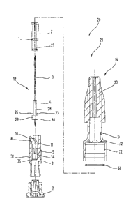
[0014] FIG. 2 is a schematic illustration of the embodiment of the
invention
disclosed herein. FIG. 2, like FIG.1, shows the system extending from the
distal probe 30 to
proximal terminals 36C, 40C and 42C.
[0015] FIG. 3 is a longitudinal view of the camera assembly 34, cable 36
and
proximal connector 36C.
[0016] FIG. 4 is a partial longitudinal sectional view of the camera
assembly 34
showing camera housing 34, focus ring 50 and laser filter 46. FIG. 4 shows the
distal recess
52 for engaging the nose 54 of the hand piece 32.
[0017] FIG. 5 is partial sectional view of the hand piece 32.
[0018] FIG. 6 is a view similar to that of FIGs. 4 and 5 showing the
probe 30 and
hand piece 32 assembled and coupled to the camera assembly 34.

CA 2832209 2017-03-21
- 5 -
WO 2011/142989
PCT/US2011/034464
Detailed Description
[0019] Except for the
prior art FIG.1, the figures are all to a single embodiment.
{0020] As shown in
FIG.!, the known laser video endoscopes have an operating
probe 24, a hand piece 22, a cable 18 which carries a laser guide 12, an
illumination guide 14
and an image guide 16. These are all fiber optic guides which extend from the
distal end of
the probe 24 to the terminals 12C, 14C and 16C. Distal of the trifurcation
zone 20, the fiber
optic guides are combined geometrically to provide a minimum diameter cable.
[0021] The laser video endoscope of this invention includes the probe 30,
a
specifically designed hand piece 32 and a camera assembly 34 coupled to the
proximal end of
the hand piece 36.
[0022] The camera assembly 34 is directly connected to the proximal end of the

hand piece 32. It has a relatively long electrical cable 36 which extends
proximally to a
terminal 36C which is coupled to an appropriate display mechanism including a
video screen
so that the operating surgeon can view the image during the course of
manipulating the probe.
[0023] A optical guide
cable 38 extends in the proximal direction from the hand
piece 32 to a bifurcation junction 39. This cable 38 carries the laser and
illumination guides
40 and 42 for conveying the laser energy and the illumination energy to the
probe 30. At the
bifurcation junction 44, the laser guide 40 and illumination guide 42 are
separated and
terminated at the terminals 40C and 42C for connection to the sources of laser
energy and
illumination energy. The image carrying electrical cable 36 is about as long
as is the optical
guide cable 38. Each cable 36, 38 can be as long as required for an
installation.
[0024] As shown by the coupling mechanism in the camera assembly 34, the
optical fibers 40 from the probe 30 and hand piece 32, which carry the image
are removably
coupled to the camera so that the camera provides an electrical image that is
transmitted

CA 2832209 2017-03-21
- 6 -
WO 2011/142989 PCT/US2011/034464
along the electric cable 36 to the terminal 36C at the base where the video
displays are
provided. The camera may be any one of a number of known type and may be
specially
designed to fit the geometry of the camera assembly
[0025] Thus by positioning the camera assembly 34 at the hand piece 32,
the
lengthy and expensive optical image guide is avoided_ The camera assembly 34
can be
uncoupled from the hand piece 32 so that the relatively expensive camera
assembly can be
reused. This combination of reuse of the camera assembly 34 and elimination of
an extensive
length of expensive fiber optic image guide means that disposability of the
probe 30 is
economically acceptable even though the hand piece 32 and the laser and
illumination guides
40, 42 in the cable 38 are also disposed of after each medical routine.
[0026] The camera assembly 34 includes a laser filter 46 to protect the
camera film
from laser energy and to permit the surgeon to observe the operation even when
laser pulses
are firing. The probe 30 and hand piece 32 are cemented together by a known
process.
[0027] The camera assembly 34 includes a manually operated spring latch
(not
shown). The latch is of a known type. It enables readily mounting the camera
assembly 34
to the hand piece 32 and, most importantly, removing the camera assembly 34
from the hand
piece 32. In addition, the camera assembly 34 includes a focus ring 50 to
assure adequate
focus of the image provided at the proximal end of the laser fiber image guide
37 in the probe
30 and hand piece 32 onto the image receptors of the camera.
[0028] As may be seen from FIGs. 4, 5 and 6, the distal end of the camera
assembly has a recess 52 which engages a nose 54 of the hand piece 32. The
latch holds the
nose 54 in place in the recess 52.
[0029] The image guide 37 in the probe 30 and hand piece 32 costs about
$8.00.
This reduction in cost from about $200.00 to $8.00 is a major factor
encouraging disposable
use of the endoscope.

CA 2832209 2017-03-21
- 7 -
wo 2011/142989 PCT/US2011/034464
[0030] A variant on the illustrated embodiment is an arrangement in which
the
uncoupling at the proximal end of the hand piece 32 will uncouple not only the
camera
assembly 34 but also the cable 38 so that only the probe 30 and the hand piece
32 would be
disposed of between each operation.
[0031] It has to be kept in mind that the positioning of the camera
assembly 34 at
the hand piece 32 permits a standard optical coupling of the image at the
proximal end of the
optical fiber image guide 37 to the camera assembly 34. It is not feasible to
provide a
mechanism that will permit coupling and uncoupling the fiber optic image guide
37 at a
junction other than the input to the camera. Coupling and uncoupling is
otherwise not
feasible because of the enormous number of optical fibers which would have to
be aligned for
such coupling to provide an image that is not degraded or useless.
[0032] While the foregoing description and drawings represent the
presently
preferred embodiments of the invention, it should be understood that those
skilled in the art
will be able to make changes and modifications to those embodiments without
departing from
the teachings of the invention and the scope of the claims.
[0033] For example, the image guide 37 within the probe 30 and hand piece
32 is a
fiber optic bundle of the type normally used. However, there are other means
to provide an
image guide. One such is the gradient index lens, often referred to as a GRIN
lens.

Representative Drawing
A single figure which represents the drawing illustrating the invention.
Administrative Status

For a clearer understanding of the status of the application/patent presented on this page, the site Disclaimer , as well as the definitions for Patent , Administrative Status , Maintenance Fee  and Payment History  should be consulted.

Administrative Status

Title Date
Forecasted Issue Date 2019-04-02
(86) PCT Filing Date 2011-04-29
(87) PCT Publication Date 2011-11-17
(85) National Entry 2013-10-03
Examination Requested 2016-04-18
(45) Issued 2019-04-02

Abandonment History

There is no abandonment history.

Maintenance Fee

Last Payment of $347.00 was received on 2024-04-19


 Upcoming maintenance fee amounts

Description Date Amount
Next Payment if standard fee 2025-04-29 $347.00
Next Payment if small entity fee 2025-04-29 $125.00

Note : If the full payment has not been received on or before the date indicated, a further fee may be required which may be one of the following

  • the reinstatement fee;
  • the late payment fee; or
  • additional fee to reverse deemed expiry.

Patent fees are adjusted on the 1st of January every year. The amounts above are the current amounts if received by December 31 of the current year.
Please refer to the CIPO Patent Fees web page to see all current fee amounts.

Payment History

Fee Type Anniversary Year Due Date Amount Paid Paid Date
Reinstatement of rights $200.00 2013-10-03
Application Fee $400.00 2013-10-03
Maintenance Fee - Application - New Act 2 2013-04-29 $100.00 2013-10-03
Maintenance Fee - Application - New Act 3 2014-04-29 $100.00 2014-04-11
Maintenance Fee - Application - New Act 4 2015-04-29 $100.00 2015-04-06
Maintenance Fee - Application - New Act 5 2016-04-29 $200.00 2016-03-30
Request for Examination $800.00 2016-04-18
Registration of a document - section 124 $100.00 2016-08-04
Registration of a document - section 124 $100.00 2016-11-01
Maintenance Fee - Application - New Act 6 2017-05-01 $200.00 2017-03-30
Maintenance Fee - Application - New Act 7 2018-04-30 $200.00 2018-04-02
Final Fee $300.00 2019-02-12
Maintenance Fee - Patent - New Act 8 2019-04-29 $200.00 2019-04-02
Maintenance Fee - Patent - New Act 9 2020-04-29 $200.00 2020-04-24
Maintenance Fee - Patent - New Act 10 2021-04-29 $255.00 2021-04-23
Maintenance Fee - Patent - New Act 11 2022-04-29 $254.49 2022-04-22
Maintenance Fee - Patent - New Act 12 2023-05-01 $263.14 2023-04-21
Maintenance Fee - Patent - New Act 13 2024-04-29 $347.00 2024-04-19
Owners on Record

Note: Records showing the ownership history in alphabetical order.

Current Owners on Record
BEAVER-VISITEC INTERNATIONAL, INC.
Past Owners on Record
ENDO OPTIKS, INC.
Past Owners that do not appear in the "Owners on Record" listing will appear in other documentation within the application.
Documents

To view selected files, please enter reCAPTCHA code :


To view images, click a link in the Document Description column. To download the documents, select one or more checkboxes in the first column and then click the "Download Selected in PDF format (Zip Archive)" or the "Download Selected as Single PDF" button.

List of published and non-published patent-specific documents on the CPD .

If you have any difficulty accessing content, you can call the Client Service Centre at 1-866-997-1936 or send them an e-mail at CIPO Client Service Centre.


Document
Description 
Date
(yyyy-mm-dd) 
Number of pages   Size of Image (KB) 
Abstract 2013-10-03 1 60
Claims 2013-10-03 2 46
Drawings 2013-10-03 6 44
Description 2013-10-03 7 199
Representative Drawing 2013-10-03 1 11
Cover Page 2013-11-22 1 42
Examiner Requisition 2017-08-24 4 252
Amendment 2018-02-23 10 360
Description 2018-02-23 7 184
Claims 2018-02-23 2 58
Change of Agent 2018-11-27 3 88
Office Letter 2018-12-17 1 22
Office Letter 2018-12-17 1 25
Final Fee 2019-02-12 2 46
Representative Drawing 2019-03-04 1 7
Cover Page 2019-03-04 1 39
Maintenance Fee Payment 2019-04-02 1 33
PCT 2013-10-03 15 802
Assignment 2013-10-03 5 145
Fees 2015-04-06 1 33
Request for Examination 2016-04-18 1 49
Response to section 37 2016-08-04 6 268
Examiner Requisition 2016-09-21 3 180
Amendment 2017-03-21 15 471
Description 2017-03-21 7 182
Claims 2017-03-21 2 50