Note: Descriptions are shown in the official language in which they were submitted.
CA 02832502 2016-07-29
. .
ILLUMINATED MICROSURGICAL INSTRUMENT INCLUDING
OPTICAL FIBER WITH BEVELED END FACE
FIELD AND BACKGROUND
[0002] Various surgical procedures, called vitreo-retinal
procedures, are commonly
performed in the posterior segment of the eye. Vitreo-retinal procedures are
appropriate to
treat many serious conditions of the posterior segment. Vitreo-retinal
procedures treat
conditions such as age-related macular degeneration (AMD), diabetic
retinopathy and
diabetic vitreous hemorrhage, macular hole, retinal detachment, epiretinal
membrane, CMV
retinitis, and many other ophthalmic conditions.
[0003] A surgeon performs vitreo-retinal procedures with a
microscope and special
lenses designed to provide a clear image of the posterior segment. Several
tiny incisions just
a millimeter or so in length are made on the sclera at the pars plana. The
surgeon inserts
microsurgical instruments through the incisions, such as a fiber optic light
source to
illuminate inside the eye; an infusion line to maintain the eye's shape during
surgery; and
instruments to cut and remove the vitreous body. A separate incision may be
provided for
each microsurgical instrument when using multiple instruments simultaneously.
[0004] During such surgical procedures, proper illumination of the
inside of the eye is
important. Typically, a thin optical fiber is inserted into the eye to provide
the illumination.
A light source, such as a halogen tungsten lamp or high pressure are lamp
(metal-halides,
Xe), may be used to produce the light carried by the optical fiber into the
eye. The light
passes through several optical elements (typically lenses, mirrors, and
attenuators) and is
transmitted to the optical fiber that carries the light into the eye.
[0005] As with most surgical procedures, there is a benefit to
minimizing the number
and size of incisions required to perform the vitreo-retinal procedure.
Incisions are typically
only made large enough to accommodate the size of the microsurgical instrument
being
inserted into the interior of the eye. Efforts to minimize the incision size
generally involve
reducing the size of the microsurgical instrument. Depending on the size of
the
microsurgical instrument employed, the incision may be small enough to render
the resulting
wound substantially self-healing, thereby eliminating the need to employ
additional
- 1 -
CA 2832502 2017-05-11
procedures to close the incision, such as sutures. Reducing the number of
incisions may be
accomplished by integrating various microsurgical instruments. For example,
the optical
fiber may be incorporated into the working end of a microsurgical instrument.
This may
eliminate the need for a separate illumination incision, and offers the
advantage of directing
the light beam, together with the microsurgical instrument, onto the target
site through a
common opening in the sclera. Unfortunately, at least some prior attempts at
integrating
illuminating optical fibers with microsurgical instruments have resulted in a
decrease in
illuminating efficiency, or otherwise adversely effected the distribution of
light emitted from
the optical fibers.
SUMMARY
[0005a] Certain exemplary embodiments can provide an illuminated
microsurgical
instrument comprising: a microsurgical instrument having a distal tip sized
and shaped for
insertion through an incision in an eye and into a vitreous region of the eye
when performing
an ophthalmic surgical procedure; and an optical fiber for delivering a beam
of light to a
surgical site of the eye, the optical fiber including a proximal end for
receiving a light beam
from a light source, and a distal end proximate to the distal tip of the
microsurgical
instrument for emitting the light beam, the distal end including a beveled end
face oriented
toward the distal tip of the microsurgical instrument at an oblique angle with
respect to an
optical axis of the optical fiber, wherein a core of the optical fiber has an
index of refraction
greater than an index of refraction of the vitreous region such that a
propagation angle of the
light beam relative to the beveled end face is greater within the vitreous
region than within
the optical fiber causing an angular distribution of the emitted light beam
away from the
optical axis of the optical fiber and away from the microsurgical instrument.
[0005b] Certain exemplary embodiments can provide an illuminated ophthalmic
microsurgical instrument comprising: a microsurgical instrument having a
distal tip; and
an optical fiber for delivering a beam of light to a surgical site in an eye,
the optical fiber
including a proximal end for receiving a light beam from a light source, and a
distal end
proximate to the distal tip of the microsurgical instrument for emitting the
light beam,
wherein the distal end includes a beveled end face oriented away from the
distal tip of the
- 2 -
CA 2832502 2017-05-11
microsurgical instrument, at an oblique angle with respect to an optical axis
of the optical
fiber, wherein a core of the optical fiber has an index of refraction greater
than an index of
refraction of a vitreous region of the eye such that a propagation path of the
light beam from
the beveled end face is towards the microsurgical instrument, when used to
perform an
ophthalmic surgical procedure.
BRIEF DESCRIPTION OF THE DRAWINGS
[0006] FIG. 1 is schematic illustration of an exemplary microsurgical
instrument
employing an exemplary integrated fiber optic illuminator, shown illuminating
an interior
region of an eye;
[0007] FIG. 2 is a schematic partial cross-sectional view of the
microsurgical
instrument and integrated fiber optic illuminator;
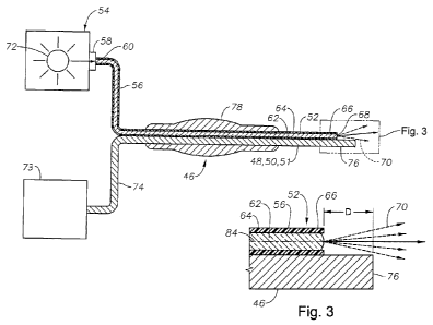
[0008] FIG. 3 is a schematic partial cross-sectional view of a distal end
of the
microsurgical instrument and integrated fiber optic illuminator shown in FIG.
2;
[0009] FIG. 4 is a schematic partial cross-sectional view of the distal end
of the fiber
optic illuminator configured to include a beveled end face;
[00101 FIG. 5 is a schematic plan view of the fiber optic illuminator shown
in FIG. 4;
[0011] FIG. 6 is a schematic partial cross-sectional view of the fiber
optic illuminator
shown in FIG. 4, employing substantially planar beveled end face;
[0012] FIG. 7 is a partial cross-sectional view of the fiber optic
illuminator shown in
FIG. 4, employing a generally convex beveled end face;
[0013] FIG. 8 is a partial cross-sectional view of the fiber optic
illuminator shown in
FIG. 4, employing a generally concave beveled end face;
[0014] FIG. 9 is a schematic partial cross-sectional view of the distal end
of the fiber
optic illuminator, with the beveled end face arranged to face generally away
from the
microsurgical instrument;
[0015] FIG. 10 is a schematic end view of an alternately configured fiber
optic
illuminator employing multiple optical fibers;
- 2a -
CA 02832502 2013-10-04
WO 2012/154435 PCT/US2012/035774
[0016] FIG. 11 is a schematic partial cross-sectional view of the fiber
optic illuminator
shown in FIG. 10;
[0017] FIG. 12 is a schematic end view of an alternately configured fiber
optic
illuminator employing multiple optical fibers; and
[0018] FIG. 13 is a schematic partial cross-sectional view of the fiber
optic illuminator
shown in FIG. 12.
DETAILED DESCRIPTION
[0019] Referring now to the discussion that follows and the drawings,
illustrative
approaches to the disclosed systems and methods are described in detail.
Although the
drawings represent some possible approaches, the drawings are not necessarily
to scale
and certain features may be exaggerated, removed, or partially sectioned to
better illustrate
and explain the present disclosure. Further, the descriptions set forth herein
are not
intended to be exhaustive, otherwise limit, or restrict the claims to the
precise forms and
configurations shown in the drawings and disclosed in the following detailed
description.
[0020] FIG. 1 illustrates an anatomy of an eye 20, which includes a cornea
22, an iris
24, a pupil 26, a lens 28, a lens capsule 30, zonules 32, ciliary body 34,
sclera 36, vitreous
region 38, retina 40, macula 42, and optic nerve 44. Cornea 22 is a clear,
dome shaped
structure on the surface of eye 20 that acts as a window, letting light into
the eye. Iris 24,
which corresponds to the colored part of the eye, is a muscle surrounding
pupil 26 that
relaxes and contracts to control the amount of light entering eye 20. Pupil 26
is a round,
central opening in iris 24. Lens 28 is a structure inside eye 20 that helps
focus light on
retina 40. Lens capsule 30 is an elastic bag that encapsulates lens 30,
helping to control
the shape of lens 28 as the eye focuses on objects at different distances.
Zonules 32 are
slender ligaments that attach lens capsule 30 to the inside of eye 20, holding
lens 28 in
place. Ciliary body 34 is a muscular area attached to lens 28 that contracts
and relaxes to
control the size of the lens for focusing. Sclera 36 is a tough, outermost
layer of eye 20
that maintains the shape of the eye. Vitreous region 38 is a large, gel-filled
section located
towards a back of eye 20 that helps maintain the curvature of the eye. Retina
40 is a light-
sensitive nerve layer at the back of eye 20 that receives light and converts
it into signals to
send to the brain. Macula 42 is an area in the back of eye 20 that includes
receptors for
detecting fine detail in a viewed image. Optic nerve 44 transmits signals from
eye 20 to
the brain.
-3 -
CA 02832502 2016-07-29
[0021] With continued reference to FIG. 1, various microsurgical
instruments 46 may
be inserted through sclera 36 into vitreous region 38 when performing an
ophthalmic surgical
procedure, such as a vitreoretinal procedure. For purposes of this
specification, a
microsurgical instrument 46 refers to any tool sized for insertion through an
incision that is
adapted to perform physical or electromagnetic manipulation of ocular tissue.
These may
include a variety of surgical instruments, such as, for example, a vitrectomy
probe 48,
infusion cannula 50 and aspiration probe 51. Microsurgical instrument 46 may
include an
integrated fiber optic illuminator 52 for illuminating an interior of eye 20.
[0022] With reference to FIG. 2, the fiber optic illuminator 52 may be
optically
connected to an illuminator 54 for producing light that may be used to
illuminate vitreous
region 38 of eye 20 during various intra-optical procedures, such as
vitreoretinal surgery.
Light produced by illuminator 54 may be transmitted to the interior region of
the eye through
an optical fiber 56. Optical fiber 56 may include a fiber optic connector 58
for optically
connecting a proximal end 60 of optical fiber 56 to illuminator 54. Fiber
optic connector 58
may be configured to releasably connect to a correspondingly configured
illuminator optical
connector operably associated with illuminator 54.
[0023] Continuing to refer to FIG. 2, optical fiber 56 may have any of a
variety of
configurations. In the exemplary configuration shown in FIG. 2, optical fiber
56 includes an
optically transmissive fiber optic core 62 surrounded by a cladding material
64 having a low
index of refraction relative to core 62. Fiber optic core 62 may be made of
various materials,
including, but not limited to, glass and plastics. Optical fiber 56 may also
include additional
layers, depending on the requirements of a particular application. For
example, optical fiber
56 may include a buffer material encasing cladding material 64, as well as an
outer protective
jacket for shielding the cable's interior components from damage. A distal end
66 of optical
fiber 56 may include an opening 68 for emitting light 70 produced by
illuminator 54.
[0024] Continuing to refer to FIG. 2, illuminator 54 may employ a light
source 72 for
generating light at a particular luminous flux and chromaticity. The light may
be emitted over
a relatively wide or narrow range of wavelengths depending on the type of
light source
employed. Light source 72 may employ various light producing technologies,
including, but
not limited to, lamp based light sources, such as halogen tungsten lamps and
high-pressure are
lamps (metal-halides and Xe). Light emitting diodes (LEDs) may also be
employed as light
source 72. Lasers may also be employed as light source 72. Lasers are
generally capable of
- 4 -
CA 02832502 2016-07-29
=
producing light having a relatively high degree of coherence, as compared to
other light
sources, such as LEDs and lamp based light sources. High coherence enables the
emitted light
to be focused to smaller spot sizes for more efficient transmission to optical
fiber 56. The
ability to focus the emitted light to small spot sizes may enable the use of
smaller optical
fibers, such as nano-scaled optica; fibers, which may in turn limit the size
of an incision
required to insert microsurgical instrument 46 into eye 20. Nano-scale optic
fibers generally
have a diameter (or other largest cross-sectional dimension) of less than 100
microns.
[0025]
Due to the small size of nano-scale optic fibers, it may be possible to
integrate
fiber optic illuminator 52 with another surgical instrument, such as
microsurgical instrument
46, to reduce the number of surgical incisions required for inserting surgical
instruments
during a vitreoretinal procedure. Continuing to refer to FIG. 2, microsurgical
instrument 46
may be suitably connected to a service source 73, for example, via conduit 74.
Service source
73 may be configured to provide various services used in connection with
operating
microsurgical instrument 46. For example, service source 73 may provide
pressure and/or
vacuum for operating microsurgical instrument 46. Vacuum may also be provided
for
aspirating fluids and materials from the interior of eye 20. Service source 73
may provide a
source of fluids used in connection with the surgical procedure.
[0026]
Microsurgical instrument 46 may have various configurations depending on
the surgical procedure performed. For example, certain ophthalmic surgical
procedures may
require the cutting and/or removal of vitreous region 38, which is a
transparent jelly-like
material that fills the posterior segment of eye 20. Vitrectomy probe 48 may
be used to resect
and remove the vitreous region. In one exemplary configuration, vitrectomy
probe 48 may
include a hollow outer cutting member, a hollow inner cutting member arranged
coaxially
with and movably disposed within the hollow outer cutting member, and a port
extending radially through the outer cutting member near a distal end 76
thereof. Vitreous
region 38 is aspirated into the open port, and the inner member is actuated to
close the
port and sever the vitreous material, which may then be aspirated away through
conduit 74. The mechanism for actuating the hollow inner member may be
enclosed within a
housing 78, which may also function as a handle for grasping microsurgical
instrument 46.
Microsurgical instrument 46 may also be configured as infusion cannula 50 for
delivering a
fluid to the interior of eye 20. The fluid may be delivered to infusion
cannula 50 through
conduit 74. Conduit 74 may also be used to connect microsurgical instrument 46
to a
- 5 -
CA 02832502 2013-10-04
WO 2012/154435 PCT/US2012/035774
vacuum source, for example, when configuring microsurgical instrument 46 as
aspiration
probe 51.
100271 Referring to Fig. 3, in certain applications, it is generally
desirable for light
beam 70 emitted from fiber optic illuminator 52 to have a relatively wide
angular
distribution to enable illumination of a corresponding wide surgical field
within eye 20.
However, a portion of the light beam 70 emitted from optical fiber may be
either absorbed
or reflected from an adjacent outer surface 80 of microsurgical instrument 46,
depending
on the positioning of distal end 66 of optical fiber 56 relative to distal end
76 of
microsurgical instrument 46. It may not always be desirable, however, to
position distal
end 66 of optical fiber 56 proximate to end 76 of microsurgical instrument 46.
Positioning
distal end 66 of optical fiber 56 a distance "D" from distal end 76 of
microsurgical
instrument 46 may, however, adversely affect the illuminating efficiency of
fiber optic
illuminator 52, particularly in instances in which a measurable portion of the
emitted light
is absorbed by outer surface 80 of microsurgical instrument 46.
100281 Referring to FIGS. 4 and 5, to help avoid a distal tip of
microsurgical
instrument 46 interfering with the propagation of light beam 70 emitted from
optical
fiber 56, distal end 66 may be provided with a beveled end face 82 arranged at
an oblique
angle relative to an optical axis 84 of optical fiber 56. For purposes of this
specification,
"beveled end face" need not refer strictly to a flat beveled surface but
rather may include
any configuration wherein a distalmost end face is arranged so that the
surface normal,
i.e., the axis perpendicular to the surface, is deviated to one side of the
optical axis 84 over
the majority of the end face, making the distalmost end face asymmetrical
relative to the
optical axis. When the beveled end face 82 is said to "point" or to be
"oriented" toward a
certain direction, this refers to the side of the optical axis 84 toward which
the beveled end
face 82 is asymmetrically deviated. Inclining end face 82 relative to optical
axis 84
generally results in light beam 70 approaching beveled end face 82 at an
oblique incidence
angle relative to the surface normal at the point of incidence. The transition
between the
two different refractive indices causes the light to refract as it transitions
the interface
between optical fiber 56 and vitreous region 38 of eye 20, thereby deflecting
a propagation
path 86 of light beam 70 away from optical axis 84 of optical fiber 56. The
amount of
refraction may be approximated using Snell's law, which provides:
n1 * Sin(01) = n2 * Sin(e2)
where:
- 6 -
CA 02832502 2013-10-04
WO 2012/154435 PCT/US2012/035774
n1 is the refractive index of fiber optic core 62
n2 is the refractive index of vitreous region 38
01 is the propagation angle of light beam 70 within fiber optic core 62
02 is the propagation angle of light beam 70 within vitreous
region 38,
where 01 and 02 are both measured relative to the surface normal of
the beveled end face 82.
[0029] Because the index of refraction of the vitreous region is lower than
that of the
fiber optic core, the light beam 70 will tend to be refracted away from the
surface normal
of the beveled end surface 82, viz., 02> 01. The angular distribution of the
rays in light
beam 70 as the rays travel through the optical fiber 56 will therefore produce
an angular
distribution in the emitted light beam 70, which will be preferentially
shifted away from
the optical axis 84 of the optical fiber 56.
[0030] While beveled end face 82 is illustrated on an optical fiber 56 of
uniform
diameter, beveled end face 82 may also be used on a fiber optic with a tapered
distal tip
that narrows to a smaller width along a path that may includes curved or
straight segments
as the fiber optic extends toward the distal tip. In particular embodiments of
the tapered
distal tip, the cladding may also be removed. The tapered distal end provides
a wider
angular distribution, which may advantageously be combined with the deflection
produced
by the beveled end face 82 to produce a wider illumination beam from the fiber
optic
selectively directed in a particular direction around the tip of the surgical
instrument.
[0031] The deflection of light beam 70 relative to microsurgical instrument
46 is at
least partially dependent on the orientation of beveled end face 82 relative
to microsurgical
instrument 46. For example, orienting beveled end face 82 to point toward
microsurgical
instrument 46, such as shown in FIG. 4, tends to shift propagation path 86 of
the light
beam away from microsurgical instrument 46. On the other hand, orienting
beveled end
face 82 to point away from microsurgical instrument 46, such as shown in FIG.
9, tends to
shift the propagation path 86 of light beam 70 toward microsurgical instrument
46.
Referring to FIG. 9, fiber optic illuminator 52 is shown with beveled end face
82 oriented
to face generally away from microsurgical instrument 46. This arrangement
generally
results in propagation path 86 of light beam 70 being shifted toward
microsurgical
instrument 46. Thus, this arrangement increases, rather than decreases, the
amount of light
reflected from microsurgical instrument 46. A wider dispersion of light
emitted from
- 7 -
CA 02832502 2013-10-04
WO 2012/154435 PCT/US2012/035774
optical fiber may be obtained by enhancing the reflectivity of outer surface
80 of
microsurgical instrument 46. Light emitted from optical fiber 56 may be
reflected from
surface 80 of microsurgical instrument 46 to provide a broader distribution of
light within
an interior region of eye 20.
[0032] FIGS. 6-8 are partial cross-sectional views taken through beveled
end face 82
(see FIG. 4) along a perspective generally parallel to end face 82. Beveled
end face 82
may include a variety of surface contours. For example, FIG. 6 shows beveled
end face 82
configured to include a planar surface. Beveled end face 82 may alternatively
be
configured to include a generally convex surface contour, such as shown in
FIG. 7.
Beveled end face 82 may also have a generally concave configuration, as shown
in FIG. 8.
These are merely a few examples of the various surface contours that may be
employed
with beveled end face 82. In practice, other contours may also be employed to
accommodate design and performance requirements of a particular application.
[0033] Referring to FIGS. 10-13, fiber optic illuminator 52 may be
configured to
include multiple bundled optical fibers 56 surrounding a distal tip of a
microsurgical
instrument 46. FIG. 10 shows an exemplary arrangement including four optical
fibers 56
bundled together. Each optical fiber may include a beveled end face 82 for
selectively
controlling a propagation path of emitted light. In the exemplary arrangement
illustrated
in FIGS. 10 and 11, beveled end face 82 of optical fibers 56 positioned at
opposite corners
of the cable bundle are shown oriented so as to generally face one another.
This particular
arrangement tends to increase the dispersion of the emitted light by shifting
propagation
path 86 of light beam 70 outward from a center axis 88 of the bundle.
100341 FIGS. 12 and 13 show an exemplary optical fiber bundle including
seven
optical fibers 56. The optical fibers are shown arranged generally in a
hexagonal pattern,
with six optical fibers positioned around a center optical fiber. Each of the
outer optical
fibers 56 may include a beveled end face 82 for selectively controlling a
propagation path
of emitted light. The single center optical fiber 56 in this exemplary
configuration does
not include a beveled end face. Beveled end faces 82 of the outer optical
fibers 56 may be
oriented so as to generally point radially inward toward a center of the
optical fiber
bundle. This particular arrangement tends to increase the dispersion of the
light emitted
from the outer optical fibers by shifting propagation path 86 of light beam 70
outward
from the center of the optical fiber bundle.
- 8 -
CA 02832502 2016-07-29
[0035] The distal end of the entire bundle is placed proximate to a distal
tip of a
microsurgical instrument 46. The central fiber optical cable and/or the
optical fibers that are
more remote from the distal tip of the microsurgical instrument 46 can have a
flat surface so
that the propagation path of light emitted from the center optical fiber tends
to coincide with
optical axis of the optical fiber. In such embodiments, light emitted from the
center optical fiber
56 may fill a light void that may exist between the light beams emitted from
the surrounding
outer optical fibers 56, while still allowing the overall amount of reflected
light from the distal
tip of the microsurgical instrument 46 to be reduced by the orientation of the
closest optical
fibers 56. For example, if the distal tip of the microsurgical instrument 46
is reflective, then the
depicted orientation of the beveled end faces 82 can advantageously provide
additional
illumination through reflection, as previously illustrated in FIG. 9.
Alternatively, in the case of
a non-reflective tip of microsurgical instrument 46, the beveled end faces 82
could be reversed
to point toward the distal tip of microsurgical instrument 46, preferentially
shifting the
illumination away from the distal tip of microsurgical instrument 46, as
illustrated in FIG. 4. In
yet another alternative embodiment, the optical fibers 56 can be placed in a
similar
configuration as illustrated in FIGS. 10-13, but centered around the distal
tip of microsurgical
instrument 46, so as to produce illumination from multiple optical fibers 56
around the
microsurgical instrument 46.
[0036] It will be appreciated that the exemplary surgical illumination
system described
herein has broad applications. The foregoing configuration were chosen and
described in order
to illustrate principles of the methods and apparatuses as well as some
practical applications.
The preceding description enables others skilled in the art to utilize methods
and apparatuses in
various configurations and with various modifications as are suited to the
particular use
contemplated. In accordance with the provisions of the patent statutes, the
principles and modes
of operation of the disclosed surgical illumination system have been explained
and illustrated in
exemplary configurations.
[0037] It is intended that the scope of the present methods and apparatuses
be defined by
the following claims. However, it must be understood that the disclosed
surgical illumination
system may be practiced otherwise than is specifically explained and
illustrated without
departing from its scope. It should be understood by those skilled in the art
that various
alternatives to the configuration described herein may be employed in
practicing the claims
without departing from the scope as defined in the following claims.
- 9 -
CA 02832502 2013-10-04
WO 2012/154435 PCT/US2012/035774
The scope of the disclosed surgical illumination system should be determined,
not with
reference to the above description, but should instead be determined with
reference to the
appended claims, along with the full scope of equivalents to which such claims
are
entitled. It is anticipated and intended that future developments will occur
in the arts
discussed herein, and that the disclosed systems and methods will be
incorporated into
such future examples. Furthermore, all terms used in the claims are intended
to be given
their broadest reasonable constructions and their ordinary meanings as
understood by
those skilled in the art unless an explicit indication to the contrary is made
herein. In
particular, use of the singular articles such as "a," "the," "said," etc.
should be read to
recite one or more of the indicated elements unless a claim recites an
explicit limitation to
the contrary. It is intended that the following claims define the scope of the
device and
that the method and apparatus within the scope of these claims and their
equivalents be
covered thereby. In sum, it should be understood that the device is capable of
modification and variation and is limited only by the following claims.
- 1 0 -