Note: Descriptions are shown in the official language in which they were submitted.
CA 02832859 2013-10-09
WO 2012/142581
PCT/US2012/033782
TREATMENT OF DISEASE WITH
POLY-N-ACETYLGLUCOSAMINE NANOFIBERS
1. INTRODUCTION
100011 This application relates to compositions comprising shortened fibers
of poly-N-
acetylglucosamine and/or a derivative thereof ("sNAG nanofibers") and the use
of such
compositions in the treatment of disease.
2. BACKGROUND
100021 Defcnsins are small (3-4 IcDa), cystcinc-rich cationic peptides
found in mammals,
insects, and plants that are classified into different families (a, p, and 0)
based on their pattern of
disulfide bonding. These small peptides are important effectors of innate
immunity and
consequently play an important role in the body's battle against various
diseases.
100031 A number of diseases are incurable at this time or have suboptimal
treatments
available, due to only partial effectiveness of such treatments or side
effects associated with such
treatments. Such diseases include, among others, cancer, some viral diseases,
some fungal
diseases, inflammatory bowel diseases (e.g., Crohn's disease), and
dermatological diseases such
as psoriasis and dermatitis. There remains a need for an effective treatment
for these diseases
that can be used alone, or in combination with a standard therapy, that is
safe and effective.
3. SUMMARY
100041 In one aspect, described herein are methods for preventing and/or
treating infections
and/or diseases for which an increase in defensin production and/or secretion
may be beneficial,
comprising administering to the subject a composition comprising shortened
fibers of poly-13-
-4-N-acetylglucosamine and/or derivatives thereof (referred to herein as "sNAG
nanofibers").
Examples of such infections and/or diseases include, but are not limited to,
solid tumor cancers,
skin cancer, viral infections, yeast infections, fungal infections,
inflammatory bowel disease,
Crohn's disease, dermatitis and psoriasis.
100051 In one embodiment, described herein is a method for treating a viral
infection in a
subject, comprising administering a composition comprising sNAG nanofibers to
a subject
having (e.g., diagnosed with) a viral infection (e.g., an HSV infection). In
another embodiment,
described herein is a method for preventing a viral disease in a human
subject, comprising
1
CA 02832859 2013-10-09
WO 2012/142581
PCT/US2012/033782
administering a composition comprising sNAG nanofibers to a subject at risk of
developing a
viral disease (e.g., a symptom of an HSV infection such as a cold sore or a
lesion). In a specific
embodiment, the sNAG nanofiber composition is topically administered to the
subject (e.g., to
the skin or mucous membrane). In specific embodiments, the subject is a human.
10006] In another embodiment, described herein is a method for treating a
solid tumor in a
subject, comprising administering a composition comprising sNAG nanofibers to
a subject
diagnosed with a solid tumor. In a specific embodiment, all or part of the
solid tumor has been
removed from the subject (e.g., surgically removed), and the sNAG nanofibers
are administered
to the site of the solid tumor before, during, and/or after the removal of all
or part of the solid
tumor. In specific embodiments, the subject is a human.
100071 In another embodiment, described herein is a method for treating a
skin cancer in a
subject, comprising topically administering a composition comprising sNAG
nanofibers to a
human subject diagnosed with a skin cancer. In a specific embodiment, all or
Part of the skin
cancer has been removed from the subject (e.g., surgically removed), and the
sNAG nanofibers
are administered to the site of the skin cancer before, during, and/or after
the removal of all or
part of the skin cancer. In specific embodiments, the subject is a human.
100081 In another embodiment, provided herein is a method for treating
inflammatory bowel
disease in a subject, comprising administering a composition comprising sNAG
nanofibers to a
subject with inflammatory bowel disease (e.g., diagnosed with inflammatory
bowel disease). In
a specific embodiment, described herein is a method for treating Crohn's
disease in a subject,
comprising administering a composition comprising sNAG nanofibers to a subject
with Crohn's
disease (e.g., a subject diagnosed with Crohn's disease). In a specific
embodiment, the sNAG
nanofiber composition is topically administered to the subject (e.g., rectally
via a suppository).
In specific embodiments, the subject is a human.
[0009] The sNAG nanofibers contemplated in the methods described herein may
be of
varying lengths, widths and molecular weights as described in Section 5.1,
infra. In certain
embodiments, the majority (and in certain embodiments, at least or more than
60%, 70%, 80%,
90%, 95% or 99%) of the sNAG nanofibers, or 100% of the sNAG nanofibers, are
between
about 1 to 15 Am in length. In some embodiments, the majority (and in certain
embodiments, at
least or more than 60%, 70%, 80%, 90%, 95% or 99%) of the sNAG nanofibers, or
100% of the
sNAG nanofibers, are between about 2 to 10 p.m, 4 to 7 pm, 4 to 10 i.tm, or 5
to 10 i.tm in length.
2
CA 02832859 2013-10-09
WO 2012/142581
PCMJS2012/033782
The sNAG nanofibers of the described length can be obtained, for example, as
described below
in Section 5.2, infra.
100101 In certain embodiments, the sNAG nanofibers were produced by
irradiation, e.g.,
gamma irradiation, of poly-N-acetylglucosamine or a derivative thereof. In
some embodiments,
the sNAG nanofibers are produced by irradiation of the poly-13-1-4-N-
acetylglucosamine in the
form of dried fibers (e.g., at 500-2,000 kgy), or irradiation of the poly-13-1-
-4-N-
acetylglueosamine in the form of wet fibers (e.g., at 100-500 kgy).
10011] In certain embodiments, the sNAG nanofibers are derived from
microalgae. In
another embodiment, the sNAG nanofibers are not derived from crustaceans. In
yet another
embodiment, the sNAG nanofibers may be derived from microalgae, crustaceans
(e.g., shrimp),
fungus or any other source.
100121 In one embodiment, the sNAG nanofibers comprise N-acetylglucosamine
monosaccharides and/or glueosamine monosaccharides, wherein more than 60%,
70%, 80%,
90%, 95%, or 99% of the monosaccharides of the sNAG nanofibers are N-
acetylglucosamine
monosaccharides. In another embodiment, the sNAG nanofibers comprise N-
acetylglucosamine
monosaccharides and/or glucosamine monosaccharides, wherein more than 70% of
the
monosaccharides of the sNAG nanofibers are N-acetylglucosamine
monosaccharides.
100131 In certain embodiments, the sNAG nanofibers used in the methods
described
herein are non-reactive in a biocompatibility test or tests. For example, the
sNAG nanofibers
used in the methods described herein may be non-reactive when tested in an
elution test, an
intramuscular implantation test, an intracutaneous test, or a systemic test.
In some embodiments,
the compositions described herein are non-reactive when tested in an elution
test, an
intramuscular implantation test, an intracutaneous test, or a systemic test.
In other embodiments,
the sNAG nanofibers used in the methods described herein have Grade 0 or Grade
1 when tested
in an elution test, an intramuscular implantation test, an intracutaneous
test, or a systemic test. In
yet another embodiment, the sNAG nanofibers used in the methods described
herein are at most
mildly reactive when tested in an elution test, an intramuscular implantation
test, an
intracutaneous test, or a systemic test. In one embodiment, the sNAG
nanofibers or
compositions comprising such nanofibers are non-reactive as determined by an
intramuscular
implantation test. In certain embodiments, the compositions described herein
do not cause an
allergenic reaction or an irritation, e.g., at the site of application. In
other embodiments, the
3
CA 02832859 2013-10-09
WO 2012/142581
PCT/US2012/033782
compositions described herein cause at most a mild allergenic reaction or a
mild irritation, e.g.,
at the site of application.
10014] In certain embodiments, the sNAG nanofibers used in the methods
described
herein increase the metabolic rate of serum-starved human umbilical cord vein
endothelial cells
in a MTT assay and/or do not rescue apoptosis of serum-starved human umbilical
cord vein
endothelial cells in a trypan blue exclusion test. In some embodiments, the
sNAG nanofibers
increase the metabolic rate of serum-starved human umbilical cord vein
endothelial cells in a
MTT assay and do not rescue apoptosis of serum-starved human umbilical cord
vein endothelial
cells in a trypan blue exclusion test.
100151 The contemplated modes of administration of the compositions
described herein
are topical, e.g., topical on the skin; topical at the site of a wound, a
surgery, a viral infection, a
fungal infection, or a symptom of an infection (e.g., a swelling, a blister, a
rash, a lesion); and
topical to a body surface such as the skin, mucous membranes (e.g., vagina,
anus, throat, eyes,
ears), or the surface of other tissues. In certain embodiments, the sNAG
nanofibers or
compositions comprising such nanofibers are formulated as a dressing, a
bandage, a mat, a spray,
a liquid, a suspension (e.g., a thick suspension), a membrane, a powder, an
ointment, a cream, a
paste, a suppository, or a gel. In some embodiments, the sNAG nanofibers or
compositions
comprising such nanofibers are formulated as a suspension, a cream, a liquid
solution, a gel, an
ointment, a membrane, a powder, a spray, or a suppository. In one embodiment,
the sNAG
nanofibers or compositions comprising such nanofibers are formulated as a
suspension (e.g., a
thick suspension). In particular embodiments, compositions comprising the sNAG
nanofibers
are not solid or barrier-forming.
10016] In another aspect, described herein are compositions for use in the
methods
described herein. In a specific embodiment, the compositions comprise sNAG
nanofibers. In
certain embodiments, the compositions described herein comprise sNAG
nanofibers and one or
more additional active ingredients useful in preventing and/or treating solid
tumor cancers, skin
cancer, viral infections, viral diseases, yeast infections, fungal infections,
fungal diseases,
inflammatory bowel disease, Crohn's disease, dermatitis and psoriasis. In
certain embodiments,
the compositions described herein do not comprise any additional anti-
bacterial agent (e.g., an
antibiotic). In a specific embodiment, the compositions described herein
comprise the sNAG
nanofibcrs as the only active ingredient and do not comprise any additional
active ingredients.
4
CA 02832859 2013-10-09
WO 2012/142581
PCMJS2012/033782
100171 In certain embodiments, the compositions described herein are
administered in
conjunction with one or more additional therapies. In other embodiments, the
compositions
described herein are not administered in conjunction with any other therapy.
3.1 Terminology
100181 As used herein, the terms "sNAG nanofiber," "sNAG," "Taliderm," or
"Talymed"
(formerly known as "Taliderm") are used interchangeably to refer to shortened
fibers of poly-N-
acetylglucosamine and/or derivatives thereof. In a preferred embodiment, sNAG
nanofibers
consist entirely of shortened fibers of poly-N-acetylglucosamine and/or
derivatives thereof.
Taliderm or Talymed are examples of sNAG nanofibers which are membranes
consisting
entirely of shortened fibers of poly-N-acetylglucosamine and/or derivatives
thereof.
10019] As used herein, the term "about" means a range around a given value
wherein the
resulting value is the same or substantially the same (e.g., within 10%, 5% or
1%) as the
expressly recited value. In one embodiment, "about" means within 10% of a
given value or
range. In another embodiment, the term "about" means within 5% of a given
value or range. In
another embodiment, the term "about" means within 1% of a given value or
range.
100201 As used herein, the terms "disease" and "disorder" are used
interchangeably to refer
to a condition in a subject. Exemplary diseases/disorders that can be treated
or prevented in
accordance with the methods described herein include, without limitation,
solid tumor cancers,
skin cancers, viral diseases, yeast diseases, fungal diseases, inflammatory
bowel disease, and
Crohn's disease, psoriasis and dermatitis. In the context of viral diseases,
yeast diseases, and
fungal diseases, the disease is the pathological state resulting from
infection by a virus, a yeast,
or a fungus, respectively.
10021] As used herein, the term "infection" means the invasion by,
multiplication and/or
presence of a pathogen (e.g., a virus, yeast, or fungus) in a cell or a
subject.
100221 As used herein, the numeric term "log" refers to logio=
10023] As used herein, the term "subject" and "patient" are used
interchangeably to refer to
an animal (e.g., cow, horse, sheep, pig, chicken, turkey, quail, cat, dog,
mouse, rat, rabbit, guinea
pig, etc.). In some embodiments, the subject is a mammal such as a non-primate
and a primate
(e.g., monkey and human). In specific embodiments, the subject is a human.
100241 As used herein, the term "effective amount" in the context of
administering a sNAG
nanofiber or composition thereof to a subject refers to the amount of a sNAG
nanofiber or
CA 02832859 2013-10-09
WO 2012/142581
PCMJS2012/033782
composition thereof that results in a beneficial or therapeutic effect. In
specific embodiments, an
"effective amount" of a sNAG nanofiber or composition thereof refers to an
amount of a sNAG
nanofiber or composition thereof which is sufficient to achieve at least one,
two, three, four or
more of the following effects: (i) reduction or amelioration of the severity
of a disease in the
subject or population of subjects or a symptom associated therewith; (ii)
reduction of the
duration of a disease in the subject or population of subjects or a symptom
associated therewith;
(iii) prevention of the progression of a disease in the subject or population
of subjects or a
symptom associated therewith; (iv) regression of a disease in the subject or
population of
subjects or a symptom associated therewith; (v) prevention of the development
or onset of a
disease in the subject or population of subjects or a symptom associated
therewith; (vi)
prevention of the recurrence of a disease in the subject or population of
subjects or a symptom
associated therewith; (vii) prevention or reduction of the spread of a disease
from the subject or
population of subjects to another subject or population of subjects; (viii)
reduction in organ
failure associated with a disease in the subject or population of subjects;
(ix) reduction of the
incidence of hospitalization of the subject or population of subjects; (x)
reduction of the
hospitalization length of the subject or population of subjects; (xi) an
increase the survival of the
subject or population of subjects; (xii) elimination of a disease in the
subject or population of
subjects; (xiii) enhancement or improvement of the prophylactic or therapeutic
effect(s) of
another therapy in the subject or population of subjects; (xiv) prevention of
the spread of a
pathogen from a cell, tissue, organ of the subject to another cell, tissue,
organ of the subject; (xv)
reduction of the number of symptoms of a disease in the subject or population
of subjects; (xvi)
the clearance of an infection with a pathogen (e.g., a virus, a fungus, or an
yeast); (xvii) the
eradication of one or more symptoms associated with an infection; (xviii) the
reduction of time
required to clear an infection; (xix) the reduction of time required to clear
an infection; (xx) the
reduction or amelioration of the severity of an infection and/or one or more
symptoms associated
therewith; (xxi) the prevention of the recurrence of an infection ancUor one
or more symptoms
associated there with; (xxii) the reduction or elimination of a pathogen as
measured, e.g., by viral
count; (xxiii) the reduction or elimination in the spread of a pathogen from
one subject to another
subject, or one organ or tissue to another organ or tissue; (xxiv) the
prevention of an increase in
the pathogen numbers as measured, e.g., by viral count; (xxv) the prevention
of the development
or onset of an infection or one or more symptoms associated therewith; (xxvi)
the reduction in
6
CA 02832859 2013-10-09
WO 2012/142581
PCMJS2012/033782
the number of symptoms associated with an infection; (xxvii) the stabilization
or reduction of
inflammation associated with an infection; (xxviii) the induction of the
expression of one or
more defensin proteins and/or defensin-like proteins; (xxix) the induction of
the expression of
one or more Toll-like receptors; (xxx) the induction of the expression of one
or more proteins
that are beneficial for clearance or reduction of a pathogen infection or one
or more symptoms
associated therewith; (xxxi) the reduction in organ failure associated with a
pathogen infection or
a disease associated therewith; (xxxii) the prevention of the onset,
development or recurrence of
= a condition caused by or associated with a pathogen infection; (xxxiii)
the reduction in mortality;
(xxxiv) the inhibition of the progression of a cancer and/or one or more
symptoms associated
therewith; (xxxv) a reduction or elimination in the cancer cell population;
(xxxvi) a reduction in
the growth of a tumor or neoplasm; (xxxvii) a decrease in tumor size (e.g.,
volume or diameter);
(xxxvii) a reduction in the formation of a newly formed tumor; (xxxviii)
eradication, removal, or
control of primary, regional and/or metastatic cancer; (xxxix) a decrease in
the number or size of
metastases; (xxxx) an increase in tumor-free survival rate of patients;
(xxxxi) an increase in
relapse free survival; (xxxxii) an increase in the number of patients in
remission; (xxxxiii) the
size of a tumor is maintained and does not increase or increases by less than
the"increase of a
tumor after administration of a standard therapy as measured by conventional
methods available
to one of skill in the art, such as evaluation of PSA concentrations, digital
rectal exam,
ultrasound (e.g., transrectal ultrasound), bone scan, computed tomography (CT)
scan, magnetic
resonance imaging (MRI), dynamic contrast-enhanced MRI (DCE-MRI), or a
positron emission
tomography (PET) scan; (xxxxiv) an increase in the length of remission in
patients; (xxxxv) an
increase in symptom-free survival of cancer patients; (xxxxvi) stabilization
or reduction of a
tumor or peritumoral inflammation or edema; (xxxxvii) inhibition or decrease
in tumor
metabolism or perfusion; and/or (xxxiii) improvement in quality of life as
assessed by methods
well known in the art, e.g., a questionnaire. In specific embodiments, an
"effective amount" of a
sNAG nanofiber refers to an amount of a sNAG nanofiber composition specified
herein, e.g., in
Section 5.6, infra.
[0025] As used herein, the term "premature human infant" refers to a human
infant born at
=
less than 37 weeks of gestational age.
[0026] As used herein, the term "human infant" refers to a newborn to I
year old human.
7
CA 02832859 2013-10-09
WO 2012/142581
PCMJS2012/033782
[0027] As used herein, the term "premature human infacit" refers to a
newborn to 1 year old
year human who was born of less than 37 weeks gestational age (e.g., before 37
weeks, 36
weeks, 35 weeks, 34 weeks, 33 weeks, 32 weeks, 31 weeks, 30 weeks, 29 weeks,
28 weeks, or
less than 28 weeks of pregnancy).
100281 As used herein, the term "human toddler" refers to a human that is 1
years to 3 years
old.
[0029] As used herein, the term "human child" refers to a human that is 1
year to 18 years
old.
[0030] As used herein, the term "human adult" refers to a human that is 18
years or older.
100311 As used herein, the term "elderly human" refers to a human 65 years
or older.
100321 As used herein, the term "low expression," in the context of
expression of a gene
(e.g., based on the level of protein or peptide produced by the gene) refers
to an expression that
is less than the "normal" expression of the gene. In a specific embodiment,
"low expression"
refers to expression of a gene that is less than 99%, less than 95%, less than
90%, less than 85%,
less than 75%, less than 70%, less than 65%, less than 60%, less than 55%,
less than 50%, less
than 45%, less than 40%, less than 35%, less than 30%, less than 25%, or less
than 20% of the
"normal" expression of the gene. In another specific embodiment, "low
expression" refers to
expression of a gene that is about 20-fold, about 15-fold, about 10-fold,
about 5-fold, about 4-
fold, about 3-fold, about 2-fold, or about 1.5 fold less than the "normal"
expression of the gene.
100331 As used herein, the term "majority" refers to greater than 50%,
including, e.g., 50.5%,
51%, 55%, etc.
100341 As used herein, the terms "therapies" and "therapy" can refer to any
protocol(s),
method(s), compositions, formulations, and/or agent(s) that can be used in the
prevention and/or
treatment of an infection with a pathogen or a disease or a symptom thereof,
or a disease
described herein (such as Crohn's disease, inflammatory bowel disease,
psoriasis, dermatitis, and
solid tumor). A pathogen may be a virus, a fungus, or an yeast. In certain
embodiments, the
terms "therapies" and "therapy" refer to drug therapy, adjuvant therapy,
radiation, surgery,
biological therapy, supportive therapy, and/or other therapies useful in
treatment and/or
prevention of an infection with a pathogen or a disease, or a symptom thereof,
or a disease
described herein. In certain embodiments, the term "therapy" refers to a
therapy other than a
sNAG nanofiber or a pharmaceutical composition thereof. In specific
embodiments, an
8
CA 02832859 2013-10-09
WO 2012/142581
PCMJS2012/033782
"additional therapy" and "additional therapies" refer to a therapy other than
a treatment using a
sNAG nanofiber or a pharmaceutical composition thereof. In a specific
embodiment, a therapy
includes the use of a sNAG nanofiber as an adjuvant therapy. For example,
using a sNAG
nanofiber in conjunction with a drug therapy, biological therapy, surgery,
and/or supportive
therapy.
4. BRIEF DESCRIPTION OF FIGURES
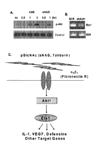
[00351 Figure 1. Nanofibers stimulate Akt 1 activation, an upstream
regulator of Etsl. (A)
Western blot analysis of phospho-Akt in response to NAG and sNAG stimulation
of serum
starved EC. (B) RT-PCR analysis of EC infected either with scrambled control
("SCR") or Akt I
shRNA lentiviruses and assessed for expression of Ets I and S26 as a loading
control. (C)
Schematic of a signal transduction pathway transducing a signal from sNAG
nanofibers to Aktl,
Etsl and Defensins.
[0036] Figure 2. Delayed wound healing in Aktl null animals is partially
rescued by
Taliderm treatment. (A) Representative images of wounded WT and AKT1 null mice
with and
without treatment of Taliderm. (B) H&E staining of representative mouse skin
sections from
day 3 wounds.
100371 Figure 3. sNAG nanofibers stimulate cytokine and defensin expression
in primary
endothelial cells. (A) Immunohistochemisty of EC treated with or without sNAG
using an
antibody directed against a-dcfensin. (B) ELISA showing that nanofiber
treatment of EC results
in the secretion of a-defensins 1-3 (serum starved, treated with 5 jig/m1 or
101.tg,/m1sNAG).
[0038] Figure 4. sNAG nanofibers stimulate defensin expression in primary
endothelial
cells in an Aktl dependent manner. (A) and (B) Quantitative RT-PCR analyses of
serum starved
EC ("ss") treated with or without sNAG ("snag"), with or without PD98059 (MAPK
inhibitor,
"PD"), Wortmannin (PI3K inhibitor, "wtm") or infected with a scrambled control
("SCR"), or
Aktl ("AKT I") shRNA lentiviruses and assessed for expression of the genes
indicated.
- [0039] Figure 5. sNAG nanofibers stimulate 3-defensin 3 expression in
mouse
keratinocytes. (A) Immunofluorescent staining with 13-defensin 3 (visible as
bright staining in
the upper right hand panel; see, e.g., thick white arrows) and Involucrin
antibodies of paraffin
embedded mouse cutaneous wound sections from WT and Aktl null animals on Day
3. (B)
Quantification of13-defensin 3 immunofluorescent staining using NIHImageJ
software
(TX=Taliderm; Akt1=Akt1 null). (C) Immunofluorescent staining of WT and Aktl
null treated
9
CA 02832859 2013-10-09
WO 2012/142581
PCT/US2012/033782
and untreated keratinocytes with P-Defensin 3 (visible as bright staining;
see, e.g., thick white
arrows) and TOPRO-3 (nuclei staining; see, e.g., thin white arrows). Notice
the increase in 13-
Defensin 3 staining in WT and Aktl Taliderm treated wounds.
100401 Figure 6. Aktl dependent transcription factor binding sites.
Schematic of Aktl
dependent transcription factor binding sites. Using Genomatix software, 500 bp
upstream of the
transcription start site was analyzed for conserved sites on the mRNA of DEF
I, 4, and 5 (ETS-
.
black ovals; FKHD-striped ovals; CREB-white ovals; NFKB-checkered ovals).
10041] Figure 7. sNAG treatment results in expression and secretion of
defensins in =vitro.
(A) RTPCR analysis of serum starved ("SS") primary endothelial cells treated
with sNAG
(50 g/m1) for the times indicated and assessed for expression of P-defensin 3
and a-defensin 1.
(B) Immunofluorescent labeling of endothelial cells either serum starved
(untreated) or treated
with sNAG nanofibers (10 g/m1 for 5hrs). Antibodies are directed against ct-
defensin 5 (F1TC,
upper left hand panel), 13-defensin 3 (Texas Red, upper right hand panel).
Nuclei are stained with
TOPRO-3 (Blue, lower left hand panel). Lower right hand panel represents
triple overlay. (C)
Immunofluorescent labeling of keratinocytes (HaCat) that are either serum
starved (untreated) or
treated with sNAG nanofibers (10 g/m1 for 5 hours). Antibodies are directed
against a-defensin
(FITC, upper left hand panel), P-defensin 3 (Texas Red, upper right hand
panel). Nuclei are
stained with TOPRO-3 (Blue, lower left hand panel).
100421 Figure 8. sNAG induced defensin expression is dependent on Aktl. (A)
-
Quantitative RT-PCR analyses using primers directed against a-defensin 1 from
total RNA
isolated from serum starved endothelial cells treated with or without sNAG for
3 hours, with or
without pretreatment with PD098059 ("PD")(501.tM), wortmannin ("WTM")(100nm).
Quantitation is relative to the S26 protein subunit. (B) Quantitation of P-
defensin 3 expression
from total RNA isolated from serum starved endothelial cells treated with or
without sNAG for 3
hours, with or without PD98059 (50am), wortmannin (100nm) and shown as
relative to S26. (C)
Western Blot analysis of phospho-Akt in serum starved endothelial cells (SS)
stimulated with
sNAG for the times indicated. Line indicates where lanes have been removed (D)
Quantitative
RT-PCR analyses of serum starved endothelial cells infected with a scrambled
control (SCR) or
Aktl shRNA lentiviruses, treated with or without sNAG and assessed for a-
defensin 4
expression. Quantitation is shown relative to S26. (E) Quantitation of P-
defensin 3 expression
from total RNA isolated from serum starved endothelial cells infected with a
scrambled control
CA 02832859 2013-10-09
WO 2012/142581
PCT/US2012/033782
(SCR) or Aktl shRNA lentiviruses, treated with or without sNAG. Quantitation
is shown
relative to S26. All experiments were done in at least triplicate and repeated
at least three
independent times and p values are shown.
100431 Figure 9. sNAG induced defensin expression in vivo requires Aktl.
(A) Paraffin
embedded sections of cutaneous wounds harvested on day 3 post wounding from
both WT (n=3)
and Aktl mice. Wounds were either untreated or treated with sNAG membrane.
Immunofluorescence was performed using antibodies directed against 13-defensin
3 (green,
visible as bright staining in the upper right hand panel; see, e.g., white
thick arrows), Involucrin
(Red), and Topro (Blue, nuclei staining; see, e.g., white thin arrows). (B)
Paraffin embedded
section from WT treated with sNAG harvested on day 3. Immunofluorescence was
performed
using antibodies directed against f3- defensin 3 (green, visible as bright
staining; see, e.g., thick
white arrows), Involucrin (Red), and Topro (Blue, nuclei staining; see, e.g.,
thin white arrows).
This lower magnification (20x) is included to better illustrate the epidermal
layers expressing f3-
defensin 3. Scale bars = 50 [tm. (C) Quantitation of f3-defensin 3 expression
from paraffin
embedded sections was performed using NIH ImageJ software. Experiments were
repeated three
independent times and p values are shown.
100441 Figure 10. sNAG treatment increases wound closure in wild type mice.
H&E
staining of wound tissue sections derived from C57B16 wild type animals either
untreated or
treated with sNAG membrane. The day post-wound is indicated to the left of
each panel. The
solid black line follows the keratinocyte cell layer indicating wound closure.
Black arrows
indicate the margin of the wound bed.
100451 Figure 11. sNAG treatment reduces bacterial infection in an Aktl
dependent
manner. (A) Tissue gram staining of S. aureus infected wounds from WT mice. WT
mice were
wounded using a 4 mm biopsy punch. Immediately after wounding mice were
inoculated with I
x 109 cfti/ml. 30 minutes post-infection, mice in the treated group were
treated with Taliderm.
Skin samples were taken 5 days post-treatment and sectioned for analysis.
Tissue gram staining
was performed. Dark purple staining indicates gram-positive bacteria and
neutrophils that have
engulfed bacteria. Sections under 20x and 40x magnification are shown. (B)
Tissue gram
staining of paraffin embedded S. aureus infected wounds from WT and Aktl null
mice (n=3).
Infected wounds were either untreated or treated with sNAG membrane and wound
beds were
harvested on day 3 and day 5 for analysis. Dark purple staining indicates the
presence of gram
11
CA 02832859 2013-10-09
WO 2012/142581
PCMJS2012/033782
=
positive bacteria in the wound bed. Black arrows indicate examples of gram
positive staining.
Note the accumulation of positive staining in untreated WT that is lacking in
WT animals treated
with sNAG. Scale bars = 5011m. (C) CFUs derived from day 5 post wounding were
quantitated
from S. aureus infected wounds using both treated and untreated WT (n= 3) and
Aktl mice
(n=3). Wild type mice that were sNAG treated show a significant (p<.01)
decrease in bacteria
load in the wound.beds as compared to Aktl null animals. All experiments were
repeated three
independent tinfes and the p values are shown. (D) CFU quantitated from
infected wounds at
day 3 post wounding in a similar fashion described in (C). sNAG treatment of
infected wounds
shows a significant decrease in CFU of both WT and Aktl null animals on day 3,
but the WT
animals show an approximate 10 fold difference compared to a 2 fold difference
in Aktl
animals. (E) Quantitation of CFUs in S. aureus cultures that were either
untreated or treated with
various amounts of sNAG nanofibers. Each experiment was performed three
independent times
and p values are shown. (F) Tissue gram staining of S. aureus infected wounds
harvested on day
3 post wound from WT mice (n=3) that were treated with or without p- defensin
3 peptide (1.0
uM). Note the decrease in gram positive staining in infected wounds that were
treated with 13-
defensin 3 peptide. (G) Quantitation of CFUs from S. aureus infected WT mice
(n=3) treated
with or without 13-defensin 3 peptide. Infected wounds that were treated with
peptide show a
significant decrease (p <.05) in CFU. Scale bars = 50 m. Each experiment was
performed three
independent times and p values are shown.
[0046] Figure 12. Rapid induction of defensin expression by sNAG treatment
of S. aureus
infected wounds. (A) Paraffin embedded tissue sections from S. aureus infected
wounds,
harvested on day 3, were subjected to immunofluorescence using antibodies
directed against 13-
defensin 3 (green, visible as bright staining in the upper right hand panel
and in the lower panel
in the middle; see, e.g., thick white arrows), Involucrin (red) to mark the
keratinocyte layer, and
Topro (blue, nuclei staining; see, e.g., thin white arrows) from both sNAG
treated WT (n=3) and
untreated WT mice (n=3). Non specific staining of keratin is indicated by the
no primary control
which was stained with secondary antibody only. Scale bar = 50htm. (B)
Quantitation of 3-
defensin 3 expression from paraffin embedded sections using Nil-1 ImageJ
software. S. aureus
infected wounds that were treated with sNAG show a significant increase
(p<.05) in 3-defensin 3
staining. Experiments were repeated three independent times and p values are
shown.
12
CA 02832859 2013-10-09
WO 2012/142581
PCMJS2012/033782
100471 Figure 13. Antibodies against P-defensin 3 impedes antibacterial
effects of sNAG
treatment. (A) Tissue gram staining of paraffin embedded S. aureus infected
wounds treated
with sNAG from WT mice (n=3) that were harvested on Day 3. sNAG treated wounds
were
treated with either p-defensin 3 antibody or isotype control goat IgG antibody
prior to sNAG
treatment. Representative images show increased accumulation gram positive
staining (black
arrows) in the wound beds of mice treated with an antibody directed against p-
defensin 3. Scale
bar 201tm. (B) Quantitation of CFUs from S. aureus infected WT mice treated
either P-
defensin 3 antibody (n=3) or control IgG antibody (n=3) prior to sNAG
treatment. p-defensin 3
application significantly increased (p <.05) CFU.
100481 Figure 14. Effect of irradiation on chemical and physical structure
of poly-N-
acetylglucosamine ("pG1cNAc") fibers. (A) Correlation between molecular weight
of pG1cNAc
and irradiation level/formulation for irradiation. (B) Infrared (IR) spectrum
of non-irradiated
pG1cNAc slurry (top line), pG1cNAc slurry irradiated at 100 kGy (bottom line),
and pG1cNAc
slurry irradiated at 200 kGy (middle line). (C) Scanning electron microscopic
(SEM) analyses of
pG1cNAc..(D) Scanning electron microscopic (SEM) analyses of sNAG.
[0049] Figure 15. pGleNAc did not affect metabolic rate. For each time
period (i.e., at 24
and 48 hours), the identity for each of the four bars (from left to right) is
as follows: serum
starvation (SS), VEGF, and pG1cNike (NAG) at 50 and 100 ng/ml.
100501 Figure 16. pGleNAc protected human umbilical vein endothelial cell
(EC) from cell
death induced by serum deprivation. For each time period (i.e., at 24, 48 and
72 hours), the
identity for each of the five bars (from left to right)=is as follows: serum
starvation (SS), VEGF,
and pG1cNAc (NAG) at 50, 100, and 250 jig/ml.
[0051] Figure 17. sNAG induced marked increase in metabolic rate. Identity
for each of the
five bars (from left to right) is as follows: serum starvation (SS), VEGF, and
sNAG at 50, 100
and 200 jig/ml.
[0052] Figure 18. sNAG did not protect EC from cell death induced by serum
deprivation.
For each time period (i.e., at 24 and 48 hours), the identity for each of the
five bars (from left to
right) is as follows: serum starvation (SS), VEGF, and sNAG at 50, 100 and 200
jig/ml.
[0053] Figure 19. Numeric Pain Intensity Scale.
13
CA 02832859 2013-10-09
WO 2012/142581
PCT/US2012/033782
[0054] Figure 20. Schematic showing experimental set up for the 3% DSS
(dextran sodium
sulphate)-induced inflammatory bowel disease (in particular, ulcerative
colitis) in a mouse
model.
100551 Figure 21. sNAG treatment decreased inflammation in an animal model
of
inflammatory bowel disease. (A) H&E staining of a section of intestinal
epithelium from a
control group of 10 mice administered 3% DSS via drinking water for 7 days
(day 0 to day 7),
and saline via rectal suppository at day 0 and day 3 (100 I). (B) H&E
staining of a section of
intestinal epithelium from a test group of 10 mice administered 3% DSS via
drinking water for 7
days (day 0 to day 7), and sNAG via rectal suppository at day 0 and day 3 (100
I total with 12
gift! sNAG). Thin arrow and bracket point to the site of edema, and thick
arrow points to the
site of leukocytic infiltration.
[0056] Figure 22. sNAG treatment decreased fibrosis in an animal model of
inflammatory
bowel disease. (A) staining for fibrosis of a section of intestinal epithelium
from a control group
of 10 mice administered 3% DSS via drinking water for 7 days (day 0 to day 7),
and saline via
rectal suppository at day 0 and day 3(100 1). (B) staining for fibrosis of a
section of intestinal
epithelium from a test group of 10 mice administered 3% DSS via drinking water
for 7 days (day
0 to day 7), and sNAG via rectal suppository at day 0 and day 3 (100 al total
with 12 g/ 1
sNAG).
5. DETAILED DESCRIPTION =
[0057] The inventors of the present invention have found that sNAG
nanofibers can
stimulate expression of defensins, which may boost the innate immune response.
It is widely
accepted that defensins are important players in innate immunity. As
demonstrated in the.
examples presented in Sections 6.1 and 6.2, infra, the inventors of the
present invention have
found that sNAG nanofibers can increase the expression of both a- and p- type
defensins in
endothelial cells and I3-type defensins in keratinocytes in vitro and in a
wound healing model in
vivo.
100581 Further, as demonstrated in the examples presented in Sections 6.1
and 6.2, infra, but
without being bound by any specific mechanism of action, Aktl appears to be
important for
sNAG-dependent defensin expression in vitro and in vivo, in a wound healing
model.
[0059] The inventors of this invention have also found that a number of
Toll-like receptors
can be up-regulated by sNAG treatment of human endothelial cells. Toll-like
receptors ("TLRs"
14
CA 02832859 2013-10-09
WO 2012/142581
PCMJS2012/033782
or "TLR") are highly conserved receptors that activate innate immunity. Recent
work has linked
human dcfcnsin expression to TLR activation, in particular, stimulation of
TLRs can lead to
increased defensin synthesis. Thus, without being bound by any mechanism of
action, sNAG
nanofibers may act as a stimulator of innate immunity.
[0060] Accordingly, described herein is the use of sNAG nanofibers as a
novel method for
preventing and/or treating of infections and diseases for which an increase in
expression and/or
secretion of one or more of defensins and Toll-like receptors may be
beneficial. In certain
embodiments, treatment of viral, yeast or fungal infections with sNAG
nanofibers decreases the
pathogen count in patients. In specific embodiments, the use of sNAG
nanofibers enhances
wound closure while simultaneously eradicating, decreasing or preventing a
viral, a fungal or an
yeast infection of the wound. In other embodiments, the sNAG nanofibers can be
used in
treating a dermatological condition such as dermatitis or psoriasis by, for
example, alleviating
one or more symptoms of such diseases. In yet another embodiment, the sNAG
nanofibers can
be used in treating an Inflammatory Bowel Disease (e.g., Crohn's disease) by,
for example,
alleviating one or more symptoms of such diseases.
100611 The inventors have, in fact, found that sNAG nanofibers can be
effective to treat viral
infections. In particular, the inventors found that sNAG nanofibers are
effective to treat HSV
infection when administered topically to human patients. Example 8, infra,
demonstrates that
topical administration of sNAG nanofibers to cold sores reduces the pain
associated with cold
sores and reduces the duration of the cold sores in human patients. Cold sores
are typically
caused by HSV-1 infection, Thus, described herein are uses of sNAG nanofibers
to treat viral
infections, in particular, topical viral infections. In specific embodiments,
described herein are
uses of sNAG nanofibers to treat an HSV infection, or to treat and/or prevent
a symptom
associated with an HSV infection (e.g., a cold sore or a lesion) by topical
administration of
sNAG nanofibers to a patient (e.g., at the site of HSV infection or the site
of a symptom of HSV
infection, or at the site where a symptom of infection is known to occur).
[0062] The inventors have also found that sNAG nanofibers can be
effective to treat
inflammatory bowel disease (IBD). In particular, the inventors found that sNAG
nanofibers are
effective to treat IBD in an animal model of IBD when administered rectally
(such as via a
suppository). Example 9, infra, demonstrates that administration of sNAG
nanofibers reduces
inflammation and fibrosis associated with IBD in a mouse model of IBD. Thus,
described herein
=
=
CA 02832859 2013-10-09
WO 2012/142581 PCT/US2012/033782
are uses of sNAG nanofibers to treat IBD, such as ulcerative colitis and
Crohn's disease. In
specific embodiments, described herein are uses of sNAG nanofibers to treat
IBD (e.g.,
ulcerative colitis, or Crohn's disease) by topical administration of sNAG
nanofibers to a patient
(e.g., to the anus or rectally via a suppository, a cream, a suspension, a
liquid solution, a gel, or =
an ointment).
5.1 sNAG Nanotibers
100631 Described herein are sNAG nanofiber compositions. The sNAG
nanofibers comprise
fibers of poly-N-acetylglucosamine and/or a derivative(s) thereof, the
majority of which are less
than 30 microns in length and at least 1 micron in length as measured by any
method known to
= one skilled in the art, for example, by scanning electron microscopy
("SEM"). Such sNAG
nanofibers may be obtained, for example, as described herein.
100641 In certain embodiments, the majority (and in certain embodiments, at
least 60%, 70%,
80%, 90%, 95%, 98%, 99%, 99.5%, 99.8%, 99.9%, or 100%, or between 55% to 65%,
55% to
75%, 65% to 75%, 75% to 85%, 75% to 90%, 80% to 95%, 90% to 95%, or 95% to
99%) of the
sNAG nanofibers are less than about 30, 25, 20, 15, 12, 10, 9, 8, 7, 6, 5, 4,
or 3 microns in
length, and at least 1 micron in length as measured by any method known to one
skilled in the
art, for example, by SEM. In specific embodiments, the majority (and in
certain embodiments,
at least 60%, 70%, 80%, 90%, 95%, 98%, 99%, 99.5%, 99.8%, 99.9%, or 100%, or
between
55% to 65%, 55% to 75%, 65% to 75%, 75% to 85%, 75% to 90%, 80% to 95%, 90% to
95%, or
95% to 99%) of the sNAG nanofibers are less than about 15 microns or less than
about 12
microns in length, and at least 1 micron in length as measured by any method
known to one
skilled in the art, for example, by SEM. In specific embodiments, all (100%)
of the sNAG
nanofibers are less than about 15 microns or less than about 10 microns in
length, and at least 1
micron in length as measured by any method known to one skilled in the art,
for example, by
SEM. In certain embodiments, the majority (and in certain embodiments, at
least 60%, 70%,
80%, 90%, 95%, 98%, 99%, 99.5%, 99.8%, 99.9%, or 100%, or between 55% to 65%,
55% to
75%, 65% to 75%, 75% to 85%, 75% to 90%, 80% to 95%, 90% to 95%, or 95% to
99%) of the
sNAG nanofibers are equal to or less than 14, 13, 12, 11, 10, 9, 8 or 7
microns in length, and at
least 1 micron in length as measured by any method known to one skilled in the
art, for example,
by SEM. In some embodiments, the majority (and in certain embodiments, at
least 60%, 70%,
80%, 90%, 95%, 98%, 99%, 99.5%, .99.8%, 99.9%, or 100%, or between 55% to 65%,
55% to
16
CA 02832859 2013-10-09
WO 2012/142581
PCMJS2012/033782
75%, 65% to 75%, 75% to 85%, 75% to 90%, 80% to 95%, 90% to 95%, or 95% to
99%) of the
sNAG nanofibers are between Ito 15, 2 to 15,2 to 14, Ito 12,2 to 12, 1 to 10,2
to 10, 3 to 12,
3 to 10, 4 to 12, 4 to 10, 5 to 12, 5 to 10, 1 to 9, 2 to 9, 3 to 9, 1 to 8, 2
to 8, 3 to 8, 4 to 8, 1 to 7,
2 to 7, 3 to 7, 4 to 7, 1 to 6, 1 to 5, 1 to 4, or 1 to 3 microns in length as
measured by any method
known to one skilled in the art, for example, by SEM.
100651 In a specific embodiment, the majority (and in certain embodiments,
at least 60%,
70%, 80%, 90%, 95%, 98%, 99%, 99.5%, 99.8%, 99.9%, or 100%, or between 55% to
65%,
55% to 75%, 65% to 75%, 75% to 85%, 75% to 90%, 80% to 95%, 90% to 95%, or 95%
to
99%) of the sNAG nanofibers are about 8, 7, 6, 5, 4, 3 or 2 microns in length
as measured by any
method known to one skilled in the art, for example, by SEM. In another
specific embodiment,
the majority (and in certain embodiments, at least 60%, 70%, 80%, 90%, 95%,
98%, 99%,
99.5%, 99.8%, 99.9%, or 100%, or between 55% to 65%, 55% to 75%, 65% to 75%,
75% to
85%, 75% to 90%, 80% to 95%, 90% to 95%, or 95% to 99%) of the sNAG nanofibers
are
between about 2 to about 10 microns, about 3 to about 8 microns, about 4 to
about 7 microns,
about 4 to about 10 microns, or about 5 to about 10 microns in length as
measured by any
method known to one skilled in the art, for example, by SEM. In another
specific embodiment,
all (100%) of the sNAG nanofibers are between about 2 to about 10 microns,
about 3 to about 8
microns, about 4 to about 7 microns, about 4 to about 10 microns, or about 5
to about 10 microns
in length as measured by any method known to one skilled in the art, for
example, by SEM.
100661 In certain embodiments, the sNAG nanofibers fibers are in a range
between 0.005 to 5
microns in thickness and/or diameter as determined by electron microscopy. In
specific
embodiments, the sNAG nanofibers are about 0.01, 0.02, 0.03, 0.04, 0.05, 0.06,
0.07, 0.08, 0.09,
0.1, 0.2, 0.25, 0.3, 0.35, 0.4, 0.45, 0.5, 0.55, 0.6, 0.65, 0.7, 0.75, 0.8,
0.85, 0.9, 1, 1.1, 1.2, 1.3,
1.4, 1.5, 1.6, 1.7, 1.8, 1.9, 2, 2.2, 2.4, 2.6, 2.8, 3 or 4 microns in
thickness and/or diameter: on
average, or any range in between (e.g., 0.02 to 2 microns, 0.02 to 1 microns,
0.02 to 0.75
microns, 0.02 to 0.5 microns, 0.02 to 0.5 microns, 0.05 to 1 microns, 0.05 to
0.75 microns, 0.05
to 0.5 microns, 0.1 to 1 microns, 0.1 to 0.75 microns, 0.1 to 0.5 microns,
etc.). In specific =
embodiments, the majority (and in certain embodiments, at least 60%, 70%, 80%,
90%, 95%,
98%, 99%, 99.5%, 99.8%, 99.9%, or 100%, or between 55% to 65%, 55% to 75%, 65%
to 75%,
75% to 85%, 75% to 90%, 80% to 95%, 90% to 95%, or 95% to 99%) of the sNAG
nanofibers
have a thickness or diameter of about 0.02 to 1 microns. In other specific
embodiments, the
17
CA 02832859 2013-10-09
WO 2012/142581
PCT/US2012/033782
majority (and in certain embodiments, at least 60%, 70%, 80%, 90%, 95%, 98%,
99%, 99.5%,
99.8%, 99.9%, or 100%, or between 55% to 65%, 55% to 75%, 65% to 75%, 75% to
85%, 75%
to 90%, 80% to 95%, 90% to 95%, or 95% to 99%) of the sNAG nanofibers have a
thickness or
diameter of about 0.05 to 0.5 microns. In specific embodiments, all (100%) of
the sNAG
nanofibers have a thickness or diameter of about 0.02 to 1 microns or about
0.05 to 0.5 microns.
In certain embodiments, the majority (and in certain embodiments, at least
60%, 70%, 80%,
90%, 95%, 98%, 99%, 99.5%, 99.8%, 99.9%, or 100%, or between 55% to 65%, 55%
to 75%,
65% to 75%, 75% to 85%, 75% to 90%, 80% to 95%, 90% to 95%, or 95% to 99%) of
the sNAG
nanofibers have a thickness or diameter of about 0.02 to 2 microns, 0.02 to 1
microns, 0.02 to
0.75 microns, 0.02 to 0.5 microns, 0.02 to 0.5 microns, 0.05 to 1 microns,
0.05 to 0.75 microns,
0.05 to 0.5 microns, 0.1 to 1 microns, 0.1 to 0.75 microns, or 0.1 to 0.5
microns.
[0067] In certain embodiments, the majority (and in certain embodiments, at
least 60%, 70%,
80%, 90%, 95%, 98%, 99%, 99.5%, 99.8%, 99.9%, or 100%, or between 55% to 65%,
55% to
75%, 65% to 75%, 75% to 85%, 75% to 90%, 80% to 95%, 90% to 95%, or 95% to
99%) of the
sNAG nanofibers are between 1 and 15 microns, or between (or in the range of)
1 to 10 microns,
2 to 10 microns, 3 to 10 microns, 4 to 10 microns, 4 to 7 microns, 5 to 10
microns, or 5 to 15
microns in length and have a thickness or diameter of about 0.02 to 1 microns.
[0068] In certain embodiments, the molecular weight of the sNAG nanofibers
is less than
100kDa, 90kDa, 80kDa, 75kDa, 70 kDa, 65 kDa, 60IcDa, 55kDa, 50kDa, 45 kDA,
40kDa,
35kDa, 30kDa, or 25kDa. In certain embodiments, the majority (and in certain
embodiments, at
least 60%, 70%, 80%, 90%, 95%, 98%, 99%, 99.5%, 99.8%, 99.9%, or 100%, or
between 55%
to 65%, 55% to 75%, 65% to 75%, 75% to 85%, 75% to 90%, 80% to 95%, 90% to
95%, or 95%
to 99%) of the sNAG nanofibers have a molecular weight of less than 100kDa,
90kDa, 80kDa,
75kDa, 70 kDa, 65 kDa, 60kDa, 55kDa, 50kDa, 45 kDA, 40kDa, 35kDa, 30kDa, or
25kDa. In
other embodiments, the majority (and in certain embodiments, at least 60%,
70%, 80%, 90%,
95%, 98%, 99%, 99.5%, 99.8%, 99.9%, or 100%, or between 55% to 65%, 55% to
75%, 65% to
75%, 75% to 85%, 75% to 90%, 80% to 95%, 90% to 95%, or 95% to 99%) of the
sNAG
nanofibers have a molecular weight between about 5kDa to 100kDa, about 10kDa
to 100kDa,
about 20kDa to 100kDa, about 10kDa to 80kDa, about 20kDa to 80kDa, 20kDa to
75kDa, about
25kDa to about 75kDa, about 30kDa to about 80kDa, about 30kDa to about 75kDa,
about 40kda
to about 80kDa, about 40kDa to about 75kDa, about 40kDa to about 70kDa, about
50kDa to
18
CA 02832859 2013-10-09
WO 2012/142581
PCT/US2012/033782
=
about 70kDa, or about 55kDa to about 65kDa. In one embodiment, the majority
(and in certain
embodiments, at least 60%, 70%, 80%, 90%, 95%, 98%, 99%, 99.5%, 99.8%, 99.9%,
or 100%,
or between 55% to 65%, 55% to 75%, 65% to 75%, 75% to 85%, 75% to 90%, 80% to
95%,
90% to 95%, or 95% to 99%) of the sNAG nanofibers have a molecular weight of
about 60kDa. =
[00691 In certain embodiments, 1% to 5%, 5% to 10%, 5% to 15%, 20% to 30%
or 25% to
30% of the sNAG nanofibers are deacetylated. In some embodiments, 1%, 5%, 10%,
15%, 20%,
25%, or 30% of the sNAG nanofibers are deacetylated. In other embodiments,
less than 30%,
25%, 20%, 15%, 10%, 5%, 4%, 3%, 2% or 1% of the sNAG nanofibers are
deacetylated. In
some embodiments, equal to or more than 1%, 5%, 10%, 15%, 20%, 25%, 30%, 35%,
40%,
45%, 50%, 55%, 60%, 65%, 70%, 75%, 80%, 85%, 90%, 95% or 99%, or all (100%),
of the
sNAG nanofibers are deacetylated. In other embodiments, less than 1%, 5%, 10%,
15%, 20%,
25%, 30%, 35%, 40%, 45%, 50%, 55%, 60%, 65%, 70%, 75%, 80%, 85%, 90%, 95%,
99%, or
100% of the sNAG nanofibers are deacetylated.
[00701 In certain embodiments, 70% to 80%, 75% to 80%, 75% to 85%, 85% to
95%, 90%
to 95%, 90% to 99% or 95% to 100% of the sNAG nanofibers are acetylated. In
some
embodiments, 70%, 75%, 80%, 85%, 90%, 95%, 98%, 99% or 100% of the sNAG
nanofibers
are acetylated. In other embodiments, more than 70%, 75%, 80%, 85%, 90%, 95%,
97%, 98%,
99%, 99.5% or 99.9% of the sNAG nanofibers are acetylated. In some
embodiments, equal to or
more than 1%, 5%, 10%, 15%, 20%, 25%, 30%, 35%, 40%, 45%, 50%, 55%, 60%, 65%,
70%,
75%, 80%, 85%, 90%, 95%, 97%, 98% or 99%, or all (100%), of the sNAG
nanofibers are
acetylated. In other embodiments, less than 1%, 5%, 10%, 15%, 20%, 25%, 30%,
35%, 40%,
45%, 50%, 55%, 60%, 65%, 70%, 75%, 80%, 85%, 90%, 95%, 97%, 98%, 99%, or 100%
of the
sNAG nanofibers are acetylated.
[00711 In some embodiments, the majority (and in certain embodiments, at
least 60%, 70%,
80%, 90%, 95%, 98%, 99%, 99.5%, 99.9%, or 100%) of the sNAG nanofibers are
between (or in
the range of) 2 to 12 microns, 2 to 10 microns, 4 to 15 microns, 4 to 10
microns, 5 to 15 microns,
or 5 to 10 microns, and such sNAG nanofibers are at least 70%, 75%, 80%, 85%,
90%, 95%,
97%, 98%, 99% or 100% acetylated.
[00721 In some embodiments, the sNAG nanofibers comprise at least one
glucosamine
monosaccharide, and may further comprise at least 10%, 20%, 30%, 40%, 50%,
60%, 70%,
80%, 90%, 95% or 99% of the N-acetylglucosaminc monosaccharides. In other
embodiments,
19
CA 02832859 2013-10-09
WO 2012/142581
PCMJS2012/033782
the sNAG nanofibers comprise at least one N-acetylglucosamine monosaccharide,
and may
further comprise at least 10%, 20%, 30%, 40%, 50%, 60%, 70%, 80%, 90%, 95% or
99% of
glucosamine monosaccharides.
100731 In one aspect, the sNAG nanofibers increase the metabolic rate of
serum-starved
human umbilical cord vein endothelial cells ("EC") in a MTT assay. A MTT assay
is a
laboratory test and a standard colorimetric assay (an assay which measures
changes in color) for
measuring cellular proliferation (cell growth). Briefly, yellow MTT (3-(4,5-
Dimethylthiazol-2-
y1)-2,5-diphenyltetrazolium bromide, a tetrazole) is reduced to purple
formazan in the
mitochondria of living cells. This reduction takes place only when
mitochondrial reductase
enzymes are active, and therefore conversion can be directly related to the
number of viable
(living) cells. The MTT assay is described in Example 6, infra, where it is
utilized to assess the
effect of sNAG nanofibers on the metabolic rate of EC cells. The metabolic
rate of cells may
also be determined by other techniques commonly known to the skilled artisan.
100741 In another aspect, the sNAG nanofibers do not rescue apoptosis of
serum-starved EC
in a trypan blue exclusion test. A trypan blue exclusion test is a dye
exclusion test used to
determine the number of viable cells present in a cell suspension. It is based
on the principle that
live cells possess intact cell membranes that exclude certain dyes, such as
trypan blue, Eosin, or
propidium, whereas dead cells do not. The trypan blue assay is described in
Example 6, infra,
where it is utilized to assess the effect of sNAG nanofibers on cell viability
of EC cells. The
viability of cells may also be determined by other techniques commonly known
to the skilled
artisan.
[0075] In certain embodiments, compositions comprising the sNAG nanofibers
are
described, wherein the sNAG nanofibers increase the metabolic rate of serum-
starved human
umbilical cord vein endothelial cells in a MTT assay and/or do not rescue
apoptosis of serum-
starved human umbilical cord vein endothelial cells in a trypan blue exclusion
test. In some
embodiments, the sNAG nanofibers increase the metabolic rate of serum-starved
human
umbilical cord vein endothelial cells in a MTT assay and do not rescue
apoptosis of serum-
starved human umbilical cord vein endothelial cells in a trypan blue exclusion
test.
100761 In a specific embodiment, the sNAG nanofibers are biocompatible.
Biocompatibility
may be determined by a variety of techniques, including, but not limited to
such procedures as
the elution test, intramuscular implantation, or intracutaneous or systemic
injection into animal
subjects. Such tests are described in U.S. Patent No. 6,686,342 (see, e.g.,
Example 10),
Some of the biocompatibility tests are described in Example 7, infra, which
show that sNAG
nanofibers arc non-reactive in such tests.
100771 In certain embodiments, the sNAG nanofibers used in the methods
described herein
are non-reactive in a biocompatibility test or tests. For example, the sNAG
nanofibers used in
the methods described herein may be non-reactive when tested in an elution
test, an
intramuscular iMplantation test, an intracutaneous test, and/or a systemic
test. In other
embodiments, the sNAG nanofibers used in the methods described herein have
Grade 0 or Grade
1 test score when tested in an elution test, an intramuscular implantation
test, an intracutaneous
test, or a systemic test. In yet another embodiment, the sNAG nanofibers used
in the methods
described herein are at most mildly reactive when tested in an elution test,
an intramuscular
implantation test, an intracutaneous test, and/or a systemic test. In certain
embodiments, the
compositions described herein do not cause an allergenic reaction or an
irritation. In other
embodiments, the compositions described herein cause at most a mild allergenic
reaction or a
mild irritation, e.g., at the site of application. The relevant tests and
evaluation of test results are
described in, e.g., U.S. Patent No. 6,686,342 and in Section 6.8, infra.
100781 In a specific embodiment, the sNAG nanofibers are non-reactive
when tested in an
intramuscular implantation test. In one aspect, an intramuscular implantation
test is an
intramuscular implantation test¨ ISO 4 week.implantation, as described in
Section 6.8.3, infra.
In certain embodiments, the sNAG nanofibers display no biological reactivity
as determined by
an elution test (Elution Test Grade = 0). In some embodiments, the sNAG
nanofibers have a test
score equal to ''0" and/or are at most a negligible irritant as determined by
intracutaneous
injection test. In some embodiments, the sNAG nanofibers elicit no intradermal
reaction (i.e.,
Grade l reaction) in Kligman test and/or have a weak allergenic potential as
determined by
Kligman test. Example 7, infra, shows that sNAG nanofibers are non-reactive in
an
intramuscular implantation test, an intracutaneous injection test, and Kligman
test.
100791 In certain aspects, the sNAG nanofibers arc immunoneutral (i.e.,
they do not elicit an
immune response).
10080] In some embodiments, the sNAG nanofibers are biodegradable. The
sNAG
nanofibers preferably degrade within about I day, 2 days, 3 days, 5 days, 7
days (I week), 8
21
CA 2832859 2 0 1 8 ¨1 1 ¨13
CA 02832859 2013-10-09
WO 2012/142581
PCT/1JS2012/033782
days, 10 days, 12 days, 14 days (2 weeks), 17 days, 21 days (3 weeks), 25
days, 28 days (4
weeks), 30 days, 1 month, 35 days, 40 days, 45 days, 50 days, 55 days, 60
days, 2 months, 65
days, 70 days, 75 days, 80 days, 85 days, 90 days, 3 months, 95 days, 100 days
or 4 months after
administration or implantation into a patient.
100811 In certain embodiments, the sNAG nanofibers do not cause a
detectable foreign body
reaction. A foreign body reaction, which may occur during wound healing,
includes
accumulation of exudate at the site of injury, infiltration of inflammatory
cells to debride the
area, and the formation of granulation tissue. The persistent presence of a
foreign body can
inhibit full healing. Rather than the resorption and reconstruction that
occurs in wound healing,
the foreign body reaction is characterized by the formation of foreign body
giant cells,
encapsulation of the foreign object, and chronic inflammation. Encapsulation
refers to the firm,
generally avascular collagen shell deposited around a foreign body,
effectively isolating it from
the host tissues. In one embodiment, treatment of a site (e.g., a wound or a
site of a bacterial
infection in a wound) with the sNAG nanofibers does not elicit a detectable
foreign body
reaction in I day, 3 days, 5 days, 7 days, 10 days or 14 days after treatment.
In one such
embodiment, treatment of a site (e.g., a wound) with the sNAG nanofibers does
not elicit a
foreign body encapsulations in I day, 3 days, 5 days, 7 days, 10 days or 14
days after treatment.
100821 In some embodiments, the sNAG nanofibers (i) comprise fibers,
wherein majority of
the fibers are between about 1 and 15 microns in length, and (ii) (a) increase
the metabolic rate
of serum-starved EC in a MIT assay and/or do not rescue apoptosis of serum-
starved EC in a
trypan blue exclusion test, and (b) are non-reactive when tested in an
intramuscular implantation
test. In certain embodiments, the sNAG nanofibers (i) comprise fibers, wherein
majority of the
fibers are between about 1 and 12 microns in length, and (ii) (a) increase the
metabolic rate of
serum-starved EC in a MTT assay and/or do not resew apoptosis of serum-starved
EC in a
trypan blue exclusion test, and (b) are non-reactive when tested in an
intramuscular implantation
test. In some embodiments, the sNAG nanofibers (i) comprise fibers, wherein
majority of the
fibers are between (or in the range of) 1 to 10 microns, 2 to 10 microns, 4 to
10 microns, 5 to 10
microns, or 5 to 15 microns in length, and (ii) (a) increase the metabolic
rate of serum-starved
EC in a MIT assay and/or do not rescue apoptosis of serum-starved EC in a
trypan blue
exclusion test, and (b) are non-reactive when tested in an intramuscular
implantation test. In
some embodiments, the sNAG nanofibers (i) comprise fibers, wherein majority of
the fibers are
22
CA 02832859 2013-10-09
WO 2012/142581
PCMJS2012/033782
between about 4 and 10 microns in length, and (ii) (a) increase the metabolic
rate of serum-
starved EC in a MTT assay and/or do not rescue apoptosis of serum-starved EC
in a trypan blue
exclusion test, and (b) are non-reactive when tested in an intramuscular
implantation test. In
certain embodiments, the sNAG nanofibers (i) comprise fibers, wherein majority
of the fibers are
between about 4 and 7 microns in length, and (ii) (a) increase the metabolic
rate of serum-starved
EC in a MTT assay and/or do not rescue apoptosis of serum-starved EC in a
trypan blue
exclusion test, and (b) are non-reactive when tested in an intramuscular
implantation test.
100831 In certain embodiments, the sNAG nanofibers do not have a direct
effect on the
growth or survival of bacteria, such as S. aureus, as determined by one
skilled in the art. In other
embodiments, sNAG nanofibers do not have a direct effect on the growth or
survival of bacteria,
such as S. aureus, as determined by the methods set forth in Section 6.2.2.5,
infra. In some
embodiments, the sNAG =nanofibers do not have a direct effect in vitro on
bacterial growth or
survival. In one embodiment, the sNAG nanofibers do not have a direct effect
(e.g., in vitro) on
growth or survival of gram-negative bacteria. In another embodiment, the sNAG
nanofibers do
not have a direct effect (e.g., in vitro) on growth or survival of gram-
positive bacteria. In yet
another embodiment, the sNAG nanofibers do not have a direct effect (e.g., in
vitro) on growth
or survival of either gram-positive or gram-negative bacteria.
100841 In some embodiments, the sNAG nanofibers (i) comprise fibers,
wherein majority of
the fibers are between (or in the range of) about 1 and 15 microns, 1 and 12
microns, 1 and 10
microns, 4 and 10 microns, 4 and 15 microns, 5 and 10 microns, 5 and 15
microns, or 4 and 7
microns in length, (ii) do not have an effect on bacterial growth or survival
of Staphylococcus
aureus bacterial cultures in vitro, and (iii) are non-reactive when tested in
a biocompatibility test
(e.g., an intramuscular implantation test).
[00851 In certain embodiments, the sNAG nanofibers induce a certain pattern
of gene
expression (RNA or protein expression as determined by, e.g., RT-PCR,
microarray or ELISA)
in a cell, tissue or organ treated with or exposed to a sNAG nanofiber
composition. Specifically,
in some embodiments, the sNAG nanofibers or a composition comprising the sNAG
nanofibers
induce expression of one or more defensin proteins, one or more defensin-like
proteins, and/or
one or more Toll-like receptors. In yet other embodiments, the sNAG nanofibers
or a
composition comprising the sNAG nanofibers induce expression of one or more
proteins that are
known to have an anti-bacterial effect.
= 23
CA 02832859 2013-10-09
WO 2012/142581
PCT/US2012/033782
[0086] In certain embodiments, the sNAG nanofibers or a composition
comprising the sNAG
nanofibers induce expression of one or more a-defensins (e.g., DEFA1 (i.e., a-
defensin 1),
DEFA1B, DEFA3, DEFA4, DEFA5, DEFA6), one or more P-defensins (e.g., DEFB1
(i.e., 13-
defensin 1), DEFB2, DEFB4, DEFB103A, DEFB104A, DEFB105B, DEFB107B, DEFB108B,
DEFB I 10, DEFB I 12, DEFB114, DEFB118, DEFB119, DEFB123, DEFB124, DEFB125,
DEFB126, DEFB127, DEFB128, DEFB129, DEFB131, DEFB136), and/or one or more 0-
defensins (e.g., DEFT1P). In some embodiments, the sNAG nanofibers or a
composition
comprising the sNAG nanofibers induce expression of one or more of DEFA1,
DEFA3, DEFA4,
DEFA5, DEFB I, DEFB3, DEFB103A, DEFB104A, DEFB108B, DEFB112, DEFB114,
DEFB118, DEFB119, DEFB123, DEFB124, DEFB125, DEFB126, DEFB128, DEFB129 and
DEFB131. In certain embodiments, the sNAG nanofibers or a composition
comprising the
sNAG nanofibers induce expression of one or more Toll receptors (e.g., TLR1,
TLR2, TLR3,
TLR4, TLR5, TLR6, TLR7, TLR8, TLR9, TLRIO, TLR11, and/or TLR12). In other
embodiments, the sNAG nanofibers or a composition comprising the sNAG
nanofibers induce
expression of one or more of IL-1, CEACAM3, SPAG11, SIGIRR (ILI-like
receptor), IRAK1,
1RAK2, IRAK4, TBK I , TRAF6 and 1KKi. In some embodiments, the sNAG nanofibers
or a
composition comprising the sNAG nanofibers induce expression of one or more of
IRAK2,
SIGIRR, TLR1, TLR2, TLR4, TLR7, TLR8, TLRIO and TRAF6. In one embodiment, the
sNAG nanofibers or a composition comprising the sNAG nanofibers induce
expression of at
least one of the above-listed gene products.
100871 In some embodiments, the sNAG nanofibers or a composition comprising
the sNAG
nanofibers induce expression of one or more of the above-listed genes in the
amount equal to or
more than about 0.25 fold, 0.5 fold, I fold, 1.5 fold, 2 fold, 2.5 fold, 3
fold, 3.5 fold, 4 fold, 4.5
fold, 5 fold, 6 fold, 7 fold, 8 fold, 9 fold, 10 fold, 12 fold, 15 fold or 20
fold as compared to the
level of expression of the one or more of the above-listed genes in a cell,
tissue or organ of a
subject before treatment with the sNAG nanofibers (e.g., a known average level
of expression of
the one or more of the above-listed genes). In some embodiments, the sNAG
nanofibers or a
composition comprising the sNAG nanofibers induce expression of one or more of
the above-
listed genes in the amount equal to or more than about 10%, 25%, 50%, 75%,
100%, 125%,
150%, 175%, 200%, 225%, 250%, 275%, 300%, 350%, 400%, 450%, 500%, 550%, 600%,
650%, 700%, 750%, 800%, 900% or 1000% the level of expression of the one or
more of the
24
CA 02832859 2013-10-09
WO 2012/142581
PCMTS2012/033782
above-listed genes in a cell, tissue or organ of a subject before treatment
with the sNAG
nanofibers (e.g., a known average level of expression of the one or more of
the above-listed
genes).
100881 In some embodiments, the sNAG nanofibers but not long poly-N-
acetylglucosamine,
chitin and/or chitosan induce expression of the one or more genes listed
above, as determined by
a method known to one skilled in the art, or described herein. In some of
these embodiments,
long poly-N-acetylglucosarnine, chitin and/or chitosan do not induce
expression of the one or
more genes listed above or induce lower level (e.g., more than 1.25 fold, 1.5
fold, 2 fold, 2.5
fold, 3 fold, 3.5 fold, 4 fold, 4.5 fold, 5 fold, 6 fold, 7 fold, 8 fold, 9
fold, or 10 fold lower) of
expression of the one or more genes listed above as compared to the level of
expression of the
one or more genes listed above induced by the sNAG nanofibers, as determined
by a method
known to one skilled in the art, or described herein.
[0089] In certain embodiments, the sNAG nanofibers or a composition
comprising the sNAG
nanofibers induce a gene expression profile that is consistent with, similar
to, about the same as,
or equivalent to one or more gene expression profiles demonstrated in Tables
I, II, III, V, VIII
and IX, Sections 6.2-6.5, infra. In some embodiments, the sNAG nanofibers or a
composition
comprising the sNAG nanofibers induce expression of one or more of the genes
shown to be
upregulated by sNAG treatment in Tables I, II, III, V, VIII and IX, Sections
6.2-6.5, infra. In
some embodiments, the sNAG nanofibers or a composition comprising the sNAG
nanofibers
induce expression of the majority or all of the genes shown to be upregulated
by sNAG treatment
in Tables I, II, III, V, VIII and IX, Sections 6.2-6.5, infra. In some of
these embodiments, gene
expression levels are measured at 1 hour, 2 hours, 4 hours, 5 hours, 6 hours,
8 hours, 10 hours,
12 hours, 14 hours, 16 hours, 18 hours, 20 hours, 24 hours, 48 hours, 3 days
or 5 days after
treatment of a cell, tissue or organ with a sNAG nanofiber composition by a
method known to
one skilled in the art, or described herein.
[0090] In certain embodiments, the sNAG nanofibers or a composition
comprising the sNAG
nanofibers induce a gene expression profile that differs from the profile
induced by long poly-N-
acetylglucosamine polymers or fibers. In specific embodiments, a gene
expression profile
induced by the sNAG nanofibers is consistent with, similar to, about the same
as, or equivalent
to that shown in Tables I, II, III, V, VIII and IX, Sections 6.2-6.5, infra,
whereas gene expression
profile induced by long poly-N-acetylglucosamine polymers or fibers is
consistent with, similar
to, about the same with, or equivalent to that shown in Table VIII ancUor IX,
Section 6.5, infra.
In other embodiments, the sNAG nanofibers or a composition comprising the sNAG
nanofibers
induce a gene expression profile that differs from the gene expression profile
induced by chitin
or chitosan.
[0091] In a specific embodiment, the sNAG nanofibers are obtained by
irradiating poly-N-
acetylglucosamine and/or a derivative thereof. See Section 5.1.1, infra,
regarding poly-N-
acetylglucosamine and derivatives thereof and Section 5.2, infra, regarding
methods for
producing the sNAG nanofibers using irradiation. Irradiation may be used to
reduce the length
of poly-N-acetylglucosamine fibers and/or poly-N-acetylglucosamine derivative
fibers to form
shortened poly-0-1--t4-N-acetylgiucosamine fibers ancVor shortened poly-N-
acetylglucosamine
derivative fibers, i.e. sNAG nanofibers. Specifically, irradiation may be used
to reduce the
length and molecular weight of poly-N-acctylglucosamine or a derivative
thereof without
disturbing its microstructure. The infrared spectrum (IR) of sNAG nanofibers
is similar to, about
the same as, or equivalent to that of the non-irradiated poly-I3-1---t4-N-
acetylgulcosamine or a
derivative thereof.
[00921 In one embodiment, the sNAG nanotibers are not derived from chitin
or chitosan.
Whereas in another embodiment, the compositions described herein may be
derived from chitin
or chitosan, or the sNAG nanofibers may be derived from chitin or chitosan.
5.1.1 Poly-N-Aeetylglueosamine and Derivatives Thereof
100931 U.S. Patent Nos. 5,622,834; 5,623,064; 5,624,679; 5,686,115;
5,858,350; 6,599,720;
6,686,342; 7,115,588 and U.S. Patent Pub. 2009/0117175
describe the poly-N-acetylglucosamine and derivatives thereof, and methods of
producing the same. In some embodiments, the poly-N-acetylglucosamine has a 3-
I ¨*4
configuration. In other embodiments, the poly-N-acetylglucosamine has a a-1-04
configuration.
The poly-N-acetylglucosamine and derivatives thereof may be in the form of a
polymer or in the
form of a fiber.
100941 Poly-N-acetylglucosainine can, for example, be produced by, and
may be purified
from, microalgae, preferably diatoms. The diatoms which may be used as
starting sources for
the production of the poly-N-acetylglucosarnine include, but are not limited
to members of the
Coscinodiscus genus, the Cyclotella genus, and the Thalassiosira genus. Poly-N-
acetylglucosamine may be obtained from diatom cultures via a number of
different methods,
26
CA 2832859 2018-11-13
including the mechanical force method and chemical/biological method known in
the art (see,
e.g., U.S. Patent Nos. 5,622,834; 5,623,064; 5,624,679; 5,686,115; 5,858,350;
6,599,720;
6,686,342; and 7,115,588),
In certain embodiments, the poly-N-acetylglucosarniue is not derived from one
or more of the
following: a shell fish, a crustacean, an insect, a fungi or yeasts.
[0095] In one embodiment, poly-D-1---*4-N-acetylglucosamine is derived
from a process
comprising a) treating a rnicroalgae comprising a cell body and a poly-P-1-44-
N-
acetylglocosamine polymer fiber with a biological agent (such as hydrofluoric)
capable of
separating the N-acetylglucosamine polymer fiber from the cell body for a
sufficient time so that
the poly-43-1-4-N-acetylglucosarnine polymer fiber is released from the cell
body; b)
segregating the poly-11-1--4-N-acetylglucosamine polymer fiber from the cell
body; and c)
removing contaminants from the segregated poly-13-1--.4-N-acetylglucosamine
polymer fiber, so
that the poly-(3-1---.4-N-acetylglucosamine polymer is isolated and purified.
(00961 In other embodiments, the po1y-(3-1-4-N-acetylglueosamine may be
derived from
one or more of the following: a shell fish, a crustacean, an insect, a fungi
or yeasts. hi certain
embodiments, the compositions described herein do not comprise chitin or
chitosan.
[0097] One or more of the monosaccharide units of the poly-N-
acetylglucosaminc may be
deacetylated. In certain embodiments, I% to 5%, 5% to 10%, 5% to 15%, 20% to
30% or 25%
to 30% of the poly-N-acetylglucosarnine is deacetylated. In some embodiments,
1%, 5%,
10%,15%, 20%, 25%, or 30% of the poly-N-acetylglucosamine is deacetylated. In
other
embodiments, less than 30%, 25%, 20%, 15%, 10%, 5%, 4%, 3%, 2% or I% of the
poly-N-
acetylglucosamine is deacetylated. In some embodiments, equal to or more than
1%, 5%, 10%,
15%, 20%, 25%, 30%, 35%, 40%, 45%, 50%, 55%, 60%, 65%, 70%, 75%, 80%, 85%,
90%,
95% or 99%, or all (100%), of the poly-N-acetylglucosamine is deacetylated. In
other
embodiments, less than 1%, 5%, 10%, 15%, 20%, 25%, 30%, 35%, 40%, 45%, 50%,
55%, 60%,
65%, 70%, 75%, 80%, 85%, 90%, 95%, 99%, or 100% of the poly-N-
acetylglucosamine is
deacetylated.
[0098] In certain embodiments, a poly-N-acetylglucosamine composition
comprises 70% to
80%, 75% to 80%, 75% to 85%, 85% to 95%, 90% to 95%, 90% to 99% or 95% to 100%
of
acetylated glucosamine (i.e., N-acetylglucosamine) monosaccharides. In some
embodiments, a
poly-N-acetylglucosarnine composition comprises 70%, 75%, 80%, 85%, 90%, 95%,
97%, 98%,
27
CA 2832859 2018-11-13
99% or 100% of acetylated glucosamine (i.e., N-acetylglucosamine)
monosaccharides. In other
embodiments, a poly-N-acetylglucosarnine composition comprises more than 70%,
75%, 80%,
85%, 90%, 95%, 97%, 98%, 99%, 99.5% or 99.9% of the acetylated glucosamine. In
some
embodiments, a poly-N-acetylglucosamine composition comprises equal to or more
than 1%,
5%, 10%, 15%, 20%, 25%, 30%, 35%, 40%, 45%, 50%, 55%, 60%, 65%, 70%, 75%, 80%,
85%,
90%, 95%, 97%, 98%, or 99%, or all (100%), of the acetylated glucosamine. In
other
embodiments, a poly-N-acetylglucosamine composition comprises less than 1%,
5%, 10%, 15%,
20%, 25%, 30%, 35%, 40%, 45%, 50%, 55%, 60%, 65%, 70%, 75%, 80%, 85%, 90%,
95%,
99%, or 100% of the acetylated glucosamine.
[0099] In some embodiments, a poly-N-acetylglucosamine composition
comprises at least
one glucosamine monosaccharide, and may further comprise at least 10%, 20%,
30%, 40%,
50%, 60%, 70%, 80%,90%, 95% or 99% of N-acctylglucosamine monosaccharides. In
other
embodiments, a poly-N-acetylglucosamine composition comprises at least one N-
acetylglucosamine monosaccharide, and may further comprise at least 10%, 20%,
30%, 40%,
50%, 60%, 70%, 80%, 90%, 95% or 99% of glucosamine monosaccharides.
1001001 Derivatives of poly-N-acctylglucosamice may also be used in a
composition
described herein. Derivatives of poly-N-acetylglucosamine and methods of
making such
derivatives are described in U.S..Patent No. 5,623,064 (see, e.g., Section
5.4).
Derivatives of poly-N-acetylglueosamine may
include, but are not limited to, partially or completely deacetylated poly-N-
acetylglucosamine, or
its deacetylated derivatives. Further, poly-N-acetylglucosamine may be
derivatized by being
sulfated, phosphorylated and/or nitrated. Poly-N-acetylglucosamine derivatives
include, e.g.,
sulfated poly-N-acetylglucosamine derivatives, phosphorylated poly-N-
acetylglucosamine
derivatives, or nitrated poly-N-acetylglucosamine derivatives. Additionally,
one or more of the
monosaccharide units of the poly-N-acerylglucosarnine may contain one or more
sulfonyl groups
one or more 0-acyl groups. In addition, one or more dthe monosaccharides of
the deacetylated
poly-N-acerylglueosamine may contain an N-acyl group. One or more of the
monosaccharides
of the poly-N-acetylglucosamine or of its deacetylated derivative, may contain
an 0-alkyl group.
One or more of the monosaccharide units of the poly -N-acetylglucosarnine may
be an alkali
derivative. One or more of the monosaccharide units of the deacetylated
derivative of poly-N-
acetylglucosamine may contain an N-alkyl group. One or more of the
monosaccharide units of
28
CA 2832859 2018-11-13
the deacetylated derivative of poly-N-acetylglucosamine may contain at least
one deoxyhalogen
derivative. One or more of the monosaccharide units of the deacetylatcd
derivative of poly- N-
acetylglucosamine may form a salt. One or more of the monosaccharide units of
the
deacetylated derivative of poly-N-acetylglucosamine may form a metal chelate.
In a specific
embodiment, the metal is zinc. One or more of the monosaccharide units of the
deacetylated
=derivative of poly-N-acetylglucosamine may contain an N-alkylidene or an N-
arylidene group.
In one embodiment, the derivative is an acetate derivative. In another
embodiment, the
derivative is not an acetate derivative. In one embodiment the poly-N-
acetylglucosamine or
deacetylated poly-N-acetylglucosamine is derivatized with lactic acid,
Wherein, in another
embodiment, the derivative is not derivatized with lactic acid.
5.2 Methods of Producine sNAG Nanofibers
[00101] The poly-N-acetylglucosamine polymers or fibers, and any derivatives
of poly-N-
acetylglucosamine polymers or fibers described above, can be irradiated as dry
polymers or
fibers or polymer or fiber membranes. Alternatively, poly-N-acetylglucosamine
polymers or
fibers, and any derivatives of poly-N-acetylglucosamine polymers or fibers
described above, can
be irradiated when wet. The methods of making sNAG nanofibers by irradiation
and the sNAG
nanofibers so produced have been described in U.S. Patent Pub. No.
2009/0117175.
100102] In certain embodiments, the poly-N-acetylglucosamine polymers or
fibers are
formulated into a suspension/slurry or wet cake for irradiation. Irradiation
can be performed
prior to, concurrently with or following the formulation of the polymers or
fibers into its final
formulation, such as a dressing. Generally, the polymer or fiber content of
suspensions/slurries
and wet cakes can vary, for example from about 0.5 mg to about 50 mg of
polymer or fiber per 1
ml of distilled water are used for slurries and from about 50 mg to about 1000
mg of polymer or
fiber per 1 ml of distilled water are use for wet cake formulations. The
polymer or fiber may
first be lyophilized, frozen in liquid nitrogen, and pulverized, to make it
more susceptible to
forming a suspension/slurry or wet cake. Also, the suspensions/slurries can be
filtered to remove
water such that a wet cake is formed. In certain aspects, the polymer or fiber
is irradiated as a
suspension comprising about 0.5 mg, 1 mg, 2 mg, 3 mg, 4 mg, 5 mg, 6 mg, 7 mg,
8 mg, 9 mg, 10
mg, 12 mg, 15 mg, 18 mg, 20 mg, 25 mg or 50 mg of polymer or fiber per ml of
distilled water,
or any range in between the foregoing embodiments (e.i, 1-10 mg/ml, 5-15
mg/ml, 2-8 mg/ml,
29
CA 2832859 2018-11-13
CA 02832859 2013-10-09
WO 2012/142581
PCMJS2012/033782
20-50 mg/ml, etc.). In other aspects, the polymer or fiber is irradiated as a
wet cake, comprising
about 50-1,000 mg polymer or fiber per 1 ml of distilled water. In specific
embodiments, the wet
cake comprises about 50, 100, 200, 300, 400, 500, 600, 700, 800, 900 or 1000
mg of polymer or
fiber per 1 ml distilled water, or any range in between (e.g., 100-500 mg/ml,
300-600 mg/ml, 50-
1000 mg/ml, etc.).
1001031 The irradiation is preferably in the form of gamma radiation, e-
beam radiation, or x-
rays. Two sources of irradiation are preferred: radioactive nuclides and
electricity. In specific
embodiment, the radioactive nuclides are cobalt-60 and cesium-137. Both of
these nuclides emit
gamma rays, which are photons containing no mass. The gamma rays have energies
from 0.66
to 1.3 MeV. Using electricity, electrons are generated and accelerated to
energies up to 10 MeV
or higher. When irradiating polymers or fibers to reduce their size, a
consideration to take into
account is that the depth of penetration of materials with densities similar
to water by 10 MeV
electrons is limited to about 3.7 cm with one-sided exposure or about 8.6 cm
with two-sided
exposure. Depth of penetration decreases at lower electron energies. Electron
energy can be
converted to x-rays by placing a metal (usually tungsten or tantalum) target
in the electron beam
path. Conversion to x-rays is limited to electrons with energies up to 5 MeV.
X-rays are photons
with no mass and can penetrate polymers or fibers similar to gamma rays. There
is only about
8% efficiency in the conversion of electron energy to x-ray energy. High
powered electron beam
'machines are needed in x-ray production facilities to account for the low
conversion efficiency.
10010411 In a specific embodiment, the irradiation is gamma irradiation.
1001051 The absorbed dose of radiation is the energy absorbed per unit weight
of product,
measured in gray (gy) or kilogray (kgy). For dried polymers or fibers, the
preferred absorbed
dose is about 500-2,000 kgy of radiation, most preferably about 750-1,250 kgy
or about 900-
1,100 kgy of radiation. For wet polymers or fibers, the preferred absorbed
dose is about 100-500
kgy of radiation, most preferably about 150-250 kgy or about 200-250 kgy of
radiation.
1001061 The dose of radiation can be described in terms of its effect on
the length of the
polymers or fibers. In specific embodiments, the dose of radiation used
preferably reduces the
length of the polymer or fiber by anywhere from about 10% to 90% of the
starting length of the
polymer or fiber, respectively. In specific embodiments, the average length is
reduced by about
10%, by about 20%, by about 30%, by about 40%, by about 50%, by about 60%, by
about 70%,
CA 02832859 2013-10-09
WO 2012/142581
PCT/US2012/033782
by about 80%, or by about 90%, or any range in between (e.g., 20-40%, 30-70%,
and so on and
so forth). Alternatively, the dose of radiation used preferably reduces the
length of the polymer
or fiber to anywhere from 1 to 100 microns. In specific embodiments, and
depending on the
starting fiber length, the average length of the polymer or fiber is reduced
to less than about 15
microns, less than about 14 microns, less than about 13 microns, less than
about 12 microns, less
than about 11 microns, less than about 10 microns, less than about 8 microns,
less than about 7
microns, less than about 5 microns, less than about 4 microns, less than about
3 microns, less
than 2 microns, or less than 1 microns. In certain embodiments, the length of
the majority (and in
certain embodiments, at least 60%, 70%, 80%, 90%, 95%, 98%, 99%, 99.5%, 99.8%,
99.9%, or
100%, or between 55% to 65%, 55% to 75%, 65% to 75%, 75% to 85%, 75% to 90%,
80% to
95%, 90% to 95%, or 95% to 99%) of the polymers or fibers is reduced to no
greater than about
20 microns, no greater than about 15 microns, no greater than about 12
microns, no greater than
about 10 microns, no greater than about 8 microns, no greater than about 7
microns, or no greater
than about 5 microns. In certain embodiments, irradiation of the polymers or
fibers reduces the
length of the majority (and in certain embodiments, at least 60%, 70%, 80%,
90%, 95%, 98%,
99%, 99.5%, 99.8%, 99.9%, or 100%, or between 55% to 65%, 55% to 75%, 65% to
75%, 75%
to 85%, 75% to 90%, 80% to 95%; 90% to 95%, or 95% to 99%) of the fibers to
anywhere
between about Ito 20 microns, between about Ito 15 microns, between about 2 to
15 microns,
between about 1 to 12 microns, between about 2 to 12 microns, between about 1
to 10 microns,
between about 2 to 10 microns, between about 1 to 8 microns, between about 2
to 8 microns,
between about 1 to 7 microns, between about 2 to 7 microns, between about 3 to
8 microns,
between about 4 to 10 microns, between about 4 to 7 microns, between about 5
to 10 microns,
between about 1 to 5 microns, between about 2 to 5 microns, between about 3 to
5 microns,
between about 4 to 10 microns, or any ranges between the foregoing lengths,
which are also
encompassed.
[00107] The dose of radiation can also be described in terms of its effect on
the molecular
weight of the polymer or fiber. In specific embodiments, the dose of radiation
used preferably
reduces the molecular weight of the polymer or fiber by anywhere from, about
10% to 90% of the
starting weight of the polymer or fiber. In specific embodiments, the average
molecular weight
is reduced by about 10%, by about 20%, by about 30%, by about 40%, by about
50%, by about
60%, by about 70%, by about 80%, or by about 90%, or any range in between
(e.g., 20-40%, 30-
31
CA 02832859 2013-10-09
WO 2012/142581
PCMJS2012/033782
70%, and so on and so forth). Alternatively, the dose of radiation used
preferably reduces the
molecular weight of the polymer or fiber to anywhere from 1,000 to 1,000,000
daltons. In
specific embodiments, and depending on the starting molecular weight, the
average molecular
weight of the polymer or fiber is reduced to less than 1,000,000 daltons, less
than 750,000
daltons, less than 500,000 daltons, less than 300,000 daltons, less than
200,000 daltons, less than
100,000 daltons, less than 90, 000 daltons, less than 80,000 daltons, less
than 70,000 daltons, less
than 60,000 daltons, less than 50,000 daltons, less than 25,000 daltons, less
than 10,000 daltons,
or less than 5,000 daltons. In certain embodiments, the average molecular
weight is reduced to
no less than 500 daltons, no less than 1,000 daltons, no less than 2,000
daltons, no less 3,500
daltons, no less than 5,000 daltons, no less than 7,500 daltons, no less than
10,000 daltons, no
less than 25,000 daltons, no less than 50,000 daltons, no less than 60, 000
daltons or no less than
100,000 daltons. Any ranges between the foregoing average molecular weights
are also
encompassed; for example, in certain embodiments, irradiation of the polymer
or fiber reduces
the average molecular weight to anywhere between 10,000 to 100,000 daltons,
between 1,000
and 25,000 daltons, between 50,000 and 500,000 daltons, between 25,000 and
100,000 daltons,
between 30,000 and 90,000 daltons, between about 40,000 and 80,000 daltons,
between about
25,000 and 75,000 daltons, between about 50,000 and 70,000 daltons, or between
about 55,000
and 65,000 daltons and so on and so forth. In certain embodiments, irradiation
of the polymers
or fibers reduces the molecular weight of the majority and in certain
embodiments, at least 60%,
70%, 80%, 90%, 95%, 98%, 99%, 99.5%, 99.8%, 99.9%, or 100%, or between 55% to
65%,
55% to 75%, 65% to 75%, 75% to 85%, 75% to 90%, 80% to 95%, 90% to 95%, or 95%
to
99%) of the fibers to anywhere between about 20,000 and 100,000 daltons, about
25,000 and
75,000 daltons, about 30,000 and 90,000 daltons, about 40,000 and 80,000
daltons, about 50,000
and 70,000 daltons, or about 55,000 and 65,000 daltons. In certain
embodiments, irradiation of
the polymers or fibers reduces the molecular weight of the majority and in
certain embodiments,
at least 60%, 70%, 80%, 90%, 95%, 98%, 99%, 99.5%, 99.8%, 99.9%, or 100%, or
between
55% to 65%, 55% to 75%, 65% to 75%, 75% to 85%, 75% to 90%, 80% to 95%, 90% to
95%, or
95% to 99%) of the fibers to about 60,000 daltons.
[00108] Following irradiation, slurries can be filtered and dried, and wet
cakes can be dried, to
form compositions (e.g., dressings and other compositions described herein)
that are useful in the
practice of the invention.
32
CA 02832859 2013-10-09
WO 2012/142581
PCMJS2012/033782
5.3 Compositions Comprising sNAG Nanofibers
1001091 The sNAG nanofibers may be formulated in a variety of compositions for
topical
administration as described herein.
100110] A composition comprising the sNAG nanofibers may be formulated as a
cream, a
membrane, a film, a liquid solution, a suspension (e.g., a thick suspension),
a powder, a paste, an
ointment, a suppository, a gelatinious composition, an aerosol, a gel, or a
spray. In one
embodiment, a composition comprising the sNAG nanofibers is formulated as an
ultra-thin
membrane. In some embodiments, a composition comprising the sNAG nanofibers is
formulated
as a dressing, a mat, or a bandage. In particular embodiments, compositions
comprising sNAG
nanofibers are not solid or barrier-forming. Solid formulations suitable for
solution in, or
suspension in, liquids prior to administration are also contemplated. It is
also possible that such
compositions arc incorporated in or coated on implantable devices, such as
orthopedic implants
(for hip, knee, shoulder; pins, screws, etc.), cardiovascular implants
(stents, catheters, etc.) and
=
the like where the antibacterial activity would be of benefit.
[00111] A composition comprising the sNAG nanofibers may include one or more
of
pharmaceutically acceptable excipients. Suitable excipients may include water,
saline, salt
solution, dextrose, glycerol, ethanol and the like, or combinations thereof.
Suitable excipients
also include starch, glucose, lactose, sucrose, gelatin, malt, rice, flour,
chalk, silica gel, sodium
stearate, glycerol monostearate, oil (including those of petroleum, animal,
vegetable or synthetic
origin, such as peanut oil, soybean oil, mineral oil, sesame oil and the
like), talc, sodium
chloride, dried skim milk, propylene, glycol and the like. In addition, a
composition comprising
= the sNAG nanofibers may include one or more of wetting agents,
emulsifying agents, pH
buffering agents, and other agents. The sNAG nanofiber compositions may also
be incorporated
in a physiologically acceptable carrier, for example in a physiologically
acceptable carrier
suitable for topical application. The term "pharmaceutically acceptable" means
approved by a
regulatory agency of the Federal or a state government or listed in the U.S.
Pharmacopeia or
other generally recognized pharmacopeia for use in animals, and more
particularly in humans.
Examples of suitable pharmaceutical carriers are described in "Remington's
Pharmaceutical
Sciences" by E.W. Martin.
1001121 The final amount of the sNAG nanofibers in a composition may vary. For
example,
the amount of the sNAG nanofibers in a composition (e.g., prepared for
administration to a
33
CA 02832859 2013-10-09
WO 2012/142581 PCMJS2012/033782
patient) may be greater than or equal to about 50%, about 60%, about 70%,
about 75%, about
80%, about 85%, about 90%, about 95%, about 98%, or about 99% weight by
volume. In one
embodiment, the amount of the sNAG nanofibers in a composition is about 95%,
about 98%,
about 99, or about 100%. Also, the amount of the sNAG nanofibers in a
composition (e.g.,
prepared for administration to a patient) may be about 50%100%, about 60%-
100%, about 70%-
100%, about 75%-100%, about 80%-100%, about 90%-100%, about 95%100%, about 70%-
95%, about 75%-95%, about 80%-95%, about 90%-95%, about 70%-90%, about 75%-
90%, or
about 80%-90% weight/volume. A composition may comprise more than 30%, 40%,
50%, 60%,
70%, 75%, 80%, 90%, 95% or 99% solution of the sNAG nanofibers.
1001131 A sNAG nanofiber composition may be formulated into a wound dressing.
In certain
embodiments, a sNAG nanofiber composition is formulated as a wound dressing in
the form of a
barrier, a membrane, or a film. Alternatively, a sNAG nanofiber composition
may be added to
dressing backings, such as barriers, membranes, or films. A barrier, membrane,
or film can be
supplied in a variety of standard sizes, which can be further cut and sized to
the area being
treated. The backing can be a conventional dressing material, such as a
bandage or gauze to
which a polymer or fiber is added or coated on, prior to application to the
patient. Alternatively,
the sNAG nanofibers can be formulated as a barrier, membrane, or film made out
of strings,
microbeads, microspheres, or microfibrils, or the composition can be
formulated as a barrier-
.
=
forming mat. In certain embodiments, at least 75%, at least 85%, at least 90%,
or at least 95% of
a dressing is composed of the sNAG nanofibers. In certain aspects, a dressing
does not contain
a conventional dressing material such as a gauze or bandage. In such
embodiments, the sNAG
nanofiber itself is formulated as a wound dressing.
[00114] A composition comprising the sNAG nanofibers may further comprise any
suitable
natural or synthetic polymers or fibers. Examples of suitable polymers or
fibers include
cellulose polymers, xanthan, polyaramides, polyamides, polyimides,
polyamide/imides,
polyamidehydrazides, polyhydrazides, polyimidazoles, polybenzoxazoles,
polyester/amide,
polyester/imide, polycarbonate/amides, polycarbonate/imides,
polysulfone/amides, polysulfone
imides, and the like, copolymers and blends thereof. Other suitable classes of
polymers or fibers
include polyvinyledene fluorides and polyacrylonitriles. Examples of these
polymers or fibers
include those described in U.S. Patent Nos. RE 30,351; 4,705,540, 4,717,393;
4,717,394;
4,912,197; 4,838,900; 4,935,490; 4,851,505; 4,880,442; 4,863,496; 4,961,539;
and European
34
=
Patent Application 0 219 878. The polymers or fibers
can include at least one of either of cellulose polymers, polyamides,
polyaramidcs,
polyamiddimides or polyimides. In certain embodiments, the polymers or fibers
include
polyaramides, polyester, urethan and polytetrafluoroethylene. In one
embodiment, the
compositions described herein comprise more than one type of polymer (e.g.,
the sNAG
nanofiber and cellulose).
[00115] In certain aspects, the sNAG nanofiber is the only active
ingredient in a composition.
[00116] In other embodiments, a composition comprises one or more
additional active
ingredients, e.g., an anti-viral agent, an anti-fungal agent, an anti-yeast
agent, a chemotherapeutic
agent or any other agent. In some embodiments, the additional active
ingredient is one or more
of an anti-viral agent, an anti-fungal agent, an anti-yeast agent, a defensin
peptide, a defensin-
like peptide, or a Toll-receptor-like peptide), or a growth factor. In
specific embodiments, the
additional active ingredient is a growth factor such as one or more of PDGF-
AA, PDGF-AB,
PDGF-BB, PDGF-CC, PDGF-DD, FGF-1, FGF-2, FGF-5, FGF-7, FGF-10, EGF, TGF-a, (HB-
EGF), amphiregulin, epiregulin, betacellulin, neureg-ulins, epigen, VEGF-A,
VEGF-B, VEGF-C,
VEGF-D, VEGF-E, placenta growth factor (PLGF), angiopoietin-1, angiopoietin-2,
IGF-I, IGF-
II, hepatocyte growth factor (HGF), and macrophage-stimulating protein (MSP).
In other
embodiments, the additional active ingredient is an agent that boost the
immune system, a pain
relief agent, or a fever relief agent.
[00117] In certain embodiments, the additional active ingredient is an
anti-viral agent. Any
anti-viral agents well-known to one of skill in the art may be used in a sNAG
nanofiber
composition. Non-limiting examples of anti-viral agents include proteins,
polypeptides,
peptides, fusion proteins antibodies, nucleic acid molecules, organic
molecules, inorganic
molecules, and small molecules that inhibit and/or reduce the attachment of a
virus to its
receptor, the internalization of a virus into a cell, the replication of a
virus, or release of virus
from a cell. In particular, anti-viral agents include, but are not limited to,
nucleoside analogs
(e.g., zidovudine, acyclovir, gangcyclovir, vidarabinc, idoxuridine,
trifluridine, and ribavirin),
foscamet, amantadine, peramivir, rimantadine, saquinavir, indinavir,
ritonavir, alpha-interferons
and other interferons, AZT, zanamivir (Relenza0), and oseltamivir (Tamillu0).
Other anti-viral
agents include influenza virus vaccines, e.g., Fluarix (GlaxoSmithlUine),
FluMist
CA 2832859 2018-11-13
(MedImmune Vaccines), Fluvirin (Chiron Corporation), Flulaval
(GlaxoSmithKline),
Minna (CSL Biotherapies Inc.), Agriflu (Novartis) or Fluzone (Aventis
Pasteur).
(1301181 In certain embodiments, the additional active ingredient is an
anti-cancer agent. In a
specific embodiment, the anti-cancer agent is a chemotherapeutic agent. Any
anti-cancer agents
known to one of skill in the art may be used in a sNAG nanofiber composition.
Exemplary anti-
cancer agents include: acivicin; anthracyclin; anthramycin; azacitidine
(Vidaza);
bisphosphonates (e.g., pamidronate (Aredria), sodium ciondronate (Bonefos),
zoledronic acid
(Zometa), alendronate (Fosamax), etidronate, ibandornate, cimadronate,
risedromate, and
tiludromate); carboplatin; chlorarnbucil; cisplatin; cytarabine (Ara-C);
daunorubicin
hydrochloride; decitabine (Dacogen); demethylation agents, docetaxel;
doxorubicin; EphA2
inhibitors; etoposide; fazarabine; fluorouracil; gemcitabine; histone
deacetylase inhibitors
(HDACs); interleukin II (including recombinant interlculcin II, or rIL2),
interferon alpha;
interferon beta; interferon gamma; lenalidornide (Revlimid); anti-CD2
antibodies (e.g.,
siplizumab (MedImmune Inc.; International Publication No. WO 027098370));
inelphalan; methotrexate; mitomycin;
oxaliplatin; paclitaxel; puromycin; riboprine; spiroplatin; tegafur;
teniposide; vinblastine sulfate;
vincristine sulfate; vorozole; zeniplatin; zinostatin; and zorubicin
hydrochloride.
1001191 Other examples of additional active ingredients that may be used
in a sNAG
nanofiber composition include, but are not limited to angiogenesis inhibitors;
antisense
oligonucleotides; apoptosis gene modulators; apoptosis regulators; BCR/ABL
antagonists; beta
lactam derivatives; casein kinase inhibitors (ICOS); estrogen agonists;
estrogen antagonists;
glutathione inhibitors; HMG CoA reductase in immunostimulant
peptides; insulin-like
growth factor-1 receptor inhibitor; interferon agonists; interferons;
interleukins; lipophilic
platinum compounds; matrilysin inhibitors; matrix metalloproteinase
inhibitors; mismatched
double stranded RNA; nitric oxide modulators; oligonucleotides; platinum
compounds; protein
kinase C inhibitors, protein tyrosine phosphatase inhibitors; purine
nucleoside phosphorylase
inhibitors; raf antagonists; signal transduction inhibitors; signal
transduction modulators;
translation inhibitors; tyrosine kinase inhibitors; and urokinase receptor
antagonists.
1001201 In some embodiments, the additional active ingredient is an anti-
angiogenic agent.
Non-limiting examples of anti-angiogcnic agents include proteins,
polypeptides, peptides,
conjugates, antibodies (e.g., human, humanized, chimeric, monoclonal,
polyclonal, Fvs, ScFvs,
36
CA 2832859 2 0 1 8 ¨1 1-13
Fab fragments, F(ab)2 fragments, and antigen-binding fragments thereof) such
as antibodies that
specifically bind to TNF-a, nucleic acid molecules (e.g., antisense molecules
or triple helices),
organic molecules, inorganic molecules, and small molecules that reduce or
inhibit angiogenesis.
Other examples of anti-angiogenie agents can be found, e.g., in U.S.
Publication No.
2005/0002934 Al at paragraphs 277-282.
In other embodiments, the additional active ingredient is not an anti-
angiogenic agent.
[001211 In some embodiments, the additional active ingredient is an anti-
inflammatory agent.
Non-limiting examples of anti-inflammatory agents include non-steroidal anti-
inflammatory
drugs (NSAIDs) (e.g., celecoxib (CELEBREXTm), diclofenac (VOLTA_RENTm),
etodolac
(LODINET"), fcnoprofen (NALFON"), indomethacin (INDOCINTm), ketoralac
(TORADOLTm), oxaprozin (DAYPRO"), nabumentone (RELAFENTm), sulindac
(CL1NORILTm), tolmentin (TOLECTINT"), rofecoxib (VIOXXTm), naproxen (ALEVETM,
NAPROSYN"), ketoprofen (ACTRONTm) and nabumetone (RELAFEN")), steroidal anti-
inflammatory drugs (e.g., glucocorticoids, dexamethasone (DECADRON"),
corticosteroids
(e.g., methylprednisolone (MEDROLT")), cortisone, hydrocortisone, prednisone
(PREDNISONE" and DELTASONE"), and prednisolone (PRELONETM and
PEDIAPREDTm)), anticholinergics (e.g., atropine sulfate, atropine
methylnitratc, and
ipratropium bromide (ATROVENT")), beta2-agonists (e.g., abuterol (VENTOLIN"
and
PROVENTILTTM), bitolterol (TORNALATETTM), levalbuterol (XOPONEXTK),
metaproterenol
(ALUPENTTm), pirbuterol (MAXAIRTm), terbutlaine (BRETHAIRE" and BRETHTNETm),
albuterol (PROVENTILTm, REPETABSTm, and VOLMAXTm), formoterol (FORADIL
AEROLIZERTm), and salmeterol (SEREVENT" and SERE VENT DISKUSTTM)), and
methybcanthines (e.g., theophylline (UNIPHYLTM, THEO-DURTm, SLOBlDTM, AND TEHO-
42Tm)).
100122) In certain embodiments, the additional active ingredient is an
allcylating agent, a
nitrosourea, an antimetabolite, an anthracyclin, a topoisomerase II inhibitor,
or a mitotic
inhibitor. Alkylating agents include, but are not limited to, busulfan,
cisplatin, carboplatin,
cholormbucil, cyclophosphamide, ifosfamide, decarbazine, mechlorethamiiae,
mephalen, and
themozolomide. Nitrosoureas include, but are not limited to cammstine (BCNU)
and lomustine
(CCNU). Antimetabolites include but are not limited to 5-
fluorouracil,'capecitabine,
methotrexate, gemcitabine, cytarabine, and fludarabine. Anthracyclins include
but are not
37
CA 2832859 2018-11-13
CA 02832859 2013-10-09
WO 2012/142581
PCT/1TS2012/033782
limited to daunorubicin, doxorubicin, epirubicin, idarubicin, and
mitoxantrone. Topoisomerase
II inhibitors include, but are not limited to, topotecan, irinotecan,
etopiside (VP-16), and
teniposide. Mitotic inhibitors include, but are not limited to taxanes
(paclitaxel, docetaxel), and
the vinca alkaloids (vinblastine, vincristine, and vinorelbine).
[00123] A sNAG nanofiber composition may contain collagen, although in certain
aspects a
sNAG nanofiber composition does not contain collagen.
[00124] In certain embodiments, a sNAG nanofiber composition does not comprise
any
additional therapy. In certain embodiments, a sNAG nanofiber composition does
not comprise
any additional anti-viral agent, anti-cancer agent, anti-fungal agent, anti-
yeast agent, anti-
inflammatory agent, chemotherapeutic agent, anti-angiogenic agent, a defensin
peptide, a
defensin-like peptide, a Toll-receptor-like peptide, or a growth factor.
[00125] In some embodiments, the additional active ingredient is not an anti-
bacterial agent
(e.g., an antibiotic, a defensin peptide, a defensin-like peptide, or a Toll-
receptor-like peptide), or
a growth factor. In specific embodiments, the additional active ingredient is
not a growth factor,
such as PDGF-AA, PDGF-AB, PDGF-BB, PDGF-CC, PDGF-DD, FGF-1, FGF-2, FGF-5, FGF-
7, FGF-10, EGF, TGF-a, (HB-EGF), amphiregulin, epiregulin, betacellulin,
neuregulins, epigen,
VEGF-A, VEGF-B, VEGF-C, VEGF-D, VEGF-E, placenta growth factor (PLGF),
angiopoietin-
1, angiopoietin-2, IGF-I, IGF-II, hepatocyte growth factor (HGF), and
macrophage-stimulating
protein (MSP). In certain embodiments, the additional active ingredient is not
an agents that
boost the immune system, a pain relief agent, or a fever relief agent.
[00126] In certain embodiments, the additional active ingredient is not an
antibiotic from
one of the following classes of antibiotics: microlides (e.g., erythromycin,
azithromycin),
aminoglycosides (e.g., amikacin, gentamicin, neomycin, streptomycin),
cephalosporins (e.g.,
cefadroxil, cefaclor, cefotaxime, cefepime), fluoroquinolones (e.g.,
ciprofloxacin, levofloxacin),
penicillins (e.g., penicillin, ampicillin, amoxicillin), tetracyclines (e.g.,
tetracycline,
doxycycline), and carbapenems (e.g., meropenem, imipenem). In some
embodiments, the
additional active ingredient is not vancomycin, sulfa drug (e.g., co-
trimoxazole/trimethoprim-
sulfamethoxazole), tetracycline (e.g., doxycycline, minocycline), clindamycin,
oxazolidinones
(e.g., linezolid), daptomycin, teicoplanin, quinupristin/dalfopristin
(synercid), tigecycline, allicin,
bacitracin, nitrofurantoin, hydrogen peroxide, novobiocin, netilmicin,
methylglyoxal, bee
38
CA 02832859 2013-10-09
WO 2012/142581
PCT/US2012/033782
defensin-1, tobramycin, chlorhexidine digluconate, chlorhexidine gluconate,
levofloxacin, zinc,
and/or silver.
[00127] In other aspects, a sNAG nanofiber composition does not comprise a
significant
amount of protein material. In specific embodiments, the protein content of a
sNAG nanofiber
composition is no greater than 0.1%, 0.5% or 1% by weight. In other
embodiments, the protein
content of the composition is undetectable by Coomassie staining. =
[00128] In one embodiment, zinc is also included in a sNAG nanofiber
composition. In
addition to its antimicrobial properties, zinc also plays a role in wound
healing (see Andrews et
al., 1999, Adv Wound Care 12:137-8). The zinc is preferably added in the form
of a salt, such as
zinc oxide, zinc sulphate, zinc acetate or zinc gluconate.
5.4 PrsphvIactic and Therapeutic Uses
[00129] In certain embodiments, the compositions described herein can be used
to prevent
and/or treat infections and/or diseases for which an increase in defensin
production and/or
secretion is beneficial. Such diseases may be the result of a defensin
deficiency or may derive =
benefit from increased presence of defensins.
[00130] In a specific embodiment, the compositions described herein are used
to treat and/or
prevent a disease which is associated with no or low level of expression of
one or more defensin
peptides; or a mutation/deletion/low gene copy number ("GCN") in a gene or
genes encoding
one or more of defensin peptides. Exemplary defensin genes that may be
mutated/deleted/have
low GCN/not expressed or whose expression may be low or altered include any of
the known a-
defensins (e.g., DEFA1, DEFA1B, DEFA3, DEFA4, DEFA5, DEFA6), any of the known
13-
defensins (e.g., DEFB1, DEFB2, DEFB4, DEFB103A, DEFB104A, DEFB105B, DEFB107B,
DEFB108B, DEFB110, DEFB112, DEFB114, DEFB118, DEFB119, DEFB123, DEFB124,
DEFB125, DEFB126, DEFB127, DEFB128, DEFB129, DEFB131, DEFB136), and any of the
known 0-defensins (e.g., DEFT1P). In some embodiment, the compositions
described herein are
used to treat or prevent a disease or infection which is associated with no,
low, or altered level of
expression of or a mutation/deletion/low GCN of one or more of the above-
listed genes. In a
specific embodiment, the compositions described herein are used to treat or
prevent a disease or
infection which is associated with no, low, or altered level of expression of
or a
mutation/deletion/low GCN of one or more of DEFA1, DEFA3, DEFA4, DEFA5, DEFB1,
DEFB3, DEFB103A, DEFB104A, DEFB108B, DEFB112, DEFB114, DEFB118, DEFB119,
39
CA 02832859 2013-10-09
WO 2012/142581
PCT/US2012/033782
DEFB123, DEFB124, DEFB125, DEFB126, DEFB128, DEFB129 and DEFB131. In some
embodiments, the compositions described herein are used to treat or prevent a
disease or
infection which is associated with no, low, or altered level of expression of
or a
mutation/deletion/low GCN of one or more Toll receptors (e.g., TLR1, TLR2,
TLR3, TLR4,
TLR5, TLR6, TLR7, TLR8, TLR9, TLRIO, TLR11, and/or TLR12). In yet other
embodiments,
the compositions described herein are used to treat or prevent a disease or
infection which is
associated with no, low, or altered level of expression of or a
mutation/deletion/low GCN of one
or more of IL-1, CEACAM3, SPAG11, SIGIRR (IL1-like receptor), IRAK1, IRAIC2,
IRAK4,
TBKI , TRAF6 and 11(.1Ci. In some embodiments, the compositions described
herein are used to
treat or prevent a disease or infection which is associated with no, low, or
altered level of
expression of or a mutation/deletion/low GCN of one or more of IRAIC2, SIGIRR,
TLR I, TLR2,
TLR4, TLR7, TLR8, TLRIO and TRAF6.
1001311 A low level of expression of a gene is a level that is lower (e.g.,
more than 1.25 fold,
1.5 fold, 2 fold, 2.5 fold, 3 fold, 3.5 fold, 4 fold, 4.5 fold, 5 fold, 6
fold, 7 fold, 8 fold, 9 fold, 10
fold lower) than the "normal" level of expression. An altered level of
expression of a gene is a
level that differs (e.g., by more than 20%, 25%, 30%, 50%, 75%, 100%, 150%,
200%, 250%,
300%) from the "normal" level of expression. In certain embodiments, the
expression of one or
more defensin genes (e.g., above-listed defensin genes) in a patient to be
administered a
composition described herein may be less than less than 90%, less than 75%,
less than 60%, less
than 50%, less than 30% or less than 20% of the "normal" expression of one or
more defensin
genes. Wherein the "normal" expression of one or more defensin genes is: (i)
the average
expression level known to be found in subjects not displaying symptoms or not
diagnosed with
the disease or infection to be treated; (ii) the average expression level
detected in three, five, ten,
twenty, twenty-five, fifty or more subjects not displaying symptoms or not
diagnosed with the
disease or infection to be treated; and/or (iii) the level of expression
detected in a patient to be
administered a composition described herein before the onset of the disease or
infection.
[00132] In another specific embodiment, the compositions described herein are
used to treat a
solid tumor cancer. Without being bound by any mechanism of action, the
ability of sNAG
nanofibers to induce alpha and beta defensins (e.g., beta-defensin 1) may
contribute to the anti-
cancer activity of the sNAG nanofibers. Human alpha and beta defensins (e.g.,
beta-defensin 1)
have been shown to have anti-cancer activity. Exemplary solid tumor cancers
that can be treated
CA 02832859 2013-10-09
WO 2012/142581
PCT/US2012/033782
with the compositions described herein include, without limitation, bone and
connective tissue
sarcomas, brain cancer, breast cancer, ovarian cancer, kidney cancer,
pancreatic cancer,
esophageal cancer, stomach cancer, ovarian cancer, lung cancer (e.g., small
cell lung cancer
(SCLC), non-small cell lung cancer (NSCLC), throat cancer, and mesothelioma),
liver cancer,
and prostate cancer. In some embodiments, the compositions described herein
are used to treat a
cancer caused by or associated with a viral infection. In a specific
embodiment, the
compositions described herein are used to treat Kaposi's sarcoma. In certain
embodiments,
treatment of a subject having a solid tumor by administration of a composition
described herein
results in one or more of the following: reduction in the size of the solid
tumor; prevention of
the metastasis of the solid tumor; prevention of the recurrence of the solid
tumor; reduction in the
duration and/or severity of one or more symptoms associated with the solid
tumor; reduction in
the number of symptoms associated with the solid tumor; prevention of the
increase in the size of
the solid tumor; reduction/inhibition of proliferation of cancer cells of the
solid tumor; reduction
in organ failure associated with the solid tumor; reduction of the incidence
of hospitalization of
the subject; reduction of the hospitalization length of the subject; an
increase the survival of the
subject; and/or enhancement or improvement of the prophylactic or therapeutic
effect(s) of
another therapy in the subject.
[00133] In another specific embodiment, the compositions described herein are
used to treat
skin cancer. Exemplary skin cancers that can be treated with the compositions
described herein
include, without limitation, melanoma, basal cell carcinoma, and squamous cell
carcinoma. In
certain embodiments, treatment of a subject having a skin cancer by
administration of a
composition described herein results in one or more of the following:
reduction in the size of the
skin cancer; prevention of the metastasis of the skin cancer; prevention of
the recurrence of the
skin cancer; reduction in the duration and/or severity of one or more symptoms
associated with
the skin cancer; reduction in the number of symptoms associated with the skin
cancer; reduction
in organ failure associated with the skin cancer; reduction of the incidence
of hospitalization of
the subject; reduction of the hospitalization length of the subject; an
increase the survival of the
subject; and/or enhancement or improvement of the prophylactic or therapeutic
effect(s) of
another therapy in the subject.
[00134] In another specific embodiment, the compositions described herein are
used to treat
inflammatory bowel disease (IBD). Without being bound by any mechanism of
action, the
41
CA 02832859 2013-10-09
WO 2012/142581
PCT/US2012/033782
ability of sNAG nanofibers to induce alpha and beta defensins may contribute
to the anti-IBD
activity of the sNAG nanofibers. Alpha and beta defensins have been shown to
have anti-IBD
activity. IBD includes, but is not limited to, Crohn's disease and ulcerative
colitis. In certain
embodiments, treatment of a subject having IBD by administration of a
composition described
herein results in one or more of the following: prevention of the recurrence
of IBD; reduction in
the duration and/or severity of one or more symptoms associated with IBD;
reduction in the
number of symptoms associated with the IBD; reduction of the incidence of
hospitalization of
the subject; reduction of the hospitalization length of the subject; and/or
enhancement or
improvement of the prophylactic or therapeutic effect(s) of another therapy in
the subject. Some
of the symptoms of IBD include abdominal pain, vomiting, diarrhea, rectal
bleeding, severe
internal cramps/muscle spasms in the region of the pelvis, weight loss and
various associated
complaints or diseases like arthritis, pyoderma gangrenosum, and/or primary
sclerosing
cholangitis. In some embodiments, the compositions described herein prevent
the onset or
development of one or more of the above-listed symptoms or other symptoms
known in the art,
or reduce duration and/or severity of one or more of these symptoms. In one
embodiment, the
compositions described herein are used to treat ulcerative colitis. Symptoms
of ulcerative colitis
may include above listed symptoms of IBD, and may also include defecation
often mucus-like
and with blood, tenesmus, and/or fever. Example 9, infra, shows that sNAG
nanofibers are
effective to treat IBD based on the data obtained in an animal model of IBD. A
composition
comprising sNAG nanofibers that can be used to treat IBD can be any sNAG
composition
described herein. In one embodiment, a composition comprising sNAG nanofibers
that can be
used to treat IBD is the same or similar to the composition described in
Example 9.
[00135] In a specific embodiment, the compositions described herein are used
to treat Crohn's
disease (e.g., ileal Crohn's disease). Without being bound by any mechanism of
action, the
ability of sNAG nanofibers to induce alpha and beta defensins may contribute
to the anti-Crohn's
disease activity of the sNAG nanofibers. Alpha and beta defensins have been
shown to have
anti-Crohn's disease activity. In certain embodiments, treatment of a subject
having Crohn's
disease by administration of a composition described herein results in one or
more of the
following: prevention of the recurrence of the Crohn's disease; reduction in
the duration and/or
severity of one or more symptoms associated with the Crohn's disease;
reduction in the number
of symptoms associated with the Crohn's disease; reduction of the incidence of
hospitalization of
42
CA 02832859 2013-10-09
WO 2012/142581
PCT/US2012/033782
the subject; reduction of the hospitalization length of the subject; and/or
enhancement or
improvement of the prophylactic or therapeutic effect(s) of another therapy in
the subject. Some
of the symptoms of Crohn's disease include abdominal pain, vomiting, diarrhea,
rectal bleeding,
severe internal cramps/muscle spasms in the region of the pelvis, weight loss
and various
associated complaints or diseases like arthritis, pyoderma gangrenosum, and
primary sclerosing
cholangitis. Symptoms of Crohn's disease also may include defecation often
porridge-like and
sometimes steatorrhea, fever, fistulae, flatulence, bloating, perianal
discomfort such itchiness and
pain, fecal incontinence, aphthous ulcers of the mouth, and/or weight loss. In
some
embodiments, the compositions described herein prevent the onset or
development of one or
more of the above-listed symptoms or other symptoms known in the art, or
reduce duration
and/or severity of one or more of these symptoms.
[001361 In some embodiments, the compositions described herein are used to
prevent and/or
treat mucositis. Mucositis is the painful inflammation and ulceration of the
mucous membranes
lining the digestive tract (e.g., as an adverse effect of chemotherapy or
radiotherapy treatment for
cancer). Mucositis can occur anywhere along the gastrointestinal tract, for
example, in the
mouth (i.e., oral mucositis). Accordingly, in some embodiments, the
compositions described
herein are administered topically to a patient (e.g., a patient diagnosed with
or displaying
symptoms of mucositis) to treat mucositis (e.g., administered topically on the
inflamed or
ulcerated area of the mouth, or administered topically to the anus or rectal
area such as via a
cream, a suppository, a suspension, a liquid solution, a gel, or an ointment).
In some
embodiments, the compositions described herein are administered at the site,
or in proximity to
the site, of an inflammation or ulcer caused by or associated with mucositis
(e.g., in the mouth).
In specific embodiments, the compositions described herein can be administered
daily (e.g., once
or twice a day) until the symptoms of mucositis subside (e.g., for 2, 3, 4, 5,
6, 7, 8, 9, 10, 11, 12
days, 2 weeks, 3 weeks, 4 weeks, or more than 4 weeks). In certain
embodiments, treatment of a
subject having mucositis by administration of a composition described herein
results in one or
more of the following: prevention or reduction of frequency of the recurrence
of the mucositis;
reduction in the duration and/or severity of one or more symptoms associated
with mucositis
(e.g., pain, ulceration); reduction in the number of symptoms associated with
mucositis;
reduction of the incidence of hospitalization of the subject; reduction of the
hospitalization
43
CA 02832859 2013-10-09
WO 2012/142581
PCT/US2012/033782
length of the subject; and/or enhancement or improvement of the prophylactic
or therapeutic
effect(s) of another therapy in the subject.
1001371 In another specific embodiment, the compositions described herein are
used to
prevent and/or treat a viral infection or a disease caused by or associated
therewith. Without
being bound by any mechanism of action, the ability of sNAG nanofibers to
induce beta
defensins (e.g., beta-defensin 1) may contribute to the anti-viral activity of
the sNAG nanofibers.
Beta defensins (e.g., beta-defensin 1) have been shown to have anti-viral
activity. Exemplary
viruses which can cause infection or disease to be prevented and/or treated
with the compositions
described herein include, without limitation, respiratory syncytial virus
(RSV), influenza virus
(influenza A virus, influenza B virus, or influenza C virus), human
metapneumovirus (H1YLPV),
rhinovirus, parainfluenza virus, SARS Coronavirus, human immunodeficiency
virus (HIV),
hepatitis virus (A, B, C), ebola virus, herpes simplex virus (e.g., HSV-1, HSV-
2, HSV-6, HSV-
7), varicella, varicella zoster virus, human papillomavirus (HPV), parapox
virus, morbilli,
echovirus, adenovirus, Epstein Barr virus, Coxsackie virus, enterovirus,
rubella, variola major,
and variola minor. In certain embodiments, prevention of a viral infection in
a subject or a
disease caused by or associated therewith by administration of a composition
described herein
results in one or more of the following: prevention of the development or
onset of a disease
caused by or associated with viral infection; and/or prevention of the spread
of a viral infection
or a disease caused by or associated therewith from the subject to another
subject or population
of subjects. In certain embodiments, treatment of a subject having a viral
infection or a disease
caused by or associated therewith by administration of a composition described
herein results in
one or more of the following: prevention of the recurrence of the viral
infection or a disease
caused by or associated therewith; reduction in the number of symptoms
associated with the viral
infection or a disease caused by or associated therewith; reduction in organ
failure associated
with the viral infection or a disease caused by or associated therewith;
reduction of the severity
and/or duration of the viral infection or a disease caused by or associated
therewith; reduction of
the severity and/or duration of one or more symptoms of the viral infection or
a disease caused
by or associated therewith; reduction in viral load or count (e.g., by more
than about 0.25 log, 0.5
= log, 0.75 log, I log, 1.5 log, 2 logs, 2.5 logs, 3 logs, 4 logs, 5 logs,
6 logs, 7 logs, 8 logs, 9 logs,
or 10 logs); reduction of the incidence of hospitalization of the subject;
reduction of the
hospitalization length of the subject; an increase the survival of the
subject; enhancement or
44
CA 02832859 2013-10-09
WO 2012/142581
PCMJS2012/033782
improvement of the prophylactic or therapeutic effect(s) of another therapy in
the subject;
prevention of the spread of a virus from a cell, tissue, organ of the subject
to another cell, tissue,
organ of the subject; prevention of the development or onset of a disease
caused by or associated
with the viral infection, or one or more symptom thereof; and/or prevention of
the spread of a
viral infection or a disease caused by or associated therewith from the
subject to another subject
or population of subjects.
[00138] In one specific embodiment, the compositions described herein are not
used to
prevent and/or treat an HIV infection or a disease caused by or associated
with an HIV infection.
[00139] Symptoms of a viral infection may include but are not limited to
fever, chills,
headache, stiff neck, irritability, enlarged glands, diarrhea, nausea,
vomiting, a skin or a mucous
membrane abnormality associated with a viral infection (e.g, a rash, an
ulceration, a cold sore, a
lesion, a swelling, redness, itching, a papule, a vesicle, a pustule, a
blister, a crust) and/or pain
associated with such abnormality, abdominal pain, sore throat, ear pain,
cough, weight loss,
fatigue, body aches, and/or other flu-like symptoms. In some embodiments, the
compositions
described herein prevent the onset or development of one or more of the above-
listed symptoms
or other symptoms known in the art, or reduce duration and/or severity of one
or more of these
symptoms.
[00140] In another specific embodiment, the compositions described herein are
used to
prevent and/or treat a viral infection of a wound (e.g., an open wound such as
an incision, a
laceration, a penetration, an abrasion, or a burn). In another specific
embodiment, the
compositions described herein are not used to prevent and/or treat a viral
infection of a wound.
There are two types of wounds, open and closed. Open wounds are classified
according to the
object that caused the wound. For example, incisions or incised wounds
(including surgical
wounds) are caused by a clean, sharp-edged object such as a knife, a razor or
a glass splinter.
Lacerations are irregular wounds caused by a blunt impact to soft tissue which
lies over hard
tissue (e.g., laceration of the skin covering the skull) or tearing of skin
and other tissues such as
caused by childbirth. Abrasions or grazes are superficial wounds in which the
topmost layer of
the skin (the epidermis) is scraped off. Puncture wounds are caused by an
object puncturing the
skin, such as a nail or needle. Penetration wounds are caused by an object
such as a knife
entering the body. Gunshot wounds are caused by a bullet or similar projectile
driving into (e.g.,
entry wound) and/or through the body (e.g., exit wound). In a medical context,
all stab wounds
CA 02832859 2013-10-09
WO 2012/142581
PCMJS2012/033782
and gunshot wounds are considered open wounds. Open wounds also include burn
wounds
induced by thermal, chemical, or electrical injury. Closed wounds include
contusions (more
commonly known as a bruise, caused by blunt force trauma that damages tissue
under the skin),
hematoma (also called a blood tumor, caused by damage to a blood vessel that
in turn causes
blood to collect under the skin), and crushing injuries (caused by a great or
extreme amount of
force applied over a long period of time).
[00141] In certain embodiments, the sNAG compositions described herein are
used to prevent
and/or treat a topical viral infection in a patient (e.g., in a patient
diagnosed with viral infection
or displaying a symptom of a viral infection). In some embodiments, the
compositions described
herein are used to prevent and/or treat a viral infection or a symptom of a
viral infection on the
skin, mucous membranes (e.g., eyes, ears, throat, vagina, anus), or the
surface of other tissues.
In certain embodiments, the compositions described herein are administered
directly to the skin,
mucous membrane (e.g., eyes, ears, throat, oral cavity, vagina, anus), or the
surface of other
tissues. In some embodiments, the compositions described herein are used to
treat vesicular
(such as a vesicle, a pustule or a blister), ulcer or crust stages of a viral
infection (e.g., herpes
simplex virus infection or varicella zoster infection). In other embodiments,
the compositions
described herein are used to treat prodrome, erythema/macule or papule/edema
stages of a viral
infection (e.g., herpes simplex virus infection or varicella zoster
infection). In certain
embodiments, the compositions described herein are administered at the site or
in the proximity
to the site of a viral infection or at the site or in the proximity to the
site of a symptom of a viral
infection (e.g., to a cold sore, lesion, blister, pustule, ulcer, rash,
swelling, or crust associated
with a viral infection). In some embodiments, treatment of a subject having a
topical viral
infection by administration of a composition described herein results in one
or more of the
following: reduction of the severity and/or duration of a symptom of a topical
viral infection
(e.g., an itching, a lesion, an ulcer, a blister, a papule, a rush, a crust,
or any other symptom of a
topical viral infection described herein or known in the art); reduction of
pain associated with a
symptom of a topical viral infection; reduction in the number of symptoms
associated with a
topical viral infection; prevention or reduction of frequency of the
recurrence of a symptom of a
topical viral infection; prevention of the spread of a topical viral infection
from the subject to
another subject; prevention of the onset or development of one or more of the
symptoms of a
topical viral infection described herein or known in the art; and/or
enhancement or improvement
46
CA 02832859 2013-10-09
WO 2012/142581
PCMJS2012/033782
of the prophylactic or therapeutic effect(s) of another therapy (e.g., another
anti-viral therapy) in
the subject. In particular embodiments, the sNAG compositions described herein
are formulated
in a non-barrier form for use in the treatment of topical viral infections.
For example, the
compositions described herein are formulated in the form of a liquid solution,
a suspension (e.g.,
a thick suspension), a cream, or an ointment for use in the treatment of
topical viral infections.
In one embodiment, the sNAG compositions described herein are not in a solid
form when used
in the treatment of topical viral infections. In yet other embodiments, the
sNAG compositions
described herein are barrier-forming and/or solid for use in the treatment of
topical viral
infections. Example 8, infra, shows that sNAG nanofibers are effective to
treat topical viral
infections based on the data obtained on human patients. In particular,
Example 8 shows that
sNAG nanofibers are effective to treat an HSV infection, such as a cold sore
caused by or
associated with an HSV infection, in human patients. A composition comprising
sNAG
nanofibers that can be used to treat a topical viral infection can be any sNAG
composition
described herein. In one embodiment, a composition comprising sNAG nanofibers
that can be
used to treat a topical viral infection (e.g., HSV) can be the same or similar
to the composition
described in Example 8.
[00142] Viral infections and diseases/conditions of the skin associated
with viral infections
that can be topically treated using the compositions described herein include,
but are not limited
to, measles (morbilli), German measles (rubella), chickenpox (varicella),
fifth disease (erythema
infectiosum, due to parvovirus), Roseola, infectious mononucleosis or
glandular fever (Epstein
Barr virus), enterovirus infections, Pityriasis rosea (possibly caused by
herpes 6 or 7), hand, foot
and mouth disease (due to Coxsackie infection), Gianotto-Crosti syndrome
(papular
acrodermatitits occurring in children; most often caused by infectious
mononucleosis due to
Epstein Barr virus or hepatitis B), Laterothoracic exanthem (asymmetric
periflexural exanthem
of childhood or APEC), smallpox, cowpox, epidermodysplasia verniciformis, skin
conditions
caused by or associated with HIV infections and/or Kaposi's sarcoma,
Rickettsial diseases,
yellow fever (due to flavivirus infection), herpes simplex (cold sores and
genital herpes), eczema
herpetcum, herpes zoster (shingles), herpangina/vesicular stomatitis (oral
ulcers), molluscum
contagiosum, viral warts (e.g., verrucas, genital warts or condylomas,
squamous cell papillomas),
herpetic whitlow, herpes gladiatorum, Orf, and Milker's nodules.
47
CA 02832859 2013-10-09
WO 2012/142581
PCMJS2012/033782
=
[00143] In a specific embodiment, the compositions described herein are used
to prevent
and/or treat an infection with a herpes simplex virus (e.g., HSV-1, HSV-2), or
a disease or
condition caused by a herpes simplex virus (e.g., HSV-1, HSV-2). Symptoms of
Herpes simplex
virus type 1 (HSV-1) may include blisters or lesions in the mouth, throat,
lips (e.g., pen-oral cold
sores), and symptoms of herpes simplex virus type 2 (HSV-2) may include
blisters or lesions
(e.g., papules and/or vesicles) on the outer surface of genitals. Both types
of HSV reside in a
latent state in the sensory nerves of the skin. During an attack, the virus
spreads down the nerves
and out into the skin or mucous membranes where it multiplies, causing the
clinical lesion. After
each attack it recedes up the nerve fiber and becomes dormant again. During
the active phase,
there is considerable shedding of virus and the lesions are highly contagious.
Primary infections
of type 1 occur mainly in infants and young children and are usually mild or
subclinical. In
crowded, underdeveloped areas of the world up to 100% of children have been
infected by the
age of 5. Type 2 is usually sexually acquired, after puberty and is less often
asymptomatic. The
virus is shed in saliva and genital secretions, during a clinical attack and
for some days or weeks
afterwards. The amount shed from active lesions is 100 to 1,000 times greater
than when it is
inactive. Spread of HSV is usually by direct contact with infected secretions.
Where immunity
is deficient infections tend to occur more frequently and to be more
pronounced and persistent.
Recurrence may be triggered by: minor trauma; other infections including
coryza, ultraviolet
radiation (sun exposure); hormonal factors (premenstrual flares occur);
emotional stress;
operations or procedures performed on the face (including dentistry).
Accordingly, in some
embodiments, the compositions described herein are administered topically to
treat HSV-1
infection or a lesion or a cold sore associated with HSV-1 infection (e.g.,
administered orally or
pen-orally). In other embodiments, the compositions described herein are
administered topically
to treat HSV-2 infection or a genital lesion associated with HSV-2 infection
(e.g., administered
topically in the genital area, such as vaginally). In some embodiments,
treatment of a subject
having an HSV infection or a disease caused by or associated therewith by
administration of a
composition described herein results in one or more of the following:
reduction of the severity
and/or duration of a symptom of an HSV infection (e.g., a cold sore, a lesion,
or any other
symptom of an HSV infection described herein or known in the art); reduction
in the number of
symptoms associated with an HSV infection, reduction of pain associated with a
symptom of an
HSV infection (e.g., a cold sore or a lesion); prevention or reduction of
frequency of the
48
CA 02832859 2013-10-09
WO 2012/142581
PCT/1JS2012/033782
recurrence of a symptom of an HSV infection; prevention of the spread of an
HSV infection
from the subject to another subject; prevention of the onset of development of
one or more
symptoms of an HSV infection; and/or enhancement or improvement of the
prophylactic or
therapeutic effect(s) of another therapy in the subject. In particular
embodiments, a composition
described herein is administered to a human infant, a human toddler, a human
child, a human
adult, and/or an elderly human who has an HSV infection or a symptom of an HSV
infection. In
some embodiments, the compositions described herein are administered topically
on a surface
area (e.g., pen-oral, oral or genital area of the skin or mucous membranes) at
the time when the
surface area starts to tingle, itch or swell (wherein tingling, itching or
swelling at the surface area
is associated with an HSV infection (e.g., HSV-1 or HSV-2)). In some
embodiments, the
compositions described herein are administered topically at the site, or in
proximity to the site, of
a cold sore or lesion (e.g., a pen-oral, oral or genital lesion on the skin or
on a mucous
membrane) (wherein the cold sore or lesion is associated with an HSV infection
(e.g., HSV-1 or
HSV-2)). Example 8 shows that a sNAG nanofiber composition was effective to
treat cold sores
associated with HSV infection in human patients when applied topically at the
site of the cold
sore in a patient, and that treatment of HSV-associated cold sores with a sNAG
nanofiber
composition resulted in reduction of the severity and duration of the cold
sores, and of the pain
associated with the cold sores. In some embodiments, the compositions
described herein are
used to treat vesicular (such as a vesicle, a pustule or a blister), ulcer or
crust stages of an HSV
(e.g., HSV-1 or HSV-2) infection. In other embodiments, the compositions
described herein are
used to treat prodrome, erythema/macule or papule/edema stages of an HSV
(e.g., HSV-1 or
HSV-2) infection. In certain embodiments, compositions described herein are
administered in
combination with an anti-viral drug (e.g., acyclovir or any other anti-viral
drug described herein
or known in the art) in the treatment of an HSV infection. In specific
embodiments, the
compositions described herein can be administered daily (e.g., once or twice a
day) until the
symptoms of an HSV infection subside (e.g., for 2, 3, 4, 5, 6, 7, 8, 9, 10,
11, 12 days, 2 weeks, 3
weeks, 4 weeks, or more than 4 weeks).
[001441 In a specific embodiment, the compositions described herein are used
to prevent
and/or treat an infection with varicella virus, or a disease or condition
caused by varicella virus
(herpes zoster, shingles, or chickenpox). During varicella infection (see
Shingles, Clinical
Knowledge Summaries (2008)), which usually occurs iii childhood, virus is
seeded to nerve
49
CA 02832859 2013-10-09
WO 2012/142581
PCT/US2012/033782
cells, usually sensory cells. Herpes zoster or shingles is characterized by
distribution in a single
dermatome. It may not affect all of the dermatome but usually it is confined
to the area of one
dermatome and does not therefore cross the midline. Symptoms of herpes zoster
include,
without limitation, a rash, which consists of macules and papules, and
develops into vesicular
lesions in a dermatomal distribution (most commonly on the chest), and pain.
The rash tends to
last 7-10 days, and healing can take 2-4 weeks. More extensive disease may
occur in immune
compromised patients (for example, with lymphomas and HIV). Herpes zoster can
occur at any
age, but it is more common in the elderly, and slightly more common in females
(although
chickenpox affects both sexes equally). Skin complications can include
secondary infection,
scarring and changes in pigmentation. The elderly are more likely to have
complications due to
herpes zoster (especially postherpetic neuralgia). Accordingly, in some
embodiments, the
compositions described herein are administered topically to treat herpes
zoster infection or
chickenpox (e.g., administered on the skin). In some embodiments, the
compositions described
herein are administered at the site, or in proximity to the site, of a rash
(e.g., a macule or papule
on the skin) (wherein the rash is associated with herpes zoster infection). In
some embodiments,
treatment of a subject having herpes zoster infection or a disease caused by
or associated
therewith by administration of a composition described herein results in one
or more of the
following: reduction of the severity and/or duration of a symptom of herpes
zoster infection (e.g.,
a rash, or any other symptom of herpes zoster infection listed herein or known
in the art);
reduction in the number of symptoms associated with herpes zoster infection;
reduction of pain
associated with a symptom of herpes zoster infection; prevention or reduction
of frequency of the
recurrence of a symptom of herpes zoster infection; prevention of the spread
of herpes zoster
infection from the subject to another subject; prevention of the onset or
development of a
symptom of herpes zoster infection; and/or enhancement or improvement of the
prophylactic or
therapeutic effect(s) of another therapy (e.g., another anti-viral therapy) in
the subject. In
particular embodiments, a composition described herein is administered to a
human infant, a
human toddler, a human child, a human adult, and/or an elderly human who has
herpes zoster
infection or a symptom of herpes zoster infection. In certain embodiments,
compositions
described herein are administered in combination with an anti-viral drug
(e.g., acyclovir or any
other anti-viral drug described herein or known in the art) in the treatment
of herpes zoster
infection. In specific embodiments, the compositions described herein can be
administered daily
CA 02832859 2013-10-09
WO 2012/142581
PCT/US2012/033782
(e.g., once or twice a day) until the symptoms of herpes zoster infection
subside (e.g., for 2, 3, 4,
5, 6, 7, 8, 9, 10, 11, 12 days, 2 weeks, 3 weeks, 4 weeks, or more than 4
weeks).
[00145] In another specific embodiment, the compositions described herein are
used to
prevent and/or treat molluscum contagiosum infection. Molluscum contagiosum is
common and
usually affects infants and young children, and, only rarely, adults (see
Molluscum contagiosum.
Clinical Knowledge Summaries (2008)). Symptoms of molluscum contagiosum
include, but are
not limited to, clusters of small papules, particularly, in the warm moist
places such as the axilla,
groin or behind the knees. The papules range in size from 1-6 nun and may be
white, pink or
brown. They often have a waxy, pinkish look and are.umbilicated (a central
depression of the
surface). As they resolve, they may become inflamed, crusted or scabby. They
may number a
few or several hundred on any individual. The disease may persist for months
or occasionally
for a couple of years. Rarely, it may leave tiny pit-like scars (induration).
Molluscum
contagiosum can be spread from person to person, usually among children, by
direct skin
contact; and sexual contact in adults may transmit infection. Lesions tend to
be more numerous
and more persistent in children with atopic eczema and in HIV-infected
patients. In children,
lesions are common on the face and trunk. Accordingly, in some embodiments,
the
compositions described herein are administered topically to treat molluscum
contagiosum
infection (e.g., administered on the skin). In some embodiments, the
compositions described
herein are administered at the site, or in proximity to the site, of a rash
(e.g., a macule or papule
on the skin) (wherein the rash is associated with molluscum contagiosum
infection). In some
embodiments, treatment of a subject having molluscum contagiosum or a disease
caused by or
associated therewith by administration of a composition described herein
results in one or more
of the following: reduction of the severity and/or duration of a symptom of
molluscum
contagiosum (e.g., a rash, or any other symptom of molluscum contagiosum
listed herein or
known in the art); reduction in the number of symptoms of molluscum
contagiosum; reduction of
pain associated with a symptom of molluscum contagiosum; prevention or
reduction of
frequency of the recurrence of a symptom of molluscum contagiosum; prevention
of the spread
of molluscum contagiosum from the subject to another subject; prevention of
the onset or
development of a symptom of molluscum contagiosum; and/or enhancement or
improvement of
the prophylactic or therapeutic effect(s) of another therapy in the subject.
In particular
embodiments, a composition described herein is administered to a human infant,
a human
51
CA 02832859 2013-10-09
WO 2012/142581
PCT/US2012/033782
toddler, a human child, a human adult, and/or an elderly human who has
molluscum
contagiosum or a symptom of molluscum contagiosum. In certain embodiments,
compositions
described herein are administered in combination with a known therapy (e.g.,
squeezing,
piercing, curettage, cryotherapy, wart paints such as salicylic acid and
podophyllin;
inununomodulatory agent such as imiquimod cream; 1% hydrocortisone cream; or
fusidic acid
cream 2%) in the treatment of molluscum contagiosum infection. In specific
embodiments, the
compositions described herein can be administered daily (e.g., once or twice a
day) until the
symptoms of molluscum contagiosum infection subside (e.g., for 2, 3, 4, 5, 6,
7, 8, 9, 10, 11, 12
days, 2 weeks, 3 weeks, 4 weeks, or more than 4 weeks).
1001461 In another specific embodiment, the compositions described herein are
used to
prevent and/or treat human papillomavirus or warts or verrucae associated with
human
papillomavirus. More than 80 HPV subtypes are known (see Warts and verrucae,
Clinical
Knowledge Summaries (June 2009)), of which 20 can affect the genital tract.
The presentation
and appearance of HPV infection varies according to the site of infection. For
example, plantar
warts occur on pressure-bearing areas and are flattened rather than raised.
Warts are most
common in childhood and are spread by direct contact or auto-inoculation; it
may take up to 12
months for the wart to appear. HPV warts are more frequent and more
troublesome in
association with immunosuppression, and are more infectious when they are wet
or when they
bleed from trauma (e.g. scratching). HPV infection is more persistent in
adults than in children.
Accordingly, in some embodiments, the compositions described herein are
administered
topically to treat HPV infection or warts or verrucae associated with HPV
infection (e.g.,
administered on the skin). In some embodiments, the compositions described
herein are
administered at the site, or in proximity to the site, of a wart or verricuae
(wherein the wart or
verricuae is caused by or associated with HPV). In some embodiments, treatment
of a subject
having HPV infection or a disease caused by or associated therewith by
administration of a
composition described herein results in one or more of the following:
reduction of the severity
and/or duration of a symptom of HPV infection (e.g., a wart, a verricuae, or
any other symptom
of HPV infection described herein or known in the art); reduction in the
number of symptoms
associated with HPV infection; reduction of pain associated with a symptom of
HPV infection;
prevention or reduction of frequency of the recurrence of a symptom of HPV
infection;
prevention of the spread of HPV infection from the subject to another subject;
prevention of the
52
CA 02832859 2013-10-09
WO 2012/142581
PCT/US2012/033782
onset or development of a symptom of ITPV infection; and/or enhancement or
improvement of
the prophylactic or therapeutic effect(s) of another therapy in the subject. .
In particular
embodiments, a composition described herein is administered to a human infant,
a human
toddler, a human child, a human adult, and/or an elderly human who has HPV
infection or a
symptom of HPV infection. In specific embodiments, the compositions described
herein can be
administered daily (e.g., once or twice a day) until the symptoms of HPV
infection subside (e.g.,
for 2, 3, 4, 5, 6, 7, 8, 9, 10, 11, 12 days, 2 weeks, 3 weeks, 4 weeks, or
more than 4 weeks).
[00147] In another specific embodiment, the compositions described herein are
used to
prevent and/or treat orf. Orf is contracted from sheep and goats (see Orf,
Health Protection
Agency (2010)). It is caused by a parapox virus, which infects mainly young
lambs and goats
that contract the infection from one another (or possibly from persistence of
the virus in the
pastures). Human lesions are caused by direct inoculation of infected
material. It may occur in
farmers, butchers, vets, children who bottle-feed lambs and possibly even
children who play in
pastures where sheep have grazed. The incubation period of parapox virus is 5
or 6 days. Orf
lesions are usually solitary but multiple lesions do occur. Orf lesions are
usually small, firm, red
or reddish-blue, forming a lump that enlarges to form a flat-topped, blood-
tinged pustule or
blister. The fully developed lesion is usually 2 or 3 cm in diameter but may
be as large as 5 cm;
and although there appears to be pus under the white skin, incising this will
reveal firm, red
tissue underneath. The lesion is sometimes irritable during the early stages
and is often tender.
They usually occur on the fingers, hands or forearms, but may also occur on
the face. Red
= lymph lines may occur on the medial side of the elbow up to the axilla.
There may be a mild
fever associated with orf. Accordingly, in some embodiments, the compositions
described
herein are administered topically to treat orf (e.g., administered on the
skin). In some
embodiments, the compositions described herein are administered at the site,
or in proximity to
the site, of a lesion, a pustule or a blister (wherein the lesion, pustule or
blister is caused by or
associated with orf). In some embodiments, treatment of a subject having orf
by administration
of a composition described herein results in one or more of the following:
reduction of the
severity and/or duration of a symptom of orf (e.g., a lesion, a pustule, a
blister, or any other
symptom of orf described herein or known in the art); reduction in the number
of symptoms
associated with orf; reduction of pain associated with a symptom of orf;
prevention or reduction
of frequency of the recurrence of a symptom of orf; prevention of the spread
of orf from the
53
CA 02832859 2013-10-09
WO 2012/142581
PCT/US2012/033782
subject to another subject; prevention of the onset or development of a
symptom of orf; and/or
enhancement or improvement of the prophylactic or therapeutic effect(s) of
another therapy in
the subject. In particular embodiments, a composition described herein is
administered to a
human infant, a human toddler, a human child, a human adult, and/or an elderly
human who has
orf or a symptom of oil. In specific embodiments, the compositions described
herein can be
administered daily (e.g., once or twice a day) until the symptoms of orf
subside (e.g., for 2, 3,4,
5, 6, 7, 8, 9, 10, 11, 12 days, 2 weeks, 3 weeks, 4 weeks, or more than 4
weeks).
1001481 In certain embodiments, the compositions described herein are used to
treat viral
infections that produce rashes. Examples of viral infections that produce
rashes include, but are
not limited to, measles (morbilli), German measles (rubella), chickenpox
(varicella virus), fifth
disease (erythema infectiosum, due to parvovirus), Roseola (erythema subitum,
due to herpes
virus 6), Pityriasis rosea (the cause is unknown but it may be caused by
herpes virus types 6 and
7), echovirus and adenovirus infections, Epstein Barr virus of infectious
mononucleosis or
glandular fever, and primary HIV infection. In certain embodiments, the
compositions described
herein are used to treat nonspecific rashes associated with viral infections
(e.g., erythematous
rash such as erythematous blotchy eruption). In some embodiments, the
compositions described
herein are administered at the site of a rash caused by or associated with a
viral infection or in
proximity to the site of a rash caused by or associated with a viral
infection. In certain
embodiments, the compositions described herein reduce the severity of rashes,
the duration of
rashes, and/or pain associated with rashes caused by a viral infection.
100149] In another specific embodiment, the compositions described herein are
used to
prevent and/or treat hand, foot and mouth disease or an infection caused by
Coxsackie virus or
enterovirus. Hand, foot and mouth disease is common, mild and brief, most
often affecting
young children during the summer months (see Hand, foot and mouth disease,
Clinical
Knowledge Summaries (March 2010)). Hand, foot and mouth disease is caused by
Coxsackie
virus A16, although it can also be due to enterovirus 71. Incubation period of
such viruses is 3-5
days. Symptoms of hand, foot and mouth disease include small, flat blisters on
the hands and
feet, oral ulcers, that are sometimes painful, and the disease may be
accompanied by a mild fever
or a rash on the buttocks (in young children). Accordingly, in some
embodiments, the
compositions described herein are administered topically to treat hand, foot
and mouth disease or
a condition caused by or associated with Coxsackie virus or enterovirus
infection (e.g.,
54
CA 02832859 2013-10-09
WO 2012/142581
PCT/US2012/033782
administered on the skin). In some embodiments, the compositions described
herein are
administered at the site, or in proximity to the site, of a blister, ulcer or
rash (wherein the blister,
ulcer or rash is associated with hand, foot and mouth disease or a condition
caused by or
associated with Coxsackie virus or enterovirus infection). In some
embodiments, treatment of a
subject having hand, foot and mouth disease by administration of a composition
described herein
results in one or more of the following: reduction of the severity and/or
duration of a symptom of
hand, foot and mouth disease (e.g., a rash, a blister, an ulcer or any other
symptom of hand, foot
and mouth disease described herein or known in the art); reduction in the
number of symptoms
of hand, foot and mouth disease; reduction of pain associated with a symptom
of hand, foot and
mouth disease; prevention or reduction of frequency of the recurrence of a
symptom of hand,
foot and mouth disease; prevention of the spread of hand, foot and mouth
disease from the
subject to another subject; prevention of the onset or development of a
symptom of hand, foot
and mouth disease; and/or enhancement or improvement of the prophylactic or
therapeutic
effect(s) of another therapy in the subject. In particular embodiments, a
composition described
herein is administered to a human infant, a human toddler, a human child, a
human adult, and/or
an elderly human who has hand, foot and mouth disease or a symptom of hand,
foot and mouth
disease. In specific embodiments, the compositions described herein can be
administered daily
(e.g., once or twice a day) until the symptoms of hand, foot and mouth disease
subside (e.g., for
2, 3, 4, 5, 6, 7, 8,9, 10, 11, 12 days, 2 weeks, 3 weeks, 4 weeks, or more
than 4 weeks).
1001501 In another specific embodiment, the compositions described herein are
used to
prevent and/or treat Crosti-Gianotti syndrome. Crosti-Gianotti syndrome is a
response of the skin
to viral infection with a papular rash which lasts for several weeks. This
condition is also known
as papulovesicular acrodermatitis of childhood, papular acrodermatitis of
childhood and
acrodermatitis papulosa infantum. Crosti-Gianotti syndrome can be caused by
Hepatitis B virus,
Epstein Barr virus, Coxsackie viruses, Echoviruses, or Respiratory syncytial
virus. It affects
children between 6 and 12 months. In this condition, a profuse eruption of
dull red spots may
develop over 3 or 4 days. They usually appear first on the thighs and
buttocks, then on the outer
aspects of the arms and finally on the face, often in an asymmetrical pattern
(see Chuh, Cutis
68(3):207-13 (2001)). The spots may be 5-10 mm in diameter, may have a deep
red color or
purple color (especially on the legs, due to leakage of blood from the
capillaries), and may
develop into fluid-filled blisters. Accordingly, in some embodiments, the
compositions
CA 02832859 2013-10-09
WO 2012/142581
PCMTS2012/033782
described herein are administered topically to treat Crosti-Gianotti syndrome
(e.g., administered
on the skin). In some embodiments, the compositions described herein are
administered at the
site, or in proximity to the site, of a red spot, eruption or rash (wherein
the red spot, eruption or
rash is associated with Crosti-Gianotti syndrome or an infection caused by
Hepatitis B virus,
Epstein Barr virus, Coxsackie viruses, Echoviruses, or Respiratory syncytial
virus). In some
embodiments, treatment of a subject having Crosti-Gianotti syndrome by
administration of a
composition described herein results in one or more of the following:
reduction of the severity
and/or duration of a symptom of Crosti-Gianotti syndrome (e.g., a red spot, an
eruption, a rash or
any other symptom of Crosti-Gianotti syndrome described herein or known in the
art); reduction
in the number of symptoms associated with Crosti-Gianotti syndrome; reduction
of pain
associated with a symptom of Crosti-Gianotti syndrome; prevention or reduction
of frequency of
the recurrence of a symptom of Crosti-Gianotti syndrome; prevention of the
spread of Crosti-
Gianotti syndrome from the subject to another subject; prevention of the onset
or development of
a symptom of Crosti-Gianotti; and/or enhancement or improvement of the
prophylactic or
therapeutic effect(s) of another therapy in the subject. In particular
embodiments, a composition
described herein is administered to a human infant (particularly, to an infant
between 6 and 12
months of age), a human toddler, a human child, a human adult, and/or an
elderly human who
has Crosti-Gianotti syndrome or a symptom thereof. In specific embodiments,
the compositions
described herein can be administered daily (e.g., once or twice a day) until
the symptoms of
Crosti-Gianotti syndrome subside (e.g., for 2, 3, 4, 5, 6, 7, 8, 9, 10, 11, 12
days, 2 weeks, 3
weeks, 4 weeks, or more than 4 weeks).
1001511 In another specific embodiment, the compositions described herein are
used to
prevent and/or treat herpes gladiator= or "scrum pox." Herpes gladiatorum is
primarily
transmitted by direct skin-to-skin contact and abrasions may facilitate a
portal of entry (see
Becker et al., Am J Sports Med. 16(6):665-9 (1988)). The majority of lesions
due to herpes
gladiatorum occur on the head or face, followed by the trunk and extremities.
Symptoms of
herpes gladiatorum include, but are not limited to, prodromal itching or
burning sensation, which
may be followed by clustered vesicles on an erythematous base which heal with
crusts over
about 1 to 2 weeks. Other, less common, symptoms of herpes gladiatorum
include, without
limitation, headache, malaise, sore throat and fever. Recurrent episodes may
follow the initial
infection. Accurate diagnosis can be made by viral immunofluorescence, and
cultures can be
56
CA 02832859 2013-10-09
WO 2012/142581
PCT/US2012/033782
obtained by gently breaking an intact vesicle and firmly rubbing the swab tip
across the base of
the erosion. Accordingly, in some embodiments, the compositions described
herein are
administered topically to treat herpes gladiator= infection (e.g.,
administered on the skin). In
some embodiments, the compositions described herein are administered at the
site, or in
proximity to the site, of an itching, a lesion, a vesicle or a crust (wherein
the site of itching, a
lesion, a vesicle or a crust is caused by or associated with herpes
gladiatorum infection). In
certain embodiments, compositions described herein are administered in
combination with an
anti-viral drug (e.g., acyclovir or any other anti-viral drug described herein
or known in the art)
in the treatment of herpes gladiatorum infection. In some embodiments,
treatment of a subject
having herpes gladiatorum by administration of a composition described herein
results in one or
more of the following: reduction of the severity and/or duration of a symptom
of herpes
gladiatorum (e.g., an itching, a lesion, a vesicle, a crust, or any other
symptom of HPV infection
described herein or known in the art); reduction in the number of symptoms
associated with
herpes gladiatorum; reduction of pain associated with a symptom of herpes
gladiatorum;
prevention of the recurrence of a symptom of herpes gladiatorum; prevention of
the spread of
herpes gladiatorum from the subject to another subject; prevention of the
onset or development
of a symptom of herpes gladiatorum; and/or enhancement or improvement of the
prophylactic or
therapeutic effect(s) of another therapy in the subject. In particular
embodiments, a composition
described herein is administered to a human infant, a human toddler, a human
child, a human
adult, and/or an elderly human who has herpes gladiatorum or a symptom of
herpes gladiatorum.
In specific embodiments, a composition described herein is administered to an
athlete (e.g.,
professional athlete) for a prophylactic and/or therapeutic purpose. In
specific embodiments, the
compositions described herein can be administered daily (e.g., once or twice a
day) until the
symptoms of herpes gladiatorum infection subside (e.g., for 2, 3,4, 5, 6, 7,
8, 9, 10, 11, 12 days,
2 weeks, 3 weeks, 4 weeks, or more than 4 weeks).
[00152] In another specific embodiment, the compositions described herein are
used to
prevent and/or treat common warts associated with a viral infection (e.g.,
plantar warts, warts in
calluses). In some embodiments, the compositions described herein are
administered at the site,
or in proximity to the site, of a wart caused by or associated with a viral
infection or in proximity
to the site of a wart caused by or associated with a viral infection. In
certain embodiments, the
57
CA 02832859 2013-10-09
WO 2012/142581
PCT/US2012/033782
compositions described herein reduce the number of warts, the severity of
warts, the duration of
warts and/or the pain associated with warts caused by a viral infection.
1001531 In certain embodiments, the compositions described herein are used to
prevent and/or
treat viral infections in immunocompromised patients (e.g., HIV-infected
patients).
1001541 In some embodiments, the compositions described herein are used to
treat
gastroenteritis (e.g., gastroenteritis caused by or associated with a viral
infection, or
gastroenteritis caused by or associated with a protozoal infection). Examples
of viruses that can
cause gastroenteritis include but are not limited to rotavinis, noravirus,
adenovirus, and
astrovirus. Examples of protozoa that can cause gastroeneteritis include but
are not limited to
Giardia lamblia, cryptosporidium, and Entamoeba histolytica. In one
embodiment, the
compositions described herein are administered rectally (e.g., as a cream or
suppository) to treat
gastroenteritis. In certain embodiments, treatment of a subject having
gastroenteritis by
administration of a composition described herein results in one or more of the
following:
prevention of the recurrence of gastroenteritis; reduction in the duration
and/or severity of one or
more symptoms associated with gastroenteritis; reduction in the number of
symptoms associated
with gastroenteritis; reduction of the incidence of hospitalization of the
subject; reduction of the
hospitalization length of the subject; and/or enhancement or improvement of
the prophylactic or
therapeutic effect(s) of another therapy in the subject.
1001551 In another specific embodiment, the compositions described herein are
used to
prevent and/or treat a fungal infection or a disease caused by or associated
therewith. Without
being bound by any mechanism of action, the ability of sNAG nanofibers to
induce beta
defensins (e.g., beta-defensin 1) may contribute to the anti-fungal activity
of the sNAG
nanofibers. Beta defensins (e.g., beta-defensin I) have been shown to have
anti-fungal activity.
Exemplary fungi which can cause infection or disease to be prevented and/or
treated with the
compositions described herein include, without limitation, Blastomyces,
Paracoccidiodes,
Sporothrix, Cryptococcus, Candida, Aspergillus, Histoplasma, Cryptococcus,
Bipolaris,
Cladophialophora, Cladosporium, Drechslera, Exophiala, Fonsecaea, Phialophora,
Xylohypha,
Ochroconis, Rhinocladiella, Scolecobasidium, and Wangiella. In certain
embodiments,
prevention of a fungal infection in a subject or a disease caused by or
associated therewith by
administration of a composition described herein results in one or more of the
following:
prevention of the development or onset of a disease caused by or associated
with fungal
58
CA 02832859 2013-10-09
WO 2012/142581
PCT/1TS2012/033782
infection; and/or prevention of the spread of a fungal infection or a disease
caused by or
associated therewith from the subject to another subject or population of
subjects. In certain
embodiments, treatment of a subject having a fungal infection or a disease
caused by or
associated therewith by administration of a composition described herein
results in one or more
of the following: prevention of the recurrence of the fungal infection or a
disease caused by or
associated therewith; reduction in the number of symptoms associated with the
fungal infection
or a disease caused by or associated therewith; reduction in organ failure
associated with the
fungal infection or a disease caused by or associated therewith; reduction of
the duration and/or
severity of the fungal infection or a disease caused by or associated
therewith; reduction of the
duration and/or severity of one or more symptoms of the fungal infection or a
disease caused by
or associated therewith; reduction in fungal cell count; reduction of the
incidence of
hospitalization of the subject; reduction of the hospitalization length of the
subject; an increase
the survival of the subject; enhancement or improvement of the prophylactic or
therapeutic
effect(s) of another therapy in the subject; prevention of the spread of a
fungus from a cell,
tissue, organ of the subject to another cell, tissue, organ of the subject;
prevention of the
development or onset of a disease caused by or associated with the fungal
infection, or one or
more symptoms thereof; and/or prevention of the spread of a fungal infection
or a disease caused
by or associated therewith from the subject to another subject or population
of subjects.
1001561 Symptoms of a fungal infection with jock itch may include itching in
the groin are
and/or a red scaly rash. Symptoms of athlete's foot may include scaling,
flaking of the skin,
itching of the feet and/or yelowish toenails. Symptoms of a vaginal infection
may include
itching and irritation, burning with urination, and/or thick vaginal
discharge. Symptoms of
fungal gastroenteritis may include vomiting and/or diarrhea. Symptoms of a
fungal infection of
the lungs may include fever, cough and/or symptoms of pneumonia. Symptoms of
mouth yeast
infection (thrush, Candidiasis) may include yellow-white patchy lesions/sores
in the mouth or
tongue. Symptoms of Candidiasis of the genitalia (e.g., vagina, vulvae, penis)
include itching,
burning, soreness, irritation and/or discharge. In some embodiments, the
compositions described
herein prevent the onset or development of one or more of the above-listed
symptoms or other
symptoms known in the art, or reduce duration and/or severity of one or more
of these
symptoms.
59
CA 02832859 2013-10-09
WO 2012/142581
PCMJS2012/033782
[00157] In some embodiments, the compositions described herein are used to
prevent and/or
treat Athlete's foot, jock itch, ringworm, or a fungal infection of nail,
scalp or hair. These fungal
infections can cause reddening, peeling, blistering, and scaling of the skin,
itching, deformation
and brittleness of affected nails, and/or brittle hair. They are caused by
dermatophytes, a group
of fungi that includes Trichophyton, Microsporum, and Epidermophyton species.
Dermatophytes feed on keratin and rarely penetrate below the skin. Athlete's
foot (tinea pedis)
is found between the toes and sometimes covers the bottom of the foot. Jock
itch (tinea cruris)
may extend from the groin to the inner thigh. Scalp and hair infection (tinea
capitis) affects hair
shaft, primarily in children. Finger or toenail infection (tinea unguium)
typically affects toenails
but may also affect fingernails. Ringworm of the body (tinea corporis) can be
found anywhere
on the body. Barber's itch (tinea barbae) affects the bearded portion of the
face. In particular
embodiments, the compositions described herein are administered topically to
treat any of the
above-listed fungal infections (such as Athlete's foot, jock itch, nail, scalp
and hair infections).
In one embodiment, the compositions described herein are administered at the
site, or in the
proximity to the site, of reddening, peeling, blistering, or scaling of the
skin, or itching,
deformation or brittleness of the nail (wherein such symptoms are associated
with a fungal
infection). In some embodiments, treatment of a subject having one of the
above-listed fungal
infections by administration of a composition described herein results in one
or more of the
following: reduction of the severity and/or duration of a symptom of the
fungal infection (e.g.,
itching, reddening, peeling, blistering, or any other symptom of the fungal
infection described
herein or known in the art); reduction in the number of symptoms of the fungal
infection;
reduction of pain associated with a symptom of the fungal infection;
prevention of the recurrence
of a symptom of the fungal infection; prevention of the spread of the fungal
infection from the
subject to another subject; prevention of the onset or development of a
symptom of the fungal
infection; and/or enhancement or improvement of the prophylactic and/or
therapeutic effect(s) of
another therapy (e.g., anti-fungal therapy) in the subject. In particular
embodiments, a
composition described herein is administered to a human infant, a human
toddler, a human child,
a human adult, and/or an elderly human who has one of the above-listed fungal
infections or a
symptom thereof In specific embodiments, the compositions described herein can
be
administered daily (e.g., once or twice a day) until the symptoms of a fungal
infection subside
(e.g., for 2, 3, 4, 5, 6, 7, 8, 9, 10, 11, 12 days, 2 weeks, 3 weeks, 4 weeks,
or more than 4 weeks).
CA 02832859 2013-10-09
WO 2012/142581
PCMJS2012/033782
[00158] In some embodiments, the compositions described herein are used to
prevent and/or
treat Sporotrichosis. Sporotrichosis is a condition caused by the fungus
Sporothrix schenckii,
which is not a dermatophyte. It is an infection of the skin and subcutaneous
tissue that has been
abraded by thorny plants, pine needles, and sphagnum moss where this fungus
normally resides.
In particular embodiments, the compositions described herein are administered
topically to treat
Aporotrichosis. In one embodiment, the compositions described herein are
administered at the
site, or in the proximity to the site, of Sporotrichosis infection. In some
embodiments, treatment
of a subject having Sporotrichosis by administration of a composition
described herein results in
one or more of the following: reduction of the severity and/or duration of a
symptom of
Sporotrichosis, reduction in the number of symptoms of Sporotrichosis;
reduction of pain
associated with a symptom of Sporotrichosis; prevention or reduction of
frequency of the
recurrence of a symptom of Sporotrichosis; prevention of the spread of
Sporotrichosis from the
subject to another subject; prevention of the onset or development of a
symptom of
Sporotrichosis; and/or enhancement or improvement of the prophylactic and/or
therapeutic
effect(s) of another therapy (e.g., anti-fungal therapy) in the subject. In
specific embodiments,
the compositions described herein can be administered daily (e.g., once or
twice a day) until the
symptoms of Sporotrichosis subside (e.g., for 2, 3,4, 5;6, 7, 8, 9, 10, 11, 12
days, 2 weeks, 3
weeks, 4 weeks, or more than 4 weeks).
[00159] In another specific embodiment, the compositions described herein are
used to
prevent and/or treat a fungal infection of a wound (e.g., an open wound such
as an incision, a
laceration, a penetration, an abrasion, or a burn). In another specific
embodiment, the
compositions described herein are not used to prevent and/or treat a fungal
infection of a wound.
[00160] In another specific embodiment, the compositions described herein are
used to
prevent and/or treat a yeast infection or a disease caused by or associated
therewith. Without
being bound by any mechanism of action, the ability of sNAG nanofibers to
induce beta
defensins may contribute to the anti-yeast activity of the sNAG nanofibers.
Beta defensins have
been shown to have anti-yeast activity. Exemplary yeast which can cause
infection or disease to
be prevented and/or treated with the compositions described herein include,
without limitation,
Aciculoconidium, Botryoascus, Brettanomyces, Bullera, Bulleromyces, Candida,
Citeromyces,
Clavispora, Cryptococcus, Cystofilobasidium, Debaromyces, Debaryomyces,
Dekkera,
Dipodascus, Endomyces, Endomycopsis, Erythrobasidium, Fellomyces,
Filobasidium,
61
CA 02832859 2013-10-09
WO 2012/142581
PCT/1TS2012/033782
Guilliermondella, Hanseniaspora, Hansenula, Hasegawaea, Hyphopichia,
Issatchenkia,
Kloeckera, Kluyveromyces, Komagataella, Leucosporidium, Lipomyces,
Lodderomyces,
Malassezia - Mastigomyces, Metschnikowia, Mrakia, Nadsonia, Octosporomyces,
Oosporidium,
Pachysolen, Petasospora, Phaffia, Pichia, Pseudozyma, Rhodosporidium,
Rhodotorula,
Saccharomyces, Saccharomycodes, Saccharomycopsis, Schizoblastosporion,
Schizosaccharomyces, Schwanniomyces, Selenotila, Sirobasidium, Sporidiobolus,
Sporobolomyces, Stephanoascus, Sterigmatomyces, Syringospora, Torulaspora,
Torulopsis,
Tremelloid, Trichosporon, Trigonopsis, Udeniomyces, Waltomyces, Wickerhamia,
Williopsis,
Wingea, Yarrowia, Zygofabospora, Zygolipomyces, or Zygosaccharomyces. In
certain
embodiments, prevention of a yeast infection of a subject or a disease caused
by or associated
therewith by administration of a composition described herein results in one
or more of the
following: prevention of the development or onset of a disease caused by or
associated with a
yeast infection; and/or prevention of the spread of a yeast infection or a
disease caused by or
associated therewith from the subject to another subject or population of
subjects. In certain
embodiments, treatment of a subject having a yeast infection or a disease
caused by or associated
therewith by administration of a composition described herein results in one
or more of the
following: prevention of the recurrence of the yeast infection or a disease
caused by or
associated therewith; reduction in the number of symptoms associated with the
yeast infection or
a disease caused by or associated therewith; reduction in organ failure
associated with the yeast
infection or a disease caused by or associated therewith; reduction of the
duration and/or severity
of the yeast infection or a disease caused by or associated therewith;
reduction of the duration
and/or severity of one or more symptoms of the yeast infection or a disease
caused by or
associated therewith; reduction in yeast cell count; reduction of the
incidence of hospitalization
of the subject; reduction of the hospitalization length of the subject; an
increase the survival of
the subject; enhancement or improvement of the prophylactic or therapeutic
effect(s) of another
therapy in the subject; prevention of the spread of a yeast from a cell,
tissue, organ of the subject
to another cell, tissue, organ of the subject; prevention of the development
or onset of a disease
caused by or associated with the yeast infection, or one or more symptoms
thereof; and/or
prevention of the spread of a yeast infection or a disease caused by or
associated therewith from
the subject to another subject or population of subjects.
62
CA 02832859 2013-10-09
WO 2012/142581
PCT/US2012/033782
[00161] In some embodiments, the compositions described herein are used to
prevent and/or
treat Candidiasis. Candidiasis is a common yeast infection that is due
primarily to the
overgrowth of Candida albicans and other species of Candida, which are part of
the normal
flora. In the mouth, candidiasis causes redness and white patches and is
called "thrush." In
children, Candida can cause diaper rash. In women, it can cause genital
itching and vaginal
discharge that is referred to as a "yeast infection." Candidiasis can also
cause a variety of other
infections, including nail infections, and can become systemic ¨ especially in
those who are
inununocompromised. It is currently the fourth most common cause of hospital-
acquired
septicemia in the United States. In particular embodiments, the compositions
described herein
are administered topically to treat Candidiasis (e.g, administered topically
to the skin or topically
to the genital area, such as intravaginally). In one embodiment, the
compositions described
herein are administered at the site, or in the proximity to the site, of
redness, white patches or
genital itching (wherein such symptoms are associated with Candidiasis). In
some embodiments,
treatment of a subject having Candidiasis by administration of a composition
described herein
results in one or more of the following: reduction of the severity and/or
duration of a symptom of
Candidiasis, reduction in the number of symptoms of Candidiasis; reduction of
pain associated
with a symptom of Candidiasis; prevention or reduction of frequency of the
recurrence of a
symptom of Candidiasis; prevention of the spread of Candidiasis from the
subject to another
subject; prevention of the onset or development of a symptom of Candidiasis;
and/or
enhancement or improvement of the prophylactic and/or therapeutic effect(s) of
another therapy
(e.g., anti-yeast therapy) in the subject. In specific embodiments, the
compositions described
herein can be administered daily (e.g., once or twice a day) until the
symptoms of a Candida
infection subside (e.g., for 2, 3, 4, 5, 6, 7, 8, 9, 10, 11, 12 days, 2 weeks,
3 weeks, 4 weeks, or
more than 4 weeks).
[00162] In some embodiments, the compositions described herein are used to
prevent and/or
treat Tinea versicolor. Symptoms of Tinea versicolor include, but are not
limited to,
multicolored patches or lesions on the skin. It is a condition that is common
in young adults. In
particular embodiments, the compositions described herein are administered
topically to treat
Tinea versicolor. In one embodiment, the compositions described herein are
administered at the
site, or in the proximity to the site, of Tinea versicolor infection. In some
embodiments,
treatment of a subject having Tinea versicolor by administration of a
composition described
63
CA 02832859 2013-10-09
WO 2012/142581
PCT/1JS2012/033782
herein results in one or more of the following: reduction of the severity
and/or duration of a
symptom of Tinea versicolor, reduction in the number of symptoms of Tinea
versicolor;
reduction of pain associated with a symptom of Tinea versicolor; prevention or
reduction of
frequency of the recurrence of a symptom of Tinea versicolor; prevention of
the spread of Tinea
versicolor from the subject to another subject; prevention of the onset or
development of a
symptom of Tinea versicolor; and/or enhancement or improvement of the
prophylactic and/or
therapeutic effect(s) of another therapy (e.g., anti-yeast therapy) in the
subject. In specific
embodiments, the compositions described herein can be administered daily
(e.g., once or twice a
day) until the symptoms of a Tinea versicolor infection subside (e.g., for 2,
3, 4, 5, 6, 7, 8, 9, 10,
11, 12 days, 2 weeks, 3 weeks, 4 weeks, or more than 4 weeks).
[00163] In some embodiments, the compositions described herein are used to
prevent and/or
treat osteomyelitis. Osteomyelitis is an infection of the bone or bone marrow,
which can be
caused by a bacteria or a fungus Osteomyeltitis can be diagnosed based on
radiologic results
showing a lytic center with a ring of sclerosis, and culture of material can
taken from a bone
biopsy to identify the specific pathogen. Accordingly, in some embodiments,
the compositions
described herein are administered to a patient having (e.g., diagnosed with)
osteomyelitis to treat
osteomyelitis. In specific embodiments, the compositions described herein are
used to treat
osteomyelitis caused by a fungal infection. In a particular embodiment, the
compositions
described herein are used to treat an infection, wherein the infection is not
caused by a bacteria.
Yet in other embodiments, the compositions described herein are used to treat
osteomyelitis
caused by any infection (including, but not limited to, a bacterial
infection). In specific
embodiments, the compostions described herein are administered topically to a
surface of a
tissue (e.g., to the surface of a bone or in proximity to the surface of a
bone) of a patient after
surgery. For example, the compositions described herein can be administered to
an area of the
knee after knee replacement surgery, to an area of the hip after hip
replacement surgery, or to an
area of the elbow after elbow replacement surgery. In certain embodiments,
treatment of a
subject having osteomyelitis by administration of a composition described
herein results in one
or more of the following: reduction in the duration and/or severity of one or
more symptoms
associated with osteomyelitis (e.g., pain, inflammation); reduction in the
number of symptoms
associated with osteomyelitis; prevention or reduction of frequency or
recurrence of
osteomyelitis; reduction of the incidence of hospitalization of the subject;
reduction of the
64
CA 02832859 2013-10-09
WO 2012/142581
PCT/US2012/033782
hospitalization length of the subject; and/or enhancement or improvement of
the prophylactic or
therapeutic effect(s) of another therapy in the subject.
[00164] In another specific embodiment, the compositions described herein are
used to
prevent and/or treat a yeast infection of a wound (e.g., an open wound such as
an incision, a
laceration, a penetration, an abrasion, or a bum). In another specific
embodiment, the
compositions described herein are not used to prevent and/or treat a yeast
infection of a wound.
[00165] In specific embodiments, a subject treated for a viral infection, a
fungal infection or
an yeast infection (such as any of the viral, fungal or yeast infections
described herein) using the
sNAG compositions described herein does not have a bacterial infection. In
other embodiments,
a subject treated for a viral, a fungal, or an yeast infection (such as any of
the viral, fungal or
yeast infections described herein) using the sNAG compositions described
herein has both a
bacterial infection and a viral, a fungal or an yeast infection. In some
embodiments, such
infections are in the same location in the subject's organism. Inother
embodiments, such
infections are in different locations in the subject's organism.
[00166] In certain embodiments, the compositions described herein are used to
treat skin
diseases. Without being bound by any mechanism of action, the ability of sNAG
nanofibers to
induce beta defensins may contribute to the activity of the sNAG nanofibers in
treatment of skin
diseases. Beta defensins have been shown to have activity in skin diseases. In
a specific
embodiment, the compositions described herein are used to treat dermatitis
(e.g., atopic
dermatitis). In a specific embodiment, the compositions are used to prevent
and/or treat atopic
dermatitis in a premature human infant, a human infant, a human toddler, or a
human child. In
another specific embodiment, the compositions described herein are used to
treat psoriasis (e.g.,
Psoriasis vulgaris, Psoriasis erythroderma, Pustular psoriasis, nail
psoriasis, or guttate psoriasis).
In a specific embodiment, the compositions are used to treat psoriasis in a
premature human
infant, a human infant, a human toddler, or a human child. In certain
embodiments, treatment of
a subject having a skin disease by administration of a composition described
herein results in one
or more of the following: prevention of the recurrence of the skin disease;
reduction in the
number of symptoms associated with the skin disease; reduction in the severity
or duration of
one or more symptoms associated with the skin disease; reduction of the
incidence of
hospitalization of the subject; reduction of the hospitalization length of the
subject; and/or
CA 02832859 2013-10-09
WO 2012/142581
PCT/US2012/033782
enhancement or improvement of the prophylactic or therapeutic effect(s) of
another therapy in
the subject.
[00167] Symptoms of dermatitis include but are not limited to rashes (e.g., a
bumpy rash),
blisters, redness of the skin, swelling, itching, skin lesions, oozing, and/or
scarring. Such
symptoms often appear on the neck, wrist, forearm, thigh or ankle; but may
also appear on the
genital area. Common symptoms of atopic dermatitis include but are not limited
to dry, itchy,
and/or red skin. Symptoms of psoriasis include but are not limited to plaques
(e.g., raised areas
of inflamed skin covered with silvery white scaly skin), itching, swelling,
pain, pustules (e.g.,
raised bumps filled with noninfectious pus), smooth inflamed patches of skin,
small scaly
lesions, and/or thickening and discoloring of the nails. In some embodiments,
the compositions
described herein prevent the onset or development of one or more of the above-
listed symptoms
or other symptoms known in the art, or reduce duration and/or severity of one
or more of these
symptoms.
[00168] In a specific embodiment, a composition described herein is not used
to prevent
and/or treat a bacterial infection or a disease caused by or associated
therewith. In one
embodiment, composition described herein is not used to prevent and/or treat
S. aureus infection
or a disease caused by or associated with such infection. In another specific
embodiment, a
composition described herein is not used to prevent and/or treat a bacterial
infection of a wound
(e.g., an open wound such as an incision, a laceration, a penetration, an
abrasion, or a burn).
[00169] In a specific embodiment, the disease to be treated and/or prevented
by administration
of a composition described herein is not a wound (e.g., an open wound such as
an incision, a
laceration, a penetration, an abrasion, or a bum).
5.5 Patient Populations
[00170] In certain embodiments, a composition described herein may be
administered to a
naïve subject, i.e., a subject that does not have a disease or infection. In
one embodiment, a
composition described herein is administered to a naïve subject that is at
risk of acquiring a
disease or infection.
[00171] In one embodiment, a sNAG nanofiber composition described herein may
be
administered to a patient who has been diagnosed with a disease or infection.
In another
embodiment, a composition described herein may be administered to a patient
who displays one
66
CA 02832859 2013-10-09
WO 2012/142581
PCT/US2012/033782
or more symptoms of a disease or infection. In certain embodiments, a patient
is diagnosed with
a disease or infection prior to administration of a composition described
herein
[00172] In certain embodiments, the compositions described herein are
administered to
patients diagnosed with an infection. For example, the compositions described
herein may be
administered to a patient when a pathogen (e.g., virus, fungi or yeast) is
detected in a biological
sample taken from the patient. In one embodiment, a biological sample is
obtained from the site
or area to be treated by the compositions described herein or an area to which
the compositions
described herein are to be administered. In one embodiment, a swab is used to
collect cells or
pus from the site of the suspected infection to detect an infection. In
another embodiment, a
fluid is aspirated from the suspected site of an infection (e.g., a wound) to
detect an infection. In
yet another embodiment, a tissue biopsy is performed to detect an infection.
In an embodiment
where the suspected site of an infection is a wound, a wound culture may be
performed to detect
an infection. In another embodiment, the biological sample is obtained from
blood, urine,
sputum or feces of the patient. In some embodiments, a blood or a urine test
may be performed
to detect an infection (e.g., when an infection is suspected to have spread
into the blood or other
tissues/organs). In some embodiments, the collected sample (e.g., cells,
tissues or fluid) is tested
using DNA detection methods such as PCR for presence of one or more types of
bacteria. In
other embodiments, inununofluorescence analysis, serology, culture (e.g.,
blood agar culture), or
any other test known and/or practiced in the art may be used for laboratory
diagnosis of an
infection.
[00173] In other specific embodiments, the compositions described herein may
be
administered to a patient diagnosed with or displaying one or more symptoms of
a disease, e.g., a
= cancer, an IBD, Crohn's disease, dermatitis, psoriasis or an infection
(e.g., viral, yeast or fungi
infection). In certain embodiments, a patient is diagnosed with a disease
(e.g., one of the
diseases listed above) or displays one or more symptoms of a disease prior to
administration of a
composition described herein. A disease may be diagnosed by any method known
to a skilled
artisan, including evaluation of the patient's symptoms and/or detection of a
pathogen in a
biological sample of the patient (e.g., as described above). In one example,
the compositions
described herein may be administered to a patient diagnosed with a disease by
a treating
physician or another medical professional. In another example, a patient may
use the
compositions described herein upon detection of one or more symptoms of a
disease.
67
CA 02832859 2013-10-09
WO 2012/142581
PCT/US2012/033782
1001741 In certain embodiments, a subject to be administered a composition
described herein
is a subject with no or low level of expression of one or more defensin
peptides or a
mutation/deletion in a gene or genes encoding one or more defensin peptides.
In some
embodiments, a subject to be administered a composition described herein is a
subject with no or
low or altered level of expression of one or more a-defensins (e.g., DEFAI,
DEFA1B, DEFA3,
DEFA4, DEFA5, DEFA6), one or more p-defensins (e.g., DEFBI, DEFB2, DEFB4,
DEFB103A, DEFB104A, DEFB105B, DEFB107B, DEFB108B, DEFB110, DEFB112,
DEFB114, DEFB118, DEFB119, DEFB123, DEFB124, DEFB125, DEFB126, DEFB127,
DEFB128, DEFBI29, DEFB131, DEFB136), and/or one or more 0-defensins (e.g.,
DEFT1P).
In some embodiment, a subject to be administered a composition described
herein is a subject
with no or low or altered level of expression of one or more of DEFA1, DEFA3,
DEFA4,
DEFA5, DEFB1, DEFB3, DEFB103A, DEFB104A, DEFB108B, DEFB112, DEFB114,
DEFB118, DEFB119, DEFB123, DEFB124, DEFB125, DEFB126, DEFB128, DEFB129 and
DEFB131. In certain embodiments, a subject to be administered a composition
described herein
is a subject with no or low or altered level of expression of one or more Toll
receptors (e.g.,
TLR1, TLR2, TLR3, TLR4, TLR5, TLR6, TLR7, TLR8, TLR9, TLR10, TLR11, and/or
TLR12). In yet other embodiments, a subject to be administered a composition
described herein
is a subject with no or low or altered level of expression of one or more of
IL-1, CEACAM3,
SPAG11, SIGIRR (IL1-like receptor), IRAK1, IRAK2, IRAK4, TBK1, TRAF6 and IKKi.
In
some embodiments, a subject to be administered a composition described herein
is a subject with
no or low or altered level of expression of one or more of IRAK2, SIGIRR,
TLR1, TLR2, TLR4,
TLR7, TLR8, TLRIO and TRAF6. A low level of expression of a gene is a level
that is lower
(e.g., more than 1.25 fold, 1.5 fold, 2 fold, 2.5 fold, 3 fold, 3.5 fold, 4
fold, 4.5 fold, 5 fold, 6
fold, 7 fold, 8 fold, 9 fold, 10 fold lower) than the normal level of
expression. An altered level of
expression of a gene is a level that differs (e.g., by more than 20%, 25%,
30%, 50%, 75%, 100%,
150%, 200%, 250%, 300%) from the normal level of expression. Wherein the
"normal"
expression of one or more defensin genes is: (i) the average expression level
known to be found
in subjects not displaying symptoms or not diagnosed with the disease or
infection to be treated;
(ii) the average expression level detected in three, five, ten, twenty, twenty-
five, fifty or more
subjects not displaying symptoms or not diagnosed with the disease or
infection to be treated;
68
CA 02832859 2013-10-09
WO 2012/142581
PCT/US2012/033782
and/or (iii) the level of expression detected in a patient to be administered
a composition
described herein before the onset of the disease or infection.
[00175] In certain embodiments, a composition described herein is administered
to a patient
who has been diagnosed with a solid tumor cancer, such as bone and connective
tissue sarcomas,
brain cancer, breast cancer, ovarian cancer, kidney cancer, pancreatic cancer,
esophageal cancer,
stomach cancer, lung cancer (e.g., small cell lung cancer (SCLC), non-small
cell lung cancer
(NSCLC), throat cancer, and mesothelioma), liver cancer, and prostate cancer.
In a specific
embodiment, a composition described herein is administered to a patient who
has been diagnosed
with Kaposi's sarcoma.
[00176] In certain embodiments, a composition described herein is administered
to a patient
who has been diagnosed with a skin cancer, such as melanoma, basal cell
carcinoma, and
squamous cell carcinoma.
[00177] In certain embodiments, a composition described herein is administered
to a patient
who has (e.g., has been diagnosed with) inflammatory bowel disease (e.g.,
ulcerative colitis) or
displays one, two or more symptoms of inflammatory bowel disease.
[00178] In certain embodiments, a composition described herein is administered
to a patient
who has (e.g., has been diagnosed with) Crohn's disease (e.g., ileal Crohn's
disease) or displays
one or more symptoms of Crohn's disease.
[00179] In certain embodiments, a composition described herein is administered
to a patient
who has (e.g., has been diagnosed with) a disease caused by a virus or an
infection associated
with a virus (such as any disease caused by a virus or an infection associated
with a virus
described herein), e.g., the patient has been infected by respiratory
syncytial virus (RSV),
influenza virus (influenza A virus, influenza B virus, or influenza C virus),
human
metapneumovirus (HMPV), rhinovirus, parainfluenza virus, SARS Coronavirus,
human
immunodeficiency virus (HIV), hepatitis virus (A, B, C), ebola virus, herpes
simplex virus (e.g.,
HSV-1, HSV-2), rubella, variola major, and/or variola Minor. In certain
embodiments, a
composition described herein is administered to a patient who displays one,
two or more
symptoms of a disease caused by a virus or an infection associated with a
virus (such as any
disease caused by a virus or an infection associated with a virus described
herein).
[00180] In certain embodiments, a composition described herein is administered
to a patient
who has (e.g., has been diagnosed with) a wound (e.g., an open wound such as
an incision, a
69
CA 02832859 2013-10-09
WO 2012/142581 PCT/US2012/033782
laceration, a penetration, an abrasion, or a burn) that has been infected by a
virus. In a specific
embodiment, a composition described herein is not administered to a patient
who has been
diagnosed with a wound that has been infected by a virus.
=
[00181] In certain embodiments, a composition described herein is administered
to a patient
who has (e.g., has been diagnosed with) a disease caused by a fungus or an
infection associated
with a fungus (any disease caused by a fungus or an infection associated with
a fungus described
herein); e.g., the patient has been infected by Blastomyces, Paracoccidiodes,
Sporothrix,
Cryptococcus, Candida, Aspergillus, Histoplasma, Cryptococcus, Bipolaris,
Cladophialophora,
Cladosporium, Drechslera, Exophiala, Fonsecaea, Phialophora, Xylohypha,
Ochroconis,
Rhinocladiella, Scolecobasidium, and/or Wangiella. In certain embodiments, a
composition
described herein is administered to a patient who displays one, two or more
symptoms of a
disease caused by a fungus or an infection associated with a fungus (such as
any disease caused
by a virus or an infection associated with a fungus described herein).
[00182] In certain embodiments, a composition described herein is administered
to a patient
who has (e.g., has been diagnosed with) a wound (e.g., an open wound such as
an incision, a
laceration, a penetration, an abrasion, or a burn) that has been infected by a
fungus. In a specific
embodiment, a composition described herein is not administered to a patient
who has been
diagnosed with a wound that has been infected by a fungus.
[00183] In certain embodiments, a composition described herein is administered
to a patient
who has (e.g., has been diagnosed with) a disease causea by a yeast or an
infection associated
with a yeast, e.g., the patient has been infected by Aciculoconidium,
Botryoascus,
Brettanomyces, Bullera, Bulleromyces, Candida, Citeromyces, Clavispora,
Cryptococcus,
Cystofilobasidium, Debaromyces, Debaryomyces, Deklcera, Dipodascus, Endomyces,
Endomycopsis, Erythrobasidium, Fellomyces, Filobasidium, Guilliermondella,
Hanseniaspora,
Hansenula, Hasegawaea, Hyphopichia, Issatchenlcia, Kloeckera, Kluyveromyces,
Komagataella,
Leucosporidium, Lipomyces, Lodderomyces, Malassezia - Mastigomyces,
Metschnikowia,
Mrakia, Nadsonia, Octosporomyces, Oosporidium, Pachysolen, Petasospora,
Phaffia, Pichia,
Pseudozyma, Rhodosporidium, Rhodotorula, Saccharomyces, Saccharomycodes,
Saccharomycopsis, Schizoblastosporion, Schizosaccharomyces, Schwanniomyces,
Selenotila,
Sirobasidium, Sporidiobolus, Sporobolomyces, Stephanoascus, Sterigmatomyces,
Syringospora,
Torulaspora, Torulopsis, Tremelloid, Trichosporon, Trigonopsis, Udeniomyces,
Waltomyces,
=
CA 02832859 2013-10-09
WO 2012/142581
PCT/US2012/033782
Wickerhamia, Williopsis, Wingea, Yarrowia, Zygofabospora, Zygolipomyces,
and/or
Zygosaccharomyces.
[00184] In certain embodiments, a composition described herein is administered
to a patient
who has (e.g., has been diagnosed with) a wound (e.g., an open wound such as
an incision, a
laceration, a penetration, an abrasion, or a bum) that has been infected by a
yeast. In a specific
embodiment, a composition described herein is not administered to a patient
who has been
diagnosed with a wound that has been infected by a yeast.
[00185] In certain embodiments, a composition described herein is administered
to a patient
who has (e.g., has been diagnosed with) a skin disease or displays one, two or
more symptoms of
a skin disease. In a specific embodiment, a composition described herein is
administered to a
patient who has been diagnosed with dermatitis (e.g., atopic dermatitis) or
displays one, two or
more symptoms of dermatitis. In another specific embodiment, a composition
described herein
is administered to a patient who has been diagnosed with psoriasis or displays
one, two or more
symptoms of psoriasis.
[00186] In some embodiments, a composition described herein is administered to
an
immunosuppressed patient, and/or a patient susceptible to acute or chronic
disease or infection
(e.g., an HIV positive patient, or a patient immunosuppressed as a result of
cancer treatment or a
transplantation procedure). In one embodiment, a composition described herein
is administered
to a patient diagnosed with cystic fibrosis.
[00187] In some embodiments, a composition described herein is administered to
a patient
with a disease or infection before symptoms of the disease or infection
manifest or before
symptoms of the disease or infection become severe (e.g., before the patient
requires treatment or
hospitalization). In some embodiments, a composition described herein is
administered to a
patient with a disease or infection after symptoms of the disease or infection
manifest or after
symptoms of the disease or infection become severe (e.g., after the patient
requires treatment or
hospitalization).
[00188] In some embodiments, a subject to be administered a composition
described herein is
an animal. In certain embodiments, the animal is a bird. In certain
embodiments, the animal is a
canine. In certain embodiments, the animal is a feline. In certain
embodiments, the animal is a
horse. In certain embodiments, the animal is a cow. In certain embodiments,
the animal is a
mammal, e.g., a horse, swine, mouse, or primate, preferably a human.
71
CA 02832859 2013-10-09
WO 2012/142581
PCMJS2012/033782
[00189] In certain embodiments, a subject to be administered a composition
described herein
is a human adult. In certain embodiments, a subject to be administered a
composition described
herein is a human adult more than 50 years old. In certain embodiments, a
subject to be
administered a composition described herein is an elderly human subject.
[00190] In certain embodiments, a subject to be administered a composition
described herein
is a human toddler. In certain embodiments, a subject to be administered a
composition
described herein is a human child. .In certain embodiments, a subject to be
administered a
composition described herein is a human infant. In certain embodiments, a
subject to be
administered a composition described herein is a premature human infant.
[00191] In a specific embodiment, a composition described herein is not
administered to a
subject to prevent and/or treat a bacterial infection or a disease caused by
or associated therewith.
In one embodiment, a composition described herein is not administered to a
subject to prevent
and/or treat S. aureus infection or a disease caused by or associated with
such infection. In
another specific embodiment, a composition described herein is not
administered to a subject to
prevent and/or treat a bacterial infection of a wound (e.g., an open wound
such as an incision, a
laceration, a penetration, an abrasion, or a bum).
[00192] In a specific embodiment, a composition described herein is not
administered to a
subject to treat a wound (e.g., an open wound such as an incision, a
laceration, a penetration, an
abrasion, or a bum).
5.6 Modes of Administration
[00193] In certain embodiments, methods are described herein for treating or
preventing an
infection and/or a disease or a symptom thereof, wherein a composition
comprising the sNAG
nanofibers is topically administered to a patient in need of such treatment.
In some
embodiments, a sNAG nanofiber composition is applied topically to tissue or
organ which has an
increased risk of an infection or a disease.
[00194] In some embodiments, an effective amount of the sNAG nanofibers and/or
a sNAG
nanofiber composition is administered to a subject.
[00195] In some embodiments, a composition comprising the sNAG nanofibers is
administered topically to the site of an infection or a disease in a patient
or to the site affected by
an infection or a disease. In yet other embodiments, a composition comprising
the sNAG
nanofibers is administered topically to the site and around the site of an
infection or a disease in a
72
CA 02832859 2013-10-09
WO 2012/142581
PCT/US2012/033782
patient or to the site affected by an infection or a disease. In yet other
embodiments, a
composition comprising sNAG nanofibers is applied in proximity to the site of
an infection or
disease in a patient or in proximity to the site affected by an infection or a
disease. In yet another
embodiment, a composition comprising the sNAG nanofibers is administered
topically to the site
at high risk of an infection or a disease associated with such infection.
[00196] The sNAG nanofiber compositions described herein may be administered
by any of
the many suitable means of topical administration which are well known to
those skilled in the
art, including but not limited to topically to the skin, topically to any
other surface of the body
(e.g., mucosal surface), by inhalation, intranasally, vaginally, rectally,
buccally, or sublingually.
The mode of topical administration may vary depending upon the infection or
disease to be
treated or prevented. The sNAG nanofiber compositions can be formulated for
the various types
of topical administration.
[00197] In a specific embodiment, the compositions disclosed herein are
applied topically, for
example to the skin of a patient in need of such treatment or to another
tissue of a patient in need
of such treatment. In some embodiments, the compositions may be applied
directly to the site of
a disease or infection and/or in the proximity to the site of a disease or
infection. In some
embodiments, the compositions may be applied directly to a site where a
disease or infection
might potentially develop (e.g., to an open wound).
[00198] In one embodiment, a composition comprising sNAG nanofibers is applied
to the skin
of a patient. For example, such a composition may be applied topically to the
skin of a patient
for treating or preventing a disease or infection of the skin.
[00199] In another embodiment, a composition described herein may be applied
topically to a
mucosal surface of a patient. For example, such a composition may be applied
topically to the
oral mucosa for treating or preventing a disease or infection of the mouth or
gums.
[00200] In some embodiments, a composition described herein may be applied
topically to a
genital, urinal or anal surface/area of a patient. For example, such a
composition may be applied
topically to genital, urinal or anal surface/area for treating or preventing a
genital, urinal or anal
disease or infection.
[00201] The above-listed methods for topical administration may include
administration of a
sNAG nanofiber in the form of a suspension (e.g., a thick suspension), a
cream, anointment, a
gel, a liquid solution, a membrane, a spray, a paste, a powder or any other
formulation described
73
CA 02832859 2013-10-09
WO 2012/142581
PCT/US2012/033782
herein or known in the art. A sNAG nanofiber may also be applied in a dressing
or a bandage,
for example to treat localized diseases or infections on the skin of a
patient. In particular
embodiments, compositions comprising sNAG nanofibers are not solid or barrier-
forming.
[00202] In some embodiments, a composition described herein may be applied as
a spray into
the oral cavity and/or respiratory system of a patient. For example, such a
composition may be
applied as a spray for treating or preventing a disease or infection of the
mouth, nose, gums,
throat or lungs. In one such embodiment, the composition may be formulated to
be administered
as an inhaler.
[00203] In some embodiments, a composition described herein may be applied as
a
suppository in the rectum, vagina or urethra of a patient. For example, such
composition may be
applied as a suppository for treating or preventing a disease or infection of
the digestive tract,
urinary tract or reproductive tract.
[00204] In some embodiments, a composition described herein may be applied
topically with
a syringe or another type of applicator (e.g., a spatula, a cotton swab, a
tube such as a squeeze
tube) suitable for topical delivery of the composition to the patient. For
example, a composition
described herein formulated as a suspension (e.g., thick suspension), a liquid
solution, a cream,
an ointment, or a gel can be administered topically to the skin, mucous
membrane or other
surface tissue of a patient via an applicator (e.g., syringe).
[00205] In another embodiment, a composition described herein may be applied
at the site of
a surgical procedure. For example, such composition may be sprayed, applied as
a cream,
suspension (e.g., a thick suspension), liquid solution, ointment, gel,
membrane, or powder, or
coated on the surface of the tissue or organ to be subjected to a surgical
procedure or that has
been subjected to the surgical procedure. In one embodiment, a composition
described herein is
applied at the site of the surgical incision, at the site .of the excised
tissue, or at the site of surgical
stitches or sutures. Such administration of a composition described herein may
prevent a post-
surgical infection or may prevent recurrence of a disease for which the
surgery was indicated.
For example, a composition described herein may be used during or after a
surgical procedure
which is known to pose high risk of a viral, yeast, or fungal infection.
Surgical procedures that
are known to pose high risk of an infection include bowel resection,
gastrointestinal surgical
procedures, kidney surgery, etc. A composition deseribed herein may be applied
at the site of
any of the above-listed or other surgical procedures.
74
CA 02832859 2013-10-09
WO 2012/142581
PCMJS2012/033782
[00206] In yet other embodiments, a composition described herein may be coated
on a device,
for example an oral hygiene product, a catheter, a surgical instrument or
another product, to be
used in or inserted into a patient, in order to treat or prevent a disease or
infection in a patient.
[00207] In a specific embodiment, the compositions disclosed herein are
applied topically to
the site of a solid tumor or skin cancer. In some embodiments, the
compositions are applied
directly to the solid tumor or skin cancer itself. In some embodiments, the
compositions are
applied directly to the site of a solid tumor or skin cancer, wherein all or
part of the tumor or skin
cancer has been removed (e.g., surgically removed). In some embodiments, the
compositions
disclosed herein are topically administered to the site and/or around the site
from which a solid
tumor or skin cancer has been excised or removed. In some of these
embodiments, such
compositions may be sprayed, applied as a suspension, liquid solution, cream,
ointment, gel,
membrane, or powder, or'coated on the surface of an organ or tissue from which
a solid tumor
was excised. In specific embodiments, the compositions described herein are
sprayed or coated
at and around the site or sites of removed or excised solid tumor or skin
cancer.
[00208] In some embodiments, methods contemplated herein include a step that
includes
detection/diagnosis of a disease or an infection in a patient. In some
embodiments,
detection/diagnosis involves a test or assay for one or more pathogen (e.g., a
virus, a fungi, or an
yeast) in a biological sample of the patient. In other embodiments, diagnosis
involves assessing
whether the patient has one or more symptoms of a disease (e.g., IBD, Crohn's
disease, cancer,
dermatitis, psoriasis, or a disease associated with a viral, fungal, or yeast
infection).
[00209] The compositions described herein may exhibit sustained release
properties and/or
may be administered in a formulation resulting in a sustained release of such
compositions. In
some embodiments, the sNAG nanofibers biodegrade over time as described in
Section 5.1,
supra, and these properties of sNAG nanofibers may lead to or contribute to
sustained release of
the compositions described herein. In yet other embodiments, the compositions
described herein
are formulated to display sustained release capabilities using any methods
known in the art. The
compositions described herein may exhibit sustained release over a time period
equal to or more
than about 6 hours, 12 hours, 18 hours, 24 hours (1 day), 2 days, 3 days, 5
days, 7 days (1 week),
days, 14 days (2 weeks), 3 weeks or 4 weeks after administration of the
composition to the
patient.
CA 02832859 2013-10-09
WO 2012/142581
PCMJS2012/033782
[00210] Contemplated treatment regimes include a single dose or a single
application of a
sNAG nanofiber composition; two doses or two applications of a sNAG nanofiber
composition;
or a regiment of multiple doses or multiple applications of a sNAG nanofiber
composition. A
dose or an application may be administered hourly, daily, weekly or monthly.
For example, a
dose of a sNAG nanofiber composition may be administered once a day, twice a
day, three times
a day, four times a day, once a week, 2 times a week, 3 times a week, every
other day, once in 2
weeks, once in 3 weeks, once in 4 weeks, once a month, or once in two months.
[00211] A sNAG nanofiber composition may be administered for a duration equal
to or
greater than 2 days, 3 days, 4 days, 5 days, 1 week, 2 weeks, 3 weeks, 1
month, 2 months, 3
months, 4 months, 5 months, 6 months, 9 months, 1 year, 1.5 years, 2 years,
2.5 years, 3 years, 4
years, 5 years, 7 years, 10 years or more. In some embodiments, a sNAG fiber
composition is
administered to a patient once or twice a day for a duration equal to or
greater than 2 days, 3
days, 4 days, 5 days, 1 week, 2 weeks, 3 weeks, 1 month, 2 months, 3 months, 4
months, 5
months, 6 months, 9 months, or 1 year. In one such embodiment, a sNAG
nanofiber
composition does not cause any side effects or causes only mild side effects
during the duration
of the treatment. In another embodiment, a sNAG nanofiber composition does not
cause
irritation (e.g., moderate or severe irritation) or allergy (e.g., moderate or
severe allergy).
[00212] The concentration of sNAG nanofibers in a composition may vary. In
general, an
effective amount of sNAG nanofibers are used in the compositions described
herein to treat the
diseases described herein. An effective amount may be an amount sufficient to
achieve one or
more of the effects described herein, for example an amount effective to treat
a disease or reduce
or eradicate one or more symptoms of a disease. For example, a composition may
comprise
about 0.2 to 20 mg/cm2 of sNAG nanofibers per dose/application of the
composition in a form
suitable for topical delivery to a patient. In certain embodiments, a
composition described herein
comprises about 0.25 to 20 mg/cm2, about 0.5 to 20 mg/cm2, about Ito 20
mg/cm2, about 1 to 15
mg/cm2, about 1 to 12 mg/cm2, about 1 to 10 mg/cm2, about 1 to 8 mg/cm2, about
1 to 5 mg/cm2,
about 2 to 8 mg/cm2, or about 2 to 6 mg/cm2 of sNAG nanofibers per
dose/application of the
composition in a form suitable for topical delivery to a patient. In some
embodiments,
compositions described herein can comprise about 5 to 50 mg/ml of sNAG
nanofibers per
dose/application of the composition in a form suitable for topical delivery to
a patient. In certain
embodiments, a composition described herein comprises about 5 to 40 mg/ml,
about 5 to 35
76
CA 02832859 2013-10-09
WO 2012/142581
PCT/US2012/033782
mg/ml, about 10 to 50 mg/ml, about 10 to 40 mg/ml, about 10 to 35 mg/ml, about
10 to 30
mg/ml, about 15 to 40 mg/ml, about 15 to 35 mg/ml, about 15 to 30 mg/ml, or
about 20 to 30
mg/ml of sNAG nanofibers per dose/application of the composition in a form
suitable for topical
delivery to a patient. In specific embodiments, a composition described herein
comprises about
mg/ml, 12 mgJml, 15 mg/ml, 20 mg/ml, 25 mg/ml or 30 mg/ml of sNAG nanofibers
per
dose/application of the composition in a form suitable for topical delivery to
a patient. In certain
embodiments, compositions described herein can comprise an amount of total
solution or
suspension (comprising sNAG nanofibers) in the range of about 50 to 100 1, 50
to 200 I, 50 to
250 I, 50 to 300 I, 50 to 350 I, 50 to 400 I, 50 to 450 I, 50 to 500 1,
100 to 200 1, 100 to
300 1, 100 to 400 1.1, 100 to 500 1 per 0.5 cm2or 1 cm2of the surface to be
treated in a patient
(e.g., skin, mucosal surface or other tissue surface). The total solution or
suspension can
comprise saline, buffer, solution (e.g., Hank buffer solution), or any other
physiologically
compatible solution.
5.7 Combination Therapy
1002131 In various embodiments, the sNAG nanofibers described herein or
compositions
thereof may be administered to a subject in combination with one or more other
therapies. The
one or more other therapies may be beneficial in the treatment or prevention
of a disease or may
ameliorate a symptom or condition associated with a disease. In certain
embodiments, the
therapies are administered less than 5 minutes apart, less than 30 minutes
apart, 1 hour apart, at
about 1 hour apart, at about 1 to about 2 hours apart, at about 2 hours to
about 3 hours apart, at
about 3 hours to about 4 hours apart, at about 4 hours to about 5 hours apart,
at about 5 hours to
about 6 hours apart, at about 6 hours to about 7 hours apart, at about 7 hours
to about 8 hours
=
apart, at about 8 hours to about 9 hours apart, at about 9 hours to about 10
hours apart, at about
10 hours to about 11 hours apart, at about 11 hours to about 12 hours apart,
at about 12 hours to
18 hours apart, 18 hours to 24 hours apart, 24 hours to 36 hours apart, 36
hours to 48 hours apart,
48 hours to 52 hours apart, 52 hours to 60 hours apart, 60 hours to 72 hours
apart, 72 hours to 84
hours apart, 84 hours to 96 hours apart, or 96 hours to 120 hours part. In
specific embodiments,
two or more therapies are administered within the same patent visit.
[00214] In certain embodiments, the one or more therapies is surgery. In a
specific
embodiment, surgery is performed to remove all or part of a solid tumor or
skin cancer, and a
77
composition described herein is administered to the site of the tumor before,
during, and/or after
the surgery. In certain embodiments, the one or more therapies is radiation
therapy.
[002151 In certain embodiments, the one or more therapies is an anti-viral
agent. Any anti-
viral agents well-known to one of skill in the art may used in combination
with the sNAG
nanofibers described herein or compositions thereof, Non-limiting examples of
anti-viral agents
include proteins, polypeptides, peptides, fusion proteins antibodies, nucleic
acid molecules,
organic molecules, inorganic molecules, and small molecules that inhibit
and/or reduce the
attachment of a virus to its receptor, the internalization of a virus into a
cell, the replication of a
virus, or release of virus from a cell. In particular, anti-viral agents
include, but are not limited
to, nucleoside analogs (e.g., zidovudine, acyclovir, gangcyclovir, vidarabine,
idoxuridinc,
trifluridine, and ribavirin), foscamet, amantadine, peramivir, rimantadine,
saquinavir, indinavir,
ritonavir, alpha-interferons and other interferons, AZT, zanamivir (Relenza0),
and oseltamivir
(Tamiflu0). Other anti-viral agents include influenza virus vaccines, e.g.,
Fluarix
(GlaxoSmithKline), FluMist (Medgnunune Vaccines), Fluvirin (Chiron
Corporation),
Flulaval (GlaxoSmithlaine), Afluria (CSL Biotherapies Inc.), Agriflu
(Novartis) or
Fluzonee (Aventis Pasteur).
[00216] In certain embodiments, the one or more therapies is an anti-cancer
agent. In a
specific embodiment, the anti-cancer agent is a chemotherapeutic agent. Any
anti-cancer agents
known to one of skill in the art may used in combination with the sNAG
nanofibers described
herein or compositions thereof. Exemplary anti-cancer agents include:
acivicin; anthracyclin;
anthramycin; azacitidine (Vidaza); bisphosphonates (e.g., pamidronate
(Aredria), sodium
clondronate (Bonefos), zoledronic acid (Zometa), alendronate (Fosamax),
etidronate,
ibandomate, cimadronate, risedromate, and tiludromate); carboplatin;
chlorambucil; cisplatin;
cytarabine (Ara-C); daunorubicin hydrochloride; decitabine (Dacogen);
demethylation agents,
docetaxel; doxorubicin; EphA2 inhibitors; etoposide; fazarabine; fluorouracil;
gemcitabine;
histone deacetylase inhibitors (HDACs); interleulcin II (including recombinant
interleukin II, or
rIL2), interferon alpha; interferon beta; interferon gamma; lenalidomide
(Revlimid); anti-CD2
antibodies (e.g., siplinunab (Medlnununc Inc.; International Publication No.
WO 02/098370)); mclphalan; methotrexate; mitomycin;
oxaliplatin; paclitaxel; puromycin; riboprine; spiroplatin; tegafur;
teniposidc; vinblastine sulfate;
vincristine sulfate; vorczole; zeniplatin; zinostatin; and zonibicin
hydrochloride.
78
CA 2832859 2018-11-13
1002171 Other examples of cancer therapies include, but are not limited to
angiogenesis
inhibitors; antisense oligonucleotides; apoptosis gene modulators; apoptosis
regulators;
BCR/ABL antagonists; beta lactam derivatives; casein kinase inhibitors (ICOS);
estrogen
agonists; estrogen antagonists; glutathione inhibitors; I-EvIG CoA reductase
inhibitors;
immunostimulant peptides; insulin-like growth factor-1 receptor inhibitor;
interferon agonists;
interferons; interleukins; lipophilic platinum compounds; matrilysin
inhibitors; matrix
metalloproteinase inhibitors; mismatched double stranded RNA; nitric oxide
modulators;
oligonucleotides; platinum compounds; protein kinasc C inhibitors, protein
tyrosine phosphatase
inhibitors; purine nucleoside phosphorylasc inhibitors;raf antagonists; signal
transduction
inhibitors; signal transduction modulators; translation inhibitors; tyrosine
kinase inhibitors; and
urokinase receptor antagonists.
[002181 In some embodiments, the therapy(ies) used in combination with the
sNAG
nanofibers described herein or compositions thereof is an anti-angiogenic
agent. Non-limiting
examples of anti-angiogenic agents include proteins, polypeptides, peptides,
conjugates,
antibodies (e.g., human, humanized, chimeric, monoclonal, polyclonal, Fvs,
ScEvs, Fab
fragments, F(ab)2 fragments, and antigen-binding fragments thereof) such as
antibodies that
specifically bind to TNF-a, nucleic acid molecules (e.g., antisense molecules
or triple helices),
organic molecules, inorganic molecules, and small molecules that reduce or
inhibit angiogenesis.
Other examples of anti-angiogcnic agents can be found, e.g., in U.S.
Publication No.
2005/0002934 Al at paragraphs 277-282.
In other embodiments, the therapy(ies) used in accordance.with the invention
is not an anti-
angiogenic agent.
1002191 In some embodiments, the therapy(ies) used in combination with the
sNAG
nanofibers described herein or compositions thereof is an anti-inflammatory
agent. Non-limiting
examples of anti-inflammatory agents include non-steroidal anti-inflammatory
drugs (NSAIDs)
(e.g., celecoxib (CELEBREXTm), diclofenac (VOLTARENTm), etodolac (LODINETm),
fenoprofen (NALFON"), indomethacin ketoralac
(TORADOLTm), oxaprozin
(DAYPROTT"), nabumentone (RELAFEN"), sulindac (CLINORILTm), toimentin
(TOLECTIN"), rofecoxib (VIOXXTm), naproxcn (ALEVETm, NAPROSYN"), ketoprofcn
(ACTRON") and nabumetone (RELAFENTm)), steroidal anti-inflammatory drugs
(e.g.,
glucocorticoids, dexamethasone (DECADRONTm), corticosteroids (e.g.,
methylprednisolone
79
CA 2832859 2018-11-13
CA 02832859 2013-10-09
WO 2012/142581
PCMJS2012/033782
(MEDROLTm)), cortisone, hydrocortisone, prednisone (PREDNISONETM and
DELTASONETm),
and prednisolone (PRELONETm and PEDIAPREDTm)), anticholinergics (e.g.,
atropine sulfate,
atropine methylnitrate, and ipratropium bromide (ATROVENTTm)), beta2-agonists
(e.g.,
abuterol (VENTOLINTm and PROVENTILTm), bitolterol (TORNALATETm), levalbuterol
(XOPONEXTm), metaproterenol (ALUPENTTm), pirbuterol (MAXAIRTm), terbutlaine
(BRETHAIRETm and BRETHINETm), albuterol (PROVENTILTm, REPETABSTm, and
VOLMAXTm), formoterol (FORADIL AEROLIZERTm), and salmeterol (SEREVENTTm and
SERE VENT DISKUSTm)), and methylxanthines (e.g., theophylline (UNIPHYLTm, THEO-
DURTm, SLOBIDTM, AND TEHO-42Tm)).
[00220] In certain embodiments, the therapy(ies) used in combination with the
sNAG
nanofibers described herein or compositions thereof is an allcylating agent, a
nitrosourea, an
antimetabolite, an anthracyclin, a topoisomerase II inhibitor, or a mitotic
inhibitor. Alkylating
agents include, but are not limited to, busulfan, cisplatin, carboplatin,
cholormbucil,
cyclophosphamide, ifosfamide, decarbazine, mechlorethamine, mephalen, and
themozolomide.
Nitrosoureas include, but are not limited to carmustine (BCNU) and lomustine
(CCNU).
Antimetabolites include but are not limited to 5-fluorouitacil, capecitabine,
methotrexate,
gemcitabine, cytarabine, and fludarabine. Anthracyclins include but are not
limited to
daunorubicin, doxorubicin, epirubicin, idarubicin, and mitoxantrone.
Topoisomerase II
inhibitors include, but are not limited to, topotecan, irinotecan, etopiside
(VP-16), and teniposide.
Mitotic inhibitors include, but are not limited to taxanes (paclitaxel,
docetaxel), and the vinca
alkaloids (vinblastine, vincristine, and vinorelbine).
[00221] In some embodiments, the therapy(ies) used in combination with the
sNAG
nanofibers described herein or compositions thereof is an anti-pain medication
(e.g., an
analgesic). In some embodiments, the therapy(ies) used in combination with the
sNAG
nanofibers described herein or compositions thereof is an anti-fever
medication.
5.8 Kits
[00222] Also provided herein is a pharmaceutical pack or kit comprising one or
more of the
sNAG nanofiber compositions described herein. The pack or kit may comprise one
or more
containers filled with one or more ingredients comprising the compositions
described herein.
The composition is preferably contained within a sealed, water proof, sterile
package which
facilitates removal of the composition without contamination. Materials from
which containers
CA 02832859 2013-10-09
WO 2012/142581
PCT/US2012/033782
may be made include aluminum foil, plastic, or another conventional material
that is easily
sterilized. The kit can contain material for a single administration or for
multiple administrations
of the composition, preferably wherein the material for each administration is
provided in a
separate, waterproof, sterile package.
[00223] In another embodiment, a container having dual compartments is
provided. A first
compartment contains any of the above-described sNAG nanofiber compositions
described
herein, while the second compartment contains another active agent such as
another agent to be
used in combination with the sNAG nanofiber composition. In the field or the
clinic, the
composition in the first compartment can be readily combined with the agent in
the second
compartment for subsequent administration to a patient.
[00224] The kit can also contain an applicator for administration of one or
more of the sNAG
nanofiber compositions described herein, and/or for administration of another
active agent such
as another agent to be used in combination with the sNAG nanofiber
composition. In one
embodiment, the kit comprises an applicator for topical administration of a
sNAG nanofiber
composition. Examples of applicators for topical administration of a sNAG
nanofiber
composition include, without limitation, a syringe, a spatula, a tube (a
squeeze tube), and a
cotton swab.
[00225] Additionally, a kit designed for emergency or military use can also
contain disposable
pre-sterilized instruments, such as scissors, scalpel, clamp, tourniquet,
elastic or inelastic
bandages, or the like.
[00226] Optionally associated with such kit or pack can be a notice in the
form prescribed by
a governmental agency regulating the manufacture, use or sale of
pharmaceuticals or biological
products, which notice reflects approval by the agency of manufacture, use or
sale for human
administration. For example, a kit can comprise a notice regarding FDA
approval and/or
instructions for use.
1002271 The kits encompassed herein can be used in the above applications and
methods.
81
CA 02832859 2013-10-09
WO 2012/142581
PCMJS2012/033782
6. EXAMPLES
6.1 Example 1: sNAG Nanofibers From a Marine Diatom Promote Wound
Healing and
Defensin Expression via an Aktl/Etsl-Dependent Pathway
[00228] This example demonstrates that sNAG nanofibers promote cutaneous wound
healing
and expression of defensins, and that the Aktl---+Ets1 pathway plays a central
role in the
regulation of cutaneous wound healing by sNAG nanofibers.
6.1.1 Materials and Methods
[00229] sNAG nanofibers (in particular, Taliderm) are produced and supplied by
Marine
Polymer Technologies and formed into suitable patches for wound treatment.
Wildtype C57
Black and Aktl null mice were housed at the Medical University of South
Carolina animal
facilities. Wildtype and Aktl null mice, ages ranging from eight to 12 weeks,
were anesthetized
with 50% pure oxygen and 50% isoflurane gas. Immediately before wounding, Nair
Hair
Removal Lotion was applied to their dorsum to remove any unwanted hair. A
dorsal 4mm
circular area of skin was removed using an excision biopsy punch. Taliderm was
placed onto
each wound at day 0 or wounds were left untreated. At days 1,3,5, and 7 the
wounds were
photographed, measured, and excised using an 8 mm biopsy punch to ensure
complete removal
of the wound and surrounding skin. Wildtype and Aktl null wounds with and
without Taliderm
treatment were embedded in paraffin in preparation for H&E and
immunofluorescent staining.
[00230] Paraffin-embedded sections were sectioned and placed on microscope
slides for
staining. Slides were washed with xylenes to remove paraffin and rehydrated
through a series of
t.
graded alcohols. The sections were then incubated in 0.1% Triton x100 for
permeabilization.
Sections were incubated in a boiling Antigen Retrival solution. 1% Animal
serum was used for
blocking before incubating in the primary goat antibody,I3-defensin 3 1:400
dilution. The
sections were then incubated in the primary antibody overnight at 4 in a
humidity chamber. An
immunofluorescent secondary Donkey a-goat 488 antibody 1:200 dilution was
used, followed by
nuclear staining with TOPRO-3. Images were captured using confocal microscopy.
[00231] Hematoxylin and eosin staining was used to visualize basic structures
such as the
epidermis, dermis, muscle, and blood vessels and to determine the orientation
and approximate
location in the wound. H&E staining was also used to begin to identify which
cell types are
stimulated by Taliderm in an Aktl-independent manner.
82
CA 02832859 2013-10-09
WO 2012/142581
PCMJS2012/033782
[00232] Other materials and methods are described in figure descriptions and
in the results
section below, and performed in accordance with the methods known in the art.
6.1.2 Results
[00233] sNAG Nanofibers stimulate Akt I activation, an upstream regulator of
Etsl. Figure
1A shows a Western blot analysis of phospho-Akt in response to NAG and sNAG
stimulation of
serum starved EC. Figure 1B shows RT-PCR analysis of EC infected either with
scrambled
control or Aktl shRNA lentiviruses and assessed for expression of Etsl and S26
as a loading
control. Figure 1C illustrates a signal transduction pathway transducing a
signal from sNAG
nanofibers to Aktl, Etsl and Defensins.
[00234] Delayed wound healing in Aktl null animals is partially rescued by
Taliderm (sNAG)
treatment. Figure 2A shows representative images of wounded WT and AKT1 null
mice with
and without treatment of Taliderm. Figure 2B shows H&E staining of
representative mouse
skin sections from day 3 wounds. H&E staining of wildtype and Aktl wound
exicisons indicate
a Taliderm depdendent increase in keratinocyte proliferation and migration.
The dashed lines
indicate the area of keratinocyte proliferation across the wound margin. In
both the wildtype and
Aktl treated wounds there is an an evident increase in reepithelization across
the wound margin
compared to the wildytpe and Aktl control. This indicates that Taliderm
increases kertainoetye
recruitment indepdent of the Akt 1 pathway. Although Taliderm induces a
complete
reepithlization of the epidermis across the wound margin, there is still
substanial lack of
revascularization in the underlying tissue compared to the wildtype. This is
evident by
substantial hemorrhaging and infiltration of red blood cells in the Aktl
aminals.
[00235] sNAG nanofibers stimulate cytokine and defensin expression in primary
endothelial
cells. Figure 3A shows immunohistochemisty of EC treated with or without sNAG
using an
antibody directed against a-defensin. Figure 3B presents ELISA showing that
nanofiber
treatment of EC results in the secretion of a-defensins 1-3.
[00236] sNAG nanofibers stimulate defensin expression in primary endothelial
cells in an
Aktl dependent manner. Figures 4A and 4B show quantitative RT-PCR analyses of
serum
starved EC treated with or without sNAG, with or without PD98059 (MAPK
inhibitor),
Wortmannin (1)13K inhibitor) or infected with a scrambled control or Aktl
shRNA lentiviruses
and assessed for expression of the genes indicated.
83
CA 02832859 2013-10-09
WO 2012/142581
PCT/US2012/033782
[00237] sNAG nanofibers stimulate fl-defensin 3 expression in mouse
keratinocytes. Figure
5A shows immunofluorescent staining with -defensin 3 and Involucrin antibodies
of paraffin
embedded mouse cutaneous wound sections from WT and Aktl null animals on Day
3. A
cutaneous wound healing model was developed in both WT and Aktl null mice to
assess the
effects of Taliderm in vivo. These findings show that I3-defensin 3 expression
increases in
Taliderm treated animals in an Alct 1-dependent manner. The ability of
Taliderm to increase
defensin expression in a healing wound has important implications for treating
and controlling
wound infection. Figure 5B shows quantification of13-,defensin 3
immunofluorescent staining
using NIHImageJ software. Figure 5C shows immunofluorescent staining of WT and
Aktl null
treated and untreated keratinocytes with 13-Defensin 3 and TOPRO-3. Notice the
increase in
green 0-Defensin 3 staining in WT and Aktl Taliderm treated wounds. The
immunofluorescent
labeling of wound sections illustrates that Taliderm treated wounds show an
increase in 13-
defensin 3 expression in an Aka dependent manner. Although the Aktl treated
wounds show a
reasonable increase in 13-defensin 3, the wildtype treated wounds illustrate a
more remarkable
increase. This indicates that I3-defensin 3 expression is not only increased
by application of the
nanofiber, but is at least partially dependent on the Aktl pathway. 3-defensin
3 expression seems
limited to the keratinocytes indicating this expression is keratinocyte
specific.
[00238] Aktl dependent transcription factor binding sites. Figure 6 shows
schematic of Aktl
dependent transcription factor binding sites. Using Genomatix software, 500 bp
upstream of the
transcription start site was analyzed for conserved sites on the mRNA of DEF1,
4, and 5.
6.1.3 Conclusions
[00239] The provided data show that sNAG nanofiber stimulation of Etsl results
from the
activation of Aka by these nanofibers. Nanofiber treatment resulted in marked
increases in the
expression of genes involved in cellular recruitment, such as IL-1 (a known
Etsl target), VEGF
and several defensins (33, al, a4, and a5), small anti-microbial peptides
recently shown to act as
chemoattractants. Both pharmacological inhibition of the PI3KJAkt1 pathway and
Aktl
knockdown using shRNAs resulted in decreased expression of these chemotactic
factors. Aktl
null mice exhibited a delayed wound healing phenotype that is partially
rescued by Taliderm
nanofibers. Taliderm treated wounds also showed an increase in defensin
expression that is Aktl
dependent.
84
CA 02832859 2013-10-09
WO 2012/142581
PCT/US2012/033782
[00240] The increase of 13-defensin 3 expression and keratinocyte
proliferation in Taliderm
treated wounds demonstrates the beneficial use of Taliderm as an effective
wound healing
product. Taliderm acts to increase anti-microbial peptide expression in
keratinocytes in an Aktl
dependent manner suggesting the essential role of Aktl in the function of sNAG
nanofibers.
This correlates with the results from other studies in the laboratory (Buff,
Muise-Helmericks,
unpublished) that inhibition of the PI3K/Akt1 pathway and Aktl knockdown using
shRNAs
results in decreased expression of these chemotactic factors.
[00241] Although the increased expression of 11-defensin 3 is Aktl-
dependent, H&E
staining of 8 mm wound excisions (Figure 2B) indicated that Taliderm acts
independent of Aktl
in wound reepithelization. Even though the new keratinocytes span the entire
wound margin, the
underlying tissue did not demonstrate the same stimulation in vascular growth.
This indicates
the that absence of Aktl is responsible for leaky blood vessels and the large
amount of floating
red blood cells in the dermis. This suggests that Taliderm is dependent on the
Aktl pathway for
an increase in vascularization.
[00242] In summary, (i) sNAG nanofibers (such as Taliderm) increase wound
healing in part
by stimulating angiogenesis; (ii) sNAG nanofibers treatment of endothelial
cells activate an
Aktl/Etsl dependent pathway leading to changes in cell motility and cytokine
secretion; (iii)
Taliderm treated wounds show increased expression of 13-defensin 3 in an AKT I
dependent
manner; (iv) treatment of Aktl null animals with Taliderm partially rescues
the phenotype,
leading to markedly increased keratinocyte proliferatithilmigation; and (v)
bioinformatics
analysis indicates that ETS1 is likely involved in the sNAG activated pathway
leading to
increased wound healing and cytokine secretion.
[00243] Taken together these findings suggest a central role of the Akt1¨)Ets1
pathway in the
regulation of cutaneous wound healing by sNAG nanofibers and support the use
of these
nanofibers as a novel and effective method for enhancing wound healing.
6.2 Example 2: sNAG Nanofibers Incre.Use-Defensin Expression, Increase
Kinetics of
Wound Closure, and Have an Indirect Defensin-Dependent Anti-Bacterial Effect.
[00244] This example demonstrates that sNAG nanofibers have a potent anti-
bacterial effect
against Staphylococcus aureus in vivo, which is indirect and defensin-
dependent. This example
also shows that sNAG nanofibers induce expression of defensins in vitro in
keratinocytes and
CA 02832859 2013-10-09
WO 2012/142581
PCMJS2012/033782
endothelial cells and in vivo in cutaneous wounds, in an Akt-1 dependent
manner, and increase
the kinetics of wound closure.
6.2.1 Materials and Methods
100245] Tissue Culture, Pharmacological Inhibition, ELISA: Human umbilical
cord vein EC
(Lonza) were maintained at 37 with 5% CO2 in endothelial basal medium 2
(Lonza).
Endothelial basal medium 2 (EBM2) was supplemented with EC growth medium 2
SingleQuots
as described by Lonza procedures and 1% penicillin/streptomycin (Invitrogen).
Serum starvation
was performed at 80-90% confluency in EBM2 supplemented with 0.1% fetal calf
serum (Valley
Biomedical) for 24 hours followed by stimulation with highly purified pG1cNAc
(50 g/m1)
nanofibers (sNAG) in sterile water (provided by Marine Polymer Technologies,
Inc., Danvers,
Mass., USA). The pG1cNAc diatom-derived nanofibers used in this study are
short
biodegradable fibers derived from a longer form (NAG), and have an average
length of 4-71.xm
and a polymer molecular weight of approximately 60,000Da. For inhibition using
PD098059
(501iM) or wortmannin (100nM), cells were pre-treated for 45 minutes prior to
3 hour
stimulation with sNAG (50 g/m1).
[00246] Statistical Analysis: Each quantitative experiment was performed at
least in triplicate
at least three independent times. All statistical analyses were performed
using Microsoft Excel to
calculate means, standard deviations and student t-test
[00247] Lentiviral Infection: Mission shRNA lentiviral constructs directed
against Aktl were
purchased from Sigma/Aldrich. A scrambled pLK0.1 shRNA vector was purchased
from
Addgene. Lentiviruses were propagated in 293T cells, maintained in DMEM
supplemented as
above. Lentiviral production was performed using psPAX2 and pMD2.G packaging
vectors
purchased from Addgene using the protocol for producing lentiviral particles
from Addgene. For
infection of target cells, 7.5 X 10⁵ cells were plated on 100 nun2 plates and
allowed to incubate
overnight. The next day, cells were transduced using a,fmal concentration of 1
ug/mIpolybrene
and either scrambled control or Aktl shRNA lentiviruses. After transduction,
endothelial cells
were serum starved overnight and stimulated with sNAG (50g/m1) for 3 hours.
All infections
were monitored for appropriate knockdown by RT-PCR.
[00248] RT-PCR: For semi-quantitative RT-PCR, RNA was extracted with RNAsol
(Teltest,
Inc.) following manufacturer's instructions. cDNA was synthesized from 2 Lig
total RNA with a
Superscript First Strand Synthesis Kit (Invitrogen), using Oligo(dT) following
the
86
CA 02832859 2013-10-09
WO 2012/142581
PCMJS2012/033782
manufacturer's instructions. PCR reactions contained equal amounts of cDNA and
1.25 tiM of
the appropriate primer pair (Sigma- Proligo, St. Louis, MO, USA). All primer
sequences used in
these analyses are as follows:
Aktl F 5' GAGGCCGTCAGCCACAGTCTG 3'
Aktl R 5' ATGAGCGACGTGGCTATTGTG 3'
f3-Defensin3 F 5' GTGGGGTGAAGCCTAGCAG 3'
P-Defensin 3 R 5' TTTCTTTCTTCGGCAGCATT 3'
a-Defensinl F 5' CACTCCAGGCAAGAGCTGAT 3'
a-Defensinl R 5' TCCCTGGTAGATGCAGGTTC 3'
S26 F 5' CTCCGGTCCGTGCCTCCAAG 3'
S26 R 5' CAGAGAATAGCCTGTCTTCAG 3'
1002491 Cycling conditions were: 94 C for 5 min; 30-35 cycles of 94 C for I
min, 55-65 C
(based on primer TO for 1 min, 72 C for 1 min; 72 C for 7 min and cooled to 4
C. Cycle
number was empirically determined to be within the linear range of the assay
for each primer
pair used. All semi-quantitative RT-PCR was performed with the ribosomal
protein subunit S26
primers as internal controls. Products were visualized on a BioRad Molecular
Imaging System
(Hercules, CA, USA). Real time PCR was performed using a Brilliant CYBR green
QPCR kit in
combination with an Mx3000P Real-Time PCR system both purchased from
Stratagene. Primers
detecting the ribosomal subunit S26 were used as internal controls.
1002501 Excisional Wound Healing Model: Wild Type C57B1/6 and Aktl-/- [43]
were used in
all experiments. The Aktl null animals were created using an insertional
mutagenesis strategy at
the translational start site that blocks expression of the entire protein.
Wounding was performed
on anesthetized adult male mice between 8-12 weeks old. Two full thickness
cutaneous wounds
were created using a 4mm biopsy punch (Miltex), to create two identical wounds
on each flank.
==
Mice were anesthetized using an Oillsoflurane vaporizing anesthesia machine
(VetEquip, Inc.).
Isoflurane was used at 4% for induction; 2% for surgery. Prior to surgery hair
was removed by
depilation and the area was washed and sterilized using 70% ethanol. Wounds
were either treated
with sNAG membrane moistened with distilled water or left untreated. On days 3
and 5 animals
were euthanized and entire wounds were harvested including the surrounding
skin using an 8
mm biopsy punch (Miltex). Wounds were fixed in 4% paraformaldehyde overnight
at 4 ,
embedded in paraffin, and sectioned for analysis.
87
CA 02832859 2013-10-09
WO 2012/142581
PCT/US2012/033782
1002511 Hematoxylin and Eosin Staining (H&E): All H&E staining was performed
in the
Histology Core Facility at the Medical University of South Carolina,
Department of
Regenerative Medicine and Cell Biology. Briefly, sections were cleared in
xylene, rehydrated
through a series of graded alcohols, placed in Hematoxylin followed by acid
alcohol. Samples
were then placed in ammonia water, rinsed in ethanol and exposed to Eosin
before dehydrating
through graded alcohols and clearing in xylene. Sections were mounted using
Cytoseal-XYL
(Richard-Allan Scientific). H&E sections were visualized using an Olympus BX40
microscope
(4x objective lens, 0.13) and captured using an Olympus Camera (Model DP25)
and DP2-BSW
acquisition software.
1002521 Bacterial Inoculation, Tissue Gram Staining, Colony Forming Unit
Quantitation:
Male mice between 8-12 weeks were wounded as described above. Single colonies
of
Staphylococcus aureus (ATCC 25923) were picked and cultured overnight at 37
and adjusted to
an absorbance of ()Duo= 0.53. One inL of S. aureus was spun at 10,000rpm, re-
suspended in
sterile PBS, and 15 1 was used to innoculate each wound. sNAG membranes were
applied to the
treated group thirty minutes post inoculation. Mice were euthanized on day 3
and 5 post
wounding and wounds were harvested using an 8 mm biopsy punch. One wound per
animal was
fixed overnight in 4% paraformaldehyde at 4=C and the other wound was cultured
and plated on
LB media without antibiotic for bacterial quantitation (see below). Wounds for
tissue gram
staining were embedded in paraffin and sectioned. Sections were cleared in
xylene and
rehydrated through a series of alcohol and were stained using a tissue gram
stain (Sigma-
Aldrich) by procedures described by the manufacturer.
1002531 For culturing, wound sections were placed in 0.5m1 bacterial media an
incubated for
30 min at 37oC while shaking. Colony forming units (CFU) were quantitated
using a dilution
series plated overnight at 37 C. Number of colonies per plate/per dilution
were counted and
CFU/ml were calculated.
1002541 To determine CFU/ml from sNAG treated bacterial cultures, S. aureus
cultures in
solution were treated with varying concentrations of sNAG (10111 and 20 I of
10.8 mg/ml
sNAG) for three hours. Cultures were then plated overnight at yr and CFU/ml
were determined.
1002551 fl-defensin 3 Peptide Application: Three test concentrations (1.0 M,
2.51tM, 5.0 M)
of biologically active human p-defensin 3 peptide (Peptide Institute, Inc.)
were tested for their
effect on bacterial growth in the infected wound healirii.model described
above. Each
88
CA 02832859 2013-10-09
WO 2012/142581
PCT/US2012/033782
concentration negatively affected bacterial growth so the lowest concentration
was chosen for
analyses. After each wound was infected with S. aureus,10u1 of peptide was
applied. After three
days, wounds were harvested, embedded for sectioning and gram staining, or
cultured for
CFU/ml quantitation as described above.
1002561 I3-defensin 3 Antibody Blockade: Wild Type male mice were wounded and
infected
with 15u1 of S. aureus as described above. After inoculation, one wound was
treated with
0.2ug/mL of f3-defensin 3 antibody (Santa Cruz) while the other was treated
with 0.2ug/mL of
normal goat IgG control antibody (Santz Cruz). sNAG membranes were applied to
all mice after
antibody treatment on day 0. Antibody was applied every 24 hours. Mice were
euthanized on day
3 and wounds were harvested using an 8 mm biopsy punch. Wounds were fixed
overnight in 4%
paraformaldehyde at 4*C, embedded in paraffin, sectioned, and analyzed using
tissue gram stain.
CFU/ml quantitation was performed from wounds harvested on day 3 as described
above.
[00257] Immunofluoresence, Microscopy: Paraffin embedded tissue sections were
re-
hydrated through xylene and a series of graded alcohols. Sections were treated
with 0.01%
Triton-X100 and subjected to antigen retrieval using antigen unmasking
solution (Vector
Laboratories) in a pressure cooker for 5min and allowed to cool. Skin sections
were labeled with
ft-defensin 3 goat polyclonal antibody (Santa Cruz), involucrin rabbit
polyclonal antibody (Santa
Cruz), and TO-PRO 3-iodide (Molecular Probes). Sections were incubated in
primary antibody
overnight at 4 and appropriate secondary immunofluorescent antibodies
(Invitrogen) for 1 hour
at room temperature. Control sections for each antibody were stained without
primary antibody.
Tissue sections were visualized using an Olympus FluroView laser scanning
confocal
microscope (Model IX70) and captured at ambient temperature using an Olympus
camera
(Model FV5-ZM) and Fluoview 5.0 acquisition software. All tissue sections were
imaged using
60x oil immersion lens (Olympus Immersion Oil)
[00258] HUVECs were either serum starved or treated with sNAG for 5 hours in
culture and
stained with antibodies directed against a-defensin 5 (FITC), ft-defensin 3
(Texas Red), or
TOPRO 3 (Blue). Images were taken using immunofluorescent microscopy. Cell
culture
defensin expression was visualized using a Zeiss Axiovert 100M confocal
microscope and was
captured at ambient temperature, using water as the medium, using LSM 510
camera (Zeiss
Fluor 63xW/1.2A objective).
89
CA 02832859 2013-10-09
WO 2012/142581
PCT/US2012/033782
[00259] Western Blot
Analysis: Endothelial cells were serum starved prior to stimulation with
sNAG (501t1/m1) for a given time course. Cells were then lysed and subjected
to Western blot
analysis. The antibodies used for Western blot analysis are as follows: anti-
p85 subunit of PI3K
and phosphospecific Akt antibody (Cell Signaling Technologies).
6.2.2 Results
6.2.2.1. Keratinocytes and Endothelial Cells Express and Secrete Defensins
When Stimulated With sNAG
[00260] This example demonstrates that sNAG treatment modulates the expression
of
defensins, small anti-microbial peptides that are part of the innate immune
response.
[00261] To investigate the affect of sNAG treatment on defensin expression in
vitro, primary
human umbilical vein endothelial cells in culture were used. Endothelial cells
express both a-
type and 11-type defensins when stimulated with sNAG.' As shown in Figure 7A
endothelial
cells treated with sNAG show an up-regulation of I3-defensin 3 and a-defensin
1 mRNA
expression within 1 hour of stimulation. Similar up-regulation of a-defensin 4
and 5 by sNAG
treatment was also observed (data not shown). Custom gene arrays containing
over 25 different
defensin genes were used to confirm the expression of the a-type defensins in
primary
endothelial cells and the 13-type defensins in keratinocytes. sNAG stimulation
of endothelial
cells was shown to increase the expression specifically Of a-defensins 1, 4
and 5 and fl-defensin
3. Additionally, sNAG stimulation of human keratinocytes increased expression
of fl-defensin
like genes, several of which are listed in Table 1. These findings suggest
that at least three a-
defensin genes and 13-defensin 3 are expressed in primary endothelial cells
and multiple t3-
defensin genes are expressed in primary keratinocytes in response to sNAG
stimulation.
Table I: Gene array analysis reveals numerous defensin genes upregulated by
sNAG
Fold ..,= _________________________
HINE C Gene Name Keratinocvte Gene Name ..
Fold Change
Change
a-defensin 1 +1.36 fl-defensin 1 +1.4
a-defensin 4 +2.74 13-defensin 126 +1.73
a-defensin 5 +2.46 I3-defensin 105B +2.55
fl-defensin 1 +2.19 3-defensin 123 +1.65
13-defensin 4 +3.06 13-defensin 129 +1.46
CA 02832859 2013-10-09
WO 2012/142581
PCT/US2012/033782
1002621 To test whether the sNAG-dependent defensin expression also occurred
on the
protein level, sNAG stimulated endothelial cells were subjected to
inimunofluorescence using
antibodies directed against both a and f3 defensins. As shown in Figure 7B,
both p-defensin 3
and a- defensin 5 are up-regulated upon sNAG stimulation in this cell type.
However,
stimulation of primary human keratinocytes (HaCat) with sNAG did not cause
increased
.
expression of a- defensin but does cause an increase in the expression of P-
defensin 3 (Figure
7C). Taken together, these experiments suggest that sNAG stimulation results
in an up-
regulation of defensin peptides in both primary keratinocytes and primary
endothelial cells.
6.2.2.2. sNAG-Dependent Defensin Expression Requires Aka
1002631 Previously published data show that sNAG stimulation of primary
endothelial cells
results in increased integrin activation, Etsl expression and MAP kinase
activation. (Voumakis,
J.N., et al., 2008, J Vase Res. 45(3):222-32.) Findings position Aktl upstream
of Etsl in
endothelial cells and in Drosophila. (Lavenburg, K.R., et al., 2003, FASEB
J.17(15): 2278-80.)
To begin to determine the signaling pathway responsible for the expression of
defensins,
endothelial cells were serum starved and pre-treated with pharmacological
inhibitors directed
against PI3K (wortinannin) or MAP kinase (PD098059) prior to sNAG stimulation.
Quantitative
real time PCR analysis shows that a-defensin 1 mRNA levels are greatly
diminished after
inhibition of either the PI3K/Akt pathway or the MAP kinase pathway (Figure
8A). RT-PCR
analysis of P-defensin 3 also shows that levels are decreased by the
inhibition of these pathways
as well (Figure 8B). sNAG treatment of endothelial cells for a short time
course leads to
phosphorylation of Akt 1, a standard indicator of its activation (Figure 8C).
To confirm that
Aktl is indeed required for defensin expression, lentiviral delivery of shRNA
directed against
Aktl was used. Quantitative RT-PCR of serum starved endothelial cells infected
with scrambled
(SCR) control or Aktl shRNA followed with sNAG treatment confirms that Aktl
expression is
required for sNAG-dependent a-defensin expression (Figure 8D). Since P-
defensins are known
to be expressed in epithelial cells, lentiviral delivery of shRNA directed
against Aktl was used in
human keratinocytes (HaCat). sNAG treatment of serum starved keratinocytes
infected with
scrambled (SCR) control leads to a significant increase in 3-defensin 3
expression that is
abrogated by Aktl knockdown (Figure 8E). These results illustrate that sNAG
treatment
91
CA 02832859 2013-10-09
WO 2012/142581
PCMJS2012/033782
activates Aktl in endothelial cells and strongly suggest that sNAG-dependent
defensin
expression requires Aktl in both endothelial cells and keratinocytes.
6.2.2.3. sNAG Treatment of Cutaneous Wounds Increase Defensin Expression
In Vivo
[00264] To confirm the dependence of Aktl for the expression of defensins in
vivo, wild type
and Aka null animals were used in an excisional wound healing model. Although
most
mammalian leukocytes express a-defensins (human, rabbit, rat, and hamster),
mouse leukocytes
do not express a-defensins. Therefore, 3-defensin expression in these mouse
models was
focused on. Treatment of cutaneous wounds with a dried form of sNAG, a thin
biodegradable
membrane, for three days results in a statistically significant increase in 3-
defensin 3 expression
in keratinocytes of wild type animals (Figure 9A). Involucrin (Watt, F.M.,
1983, J Invest
Dermatol. 81(1 Suppl):100s-3s) staining (red) was used to mark the
keratinocyte cell layers and
show that the expression of P-defensin 3 is confined to the epidermal layer.
To assess if sNAG-
dependent defensin expression is dependent on Alai, a similar assay was
performed using an
Aktl null animal model. Wounds from Aktl null mice treated with sNAG membranes
show a
markedly reduced induction of 13-defensin 3 expression (Figure 9A). To better
visualize the
epidermal layers that are expressing J3-defensin 3, Figure 9B shows a
representative image of a
sNAG treated wild type wound harvested on day 3. sNAG treatment of cutaneous
wounds
induced P-defensin 3 expression mainly in the suprabasal layers of skin
(Figure 9B).
Quantitative analyses shown in Figure 9C shows an approximate 5-fold increase
in p-defensin 3
expression in sNAG treated wild type animals and that Aktl is required for
this increase.
6.2.2.4. sNAG Treatment Increases the Kinetics of Wound Closure in WT
Animals
[00265] Previous results have shown an increased kinetics of wound closure in
diabetic mouse
models in response to sNAG treatment. sNAGs were tested for a similar affect
in wild type
animals. Excisional wounds were created in wild type animals which were either
treated with
the membrane form of sNAG or left untreated. Tissue sections were taken at 1,
3 and 5 days
post wounding and subjected to H&E staining. As shown in Figure 10, sNAG
treatment of wild
type wounds results in complete closure, as visualized by the solid line, at
day 3 post wounding.
This occurs two days earlier than in the control wounds. Aktl null animals
display a delay in
wound closure; these animals do not fully close the wound until 7 days post
wounding. The
delay in wound closure in the Aktl null animals is not rescued by sNAG
treatment (data not
92
CA 02832859 2013-10-09
WO 2012/142581
PCT/ITS2012/033782
shown). These findings suggest that sNAG not only induces defensin expression
but also
increases wound healing kinetics in wild type mice and may be a novel and
effective therapeutic.
6.2.2.5. sNAG is an Effective Antimicrobial Against S.Aureus
[00266] Defensin peptides are known to possess antimicrobial properties that
are active
against gram-positive and gram-negative bacteria. Since treatment of
endothelial cells with
sNAG increases defensin expression (both a- and 0-type) and treatment of
cutaneous wounds
with sNAG dramatically increases p- defensin 3 expression in vivo, the
antimicrobial efficacy of
sNAG treatment in bacterially infected wounds was assessed.
[00267] To determine if sNAG decreases bacterial load in cutaneous wounds,
wild type and
Aktl null animals were subjected to cutaneous wound healing, followed by
infection with
Staphylococcus aureus. Infected wounds were either treated with sNAG or left
untreated for 3
and 5 days post infection. As shown by the tissue gram staining in Figures 11A
and 11B, wild
type animals treated with sNAG show a significant reduction in gram positive
staining by day 5
post wounding as compared with untreated wounds. In contrast, gram stained
tissue derived
from untreated wounds in Aktl null animals at 5 days post wounding show an
accumulation of
neutrophils which stain gram positive (Figure 11B), indicating a potential
lack of bacterial
clearance in these animals that is not rescued by sNAG treatment. These
findings suggest that
Aktl null animals have a defect in immune clearance mechanisms which is not
rescued by sNAG
treatment.
[00268] To quantitate sNAG-specific bacterial changes in colony forming units
(CFU),
infected wounds from both wild type and Aktl null mice either sNAG treated or
untreated were
harvested and cultured. As shown in Figure 11C, at 5 days post wounding
bacterial number is
markedly reduced (10-fold) in wild type animals treated with sNAG. However,
although the
number of bacteria detected in the Aka null animals is reduced in comparison
to wild type,
sNAG treatment had a little effect on absolute bacterial number in the Aktl
null animals. At 3
days post-infection (Figure 11D), there is a similar 10-fold decrease in CFU
in sNAG treated
wild type mice as compared to untreated controls. The sNAG treated Aktl null
animals show a
2-fold decrease in CFU as compared to untreated Aktl null animals. In general,
the Aktl null
animals have a lower bacterial load per wound which may be reflective of an
Alal -dependent
effect on other processes in addition to defensin expression. These findings
suggest that sNAG
treatment results in a marked reduction in bacterial load in infected
cutaneous wounds in wild
93
CA 02832859 2013-10-09
WO 2012/142581
PCT/US2012/033782
type mice but not in Aktl null mice, suggesting the possibility that defensins
are mediating the
anti-bacterial response.
[00269] To show that the antibacterial effect of sNAG treatment is not due to
a direct effect of
the nanofibers on bacterial growth or on their survival, S. aureus bacterial
cultures were treated
. õ
in solution with different amounts of sNAG, for 3 hours and colony forming
units were
determined. As shown in Figure 11E, sNAG treatment had no direct effect on the
growth of S.
aureus, indicating that sNAG is not directly inhibiting bacterial growth and
may then be working
via the up-regulation of defensins.
6.2.2.6. Application of Defensin Peptide Mimics the sNAG Antibacterial Effect
[00270] To determine whether addition of defensin peptide can block bacterial
infection
similarly to that shown for sNAG treatment, wild type mice were wounded and
inoculated with
S. aureus as described above and then treated with biologically active human
13-defensin 3
peptide (1.0um) for three days. Tissue biopsies were stained using a tissue
gram stain and CFU
was quantitated. Figure 11 F-G shows the results of these experiments.
Infected mice treated
with 3-defensin 3 peptide have a decreased bacterial load, an approximate 7.5
fold decrease in
viable bacteria (Figure 11G), similar to that shown in wild type mice treated
with sNAG.
1002711 One of the mechanisms by which defensin expression is induced is
through
stimulation by bacterial LPS, possibly through the activation of Toll like
receptors. (Selsted,
M.E. and A.J. Ouellette, 2005, Nat Immunol. 6(6):551-7.) To test whether
bacterial infection
alone is able to induce 3-defensin expression within the time periods tested,
expression of 13-
defensin was assessed in infected wounds from wild type animals after three
days post
wounding. As shown in Figure 12A, bacterial infection alone does not induce
the expression of
13-defensin within 3 days of infection, as is shown with sNAG treatment.
However, in wild type
animals, sNAG treatment of infected wounds causes approximate 3- to 5-fold
increase in the
expression of 3-defensin within a similar time period (Figure 12B). These
findings suggest that
sNAG treatment rapidly induces the expression of defensin expression resulting
in marked
bacterial clearance in S. aureus infected wounds.
6.2.2.7. Antibodies Directed Against II-Defensin 3 Block the Antibacterial
Effect of sNAG
[00272] Since defensins are secreted proteins, the inventors hypothesized that
antibodies
directed against 0- defensin 3 may be able to block the antibacterial
activities. To test this
94
CA 02832859 2013-10-09
WO 2012/142581
PCMJS2012/033782
hypothesis, wounds were created, infected with S. aureus and treated with sNAG
as described
above. The wounds were either treated with a 0-defensin 3 antibody or an
isotype control; one
application each day for three days. Wound sections were obtained and stained
for gram positive
bacteria. As shown in Figure 13A, sections derived from wounds treated with 13-
defensin
antibody have more gram positive bacteria than those treated with isotype
control antibodies. E
ach section shown was derived from the wound area directly under the scab.
Quantitation of
CFU in these wounds shows that neutralization of f3-defensin 3 prior to sNAG
treatment in S.
aureus infected wounds results in a significant increase in bacteria. Animals
that were treated
with an IgG isotype control show an approximate 5-fold reduction in viable
bacteria (Figure
13B). Taken together, these results suggest that sNAG treatment not only
results in the increased
kinetics of wound healing but also promotes an endogenous anti-bacterial
response and supports
the use of this nanofiber as novel therapy to enhance wound healing while
concurrently
decreasing wound infection.
6.2.3 Conclusions
1002731 The findings presented here demonstrate that a marine diatom derived
nanofiber,
sNAG, may be used as a novel and effective method to enhance wound healing
while
concurrently decreasing wound infection. The data demonstrates that this FDA
approved
material, which is presently used for hemostasis, stimulates the expression of
both a-type and J3-
type defensins in primary endothelial cells and an up-regulation of the 0-type
in primary
keratinocytes.
1002741 Defensins are an essential component of the innate immune system.
These peptides
possess anti-microbial properties that are active against gram-positive and
negative bacteria,
fungi, and many viruses. Defensins are small (3-4 IcDa), cysteine-rich
cationic peptides found in
mammals, insects, and plants that are classified into different families (a,
p, and 0) based on their
pattern of disulfide bonding. a-defensins are thought to be specific to
neutrophils, are found in
very high concentrations (comprising approximately 5-7% of the total cellular
protein) (Ganz, T.
and R.I. Lehrer, 1994, Curr Opin Immunol. 6(4):584-9), and are secreted during
anti-microbial
responses (Ganz, T., 1987, Infect Immun. 55(3):568-71). It has also been shown
that rabbit
alveolar macrophages possess a-defensins in levels comparable to rabbit
neutrophils. (Ganz, T.,
et al., 1989, J Immunol. 143(4):1358-65.) 0-defensins are found in epithelial
cell types such as
keratinocytes, mucosal epithelial cells (Harder, J., et al., 1997, Nature
387(6636):861; and
CA 02832859 2013-10-09
WO 2012/142581
PCT/US2012/033782
Harder, J., et al., 2001, J Biol Chem. 276(8):5707-13) ,oral cavity tissues
and salivary secretions
(Mathews, M., et al., 1999, Infect Inunun. 67(6):2740-5), and kidney where
they can be up-
regulated in response to infectious or inflammatory stimuli (Ganz, T. and R.I.
Lehrer, 1994, Curr
Opin Inununol. 6(4):584-9). Human 3-defensin 1 (hDEFB I) is one of the most
important
antimicrobial peptides in epithelial tissues. Defensin expression and
secretion could be
extremely important for creating wound therapeutics. The anti-microbial action
by defensins is
considered part of innate immunity and is non-specific and broad spectrum.
Therefore acquired
bacterial resistance, as seen with the overuse of antibiotics, is not an
issue.
[00275] The data presented here also demonstrate that both in vitro and in
vivo Aktl is
required for defensin expression. sNAG treatment decreases Staph aureus
infection of cutaneous
wounds in wild type control animals but not in similarly treated Aktl null
animals. It is also
important to note that sNAG stimulation of wild type cutaneous wounds results
in an increased
kinetics of wound closure. Antibody blockade of P-defensin results in a
reduction in the sNAG-
antibacterial activity. Taken together these findings suggest a central role
for Aktl in the
regulation of defensin expression that is responsible for the clearance of
bacterial infection and
that sNAG treatment activates these pathways in wild type animals.
[00276] The data that suggests that sNAG treatment of infected wounds could
drastically
decrease bacterial load in patients, at least in part, by the induction of
defensin expression.
Staphylococcus aureus is a bacterium frequently found colonizing the skin and
in the nose. It is
still a common cause of nosocomial infections, often causing postsurgical
wound infections. S.
aureus infections in hospitals have plagued healthcaie Workers for years and
the widespread
usage of antibiotics for treatment has lead to antibiotic resistant strains.
The data presented
herein shows that treatment of Staph infected wounds with sNAG dramatically
decreased the
bacterial load. For example, the lack of dark purple gram staining in the
treated WT mice in
Figures 11A and 11B indicates that the S. aureus infection has been cleared
from these wounds.
Both the in vitro and in vivo data provides strong evidence for the use of
sNAG (in particular,
Taliderm) in the treatment of wounds to decrease bacterial infection and
therefore enhance
wound healing.
[00277] Control experiments indicate that the antibacterial effect of sNAG is
not due to a
direct interaction of the material with the bacteria but is due to downstream
affects such as the
regulation of defensins by Aktl activation. It is widely accepted that
defensins are important
96
CA 02832859 2013-10-09
WO 2012/142581
PCT/US2012/033782
players in innate immunity and function in antimicrobial activities. Most of
the evidence for
their function is the direct killing of bacteria by in vitro mixing
experiments with purified
defensin peptides (Selsted, M.E. and A.J. Ouellette, 2005, Nat Iffununol.
6(6):551-7) or in
similar experiments as shown in Figure 11 with direct application of the
purified active peptide.
The data here show that an induction of defensin expression in wild type
animals using a topical
application of sNAG results in an antibacterial response. It has recently been
shown that
transgenic mouse models expressing the human defensin 5 gene are resistant to
S. typhimurium,
an infection that results in death of wild-type animals (Salzman, N.H., et
al., 2003, Nature
422(6931):522-6) again suggesting the importance of defensins in the
regulation of the
antimicrobial response.
1002781 It has been accepted that the a-subtype of defensins are specifically
expressed in
neutrophils, whereas the 13-type defensins are epithelial in origin. 13-type
defensin expression
induced in response to sNAG in human keratinocytes both in culture and in the
cutaneous wound
healing model was detected. The in vivo data illustrates that i3-defensin 3 is
mainly expressed in
the suprabasal layers after treatment with sNAG. This is consistent with
previous data which
localized human I3-defensin 2 to the spinous and granular layers of the skin.
(Oren, A., et al.,
2003, Exp Mol Pathol. 74(2):180-2.) The skin is in constant contact with
injury and infection
and functions not only as a mechanical barrier but also maintains the ability
to mount an active
defense against infection. The expression of p- defensin in the outer layers
of skin supports their
role in cutaneous innate immunity. However, the data show that sNAG
specifically stimulates
the expression of three different a-defensins (1, 4 and 5) in endothelial
cells. This is shown by
RT-PCR, gene array analysis, immunofluorescence and ELISA (data not shown).
The
interaction between endothelial cells and leukocytes in tissue repair is one
of the initial and most
important steps in wound healing. The process of extravasation of leukocytes
from the
vasculature is initiated by chemotactic factors, therefore; it is interesting
that a-defensins are
induced by sNAG and may contribute to the necessary neutrophiUendothelial
cellular
interactions. More recently, it has come to light that defensins exhibit
biological activities
beyond the inhibition of microbial cells, including their contribution to the
adaptive immune
response by exhibiting chemotactic activity on dendritic (Hubert, P., et al.,
2007, FASEB
21(1 1):2765-75) and T cells, monocytes, and macrophages (Garcia, J.R., et
al., 2001, Cell Tissue
Res. 306(2):257-64) and keratinocytes (Niyonsaba, F., et al., 2007, J Invest
Dermatol.
97
CA 02832859 2013-10-09
WO 2012/142581
PCT/US2012/033782
127(3):594-604). Previous work shows that human beta defensins 1 and 2 have
the ability to
chemoattract immature dendritic cells and T cells through the CC-chemolcine
receptor 6 (CCR6)
(Yang, D., et al., 1999, Science 286(5439):525-8), and that human beta
defensin 2 can
chemoattract TNFa treated neutrophils via the CCR6 receptor (Niyonsaba, F., H.
Ogawa, and I.
Nagaoka, 2004, Immunology 111(3):273-81). Human -defensin 2 and 3 have also
been shown
to induce chemotaxis by interacting with CCR2, a receptor expressed on
macrophages,
monocytes, and neutrophils. (Rohrl, J., et al., 2010, J Itrununol, 2010.)
Interestingly, the data
show that sNAG treatment induces both a and 13-defensin expression in
endothelial cells. Taken
together, the recent data suggest that defensins may mediate wound healing not
only by their
antimicrobial properties, but also by being chemotactic for other cell types
necessary for proper
healing. However, application of f3-defensin 3 alone did not result in an
increase in wound
closure (data not shown) implying that topical application of a single
defensin does not sustain
the cellular interactions required for increased chemo attraction, cellular
recruitment and wound
closure.
[00279] The in vivo data using both wild type and Aktl knockout animals
confirms the
requirement for Aka in sNAG-induced 3-defensin 3 expression. Since mouse
leukocytes do not
express a- defensins like most other mammalian leukocytes (Ganz, T., 2004, C R
Biol.
327(6):539-49) in vivo a-defensin staining of infiltrating immune cells was
not possible.
Treatment of airway epithelial cells in vitro with alpha defensins 1-3 causes
a dose and time-
dependent increased cell migration that requires activation of PI3K and MAPK
pathways.
(Aarbiou, J., et al., 2004, Am J Respir Cell Mol Biol. 30(2):193-201.) sNAG
stimulation of
endothelial cells has been shown to result in the activation of MAPK
(Vournakis, J.N., et al.,
2008, J Vasc Res. 45(3):222-32) and in data presented here, pharmacological
inhibition of MEK
also inhibits the expression of the defensins in vitro. These findings suggest
that both pathways
impinge on the regulation of defensin expression by sNAG, however, Aktl
ablation results in a
marked reduction of its expression both in vitro and in vivo. In myeloid
cells, p-defensin 1
expression is controlled at the level of transcription, in part, by the Ets-
family member PU.1.
(Yaneva, M., et al., 2006, J Immunol. 176(11):6906-17; and Ma, Y., Q. Su, and
P. Tempst, 1998,
J Biol Chem. 273(15):8727-40.) PU.1 is a downstream target of Aktl in the B-
cell lineage.
(Rieske, P. and J.M. Pongubala, 2001, J Biol Chem. 276(I1):8460-8.) In primary
endothelial
cells it has been shown that Akt 1 is upstream of Etsl both in vitro and in
vivo during Drosophila
98
CA 02832859 2013-10-09
WO 2012/142581
PCT/ITS2012/033782
tracheal development. (Lavenburg, K.R., et al., 2003, FASEB J. 17(15):2278-
80.) sNAG
stimulation of endothelial cells results in increased expression of Ets I
(probably through Aktl)
-
which is required for the migration of endothelial cells. (Voumakis, J.N., et
al., 2008, J Vasc
Res. 45(3):222-32.)
[00280] Thus far, sNAG treatment has resulted in a series of downstream
activities;
hemostasis, cell migration, cell proliferation, increased wound closure, and
as described here,
stimulation of the innate immune response resulting in anti-bacterial
functions.
[00281] Given the dramatic increase of diabetic patients within the population
who present
with chronic wounds and complications due to wound infection, new clinical
treatments are in
high demand. Here, marine derived pG1cNAc nanofibers are described that not
only increase the
kinetics of wound healing but act to stimulate innate immunity thus providing
anti-bacterial
activity. The obvious importance of these observations is the application to
nosocomial
infections. Of the nosocomial infections, surgical wound infections
predominate; with statistics
showing up to 8% of all surgical patients. The direct cost of these types of
infections is
approximately 4.5 billion dollars per year. Given that defensins are part of
the innate immune
system, activation of these pathways will preclude the generation of resistant
organisms as well
as allow for the antibiotic-independent clearance of bacterial infection. Use
of sNAG in a
hospital setting would defray much of the cost and markedly reduce the
production of antibiotic
resistant species. Taken together, these findings suggest that these marine
derived pGleNAc
nanofibers will be highly beneficial in the clinical arena.
6.3 Example 3: sNAG Nanofibers Upre2ulate Expression of a Number of
Defensins and
Toll Receptor Genes.
[00282] This example demonstrates that a number of defensins and Toll-like
receptors are up-
regulated by sNAG treatment of human endothelial cells.
[00283] Materials and Methods: Human Chip probes were printed on epoxy slides.
HUVEC
cells were cultured as described in section 6.2, and treated with sNAG
nanofibers ("sNAG") for
hours. RNA was extracted with RNAsol (Teltest, Inc.) following manufacturer's
instructions,
amplified using Amino Allyl MessageAlVIPTm II aRNA amplification kit (Applied
Biosystems),
and labeled. The slides were prepared for hybridization with aRNA by soaking
in blocking
solution (Sigma Iris-buffered saline pH8.0, in 1000m1 dH20, 1% BSAw/v, NaN3 to
0.05%) at
RT 0/N, then rinsed and dryed. Samples containing labeled target aRNA from
sNAG-treated
99
CA 02832859 2013-10-09
WO 2012/142581 PCT/1TS2012/033782
cells were hybridized with the slides (650slide; denatured at 95 C for 5 min;
hybridized for 48
hours at 37 C in 0.1% SDS and 5 X SSC and 1% BSA), rinsed and dryed. The
slides were
scanned and hybridization detected using Perkin-Elmer Scan Array equipment and
ScanArray
Express software V3.0, updated. To identify up-regulated genes, microarray
data was analyzed
using Agilent GeneSpring GX v.11 Bioinformation Data Analysis.
1002841 Genes of interest analyzed: IL-1, CEACAM3, SPAG11, defensins ("DEFA"=a-
defensin, and "DEFB"=13-defensin); Toll-like receptors ("TLR"), SIGIRR (Single
IG IL-1- =
related receptor), and TRAF6 (TNF receptor associated factor 6). Positive
controls: 1433Z
(Tyrosine-3-monohydrogenase/tryptophan 5 monohydrogenase actition protein);
GAPD
(glyceraldehydes-3-phosphate dehydrogenase); RPL13A (Ribosomal protein L13a);
UBC
(Ubiquitin C); ACTB (Actin B).
1002851 Results: Results of the microarray gene chip analyses and Q-PCR
validation of
microarray results are presented in Tables H-VI below. Using a custom gene
chip it was
determined that a number of defensins and Toll-like receptors are up-regulated
by sNAG
treatment of human endothelial cells.
1002861 Toll-like receptors (TLRs) are highly conserved receptors that
recognize specific
molecular patterns of bacterial components leading to activation of innate
immunity.
Interestingly, Drosophila lack an adaptive immune system but are still
resistant to microbial
infections. (Imler, J.L. and J.A. Hoffmann, 2000, Curr Opin Microbiol, 3(1):16-
22.) This host
defense is the result of an innate immune system that provides protection by
synthesizing the
antimicrobial peptides dToll and 18-wheeler which are induced by TLRs.
(Lemaitre, B., et al.,
1996, Cell 86(6):973-83; and Williams, M.J., et al., 1997, EMBO J. 16(20):6120-
30.) Recent
work has also linked human defensin expression to TLR activation. Human J3-
defensin 2 was
shown to be induced in airway epithelial cells in a TLR-2 dependent manner.
(Hertz, C.J., et al.,
2003, J Imrnunol. 171(12): p. 6820-6.) Toll-like receptor 4 has been shown to
mediate human 3-
defensin 2 inductions in response to Chlamydia pneumonia in monocytes. (Romano
Carratelli,
C., et al., 2009, FEMS Immunol Med Microbiol. 57(2):116-24.) Importantly, the
PI3K/Akt
pathway is a key component in TLR signal transduction, controlling cellular
responses to
pathogens. (Weichhart, T. and M.D. Saemann, 2008, Ann Rheum Dis. 67 Suppl
3:iii70-4.)
Since it is known that stimulation of TLRs can lead to increased defensin
synthesis, this work
100
CA 02832859 2013-10-09
WO 2012/142581 PCT/US2012/033782
suggests the potential for sNAG as a stimulator of innate immunity and
bacterial clearance via
the activation of Aktl.
Table II: List of some genes up-regulated in response to sNAG stimulation
Gene Function
IL-1 Pro-inflammatory cytokine involved in immune defence
CEACAM3 Cell adhesion molecule which directs phagocytosis of several bacterial
species
SPAG11 8-defensin-3 like molecule that exhibits antimicrobial properties
Defensins A series of defensins that exhibit antimicrobial activity
TLRs Toll-like receptors: important for stimulation of cellular responses
toward infection
GENE LIGAND/FUNCTION FOLD
INDUCTION
TLR1 Triacyl lipopeptides from bacteria and mycobacteria 7.6
TLR4 LPS, viral proteins, Hsp60 (Chlamydia) 5.064
TLR7 synthetic compounds 3.271
TLR8 synthetic compounds 2.067
TRAF6 Downstream signalling modulator 6.167
SIGRR IL-1 receptor related TLR modulator 5.895
101
CA 02832859 2013-10-09
WO 2012/142581
PCT/US2012/033782
Table III: Defensin Microarray Gene Expression
(HUVEC Response to sNAG bug/m1 5 hours)
Gene Name [Oligo ID] HUVEC_10s_48h37C normalized(Fold)
D107A_HUMAN [H300005354] 4.2 (2.6 to 5.2)
DEFA4 [H200000646] 4.2 (3.243 to
4.946)
DEFA5 [H200005803] 4.8 (3.664 to
6.123)
DEFB1 [H200004191] 2.7 (1.7 to 3.7)
DEFB103A [H300008014] 9.8 (7.4 to
12.5)
DEFB118 [H200017001] 2.7 (1.502 to
4.779)
DEFB119 [H300002796] 6.2 (4.68 to
8.04)
DEFB123 [H300009262] 8.9 (7.791 to
11.1)
DEFB124 [H300001942] 3.8 (1.6 to 5.1)
DEFB126 [H200012496] 9.2 (8.286 to
10)
DEFB129 [H300005026] 5.2 (4.338 to
6.277)
ACTB_HUMAN [H300006234] 6.8 (6.603 to
7.284)
GAPD [H200007830] 16.9 (12.81 to
21.13)
RP L 13A [op H sVO4TC000041] 9.4 (7.311 to 1201).
UBC [H200014214] 7.2 (5.789 to
9.979)
1433Z HUMAN
[opHs\704TC000038] 0.6 (0.4 to
0.844)
102
=
CA 02832859 2013-10-09
WO 2012/142581
PCMJS2012/033782
Table IV: DEFCB3 Microarray Gene Validation
(AB Prism 7000; sNAG (lOug/m1), HUVEC for 5 h)
= = Fold difference
ACt A.ACt
in DEFB3
Sample DEFB3 1433z DEFB3 - ACttreated - relative to
1433z ACt untreated
untreated
untreated 37.41 0.74 14.71 0.26 22.7 0.78 0.00 0.78
1.4(1.22-1.7)
treated 40.30 1.0 17.84 0.07 22.46 1.0 -0.24 1.0 1.8(1.24-2.36)
Table V: Toll-Like Receptors Microarray Gene Expression
Gene Name [Ong ID] Fold Change
SIGIRR [opHsV04000132471] 5.895 (3.916 to 7.926)
TLR1 [H300000701] 7.612 (3.796 to 11.33) _
TLR4 (H200007406] 5.064(1.085 to 10.66)
TLR7 (H200008345] 3.272 (1.938 to 3.938)
TLR7 [H300006695] 2.2 (1.5 tO 2.7)
TI.R8 [H2000169151 2.067 (1.8 to 2.2)
TRAF6 [H200010465) 6.167 (5.2 LO
1433Z_HU MAN [opHsVO4TC0000381 0.573 (0.4 to 0.844)
103
Table VI: Real Time Q-PCR Gene Validation of TLR1 & 4
rõ)
(HUVEC, lOug/m1 sNAG for 5 h)
k=.1
JI
rõ)
oe
tueict =act
ACT = ACT sd = Fold
õ, Fold
Reference Reference up
Target Target 2+ TargetCT (S snmnieftreated) AACt
sd -
down
Fold
2
Sample CT ave CT sd 11433z) CT 44334 LT
darICt-
target - ACt ACt sd
ave
ave sd 1/2 Oat., 2
1433zcr sreference ) Calibrator(untre
ated)
TLI21
0
Ni
CO
untreated 31.12 1.2 17.84 0.34 13.28 , 1.25 0
1.25 0.42 2.37 0.83 Ni
CO
treated 28.54 0.37 17.53 0.2 11.01 0.42 -2.27 0.42
3.60 6.46 5.03
0
TLR4
0
0
1.0
untreated 26.97 0.44 27.84 0.34 9.13 0.56
0 0.56 o.68 2.47 0.62
treated 25.04. 0.38 17.53 0.2 7.51 0.43 -1.62 0.43
2.28 4.24 3.21
ci)
Car)
C.44
00
CA 02832859 2013-10-09
WO 2012/142581
PCT/US2012/033782
6.4 Example 4: sNAG and Lone Fiber NAG Differ in Their Gene Expression
Profiles.
[00287] This example demonstrates that sNAG nanofibers differ from long p-
G1cNAc fibers
in their effect on gene expression, and specifically in their effect on
expression of some of the
defensins and Toll-like receptors.
1002881 Materials and Methods: Human Defensin Chip probes (concentration:
20uM,
quantity 18-20, solvent: SSC based spotting buffer) were printed on epoxy
slides using standard
techniques. HUVEC and HaCat cells were cultured as described in section 6.2,
and treated with
either long fibers ("LNAG") or sNAG nanofibers ("sNAG"), for 2 hours or 20
hours. RNA was
extracted with RNAsol (Teltest, Inc.) following manufacturer's instructions,
and amplified using
Amino Allyl MessageAMPTm II aRNA amplification kit (Applied Biosystems).
During RNA
amplification, aRNA from cells treated with LNAG and aRNA from cells treated
with sNAG
was differentially labeled with Cy3 or Cy5 fluorescent dyes. The slides were
prepared for
hybridization with aRNA by soaking in blocking solution (Sigma Tris-buffered
saline pH8.0, in
1000m1dH20, 1%BSAw/v, NaN3 to 0.05%) at RT 0/N, then rinsed and dryed. Samples
containing equal amounts of differentially labeled target aRNA from LNAG and
sNAG-treated
cells were mixed, hybridized with the slides (65u1/slide; denatured at 95 C
for 5 min; hybridized
for 48 hours at 37 C in 0.1% SDS and 5 X SSC and 1% BSA), rinsed and dryed.
The following
exemplary graphs in Table VII illustrate experimental set up:
105
TABLE VII:
Labeling of aRNA
0
t.)
=
, ___________________________________________ 20ug total
aRNA ' labeled
260/280 for dye labeled
Name ngjul conc. 260/280
r.)
nm label used aRNA i fA
(100u1) Pmoljul
00
(ul) (20u1) II .
HaCat_e14d3_ctr 897.42 2.09 22.29 cy3 851.58
1.34 17031.6 1
i
HaCat e14d3 LNAG100 1339.08 2.07 14.94 cy5 687.01 1.87
13740.2 1 _ -- _ -- ,
HaCat_e14d3_sNAG100 1515.62 2.05 13.20 cy5
519.15 1.93 10383 Ii
HUVEC_e18d4_ctr 1656.37 2.05 12.07 cy3 529.11
1.88 19577.07 11 37u1
;
HUVEC_e18d4_LNAG100 1078.63 2.07 18.54 cy5
760.26 1.9 15205.2 ; n
HUVEC el8d4 sNAG100 1447.87 2.06 13.81 cy5
617.57 1.84 12351.4 i _ _ 0
iv
co
u..)
iv
Labeled aRNA Hybridization
co
ul
a,
chip __ ,
iv
Total aRNA : Total 10%
20xSSC D H20 Total ID Chip ID o
Sample ID aRNA/slide conc. : vol. SOS
uJ
(111) Vol (m1)
37C 37C 48h 1 1
(r18) (ngiuh , (41) (o) (0)
48h 1-
0
1
Actr = , 800 851.58 I 0.9 2 50 125.9
200 DJ _D_
H aCat
0
,
Lo
ALNAG100 = (Mix 1) 800 687.01 1.2 0 0
Actr 800 851.58 .. 0.9 2 50 125.5
200 01037 D1033
AsNAG100 (Mix 2) 800 519.15 1.5 0 0
'011? 800 529.11 1.5 2 50 125.4
200 01036 D1032
' VLNAG100 (Mix 3,) 800 760.26
______________________________ 1.1
HUVEC
n
Vctr 800 529.11 1.5 2 50 125.2
200 01035 01031
VsNAG100 (Mix4) 800 617.57 1.3
ci)
,
t.)
=
1J
-I.
Co4
C.44
--.1
W
Is.)
CA 02832859 2013-10-09
WO 2012/142581
PCT/US2012/033782
[002891 The slides were scanned and hybridization detected using Perkin-Elmer
Scan Array
equipment and ScanArray Express software V3.0, updated. For each slide, Cy5,
Cy3 and
composite fluorescence was visualized. To identify up-regulated and down-
regulated genes
microarray data was analyzed using Agilent GeneSpring GX v.11 Bioinformation
Data Analysis.
Genes of interest analyzed: DEFA1, DEFA3, DEFA4, DEFA5, DEFA6, DEFB1,
DEFB013A,
DEFB104A, DEFB105B, DEFB108B, DEFB112, DEFB114, DEFB118, DEFB119, DEFB123,
DEFB124, DEFB125, DEFB126, DEFB127, DEFB128, DEFB129, DEFB131, and DEFB4
("DEFA"=a-defensin, and "DEFB"=13-defensin); TLRI, TLR10, TL2, TLR3, TLR4,
TLR5,
TLR6, TLR7 and TLR8 ("TLR"=Toll receptor); SIGIRR (Single IG IL-1-related
receptor);
IRAK2 (IL-1 receptor-associated Icinase 1); TRAF6 (TNF receptor associated
factor 6); Dl 06A
(0-defensin 106), D107A (I3-defensin 107). Negative controls: three random
sequences (1, 2, 3).
Positive controls: 1433Z (Tyrosine-3-monohydrogenase/tryptophan 5
monohydrogenase actition
protein); GAPD (glyceraldehydes-3-phosphate dehydrogenase); RPL13A (Ribosomal
protein
L13a); UBC (Ubiquitin C); ACTB (Actin B).
[002901 Results: Results of the microarray gene chip analyses are presented in
Tables VIII
and IX below. Table VIII shows gene expression in human umbilical vein
endothelial cells
("HUVEC") after 2h or 24 h exposure to either LNAG fibers or sNAG nanofibers.
Table IX
shows gene expression in human keratinocyte cell line (HaCat) after 2h or 24 h
exposure to
either LNAG fibers or sNAG nanofibers. The results demonstrate that gene
expression profile
induced by long poly-N-acetylglucosamine fibers ("LNAG") differs from the gene
expression
profile induced by sNAG nanofibers ("sNAG"). Specifically, LNAG and sNAG
differ in their
effect on expression of defensin genes and Toll receptor genes.
=
107
CA 02832859 2013-10-09
WO 2012/142581
PCT/1JS2012/033782
Table VIII: Microarray Defensin Gene Expression in Human Umbilical Vein
Endothelial Cells (HUVEC), Fold Change
[2h, [2h, [20h, [20h,
Name Name
LNAG] SNAG] LNAG] SNAG]
1433Z_HUMAN 0.039 0.329 1433Z HUMAN -0.046 _ -0.180
ACTB_HUMAN -0.140 0.032 ACTB HUMAN _ 0.874 -0.413
D106A_HUMAN -1.376 -0.195 D106A HUMAN _ 1.107 0.522
D107A_HUMAN 1.825 1.431 0107A _HUMAN -1.007 _ 0.372
DEFA1 0.407 -1.107 DEFA1 -0.333 0.384
DEFA3 0.000 0.528 DEFA3 1.195 -2.335
DEFA4 -1.007 -0.123 DEFA4 0.496 2.636
DEFA5 -0.863 0.451 DEFA5 -0.287 -0.476
DEFA6 1.969 0.805 DEFA6 0.333 -1.402
DEFB1 0.315 1.441 DEFB1 1.933 0.413
DEFB103A 1.426 1.486 DEFB103A 0.628 1.348
DEFB104A 1.296 2.260 DEFB104A 1.543 0.344
DEFB105B 0.616 0.667 DEFB105B 0.723 -0.162
DEFB108B 2.210 0.441 DEFB108B 0.351 1.895
DEFB112 0.000 -0.528 DEFB112 -0.862 1.107
DEFB114 0.000 0.667 DEFB114 -0.862 1.799
DEFB118 -0.142 0.631 DEFB118 0.456 0.577
DEFB119 0.137 1.472 DEFB119 0.808 -1.530
DEFB123 1.664 1.814 DEFB123 0.390 -0.375
DEFB124 1.242 1.533 DEFB124 1.113 1.357
DEFB125 1.169 1.969 DEFB125 1.269 -2.053
DEFB126 -0.064 0.801 DEFB126 1.818 0.385
DEFB127 1.723 0.000 DEFB127 0.000 1.085
DEFB128 1.602 -0.528 DEF8128 0.805 2.238
DEFB129 1.528 0.407 DEFB129 1.936 -0.005
DEFB131 -0.333 0.636 DEFB131 -0.723 -0.608
DEFB4 0.406 0.567 DEFB4 0.401 -0.190
GAPD 0.420 0.602 GAPD 0.616 0.324
IRAK2 -0.035 1.106 IRAK2 1.084 0.984
RPL13A 0.671 1.329 RPL13A 0.789 0.208
SIGIRR 0.358 1.481 SIGIRR 1.870 -0.050
TLR1 -0.194 1.089 TLR1 0.196 -0.631
TLR10 0.000 -0.333 TLR10 -0.528 0.644
TLR2 0.653 2.078 TLR2 1.848 4.494
TLR3 -0.528 -0.333 TLR3 -1.484 -1.361
TLR4 0.613 2.073 TLR4 2.616 0.634
TLR5 1.723 1.181 TLR5 0.723 -0.417
108
______ enw ...,12/kb 1 .-t.--- - -.------ ,-Nx. . .
. . ,i, sNL,G1 ,. '1. , -', :, ' - 1' v====;-= -,---. ,
. - N . ] . ., ,NAG1 ,:' .
TLR6 1.333 0.528 TLR6 0.246 -0482
TLR7 Virliri 1.274 TLR7 -0.160 0.199
TLR8 -0.033 0.343 TLR8 -0.371 1.219
TRAF6 RPM 0.472 TRAF6 0.731 girEral
UBC -0.285 0.072 UBC -0.009 -0.265
Table IX: Microarray Defensin Gene Expression in Human Keratinocyte
Cell Line (HaCat), Fold Change
Name 2h, LNAG 2h, sNAG Name 20h, LNAG 20h, sNAG
1433Z 0.255 -0.282 1433Z 0.000 ' 0.205
GAPD 0.041 -0.191 GAPD 0.000 0.378
RPLI3A -0.532 0.698 RPL13A 0.000 -1.187
UBC 0.136 -0.065 UBC 0.834 -0.023
ACTB 0.130 0.447 ACTB 0.333 0.988
Negative 0.000 0.000 ' Negative 0.000 0.000
Control Control
Negative 0.000 0.000
1 Negative 0.000 0.000
Control Cont.1.3.1
Negative 0.000 0.000 Negative 0.000 0.000
Control Control
DEFB I -0.647 1.390 . DEFB1 I -0.333 - -0.426
DEFE1126 0.348 1.737 DEFB126 1.000 0.744
I
DEFB I 29 0382 1.464 DEFB129 I -0.528 -0.931
i
6.5 Ex a m pi e 5: Effect of Irradiation on sNAG Membranes
100291) Method of Preparation ofsNAG Membrane. The sNAG membrane is derived
from
microalgal pG1cNAe fibers produced as previously described (see Vournakis
etal. U.S. Patent
Nos. 5,623,064; and 5,624,679).
Briefly, microalgae were cultured in unique bioreactor conditions using a
defined
growth media. Following the harvest of microalgae from high-density cultures,
fibers were
109
CA 2832859 2018 -11 -13
CA 02832859 2013-10-09
WO 2012/142581
PCT/US2012/033782
isolated via a stepwise separation and purification process resulting in
batches of pure fibers
suspended in water for injections (wfi). Fibers were formulated into patches
by concentration
and oven drying, and were packaged and sterilized by gamma-irradiation. Fiber
dimensions
average 20-50nm x 1-2nm x 100m. Batches of fibers were individually quality
controlled
using chemical and physical test parameters, and each batch met strict purity
criteria prior to
release. Final batches were required to be substantially free of proteins,
metal ions, and other
components. The fibers were then shortened by irradiation to produce sNAG
membranes.
Briefly, the starting material contained 60 g of pG1cNAc slurry at a
concentration of 1 mg/mL.
The concentration of the pGIcNAc slurry was confirmed by filtering 5 mL into a
0.2 um filter.
15 L of pG1cNAc slurry containing 15 g pGIcNAc was .filtered until formation
of a wet cake.
The wake cake was then transferred into a foil pouch, which is a gamma
radiation compatible
container, and subjected to 200 kGy gamma radiation. Other irradiation
conditions were tested
for their effects on pG1cNAc compositions, as reflected in Figure 14A.
[00292] Effect of Irradiation on pGleNAe Membranes. While irradiation reduces
the
molecular weight of pG1cNAc, irradiation did not disturb the microstructure of
the fibers.
pG1cNAc was irradiated under different conditions: as a dry, lyophilized
material; as a dry
membrane; as a concentrated slurry (30:70 weight by volume); and as a dilute
slurry (5 mg/ml).
A suitable molecular weight reduction (to a molecular weight of 500,000-
1,000,000 daltons) was
achieved at an irradiation dose of 1,000 kgy for dry polymer, and 200 kgy for
wet polymer
(Figure 14A).
[00293] The chemical and physical structure of the fibers was maintained
throughout
irradiation as verified by infrared (IR) spectrum (Figure 14B), elemental
assay, and scanning
electron microscopes (SEMs) analysis. Microscopic observation of irradiated
fibers showed a
decrease in the particle length (Figures 14C and 14D). The majority of the
fibers are less than
about 15 pm in length, with an average length of about 4 urn.
6.6 Example 6: sNAG Nanofibers and Lone Form p-GleNAe Fibers Differ in
Their
Effects on Metabolic Rate and Serum Deprivation of Umbilical Cord Vein
Endothelial Cells
[00294] Materials and Methods. Pooled, multiple-donor human umbilical cord
vein
endothelial cells (EC) (Cambrex) were maintained at 37 C with 5% CO2 in
endothelial basal
medium 2 (Cambrex) supplemented with EC growth medium 2 SingleQuots as
described by
110
CA 02832859 2013-10-09
WO 2012/142581
PCT/US2012/033782
Cambrex procedures. Serum starvation was performed at 80-90% confluency in
RPMI-1640
supplemented with 0.1% fetal calf serum (Gibco BRL) for 24 h followed by
stimulation with
VEGF 165 (20 ng/ml, R&D Systems) or with highly purified pG1cNAc nanofibers or
sNAG
nanofibers in sterile water (provided by Marine Polymer Technologies, Inc.,
Danvers, Mass.,
USA) with the amounts indicated in the figure descriptions. For cellular
proliferation/viability
assessment, 2 different assays were used: trypan blue exclusion by direct cell
counts using a
hemacytometer and an MIT [3-(4,5-dimethylthiazol-2y1)-2,5-diphenyltetrazolium
bromide]
assay in procedures described by the manufacturer (Promega).
[00295] Results¨pG1cNAc:
[00296] pG1cNAc did not affect metabolic rate. As shown in Figure 15, pG1cNAc
did not
result in a higher metabolic rate as measured by MIT assays, indicating that
this polymeric
material was not causing marked increases in cellular proliferation.
[00297] pG1cNAc Protected ECfrom Cell Death Induced by Serum Starvation. To
test if
pG1cNAc fibers had a direct effect on EC, serum-starved EC cells were treated
with VEGF or
with different concentrations of pG1cNAc fibers. As shown in Figure 16 at 48 h
and 72 h after
serum starvation, as compared with the total number of cells plated (control),
there was about 2-
fold reduction in the number of cells after 48 h or 72 h. At 48 h, this
decrease in cell number was
rescued by the addition of VEGF or by the addition of pG1cNAc fibers at either
50 or 100 g/ml.
At 72 h, the decrease in cell number was rescued by the addition of VEGF or
largely rescued by
the addition of pG1cNAc fibers at 100 g/ml. These results indicated that like
VEGF, pGIcNAc
fiber treatment prevented cell death induced by serum deprivation.
[00298] Results¨sNAG:
[00299] sNAG Induced Marked Increase in Metabolic Rate. As measured by MIT
assays,
sNAG at 50, 100 or 200 g/m1 resulted in a higher metabolic rate of EC than
VEGF (Figure 17).
[00300] sNAG did not protect EC from cell death induced by serum deprivation.
To test if
sNAG fibers had a direct effect on EC, serum-starved EC cells were treated
with VEGF or with
different concentrations of sNAG fibers. As shown in Figure 18, at 48 h after
serum starvation,
as compared with the total number of cells plated (control), there was about 2-
fold reduction in
the number of cells. This decrease in cell number was rescued by the addition
of VEGF but not
= 111
CA 02832859 2013-10-09
WO 2012/142581
PCT/US2012/033782
by the addition of sNAG fibers at 50, 100 or 200 pig/ml. These results
indicated that not like
VEGF, sNAG fiber treatment did not prevent cell death induced by serum
deprivation.
1003011 Conclusion: The above results demonstrate that sNAG, unlike long form
pG1cNAc,
increases the metabolic rate of serum-starved EC in a MTT assay and does not
rescue apoptosis
of serum-starved EC in a trypan blue exclusion test.
6.7 Example 7. Preclinical Testing of sNAG
6.7.1 Test Article
1003021 A test article comprising sNAG produced as previously described in
Section 6.2.1
supra. was utilized. The test article was supplied sterile by Marine Polymer
Technologies, Inc.
6.7.2 Biocompatibility Testing ¨ L929 MEM Elusion Test ¨ ISO 10993-5
1003031 Biocompatibility of the test article was tested in mouse fibroblast
L929 mammalian
cells. No biological reactivity (Grade 0) was observed in the L929 cells at 48
hours, post
exposure to the test article. The observed cellular response obtained from the
positive control
article (Grade 4) and negative control article (Grade 0) confirmed the
suitability of the test
system. Based on the criteria of the protocol, the test article is considered
non-toxic and meets
the requirements of the Elution Test, International Organization for
Standardization (ISO)
10993-5 guidelines. See Table X below.
112
CA 02832859 2013-10-09
WO 2012/142581 PCT/US2012/033782
Table X:
RE..A.CTINTTY GRADES
Controls
= Te.st Article
Time 'Medium Negative Positive
A /3 CA BCA BC A BC
0 liom7. 0 0 0 0 0 0 0 0 0 0 0 0
24 Hours 0 0 0 0 0 0 0 0 0 3
48 Etna 0 0 0 0 0 0 0 0 0 4 4
¨ -
Grade Reactivity DrAription of Reactisity Zone
None Disaete tnuacytoplasnac _mum., les; no cell
tyds
Not more than 20:o of Me cells are mond, loosely an had. and without
Slips
incracytoplaunic grznales; =ado:al lysed cat are present
Nrad. Not more dna 51tFil of the cells tre rtamd aad devoid of
intracmopaamic
pamdest no extensive cell lysis and empty ifte35 between cells
3 Moderate Not more than MO of
the cell layers cocain: ms.ct, *ed cells rare lysed
4 Sevee Nearly complete destruction of the cell layers
6.7.3 Intramuscular Implantation Test ¨ ISO ¨4 Week Implantation
6.7.3.1. .. Materials and Methods
[00304] To evaluate the
potential of the test article to induce local toxic effects, the
Intramuscular Implantation Test ¨ ISO ¨4 Week Implantation ("Intramuscular
Implantation
Test") was used. Briefly, the test article was implanted in the paravertebral
muscle tissue of New
Zealand White rabbits for a period of 4 weeks. The test article was then
evaluated separately
using two control articles: positive control Surgicel (Johnson and Johnson,
NJ) and negative
control High Density Polyethylene (Negative Control Plastic).
[00305] Preparation of Test and Control Articles. The test article measured
approximately 1
mm to in width and 10 mm in length. The two control articles were prepared.
The positive
control, Surgicel (C1), measured approximately 1 mm in width by 10 mm in
length and was
received sterile. Negative Control Plastic (C2), measured approximately 1 mm
in width by 10
mm in length and was sterilized by dipping in 70% ethanol.
=
113
CA 02832859 2013-10-09
WO 2012/142581
PCMJS2012/033782
[00306] Pre-Dose Procedure. Each animal was weighed prior to implantation. On
the day of
the test, the dorsal sides of the animals were clipped free of fur and loose
hair was removed by
means of a vacuum. Each animal was appropriately anesthetized. Prior to
implantation, the area
was swabbed with a surgical preparation solution.
[00307] Dose Administration. Four test article strips were surgically
implanted into each of
the paravertebral muscles of each rabbit, approximately 2.5 cm from the
midline and parallel to
the spinal colt= and approximately 2.5 cm from each other. The test article
strips were
implanted on one side of the spine. In a similar fashion, positive control
article strips (Surgicel)
were implanted in the contralateral muscle of each animal. Two negative
control strips
(Negative Control Plastic) were implanted caudal (toward the tail) to the test
article and to Cl
control implant sites on either side of the spine (total of four strips). A
total of at least eight test
article strips and eight of each control article strips are required for
evaluation.
[00308] Post-Dose Procedures. The animals were maintained for a period of 4
weeks. The
animals were observed daily for this period to ensure proper healing of the
implant sites and for
clinical signs of toxicity. Observations included all clinical manifestations.
At the end of the
observation period, the animals were weighed. Each animal was sacrificed by an
injectable
barbiturate. Sufficient time was allowed to elapse for the tissue to be cut
without bleeding.
[00309] Gross Observations. The paravertebral muscles in which the test or
control articles
were implanted were excised in toto from each animal. The muscle tissue was
removed by
carefully slicing around the implant sites with .a scalpel and lifting out the
tissue. The excised
implant tissues were examined grossly, but without using excessive invasive
procedures that
might have disrupted the integrity of this tissue for histopathological
evaluation. The tissues
were placed in properly labeled containers containing 10% neutral buffered
formalin.
[00310] Histopathology. Following fixation in formalin, each of the implant
sites was excised
from the larger mass of tissue. The implant site, containing the implanted
material, was
examined macroscopically. Each site was examined for signs of inflammation,
encapsulation,
hemorrhaging, necrosis, and discoloration using the following scale:
0 = Normal
1= Mild
2 = Moderate
114
CA 02832859 2013-10-09
WO 2012/142581
PCT/US2012/033782
3 = Marked
After macroscopic observation, the implant material was left in-situ and a
slice of tissue
containing the implant site was processed. Histologic slides of hematoxylin
and eosin stained
sections were prepared by Toxikon. The slides were evaluated and graded by
light microscopic
examination.
1003111 Pathological Assessment of the Effects of the Implant. The following
categories of
biological reaction were assessed by microscopic observation for each implant
site:
1. Inflammatory Responses: - 4
a. Polymorphonuclear leukocytes
b. Lymphocytes
c. Eosinophils
d. Plasma cells
e. Macrophages
f. Giant cells
g. Necrosis
h. Degeneration
2. Healing Responses:
a. Fibrosis
b. Fatty Infiltrate
1003121 Each category of response was graded using the following scale:
0 = Normal
0.5 = Very Slight
1= Mild
2 = Moderate
3 = Marked
1003131 The relative size of the involved area was scored by assessing the
width of the area
from the implant/tissue interface to unaffected areas which have the
characteristics of normal
tissue and normal vascularity. Relative size of the involved area was scored
using the following
scale:
0 = 0 nun, No site
115
CA 02832859 2013-10-09
WO 2012/142581
PCMJS2012/033782
0.5 = up to 0.5 mm, Very slight
1 = 0.6 - 1.0 mm, Mild
2 = 1.1 -2.0 mm, Moderate
3 = > 2.0 mm, Marked
[00314] The Intramuscular Implantation Test was conducted based upon the
following
references:
1. ISO 10993-6, 1994, Biological Evaluation of Medical Devices ¨ Part 6: Tests
for Local
Effects After Implantation.
2. ISO 10993-12, 2002, Biological Evaluation of Medical Devices ¨ Part 12:
Sample
Preparation and Reference Materials.
3. ASTM F981-04, 2004, Standard Practice for Assessment of Compatibility of
Biomaterials for Surgical Implants with Respect to Effect of Materials on
Muscle and Bone.
4. ASTM F763-04, 2004, Standard Practice for Short Term Screening of Implant
Materials.
5. ISO/IEC 17025, 2005, General Requirements for the Competence of Testing and
Calibration Laboratories.
1003151 The results of the Intramuscular Implantation Test were evaluated
based upon the
following criteria:
1. Calculated Rating: For each implanted site, a total score is determined.
The average
score of the test sites for each animal is compared to the average score of
the control sites for that
animal. The average difference between test and control sites for all animals
is calculated and
the initial Bioreactivity Rating is assigned as follows:
0 - 1.5 No Reaction*
> 1.5 - 3.5 Mild Reaction
> 3.5 - 6.0 Moderate Reaction
> 6.0 Marked Reaction
* A negative calculation is reported as zero (0).
2. Modification of the Rating: The pathology observer reviews the calculated
level of
bioreactivity. Based on the observation of all factors (e.g., relative size,
pattern of response,
inflammatory vs. resolution), the pathology observer may revise the
Bioreactivity Rating.
Justification for the modification to the rating is presented in the narrative
report (A descriptive
116
CA 02832859 2013-10-09
WO 2012/142581
PCT/1JS2012/033782
narrative report regarding the biocompatibility of the test material is
provided by the pathology
observer).
6.7.3.2. Results
[00316] The results indicated that the test article was non-reactive when
implanted for 4
weeks (Bioreactivity Rating of 0.2) when compared to positive control
Surgicel; and non-
reactive (Bioreactivity Rating of 0.0) when compared to negative control High
Density
Polyethylene (Negative Control Plastic).
[00317] Clinical observation. Table XI below shows results of the macroscopic
evaluation of
the test article and control implant sites indicated no significant signs of
inflammation,
encapsulation, hemorrhage, necrosis, or discoloration at the 4 week time
period. Some test sites
and the majority of the positive control, Surgicel, were not seen
macroscopically and serial
sections were submitted for microscopic evaluation.
117
Table XI:
Macroscopic Observations
4 Week Implantation
Animal No.: 60959
Test Control Control
Tissue Site: TI T2 T3 T4
Ave. CIA C1-2 CI-3 CI-4Cl Ave C2-1 C2-2 C2-3 C2-4 C2 Ave.
Inflammation 0 NSF 0 NSF , 0 NSF NSF 1 NSF NSF N/A 0 0 0
0 0
Encapsulation 0 NSF 0 NSF 0 NSF NSF ! NSF , NSF N/A 0
0 0 0 0
Hemorrhage 0 NSF 0 NSF 0 NSF NSF NSF , NSF NIA 0 0
0 0 0
Necrosis 0 NSF 0 NSF 0 NSF NSF NSF NSF N/A 0 0 0 0 0
Discoloration 0 NSF 0 NSF 0 NSF NSF NSF NSF N/A 0 0 0 0 0
Total 0 N/A 0 N/A I N/A N/A N/A N/A 0_ 0 0 o
Animal No.: 60961
Tissue Site: Ti T2 T3 14 AT eyset.
C2-I C1-2 C2-3 CI-4 CcoinAt
C2-1 C2-2 C2-3 C2-4 cC 2o nAt ry
Inflammation NSF NSF NSF NSF N/A NSF NSF NSF NSF N/A 0 0 NSF 0 0
Encapsulation NSF NSF NSF NSF N/A NSF NSF NSF NSF N/A 0 0 NSF 0 0
Hemorrhage NSF NSF NSF NSF N/A NSF NSF NSF NSF N/A 0 0 NSF 0 0
Necrosis NSF NSF NSF NSF N/A NSF NSF NSF NSF N/A 0 , 0 NSF 0 0
Discoloration NSF NSF NSF NSF N/A NSF NSF NSF NSF N/A 0 I 0 NSF 0 0
Total N/A N/A N/A N/A N/A N/A N/A N/A 0 0 N/A 0
Animal No.: 60968
Test Control Control
Tissue Site: TI T2 T3 T4
Ave. C1-I CI-2 C1-3 CI-4 CI Ave C2-I C2-2 C2-3 C2-4 C2 Ave.
Inflammation NSF NSF NSF NSF N/A NSF NSF NSF NSF N/A 0 0 0 0 0
Encapsulation NSF NSF NSF NSF N/A NSF NSF NSF NSF N/A 0 0 0 0 0
Hemorrhage NSF NSF NSF NSF N/A NSF NSF NSF NSF N/A 0 0 0 0 0
Necrosis NSF NSF NSF NSF N/A NSF NSF NSF NSF N/A 0 0 0 0 0
Discoloration NSF NSF NSF NSF N/A NSF NSF NSF NSF N/A 0 0 0 0 0
Total N/A N/A N/A N/A N/A N/A N/A N/A 0 0 0 0
T = test site (representative sections were submitted for microscopic
assessment)
Cl = Surgicel (Due to the nature of the material, representative sections were
submitted for microscopic assessment)
C2 =Negative Control High Density Polyethylene (Negative Control Plastic)
Grading Scale
0 ¨ no reaction 2 = moderate reaction NSF= No Site Found
= mild reaction 3 = marked reaction N/A= Not Applicable
[00318] Implantation Site Observations (Microscopic). Table XII below shows
results of the
microscopic evaluation of the test article implant sites indicated no
significant signs of =
inflammation, fibrosis, hemorrhage, necrosis, or degeneration as compared to
each of the control
article sites. The Bioreactivity Rating for the 4 week time period (average of
three animals) was
0.2, (Cl ¨ Surgicel) and 0.0 (C2 ¨ Negative Control Plastic) indicating no
reaction as compared
to either of the control implant sites. The pathologist noted there was a
moderate polymorphic
and histiocytic (macrophages) infiltrate around the in situ test article that
was not unexpected
given the nature of the test material.
118
CA 2832859 2018-11-13
Table XII:
Microscopic Observations
4 Week Implantation
Animal No.: 60959
Categories Test Sites** Control Sites
Reaction T1 T2 13 , C1-1, C1-2 C1-3 C1-4 C2-1 C2-2 C2-3
C2-4
Foreign Debris 0.0 0.0 0.0 0.0 0.0 0.0 0.0 0.0
0.0 0.0 0.0
Rel. Size of Involved area 0.5 0.5 0.5 0.5 0.5 0.5 0.5
0.5 0.5 0.5 0.5
* Polymorphs 0.0 0.5 0.5 0.0 0.0 0.0 0.0 0.0
0.0 0.0 0.0
* Lymphocytes 0.0 0.0 0.0 0.0 0.0 0.0 0.0 0.0
0.0 0.0 0.0
* Eosinophils 0.0 0.0 0.0 0.0 0.0 0.0 0.0 0.0
0.0 0.0 0.0
* Plasma Cells 0.0 0.0 0.0 0.0 0.0 0.0 0.0 0.0
0.0 0.0 0.0
* Macrophages 0.5 0.5 0.5 0.5 0.5 0.5 0.5 0.5
0.5 0.5 0.5
* Giant Cells 0.0 0.0 0.0 0.0 0.0 0.0 0.0 0.0
0.0 0.0 0.0
* Degeneration 0.5 0.5 0.5 0.5 0.0 0.5 0.0 0.0
0.0 0.0 0.0
*Necrosis 0.0 0.0 0.0 0.0 0.0 0.0 0.0 0.0
0.0 0.0 , 00
* Fibrosis 0.5 0.5 0.5 0.5 0.5 0.5 0.5 0.5
0.5 0.5 0.5
Fatty Infiltrate 0.0 0.0 , 0.5 0.0 0.5 0.0 0.5 0.5
0.5 0.0 0.5
Total 1.5 2.0 2.5 1.5 1.5 1.5 1.5 1.5 1.5 1.0 1.5
T = Test Site
C I = Surgicel
C2 =Negative Control High Density Polyethylene (Negative Control Plastic)
Animal Test Score (Average') = 2.0
Animal Cl Score (Average*) = 1.5
Animal C2 Score (Average) = 1.4
Animal Score (Average Test Score - Average Cl Score) = 0.5
Animal Score (Average Test Score - Average C2 Score) = 0.6
*Used in calculation of Biorcactivity Rating.
"No site found in 14.
119
CA 2832859 2018-11-13
Table XII:
Microscopic Observations (Cont.)
4 Week Implantation
Animal No.: 60961
Categories Test Sites" Control Sites**
Reaction , Ti T3 T4 CI-I Cl-3 CI-4 C2-I C2-2 C2-3
Foreign Debris , 0.0 0.0 0.0 0.0 0.0 0.0 0.0 ,
0.0 0.0
Rel. Size of Involved area 0.5 0.5 0.5 0.5 0.5 0.5 0.5
0.5 0.5
* Polymornhs 0.0 0.0 0.5 0.5 0.0 _ 0.5 0.5 0.5
0.5
* Lymphocytes 0.0 - 0.0 0.0 0.0 0.0 0.0 0.0 0.0
0.0
* Eosinophils 0.0 0.0 0.0 0.0 0.0 0.0 0.0 0.0
0.0 ,
* Plasma Cells 0.0 0.0 0.0 0.0 0.0 0.0 0.0 0.0
0.0 õ.
* Macrophages 0.5 0.5 0.5 0.5 0.5 0.5 0.5 0.5
0.5
* Giant Cells 0.0 0.0 0.0 0.0 0.0 0.0 0.0 0.0
0.0
* Degeneration 0.5 0.5 0.5 0.5 0.5 0.5 0.5 0.5
0.5
*Necrosis 0.0 0.0 0.0 0.0 0.0 0.0 9.0 0.0
õ, 0,0
* Fibrosis 0 5 0.5 0.5 0.5 0.5 0.5 0.5 0.5
05
_ .
*Fatty Infiltrate 0.0 0,5 0.0 0.5 0.0 0.5 0.5 0.5
0.5
Total 1.5 2.0 2.0 2.5 1.5 2.5 2.5 2.5 2.5
T = Test Site
Cl Surgicel
C2 = Negative Control High Density Polyethylene (Negative Control Plastic)
Animal Test Score (Average*) = 1.8
Animal Cl Score (Average+) = 2.2
Animal C2 Score (Average*)= 2.5
Animal Score (Average Test Score - Average Cl Score) = -0.4
Animal Score (Average Test Score - Average C2 Score) = -0.7
*Used in calculation of Bioreactivity Rating.
**No site found in T2, C1-2, and C2-4.
120
CA 2832859 2018-11-13
Table XII:
Microscopic Observations (Cont.)
4 Week Implantation
Animal No.: 60968
Categories Test Sites Control Sites**
Reaction Ti T2 T3 T4 C1-1 C1-2 C1-3 C2-1 C2-2 C2-3 C2-4
Foreign Debris 0.0 0.0 0.0 0.0 0.0 0.0 0.0 0.0
0.0 0.0 0.0
Rel. Size of Involved area 0.5 0.5 0.5 0.5 0.5 0.5 0.5
0.5 0.5 0.5 0.5
=
* Polymorphs 0.0 0.5 0.0 0.5 0.0 0.0 0.0 0.5
0.5 0.0 0.5
* Lymphocytes 0.0 0.0 0.0 0.0 0.0 0.0 0.0 0.0
0.0 0.0 0.0
Cosinophils 0.0 0.0 0.0 0.0 0.0 0.0 0.0 0.0
0.0 0.0 0.0
* Plasma Cells 0.0 0.0 0.0 0.0 0.0 0.0 0.0 0.0
0.0 0.0 0.0
* Macrophages , 0.5 0.5 0.5 0,5 0.5 0.5 0.5 0.5
0.5 0.5 0.5
* Giant Cells 0.0 0.0 0.0 0.0 0.0 0.0 0.0 0.0
0.0 0.0 1 0.0
* Degeneration 0.5 0.5 0.5 0.5 0.5 0.5 0.5 0.5
0.5 0.5 0.5
*Necrosis 0.0 0.0 0.0 0.0 0.0 0.0 0.0 0.0
0.0 0.0 I 0.0
* Fibrosis 0.5 0.5 0.5 0.5 0.5 _ 0.5 0.5 0.5
0.5 0.5 0.5
*Fatty Infiltrate 0.5 0.5 0.5 0.5 0.5 0.0 0.5 0.5
0.5 0.5 0.5
Total [2.0 J 2.5 J 2.0 I 2.5 2.0 1.5 2.0 [2.5 I 2.5
1 2.0 2.5
T = Test Site
Cl = Surgical
C2 = Negative Control High Density Polyethylene (Negative Control Plastic)
Animal Test Score (Average) = 2.3
Animal Cl Score (Average*) = 1.8
Animal C2 Score (Average*) = 2.4
Animal Score (Average Test Score.- Average Cl Score) = 0.5
Animal Score (Average Test Score - Average C2 Score) = -0.1
*Used in calculation of 13ioreactivity Racing.
**No site found in CI-4.
Cl C2
Animal Score 60759 = 0.5 0.6
Animal Score 60961 = -0.4 -0.7
Animal Score 60968 ---- 0.5 -0.1
Biureactivity Rating = 0.2 = No Reaction
Bioreactivity Rating = -0.1 =No Reaction
6.7.4 Intraeutaneous Injection Test - ISO 10993-10
[00319] USP 0.9% Sodium Chloride for Injection (NaC1) and Cottonseed Oil
(CSO) extracts
of the test article were evaluated for their potential to produce irritation
after intracutaneous
injection in New Zealand White rabbits. The test article sites did not show a
significantly greater
biological reaction than the sites injected with the control article. Based on
the criteria of the
protocol, the test article is considered a negligible irritant and meets the
requirements of the ISO
10993-10 guidelines. Results are shown below in Table XIII.
121
CA 2832859 2018-11-13
CA 02832859 2013-10-09
WO 2012/142581 PCT/US2012/033782
Table XIII:
Intracutaneous Test Skin Reaction Scores
NaC1 Extract
Site Numbers
Animal # Vehicle lime Scoring ER/ED)
T-1 T-2 T-3 T-4 T-5 C-1 C-2 C-3 C-4 C-5
0 hourst 0/0 0/0 0/0 0/0 0/0 0/0 0/0 0/0 0/0
0/0
61917 N aa 24 hours 0/0 0/0 0/0 0/0 0/0 0/0 0/0
0/0 0/0 0/0
48 hours 0/0 0/0 0/0 0/0 0/0 0/0 0/0 0/0 0/0
0/0
72 hours 0/0 0/0 0/0 0/0 0/0 0/0 0/0 0/0 0/0
0/0
0 hourst 0/0 0/0 0/0 0/0 0/0 0/0 0/0 0/0 0/0
0/0
61919 N 24 hours 0/0 0/0 0/0 0/0 0/0 0/0 0/0 0/0
0/0 0/0
aCI
48 hours 0/0 0/0 0/0 0/0 0/0 0/0 0/0 0/0 0/0
0/0
72 hours 0/0 0/0 0/0 0/0 0/0 0/0 0/0 0/0 0/0
0/0
Total 0.0 0.0
1 = Immediately after injection, not used for the evaluation criteria.
Overall Mean Score. for Test Article = 0.0
Overall Mean Score. for Control Article = 0.0
Difference between Test Article and Control Article Overall Mean Score = 0.0-
0.0= 0.0
CSO Extract
Site Numbers
Animal # Vehicle Time Scoring ,ER/ED)
T-1 T-2 T-3 T-4 T-5 C-1 C-2 C-3 C-4 C-5
0 hourst 0/0 0/0 0/0 0/0 0/0 0/0 0/0 0/0 0/0
0/0
24 hours 0/0 0/0 0/0 0/0 0/0 0/0 0/0 0/0 0/0
0/0
61917 CSO
48 hours 0/0 0/0 0/0 0/0 0/0 0/0 0/0 0/0 0/0
0/0
72 hours 0/0 0/0 0/0 0/0 0/0 0/0 0/0 0/0 0/0
0/0
0 hourst 0/0 0/0 0/0 0/0 0/0 0/0 0/0 0/0 0/0
0/0
24 hours 0/0 0/0 0/0 0/0 0/0 0/0 0/0 0/0 0/0 0/0
61919 CSO
48 hours 0/0 0/0 0/0 0/0 0/0 0/0 0/0 0/0 0/0
0/0
72 hours 0/0 0/0 0/0 0/0 0/0 0/0 0/0 0/0 0/0
0/0
Total 0.0 0.0 .
I = Immediately after injection, not used for the evaluation criteria.
Overall Mean Score' for Test Article = 0.0
Overall Mean Score' for Control Article = 0.0
Difference between Test Article and Control Article Overall Mean Score = 0.0-
0.0 = 0.0
ER = Erythema; ED=Edema; T = Test Sites; C = Control Sites
= Overall Mean Score = Total erythema plus edema scores divided by 12
(2 animals x 3 scoring periods x 2 scoring categories)
6.7.5 Kligman Maximization Test ¨ ISO 10993-10
1003201 UPS 0.9% Sodium Chloride for Injection (NaC1) and Cottonseed Oil (CSO)
extracts
of the test article elicited no intraderrnal reaction in Hartley guinea pigs
at the challenge (0%
sensitization), following an induction phase. Therefore, as defined by the
scoring system of
Kligman, this is a Grade I reaction and the test article is classified as
having weak allergenic
.....
potential. Based on the criteria of the protocol, a Grade I sensitization rate
is not considered
122
CA 02832859 2013-10-09
WO 2012/142581
PCMJS2012/033782
significant and the test article meets the requirements of the ISO 10993-10
guidelines. Results
are shown below in Table XIV.
Table XW:
Skin Examination Data
Scores Percent
Group Animal # Sex Animals Allergenic
Potential
Day 25 Day 26 Day 27 Sensitized
1 Male 0 o 0
2 Male 0 o o
3 Male o o o
4 Male o o 0
Test Article 5 Male o o o
0% Weak
(NaCI Extract) 6 Female 0 o o
7 Female 0 o o
8 Female 0 0 o
9 Female 0 0 0
10 Female 0 0 0
11 Male 0 o 0
12 Male o o o
13 Male 0 o 0
14 Male 0 o o
Test Article 15 Male o o o
0% Weak
(CSO Extract) 16 Female 0 o o
17 Female o o o
18 Female o o o
19 Female o 0 o
20 Female o o o
21 Male 0 0 o
22 Male o 0 o
Negative
23 Female o 0 o 0% Weak
Control (NaCI)
24 Female 0 0 ' o
25 Female 0 o o
26 Male 0 o o
27 Male 0 0 0
Negative
28 Female o o o 0% Weak
Control (CSO)
29 Female 0 o o
30 Female o 0 o
31 Male 2 1 o
32 Male 2 2 I
Positive Control
33 Female 3 2 I 100% Extreme
(DNCB)
34 Female 3 2 1
35 Female 3 3 2
Sensitization Rate (%) Grade Class
0-8 I Weak
9-28 II Mild
29-64 III Moderate
65-80 IV Strong
81-100 V Extreme
The test results are interpreted based upon the percentage sensitization
observed.
6.8 Example 8: sNAG Nanofibers Are Effective to Treat Viral Infections in
Human
Patients
[00321] This example demonstrates that sNAG nanofibers have a potent anti-
viral effect, in
particular, against Herpes Simplex Virus, in vivo. Specifically, this example
shows that sNAG
123
CA 02832859 2013-10-09
WO 2012/142581
PCT/US2012/033782
nanofibers are effective to treat cold sores associated with HSV infection
when administered
topically, at the site of herpes infection, to human patients. In particular,
this example
demonstrates that topical treatment of human patients with compositions
comprising sNAG
nanofibers reduces painfulness and duration of cold sore symptoms associated
with HSV
infection.
Herpes Simplex Virus Infection
[00322] Herpes simplex labialis is a common infection that is estimated to
affect 20% to 40%
of the population (Spruance, 1992; L6whagen, 2002). The majority of these
infections are due to
herpes simplex type I, with a smaller number being attributed to herpes
simplex type II.
[00323] Most individuals suffer primary infection with herpes
gingivostomatitis early in life.
Following primary infection, the virus establishes itself in the trigeminal
sensory ganglia as a
chronic latent infection. Reactivation of the virus is common and typically
presents as herpes
labialis along the vermillion border of the lip. Primary infection with herpes
simplex is marked
by a long period of viral multiplication and shedding (Harmenberg, 2010).
After viral replication
is terminated, the lesions heal rapidly. Recurrent herpes labialis is
generally cleared more
rapidly than primary infection due to acquired immune response. Unfortunately,
the vigorous
immune response results in significant inflammation lea"iling to clinical
symptoms including
pain, redness and swelling.
[00324] Recurrent herpes is marked by distinct stages (Harmenberg, 2010). The
stages occur
in a predictable sequence as follows: prodrome, redness, papule, vesicle,
ulcer, hard crust, dry
flaking residual swelling, and normal healed skin. The disease is most severe
during the
vesicular, ulcer and crust stages that are also referred to as the ulcerative
or classical lesions.
[00325] Currently existing therapies for herpes labial's have focused
principally on decreasing
viral replication with either oral or topical antiviral medications.
Unfortunately, since the viral
replication phase is quite brief in recurrent infections these medications
have only modest
success, decreasing healing time of herpetic lesions by approximately 10%
(Harmenberg, 2010).
Study Objectives
[00326] Primary Endpoints. The primary objective of this study was to explore
the efficacy of
sNAG nanofibers in the treatment of herpes labialis pen-oral lesions, and to
explore the duration
and intensity of pain of herpes labialis pen-oral lesions in subjects treated
with sNAG
nanofibers.
124
CA 02832859 2013-10-09
WO 2012/142581
PCMJS2012/033782
Inclusion Criteria
[00327] Subjects who met all of the following inclusion criteria were eligible
for enrollment
into the study:
[00328] 1. Be a generally healthy man or woman 18 years of age or older;
[00329] 2. Have recurrent herpes labialis as defined by a history of three (3)
or more cold
sore recurrences on the lips and/or skin surrounding the lips in the previous
12 months;
[00330] 3. During 50% of recurrent episodes, develop a classic herpetic
lesion (i.e.,
vesicle, ulcer, or hard crust);
[00331] 4. Have the majority of their cold sore recurrences proceeded by a
well defined
history of prodromal symptoms including redness, pain, burning, tingling,
swelling or a tight sensation of the lip at the site of the outbreak;
[00332] 5. Primary cold sore recurrence for the study must be located on or
within 1 cm of
the lip without mucosal involvement.
Study Materials
[00333] sNAG nanofibers were supplied in five white plastic tubes containing
200 microliters
each (with sNAG concentration of 50 ing/mL).
Administration
[00334] 10 subjects (human patients) participated in the study. The sNAG
nanofibers (the
majority of the sNAG nanofibers used were between about 1 to 15 microns in
length) were
applied to the cold sore once a night for five (5) consecutive nights,
immediately prior to
bedtime.
Study Assessments and Diary
[00335] Subjects participating in the study had their herpetic ulcer
evaluated by the study
team based upon a Cold Sore Clinical Rating scale as shown in Table XV.
Table XV: Cold Sore Clinical Rating
Stage Name Description
0 Prodrome Skin appears normal. Subject reports pain, burning,
itching, tingling, swelling, or a tight sensation of the lip.
1 Erythema/Macule Redness apparent. No swelling or skin elevation.
Papule/Edema Firm raised area, generally slightly reddened. No
visible
2 fluid. May be more apparent by palpation than by
inspection.
125
CA 02832859 2013-10-09
WO 2012/142581
PCT/US2012/033782
Stage Name Description
3 Vesicle/Pustule Any presence of a blister-like elevation with fluid
visible through the stratum corneum.
Ulcer/Soft Crust Blister collapsed forming an ulcer or soft crust. Ulcer
4 floor may be moist or contain spongy or moist crusty
material.
Hard Crust Ulcer dried to form a noticeable hard consolidated mass
or scab or the fist scab has come off and a second or
third (smaller) scab has formed.
Healed Primary lesion complex has resolved. Hard crust
6 sloughed, wound essentially re-epithelialized. Residual
swelling, redness o flaking may be present
Aborted Primary lesion complex did not develop beyond Stage 2
7 (Papule/Edema). Skin appears normal and symptoms
have resolved.
[00336] Subjects were provided instructions to record the date and time of the
cold sore
recurrence and time of each treatment application and severity of pain. Pain
was assessed using
a 10 point ordinal scale (0 to 10) where 0 = no pain and 10= severe pain.
Figure 19 shows
Numeric Pain Intensity Scale utilized in the study. Table XVI shows the
Investigator End-of-
Study Global Assessment of Therapy, including the question posed to the
Investigator and the
scale by which the effectiveness of therapy was measured. Table XVII shows the
Subject End-
of-Study Global Assessment of Therapy, including the question posed to the
subject and the
scale by which the effectiveness of therapy was measured. Table XVII also
shows that the
subjects were asked "Was this cold score recurrence resolved faster than prior
events?" and the
subjects could reply "Yes" or "No" to this question.
Table XVI: Investigator End-of-Study Global Assessment of Therapy
Based on the clinical course of this cold sore recurrence, what is your
assessment of
effectiveness of therapy?
(10)
(0) =
Excellent
No
Response (1) (2) (3) (4) (5) (6) (7) (8) (9) Response
to Therapy to
Therapy
126
CA 02832859 2013-10-09
WO 2012/142581
PCT/US2012/033782
Table XVII: Subject End-of-Study Global Assessment of Therapy
Based co ek aid,paiG recurrence, WLE:(3 your assessment cif effectiveness
cetherapt.y?
(0) (10)
No Excellent
Response (1) (2) (3) (4) (5) (6) (7) (8) (9) Response
to to
Therapy Therapy
WEP @CO gCg@g3AEWMresolved itgiSe affn prior events?
No Yes
Results and Discussion
100337] Ten patients were enrolled and completed the study described above.
The research
team confirmed that the cold sores in these patients conformed to the clinical
rating and followed
the patients throughout the prescribed therapy.
1003381 Investigator End-of-Study Global Assessment of Therapy of the 10
subjects enrolled
in this study is presented in Table XVIII. At the end of the study,
Investigator was asked the
following question: based on the clinical course of this cold sore recurrence,
what is your
assessment of effectiveness of therapy? Investigator's responses are presented
in Table XVIII.
Table XVIII:
Investigator
A - 0 t
Patient 1 7
Patient 2 8
Patient 3 8
Patient 4 7
Patient 5 10
Patient 6 8
Patient 7 9
Patient 8 7
Patient 9 8
Patient 10 9
Average 8.1
Std 0.94
127
CA 02832859 2013-10-09
WO 2012/142581
PCT/US2012/033782
[00339] Accordingly, the Investigators found that topical treatment of cold
sores known to be
caused by a Herpes Simplex virus with sNAG nanofibers was a highly effective
therapy.
[00340] Assessment of the effectiveness of therapy by the 10 subjects enrolled
in this study is
presented in Table XIX. The patients undertook the topical sNAG nanofiber
administration (as
described above) and documented treatment application and severity of pain.
Patients responded
the questions and reported the results. Subjects were asked the following
questions: Based on
this cold sore recurrence, what is your assessment of effectiveness of
therapy? Was this cold
sore recurrence resolved faster than prior events? Subjects' responses are
presented in Table
XIX.
128
= .
0
1,..)
Table XIX:
c:=
,--,
r..)
,--,
Patient Second Fourth
IN)
vi
Satisfaction First Treatment Treatment Third Treatment Treatment
Fifth Treatment Pain 90
1¨,
# Faster Pre Post Pre Post Pre Post Pre Post Pre Post Reduction
,
Patient 1 7 Y 8 5 6 2 3 0 2 0
3.0
Patient 2 8 Y 5 2 2 0
2.5
Patient 3 10 Y 7 2 3 0
4.0
Patient 4 9 Y 6 2 4 1 2 0
3.0
C)
Patient 5 9 Y 5 2 3 ' 0
3.0
0
Patient 6 8 Y 7 5 6 3 4 1 2 0 I 0
2.5 OD
L..)
IV
Patient 7 9 Y 8 3 6 2
4.5 co
Lo
,
ko
Patient 8 10 Y 9 4 6 2 3 0
4.0
-- iv ,..o Patient 9 7 Y 6 4 3 1 3
0 2 0 2.3
LA
1
Patient
0
I
9 Y 7 2 4 1 3 0 3.7
o
ko
Average 8.6 1.02
,
Y means "yes," when the patient responded "yes" to the question "Was this cold
sore recurrence resolved faster than prior events?"
Patient Overall Satisfaction was assessed based on Subjects' End of Study
Global Assessment of Therapy (see Table XVII).
"Pre" assessment is assessment of pain according to Numeric Pain Intensity
Scale (see Fig. 19) before treatment with sNAG nanofibers.
"Post" assessment is assessment of pain according to Numeric Pain Intensity
Scale (see Fig. 19) within about 1 hour after treatment with sNAG
nanofibers.
od
n
.-3
C4
lµJ
0
F.
l'..)
0-,
Co4
rd.e
= -...1
00
.
l'..)
CA 02832859 2013-10-09
WO 2012/142581
PCT/US2012/033782
100341] The results of the study demonstrate that application of sNAG
nanofibers reduced the
duration and pain associated with recurrent herpes labialis. Both
investigators and patients
reported that the therapy was highly effective in treating the condition.
1003421 The results from this study apply not only to herpes labialis but
indicate the potential
of treating ulcerations caused by viruses including but not limited to genital
herpes and herpes
zoster.
1003431 Next, effectiveness of topical application of sNAG nanofibers is to be
evaluated in a
placebo-controlled study, using sNAG nanofibers at a concentration of 25
mg/ml.
References
1003441 Spruance SL. The natural history of recurrent oral-facial herpes
simplex virus
infection. Semin Dermatol 1992; 11: 200-206.
1003451 Lowhagen GB, Bonde E, Eriksson B, Nordin P, Tunback P, Krantz I. Self-
reported
herpes labialis in a Swedish population. Scand J Infect Dis 2002; 34: 664-667.
1003461 Harmenberg J, Oberg B, Spruance S. Prevention of ulcerative lesions by
episodic
treatment of recurrent herpes labialis: A literature review. Acta Derm
Venereol. 2010 Mar;
90(2):122-30.
1003471 Hull C, McKeough M, Sebastian K, Kriesel J, Spruance S. Valacyclovir
and topical
clobetasol gel for the episodic treatment of herpes labialis: a patient-
initiated, double-blind,
placebo-controlled pilot trial. J Eur Acad Dermatol Venereol. 2009 Mar;
23(3):263-7.
6.9 Example 9: sNAG Nanoflbers Are Effective to Treat Inflammatory Bowel
Disease
in Vivo.
[00348] This example shows that sNAG nanofibers are effective to treat and/or
prevent the
development of inflammatory bowel disease. In particular, this example shows
that rectal
administration of sNAG nanofibers is effective to treat and/or prevent
inflammation associated
with chemically-induced inflammatory bowel disease in an animal model of the
disease.
Inflammatory Bowel Disease
1003491 One of the common chronic inflammatory diseases with significant
impact in
morbidity and quality of life is inflammatory bowel disease (IBD) including
Crohn's disease and
ulcerative colitis (UC). There are two main questions associated with the
pathophysiology of
this disease, i.e. what is the primary trigger and how the disease progresses
towards chronic
130
CA 02832859 2013-10-09
WO 2012/142581
PCMJS2012/033782
inflammation, ineffective repair of the injured tissue and compromised
healing. Increasing
interest in the second question lately is associated with the impact new
findings can have in the
effective treatment of the disease and the improvement the quality of life.
Materials and Methods:
[00350] The model used in this study is the DSS-induced ulcerative colitis
that consists of
administration of 3% DSS (dextran sodium sulphate) via drinking water for 7
days to a mouse.
The peak of the inflammatory reaction is observed on day 7 and is followed by
a period of repair
of the injured colonic tissue and ultimately regeneration or progression to
chronic disease and
development of fibrosis.
[00351] The Chart showing experimental set up is presented in Figure 20. At
Days 0 through
7 DSS was administered via drinking water to all of the animals (mice) used in
the study. One
group of animals (N=10) was administered 100 I of sNAG nanofibers (at a
concentration of 12
mg/ml; the majority of sNAG nanofibers used were between about 1 to 15 microns
in length),
rectally, at Day 0 and Day 3 of the study (test group). Second group of
animals (N=10) was
administered saline control, rectally, at Day 0 and Day 3 of the study
(control group). All mice
were sacrificed at day 7, and their inflammatory response was evaluated by
histological analysis
of intestinal epithelium. Histological analysis was performed via staining of
the sections of
intestinal epithelium, such as H&E staining.
[00352] Protocol for H&E staining of intestinal epithelium sections. For
deparafinization,
sections were initially incubated in xylene for 30 miri followed by decreasing
concentrations of
ethanol (100%x2 for 3min, 95% for 3min, 75% for 3min, 50% for 3min). The
sections remained
in running water for 5 min to remove excess ethanol. Then, sections were
immersed in
hematoxylin for 20 sec and washed with 1-2 immersions in clean water. Sections
were
subsequently incubated in eosin for 45 sec and washed again in clean water.
Then, sections were
incubated in increasing concentrations of ethanol (80% for 30sec, 90% for
30sec and 100% for 2
min), followed by incubation in xylene for 9 min. Subsequently, sections were
mounted with
DPX mounting medium and placed under a coverslip.
Results and Discussion
[00353] Figures 21 and 22 show that in the DSS-induced mouse model of
inflammatory bowel
disease, treatment with sNAG nanofibers resulted in: significant reduction in
the inflammatory
reaction (as judged by published histological criteria) compared to the
control mice; and
131
protective effects in the subacute phase of DSS-colitis, acting in concert
with repair mechanisms
to support tissue remodeling including the intestinal epithelium,
100354] Specifically, Figure 21 shows improved histological findings
related to the
inflammatory process in the mice administered sNAG nanofibers but not in
control mice. In
particular, sNAG-treated goup mice but not control mice displayed decreased
edema (seeFigures
21A and 21B; the area of edema is indicated by a thin arrow and a bracket),
and reduced
lcukocytic infiltration (see Figures 21A and 21B; the leukocytic infiltration
is indicated by a
thick arrow).
[00355] Figure 22 shows staining for fibrosis in sections from mice treated
with sNAG
nanofibers and from control mice. The differences in the inflammatory response
between
. sNAG-treated mice and control mice are evident. In particular, the
control group shows signs of
increased fibrosis (see Fig. 22A), whereas sNAG-treated group does not (see
Fig. 22B).
[00356] The presented data show that sNAG nanofibers are effective to
treat and/or prevent
inflammation associated with inflammatory bowel disease in an animal model of
the disease.
These findings demonstrate the potential therapeutic application of sNAG
nanofibers in the
treatment of IBD patients. In addition, advantageously, sNAG nanofibers can be
applied locally
by topical application (such as rectally via a suppository), and thus avoid
systemic side-effects
(common to systemically-administered drugs).
[00357] Although the foregoing invention has been described in some detail
by way of illustration and example for purposes of clarity of understanding,
it will be readily
apparent to those of ordinary skill in the art in light of the teachings of
this invention that certain
changes and modifications may be made thereto without departing from the
spirit or scope of the
appended claims.
132
CA 2832859 2018-11-13