Note: Descriptions are shown in the official language in which they were submitted.
CA 02833409 2013-10-16
WO 2012/145630
PCT/US2012/034456
STREPTOCOCCUS BACTERIOPHAGE LYSINS FOR DETECTION
AND TREATMENT OF GRAM POSITIVE BACTERIA
FIELD OF THE INVENTION
[0001]
The present invention relates generally to methods, compositions and
articles of manufacture useful
for the prophylactic and therapeutic amelioration and treatment of gram-
positive bacteria, including Streptococcus
and Staphylococcus bacterial strains, including pathogenic and antibiotic-
resistant bacteria, and related conditions.
The invention relates to compositions and articles of manufacture
incorporating isolated Streptococcus suis
bacteriophage lysins including PlySs2 and/or PlyS sl lytic enzymes and
variants thereof, including truncations
thereof, and to methods utilizing the lysin
polypeptides and compositions.
BACKGROUND OF THE INVENTION
[0002] A major problem in medicine has been the development of drug
resistant bacteria as more antibiotics
are used for a wide variety of illnesses and other conditions. Hospital
infections are the 8th leading cause of death
in the United States, due in large part to drug-resistant and newly-emerging
pathogens. For example, there are
over 500,000 cases of Staphylococcus aureus annually in the U.S. and over 65%
of strains are multidrug resistant
(MRSA). The use of more antibiotics and the number of bacteria showing
resistance has prompted longer
treatment times. Furthermore, broad, non-specific antibiotics, some of which
have detrimental effects on the
patient, are now being used more frequently. A related problem with this
increased use is that many antibiotics do
not penetrate mucus linings easily. Additionally, the number of people
allergic to antibiotics appears to be
increasing. Accordingly, there is a commercial need for new antibacterial
approaches, especially those that operate
via new modalities or provide new means to kill pathogenic bacteria.
[0003] Gram-positive bacteria are surrounded by a cell wall containing
polypeptides and polysaccharide.
The gram-positive cell wall appears as a broad, dense wall that is 20-80 nm
thick and consists of numerous
interconnecting layers of peptidoglycan. Between 60% and 90% of the gram-
positive cell wall is
peptidoglycan, providing cell shape, a rigid structure, and resistance to
osmotic shock. The cell wall does not
exclude the Gram stain crystal violet, allowing cells to be stained purple,
and therefore "Gram-positive."
Gram-positive bacteria include but are not limited to the genera Actinomyces,
Bacillus, Listeria, Lactococcus,
Staphylococcus, Streptococcus, Enterococcus, Mycobacterium, Corynebacterium,
and Clostridium.
Medically relevant species include Streptococcus pyogenes, Streptococcus
pneumoniae, Staphylococcus
1
CA 02833409 2013-10-16
WO 2012/145630
PCT/US2012/034456
aureus, and Enterococcus faecalis. Bacillus species, which are spore-forming,
cause anthrax and
gastroenteritis. Spore-forming Clostridium species are responsible for
botulism, tetanus, gas gangrene and
pseudomembranous colitis. Corynebacterium species cause diphtheria, and
Listeria species cause meningitis.
[0004]
Antibacterials that inhibit cell wall synthesis, such as penicillins and
cephalosporins, interfere
with the linking of the interpeptides of peptidoglycan and weaken the cell
wall of both gram positive and gram
negative bacteria. Because the peptidoglycans of gram-positive bacteria are
exposed, gram-positive bacteria
are more susceptible to these antibiotics. Advantageously, eukaryotic cells
lack cell walls and are not
susceptible to these drugs or other cell wall agents.
[0005]
Attempts have been made to treat bacterial diseases through the use of
bacteriophages. However,
the direct introduction of bacteriophages into an animal to prevent or fight
diseases has certain drawbacks.
Specifically, both the bacteria and the phage have to be in the correct and
synchronized growth cycles for the
phage to attach. Additionally, there must be the right number of phages to
attach to the bacteria; if there are
too many or too few phages, there will be either no attachment or no
production of the lysing enzyme. The
phage must also be active enough. The phages are also inhibited by many things
including bacterial debris
from the organism it is going to attack. Further complicating the direct use
of a bacteriophage to treat bacterial
infections is the possibility of immunological reactions, rendering the phage
non-functional.
[0006]
Novel antimicrobial therapy approaches include enzyme-based antibiotics
("enzybiotics') such as
bacteriophage lysms. Phages use these lysins to digest the cell wall of their
bacterial hosts, releasing viral
progeny through hypotonic lysis. A similar outcome results when purified,
recombinant lysins are added
externally to Gram-positive bacteria. The high lethal activity of lysins
against Gram-positive pathogens makes
them attractive candidates for development as therapeutics. Bacteriophage
lysins were initially proposed for
eradicating the nasopharyngeal carriage of pathogenic streptococci (Loeffler,
J. M. et al (2001) Science 294:
2170-2172; Nelson, D. et al (2001) Proc Natl Acad Sci USA 98:4107-4112).
Lysins are part of the lytic
mechanism used by double stranded DNA (dsDNA) phage to coordinate host lysis
with completion of viral
assembly (Wang, I. N. et al (2000) Annu Rev Microbiol 54:799-825). Phage
encode both holins that open a
pore in the bacterial membrane, and peptidoglycan hydrolases called lysins
that break bonds in the bacterial
wall [6]. Late in infection, lysin translocates into the cell wall matrix
where it rapidly hydrolyzes covalent
bonds essential for peptidoglycan integrity, causing bacterial lysis and
concomitant progeny phage release.
[0007]
Lysin family members exhibit a modular design in which a catalytic domain is
fused to a
specificity or binding domain (Lopez, R. et al (1997) Microb Drug Resist 3:199-
211). Lysins can be cloned
from viral prophage sequences within bacterial genomes and used for treatment
(Beres, S.B. et al (2007) PLoS
ONE 2(8):1-14). When added externally, lysins are able to access the bonds of
a Gram-positive cell wall
(FIGURE 1) (Fischetti, V. A. (2008) CUrr Opinion Microbiol 11:393-400). Lysins
have been shown to
demonstrate a high lethal activity against numerous Gram-positive pathogens
(especially the bacterium from
2
CA 02833409 2013-10-16
WO 2012/145630
PCT/US2012/034456
which they were cloned), raising the possibility of their development as
therapeutics (Fischetti, V.A. (2008)
CUIT Opinion Microbiol 11:393-400; Nelson, D.L. eta! (2001) Proc Nat! Acad Sci
USA 98:4107-4112).
[0008] Bacteriophage lytic enzymes have been established as useful in the
assessment and specific
treatment of various types of infection in subjects through various routes of
administration. For example, U.S.
Patent 5,604,109 (Fischetti et al.) relates to the rapid detection of Group A
streptococci in clinical specimens,
through the enzymatic digestion by a semi-purified Group C streptococcal phage
associated lysin enzyme.
This enzyme work became the basis of additional research, leading to methods
of treating diseases. Fischetti
and Loomis patents (U.S. Patents 5,985,271, 6,017,528 and 6,056,955) disclose
the use of a lysin enzyme
produced by group C streptococcal bacteria infected with a Cl bacteriophage.
U.S. Patent 6,248,324
(Fischetti and Loomis) discloses a composition for dermatological infections
by the use of a lytic enzyme in a
carrier suitable for topical application to dermal tissues. U.S. Patent
6,254,866 (Fischetti and Loomis)
discloses a method for treatment of bacterial infections of the digestive
tract which comprises administering a
lytic enzyme specific for the infecting bacteria. The carrier for delivering
at least one lytic enzyme to the
digestive tract is selected from the group consisting of suppository enemas,
syrups, or enteric coated pills.
U.S. Patent 6,264,945 (Fischetti and Loomis) discloses a method and
composition for the treatment of
bacterial infections by the parenteral introduction (intramuscularly,
subcutaneously, or intravenously) of at
least one lytic enzyme produced by a bacteria infected with a bacteriophage
specific for that bacteria and an
appropriate carrier for delivering the lytic enzyme into a patient.
[0009] Phage associated lytic enzymes have been identified and cloned from
various bacteriophages,
each shown to be effective in killing specific bacterial strains. U.S. Patent
7,402,309, 7,638,600 and
published PCT Application W02008/018854 provides distinct phage-associated
lytic enzymes useful as
antibacterial agents for treatment or reduction of Bacillus anthracis
infections. U.S. Patent 7,569,223
describes lytic enzymes for Streptococcus pneumoniae. Lysin useful for
Enterococcus (E. faecalis and E.
faecium, including vancomycin resistant strains) are described in U.S. Patent
7,582291. US 2008/0221035
describes mutant Ply GBS lysins highly effective in killing Group B
streptococci. A chimeric lysin denoted
ClyS, with activity against Staphylococci bacteria, including Staphylococcus
aureus, is detailed in WO
2010/002959.
[00010] Streptococcus suis is a Gram-positive pathogen that infects pigs
worldwide. Reports of zoonotic
transmission from pigs to humans are increasing (Sriskandan S. et al (2006)
PLoS Medicine 3(5):585-567). S.
suis may develop a consistent presence in human populations in years to come.
Humans and pigs have been
treated with penicillin or gentamicin, but S. suis isolates resistant to these
antibiotics exist (Cantin, M. et al
(1992) J Vet Diagnostic Investig 4:170-174).
[00011] It is evident from the deficiencies and problems associated with
cuiTent traditional antibacterial
agents that there still exists a need in the art for additional specific
bacterial agents and also for broader
spectrum agents, particularly without high risks of acquired resistance. It is
notable that to date, no lysin has
3
CA 02833409 2013-10-16
WO 2012/145630
PCT/US2012/034456
been shown to demonstrate broad lytic activity against multiple distinct
species of pathogenic and clinically
relevant bacteria.
[00012] The citation of references herein shall not be construed as an
admission that such is prior art to the
present invention.
SUMMARY OF THE INVENTION
[00013] hi its broadest aspect, the present invention provides a lysin
having broad killing activity against
multiple bacteria, particularly gram-positive bacteria, including
Staphylococcus, Streptococcus, Enter ococcus
and Listeria bacterial strains. The invention provides a bacteriophage lysin
capable of killing bacteria from
distinct orders. In an apect, a lysin polypeptide is provided capable of
killing one or more bacteria from
distinct orders of Bacilli, particularly order Bacilalles and order
Lactobacillales. The present invention
provides lysin polypeptide capable of and demonstrated to kill bacteria from
two distinct orders, particularly
Bacillales and Lactobacillales, in vitro and in vivo. Lysin of the present
invention is capable of killing
Bacillales and Lactobacillales bacteria in mixed culture and in mixed
infections in vivo. The invention thus
contemplates treatment, decolonization, and/or decontamination of bacteria,
cultures or infections or in
instances wherein more than one gram positive bacteria, particularly one or
more of Staphylococcus,
Streptococcus, Enterococcus and Listeria bacteria, is suspected or present. In
particular, the invention
contemplates treatment, decolonization, and/or decontamination of bacteria,
cultures or infections or in
instances wherein more than one type of Bacilalles bacteria, more than one
type of Lactobacillales bacteria, or
at least one type of Bacillales and one type of Lactobacillales bacteria is
suspected, present, or may be present.
[00014] hi accordance with the present invention, bacteriophage lysins are
provided which are derived
from Streptococcus suis bacteria. Two exemplary distinct and unique lysins
have been isolated and
characterized, particularly PlySsl, including an active truncation thereof,
and PlySs2. The lysin polypeptides
of the present invention are unique in demonstrating broad killing activity
against multiple bacteria,
particularly gram-positive bacteria, including Staphylococcus, Streptococcus,
Enterococcus and Listeria
bacterial strains. In one such aspect, the PlySs2 lysin is capable of killing
Staphylococcus aureus strains and
bacteria in animal models, as demonstrated herein in mice. PlySs2 is effective
against antibiotic-resistant
Staphylococcus aureus such as methicillin-resistant Staphylococcus aureus
(MRSA), vancomycin
intermediate-sensitivity Staphylococcus aureus (VISA), and vancomycin
resistant Staphylococcus aureus
(VRSA). In a further such aspect, the PlySs1 lysin is capable of reducing
growth of Staphylococcus aureus
strains and bacteria, including antibiotic-resistant Staphylococcus aureus
such as methicillin-resistant
Staphylococcus aureus (MRSA), vancomycin intermediate-sensitivity
Staphylococcus aureus (VISA), or
vancomycin resistant Staphylococcus aureus (VRSA). The invention includes
compositions and articles of
4
CA 02833409 2013-10-16
WO 2012/145630
PCT/US2012/034456
manufacture comprising the lysin polypeptides and methods of prevention and
treatment of bacterial growth,
colonization and infections.
[00015] hi an aspect of the invention, a method is provided of killing gram-
positive bacteria comprising
the step of contacting the bacteria with a composition comprising an amount of
an isolated lysin polypeptide
effective to kill gram-positive bacteria, the isolated lysin polypeptide
comprising the PlySs2 lysin polypeptide
or variants thereof effective to kill gram-positive bacteria.
[00016] Thus, a method is provided of killing gram-positive bacteria
comprising the step of contacting the
bacteria with a composition comprising an amount of an isolated lysin
polypeptide effective to kill the gram-
positive bacteria, the isolated lysin polypeptide comprising the amino acid
sequence of SEQ ID NO:3 or
variants thereof having at least 80% identity, 85% identity, 90% identity, 95%
identity or 99% identity to the
polypeptide of SEQ ID NO:3 and effective to kill the gram-positive bacteria.
[00017] In an additional aspect of the above method, the composition further
comprises an effective
amount of the isolated lysin polypeptide comprising the amino acid sequence of
SEQ ID NO:1, the isolated
lysin polypeptide comprising the amino acid sequence of SEQ ID NO:2, or
variants thereof having at least
80% identity to the polypeptide of SEQ ID NO:1 or of SEQ ID NO:2 and effective
to kill the gram-positive
bacteria.
[00018] The invention provides a method of killing gram-positive bacteria
comprising the step of
contacting the bacteria with a composition comprising an amount of an isolated
lysin polypeptide effective to
kill gram-positive bacteria, the isolated lysin polypeptide comprising the
PlySs1 lysin polypeptide or
truncations or variants thereof effective to kill gram-positive bacteria. In
an aspect of this method, the
composition comprises an effective amount of the isolated lysin polypeptide
comprising the amino acid
sequence of SEQ ID NO:1, the isolated truncated lysin polypeptide comprising
the amino acid sequence of
SEQ ID NO:2, or variants thereof having at least 80% identity, 85% identity,
90% identity, 95% identity or
99% identity to the polypeptide of SEQ ID NO:1 or of SEQ ID NO:2 and effective
to kill the gram-positive
bacteria.
[00019] In an aspect of the above methods of killing gram positive bacteria,
the methods are performed in
vitro or ex vivo so as to sterilize or decontaminate a solution, material or
device, particularly intended for use
by or in a human.
[00020] The invention provides a method for reducing a population of gram-
positive bacteria comprising
the step of contacting the bacteria with a composition comprising an amount of
an isolated polypeptide
effective to kill at least a portion of the gram-positive bacteria, the
isolated polypeptide comprising the amino
acid sequence of SEQ ID NO:3 or variants thereof having at least 80% identity
to the polypeptide of SEQ ID
NO:3 and effective to kill the gram-positive bacteria. In an embodiment of
this method, the composition
further comprises an effective amount of the isolated lysin polypeptide
comprising the amino acid sequence of
SEQ ID NO:1, the isolated lysin polypeptide comprising the amino acid sequence
of SEQ ID NO:2, or
CA 02833409 2013-10-16
WO 2012/145630
PCT/US2012/034456
variants thereof having at least 80% identity to the polypeptide of SEQ ID
NO:1 or of SEQ ID NO:2 and
effective to kill the gram-positive bacteria.
[00021] The invention further provides a method for reducing a population of
gram-positive bacteria
comprising the step of contacting the bacteria with a composition comprising
an amount of an isolated
polypeptide effective to kill at least a portion of the gram-positive
bacteria, the isolated polypeptide
comprising the PlySs1 lysin polypeptide or truncations or variants thereof
effective to kill gram-positive
bacteria. In an aspect of this method, the composition comprises an effective
amount of the isolated lysin
polypeptide comprising the amino acid sequence of SEQ ID NO:1, the isolated
lysin polypeptide comprising
the amino acid sequence of SEQ ID NO:2, or variants thereof having at least
80% identity, 85% identity, 90%
identity or 95% identity to the polypeptide of SEQ ID NO:1 or of SEQ ID NO:2
and effective to kill the gram-
positive bacteria.
[00022] In an aspect of the above methods for reducing a population of gram
positive bacteria, the
methods are performed in vitro or ex vivo so as to sterilize or decontaminate
a solution, material or device,
particularly intended for use by or in a human.
[00023] The present invention further provides a method for treating an
antibiotic-resistant
Staphylococcus aureus infection in a human comprising the step of
administering to a human having an
antibiotic-resistant Staphylococcus aureus infection, an effective amount of a
composition comprising an
isolated polypeptide comprising the amino acid sequence of SEQ ID NO:3 or
variants thereof having at least
80% identity, 85% identity, 90% identity or 95% identity to the polypeptide of
SEQ ID NO:3 and effective to
kill Staphylococcus aureus, whereby the number of Staphylococcus aureus in the
human is reduced and the
infection is controlled.
[00024] In an aspect of this method, the composition may alternatively or may
further comprise an
effective amount of the isolated lysin polypeptide comprising the amino acid
sequence of SEQ ID NO:1, the
isolated lysin polypeptide comprising the amino acid sequence of SEQ ID NO:2,
or variants thereof having at
least 80% identity, 85% identity, 90% identity or 95% identity to the
polypeptide of SEQ ID NO:1 or of SEQ
ID NO:2 and effective to kill Staphylococcus aureus.
[00025] A method of the invention also includes a method for treating gram-
positive bacterial infection
caused by one or more of Staphylococcus, Streptococcus, Enterococcus or
Listeria bacteria in a human
comprising the step of administering to a subject having a bacterial
infection, an effective amount of a
composition comprising an isolated polypeptide comprising the amino acid
sequence of SEQ ID NO:3 or
variants thereof having at least 80% identity, 85% identity, 90% identity or
95% identity to the polypeptide of
SEQ ID NO:3 and effective to kill the gram-positive bacteria, whereby the
number of gram-positive bacteria
in the human is reduced and the infection is controlled.
[00026] The composition of use in the above method may alternatively or may
further comprise an
effective amount of the isolated lysin polypeptide comprising the amino acid
sequence of SEQ ID NO:1, the
6
CA 02833409 2013-10-16
WO 2012/145630
PCT/US2012/034456
isolated lysin polypeptide comprising the amino acid sequence of SEQ ID NO:2,
or variants thereof having at
least 80% identity, 85% identity, 90% identity or 95% identity to the
polypeptide of SEQ ID NO:1 or of SEQ
ID NO:2 and effective to kill the gram-positive bacteria.
[00027] The invention additionally includes a method for treating a human
subject exposed to or at risk for
exposure to a pathogenic gram-positive bacteria comprising the step of
administering to the subject a
composition comprising an amount of an isolated lysin polypeptide effective to
kill the gram-positive bacteria,
the isolated lysin polypeptide comprising the amino acid sequence of SEQ ID
NO:3 or variants thereof having
at least 80% identity, 85% identity, 90% identity or 95% identity to the
polypeptide of SEQ ID NO:3 and
effective to kill the gram-positive bacteria. In a particular aspect of this
method, wherein the subject is
exposed to or at risk of one of or one or more of Staphylococcus (such as
Staphylococcus aureus),
Streptococcus (such as Streptococcus pyogenes), Listeria (such as L.
monocytogenes), or Enterococcus (such
as E. faecalis) bacteria. The subject may be a human. The subject may be a
human adult, child, infant or
fetus.
[00028] Variants of a lysin polypeptide of use in the compositions and methods
of the invention may be
substantially identical to one or more of the lysin polypeptide(s) exemplified
herein, including to SEQ ID NO:
1, 2 or 3. Variants of a lysin polypeptide of use in the compositions and
methods of the invention may have at
least 75% identity, at least 80% identity, at least 90% identity, at least 95%
identity in amino acid sequence as
compared to the lysin polypeptide(s) exemplified herein, including to SEQ ID
NO: 1, 2 or 3.
[00029] In any such above method or methods, the susceptible, killed or
treated bacteria may be selected
from Staphylococcus aureus, Listeria monocytogenes, Staphylococcus simulans,
Streptococcus suis,
Staphylococcus epidermidis, Streptococcus equi, Streptococcus equi zoo,
Streptococcus agalactiae (GBS),
Streptococcus pyogenes (GAS), Streptococcus sanguinis, Streptococcus gordonii,
Streptococcus dysgalactiae,
Group G Streptococcus, Group E Streptococcus, Enterococcus faecalis and
Streptococcus pneumonia.
[00030] hi accordance with any of the methods of the invention, the
susceptible bacteria or bacteria being
treated or decolonized may be an antibiotic resistant bacteria. The bacteria
may be methicillin-resistant
Staphylococcus aureus (MRSA), vancomycin intermediate-sensitivity
Staphylococcus aureus (VISA), or
vancomycin resistant Staphylococcus aureus (VRSA). The susceptible bacteria
may be a clinically relevant or
pathogenic bacteria, particularly for humans. In an aspect of the method(s),
the lysin polypeptide(s) is
effective to kill Staphylococcus, Streptococcus, Enterwoccus and Listeria
bacterial strains.
[00031] In accordance with any of the methods of the invention, the
composition thereof may further
comprise a carrier, including a pharmaceutically acceptable carrier, additive
or diluent. In accordance with
any of the methods of the invention, the composition thereof may further
comprise a suitable vehicle for
delivery of the polypeptide to a site of infection. In accordance with any of
the methods of the invention, the
composition thereof may further comprise one or more antibiotic.
7
CA 02833409 2013-10-16
WO 2012/145630
PCT/US2012/034456
[00032] The invention provides compositions, including thereapeutic and
pharmaceutical compositions
comprising one or more lysin polypeptide of the invention.
[00033] The invention thus provides a pharmaceutical composition for killing
gram-positive bacteria
comprising the isolated lysin polypeptide comprising the amino acid sequence
of SEQ ID NO:3 or variants
thereof having at least 80% identity to the polypeptide of SEQ ID NO:3 and
effective to kill the gram-positive
bacteria.
[00034] In an embodiment, the pharmaceutical composition may alternatively or
may further comprise an
effective amount of the isolated lysin polypeptide comprising the amino acid
sequence of SEQ ID NO:1, the
isolated lysm polypeptide comprising the ammo acid sequence of SEQ ID NO:2, or
variants thereof having at
least 80% identity to the polypeptide of SEQ ID NO:1 or of SEQ ID NO:2 and
effective to kill the gram-
positive bacteria.
[00035] In an aspect of the invention, a pharmaceutical composition is
provided for killing gram-positive
bacteria comprising at least two isolated lysin polypeptides wherein the first
isolated polypeptide comprises
the amino acid sequence of SEQ ID NO:3 or variants thereof having at least 80%
identity to the polypeptide
of SEQ ID NO:3 and effective to kill the gram-positive bacteria, and the
second isolated polypeptide
comprises the amino acid sequence of SEQ ID NO:1, the isolated lysin
polypeptide comprising the amino acid
sequence of SEQ ID NO:2, or variants thereof having at least 80% identity to
the polypeptide of SEQ ID
NO:1 or of SEQ ID NO:2 and effective to kill the gram-positive bacteria.
[00036] In a further aspect thereof, a composition, including a therapeutic or
pharmaceutical composition,
may comprise a truncated lysin having the amino acid sequence of SEQ ID NO: 2
with a modification
whereby the truncated lysin comprises only one catalytic domain selected from
the group consisting of an
endopeptidase domain and a glucosaminidase domain. In an additional aspect of
the composition, the
truncated lysin does not include the glucosaminidase domain of SEQ ID NO:1.
The truncated lysin may
particularly have the amino acid sequence of SEQ ID NO:2, or variants thereof
having at least 80% identity to
the polypeptide of SEQ ID NO:2 and effective to kill gram-positive bacteria.
[00037] The invention includes an article of manufacture comprising a vessel
containing a composition
comprising an isolated polypeptide comprising the amino acid sequence of SEQ
ID NO:3, or variants thereof
having at least 80% identity, 85% identity, 90% identity or 95% identity to
the polypeptide of SEQ ID NO:3
and effective to kill gram-positive bacteria, and instructions for use of the
composition in treatment of a
patient exposed to or exhibiting symptoms consistent with exposure to
Staphylococcus, Streptococcus or
Listeria bacteria, where the instructions for use of the composition indicate
a method for using the
composition, the method comprising the steps of:
a) identifying the patient suspected of having been exposed to
Staphylococcus,
Streptococcus or Listeria bacteria; and
b) administering an effective amount of the composition to the patient.
8
CA 02833409 2013-10-16
WO 2012/145630
PCT/US2012/034456
[00038] In one aspect of the article of the invention, the isolated
polypeptide of the composition has the
amino acid sequence of SEQ ID NO:3. In an additional aspect of the article of
the invention, the composition
alternatively or further comprises an isolated polypeptide comprising the
amino acid sequence of SEQ ID
NO:1, the amino acid sequence of SEQ ID NO:2, or variants thereof having at
least 80% identity, 85%
identity, 90% identity or 95% identity to the polypeptide of SEQ ID NO:1 or of
SEQ ID NO:2 and effective to
kill gram-positive bacteria.
[00039] The compositions of the invention may particularly demonstrate or have
killing activity against
one or more bacteria strains, particularly selected from the group consisting
of Staphylococcus aureus,
Listeria monocytogenes, Staphylococcus simulans, Streptococcus suis,
Staphylococcus epidermidis,
Streptococcus equi, Streptococcus equi zoo, Streptococcus agalactiae (GBS),
Streptococcus pyogenes (GAS),
Streptococcus sanguinis, Streptococcus gordonii, Streptococcus dysgalactiae,
Group G Streptococcus, Group
E Streptococcus, Enterococcus faecalis and Streptococcus pneumonia.
[00040] The present invention also provides nucleic acids encoding the lysin
polypeptides of the
invention. Thus, nucleic acids are provided encoding S. suis lysins PlyS s 1,
truncated or whole lysin, and
PlySs2. Exemplary nucleic acid sequences are provided in FIGURE 3 and in
FIGURE 4. Nucleic acids
capable of encoding a polypeptide of SEQ ID NO:1, SEQ ID NO:2 or SEQ ID NO:3,
including variants
thereof are provided herein.
[00041] The present invention also relates to a recombinant DNA molecule or
cloned gene, or a
degenerate variant thereof, which encodes an S. suis lysin or lysin
polypeptide; preferably a nucleic acid
molecule, in particular a recombinant DNA molecule or cloned gene, encoding
the PlyS sl lysin polypeptide,
truncated or whole lysin, and/or the PlySs2 lysin polypeptide, has a
nucleotide sequence or is complementary
to a DNA sequence shown in FIGURE 3 and in FIGURE 4.
[00042] hi a further embodiment of the invention, the full DNA sequence of the
recombinant DNA
molecule, cloned gene, or nucleic acid sequence encoding a lysin polypeptide
hereof may be operatively
linked to an expression control sequence which may be introduced into an
appropriate host. The invention
accordingly extends to unicellular hosts, including bacterial hosts,
transformed with the nucleic acid sequence,
cloned gene or recombinant DNA molecule comprising a DNA sequence encoding the
present lysin
polypeptide(s), and more particularly, the complete DNA sequence determined
from the sequences set forth
above and in FIGURE 3 and FIGURE 4.
[00043] The present invention naturally contemplates several means for
preparation of the lysin
polypeptide(s), including as illustrated herein known recombinant techniques,
and the invention is accordingly
intended to cover such synthetic preparations within its scope. The isolation
of the DNA and amino acid
sequences disclosed herein facilitates the reproduction of the lysin
polypeptide(s) by such recombinant
techniques, and accordingly, the invention extends to expression vectors
prepared from the disclosed DNA
9
CA 02833409 2013-10-16
WO 2012/145630
PCT/US2012/034456
sequences for expression in host systems by recombinant DNA techniques, and to
the resulting transformed
hosts.
[00044] According to other preferred features of certain embodiments of the
present invention, a
recombinant expression system is provided to produce biologically active lysin
polypeptide(s). A process for
preparation of the polypeptides, particularly one or more lysin polypeptide of
the invention, is provided
comprising culturing a host cell containing an expression vector encoding one
or more lysin polypeptide(s) of
the invention or capable of expressing a lysin polypeptide(s) of the
invention, and recovering the
polypeptide(s).
[00045]
The diagnostic utility of the present invention extends to the use of the
present lysin polypeptides
in assays to screen for the presence of gram-positive bacteria, to screen for
the presence of susceptible gram-
positive bacteria, or to determine the susceptibility of bacteria to killing
or lysing by a one or more lysin
polypeptide(s) of the invention.
[00046] The present invention extends to the development of antibodies against
the lysin polypeptide(s),
or alternatively against the cleavage target of the lysin polypeptide,
including naturally raised and
recombinantly prepared antibodies. Such antibodies could include both
polyclonal and monoclonal antibodies
prepared by known genetic techniques, as well as bi-specific (chimeric)
antibodies, and antibodies including
other functionalities suiting them for additional diagnostic use conjunctive
with their capability of modulating
lysin activity.
[00047] Lysin polypeptides which are modified and are chimeric or fusion
proteins, or which are labeled,
are contemplated and provided herein. In a chimeric or fusion protein, the
lysin polypeptide(s) of the
invention may be covalently attached to an entity which may provide additional
function or enhance the use or
application of the lysin polypeptide(s), including for instance a tag, label,
targeting moiety or ligand, a cell
binding or cell recognizing motif or agent, an antibacterial agent, an
antibody, an antibiotic. Exemplary labels
14c, 321), 35 s, 36ci, 51cI., 57co, 58co, 59F e, 90y, 125/, 131-r,
include a radioactive label, such as the isotopes 3H,
and
186Re. The label may be an enzyme, and detection of the labeled lysin
polypeptide may be accomplished by
any of the presently utilized or accepted colorimetric, spectrophotometric,
fluorospectrophotometric,
amperometric or gasometric techniques known in the art.
[00048] Other objects and advantages will become apparent to those skilled in
the art from a review of the
following description which proceeds with reference to the following
illustrative drawings.
BRIEF DESCRIPTION (IF THE DRAWINGS
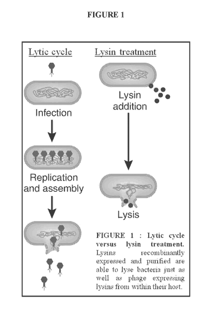
[00049] FIGURE 1 depicts the lytic cycle versus lysin treatment. Lysins
recombinantly expressed and
purified are able to lyse bacteria just as well as phage expressing lysins
from within their host.
CA 02833409 2013-10-16
WO 2012/145630
PCT/US2012/034456
[00050] FIGURE 2 depicts the PlySs2 domains. The catalytic domain corresponds
to residues 8-146.
There is a 16-residue linker. The binding domain corresponds to residues 162-
228.
[00051] FIGURE 3A and 3B provides the nucleotide and amino acid sequence of
the lysin PlySs1 as
well as a protein domain analysis. The amino acid sequence of the full length
PlySs1 (SEQ ID NO:1) and
truncated PlyS sl (SEQ ID NO:2) are provided. The endopeptidase domain (SEQ ID
NO:6), dual CPL-7
domain (SEQ ID NO:7) and glucosaminidase domain (SEQ ID NO:8) denoted.
[00052] FIGURE 4A and 4B provides the nucleotide and amino acid sequence of
the lysine PlySs2 as
well as a protein domain analysis. The amino acid sequence of PlySs2
corresponds to SEQ ID NO:3. The
CHAP domain and the SH-3 domain of the PlySs2 lysin are shaded, with the CHAP
domain starting with
LNN... and ending with ...YIT (SEQ ID NO :4) and the SH-3 domain starting with
RSY ... and ending with
...VAT (SEQ ID NO:5).
[00053] FIGURE 5 depicts the pBAD24 vector. The sequence begins with the pBAD
arabinose-inducible
promoter for the T7 polymerase and ends with PlySs2. Ampicillin serves as a
selective marker to ensure
retention of the plasmid as cells grow.
[00054] FIGURE 6 shows PlySs2 purification. All samples were run on 4-12% Bis-
Tris gels at 200 V for
¨40 mins and stained with Coomassie. A. The DEAE column flow through
containing PlySs2 at ¨26 kDa. B.
Six representative fractions of PlySs2 purified from a 10 L prep. C. A single
band at ¨26 kDa indicating the
purity of PlyS s2 after all fractions were pooled together.
[00055] FIGURE 7 depicts various aspects of PlySs2 characterization. A. To
test the optimal pH for
PlySs2 activity, 50 tiL of various phosphate/citrate buffers pH levels were
mixed with 195 id, S. suis strain
7997 cells and 5 jiL of lysin. PlySs2 had the strongest activity at pH 8Ø
PlySs2 was shown to have acute
activity up to pH 9.7. B. 195 tiL of cells, 5iLiL lysin were added to 50 tiL
of various NaC1 concentrations to
determine the optimal salt concentration for PlySs2. C. To determine the
temperature stability of PlySs2, it
was incubated for 30 minutes at various temperatures, cooled and then added to
245 L cells suspended in 15
mM Na3PO4, pH 8Ø D. PlySs2 was added to cells suspended in 15 mM Na3PO4, pH
8.0 along with various
concentrations of ethylenediaminetetraacetate (EDTA) to determine if it
requires a cofactor. hi controls, dd
1120 replaced PlySs2 for all tests.
[00056] FIGURE 8 depicts optimal pH of PlyS s2 determined against S. suis
strain 7997 in Bis-tris
propane (BTP) buffer up to a higher pH level.
[00057] FIGURE 9. The stability of purified PlyS S2 was determined by
evaluating killing effectiveness
against strain 7997 after storage at 37 C for up to 48 hours in buffer.
[00058] FIGURE 10. Killing effectiveness, assessed by 0D600 growth of strain
7997 upon treatment with
PlySs2 lysin after lysin storage at -80 C for up to 7 months in buffer.
[00059] FIGURE 11A and 11B depicts APlySs1 pH dependence. (A) Cells of host
strain 7711 were
suspended in phosphate citrate buffer (40/20 mM) at a range of pH-values from
4.6 to 8Ø APlySs1 was
11
CA 02833409 2013-10-16
WO 2012/145630
PCT/US2012/034456
added (110 in/m1) and 0D600 was measured over 60 min (horizontal axis) at 37
C. The vertical axis
represents the treated/untreated 0D600-ratio at each timepoint. For each pH-
value, the curve depicts the
running average of 3 independent experiments. Overall, activity was maximal at
the upper end of the
buffering range. (B) Here, bis-tris-propane (40 mM) was employed as the
buffering agent with a pH-range
from 7.0 to 9.7; AP1ySs1 was again added to 110 jig/mi. Each curve depicts the
running average of 3
experiments. Maximal activity was observed at pH = 9.0, although the
quantitative degree of OD-decline
was, in general, less than in phosphate-citrate.
[00060] FIGURE 12 depicts APLySS1 NaC1 dependence. S. suis 7711 cells were
suspended in
phosphate-citrate buffer pH = 7.8 (40/20 mM). NaC1 was added to the above
concentrations, followed by
APlySs1 at 110 jig/mi. Optical density at 600 nm was observed over 60 min at
37 C. In this figure, the
vertical axis represents the treated/untreated 0D600-ratio for each NaC1
concentration, averaged over 3
independent experiments.
[00061] FIGURE 13A and 13B provides assessment of AP1ySs1 DTT and EDTA
susceptibility. (A)
AP1ySs1 was pre-incubated for 1 hr with 5 mM DTT (a large molar excess) prior
to addition to 7711 cells;
activity was unchanged. (B) Here, various concentrations of EDTA were included
in the buffered suspension
of cells prior to addition of AP1ySs1 (110 jig/m1 lysin). For both images, the
vertical axis represents the
treated/untreated 0D600-ratio for each condition, averaged over 3 independent
experiments.
[00062] FIGURE 14A and 14B shows APlySs1 temperature stability. (A) A APlySs1
stock solution was
held at each of the above temperature for 30 minutes, followed by addition to
7711 cells (270 jig/m1 final
enzyme concentration, final temperature = 37 C, ideal buffering conditions).
The curves in this image
represent running averages of 3 individual experiments. In each case, complete
loss of activity was observed
between the 45 C and 50 C samples. The 3 hottest samples show a slightly
higher 0D600 reading than the
untreated control due to flocculation of APlySs1 upon denaturation. (B) The
above experiment was repeated,
but with 6 hours of heat-treatment prior to the assay. At this longer
incubation time, the 45 C sample showed
some loss of activity, though not complete. The 40 C sample maintained
essentially native activity.
[00063] FIGURE 15A and 15B (A) PlySs2 has acute lytic activity against S. suis
strain 7997 at, or above
8ug/mL. (B) Activity of PlyS s2 assessed in vitro against S. suis strain S735.
[00064] FIGURE 16A-16D provides PlySs2 activity against different species and
strains. S. suis 7997
was used as a positive control for each test. A. PlySs2 activity against
strains of S. suis. B. PlySs2 activity
against different species of bacteria and 2 strains of S. suis. C.
Streptococci and staphylococci sensitivity to
PlySs2. D. Various species tested for susceptibility to PlySs2 treatment.
[00065] FIGURE 17A and 17B shows PlySs2 activity against multiple species,
serotypes, and strains of
bacteria. In each instance the Treated/Untreated 0D600 is depicted in a bar
graph. The bars of S. aureus
strains are colored red; bars corresponding to S. suis strains are orange. The
bars of bacteria Listeria and other
bacteria of interest are shown in purple.
12
CA 02833409 2013-10-16
WO 2012/145630
PCT/US2012/034456
[00066] FIGURE 18. P1ySS2 was tested by standard MIC analysis for its ability
to kill strains of
staphylococci. Included in the testing were resistant staphylococci such as
Vancomycin resistant (VRSA),
Vancomycin intermediate (VISA) and methicillin resistant (MRSA) staphylococci.
The three VRSA strains
tested represent half of all known isolates.
[00067] FIGURE 19 provides APlySs1 bacteriolytic activity. Depicted here are
OD-drop curves for three
strains of S. suis: 7711, the serotype 7 strain from which PlySs' was
originally cloned (i.e. the host strain);
S735, a serotype 2 isolate that is the type-strain for the species; and 7997,
a virulent serotype 9 strain. Bacteria
were suspended in 20 mM phosphate buffer pH 7.8, 2 mM EDTA (defined as optimal
conditions). AP1ySs1
was added to the cells at a range of concentrations (indicated by the inset).
For each sample, optical density at
600 nm (vertical axis) was measured over the course of an hour (horizontal
axis) at 3711C. In this image, all
curves represent running averages of 3 or 4 independent experiments.
[00068] FIGURE 20 shows APlySs1 growth inhibition of S. suis 7711 APlyS sl was
added at the above
final concentrations to a dilute suspension of S. suis strain 7711 in BHI
broth. The optical density of each
sample was measured continuously overnight in 96-well plate format. Overall,
bacterial growth was delayed
in a dose-dependent manner. However, for enzyme-concentrations that were
sufficient to induce lysis in
buffered solutions (130 and 50 ug/m1), the effect was quite minimal here.
Moreover, none of the above
APlySs1 concentrations inhibited growth outright¨hence, a MIC could not be
assigned. For all of the treated
samples, one will note that the final optical densities are actually higher
than that of the untreated sample.
This is an artifact of the accumulation of aggregated bacterial debris that
occurred in the presence of lytic
enzyme.
[00069] FIGURE 21 provides a A PlySs1 bacterial strain panel. The information
provided m Figure 19
and Tables 3 and 4 is summarized graphically for two PlySs1 concentrations,
130 1.1g/m1 and 32.5 1.1g/ml. In
the image, strains of S. suis are denoted with double red
asterisks and non-suis streptococci are denoted with single black asterisks.
The optical density response
(treated-versus-untreated 0D600 ratio) after 1 hr is shown. The reader is
referred to Table 3 for the serotype
defmitions of the S. suis strains.
[00070] FIGURE 22 provides CFU killing assay results of PlySs2 bacteriolytic
activity against S. suis
strain 7997 and S735.
[00071] FIGURE 23 depicts the results of an S. aureus and S. pyogenes
resistance assay against P1ySS2
compared to antibiotic mupirocm. None of MRSA strain MW2, MSSA strain 8325,
nor S. pyogenes strain
5005 developed resistance against PlySs2 after each was exposed to
incrementally increasing concentrations
of PlyS s2. Both MW2 and 8325 developed resistance to the positive control,
mupirocin.
[00072] FIGURE 24 depicts the survival of mice with MRSA bacteremia over 10
days. PlySs2 cleared
bacteremia from 95% of mice tested. Of the controls, just 5% survived.
13
CA 02833409 2013-10-16
WO 2012/145630
PCT/US2012/034456
[00073] FIGURE 25 provides PlySs2 activity against different species and
strains. Log-phase cultures
were exposed to 32 ug/m1PlySs2 for 60 minutes in phosphate buffer. The final
0D600 of the treated samples
were divided by the final 0D600 of the untreated samples to generate the
normalized values above. Multiple
Staphylococci (including, but not limited to: MRSA, MSSA, and VISA),
Streptococci, Listeria, Enterococci,
and Bacilli were tested for susceptibility to PlySs2 activity. Escherichia and
Pseudomonas were tested as
Gram-negative controls.
[00074] FIGURE 26 depicts bactericidal effect of PlySs2 on various strains.
Bactericidal effect of 128
jig/m1 PlySs2 60 mins post-treatment. The reduction in CFU counts is presented
along a logarithmic scale.
Characteristically, PlyS s2 had significant activity against MRSA MW2. Of
note, PlyS s2 dramatically reduced
S. agalactiae and L. monocytogenes. There was no reduction in number of the
negative control E. coli.
[00075] FIGURE 27 depicts the minimum inhibitory concentration (MIC) of PlySs2
for various Gram-
positive bacteria. There was a low MIC for MRSA MW2, as expected, and a higher
MIC for S. pyogenes
5005. The MIC of PlySs2 correlates to the lytic activity and bactericidal
tests. The MIC of PlySs2 for the
negative control E. con was accordingly immeasurable.
[00076] FIGURE 28 shows PlySs2 protected mice from death caused by mixed
MRSA and S. pyogenes
infection. FVB/NJ mice were mtraperitoneally injected with 5% mucin containing
¨5x10⁵ CFU of MRSA
strain MW2, lx 107 S. pyogenes strain 5005, or combination of both bacteria
(mixed infection) from the
above inoculums at the same concentrations. Three hours post-infection, mice
in all infection groups (A-C),
received one intraperitoneal injection of 20 mM phosphate buffer control, 1 mg
of ClyS, 1 mg of PlyC, or a
combination of 1 mg of ClyS plus 1 mg of PlyC for the mixed infection. PlySs2
treatments consisted of 1 mg
for MRSA infections (A), or 2 mg for S. pyogenes and mixed infections (B-C).
Mice were monitored for
survival over ten days. The results from four independent experiments were
combined and the mice survival
data plotted with a Kaplan Meier Survival curve.
[00077] FIGURE 29 depicts activity of PlyS s2 and vancomycin against MRSA
isolates.
[00078] FIGURE 30 provides PlyS s2 enzymatic domain alignment to ClyS. The
CHAP domains of the
streptococcal lysins PlySs2 and PlyC (subunit A, GenBank no. AAP42310) are
aligned. Amino-acid identities
are indicated with underlying asterisks and highlighting. The positions of the
presumptive catalytic residues
(cysteine and histidine, for which the domain is named is named) are indicated
with arrows.
DETAILED DESCRIPTION
[00079] hi accordance with the present invention there may be employed
conventional molecular biology,
microbiology, and recombinant DNA techniques within the skill of the art. Such
techniques are explained
fully in the literature. See, e.g., Sambrook et al, "Molecular Cloning: A
Laboratory Manual" (1989); "Current
Protocols in Molecular Biology" Volumes I-III [Ausubel, R. M., ed. (1994)];
"Cell Biology: A Laboratory
14
CA 02833409 2013-10-16
WO 2012/145630
PCT/US2012/034456
Handbook" Volumes I-III [J. E. Celis, ed. (1994))]; "Cunent Protocols in
Immunology" Volumes I-III
[Coligan, J. E., ed. (1994)]; "Oligonucleotide Synthesis" (M.J. Gait ed.
1984); "Nucleic Acid Hybridization"
[B.D. Hames & S.J. Higgins eds. (1985)]; "Transcription And Translation" [B.D.
Hames & S.J. Higgins, eds.
(1984)]; "Animal Cell Culture" [R.I. Freshney, ed. (1986)]; "Immobilized Cells
And Enzymes" [IRL Press,
(1986)]; B. Perbal, "A Practical Guide To Molecular Cloning" (1984).
[00080] Therefore, if appearing herein, the following terms shall have the
definitions as provided and set
out below and in this section.
[00081] The terms "S. suis lysin(s)", "PlySs lysin(s)", "PlySs1 lysin",
"PlyS sl", "whole PlyS sl",
"truncated PlyS sl", "APlySs1", "P1ySs2 lysin", "PlySs2" and any variants not
specifically listed, may be used
herein interchangeably, and as used throughout the present application and
claims refer to proteinaceous
material including single or multiple proteins, and extends to those proteins
having the amino acid sequence
data described herein and presented in FIGURE 3 and in FIGURE 4 (SEQ ID NOS:
1, 2 and/or 3), and the
profile of activities set forth herein and in the Claims. Accordingly,
proteins displaying substantially
equivalent or altered activity are likewise contemplated. These modifications
may be deliberate, for example,
such as modifications obtained through site-directed mutagenesis, or may be
accidental, such as those
obtained through mutations in hosts that are producers of the complex or its
named subunits. Also, the terms
"S. suis lysin(s)", "PlyS s lysin(s)", "PlyS s 1 lysin", "PlyS sl", "whole
PlyS sl", "truncated PlyS sl", "APlySs 1",
"PlySs2 lysin", "PlySs2" are intended to include within their scope proteins
specifically recited herein as well
as all substantially homologous analogs, fragments or truncations, and allelic
variations.
Polypeptides and Lytic Enzymes
[00082] A "lytic enzyme" includes any bacterial cell wall lytic enzyme that
kills one or more bacteria
under suitable conditions and during a relevant time period. Examples of lytic
enzymes include, without
limitation, various amidase cell wall lytic enzymes.
[00083] A "S. suis lytic enzyme" includes a lytic enzyme that is capable of
killing at least one or more
Streptococcus suis bacteria under suitable conditions and during a relevant
time period.
[00084] A "bacteriophage lytic enzyme" refers to a lytic enzyme extracted or
isolated from a
bacteriophage or a synthesized lytic enzyme with a similar protein structure
that maintains a lytic enzyme
functionality.
[00085] A lytic enzyme is capable of specifically cleaving bonds that are
present in the peptidoglycan of
bacterial cells to disrupt the bacterial cell wall. It is also currently
postulated that the bacterial cell wall
peptidoglycan is highly conserved among most bacteria, and cleavage of only a
few bonds to may disrupt the
bacterial cell wall. The bacteriophage lytic enzyme may be an amidase,
although other types of enzymes are
possible. Examples of lytic enzymes that cleave these bonds are various
amidases such as muramidases,
CA 02833409 2013-10-16
WO 2012/145630
PCT/US2012/034456
glucosaminidases, endopeptidases, or N-acetyl-muramoyl-L-alanine amidases.
Fischetti et al (1974) reported
that the Cl streptococcal phage lysin enzyme was an amidase. Garcia et al
(1987, 1990) reported that the Cpl
lysin from a S. pneumoniae from a Cp-1 phage was a lysozyme. Caldentey and
Bamford (1992) reported that
a lytic enzyme from the phi 6 Pseudomonas phage was an endopeptidase,
splitting the peptide bridge formed
by melo-diammopimilic acid and D-alanine. The E. coil Ti and T6 phage lytic
enzymes are amidases as is the
lytic enzyme from Listeria phage (ply) (Loessner et al, 1996). There are also
other lytic enzymes known m the
art that are capable of cleaving a bacterial cell wall.
[00086] A "lytic enzyme genetically coded for by a bacteriophage" includes a
polypeptide capable of
killing a host bacteria, for instance by having at least some cell wall lytic
activity against the host bacteria.
The polypeptide may have a sequence that encompasses native sequence lytic
enzyme and variants thereof.
The polypeptide may be isolated from a variety of sources, such as from a
bacteriophage ("phage"), or
prepared by recombinant or synthetic methods, such as those described by
Garcia et al and also as provided
herein. The polypeptide may comprise a choline-binding portion at the carboxyl
terminal side and may be
characterized by an enzyme activity capable of cleaving cell wall
peptidoglycan (such as amidase activity to
act on amide bonds in the peptidoglycan) at the amino terminal side. Lytic
enzymes have been described
which include multiple enzyme activities, for example two enzymatic domains,
such as PlyGBS lysin.
Generally speaking, a lytic enzyme may be between 25,000 and 35,000 daltons m
molecular weight and
comprise a single polypeptide chain; however, this can vary depending on the
enzyme chain. The molecular
weight most conveniently can be determined by assay on denaturing sodium
dodecyl sulfate gel
electrophoresis and comparison with molecular weight markers.
[00087] "A native sequence phage associated lytic enzyme" includes a
polypeptide having the same amino
acid sequence as an enzyme derived from a bacteria. Such native sequence
enzyme can be isolated or can be
produced by recombinant or synthetic means.
[00088] The term "native sequence enzyme" encompasses naturally occurring
forms (e.g., alternatively
spliced or altered forms) and naturally-occurring variants of the enzyme. In
one embodiment of the invention,
the native sequence enzyme is a mature or full-length polypeptide that is
genetically coded for by a gene from
a bacteriophage specific for Streptococcus suis. Of course, a number of
variants are possible and known, as
acknowledged in publications such as Lopez et al., Microbial Drug Resistance
3: 199-211(1997); Garcia et
al., Gene 86: 81-88 (1990); Garcia et al., Proc. Natl. Acad. Sci. USA 85: 914-
918 (1988); Garcia et al., Proc.
Natl. Acad. Sci. USA 85: 914-918 (1988); Garcia et al., Streptococcal Genetics
(J. J. Ferretti and Curtis eds.,
1987); Lopez et al., FEMS Microbiol. Lett. 100: 439-448 (1992); Romero et al.,
J. Bacteriol. 172. 5064-5070
(1990); Ronda et al., Eur. J. Biochem. 164: 621-624 (1987) and Sanchez et al.,
Gene 61: 13-19 (1987). The
contents of each of these references, particularly the sequence listings and
associated text that compares the
sequences, including statements about sequence homologies, are specifically
incorporated by reference in
their entireties.
16
CA 02833409 2013-10-16
WO 2012/145630
PCT/US2012/034456
[00089] "A variant sequence lytic enzyme" includes a lytic enzyme
characterized by a polypeptide
sequence that is different from that of a lytic enzyme, but retains functional
activity. The lytic enzyme can, in
some embodiments, be genetically coded for by a bacteriophage specific for
Streptococcus suis having a
particular amino acid sequence identity with the lytic enzyme sequence(s)
hereof, as provided in FIGURE 3
and FIGURE 4 or in any of SEQ ID NOS: 1, 2 or 3. For example, in some
embodiments, a functionally active
lytic enzyme can kill Streptococcus suis bacteria, and other susceptible
bacteria as provided herein, including
as shown in TABLE 1 and in FIGURES 9 and 10, by disrupting the cellular wall
of the bacteria. An active
lytic enzyme may have a 60, 65, 70, 75, 80, 85, 90, 95, 97, 98, 99 or 99.5%
amino acid sequence identity with
the lytic enzyme sequence(s) hereof, as provided in FIGURE 3 and FIGURE 4 or m
any of SEQ ID NOS: 1, 2
or 3. Such phage associated lytic enzyme variants include, for instance, lytic
enzyme polypeptides wherein
one or more amino acid residues are added, or deleted at the N or C terminus
of the sequence of the lytic
enzyme sequence(s) hereof, as provided in FIGURE 3 and FIGURE 4 or in any of
SEQ ID NOS: 1, 2 or 3. In
a particular aspect, a phage associated lytic enzyme will have at least about
80% or 85% amino acid sequence
identity with native phage associated lytic enzyme sequences, particularly at
least about 90% (e.g. 90%)
amino acid sequence identity. Most particularly a phage associated lytic
enzyme variant will have at least
about 95% (e.g. 95%) amino acid sequence identity with the native phage
associated the lytic enzyme
sequence(s) hereof, as provided in FIGURE 3 and FIGURE 4 or m any of SEQ ID
NOS 1, 2 or 3.
[00090] "Percent amino acid sequence identity" with respect to the phage
associated lytic enzyme
sequences identified is defined herein as the percentage of amino acid
residues in a candidate sequence that
are identical with the amino acid residues in the phage associated lytic
enzyme sequence, after aligning the
sequences in the same reading frame and introducing gaps, if necessary, to
achieve the maximum percent
sequence identity, and not considering any conservative substitutions as part
of the sequence identity.
[00091] "Percent nucleic acid sequence identity" with respect to the phage
associated lytic enzyme
sequences identified herein is defined as the percentage of nucleotides in a
candidate sequence that are
identical with the nucleotides in the phage associated lytic enzyme sequence,
after aligning the sequences and
introducing gaps, if necessary, to achieve the maximum percent sequence
identity.
[00092] To determine the percent identity of two nucleotide or amino acid
sequences, the sequences are
aligned for optimal comparison purposes (e.g., gaps may be introduced in the
sequence of a first nucleotide
sequence). The nucleotides or amino acids at corresponding nucleotide or amino
acid positions are then
compared. When a position in the first sequence is occupied by the same
nucleotide or amino acid as the
corresponding position m the second sequence, then the molecules are identical
at that position. The percent
identity between the two sequences is a function of the number of identical
positions shared by the sequences
(i.e., % identity=# of identical positions/total # of positions×100).
[00093] The determination of percent identity between two sequences may be
accomplished using a
mathematical algorithm. A preferred, non-limiting example of a mathematical
algorithm utilized for the
17
CA 02833409 2013-10-16
WO 2012/145630
PCT/US2012/034456
comparison of two sequences is the algorithm of Karlin et al., Proc. Natl.
Acad. Sci. USA, 90:5873-5877
(1993). Such an algorithm is incorporated into the NBLAST program which may be
used to identify
sequences having the desired identity to nucleotide sequences of the
invention. To obtain gapped alignments
for comparison purposes, Gapped BLAST may be utilized as described in Altschul
et al., Nucleic Acids Res,
25:3389-3402 (1997). When utilizing BLAST and Gapped BLAST programs, the
default parameters of the
respective programs (e.g., NBLAST) may be used. See the programs provided by
National Center for
Biotechnology Information, National Library of Medicine, National Institutes
of Health. In one embodiment,
parameters for sequence comparison may be set at W=12. Parameters may also be
varied (e.g., W=5 or
W=20). The value "W" determines how many continuous nucleotides must be
identical for the program to
identify two sequences as containing regions of identity.
[00094] "Polypeptide" includes a polymer molecule comprised of multiple amino
acids joined in a linear
manner. A polypeptide can, in some embodiments, correspond to molecules
encoded by a polynucleotide
sequence which is naturally occurring. The polypeptide may include
conservative substitutions where the
naturally occurring amino acid is replaced by one having similar properties,
where such conservative
substitutions do not alter the function of the polypeptide (see, for example,
Lewin "Genes V" Oxford
University Press Chapter 1, pp. 9-13 1994).
[00095] The term "altered lytic enzymes" includes shuffled and/or chimeric
lytic enzymes.
[00096] Phage lytic enzymes specific for bacteria infected with a specific
phage have been found to
effectively and efficiently break down the cell wall of the bacterium in
question. The lytic enzyme is believed
to lack proteolytic enzymatic activity and is therefore non-destructive to
mammalian proteins and tissues
when present during the digestion of the bacterial cell wall. As shown by
Loeffler et al., "Rapid Killing of
Streptococcus pneumoniae with a Bacteriophage Cell Wall Hydrolase," Science,
294: 2170-2172 (Dec. 7,
2001), and supplemental material thereto published online by Science magazine,
which are incorporated
herein by reference in their entirety, a purified pneumococcal bacteriophage
lytic enzyme, such as Pal, is able
to kill various pneumococci. Loeffler et al. have shown through these
experiments that within seconds after
contact, the lytic enzyme Pal is able to kill 15 clinical stains of S.
pneumoniae, including the most frequently
isolated serogroups and penicillin resistant stains, in vitro. Treatment of
mice with Pal was also able to
eliminate or significantly reduce nasal carriage of serotype 14 in a dose-
dependent manner. Furthermore,
because it has been found that the action of Pal, like other phage lytic
enzymes, but unlike antibiotics, was
rather specific for the target pathogen, it is likely that the normal flora
will remain essentially intact (M. J.
Loessner, G. Wendlinger, S. Scherer, Mol Microbiol 16, 1231-41. (1995)
incorporated herein by reference). In
contrast, lysin polypeptide of the present invention has a remarkably broad
and clinically significant bacterial
killing profile. As demonstrated herein, for example, the isolated S. suis
lysin PlySs2, is effective in killing S.
suis, and also various other Streptococcus strains, including Group B
Streptococcus (GBS), Staphylococcal
18
CA 02833409 2013-10-16
WO 2012/145630
PCT/US2012/034456
strains, including Staphylococcus aureus, Enterococcus and Listeria. The lysin
of the present invention thus
demonstrates a breadth of bacterial cell killing unlike any lysin previously
reported or contemplated.
[00097] A lytic enzyme or polypeptide of the invention may be produced by the
bacterial organism after
being infected with a particular bacteriophage as either a prophylactic
treatment for preventing those who
have been exposed to others who have the symptoms of an infection from getting
sick, or as a therapeutic
treatment for those who have already become ill from the infection. In as much
the lysin polypeptide
sequences and nucleic acids encoding the lysin polypeptides are provided
herein, the lytic
enzyme(s)/polypeptide(s) may be preferably produced via the isolated gene for
the lytic enzyme from the
phage genome, putting the gene into a transfer vector, and cloning said
transfer vector into an expression
system, using standard methods of the art, including as exemplified herein.
The lytic enzyme(s) or
polypeptide(s) may be truncated, chimeric, shuffled or "natural," and may be
in combination. Relevant U.S.
Pat. No. 5,604,109 is incorporated herein in its entirety by reference. An
"altered" lytic enzyme can be
produced in a number of ways. In a preferred embodiment, a gene for the
altered lytic enzyme from the phage
genome is put into a transfer or movable vector, preferably a plasmid, and the
plasmid is cloned into an
expression vector or expression system. The expression vector for producing a
lysin polypeptide or enzyme of
the invention may be suitable for E. coli, Bacillus, or a number of other
suitable bacteria. The vector system
may also be a cell free expression system. All of these methods of expressing
a gene or set of genes are known
in the art. The lytic enzyme may also be created by infecting Streptococcus
suis with a bacteriophage specific
for Streptococcus suis, wherein said at least one lytic enzyme exclusively
lyses the cell wall of said
Streptococcus suis having at most minimal effects on other, for example
natural or commensal, bacterial flora
present.
[00098] A "chimeric protein" or "fusion protein" comprises all or
(preferably a biologically active) part of
a polypeptide of the invention operably linked to a heterologous polypeptide.
Chimeric proteins or peptides
are produced, for example, by combining two or more proteins having two or
more active sites. Chimeric
protein and peptides can act independently on the same or different molecules,
and hence have a potential to
treat two or more different bacterial infections at the same time. Chimeric
proteins and peptides also may be
used to treat a bacterial infection by cleaving the cell wall in more than one
location, thus potentially
providing more rapid or effective (or synergistic) killing from a single lysin
molecule or chimeric peptide.
[00099] A "heterologous" region of a DNA construct or peptide construct is an
identifiable segment of
DNA within a larger DNA molecule or peptide within a larger peptide molecule
that is not found in
association with the larger molecule in nature. Thus, when the heterologous
region encodes a mammalian
gene, the gene will usually be flanked by DNA that does not flank the
mammalian genomic DNA in the
genome of the source organism. Another example of a heterologous coding
sequence is a construct where the
coding sequence itself is not found in nature (e.g., a cDNA where the genomic
coding sequence contains
19
CA 02833409 2013-10-16
WO 2012/145630
PCT/US2012/034456
introns, or synthetic sequences having codons different than the native gene).
Allelic variations or naturally-
occurring mutational events do not give rise to a heterologous region of DNA
or peptide as defmed herein.
[000100] The term "operably linked" means that the polypeptide of the
disclosure and the heterologous
polypeptide are fused in-frame. The heterologous polypeptide can be fused to
the N-terminus or C-terminus of
the polypeptide of the disclosure. Chimeric proteins are produced
enzymatically by chemical synthesis, or by
recombinant DNA technology. A number of chimeric lytic enzymes have been
produced and studied. Gene E-
L, a chimeric lysis constructed from bacteriophages phi X174 and MS2 lysis
proteins E and L, respectively,
was subjected to internal deletions to create a series of new E-L clones with
altered lysis or killing properties.
The lytic activities of the parental genes E, L, E-L, and the internal
truncated forms of E-L were investigated
in this study to characterize the different lysis mechanism, based on
differences in the architecture of the
different membranes spanning domains. Electron microscopy and release of
marker enzymes for the
cytoplasmic and periplasmic spaces revealed that two different lysis
mechanisms can be distinguished
depending on penetration of the proteins of either the inner membrane or the
inner and outer membranes of
the E. coli (FEMS Microbiol. Lett. (1998) 164(1):159-67 (incorporated herein
by reference). One example of
a useful fusion protein is a GST fusion protein in which the polypeptide of
the disclosure is fused to the C-
terminus of a GST sequence. Such a chimeric protein can facilitate the
purification of a recombinant
polypeptide of the disclosure.
[000101] In another embodiment, the chimeric protein or peptide contains a
heterologous signal sequence at
its N-terminus. For example, the native signal sequence of a polypeptide of
the disclosure can be removed and
replaced with a signal sequence from another protein. For example, the gp67
secretory sequence of the
baculovirus envelope protein can be used as a heterologous signal sequence
(Current Protocols in Molecular
Biology, Ausubel et al., eds., John Wiley & Sons, 1992, incorporated herein by
reference). Other examples of
eukaryotic heterologous signal sequences include the secretory sequences of
melittin and human placental
alkaline phosphatase (Stratagene; La Jolla, Calif.). hi yet another example,
useful prokaryotic heterologous
signal sequences include the phoA secretory signal (Sambrook et al., supra)
and the protein A secretory signal
(Pharmacia Biotech; Piscataway, N.J.).
[000102] The fusion protein may combine a lysin polypeptide with a protein or
polypeptide of having a
different capability, or providing an additional capability or added character
to the lysin polypeptide. The
fusion protein may be an immunoglobulin fusion protein in which all or part of
a polypeptide of the disclosure
is fused to sequences derived from a member of the immunoglobulin protein
family. The immunoglobulin
may be an antibody, for example an antibody directed to a surface protein or
epitope of a susceptible or target
bacteria. An immunoglobulin fusion protein can be incorporated into a
pharmaceutical composition and
administered to a subject to inhibit an interaction between a ligand (soluble
or membrane-bound) and a protein
on the smface of a cell (receptor), to thereby suppress signal transduction in
vivo. The immunoglobulin fusion
protein can alter bioavailability of a cognate ligand of a polypeptide of the
disclosure. Inhibition of
CA 02833409 2013-10-16
WO 2012/145630
PCT/US2012/034456
ligandireceptor interaction may be useful therapeutically, both for treating
bacterial-associated diseases and
disorders for modulating (i.e. promoting or inhibiting) cell survival.
Moreover, an immunoglobulin fusion
protein of the disclosure can be used as an immunogen to produce antibodies
directed against a polypeptide of
the disclosure in a subject, to purify ligands and m screening assays to
identify molecules which inhibit the
interaction of receptors with ligands. Chimeric and fusion proteins and
peptides of the disclosure can be
produced by standard recombinant DNA techniques.
[000103] The fusion gene can be synthesized by conventional techniques,
including automated DNA
synthesizers. Alternatively, PCR amplification of gene fragments can be
carried out using anchor primers
which give rise to complementary overhangs between two consecutive gene
fragments which subsequently
can be annealed and reamplified to generate a chimeric gene sequence (see,
i.e., Ausubel et al., supra).
Moreover, many expression vectors are commercially available that already
encode a fusion moiety (i.e., a
GST polypeptide). A nucleic acid encoding a polypeptide of the invention can
be cloned into such an
expression vector such that the fusion moiety is linked in-frame to the
polypeptide of the invention.
[000104] As used herein, shuffled proteins or peptides, gene products, or
peptides for more than one related
phage protein or protein peptide fragments have been randomly cleaved and
reassembled into a more active or
specific protein. Shuffled oligonucleotides, peptides or peptide fragment
molecules are selected or screened to
identify a molecule having a desired functional property. This method is
described, for example, in Stemmer,
U.S. Pat. No. 6,132,970.(Method of shuffling polynucleotides); Kauffman, U.S.
Pat. No. 5,976,862 (Evolution
via Condon-based Synthesis) and Huse, U.S. Pat. No. 5,808,022 (Direct Codon
Synthesis). The contents of
these patents are incorporated herein by reference. Shuffling can be used to
create a protein that is more
active, for instance up to 10 to 100 fold more active than the template
protein. The template protein is selected
among different varieties of lysin proteins. The shuffled protein or peptides
constitute, for example, one or
more binding domains and one or more catalytic domains. Each binding or
catalytic domain is derived from
the same or a different phage or phage protein. The shuffled domains are
either oligonucleotide based
molecules, as gene or gene products, that either alone or in combination with
other genes or gene products are
translatable into a peptide fragment, or they are peptide based molecules.
Gene fragments include any
molecules of DNA, RNA, DNA-RNA hybrid, antisense RNA, Ribozymes, ESTs, SNIPs
and other
oligonucleotide-based molecules that either alone or in combination with other
molecules produce an
oligonucleotide molecule capable or incapable of translation into a peptide.
[000105] The modified or altered form of the protein or peptides and peptide
fragments, as disclosed
herein, includes protein or peptides and peptide fragments that are chemically
synthesized or prepared by
recombinant DNA techniques, or both. These techniques include, for example,
chimerization and shuffling.
When the protein or peptide is produced by chemical synthesis, it is
preferably substantially free of chemical
precursors or other chemicals, i.e., it is separated from chemical precursors
or other chemicals which are
involved in the synthesis of the protein. Accordingly such preparations of the
protein have less than about
21
CA 02833409 2013-10-16
WO 2012/145630
PCT/US2012/034456
30%, 20%, 10%, 5% (by dry weight) of chemical precursors or compounds other
than the polypeptide of
interest.
[000106] A signal sequence of a polypeptide can facilitate transmembrane
movement of the protein and
peptides and peptide fragments of the disclosure to and from mucous membranes,
as well as by facilitating
secretion and isolation of the secreted protein or other proteins of interest.
Signal sequences are typically
characterized by a core of hydrophobic amino acids which are generally cleaved
from the mature protein
during secretion in one or more cleavage events. Such signal peptides contain
processing sites that allow
cleavage of the signal sequence from the mature proteins as they pass through
the secretory pathway. Thus,
the disclosure can pertain to the described polypeptides having a signal
sequence, as well as to the signal
sequence itself and to the polypeptide in the absence of the signal sequence
(i.e., the cleavage products). A
nucleic acid sequence encoding a signal sequence of the disclosure can be
operably linked in an expression
vector to a protein of interest, such as a protein which is ordinarily not
secreted or is otherwise difficult to
isolate. The signal sequence directs secretion of the protein, such as from an
eukaryotic host into which the
expression vector is transformed, and the signal sequence is subsequently or
concurrently cleaved. The protein
can then be readily purified from the extracellular medium by art-recognized
methods. Alternatively, the
signal sequence can be linked to a protein of interest using a sequence which
facilitates purification, such as
with a GST domain.
[000107] The present invention also pertains to other variants of the
polypeptides of the invention. Such
variants may have an altered amino acid sequence which can function as either
agonists (mimetics) or as
antagonists. Variants can be generated by mutagenesis, i.e., discrete point
mutation or truncation. An agonist
can retain substantially the same, or a subset, of the biological activities
of the naturally occurring form of the
protein. An antagonist of a protein can inhibit one or more of the activities
of the naturally occurring form of
the protein by, for example, competitively binding to a downstream or upstream
member of a cellular
signaling cascade which includes the protein of interest. Thus, specific
biological effects can be elicited by
treatment with a variant of limited function. Treatment of a subject with a
variant having a subset of the
biological activities of the naturally occurring form of the protein can have
fewer side effects in a subject
relative to treatment with the naturally occurring form of the protein.
Variants of a protein of the disclosure
which function as either agonists (mimetics) or as antagonists can be
identified by screening combinatorial
libraries of mutants, i.e., truncation mutants, of the protein of the
disclosure for agonist or antagonist activity.
hi one embodiment, a variegated library of variants is generated by
combinatorial mutagenesis at the nucleic
acid level and is encoded by a variegated gene library. A variegated library
of variants can be produced by, for
example, enzymatically ligating a mixture of synthetic oligonucleotides into
gene sequences such that a
degenerate set of potential protein sequences is expressible as individual
polypeptides, or alternatively, as a set
of larger fusion proteins (i.e., for phage display). There are a variety of
methods which can be used to produce
libraries of potential variants of the polypeptides of the disclosure from a
degenerate oligonucleotide
22
CA 02833409 2013-10-16
WO 2012/145630
PCT/US2012/034456
sequence. Methods for synthesizing degenerate oligonucleotides are known in
the art (see, i.e., Narang (1983)
Tetrahedron 39:3; Itakura et al. (1984) Annu. Rev. Biochem. 53:323; Itakura et
al. (1984) Science 198:1056;
Ike et al. (1983) Nucleic Acid Res. 11:477, all herein incorporated by
reference).
[000108] In addition, libraries of fragments of the coding sequence of a
polypeptide of the disclosure can be
used to generate a variegated population of polypeptides for screening and
subsequent selection of variants,
active fragments or truncations. For example, a library of coding sequence
fragments can be generated by
treating a double stranded PCR fragment of the coding sequence of interest
with a nuclease under conditions
wherein nicking occurs only about once per molecule, denaturing the double
stranded DNA, renaturing the
DNA to form double stranded DNA which can include sense/antisense pairs from
different nicked products,
removing single stranded portions from reformed duplexes by treatment with 51
nuclease, and ligating the
resulting fragment library into an expression vector. By this method, an
expression library can be derived
which encodes N-terminal and internal fragments of various sizes of the
protein of interest. Several techniques
are known in the art for screening gene products of combinatorial libraries
made by point mutations or
truncation, and for screening cDNA libraries for gene products having a
selected property. The most widely
used techniques, which are amenable to high through-put analysis, for
screening large gene libraries typically
include cloning the gene library into replicable expression vectors,
transforming appropriate cells with the
resulting library of vectors, and expressing the combinatorial genes under
conditions m which detection of a
desired activity facilitates isolation of the vector encoding the gene whose
product was detected. Recursive
ensemble mutagenesis (REM), a technique which enhances the frequency of
functional mutants in the
libraries, can be used in combination with the screening assays to identify
variants of a protein of the
disclosure (Arkin and Yourvan (1992) Proc. Natl. Acad. Sci. USA 89:7811-7815;
Delgrave et al. (1993)
Protein Engineering 6(3):327-331) immunologically active portions of a protein
or peptide fragment include
regions that bind to antibodies that recognize the phage enzyme. In this
context, the smallest portion of a
protein (or nucleic acid that encodes the protein) according to embodiments is
an epitope that is recognizable
as specific for the phage that makes the lysin protein. Accordingly, the
smallest polypeptide (and associated
nucleic acid that encodes the polypeptide) that can be expected to bind a
target or receptor, such as an
antibody, and is useful for some embodiments may be 8, 9, 10, 11, 12, 13, 15,
20, 25, 30, 35, 40, 45, 50, 55,
60, 65, 75, 85, or 100 amino acids long. Although small sequences as short as
8, 9, 10, 11, 12 or 15 amino
acids long reliably comprise enough structure to act as targets or epitopes,
shorter sequences of 5, 6, or 7
amino acids long can exhibit target or epitopic structure in some conditions
and have value in an embodiment.
Thus, the smallest portion of the protein(s) or lysin polypeptides provided
herein, including as set out in
FIGURES 3 and 4 and in SEQ ID NOS: 1, 2 and/or 3, includes polypeptides as
small as 5, 6, 7, 8, 9, 10, 12,
14 or 16 amino acids long.
[000109] Biologically active portions of a protein or peptide fragment of the
embodiments, as described
herein, include polypeptides comprising amino acid sequences sufficiently
identical to or derived from the
23
CA 02833409 2013-10-16
WO 2012/145630
PCT/US2012/034456
amino acid sequence of the phage protein of the disclosure, which include
fewer amino acids than the full
length protein of the phage protein and exhibit at least one activity of the
corresponding full-length protein.
Typically, biologically active portions comprise a domain or motif with at
least one activity of the
corresponding protein. A biologically active portion of a protein or protein
fragment of the disclosure can be a
polypeptide which is, for example, 10, 25, 50, 100 less or more amino acids in
length. Moreover, other
biologically active portions, in which other regions of the protein are
deleted, or added can be prepared by
recombinant techniques and evaluated for one or more of the functional
activities of the native form of a
polypeptide of the embodiments.
[000110] Homologous proteins and nucleic acids can be prepared that share
functionality with such small
proteins and/or nucleic acids (or protein and/or nucleic acid regions of
larger molecules) as will be appreciated
by a skilled artisan. Such small molecules and short regions of larger
molecules that may be homologous
specifically are intended as embodiments. Preferably the homology of such
valuable regions is at least 50%,
65%, 75%, 80%, 85%, and preferably at least 90%, 95%, 97%, 98%, or at least
99% compared to the lysin
polypeptides provided herein, including as set out in FIGURES 3 and 4 and in
SEQ ID NOS: 1, 2 and/or 3.
These percent homology values do not include alterations due to conservative
amino acid substitutions.
[000111] Two amino acid sequences are "substantially homologous" when at least
about 70% of the amino
acid residues (preferably at least about 80%, at least about 85%, and
preferably at least about 90 or 95%) are
identical, or represent conservative substitutions. The sequences of
comparable lysins, such as comparable
PlyS s2 lysins, or comparable PlyS sl lysins, are substantially homologous
when one or more, or several, or up
to 10%, or up to 15%, or up to 20% of the amino acids of the lysin polypeptide
are substituted with a similar
or conservative amino acid substitution, and wherein the comparable lysins
have the profile of activities, anti-
bacterial effects, and/or bacterial specificities of a lysin, such as the
PlySs2 and/or PlySs1 lysins, disclosed
herein.
[000112] The amino acid residues described herein are preferred to be in the
"L" isomeric form. However,
residues in the "D" isomeric form can be substituted for any L-amino acid
residue, as long as the desired
fuctional property of immunoglobulin-binding is retained by the polypeptide.
NH, refers to the free amino
group present at the amino terminus of a polypeptide. COOH refers to the free
carboxy group present at the
carboxy terminus of a polypeptide. In keeping with standard polypeptide
nomenclature, J. Biol. Chem.,
243:3552-59 (1969), abbreviations for amino acid residues are shown in the
following Table of
Correspondence:
24
CA 02833409 2013-10-16
WO 2012/145630
PCT/US2012/034456
TABLE OF CORRESPONDENCE
SYMBOL AMINO ACID
1-Letter 3-Letter
Tyr tyrosine
Gly glycine
Phe phenylalanme
NI Met methionine
A Ala alanine
Ser serine
Ile isoleucine
Leu leucine
Thr threonine
V Val valine
Pro proline
Lys lysine
His histidine
Gin glutamine
Glu glutamic acid
Trp tryptophan
Arg arginine
Asp a spartic acid
Asn a sparagine
Cys cysteme
[000113] It should be noted that all ammo-acid residue sequences are
represented herein by formulae whose
left and right orientation is in the conventional direction of amino-terminus
to carboxy-terminus.
Furthermore, it should be noted that a dash at the beginning or end of an
amino acid residue sequence
indicates a peptide bond to a further sequence of one or more amino-acid
residues. The above Table is
presented to correlate the three-letter and one-letter notations which may
appear alternately herein.
[000114] Mutations can be made in the amino acid sequences, or in the nucleic
acid sequences encoding the
polypeptides and lysins herein, including in the lysin sequences set out in
Figure 3 or in Figure 4, or in active
fragments or truncations thereof, such that a particular codon is changed to a
codon which codes for a
different amino acid, an amino acid is substituted for another amino acid, or
one or more amino acids are
deleted. Such a mutation is generally made by making the fewest amino acid or
nucleotide changes possible.
A substitution mutation of this sort can be made to change an amino acid in
the resulting protein in a non-
CA 02833409 2013-10-16
WO 2012/145630
PCT/US2012/034456
conservative manner (for example, by changing the codon from an amino acid
belonging to a grouping of
amino acids having a particular size or characteristic to an amino acid
belonging to another grouping) or in a
conservative manner (for example, by changing the codon from an amino acid
belonging to a grouping of
amino acids having a particular size or characteristic to an ammo acid
belonging to the same grouping). Such
a conservative change generally leads to less change in the structure and
function of the resulting protein. A
non-conservative change is more likely to alter the structure, activity or
function of the resulting protein. The
present invention should be considered to include sequences containing
conservative changes which do not
significantly alter the activity or binding characteristics of the resulting
protein.
[000115] Thus, one of skill in the art, based on a review of the sequence of
the PlyS sl and PlyS s2 lysin
polypeptides provided herein and on their knowledge and the public information
available for other lysin
polypeptides, can make amino acid changes or substitutions in the lysin
polypeptide sequence. Amino acid
changes can be made to replace or substitute one or more, one or a few, one or
several, one to five, one to ten,
or such other number of amino acids in the sequence of the lysin(s) provided
herein to generate mutants or
variants thereof. Such mutants or variants thereof may be predicted for
function or tested for function or
capability for killing bacteria, including Staphylococcal, Streptococcal,
Listeria, or Enterococcal bacteria,
and/or for having comparable activity to the lysin(s) provided herein. Thus,
changes can be made to the
sequence of PlyS s2, for example, by modifying the amino acid sequence as set
out m Figure 4 hereof, and
mutants or variants having a change in sequence can be tested using the assays
and methods described and
exemplified herein, including in the examples. One of skill in the art, on the
basis of the domain structure of
the lysin(s) hereof can predict one or more, one or several amino acids
suitable for substitution or replacement
and/or one or more amino acids which are not suitable for substitution or
replacement, including reasonable
conservative or non-conservative substitutions.
[000116] hi this regard, and with exemplary reference to PlySs2 lysin it is
pointed out that, although the
PlySs2 polypeptide lysin represents a divergent class of prophage lytic
enzyme, the lysin comprises an N-
terminal CHAP domain (cysteine-histidine amidohydrolase/peptidase) and a C-
terminal 5H3-type 5 domain
as depicted in FIGURE 4. The domains are depicted in the amino acid sequence
in distinct shaded color
regions, with the CHAP domain corresponding to the first shaded amino acid
sequence region starting with
LNN... and the 5113-type 5 domain corresponding to the second shaded region
starting with RSY. CHAP
domains are included in several previously characterized streptococcal and
staphylococcal phage lysins. Thus,
one of skill in the art can reasonably make and test substitutions or
replacements to the CHAP domain and/or
the SH-3 domain of PlySs2. Sequence comparisons to the Genbank database can be
made with either or both
of the CHAP and/or SH-3 domain sequences or with the PlySs2 lysin full amino
acid sequence, for instance,
to identify amino acids for substitution. In FIGURE 30, the CHAP domain of
PlyS s2 is aligned with that of
the well-characterized streptococcal PlyC lysin, demonstrating conserved
catalytic residues, but only a modest
level of identity overall (28% sequence identity). In FIGURE 30 the conserved
cysteine and histidine amino
26
CA 02833409 2013-10-16
WO 2012/145630
PCT/US2012/034456
acid sequences in the CHAP domain are shown with an arrow. It is reasonable to
predict, for example, that
the conserved cysteine and histidine residues should be maintained in a mutant
or variant of PlySs2 so as to
maintain activity or capability. It is notable that a mutant or variant having
an alanine replaced for valine at
valine amino acid 19 in the PlySs2 sequence of Figure 4 and SEQ ID NO: 3 is
active and capable of killing
gram positive bacteria in a manner similar to and as effective as the Figure 4
and SEQ ID NO:3 lysm.
[000117] The following is one example of various groupings of amino acids:
Amino acids with nonpolar R groups
Alanine, Valine, Leucine, Isoleucine, Proline, Phenylalanine, Tryptophan,
Methionine
Amino acids with uncharged polar R groups
Glycine, Serine, Threonine, Cysteine, Tyrosine, Asp aragine, Glutamine
Amino acids with charged polar R groups (negatively charged at Ph 6.0)
Aspartic acid, Glutamic acid
Basic amino acids (positively charged at pH 6.0)
Lysine, Arginine, Histidine (at pH 6.0)
[000118] Another grouping may be those amino acids with phenyl groups:
Phenylalanine, Tryptophan, Tyrosine
[000119] Another grouping may be according to molecular weight e., size of R
groups):
Glycme 75 Alanme 89
Serine 105 Proline 115
Valine 117 Threonine 119
Cysteine 121 Leucine 131
Isoleucine 131 Asparagine 132
Aspartic acid 133 Glutamine 146
Lysine 146 Glutamic acid 147
Methionine 149 Histidine (at pH 6.0) 155
Phenylalanine 165 Arginine 174
Tyrosine 181 Tryptophan 204
[000120] Particularly preferred substitutions are:
- Lys for Arg and vice versa such that a positive charge may be maintained;
- Glu for Asp and vice versa such that a negative charge may be maintained;
- Ser for Thr such that a free -OH can be maintained; and
- Gin for Asn such that a free NH2 can be maintained.
[000121] Exemplary and preferred conservative amino acid substitutions include
any of:
glutamine (Q) for glutamic acid (E) and vice versa; leucme (L) for valine (V)
and vice versa; serine (S) for
threonine (T) and vice versa; isoleucine (I) for valine (V) and vice versa;
lysine (K) for glutamine (Q) and vice
27
CA 02833409 2013-10-16
WO 2012/145630
PCT/US2012/034456
versa; isoleucine (I) for methionine (M) and vice versa; serine (S) for
asparagine (N) and vice versa; leucine
(L) for methionine (M) and vice versa, lysine (L) for glutamic acid (E) and
vice versa; alanine (A) for serine
(S) and vice versa; tyrosine (Y) for phenylalanine (F) and vice versa;
glutamic acid (E) for aspartic acid (D)
and vice versa; leueme (L) for isoleucme (I) and vice versa; lysme (K) for
argmine (R) and vice versa.
[000122] Amino acid substitutions may also be introduced to substitute an
amino acid with a particularly
preferable property. For example, a Cys may be introduced a potential site for
disulfide bridges with another
Cys. A His may be introduced as a particularly "catalytic" site (i.e., His can
act as an acid or base and is the
most common amino acid in biochemical catalysis). Pro may be introduced
because of its particularly planar
structure, which induces 13-turns in the protem's structure.
[000123] A polypeptide or epitope as described herein may be used to generate
an antibody and also can be
used to detect binding to the lysin or to molecules that recognize the lysin
protein. Another embodiment is a
molecule such as an antibody or other specific binder that may be created
through use of an epitope such as by
regular immunization or by a phase display approach where an epitope can be
used to screen a library if
potential binders. Such molecules recognize one or more epitopes of lysin
protein or a nucleic acid that
encodes lysm protein. An antibody that recognizes an epitope may be a
monoclonal antibody, a humanized
antibody, or a portion of an antibody protein. Desirably the molecule that
recognizes an epitope has a specific
binding for that epitope which is at least 10 times as strong as the molecule
has for serum albumin. Specific
binding can be measured as affmity (Km). More desirably the specific binding
is at least 102, 103, 10n, 105,
106, 107, 108, or even higher than that for serum albumin under the same
conditions.
[000124] In a desirable embodiment the antibody or antibody fragment is in a
form useful for detecting the
presence of the lysin protein or, alternatively detecting the presence of a
bacteria susceptible to the lysin
protein. In a further embodiment the antibody may be attached or otherwise
associated with the lysin
polypeptide of the invention, for example in a chimeric or fusion protein, and
may serve to direct the lysin to a
bacterial cell or strain of interest or target. Alternatively, the lysin
polypeptide may serve to direct the
antibody or act in conjunction with the antibody, for example in lysing the
bacterial cell wall fully or partially,
so that the antibody may specifically bind to its epitope at the surface or
under the surface on or in the
bacteria. For example, a lysin of the invention may be attached to an anti-
Streptococcal antibody and direct
the antibody to its epitope.
[000125] A variety of forms and methods for antibody synthesis are known as
will be appreciated by a
skilled artisan. The antibody may be conjugated (covalently complexed) with a
reporter molecule or atom
such as a fluor, an enzyme that creates an optical signal, a chemilumiphore, a
microparticle, or a radioactive
atom. The antibody or antibody fragment may be synthesized in vivo, after
immunization of an animal, for
example, the antibody or antibody fragment may be synthesized via cell culture
after genetic recombination.
The antibody or antibody fragment may be prepared by a combination of cell
synthesis and chemical
modification.
28
CA 02833409 2013-10-16
WO 2012/145630
PCT/US2012/034456
[000126] An "antibody" is any immunoglobulin, including antibodies and
fragments thereof, that binds a
specific epitope. The term encompasses polyclonal, monoclonal, and chimeric
antibodies, the last mentioned
described in further detail in U.S. Patent Nos. 4,816,397 and 4,816,567. The
term "antibody" describes an
immunoglobulin whether natural or partly or wholly synthetically produced. The
term also covers any
polypeptide or protein having a binding domain which is, or is homologous to,
an antibody binding domain.
CDR grafted antibodies are also contemplated by this term. An "antibody" is
any immunoglobulin, including
antibodies and fragments thereof, that binds a specific epitope. The term
encompasses polyclonal,
monoclonal, and chimeric antibodies, the last mentioned described in further
detail in U.S. Patent Nos.
4,816,397 and 4,816,567. The term "antibody(ies)" includes a wild type
immunoglobulin (Ig) molecule,
generally comprising four full length polypeptide chains, two heavy (H) chains
and two light (L) chains, or an
equivalent Ig homologue thereof (e.g., a camelid nanobody, which comprises
only a heavy chain); including
full length functional mutants, variants, or derivatives thereof, which retain
the essential epitope binding
features of an Ig molecule, and including dual specific, bispecific,
multispecific, and dual variable domain
antibodies; Immunoglobulin molecules can be of any class (e.g., IgG, IgE, IgM,
IgD, IgA, and IgY), or
subclass (e.g., IgGl, IgG2, IgG3, IgG4, IgAl, and IgA2). Also included within
the meaning of the term
"antibody" are any "antibody fragment".
[000127] An "antibody fragment" means a molecule comprising at least one
polypeptide chain that is not
full length, including (i) a Fab fragment, which is a monovalent fragment
consisting of the variable light (VL),
variable heavy (VH), constant light (CL) and constant heavy 1 (CH1) domains;
(ii) a F(ab')2 fragment, which
is a bivalent fragment comprising two Fab fragments linked by a disulfide
bridge at the hinge region; (iii) a
heavy chain portion of an Fab (Fd) fragment, which consists of the VH and CH1
domains; (iv) a variable
fragment (Fv) fragment, which consists of the VL and VH domains of a single
arm of an antibody, (v) a
domain antibody (dAb) fragment, which comprises a single variable domain
(Ward, E.S. et al., Nature 341,
544-546 (1989)); (vi) a camelid antibody; (vii) an isolated complementarity
determining region (CDR); (viii)
a Single Chain Fv Fragment wherein a VH domain and a VL domain are linked by a
peptide linker which
allows the two domains to associate to form an antigen binding site (Bird et
al, Science, 242, 423-426, 1988;
Huston et al, PNAS USA, 85, 5879-5883, 1988); (ix) a diabody, which is a
bivalent, bispecific antibody in
which VH and VL domains are expressed on a single polypeptide chain, but using
a linker that is too short to
allow for pairing between the two domains on the same chain, thereby forcing
the domains to pair with the
complementarity domains of another chain and creating two antigen binding
sites (W094/13804; P. Holliger
et al Proc. Natl. Acad. Sci. USA 90 6444-6448, (1993)); and (x) a linear
antibody, which comprises a pair of
tandem Fv segments (VH-CH1-VH-CH1) which, together with complementarity light
chain polypeptides,
form a pair of antigen binding regions; (xi) multivalent antibody fragments
(scFv dimers, trimers and/or
tetramers (Power and Hudson, J Immunol. Methods 242: 193-204 9 (2000)); and
(xii) other non-full length
portions of heavy and/or light chains, or mutants, variants, or derivatives
thereof, alone or in any combination.
29
CA 02833409 2013-10-16
WO 2012/145630
PCT/US2012/034456
[000128] As antibodies can be modified in a number of ways, the term
"antibody" should be construed as
covering any specific binding member or substance having a binding domain with
the required specificity.
Thus, this term covers antibody fragments, derivatives, functional equivalents
and homologues of antibodies,
including any polypeptide comprising an immunoglobulin-binding domain, whether
natural or wholly or
partially synthetic. Chimeric molecules comprising an immunoglobulin binding
domain, or equivalent, fused
to another polypeptide are therefore included. Cloning and expression of
chimeric antibodies are described in
EP-A-0120694 and EP-A-0125023 and U.S. Patent Nos. 4,816,397 and 4,816,567.
[000129] An "antibody combining site" is that structural portion of an
antibody molecule comprised of light
chain or heavy and light chain variable and hypervariable regions that
specifically binds antigen.
[000130] The phrase "antibody molecule" in its various grammatical forms as
used herein contemplates
both an intact immunoglobulin molecule and an immunologically active portion
of an immunoglobulin
molecule. Exemplary antibody molecules are intact immunoglobulin molecules,
substantially intact
immunoglobulin molecules and those portions of an immunoglobulin molecule that
contains the paratope,
including those portions known in the art as Fab, Fab', F(ab')2 and F(v),
which portions are preferred for use in
the therapeutic methods described herein.
[000131] The phrase "monoclonal antibody" in its various grammatical forms
refers to an antibody having
only one species of antibody combining site capable of immunoreacting with a
particular antigen. A
monoclonal antibody thus typically displays a single binding affmity for any
antigen with which it
immunoreacts. A monoclonal antibody may therefore contain an antibody molecule
having a plurality of
antibody combining sites, each immunospecific for a different antigen; e.g., a
bispecific (chimeric)
monoclonal antibody.
[000132] The term "specific" may be used to refer to the situation in which
one member of a specific
binding pair will not show significant binding to molecules other than its
specific binding partner(s). The
term is also applicable where e.g. an antigen binding domain is specific for a
particular epitope which is
carried by a number of antigens, m which case the specific binding member
carrying the antigen binding
domain will be able to bind to the various antigens carrymg the epitope.
[000133] The term "comprise" generally used in the sense of include, that is
to say permitting the presence
of one or more features or components.
[000134] The term "consisting essentially of' refers to a product,
particularly a peptide sequence, of a
defmed number of residues which is not covalently attached to a larger
product. In the case of the peptide of
the invention hereof, those of skill in the art will appreciate that minor
modifications to the N- or C- terminal
of the peptide may however be contemplated, such as the chemical modification
of the terminal to add a
protecting group or the like, e.g. the amidation of the C-terminus.
[000135] The term "isolated" refers to the state in which the lysin
polypeptide(s) of the invention, or
nucleic acid encoding such polypeptides will be, in accordance with the
present invention. Polypeptides and
CA 02833409 2013-10-16
WO 2012/145630
PCT/US2012/034456
nucleic acid will be free or substantially free of material with which they
are naturally associated such as other
polypeptides or nucleic acids with which they are found in their natural
environment, or the environment in
which they are prepared (e.g. cell culture) when such preparation is by
recombinant DNA technology
practised in vitro or in vivo. Polypeptides and nucleic acid may be formulated
with diluents or adjuvants and
still for practical purposes be isolated - for example the polypeptides will
normally be mixed with polymers or
mucoadhesives or other carriers, or will be mixed with pharmaceutically
acceptable carriers or diluents, when
used in diagnosis or therapy.
Nucleic Acids
[000136] Nucleic acids capable of encoding the S. suis lysin polypeptide(s) of
the invention are provided
herein and constitute an aspect of the invention. Representative nucleic acid
sequences in this context are
polynucleotide sequences coding for the polypeptide of any of FIGURES 3 and 4,
the polypeptides of SEQ ID
NO:, SEQ ID NO:2 and SEQ ID NO:3, and sequences that hybridize, under
stringent conditions, with
complementary sequences of the DNA of the FIGURE 3 or 4 sequence(s). Further
variants of these sequences
and sequences of nucleic acids that hybridize with those shown in the figures
also are contemplated for use in
production of lysing enzymes according to the disclosure, including natural
variants that may be obtained. A
large variety of isolated nucleic acid sequences or cDNA sequences that encode
phage associated lysing
enzymes and partial sequences that hybridize with such gene sequences are
useful for recombinant production
of the lysin enzyme(s) or polypeptide(s) of the invention.
[000137] A "replicon" is any genetic element (e.g., plasmid, chromosome,
virus) that functions as an
autonomous unit of DNA replication in vivo; i.e., capable of replication under
its own control.
[000138] A "vector" is a replicon, such as plasmid, phage or cosmid, to which
another DNA segment may
be attached so as to bring about the replication of the attached segment.
[000139] A "DNA molecule" refers to the polymeric form of deoxyribonucleotides
(adenine, guanine,
thymine, or cytosine) in its either single stranded form, or a double-stranded
helix. This term refers only to
the primary and secondary structure of the molecule, and does not limit it to
any particular tertiary forms.
Thus, this term includes double-stranded DNA found, inter alia, in linear DNA
molecules (e.g., restriction
fragments), viruses, plasmids, and chromosomes. In discussing the structure of
particular double-stranded
DNA molecules, sequences may be described herein according to the normal
convention of giving only the
sequence in the 5' to 3' direction along the nontranscribed strand of DNA
(i.e., the strand having a sequence
homologous to the mRNA).
[000140] An "origin of replication" refers to those DNA sequences that
participate in DNA synthesis.
[000141] A DNA "coding sequence" is a double-stranded DNA sequence which is
transcribed and
translated into a polypeptide in vivo when placed under the control of
appropriate regulatory sequences. The
boundaries of the coding sequence are determined by a start codon at the 5'
(amino) terminus and a translation
31
CA 02833409 2013-10-16
WO 2012/145630
PCT/US2012/034456
stop codon at the 3' (carboxyl) terminus. A coding sequence can include, but
is not limited to, prokaryotic
sequences, cDNA from eukaryotic mRNA, genomic DNA sequences from eukaryotic
(e.g., mammalian)
DNA, and even synthetic DNA sequences. A polyadenylation signal and
transcription termination sequence
will usually be located 3' to the coding sequence.
[000142] Transcriptional and translational control sequences are DNA
regulatory sequences, such as
promoters, enhancers, polyadenylation signals, terminators, and the like, that
provide for the expression of a
coding sequence in a host cell.
[000143] A "promoter sequence" is a DNA regulatory region capable of binding
RNA polymerase in a cell
and initiating transcription of a downstream (3' direction) coding sequence.
For purposes of defining the
present invention, the promoter sequence is bounded at its 3' terminus by the
transcription initiation site and
extends upstream (5' direction) to include the minimum number of bases or
elements necessary to initiate
transcription at levels detectable above background. Within the promoter
sequence will be found a
transcription initiation site (conveniently defined by mapping with nuclease
Si), as well as protein binding
domains (consensus sequences) responsible for the binding of RNA polymerase.
Eukaryotic promoters will
often, but not always, contain "TATA" boxes and "CAT" boxes. Prokaryotic
promoters contain Shine-
Dalgamo sequences in addition to the -10 and -35 consensus sequences.
[000144] An "expression control sequence" is a DNA sequence that controls and
regulates the transcription
and translation of another DNA sequence. A coding sequence is "under the
control" of transcriptional and
translational control sequences in a cell when RNA polymerase transcribes the
coding sequence into mRNA,
which is then translated into the protein encoded by the coding sequence.
[000145] A "signal sequence" can be included before the coding sequence. This
sequence encodes a signal
peptide, N-terminal to the polypeptide, that communicates to the host cell to
direct the polypeptide to the cell
surface or secrete the polypeptide into the media, and this signal peptide is
clipped off by the host cell before
the protein leaves the cell. Signal sequences can be found associated with a
variety of proteins native to
prokaryotes and eukaryotes.
[000146] The term "oligonucleotide," as used herein in referring to the probe
of the present invention, is
defined as a molecule comprised of two or more ribonucleotides, preferably
more than three. Its exact size
will depend upon many factors which, in turn, depend upon the ultimate
function and use of the
oligonucleotide.
[000147] The term "primer" as used herein refers to an oligonucleotide,
whether occurring naturally as in a
purified restriction digest or produced synthetically, which is capable of
acting as a point of initiation of
synthesis when placed under conditions in which synthesis of a primer
extension product, which is
complementary to a nucleic acid strand, is induced, i.e., in the presence of
nucleotides and an inducing agent
such as a DNA polymerase and at a suitable temperature and pH. The primer may
be either single-stranded or
double-stranded and must be sufficiently long to prime the synthesis of the
desired extension product in the
32
CA 02833409 2013-10-16
WO 2012/145630
PCT/US2012/034456
presence of the inducing agent. The exact length of the primer will depend
upon many factors, including
temperature, source of primer and use of the method. For example, for
diagnostic applications, depending on
the complexity of the target sequence, the oligonucleotide primer typically
contains 15-25 or more
nucleotides, although it may contain fewer nucleotides.
[000148] The primers herein are selected to be "substantially" complementary
to different strands of a
particular target DNA sequence. This means that the primers must be
sufficiently complementary to hybridize
with their respective strands. Therefore, the primer sequence need not reflect
the exact sequence of the
template. For example, a non-complementary nucleotide fragment may be attached
to the 5' end of the
primer, with the remainder of the primer sequence being complementary to the
strand. Alternatively, non-
complementary bases or longer sequences can be interspersed into the primer,
provided that the primer
sequence has sufficient complementarity with the sequence of the strand to
hybridize therewith and thereby
form the template for the synthesis of the extension product.
[000149] As used herein, the terms "restriction endonucleases" and
"restriction enzymes" refer to bacterial
enzymes, each of which cut double-stranded DNA at or near a specific
nucleotide sequence.
[000150] A cell has been "transformed" by exogenous or heterologous DNA when
such DNA has been
introduced inside the cell. The transforming DNA may or may not be integrated
(covalently linked) into
chromosomal DNA making up the genome of the cell. In prokaryotes, yeast, and
mammalian cells for
example, the transforming DNA may be maintained on an episomal element such as
a plasmid. With respect
to eukaryotic cells, a stably transformed cell is one in which the
transforming DNA has become integrated
into a chromosome so that it is inherited by daughter cells through chromosome
replication. This stability is
demonstrated by the ability of the eukaryotic cell to establish cell lines or
clones comprised of a population of
daughter cells containing the transforming DNA. A "clone" is a population of
cells derived from a single cell
or common ancestor by mitosis. A "cell line" is a clone of a primary cell that
is capable of stable growth in
vitro for many generations.
[000151] Two DNA sequences are "substantially homologous" when at least about
75% (preferably at least
about 80%, and most preferably at least about 90 or 95%) of the nucleotides
match over the defined length of
the DNA sequences. Sequences that are substantially homologous can be
identified by comparing the
sequences using standard software available in sequence data banks, or in a
Southern hybridization
experiment under, for example, stringent conditions as defined for that
particular system. Defining
appropriate hybridization conditions is within the skill of the art. See,
e.g., Maniatis et al., supra; DNA
Cloning, Vols. I & II, supra; Nucleic Acid Hybridization, supra.
[000152] Many of the herein contemplated variant DNA molecules include those
created by standard DNA
mutagenesis techniques, such as M13 primer mutagenesis. Details of these
techniques are provided in
Sambrook et al. (1989) In Molecular Cloning: A Laboratory Manual, Cold Spring
Harbor, N.Y. (incorporated
herein by reference). By the use of such techniques, variants may be created
which differ in minor ways from
33
CA 02833409 2013-10-16
WO 2012/145630
PCT/US2012/034456
those disclosed. DNA molecules and nucleotide sequences which are derivatives
of those specifically
disclosed herein and which differ from those disclosed by the deletion,
addition or substitution of nucleotides
while still encoding a protein which possesses the functional characteristic
of the lysin polypeptide(s) are
contemplated by the disclosure. Also included are small DNA molecules which
are derived from the disclosed
DNA molecules. Such small DNA molecules include oligonucleotides suitable for
use as hybridization probes
or polymerase chain reaction (PCR) primers. As such, these small DNA molecules
will comprise at least a
segment of a lytic enzyme genetically coded for by a bacteriophage of
Staphylococcus suis and, for the
purposes of PCR, will comprise at least a 10-15 nucleotide sequence and, more
preferably, a 15-30 nucleotide
sequence of the gene. DNA molecules and nucleotide sequences which are derived
from the disclosed DNA
molecules as described above may also be defined as DNA sequences which
hybridize under stringent
conditions to the DNA sequences disclosed, or fragments thereof.
[000153] Hybridization conditions corresponding to particular degrees of
stringency vary depending upon
the nature of the hybridization method of choice and the composition and
length of the hybridizing DNA used.
Generally, the temperature of hybridization and the ionic strength (especially
the sodium ion concentration) of
the hybridization buffer will determine the stringency of hybridization.
Calculations regarding hybridization
conditions required for attaining particular degrees of stringency are
discussed by Sambrook et al. (1989), In
Molecular Cloning: A Laboratory Manual, Cold Spring Harbor, N.Y., chapters 9
and 11 (herein incorporated
by reference).
[000154] An example of such calculation is as follows. A hybridization
experiment may be performed by
hybridization of a DNA molecule (for example, a natural variation of the lytic
enzyme genetically coded for
by a bacteriophage specific for Bacillus anthracis) to a target DNA molecule.
A target DNA may be, for
example, the corresponding cDNA which has been electrophoresed m an agarose
gel and transferred to a
nitrocellulose membrane by Southern blotting (Southern (1975). J. Mol. Biol.
98:503), a technique well
known in the art and described in Sambrook et al. (1989) hi Molecular Cloning:
A Laboratory Manual, Cold
Spring Harbor, N.Y. (incorporated herein by reference). Hybridization with a
target probe labeled with
isotopic P32 labeled-dCTP is carried out in a solution of high ionic strength
such as 6 times SSC at a
temperature that is 20-25 degrees Celsius below the melting temperature, Tm
(described infra). For such
Southern hybridization experiments where the target DNA molecule on the
Southern blot contains 10 ng of
DNA or more, hybridization is carried out for 6-8 hours using 1-2 ng/ml
radiolabeled probe (of specific
activity equal to 109 CPM/mug or greater). Following hybridization, the
nitrocellulose filter is washed to
remove background hybridization. The washing conditions are as stringent as
possible to remove background
hybridization while retaining a specific hybridization signal. The term "Tm"
represents the temperature above
which, under the prevailing ionic conditions, the radiolabeled probe molecule
will not hybridize to its target
DNA
molecule.
The Tm of such a hybrid molecule may be estimated from the following equation:
T.=81.5 C-16.6(log10 of
34
CA 02833409 2013-10-16
WO 2012/145630
PCT/US2012/034456
sodium ion concentration)+0.41(% G+C)-0.63(% formamide)-(600/1 ) where 1=the
length of the hybrid in base
pairs. This equation is valid for concentrations of sodium ion in the range of
0.01M to 0.4M, and it is less
accurate for calculations of Tm in solutions of higher sodium ion
concentration (Bolton and McCarthy (1962).
Proc. Natl. Acad. Sci. USA 48:1390) (incorporated herein by reference). The
equation also is valid for DNA
having G+C contents within 30% to 75%, and also applies to hybrids greater
than 100 nucleotides in length.
The behavior of oligonucleotide probes is described in detail m Ch. 11 of
Sambrook et al. (1989), In
Molecular Cloning: A Laboratory Manual, Cold Spring Harbor, N.Y. (incorporated
herein by reference). The
preferred exemplified conditions described here are particularly contemplated
for use in selecting variations of
the lytic gene.
[000155] In preferred embodiments of the present disclosure, stringent
conditions may be defined as those
under which DNA molecules with more than 25% sequence variation (also termed
"mismatch") will not
hybridize. In a more preferred embodiment, stringent conditions are those
under which DNA molecules with
more than 15% mismatch will not hybridize, and more preferably still,
stringent conditions are those under
which DNA sequences with more than 10% mismatch will not hybridize.
Preferably, stringent conditions are
those under which DNA sequences with more than 6% mismatch will not hybridize.
[000156] The degeneracy of the genetic code further widens the scope of the
embodiments as it enables
major variations in the nucleotide sequence of a DNA molecule while
maintaining the amino acid sequence of
the encoded protein. For example, a representative amino acid residue is
alanine. This may be encoded in the
cDNA by the nucleotide codon triplet GCT. Because of the degeneracy of the
genetic code, three other
nucleotide codon triplets--GCT, GCC and GCA--also code for alanine. Thus, the
nucleotide sequence of the
gene could be changed at this position to any of these three codons without
affecting the amino acid
composition of the encoded protein or the characteristics of the protein. The
genetic code and variations in
nucleotide codons for particular amino acids are well known to the skilled
artisan. Based upon the degeneracy
of the genetic code, variant DNA molecules may be derived from the cDNA
molecules disclosed herein using
standard DNA mutagenesis techniques as described above, or by synthesis of DNA
sequences. DNA
sequences which do not hybridize under stringent conditions to the cDNA
sequences disclosed by virtue of
sequence variation based on the degeneracy of the genetic code are herein
comprehended by this disclosure.
[000157] Thus, it should be appreciated that also within the scope of the
present invention are DNA
sequences encoding a lysin of the present invention, including PlySs2 and PlyS
sl, which sequences code for a
polypeptide having the same amino acid sequence as provided in FIGURE 3 or 4
or in SEQ ID NO:1, 2 or 3,
but which are degenerate thereto or are degenerate to the exemplary nucleic
acids sequences provided in
FIGURE 3 or 4. By "degenerate to" is meant that a different three-letter codon
is used to specify a particular
amino acid. It is well known in the art that the following codons can be used
interchangeably to code for each
specific amino acid:
CA 02833409 2013-10-16
WO 2012/145630
PCT/US2012/034456
Phenylalanine (Phe or F) UUU or UUC
Leucine (Leu or L) UTJA or UUG or CUTJ or CUC or CUA or CUG
Isoleucine (Ile or I) ATJTJ or AUC or AUA
Methionine (Met or M) AUG
Valine (Val or V) GUTJ or GUC of GUA or GUG
Serine (Ser or S) UCU or UCC or UCA or UCG or AGU or AGC
Proline (Pro or P) CCU or CCC or CCA or CCG
Threonine (Thr or T) ACU or ACC or ACA or ACG
Alanine (Ala or A) GCU or GCG or GCA or GCG
Tyrosine (Tyr or Y) UAU or UAC
Histidine (His or H)CAU or CAC
Glutamine (Gin or Q) CAA or CAG
Asparagine (Asn or N) AAU or AAC
Lysine (Lys or K) AAA or AAG
Asp artic Acid (Asp or D) GAU or GAC
Glutamic Acid (Glu or E) GAA or GAG
Cysteine (Cys or C)UGU or UGC
Arginine (Arg or R)CGU or CGC or CGA or CGG or AGA or AGG
Glycine (Gly or G) GGU or GGC or GGA or GGG
Tryptophan (Trp or W) UGG
Termination codon UAA (ochre) or UAG (amber) or UGA (opal)
[000158] It should be understood that the codons specified above are for RNA
sequences. The
corresponding codons for DNA have a T substituted for U.
[000159] One skilled in the art will recognize that the DNA mutagenesis
techniques described here and
known in the art can produce a wide variety of DNA molecules that code for a
bacteriophage lysin of
Streptococcus suis yet that maintain the essential characteristics of the
lytic polypeptides described and
provided herein. Newly derived proteins may also be selected in order to
obtain variations on the
characteristic of the lytic polypeptide(s), as will be more fully described
below. Such derivatives include those
with variations in amino acid sequence including minor deletions, additions
and substitutions.
[000160] While the site for introducing an amino acid sequence variation may
be predetermined, the
mutation per se does not need to be predetermined. For example, in order to
optimize the performance of a
mutation at a given site, random mutagenesis may be conducted at the target
codon or region and the
expressed protein variants screened for the optimal combination of desired
activity. Techniques for making
36
CA 02833409 2013-10-16
WO 2012/145630
PCT/US2012/034456
substitution mutations at predetermined sites in DNA having a known sequence
as described above are well
known.
[000161] Amino acid substitutions are typically of single residues, or can be
of one or more, one or a few,
one, two, three, four, five, six or seven residues; insertions usually will be
on the order of about from 1 to 10
amino acid residues; and deletions will range about from 1 to 30 residues.
Deletions or insertions may be in
single form, but preferably are made in adjacent pairs, i.e., a deletion of 2
residues or insertion of 2 residues.
Substitutions, deletions, insertions or any combination thereof may be
combined to arrive at a fmal construct.
Obviously, the mutations that are made in the DNA encoding the protein must
not place the sequence out of
reading frame and preferably will not create complementary regions that could
produce secondary mRNA
structure (EP 75,444A).
[000162] Substitutional variants are those in which at least one residue in
the amino acid sequence has been
removed and a different residue inserted in its place. Such substitutions may
be made so as to generate no
significant effect on the protein characteristics or when it is desired to
finely modulate the characteristics of
the protein. Amino acids which may be substituted for an original amino acid
in a protem and which are
regarded as conservative substitutions are described above and will be
recognized by one of skill in the art.
[000163] Substantial changes m function or immunological identity may be made
by selecting substitutions
that are less conservative, for example by selecting residues that differ more
significantly in their effect on
maintaining: (a) the structure of the polypeptide backbone in the area of the
substitution, for example, as a
sheet or helical conformation; (b) the charge or hydrophobicity of the
molecule at the target site; or (c) the
bulk of the side chain. The substitutions which in general are expected to
produce the greatest changes in
protein properties will be those in which: (a) a hydrophilic residue, e.g.,
seryl or threonyl, is substituted for (or
by) a hydrophobic residue, e.g., leucyl, isoleucyl, phenylalanyl, valyl or
alanyl; (b) a cysteine or proline is
substituted for (or by) any other residue; (c) a residue having an
electropositive side chain, e.g., lysyl, arginyl,
or histadyl, is substituted for (or by) an electronegative residue, e.g.,
glutamyl or aspartyl; or (d) a residue
having a bulky side chain, e.g., phenylalanine, is substituted for (or by) one
not having a side chain, e.g.,
glycme.
[000164] The effects of these amino acid substitutions or deletions or
additions may be assessed for
derivatives or variants of the lytic polypeptide(s) by analyzing the ability
of the derivative or variant proteins
to lyse or kill susceptible bacteria, or to complement the sensitivity to DNA
cross-linking agents exhibited by
phages m infected bacteria hosts. These assays may be performed by
transfecting DNA molecules encoding
the derivative or variant proteins into the bacteria as described above or by
incubating bacteria with expressed
proteins from hosts transfected with the DNA molecules encoding the derivative
or variant proteins.
[000165] While the site for introducing an ammo acid sequence variation can be
predetermined, the
mutation per se does not need to be predetermined. For example, in order to
optimize the performance of a
mutation at a given site, random mutagenesis may be conducted at the target
codon or region and the
37
CA 02833409 2013-10-16
WO 2012/145630
PCT/US2012/034456
expressed protein variants screened for the optimal combination of desired
activity. Techniques for making
substitution mutations at predetermined sites in DNA having a known sequence
as described above are well
known.
[000166] Another feature of this invention is the expression of the DNA
sequences disclosed herein. As is
well known in the art, DNA sequences may be expressed by operatively linking
them to an expression control
sequence in an appropriate expression vector and employing that expression
vector to transform an
appropriate unicellular host. Such operative linking of a DNA sequence of this
invention to an expression
control sequence, of course, includes, if not already part of the DNA
sequence, the provision of an initiation
codon. ATG, in the correct reading frame upstream of the DNA sequence. A wide
variety of host/expression
vector combinations may be employed in expressing the DNA sequences of this
invention. Useful expression
vectors, for example, may consist of segments of chromosomal, non-chromosomal
and synthetic DNA
sequences. Suitable vectors include derivatives of 5V40 and known bacterial
plasmids, e.g., E. coil plasmids
colE1, pCR1, pBR322, pMB9 and their derivatives, plasmids such as RP4; phage
DNAS, e.g., the numerous
derivatives of phage X, e.g., NM989, and other phage DNA, e.g., M13 and
filamentous single stranded phage
DNA; yeast plasmids such as the 20 plasmid or derivatives thereof; vectors
useful in eukaryotic cells, such as
vectors useful in insect or mammalian cells; vectors derived from combinations
of plasmids and phage DNAs,
such as plasmids that have been modified to employ phage DNA or other
expression control sequences; and
the like.
[000167] Any of a wide variety of expression control sequences -- sequences
that control the expression of
a DNA sequence operatively linked to it -- may be used in these vectors to
express the DNA sequences of this
invention. Such useful expression control sequences include, for example, the
early or late promoters of
5V40, CMV, vaccinia, polyoma or adenovirus, the lac system, the trp system,
the TAC system, the TRC
system, the LTR system, the major operator and promoter regions of phage X,
the control regions of fd coat
protein, the promoter for 3-phosphoglycerate kinase or other glycolytic
enzymes, the promoters of acid
phosphatase (e.g., Pho5), the promoters of the yeast -mating factors, and
other sequences known to control the
expression of genes of prokaryotic or eukaryotic cells or their viruses, and
various combinations thereof.
[000168] A wide variety of unicellular host cells are also useful in
expressing the DNA sequences of this
invention. These hosts may include well known eukaryotic and prokaryotic
hosts, such as strains of E. coil,
Pseudornonas, Bacillus, Streptornyces, fungi such as yeasts, and animal cells,
such as CHO, R1.1, B-W and L-
M cells, African Green Monkey kidney cells (e.g., COS 1, COS 7, BSC1, BSC40,
and BMT10), insect cells
(e.g., Sf9), and human cells and plant cells in tissue culture.
[000169] It will be understood that not all vectors, expression control
sequences and hosts will function
equally well to express the DNA sequences of this invention. Neither will all
hosts function equally well with
the same expression system. However, one skilled in the art will be able to
select the proper vectors,
38
CA 02833409 2013-10-16
WO 2012/145630
PCT/US2012/034456
expression control sequences, and hosts without undue experimentation to
accomplish the desired expression
without departing from the scope of this invention.
[000170] Libraries of fragments of the coding sequence of a polypeptide can be
used to generate a
variegated population of polypeptides for screening and subsequent selection
of variants. For example, a
library of coding sequence fragments can be generated by treating a double
stranded PCR fragment of the
coding sequence of interest with a nuclease under conditions wherein nicking
occurs only about once per
molecule, denaturing the double stranded DNA, renaturing the DNA to form
double stranded DNA which can
include sense/antisense pairs from different nicked products, removing single
stranded portions from reformed
duplexes by treatment with Si nuclease, and ligating the resulting fragment
library into an expression vector.
By this method, an expression library can be derived which encodes N-terminal
and internal fragments of
various sizes of the protein of interest.
[000171] Several techniques are known in the art for screening gene products
of combinatorial libraries
made by point mutations or truncation, and for screening cDNA libraries for
gene products having a selected
property. The most widely used techniques, which are amenable to high through-
put analysis, for screening
large gene libraries typically include cloning the gene library into
replicable expression vectors, transforming
appropriate cells with the resulting library of vectors, and expressing the
combinatorial genes under conditions
in which detection of a desired activity facilitates isolation of the vector
encoding the gene whose product was
detected. Recursive ensemble mutagenesis (REM), a technique which enhances the
frequency of functional
mutants in the libraries, can be used in combination with the screening assays
to identify variants of a protein
(Arkin and Yourvan (1992) Proc. Natl. Acad. Sci. USA 89:7811-7815; Delgrave et
al. (1993) Protein
Engineering 6(3):327-331).
Compositions
[000172] Therapeutic or pharmaceutical compositions comprising the lytic
enzyme(s)/polypeptide(s) of the
invention are provided in accordance with the invention, as well as related
methods of use and methods of
manufacture. Therapeutic or pharmaceutical compositions may comprise one or
more lytic polypeptide(s), and
optionally include natural, truncated, chimeric or shuffled lytic enzymes,
optionally combined with other
components such as a carrier, vehicle, polypeptide, polynucleotide, holin
protein(s), one or more antibiotics or
suitable excipients, carriers or vehicles. The invention provides therapeutic
compositions or pharmaceutical
compositions of the lysins of the invention, including PlyS s2 and/or PlySs1
(particularly APlySs1), for use in
the killing, alleviation, decolonization, prophylaxis or treatment of gram-
positive bacteria, including bacterial
infections or related conditions. The invention provides therapeutic
compositions or pharmaceutical
compositions of the lysms of the invention, including PlySs2 and/or PlySs1
(particularly APlySs1), for use in
treating, reducing or controlling contamination and/or infections by gram
positive bacteria, particularly
including Streptococcus suis, including m contamination or infection of or via
an external smface such as
39
CA 02833409 2013-10-16
WO 2012/145630
PCT/US2012/034456
skin. Compositions are thereby contemplated and provided for topical or
dermatological applications and
general administration to the exterior, including the skin or other external
surface. Compositions comprising
PlySs2 or PlySs1 lysin, including truncations or variants thereof, are
provided herein for use in the killing,
alleviation, decolonization, prophylaxis or treatment of gram-positive
bacteria, including bacterial infections
or related conditions, particularly of Streptococcus, Staphylococcus,
Enterococcus or Listeria, including
Streptococcus pyogenes and antibiotic resistant Staphylococcus aureus.
[000173] The enzyme(s) or polypeptide(s) included in the therapeutic
compositions may be one or more or
any combination of unaltered phage associated lytic enzyme(s), truncated lytic
polypeptides, variant lytic
polypeptide(s), and chimeric and/or shuffled lytic enzymes. Additionally,
different lytic polypeptide(s)
genetically coded for by different phage for treatment of the same bacteria
may be used. These lytic enzymes
may also be any combination of "unaltered" lytic enzymes or polypeptides,
truncated lytic polypeptide(s),
variant lytic polypeptide(s), and chimeric and shuffled lytic enzymes. The
lytic enzyme(s)/polypeptide(s) in a
therapeutic or pharmaceutical composition for gram-positive bacteria,
including Streptococcus,
Staphylococcus, Enterococcus and Listeria, may be used alone or in combination
with antibiotics or, if there
are other invasive bacterial organisms to be treated, in combination with
other phage associated lytic enzymes
specific for other bacteria being targeted. The lytic enzyme, truncated
enzyme, variant enzyme, chimeric
enzyme, and/or shuffled lytic enzyme may be used in conjunction with a holin
protein. The amount of the
holin protein may also be varied. Various antibiotics may be optionally
included in the therapeutic
composition with the enzyme(s) or polypeptide(s) and with or without the
presence of lysostaphin. More than
one lytic enzyme or polypeptide may be included in the therapeutic
composition.
[000174] The pharmaceutical composition can also include one or more altered
lytic enzymes, including
isozymes, analogs, or variants thereof, produced by chemical synthesis or DNA
recombinant techniques. In
particular, altered lytic protein can be produced by amino acid substitution,
deletion, truncation,
chimerization, shuffling, or combinations thereof. The pharmaceutical
composition may contain a
combination of one or more natural lytic protein and one or more truncated,
variant, chimeric or shuffled lytic
protein. The pharmaceutical composition may also contain a peptide or a
peptide fragment of at least one lytic
protein derived from the same or different bacteria species, with an optional
addition of one or more
complementary agent, and a pharmaceutically acceptable carrier or diluent.
[000175] The present invention provides to bacterial lysins comprising a PlyS
s lysin polypeptide variant
having bacterial killing activity. The invention describes PlySs lysin
truncation mutants that contain only one
catalytic or enzymatic domain and retains gram positive antibacterial
activity. The invention describes, for
example, exemplary PlySs lysin truncation mutant that contain only one domain
selected from the predicted
alanine-amidase domain and the predicted glucosammidase domain. In the PLySS1
truncation mutant, for
example, the C terminal glucosaminidase domain is deleted, so that the
truncated lysin comprises and contains
an N-terminal enzymatic domain and a cell-wall binding domain. The APlySS1
truncation has the N-terminal
CA 02833409 2013-10-16
WO 2012/145630
PCT/US2012/034456
254 amino acids, whereas the full length PlySs1 lysin has 452 amino acids.
Thus, the invention provides S
suis lysin mutants, particularly PlySsl lysin mutants which are truncated
mutants containing only one catalytic
domain and which retain killing activity against S. suis and numerous other
bacterial strains including other
Streptococcus, as well as Staphylococcus, Listeria, and other bacteria, as
provided and demonstrated herein.
A composition is herein provided comprising a PlySs mutant lysin, including a
PlySS1 mutant lysin, having
equal or greater killing activity against Streptococcus cells, including
Streptococcus suis compared with the
full length PlySs lysin protein, including the full length PlySs1 lysin
protein, the PlySs mutant lysin having a
polypeptide variant of the amino acid sequence of SEQ ID NO:1 with a
modification selected from the group
consisting of: a) the PlySs mutant is a truncated mutant lysin containing only
one catalytic domain selected
from the group consisting of an endopeptidase domain and a glucosaminidase
domain; b) the PlySs mutant is
a truncated mutant lysin without a C-terminal enzymatic domain; c) the PlyS s
mutant has a single catalytic
domain and a cell-wall binding domain; and d) the PlySs mutant corresponds to
SEQ ID NO:2, or amino acid
variants thereof having one or more conservative substitutions.
[000176] The therapeutic composition may also comprise a holin protein. Holin
proteins (or "holins") are
proteins which produce holes in the cell membrane. Holin proteins may form
lethal membrane lesions that
terminate cellular respiration in a bacteria. Like the lytic proteins, holin
proteins are coded for and carried by a
phage. In fact, it is quite common for the genetic code of the holin protein
to be next to or even within the
code for the phage lytic protein. Most holin protein sequences are short, and
overall, hydrophobic in nature,
with a highly hydrophilic carboxy-terminal domain. In many cases, the putative
holm protein is encoded on a
different reading frame within the enzymatically active domain of the phage.
In other cases, holin protein is
encoded on the DNA next or close to the DNA coding for the cell wall lytic
protein. Holin proteins are
frequently synthesized during the late stage of phage infection and found in
the cytoplasmic membrane where
they cause membrane lesions. Holins can be grouped into two general classes
based on primary structure
analysis. Class I holins are usually 95 residues or longer and may have three
potential transmembrane
domains. Class II holins are usually smaller, at approximately 65-95 residues,
with the distribution of charged
and hydrophobic residues indicating two TM domains (Young, et al. Trends m
Microbiology v. 8, No. 4,
March 2000). At least for the phages of gram-positive hosts, however, the dual-
component lysis system may
not be universal. Although the presence of holins has been shown or suggested
for several phages, no genes
have yet been found encoding putative holins for all phages. Holins have been
shown to be present in several
bacteria, including, for example, lactococcal bacteriophage Tuc2009,
lactococcal NLC3, pneumococcal
bacteriophage EJ-1, Lactobacillus gasseri bacteriophage Nadh, Staphylococcus
aureus bacteriophage Twort,
Listeria monocytogenes bacteriophages, pneumococcal phage Cp-1, Bacillus
subtillis phage M29,
Lactobacillus delbrueckki bacteriophage LL-H lysin, and bacteriophage N 11 of
Staphyloccous aureus.
(Loessner, et al., Journal of Bacteriology, August 1999, p. 4452-4460).
41
CA 02833409 2013-10-16
WO 2012/145630
PCT/US2012/034456
[000177] For example, holin proteins can be used in conjunction with the lytic
enzymes to accelerate the
speed and efficiency at which the bacteria are killed. Holin proteins may also
be in the form of chimeric
and/or shuffled enzymes. Holin proteins may also be used alone in the
treatment of bacterial infections
according to some embodiments.
[000178] The pharmaceutical composition can contain a complementary agent,
including one or more
antimicrobial agent and/or one or more conventional antibiotics. In order to
accelerate treatment of the
infection, the therapeutic agent may further include at least one
complementary agent which can also
potentiate the bactericidal activity of the lytic enzyme. Antimicrobials act
largely by interfering with the
structure or function of a bacterial cell by inhibition of cell wall
synthesis, inhibition of cell-membrane
function and/or inhibition of metabolic functions, including protein and DNA
synthesis. Antibiotics can be
subgrouped broadly into those affecting cell wall peptidoglycan biosynthesis
and those affecting DNA or
protein synthesis in gram positive bacteria. Cell wall synthesis inhibitors,
including penicillin and antibiotics
like it, disrupt the rigid outer cell wall so that the relatively unsupported
cell swells and eventually ruptures.
Antibiotics affecting cell wall peptidoglycan biosynthesis include:
Glycopeptides, which inhibit
peptidoglycan synthesis by preventing the incorporation of N-acetylmuramic
acid (NAM) and N-
acetylglucosamine (NAG) peptide subunits into the peptidoglycan matrix.
Available glycopeptides include
vancomycin and teicoplanin.; Penicillins, which act by inhibiting the
formation of peptidoglycan cross-links.
The functional group of penicillins, the 13-lactam moiety, binds and inhibits
DD-transpeptidase that links the
peptidoglycan molecules in bacteria. Hydrolytic enzymes continue to break down
the cell wall, causing
cytolysis or death due to osmotic pressure. Common penicillins include
oxacillin, ampicillin and cloxacillin;
and Polypeptides, which interfere with the dephosphorylation of the C55-
isoprenyl pyrophosphate, a molecule
that carries peptidoglycan building-blocks outside of the plasma membrane. A
cell wall-impacting
polypeptide is bacitracin.
[000179] The complementary agent may be an antibiotic, such as erythromycin,
clarithromycin,
azithromycin, roxithromycin, other members of the macrolide family,
penicilins, cephalosporins, and any
combinations thereof in amounts which are effective to synergistically enhance
the therapeutic effect of the
lytic enzyme. Virtually any other antibiotic may be used with the altered
and/or unaltered lytic enzyme.
Similarly, other lytic enzymes may be included in the carrier to treat other
bacterial infections. Antibiotic
supplements may be used in virtually all uses of the enzyme when treating
different diseases. The
pharmaceutical composition can also contain a peptide or a peptide fragment of
at least one lytic protein, one
holin protein, or at least one holin and one lytic protein, which lytic and
holin proteins are each derived from
the same or different bacteria species, with an optional addition of a
complementary agents, and a suitable
carrier or diluent.
[000180] Also provided are compositions containing nucleic acid molecules
that, either alone or in
combination with other nucleic acid molecules, are capable of expressing an
effective amount of a lytic
42
CA 02833409 2013-10-16
WO 2012/145630
PCT/US2012/034456
polypeptide(s) or a peptide fragment of a lytic polypeptide(s) in vivo. Cell
cultures containing these nucleic
acid molecules, polynucleotides, and vectors carrying and expressing these
molecules in vitro or in vivo, are
also provided.
[000181] Therapeutic or pharmaceutical compositions may comprise lytic
polypeptide(s) combined with a
variety of carriers to treat the illnesses caused by the susceptible gram-
positive bacteria. The carrier suitably
contains minor amounts of additives such as substances that enhance
isotonicity and chemical stability. Such
materials are non-toxic to recipients at the dosages and concentrations
employed, and include buffers such as
phosphate, citrate, succinate, acetic acid, and other organic acids or their
salts; antioxidants such as ascorbic
acid; low molecular weight (less than about ten residues) polypeptides, e.g.,
polyarginme or tripeptides;
proteins, such as serum albumin, gelatin, or immunoglobulins; hydrophilic
polymers such as
polyvinylpyrrolidone; glycine; amino acids such as glutamic acid, aspaftic
acid, histidine, or arginine;
monosaccharides, disaccharides, and other carbohydrates including cellulose or
its derivatives, glucose,
mannose, trehalose, or dextrins; chelating agents such as EDTA; sugar alcohols
such as mannitol or sorbitol;
counter-ions such as sodium; non-ionic surfactants such as polysorbates,
poloxamers, or polyethylene glycol
(PEG); and/or neutral salts, e.g., NaC1, KC1, MgC12, CaC12, and
others.
Glycerin or glycerol (1,2,3-propanetriol) is commercially available for
pharmaceutical use. It may be diluted
in sterile water for injection, or sodium chloride injection, or other
pharmaceutically acceptable aqueous
injection fluid, and used m concentrations of 0.1 to 100% (v/v), preferably
1.0 to 50% more preferably about
20%. DMSO is an aprotic solvent with a remarkable ability to enhance
penetration of many locally applied
drugs. DMSO may be diluted in sterile water for injection, or sodium chloride
injection, or other
pharmaceutically acceptable aqueous injection fluid, and used in
concentrations of 0.1 to 100% (v/v). The
carrier vehicle may also include Ringer's solution, a buffered solution, and
dextrose solution, particularly
when an intravenous solution is prepared.
[000182] Any of the carriers for the lytic polypeptide(s) may be manufactured
by conventional means.
However, it is preferred that any mouthwash or similar type products not
contain alcohol to prevent
denaturing of the polyeptide/enzyme. Similarly, when the lytic polypeptide(s)
is being placed in a cough
drop, gum, candy or lozenge during the manufacturing process, such placement
should be made prior to the
hardening of the lozenge or candy but after the cough drop or candy has cooled
somewhat, to avoid heat
denaturation of the enzyme.
[000183] A lytic polypeptide(s) may be added to these substances in a liquid
form or in a lyophilized state,
whereupon it will be solubilized when it meets body fluids such as saliva. The
polypeptide(s)/enzyme may
also be in a micelle or liposome.
[000184] The effective dosage rates or amounts of an altered or unaltered
lytic enzyme/ polypeptide(s) to
treat the infection will depend in part on whether the lytic enzyme/
polypeptide(s) will be used therapeutically
or prophylactically, the duration of exposure of the recipient to the
infectious bacteria, the size and weight of
43
CA 02833409 2013-10-16
WO 2012/145630
PCT/US2012/034456
the individual, etc. The duration for use of the composition containing the
enzyme/ polypeptide(s) also
depends on whether the use is for prophylactic purposes, wherein the use may
be hourly, daily or weekly, for
a short time period, or whether the use will be for therapeutic purposes
wherein a more intensive regimen of
the use of the composition may be needed, such that usage may last for hours,
days or weeks, and/or on a
daily basis, or at timed intervals during the day. Any dosage form employed
should provide for a minimum
number of units for a minimum amount of time. The concentration of the active
units of enzyme believed to
provide for an effective amount or dosage of enzyme may be in the range of
about 100 units/ml to about
500,000 units/ml of fluid in the wet or damp environment of the nasal and oral
passages, and possibly in the
range of about 100 units/ml to about 50,000 units/ml. More specifically, time
exposure to the active enzyme/
polypeptide(s) units may influence the desired concentration of active enzyme
units per ml. Carriers that are
classified as "long" or "slow" release carriers (such as, for example, certain
nasal sprays or lozenges) could
possess or provide a lower concentration of active (enzyme) units per ml, but
over a longer period of time,
whereas a "short" or "fast" release carrier (such as, for example, a gargle)
could possess or provide a high
concentration of active (enzyme) units per ml, but over a shorter period of
time. The amount of active units
per ml and the duration of time of exposure depend on the nature of infection,
whether treatment is to be
prophylactic or therapeutic, and other variables. There are situations where
it may be necessary to have a
much higher unit/m1 dosage or a lower unit/m1 dosage.
[000185] The lytic enzyme/polypeptide(s) should be in an environment having a
pH which allows for
activity of the lytic enzyme/polypeptide(s). For example if a human individual
has been exposed to another
human with a bacterial upper respiratory disorder, the lytic
enzyme/polypeptide(s) will reside in the mucosal
lining and prevent any colonization of the infecting bacteria. Prior to, or at
the time the altered lytic enzyme is
put in the carrier system or oral delivery mode, it is preferred that the
enzyme be in a stabilizing buffer
environment for maintaining a pH range between about 4.0 and about 9.0, more
preferably between about 5.5
and about 7.5.
[000186] A stabilizing buffer may allow for the optimum activity of the lysin
enzyme/ polypeptide(s). The
buffer may contain a reducing reagent, such as dithiothreitol. The stabilizing
buffer may also be or include a
metal chelating reagent, such as ethylenediaminetetracetic acid disodium salt,
or it may also contain a
phosphate or citrate-phosphate buffer, or any other buffer. The DNA coding of
these phages and other phages
may be altered to allow a recombinant enzyme to attack one cell wall at more
than two locations, to allow the
recombinant enzyme to cleave the cell wall of more than one species of
bacteria, to allow the recombinant
enzyme to attack other bacteria, or any combinations thereof. The type and
number of alterations to a
recombinant bacteriophage produced enzyme are incalculable.
[000187] A mild surfactant can be included in a therapeutic or pharmaceutical
composition in an amount
effective to potentiate the therapeutic effect of the lytic enzyme/
polypeptide(s) may be used in a composition.
Suitable mild surfactants include, inter alia, esters of polyoxyethylene
sorbitan and fatty acids (Tween series),
44
CA 02833409 2013-10-16
WO 2012/145630
PCT/US2012/034456
octylphenoxy polyethoxy ethanol (Triton-X series), n-Octyl-.beta.-D-
glucopyranoside, n-Octyl-.beta.-D-
thioglueopyranos ide, n-D ecyl-. b eta. -D-
glucopyrano side, n-Do decyl-. b eta. -D-glucopyrano side, and
biologically occurring surfactants, e.g., fatty acids, glycerides,
monoglycerides, deoxycholate and esters of
deoxycholate.
[000188] Preservatives may also be used in this invention and preferably
comprise about 0.05% to 0.5% by
weight of the total composition. The use of preservatives assures that if the
product is microbially
contaminated, the formulation will prevent or diminish microorganism growth.
Some preservatives useful in
this invention include methylparaben, propylparaben, butylparaben,
chloroxylenol, sodium benzoate, DMDM
Hydantom, 3-Iodo-2-Propylbutyl carbamate, potassium sorbate, chlorhexidine
digluconate, or a combination
thereof
[000189] Pharmaceuticals for use in all embodiments of the invention include
antimicrobial agents, anti-
inflammatory agents, antiviral agents, local anesthetic agents, cortico
steroids, destructive therapy agents,
antifungals, and antiandrogens. In the treatment of acne, active
pharmaceuticals that may be used include
antimicrobial agents, especially those having anti-inflammatory properties
such as dapsone, erythromycin,
nnnocycline, tetracycline, clindamycin, and other antimicrobials. The
preferred weight percentages for the
antimicrobials are 0.5% to 10%.
[000190] Local anesthetics include tetracame, tetracame hydrochloride,
lidocaine, lidocaine hydrochloride,
dyclonine, dyclonine hydrochloride, dimethisoquin hydrochloride, dibucame,
dibucame hydrochloride,
butambenpicrate, and pramoxine hydrochloride. A preferred concentration for
local anesthetics is about
0.025% to 5% by weight of the total composition. Anesthetics such as
benzocaine may also be used at a
preferred concentration of about 2% to 25% by weight.
[000191] Corticosteroids that may be used include betamethasone dipropionate,
fluocinolone actinide,
betamethasone valerate, triamcinolone actinide, clobetasol propionate,
desoximetasone, diflorasone diacetate,
amcinonide, flurandrenolide, hydrocortisone valerate, hydrocortisone butyrate,
and desonide are
recommended at concentrations of about 0.01% to 1.0% by weight. Preferred
concentrations for
corticosteroids such as hydrocortisone or methylprednisolone acetate are from
about 0.2% to about 5.0% by
weight.
[000192] Additionally, the therapeutic composition may further comprise other
enzymes, such as the
enzyme lysostaphin for the treatment of any Staphylococcus aureus bacteria
present along with the susceptible
gram-positive bacteria. Mucolytic peptides, such as lysostaphin, have been
suggested to be efficacious in the
treatment of S. aureus infections of humans (Schaffner et al., Yale J. Biol. &
Med., 39:230 (1967).
Lysostaphin, a gene product of Staphylococcus simulans, exerts a
bacteriostatic and bactericidal effect upon S.
aureus by enzymatically degrading the polyglycine crosslinks of the cell wall
(Browder et al., Res. Comm.,
19: 393-400 (1965)). U.S. Pat. No. 3,278,378 describes fermentation methods
for producing lysostaphin from
culture media of S. staphylolyticus, later renamed S. simulans. Other methods
for producing lysostaphin are
CA 02833409 2013-10-16
WO 2012/145630
PCT/US2012/034456
further described in U.S. Pat. Nos. 3,398,056 and 3,594,284. The gene for
lysostaphin has subsequently been
cloned and sequenced (Recsei et al., Proc. Natl. Acad. Sci. USA, 84: 1127-
1131(1987)). The recombinant
mucolytic bactericidal protein, such as r-lysostaphin, can potentially
circumvent problems associated with
current antibiotic therapy because of its targeted specificity, low toxicity
and possible reduction of
biologically active residues. Furthermore, lysostaphm is also active against
non-dividing cells, while most
antibiotics require actively dividing cells to mediate their effects (Dixon et
al., Yale J. Biology and Medicine,
41: 62-68 (1968)). Lysostaphin, in combination with the altered lytic enzyme,
can be used in the presence or
absence of antibiotics. There is a degree of added importance in using both
lysostaphin and the lysin enzyme
in the same therapeutic agent. Frequently, when a human has a bacterial
infection, the infection by one genus
of bacteria weakens the human body or changes the bacterial flora of the body,
allowing other potentially
pathogenic bacteria to infect the body. One of the bacteria that sometimes co-
infects a body is Staphylococcus
aureus. Many strains of Staphylococcus aureus produce penicillinase, such that
Staphylococcus,
Streptococcus, and other Gram positive bacterial strains will not be killed by
standard antibiotics.
Consequently, the use of the lysm and lysostaphm, possibly m combination with
antibiotics, can serve as the
most rapid and effective treatment of bacterial infections. A therapeutic
composition may also include
mutanolysin, and lysozyme.
[000193] Means of application of the therapeutic composition comprising a
lytic enzyme/polypeptide(s)
include, but are not limited to direct, mdirect, carrier and special means or
any combination of means. Direct
application of the lytic enzyme/ polypeptide(s) may be by any suitable means
to directly bring the polypeptide
in contact with the site of infection or bacterial colonization, such as to
the nasal area (for example nasal
sprays), dermal or skin applications (for example topical ointments or
formulations), suppositories, tampon
applications, etc. Nasal applications include for instance nasal sprays, nasal
drops, nasal ointments, nasal
washes, nasal injections, nasal packings, bronchial sprays and inhalers, or
indirectly through use of throat
lozenges, mouthwashes or gargles, or through the use of ointments applied to
the nasal nares, or the face or
any combination of these and similar methods of application. The forms in
which the lytic enzyme may be
administered include but are not limited to lozenges, troches, candies,
injectants, chewing gums, tablets,
powders, sprays, liquids, ointments, and aerosols.
[000194] When the natural and/or altered lytic enzyme(s)/ polypeptide(s) is
introduced directly by use of
sprays, drops, ointments, washes, injections, packing and inhalers, the enzyme
is preferably in a liquid or gel
environment, with the liquid acting as the carrier. A dry anhydrous version of
the altered enzyme may be
administered by the inhaler and bronchial spray, although a liquid form of
delivery is preferred.
[000195] Compositions for treating topical infections or contaminations
comprise an effective amount of at
least one lytic enzyme, including PlyS sl and/or PlyS s2, according to the
invention and a carrier for delivering
at least one lytic enzyme to the infected or contaminated skin, coat, or
external surface of a companion animal
or livestock. The mode of application for the lytic enzyme includes a number
of different types and
46
CA 02833409 2013-10-16
WO 2012/145630
PCT/US2012/034456
combinations of carriers which include, but are not limited to an aqueous
liquid, an alcohol base liquid, a
water soluble gel, a lotion, an ointment, a nonaqueous liquid base, a mineral
oil base, a blend of mineral oil
and petrolatum, lanolin, liposomes, protein carriers such as serum albumin or
gelatin, powdered cellulose
carmel, and combinations thereof. A mode of delivery of the carrier containing
the therapeutic agent includes,
but is not limited to a smear, spray, a time-release patch, a liquid absorbed
wipe, and combinations thereof.
The lytic enzyme may be applied to a bandage either directly or in one of the
other carriers. The bandages
may be sold damp or dry, wherein the enzyme is in a lyophilized form on the
bandage. This method of
application is most effective for the treatment of infected skin. The carriers
of topical compositions may
comprise semi-solid and gel-like vehicles that include a polymer thickener,
water, preservatives, active
surfactants or emulsifiers, antioxidants, sun screens, and a solvent or mixed
solvent system. U.S. Pat. No.
5,863,560 (Osborne) discusses a number of different carrier combinations which
can aid in the exposure of the
skin to a medicament. Polymer thickeners that may be used include those known
to one skilled in the art,
such as hydrophilic and hydroalcoholic gelling agents frequently used in the
cosmetic and pharmaceutical
industries. CARBOPOLR TM is one of numerous cross-linked acrylic acid polymers
that are given the general
adopted name carbomer. These polymers dissolve in water and form a clear or
slightly hazy gel upon
neutralization with a caustic material such as sodium hydroxide, potassium
hydroxide, triethanolamine, or
other amine bases. KLUCELR TM is a cellulose polymer that is dispersed in
water and forms a uniform gel
upon complete hydration. Other preferred gelling polymers include
hydroxyethylcellulose, cellulose gum,
MVE/MA decadiene crosspolymer, PVM/MA copolymer, or a combination thereof.
[000196] A composition comprising a lytic enzyme/ polypeptide(s) can be
administered in the form of a
candy, chewing gum, lozenge, troche, tablet, a powder, an aerosol, a liquid, a
liquid spray, or toothpaste for
the prevention or treatment of bacterial infections associated with upper
respiratory tract illnesses. The
lozenge, tablet, or gum into which the lytic enzyme/polypeptide(s) is added
may contain sugar, corn syrup, a
variety of dyes, non-sugar sweeteners, flavorings, any binders, or
combinations thereof Similarly, any gum-
based products may contain acacia, carnauba wax, citric acid, cornstarch, food
colorings, flavorings, non-
sugar sweeteners, gelatin, glucose, glycerin, gum base, shellac, sodium
saccharin, sugar, water, white wax,
cellulose, other binders, and combinations thereof Lozenges may further
contain sucrose, cornstarch, acacia,
gum tragacanth, anethole, linseed, oleoresin, mineral oil, and cellulose,
other binders, and combinations
thereof. Sugar substitutes can also be used in place of dextrose, sucrose, or
other sugars.
[000197] Compositions comprising lytic enzymes, or their peptide fragments can
be directed to the mucosal
lining, where, in residence, they kill colonizing disease bacteria. The
mucosal lining, as disclosed and
described herein, includes, for example, the upper and lower respiratory
tract, eye, buccal cavity, nose,
rectum, vagina, periodontal pocket, intestines and colon. Due to natural
eliminating or cleansing mechanisms
of mucosal tissues, conventional dosage forms are not retained at the
application site for any significant length
of time.
47
CA 02833409 2013-10-16
WO 2012/145630
PCT/US2012/034456
[000198] It may be advantageous to have materials which exhibit adhesion to
mucosal tissues, to be
administered with one or more phage enzymes and other complementary agents
over a period of time.
Materials having controlled release capability are particularly desirable, and
the use of sustained release
mucoadhesives has received a significant degree of attention. J. R. Robinson
(U.S. Pat. No. 4,615,697,
incorporated herein by reference) provides a good review of the various
controlled release polymeric
compositions used in mucosal drug delivery. The patent describes a controlled
release treatment composition
which includes a bioadhesive and an effective amount of a treating agent The
bioadhesive is a water
swellable, but water insoluble fibrous, crosslinked, carboxy functional
polymer containing (a) a plurality of
repeating units of which at least about 80 percent contain at least one
carboxyl functionality, and (b) about
0.05 to about 1.5 percent crosslinking agent substantially free from
polyalkenyl polyether. While the polymers
of Robinson are water swellable but insoluble, they are crosslinked, not
thermoplastic, and are not as easy to
formulate with active agents, and into the various dosage forms, as the
copolymer systems of the present
application. Micelles and multilamillar micelles may also be used to control
the release of enzyme.
[000199] Other approaches involving mucoadhesives which are the combination of
hydrophilic and
hydrophobic materials, are known. Orahesive® from E.R. Squibb & Co is an
adhesive which is a
combination of pectin, gelatin, and sodium carboxymethyl cellulose in a tacky
hydrocarbon polymer, for
adhering to the oral mucosa. However, such physical mixtures of hydrophilic
and hydrophobic components
eventually fall apart. In contrast, the hydrophilic and hydrophobic domains in
this application produce an
insoluble copolymer. U.S. Pat. No. 4,948,580, also incorporated by reference,
describes a bioadhesive oral
drug delivery system. The composition includes a freeze-dried polymer mixture
formed of the copolymer
poly(methyl vinyl ether/maleic anhydride) and gelatin, dispersed in an
ointment base, such as mineral oil
containing dispersed polyethylene. U.S. Pat. No. 5,413,792 (incorporated
herein by reference) discloses paste-
like preparations comprising (A) a paste-like base comprising a
polyorganosiloxane and a water soluble
polymeric material which are preferably present in a ratio by weight from 3:6
to 6:3, and (B) an active
ingredient. U.S. Pat. No. 5,554,380 claims a solid or semisolid bioadherent
orally ingestible drug delivery
system containing a water-in-oil system having at least two phases. One phase
comprises from about 25% to
about 75% by volume of an internal hydrophilic phase and the other phase
comprises from about 23% to
about 75% by volume of an external hydrophobic phase, wherein the external
hydrophobic phase is comprised
of three components: (a) an emulsifier, (b) a glyceride ester, and (c) a wax
material. U.S. Pat. No. 5,942,243
describes some representative release materials useful for administering
antibacterial agents, which are
incorporated by reference.
[000200] Therapeutic or pharmaceutical compositions can also contain polymeric
mucoadhesives including
a graft copolymer comprising a hydrophilic main chain and hydrophobic graft
chains for controlled release of
biologically active agents. The graft copolymer is a reaction product of (1) a
polystyrene macromonomer
having an ethylenically unsaturated functional group, and (2) at least one
hydrophilic acidic monomer having
48
CA 02833409 2013-10-16
WO 2012/145630
PCT/US2012/034456
an ethylenically unsaturated functional group. The graft chains consist
essentially of polystyrene, and the main
polymer chain of hydrophilic monomeric moieties, some of which have acidic
functionality. The weight
percent of the polystyrene macromonomer in the graft copolymer is between
about 1 and about 20% and the
weight percent of the total hydrophilic monomer in the graft copolymer is
between 80 and 99%, and wherein
at least 10% of said total hydrophilic monomer is acidic, said graft copolymer
when fully hydrated having an
equilibrium water content of at least 90%. Compositions containing the
copolymers gradually hydrate by
sorption of tissue fluids at the application site to yield a very soft jelly
like mass exhibiting adhesion to the
mucosal surface. During the period of time the composition is adhering to the
mucosal surface, it provides
sustained release of the pharmacologically active agent, which is absorbed by
the mucosal tissue.
[000201] The compositions of this application may optionally contain other
polymeric materials, such as
poly(acrylic acid), poly,-(vinyl pyrrolidone), and sodium carboxymethyl
cellulose plasticizers, and other
pharmaceutically acceptable excipients in amounts that do not cause
deleterious effect upon mucoadhesivity
of the composition.
[000202] The dosage forms of the compositions of this invention can be
prepared by conventional methods.
hi cases where intramuscular injection is the chosen mode of administration,
an isotonic formulation is
preferably used. Generally, additives for isotonicity can include sodium
chloride, dextrose, mannitol, sorbitol
and lactose. In some cases, isotonic solutions such as phosphate buffered
saline are preferred. Stabilizers
include gelatin and albumin. A vasoconstriction agent can be added to the
formulation. The pharmaceutical
preparations according to this application are provided sterile and pyrogen
free.
[000203] A lytic enzyme/polypeptide(s) of the invention may also be
administered by any pharmaceutically
applicable or acceptable means including topically, orally or parenterally.
For example, the lytic
enzyme/polypeptide(s) can be administered intramuscularly, intrathecally,
subdermally, subcutaneously, or
intravenously to treat infections by gram-positive bacteria. In cases where
parenteral injection is the chosen
mode of administration, an isotonic formulation is preferably used. Generally,
additives for isotonicity can
include sodium chloride, dextrose, mannitol, sorbitol and lactose. In some
cases, isotonic solutions such as
phosphate buffered saline are preferred. Stabilizers include gelatin and
albumin. A vasoconstriction agent can
be added to the formulation. The pharmaceutical preparations according to this
application are provided sterile
and pyrogen free.
[000204] For any compound, the therapeutically effective dose can be estimated
initially either in cell
culture assays or in animal models, usually mice, rabbits, dogs, or pigs. The
animal model is also used to
achieve a desirable concentration range and route of administration. Such
information can then be used to
determine useful doses and routes for administration in humans. The exact
dosage is chosen by the individual
physician in view of the patient to be treated. Dosage and administration are
adjusted to provide sufficient
levels of the active moiety or to maintain the desired effect. Additional
factors which may be taken into
account include the severity of the disease state, age, weight and gender of
the patient; diet, desired duration
49
CA 02833409 2013-10-16
WO 2012/145630
PCT/US2012/034456
of treatment, method of administration, time and frequency of administration,
drug combination(s), reaction
sensitivities, and tolerance/response to therapy. Long acting pharmaceutical
compositions might be
administered every 3 to 4 days, every week, or once every two weeks depending
on half-life and clearance
rate of the particular formulation.
[000205] The effective dosage rates or amounts of the lytic
enzyme/polypeptide(s) to be administered
parenterally, and the duration of treatment will depend in part on the
seriousness of the infection, the weight
of the patient, particularly human, the duration of exposure of the recipient
to the infectious bacteria, the
number of square centimeters of skin or tissue which are infected, the depth
of the infection, the seriousness of
the infection, and a variety of a number of other variables. The composition
may be applied anywhere from
once to several times a day, and may be applied for a short or long term
period. The usage may last for days or
weeks. Any dosage form employed should provide for a minimum number of units
for a minimum amount of
time. The concentration of the active units of enzymes believed to provide for
an effective amount or dosage
of enzymes may be selected as appropriate. The amount of active units per ml
and the duration of time of
exposure depend on the nature of infection, and the amount of contact the
carrier allows the lytic
enzyme(s)/polypeptide(s) to have.
Methods and Assays
[000206] The bacterial killing capability, and indeed the significantly broad
range of bacterial killing,
exhibited by the lysin polypeptide(s) of the invention provides for various
methods based on the antibacterial
effectiveness of the polypeptide(s) of the invention. Thus, the present
invention contemplates antibacterial
methods, including methods for killing of gram-positive bacteria, for reducing
a population of gram-positive
bacteria, for treating or alleviating a bacterial infection, for treating a
human subject exposed to a pathogenic
bacteria, and for treating a human subject at risk for such exposure. The
susceptible bacteria are demonstrated
herein to include the bacteria from which the phage enzyme(s) of the invention
are originally derived,
Streptococcus suis, as well as various other Streptococcal, Staphylococcal,
Enterococcal and Listeria bacterial
strains. Methods of treating various conditions are also provided, including
methods of prophylactic treatment
of Streptococcal, Staphylococcal, Enterococcal or Listeria infections,
treatment of Streptococcal,
Staphylococcal, Enterococcal or Listeria infections, reducing Streptococcal,
Staphylococcal, Enterococcal or
Listeria population or carriage, treating lower respiratory infection,
treating ear infection, treating ottis media,
treating endocarditis, and treating or preventing other local or systemic
infections or conditions.
[000207] The lysin(s) of the present invention demonstrate remarkable
capability to kill and effectiveness
against bacteria from various species such as multiple Streptococcal or
Staphylococcal species, bacteria across
distinct species groups such as bacteria from each of Streptococcal,
Staphylococcal, Enterococcal and/or
Listeria, and bacterial from distinct orders. The bacterial taxonomic class of
Bacilli includes two orders,
Bacillales and Lactobacillales. The Bacillales order includes Staphylococcus,
Listeria and also Bacillus. The
CA 02833409 2013-10-16
WO 2012/145630
PCT/US2012/034456
Lactobacillales order includes Streptococcus, Enterococcus, Lactobacillus and
Lactococcus. The lysin(s) of
the present invention demonstrate anti-bacterial activity and the capability
to kill bacteria from distinct orders
of bacteria, particularly from distinct orders of Bacilli bacteria. The
lysin(s) provided herein are capable of
killing bacteria from order Bacillales and also from order Lactobacillales.
The PlySs2 lysin is demonstrated
herein to kill bacteria from two distinct orders, particularly Bacillales and
Lactobacillales, in vitro and in vivo.
Lysin of the present invention is capable of killing Bacillales and
Lactobacillales bacteria in mixed culture and
in mixed infections in vivo. The invention thus contemplates treatment,
decolonization, and/or
decontamination of bacteria, cultures or infections or in instances wherein
more than one gram positive
bacteria is suspected or present. hi particular, the invention contemplates
treatment, decolonization, and/or
decontamination of bacteria, cultures or infections or in instances wherein
more than one type of Bacilalles
bacteria, more than one type of Lactobacillales bacteria, or at least one type
of Bacillales and one type of
Lactobacillales bacteria is suspected, present, or may be present.
[000208] This invention may also be used to treat septicemia, particularly in
a human. For the treatment of
a septicemic infection, such as for pneumoniae, or bacterial meningitis, there
should be a continuous
intravenous flow of therapeutic agent into the blood stream. The concentration
of the enzymes for the
treatment of septicemia is dependent upon the bacterial count in the blood and
the blood volume.
[000209] Also provided is a method for treating Streptococcal, Staphylococcal,
Enterococcal or Listeria
infection, carriage or populations comprises treating the infection with a
therapeutic agent comprising an
effective amount of at least one lytic enzyme(s)/polypeptide(s) of the
invention, particularly PlyS s2 and/or
PlySs I, particularly PlySs2. More specifically, lytic enzyme/polypeptide
capable of lysing the cell wall of
Streptococcal, Staphylococcal, Enterococcal or Listeria bacterial strains is
produced from genetic material
from a bacteriophage specific for Streptococcus suis. In the methods of the
invention, the lysin polypeptide(s)
of the present invention, including PlyS s2 and/or PlyS s I, particularly PlyS
s2, are useful and capable in
prophylactic and treatment methods directed against gram-positive bacteria,
particularly Streptococcal,
Staphylococcal, Enterococcal or Listeria infections or bacterial colonization.
Bacterial strains susceptible and
relevant as targets in the methods of the invention include and may be
selected from Staphylococcus aureus,
Listeria monocytogenes, Staphylococcus simulans, Streptococcus suis,
Staphylococcus epidermidis,
Streptococcus equi, Streptococcus equi zoo, Streptococcus agalactiae (GBS),
Streptococcus pyogenes (GAS),
Streptococcus sanguinis, Streptococcus gordonii, Streptococcus dysgalactiae,
Group G Streptococcus, Group
E Streptococcus, Enterococcus faecalis and Streptococcus pneumonia.
[000210] The invention includes methods of treating or alleviating
Streptococcal, including S. pyogenes,
and/or Staphylococcal, including S. aureus, related infections or conditions,
including antibiotic-resistant
Staphylococcus aureus, particularly including MRSA, wherein the bacteria or a
human subject infected by or
exposed to the particular bacteria, or suspected of being exposed or at risk,
is contacted with or administered
an amount of isolated lysin polypeptide(s) of the invention effective to kill
the particular bacteria. Thus, one
51
CA 02833409 2013-10-16
WO 2012/145630
PCT/US2012/034456
or more of PlySs2 and/or PlyS sl, including truncations or variants thereof,
including such polypeptides as
provided herein in FIGURE 3 and 4 and in SEQ ID NOS: 1, 2 or 3, is contacted
or administered so as to be
effective to kill the relevant bacteria or otherwise alleviate or treat the
bacterial infection.
[000211] The term 'agent' means any molecule, including polypeptides,
antibodies, polynucleotides,
chemical compounds and small molecules. In particular the term agent includes
compounds such as test
compounds, added additional compound(s), or lysin enzyme compounds.
[000212] The term cagonisf refers to a ligand that stimulates the receptor the
ligand binds to in the broadest
sense.
[000213] The term 'assay' means any process used to measure a specific
property of a compound. A
'screening assay' means a process used to characterize or select compounds
based upon their activity from a
collection of compounds.
[000214] The term 'preventing' or 'prevention' refers to a reduction in risk
of acquiring or developing a
disease or disorder (i.e., causing at least one of the clinical symptoms of
the disease not to develop) in a
subject that may be exposed to a disease-causing agent, or predisposed to the
disease in advance of disease
onset.
[000215] The term 'prophylaxis' is related to and encompassed in the term
'prevention', and refers to a
measure or procedure the purpose of which is to prevent, rather than to treat
or cure a disease. Non-limiting
examples of prophylactic measures may include the administration of vaccines;
the administration of low
molecular weight heparin to hospital patients at risk for thrombosis due, for
example, to immobilization; and
the administration of an anti-malarial agent such as chloroquine, in advance
of a visit to a geographical region
where malaria is endemic or the risk of contracting malaria is high.
[000216] 'Therapeutically effective amount' means that amount of a drug,
compound, antimicrobial,
antibody, polypeptide, or pharmaceutical agent that will elicit the biological
or medical response of a subject
that is being sought by a medical doctor or other clinician, hi particular,
with regard to gram-positive bacterial
infections and growth of gram-positive bacteria, the term "effective amount"
is intended to include an
effective amount of a compound or agent that will bring about a biologically
meaningful decrease in the
amount of or extent of infection of gram-positive bacteria, including having a
bacteriocidal and/or
bacterio static effect. The phrase "therapeutically effective amount" is used
herein to mean an amount
sufficient to prevent, and preferably reduce by at least about 30 percent,
more preferably by at least 50
percent, most preferably by at least 90 percent, a clinically significant
change in the growth or amount of
infectious bacteria, or other feature of pathology such as for example,
elevated fever or white cell count as
may attend its presence and activity.
[000217] The term 'treating' or 'treatment' of any disease or infection
refers, in one embodiment, to
ameliorating the disease or infection (i.e., arresting the disease or growth
of the infectious agent or bacteria or
reducing the manifestation, extent or severity of at least one of the clinical
symptoms thereof). In another
52
CA 02833409 2013-10-16
WO 2012/145630
PCT/US2012/034456
embodiment 'treating' or 'treatment' refers to ameliorating at least one
physical parameter, which may not be
discernible by the subject. In yet another embodiment, 'treating' or
'treatment' refers to modulating the
disease or infection, either physically, (e.g., stabilization of a discernible
symptom), physiologically, (e.g.,
stabilization of a physical parameter), or both. In a further embodiment,
'treating' or 'treatment' relates to
slowing the progression of a disease or reducing an infection.
[000218] The phrase "pharmaceutically acceptable" refers to molecular entities
and compositions that are
physiologically tolerable and do not typically produce an allergic or similar
untoward reaction, such as gastric
upset, dizziness and the like, when administered to a human.
[000219] It is noted that in the context of treatment methods which are
carried out in vivo or medical and
clinical treatment methods in accordance with the present application and
claims, the term subject, patient or
individual is intended to refer to a human.
[000220] The terms "gram-positive bacteria", "Gram-positive bacteria", "gram-
positive" and any variants
not specifically listed, may be used herein interchangeably, and as used
throughout the present application and
claims refer to Gram-positive bacteria which are known and/or can be
identified by the presence of certain cell
wall and/or cell membrane characteristics and/or by staining with Gram stain.
Gram positive bacteria are
known and can readily be identified and may be selected from but are not
limited to the genera Listeria,
Staphylococcus, Streptococcus, Enterococcus, Mycobacterium, Corynebacteriurn,
and Clostridium, and
include any and all recognized or unrecognized species or strains thereof. In
an aspect of the invention, the
PlyS lysin sensitive gram-positive bacteria include bacteria selected from one
or more of Listeria,
Staphylococcus, Streptococcus, and Enterococcus.
[000221] The term "bacteriocidal" refers to capable of killing bacterial
cells.
[000222] The term "bacteriostatic" refers to capable of inhibiting bacterial
growth, including inhibiting
growing bacterial cells.
[000223] The phrase "pharmaceutically acceptable" refers to molecular entities
and compositions that are
physiologically tolerable and do not typically produce an allergic or similar
untoward reaction, such as gastric
upset, dizziness and the like, when administered to a human.
[000224] The phrase "therapeutically effective amount" is used herein to mean
an amount sufficient to
prevent, and preferably reduce by at least about 30 percent, more preferably
by at least 50 percent, most
preferably by at least 90 percent, a clinically significant change in the S
phase activity of a target cellular
mass, or other feature of pathology such as for example, elevated blood
pressure, fever or white cell count as
may attend its presence and activity.
[000225] One method for treating systemic or tissue bacterial infections
caused by Streptococcus or
Staphylococcus bacteria comprises parenterally treating the infection with a
therapeutic agent comprising an
effective amount of one or more lysin polypeptide(s) of the invention,
particularly PlySs2 and/or PlyS s I,
including truncations or variants thereof, including such polypeptides as
provided herein in FIGURE 3 and 4
53
CA 02833409 2013-10-16
WO 2012/145630
PCT/US2012/034456
and in SEQ ID NOS: 1, 2 or 3and an appropriate carrier. A number of other
different methods may be used to
introduce the lytic enzyme(s)/polypeptide(s). These methods include
introducing the lytic
enzyme(s)/polypeptide(s) intravenously, intramuscularly, subcutaneously,
intrathecally, and subdermally. One
skilled in the art, including medical personnel, will be capable of evaluating
and recognizing the most
appropriate mode or means of administration, given the nature and extent of
the bacterial condition and the
strain or type of bacteria involved or suspected. For instance, intrathecal
use and administration of one or
more lytic polypeptide(s) would be most beneficial for treatment of bacterial
meningitis.
[000226] Infections may be also be treated by injecting into the infected
tissue of the human patient a
therapeutic agent comprising the appropriate lytic enzyme(s)/polypeptide(s)
and a carrier for the enzyme. The
carrier may be comprised of distilled water, a saline solution, albumin, a
serum, or any combinations thereof.
More specifically, solutions for infusion or injection may be prepared in a
conventional manner, e.g. with the
addition of preservatives such as p-hydroxybenzoates or stabilizers such as
alkali metal salts of ethylene-
diamine tetraacetic acid, which may then be transferred into fusion vessels,
injection vials or ampules.
Alternatively, the compound for injection may be lyophilized either with or
without the other ingredients and
be solubilized in a buffered solution or distilled water, as appropriate, at
the time of use. Non-aqueous
vehicles such as fixed oils, liposomes, and ethyl oleate are also useful
herein. Other phage associated lytic
enzymes, along with a holin protein, may be included in the composition.
[000227] Various methods of treatment are provided for using a lytic
enzyme/polypeptide(s), such as
PlySs2 and PlySS1 as exemplified herein, as a prophylactic treatment for
eliminating or reducing the carriage
of susceptible bacteria, preventing those humans who have been exposed to
others who have the symptoms of
an infection from getting sick, or as a therapeutic treatment for those who
have already become ill from the
infection. Similarly, the lytic enzyme(s)/polypeptide(s) can be used to treat,
for example, lower respiratory
tract illnesses, particularly by the use of bronchial sprays or intravenous
administration of the enzyme. For
example, a lytic enzyme can be used for the prophylactic and therapeutic
treatment of eye infections, such as
conjunctivitis. The method of treatment comprises administering eye drops or
an eye wash which comprise an
effective amount of at least one lytic polypeptide(s) of the invention and a
carrier capable of being safely
applied to an eye, with the carrier containing the lytic enzymes. The eye
drops or eye wash are preferably in
the form of an isotonic solution. The pH of the solution should be adjusted so
that there is no irritation of the
eye, which in turn would lead to possible infection by other organisms, and
possible to damage to the eye.
While the pH range should be in the same range as for other lytic enzymes, the
most optimal pH will be in the
range as demonstrated and provided herein. Similarly, buffers of the sort
described above for the other lytic
enzymes should also be used. Other antibiotics which are suitable for use in
eye drops may be added to the
composition containing the enzymes. Bactericides and bacteriostatic compounds
may also be added. The
concentration of the enzyme(s) in the solution can be in the range of from
about 100 units/ml to about 500,000
54
CA 02833409 2013-10-16
WO 2012/145630
PCT/US2012/034456
units/ml, with a more preferred range of about 100 to about 5,000 units/mil,
and about 100 to about 50,000
units/ml. Concentrations can be higher or lower than the ranges provided.
[000228] The lytic polypeptide(s) of the invention may also be used in a
contact lens solution, for the
soaking and cleaning of contact lenses. This solution, which is normally an
isotonic solution, may contain, in
addition to the enzyme, sodium chloride, mannitol and other sugar alcohols,
borates, preservatives, and the
like. A lytic enzyme/polypeptide of the invention may also be administered to
the ear of a patient. Thus, for
instance a lytic polypeptide(s) of the invention may be used to treat ear
infections, for example caused by
Streptococcus pneumoniae. Otitis media is an inflammation of the middle ear
characterized by symptoms such
as otalgia, hearing loss and fever. One of the primary causes of these
symptoms is a build up of fluid
(effusion) in the middle ear. Complications include permanent hearing loss,
perforation of the tympanic
membrane, acquired cholesteatoma, mastoiditis, and adhesive otitis. Children
who develop otitis media in the
first years of life are at risk for recurrent acute or chronic disease. One of
the primary causes of otitis media is
Streptococcus pneumoniae. The lytic enzyme(s)/polypeptide(s) may be applied to
an infected ear by
delivering the enzyme(s) in an appropriate carrier to the canal of the ear.
The carrier may comprise sterile
aqueous or oily solutions or suspensions. The lytic enzyme(s) may be added to
the carrier, which may also
contain suitable preservatives, and preferably a smface-active agent.
Bactericidal and fungicidal agents
preferably included m the drops are phenylmercuric nitrate or acetate
(0.002%), benzalkonium chloride
(0.01%) and chlorhexidine acetate (0.01%). Suitable solvents for the
preparation of an oily solution include
glycerol, diluted alcohol and propylene glycol. Additionally, any number of
other eardrop carriers may be
used. The concentrations and preservatives used for the treatment of otitis
media and other similar ear
infections are the same as discussed for eye infections, and the carrier into
which the enzyme goes is similar
or identical to the carriers for treatment of eye infections. Additionally,
the carrier may typically includes
vitamins, minerals, carbohydrates, sugars, amino acids, proteinaceous
materials, fatty acids, phospholipids,
antioxidants, phenolic compounds, isotonic solutions, oil based solutions, oil
based suspensions, and
combinations thereof.
[000229] The diagnostic, prophylactic and therapeutic possibilities and
applications that are raised by the
recognition of and isolation of the lysin polypeptide(s) of the invention,
derive from the fact that the
polypeptides of the invention cause direct and specific effects (e.g. killing)
in susceptible bacteria. Thus, the
polypeptides of the invention may be used to eliminate, characterize, or
identify the relevant and susceptible
bacteria.
[000230] Thus, a diagnostic method of the present invention may comprise
examining a cellular sample or
medium for the purpose of determining whether it contains susceptible
bacteria, or whether the bacteria in the
sample or medium are susceptible by means of an assay including an effective
amount of one or more lysin
polypeptide(s) and a means for characterizing one or more cell in the sample,
or for determining whether or
not cell lysis has occurred or is occurring. Patients capable of benefiting
from this method include those
CA 02833409 2013-10-16
WO 2012/145630
PCT/US2012/034456
suffering from an undetermined infection, a recognized bacterial infection, or
suspected of being exposed to or
carrying a particular bacteria. A fluid, food, medical device, composition or
other such sample which will
come in contact with a subject or patient may be examined for susceptible
bacteria or may be eliminated of
relevant bacteria. In one such aspect a fluid, food, medical device,
composition or other such sample may be
sterilized or otherwise treated to eliminate or remove any potential relevant
bacteria by incubation with or
exposure to one or more lytic polypeptide(s) of the invention.
[000231] The procedures and their application are all familiar to those
skilled in the art and accordingly
may be utilized within the scope of the present invention, hi one instance,
the lytic polypeptide(s) of the
invention complex(es) with or otherwise binds or associates with relevant or
susceptible bacteria in a sample
and one member of the complex is labeled with a detectable label. The fact
that a complex has formed and, if
desired, the amount thereof, can be determined by known methods applicable to
the detection of labels. The
labels most commonly employed for these studies are radioactive elements,
enzymes, chemicals which
fluoresce when exposed to ultraviolet light, and others.A number of
fluorescent materials are known and can
be utilized as labels. These include, for example, fluorescein, rhodamine,
auramine, Texas Red, AMCA blue
and Lucifer Yellow. The radioactive label can be detected by any of the
currently available counting
3 14 32 35 36 51 57 58 59 90 125
procedures. The preferred isotope may be selected from H, C, p, s, Cl, Cr, co,
co, Fe, y, /,
131I, and 'Re. Enzyme labels are likewise useful, and can be detected by any
of the presently utilized
colorimetric, spectrophotometric, fluorospectrophotometric, amp erometric or
gasometric techniques. The
enzyme is conjugated to the selected particle by reaction with bridging
molecules such as carbodiimides,
diisocyanates, glutaraldehyde and the like. Many enzymes which can be used in
these procedures are known
and can be utilized. The preferred are peroxidase, B-glucuronidase, B-D-
glucosidase, B-D-galactosidase,
urease, glucose oxidase plus peroxidase and alkaline phosphatase. U.S. Patent
Nos. 3,654,090; 3,850,752; and
4,016,043 are referred to by way of example for their disclosure of alternate
labeling material and methods.
[000232] The invention may be better understood by reference to the following
non-limiting Examples,
which are provided as exemplary of the invention. The following examples are
presented in order to more
fully illustrate the preferred embodiments of the invention and should in no
way be construed, however, as
limiting the broad scope of the invention.
EXAMPLE 1
Cloning and Characterization of Phage Lysins From S. suis
[000233] Streptococcus suis is a Gram-positive pathogen that infects pigs
worldwide. Reports of zoonotic
transmission from pigs to humans are increasing (Sriskandan S. et al (2006)
PLoS Medicine 3(5):585-567). S.
suis may develop a consistent presence in human populations in years to come.
Humans and pigs have been
56
CA 02833409 2013-10-16
WO 2012/145630
PCT/US2012/034456
treated with penicillin or gentamicin, but S. suis isolates resistant to these
antibiotics exist (Cantin, M. et al
(1992) J Vet Diagnostic Investig 4:170-174).
[000234]
We purified and characterized two phage lysins from strains of S. suis (PlyS
sl and PlySs2) and
confirmed their in vitro activity against various S. suis strains. In
addition, the S. suis lysin, particularly PlyS s2
lysin, was shown in vitro to kill various other and distinct strains of
Streptococcus, including Group B strep.
The PlySs2 lysin also is effective in killing numerous other bacteria,
including other pathogenic and clinically
significant bacteria, particularly Staphylococcus, including Staphylococccus
aureus, even antibiotic resistant S.
aureus such as MRSA, Enterococcus, including Enterococcus faecalis, and
Listeria.
RESULTS
[000235]
PlySs1 was isolated and cloned via a functional genomic screen using S. suis
prophage
genomic DNA and PlySs2 was identified by sequence analysis of the S. suis
prophage genome sequence and
then isolated and cloned. The PlyS sl lysin was cloned through functional
shotgun screening of the genome of
S. suis 7711, a serotype 7 strain. Microgram quantities of genomic DNA (gDNA)
were briefly subjected to
restriction digestion with Tsp509I (NEB). Fragments 1.5 ¨ 4 kb in length were
isolated via agarose-gel
electrophoresis and ligated into EcoRI-linearized pBAD24 plasmid. This plasmid
confers ampicillin resistance
and allows for arabinose induction of the recombinant insert. To identify
lysin-encoding clones, libraries were
subject to a novel screening technique that relies upon the toxicity of
adjacently-encoded holin proteins
(Schmitz J.E. et al (2010) Adv Environ Microbiol 76(21):7181-7187). Briefly,
E. coli TOP10 transformants
were plated onto LB-agar supplemented with ampicillin and sheep's blood.
Following proliferation to
macroscopic colonies, the plates were exposed to a mist of arabinose to induce
recombinant transcription.
Toxic clones were revealed by the development of a surrounding zone of
hemolysis. These colonies were
identified, re-propagated and subject to a secondary screen in which they were
overlaid with heat-killed
bacteria (to assay directly for the production of lytic enzyme). For the S.
suis strain (7711) that yielded the
PlySs1 lysin, ¨3,500 clones were subjected to the original hemolysis screen;
100 of these were selected for the
secondary screen, 2 of which encoded the lytic enzyme. For the theoretical
translated protein, putative
enzymatic and binding domains assignments were made via Pfam analysis
(pfam.sanger.ac.uk). Based on this
information, primers were designed for synthesizing a truncated constructed
(hereafter referred to as PlyS sl)
with an inserted stop codon preceding the C-terminal glucosaminidase domain.
The nucleic acid and amino
acid sequences of the full length PlyS sl lysin and the amino acid sequence of
a truncated enzyme are provided
in FIGURE 3
[000236]
For the identification and cloning of PlyS s2, the genomes of 8 sequenced
isolates of S. suis were
inspected for the presence of lysin-encoding genes within integrated prophage.
These strains were: 05ZYH33
(NCBI Genome Project #17153); 98HAH33 (#17155); BM407 (#32237); GZ1 (#18737);
P1/7 (#352); SC84
(#32239); 05HAS68 05HAH33 (#17157); and 89/1591 (#12417). For each genome, the
topologically-arranged
list of annotated ORFs was manually inspected for potential prophage regions.
If a prophage was suspected,
57
CA 02833409 2013-10-16
WO 2012/145630
PCT/US2012/034456
the theoretical translations of each ORF in that region were subject to, and
putative lysin-status was assigned
based on the combination of predicted enzymatic and binding domains. The only
lysin gene identified in this
manner (PlySs2 from strain 89/1591) was PCR-cloned from genomic DNA and cloned
in to the pBad24 E. coli
expression plasmid (see below). The nucleotide and amino acid sequence of PlyS
s2 lysin are provided in
FIGURE 4.
[000237]
As described above, two S. suis lysins have been identified and cloned
through a combination of
functional recombinant screening and computational analysis of published S.
suis genomes. These lysins have
been cloned and named PlySs1 and PlySs2. Like other lysins, the S. suis
lysins, particularly PlySs2, have an N-
terminal catalytic domain and C-terminal cell-binding domain (SH-3 Type 5
binding domain in PlySs2)
(FIGURE 2). In fact, the natural structure of PlyS sl as cloned from S. suis
strain 7711 contained an additional
secondary catalytic domain downstream of the binding domain (an atypical lysin
arrangement), however this
domain was recombinantly eliminated (as described above) to conform to
standard architecture.
[000238]
The lysin-encoding gene PlySs2 was found within an integrated prophage
genome along the
sequenced genome of S. suis serotype 2 strain 89/1591 (NCBI Genome Project
#12417, GenBank accession
ZP 03625529) (Lucas, S. et al, US DOE Joint Genome Institute, direct
submission). PlySs2 was PCR-cloned
from genomic DNA from strain 89/1591 with the following primers:
AATGCTAGCCTGATACACAGTTAGAGACC¨fwd (SEQ ID NO :9)
and
CCTAAGCTTCTTTTCACAAATCATAATCCCCAG¨rev (SEQ ID NO:10). The primers include
restriction
sites (NheI and HindIII) for cloning into pBAD24. The forward primer
corresponds to a position 60 bp
upstream of the gene starting point, because several in-frame ATG triplets are
situated near one another at the
5'-end. This enables the native ribosomal binding site (instead of the
engineered RBS of pBAD24) to guide
transcription. PlyS s2 was cloned out of a prophage genome in S. suis into a
pBAD24 vector (pBAD24 PlyS s2,
FIGURE 5) and transformed it into Escherichia coli Top 10 cells. pBAD24
encodes 13-lactamase, enables tight
transcriptional control, and is induced by inexpensive arabinose. The vector-
transformed E. coli were grown
on opaque plate containing Pseudornonas peptidoglycan halos suspended in soft
agar (Wang, Y et al (2009)
CUIT Microbiol 58(6):609-615). Clearing zones appeared around the E. coil
colonies indicating expression of
active PlyS s2, which hydrolyzed the peptidoglycan within the soft agar. The
structure of PlyS s2 is quite unlike
that of LySMP.
It encodes a predicted N-terminal CHAP domain (cysteine-histidine
amidohydrolase/peptidase, PF05257) and a C-terminal 5H3-type 5 domain
(PF08460) (FIGURE 4). N-
terminal sequencing confirmed the start as "MTTVNEA..,".
[000239] Lysin Protein Production
[000240]
E. coli containing the pBAD24_PlySs2 plasmid were grown at 37 C in 10 L of
LB AMP100 and
induced for overnight expression with 0.2% arabinose at an 0D600 ¨0.8. The
cultures were spun at 10,722 rcf
for 20 mins. The pellets were resuspended in 100 mL of 15 mM Na3PO4, pH 7.4
and mixed with protease
inhibitor cocktail tablets. This mixture was homogenized, and the homogenate
was centrifuged at 1,723 rcf for
58
CA 02833409 2013-10-16
WO 2012/145630
PCT/US2012/034456
20 mins. The supernatant was ultra-centrifuged at 30,000 rpm for 1 hr. Enough
15 mM Na3PO4, pH 8.0 was
added to the supernatant to bring the pH to 7.4.
[000241] The protein was run over an anionic HiTrap Fast Flow DEAE column
(15mM Na3PO4 (PB), pH
7.4) without PlyS s2 binding (FIGURE 6A). Ammonium sulphate was added to the
flow through to a 40%
concentration. The precipitate was centrifuged and resuspended in 200 mL 15 mM
Na3PO4, pH 6.7. The
protein was dialyzed overnight in 15 mM Na3PO4, pH 6.7 with 20 pm tubing. The
dialysate was run over a
cationic HiTrap Fast Flow CM column with PlyS s2 eluting cleanly in the
shoulder of the flow through as well
as at 70 mM NaC1, 15 mM Na3PO4, pH 6.7 (FIGURE 6B). All fractions showing pure
PlySs2 were pooled
(FIGURE 6C). It is notable that there are three start codons in frame
proceeding PlySs2:
"ATGATGCGTGGAAAGGAGAAGCCTATGACAACAGTAAATGAAGCATTA... " (corresponding to:
"MMRGKEKPMT TVNEAL... '). A pure sample of the protein was submitted for
protein sequencing to
confirm c the start to be "MTTVNEAL...".
[000242] To express PlySsl, the clone was grown in Power Broth + LB-Booster
(Athena Enzyme System)
to 0D600;z--; 1.0 and induced with 0.2% arabmose. The culture was shaken for 4
hr at 37 C (inclusion bodies
would form at longer times). The expressing cells were pelleted, resuspended m
15 mM phosphate buffer pH
6.2, and lysed by three passages through an EmulsiFlex C-5 homogenizer.
Residual debris was removed by
centrifugation (1 hr, 35,000 X G), and ammonium sulfate was added at 225 g/L
(40% saturation). The
precipitated protein was pelleted and resolubilized in 15 mM phosphate pH 7.4,
and dialyzed against this
buffer overnight. The dialysate was next passed through a DEAE anion-exchange
column equilibrated against
the same buffer (fast flow resin, General Electric).
[000243] The aforementioned preparation led to a highly pure lysin
preparation in just two
chromatographic steps (FIGURE 6). With a predicted pI of 9.01, PlySs2 flowed
directly through the DEAE
column at pH = 7.4 leaving the bulk of the contaminant proteins stuck to the
DEAE column. PlyS s2 eluted
cleanly in the shoulder of the flow through and at 17 mM NaC1, being purified
from proteins that rapidly
flowed through the CM resin. This preparation yielded 60 mg of protein per
liter of E. coil culture at 1.5
mg/ml with > 99% purity. The yield increased to 150 mg per liter of E. cob
culture at 2.0 mg/ml with >
90% purity when the CM column step was forwent. If necessary, the latter
product was: dialyzed (into 5 mM
PB, 15 mM NaC1); lyophilized; reconstituted (at 10% of the original volume);
centrifuged; and filter-sterilized
(to remove any insoluble material). This generated a soluble solution of
PlySs2 at 20 mg/ml, which retained
the concentration-adjusted activity of the lower concentration starting
material. PlySs2 can be produced more
efficiently, and at a higher concentration, than many published lys ins
(Daniel A et al (2010) Antimicrob
Agents Chemother 54(4):1602-1612; Wang, Y. et al (2009) CUrr Microbiol
58(6):609-615; Nelson, D et al
(2006) Proc Natl Acad Sci USA 103(28):10765-10770).
[000244] Biochemical Characterization of PlyS s2
59
CA 02833409 2013-10-16
WO 2012/145630
PCT/US2012/034456
[000245] The S. suis lysins were further characterized and tested to
determine biochemical conditions
including optimal pH, optimal salinity, temperature stability, and the effect
of EDTA. The activity was
determined by the degree of S. suis 7997 turbidity reduction (0D600) following
the addition of PlyS s2 at 32
jig/mi. Briefly, a 5 mL brain heart infusion (BHI) S. suis 7997 overnight
culture was inoculated into 45 mL
BHI and grown at 37 C for 2 hours. The 50 mL culture was spun at 1,789 rcf for
10 min. A 50 mL culture of S.
suis 7997 was centrifuged. The pellet was washed with 50 mL double-distilled
H20 (ddH20) for the pH test, or
25 mL 15 mM Na3PO4, pH 8.0 for the other tests and centrifuged again. The
pellet was then resuspended in
enough dd H20 or 15 mM Na3PO4, pH 8.0 to bring the final 0D600 to ¨1.0 for
each test condition. In all
controls, PB replaced PlySs2. Spectrophotometric readings were taken of each
sample at 0D600 every minute
over an hour. The overall results for optimal pH, optimal salinity,
temperature stability and effect of EDTA
are depicted in FIGURE 7A-7D.The pH-dependence of the enzyme was first
addressed using two buffer sets
with adjacent pH ranges, citrate/phosphate: 4.6 ¨ 8.0; and bis-tri-propane
(BTP): 7.0 ¨ 9.7. NaC1, EDTA, and
DTT were also varied to test conditions for PlySs2 activity. To determine
optimal pH, PlySs2 activity was
tested against S. suis strain 7997 in phosphate/citrate buffer at various pH
levels (FIGURE 7A). PlySs2 had the
strongest activity at pH 8Ø We observed an extended spectrum of lysis at the
highest pH values. Optimal pH
was similarly determined against S suis strain 7997 this time using Bis-tris
propane (BTP) buffer, which
permitted assessment up to a higher pH level (FIGURE 8). PlySs2 was shown to
have acute activity up to a
pH of 9.7. In BTP, lysis was maximal at the highest pH, 9.7, but this is not a
suitable buffer for living cells.
Lysis also occurred in BTP, pH 7.0-8.0; at commensurate pH-values, however,
the magnitude of the OD-drop
was much more pronounced in citrate/phosphate (a more physiological buffer for
the growth of test cultures).
There was activity down to pH 6.0, which is significant, because the pH of
blood is approximately 7.4. To
determine the optimal salt concentration, in 1950 of cells, 50 lysin were
added to 500 of various NaCl
concentrations (FIGURE 7B). PlySs2 had the greatest activity in 0 mM NaCl. The
cells are more susceptible
to lysis within a more hypotonic solution. Salt did not enhance PlySs2-induced
lysis. At constant enzyme
concentrations, bacteriolysis decreased from 0 ¨ 1000 mM NaCl. Therefore, 0 mM
NaC1 is optimal, because
cells are more susceptible to lysis within a more hypotonic solution.
[000246] To determine the temperature stability of lysin, it was incubated
for 30 minutes at various
temperatures, cooled and then added to 245111 cells suspended in 15 mM Na3PO4,
pH 8.0 (FIGURE 7C).
Exposure of PlySs2 to an excess of DTT had no impact (either positive or
negative) on activity (data not
shown). Treatment with EDTA impeded PlyS s2-induced lysis of S. suis. Lysin
was added to cells suspended
in 15 mM Na3PO4, pH 8.0 along with various concentrations of
ethylenediaminetetraacetate (EDTA) to
determine if it requires a cofactor. In controls, dd H20 replaced lysine
(PlySs2) for all tests. Very low
concentrations of ethylenediaminetetraacetate (EDTA) diminish PlySs2 activity
(FIGURE 7D). This signifies
that PlySs2 requires a cofactor or some other modifier. Lysin (PlySs2) was
tested with EDTA at very low
concentrations to determine what level would allow some residual activity. At
that level (between 4 uM and
CA 02833409 2013-10-16
WO 2012/145630
PCT/US2012/034456
200 uM EDTA), low (5 50 uM) amounts of different divalent cations (Ca2+, Fe2+,
mg2+, mn2+, Ni2+, zn2+
-
,
) are
added to determine the cofactor.
[000247] The stability of PlySs2 was tested when incubated at: different
temperatures for 30 minutes;
37 C for hours; 4 C for days; and -80 C for months. The activity of each
aliquot (at 32 jig/m1) against S. suis
7997 was spectrophotometrically determined as outlined above. PlySs2 was also
tested after one to ten
consecutive room temperature to -80 C freeze-thaws. When incubated at 22 C ¨
85 C for 30 min, PlySs2
activity was principally unaffected through a significant temperature range,
including 55 C; at 60 C, PlySs2
activity was completely abolished. After incubation at 37 C for 24 hours,
PlySs2 retained full activity; but
after a 48-hour 37 C incubation, PlySs2 showed diminished activity. There was
no observable decrease m
activity after 15 days of 4 C incubation. In addition, PlyS s2 lasted over 7
months at -80 C without a reduction
in activity. The lysin can endure 10 consecutive room temperature to -80 C
freeze-thaws without any
observable effect on its activity. The stability of purified PlyS s2 lysin was
determined upon maintenance at
37 C for up to 48 hours in buffer. Killing effectiveness was determined
against S. suis strain 7997
periodically, as shown in FIGURE 9. The PlySs2 lysin is >90% stable up to 24
hours and maintains at least
50% activity after 48 hours Stability of the P1ySS2 lysin was evaluated on
freezer storage at -80 C. The
PlySs2 lysin retains essentially 100% activity on storage in buffer for up to
at least 7 months at -80 C
(FIGURE 10).
[000248] Investigations to determine the bond catalyzed by PlyS s2 lysin
have been undertaken. PlyS s2
was incubated with purified S. suis peptidoglycan stripped of lipotechoic acid
and carbohydrates overnight at
37 C and the product submitted for mass spectroscopy. Data suggest that the
cleavage is an N-
acetylmuramoy1,-L-alanine amida se.
[000249] Biochemical Characterization of PlyS sl
[000250] A prophage lytic enzyme was cloned from a functional genomic screen
of S. suis strain 7711, a
serotype 7 isolate originating from the Netherlands1. The complete PlySs1
lysin gene encodes a 452-residue
protein: Pfam analysis predicts a type 5 alanine-amidase domain (PF05832) at
the N-terminus, followed by a
double CPL-7 cell-wall binding domain (PF08230) in the central region, and a
secondary glucosaminidase
domain (PF01832) at the C-terminus. Architecturally, the domain arrangement of
the cloned lysin is highly
atypical. Gram-positive lysins typically consist of an N-terminal enzymatic
domain and a C-terminal binding
domain. While occasionally lysins are seen with two N-terminal lytic domains,
it is rare for a second
enzymatic functionally to be encoded after the binding domain. One example is
the LambdaSa2 lysin of S.
agalactiae (Pritchard DG et al (2007) Appl Environ Microbiol 73(22):7150-
7154). Working with LambdaSa2,
Donovan and Foster-Frey surprisingly observed increased enzymatic activity
following removal of the C-
terminal glucosaminidase domain (Donovan DM and Foster-Frey J (2008) FEMS
Microbiol Lett 287(1):22-
33). With this motivation, we engineered a truncated construct of the cloned
lysin with only the N-terminal
enzymatic and central binding domains. This truncated construct was expressed
and purified for subsequent
61
CA 02833409 2013-10-16
WO 2012/145630
PCT/US2012/034456
functional analysis; activity and characterization studies described herein
were based on the truncated PlySS1,
herein it is referred to as truncated PlySs1 or APlySsl. As above noted, the
structure and amino acid sequence
of the full length and truncated PlyS sl lysin is depicted in FIGURE 3.
[000251] The optimal biochemical conditions for PlyS sl were determined
against live cells of the encoding
S. suis strain (7711). For these experiments, activity was gauged through the
degree of turbidity reduction
(0D600) of an aqueous bacterial suspension following the addition of lysin.
The pH-dependence of the enzyme
was first addressed using two buffer sets with adjacent pH ranges,
citrate/phosphate: 4.6 ¨ 8.0; and bis-tris-
propane (BTP): 7.0 ¨ 9.7. An extended spectrum of lysis was observed, from 5.4
¨ 9.4 (FIGURE 11A). In
BTP, lysis was maximal from 8.2 ¨ 9.0; at commensurate pH-values, however, the
magnitude of the OD-drop
was slightly more pronounced in citrate/phosphate (FIGURE 11B).
[000252] The role of salt concentration was likewise considered, although
it did not greatly affect
PlySsl-induced lysis. At constant enzyme concentrations, bacteriolysis varied
little from 0 ¨ 1000 mM NaC1,
with only small numeric increases under the most hypotonic conditions (FIGURE
12). Exposure of PlySs1 to
an excess of DTT or EDTA did not negatively impact activity, indicating that
the enzyme does not rely upon
mtramolecular disulfide bridges or chelatable cations as cofactors (FIGURE 13A
and 13B). The thermal
stability of PlyS sl was examined by incubating the enzyme at various elevated
temperatures prior to use (the
OD-drop experiment itself was always conducted at 37 C). When held at 35 C ¨
60 C for 30 min, lysin
activity was virtually unaffected until 50 C, at which point it was completely
abolished (FIGURE 14A). For
the 6-hr incubation, a partial decrease m activity was observed at 45 C, while
the 40 C sample was unaffected
(FIGURE14B). The latter corresponds to typical porcine body temperature.
[000253] To determine the bond specificity of the enzymatic domain of PlyS
sl, purified S. suis cell walls
(from type strain S735) were subject to double digestion with HEWL (a
muramidase) and PlySs 1. The two
predominant peaks were m/z = 718 and m/z = 734. This corresponds exactly to
the predicted masses of the
[Na-M]+ and [K-M]+ adducts of GleNAc-MurNAc-LAla-D-Gln. This suggests that
PlySs1 possesses gamma-
endopeptidase activity, cleaving the peptidoglycan stem between D-Gln and L-
Lys as characteristic of a y-D-
glutammyl-L-lysine endopeptidase. When a mass spectrum was taken of undigested
cell wall, the above two
peaks were absent.
EXAMPLE 2
In Vitro Testing of Lysin Specific Activity
PlySs2 Activity
[000254] To determine PlyS s2 lysin activity against different cell types,
5 ILL of 1.6 1.1.g/RL (8 jig) of
PlySs2 was added in a microtiter well to 245 !IL of cells (suspended in 15 mM
Na3PO4, pH 8.0). In a
corresponding well as control, 5 !IL dd H20 was added to 245 !IL of cells.
Readings (0D600) were taken for
62
CA 02833409 2013-10-16
WO 2012/145630
PCT/US2012/034456
each well in a spectrophotometer every minute over an hour. The OD density
indicates the amount of bacterial
cell growth in the microtiter well.
[000255] This activity test was first determined for the pathogenic S. suis
strain 7997 with various
concentrations of PlySs2 (FIGURE 15A). Specific activity of purified PlySs2
lysin was also assessed in vitro
against S. suis strain S735 (FIGURE 15B). This test was then performed using
32 ug/mL PlySs2 to determine
PlySs2 activity against other species of bacteria (it was found that based on
lytic assay this was a good
concentration for killing studies in vitro against other organisms) (FIGURE
16A through 16D). Further strain
killing results are shown in FIGURE 17A and 17B. Additional results are
tabulated below in TABLE 1.
[000256] As demonstrated and depicted in the above results, the PlyS s2
lysin enzyme has broad activity
killing not only against S. suis, but other pathogens particularly including
S. aureus, S. pyogenes, Listeria and
Group B streptococci. The results shown demonstrate reduction in growth and
killing of methicillin resistant
Staphylococcus aureus strains (MRSA). In comparable in vitro tests, PlySs2 is
additionally and similarly
effective against vancomycin intermediate sensitivity Staphylococcus aureus
(VISA) and vancomycm resistant
Staphylococcus aureus (VRSA) strains (FIGURE 18).
[000257] This S. suis lysin is similar to previously identified and
characterized lysins in its ability to kill
pathogenic bacteria quickly. However, it is unusual and remarkable in its
broad activity against major
pathogens. It is also notable that the lysin can be produced and purified
readily, as shown above, and is stable
in various relevant temperatures, pH and salinity, making it an attractive
candidate therapeutic enzyme.
TABLE 1
PlyS s2 Reduction in Growth (Optical Density) of Different Bacteria
None S lig ht Moderate Acute
(0.3-0.8 drop (0.05-0.3 drop (0.3-0.8 drop ( >0.8 drop in 0D600)
in 0D600) in 0D600) in 0D600)
Bacillus thuringiensis Streptococcus
sohrinus Enterococcus faecalis Streptococcus suis,
Strain (Serotypes): 10 (2), 735 (21
6112 (1), 6388 (1),
7997 (9), 8067 (9)
Bacillus cereus Streptococcus rattus Streptococcus
Staphylococcus epidermicfis
dysgalactiae - GGS
Bacillus sub Ellis Streptococcus Staphylococcus
simulans
agaktctiae - GBS - 090R
Bacillus anthracis Streptococcus Staphylococcus aureus
pyogenes ¨ GAS
1-scherichia col/ Streptococcus Lysteria
monocytogenes
agalactiae - GIBS - Type II
Enterococcus faeci urn
63
CA 02833409 2013-10-16
WO 2012/145630
PCT/US2012/034456
PlySs1 Activity
[000258] Truncated PlySsl(APlySs1) lysin activity was determined against
different cell types. Given the
above experiments, the following optimal buffering conditions were employed
for all further in vitro
experiments with APlySsl: 20 mM phosphate buffer, pH = 7.8, 2 mM EDTA. A range
of lysin concentrations,
from 6.5 ¨ 130 tig/ml, were introduced to live S. suis cells in this buffer.
Three strains were considered
particularly relevant: 7711, the serotype 7 strain that encodes PlySs1; S735,
the serotype 2 reference strain;
and 7997, a highly virulent serotype 9 strain. For each of these strains, the
time-dependent 0D600 response at
various PlySs1 dosages is given in FIGURE 19. In terms of bacterial viability,
only the highest PlySs1-
concentration (130 g/ml) led to a >90% decrease in CFUs for 7711, S735, and
7997 after 1 hr treatment
(TABLE 2). The lysin was also tested against actively-dividing cells in broth
culture (strain 7711) (FIGURE
20). Although it delayed bacterial proliferation in a dose-dependent manner,
these effects were generally mild
and APlySs1 could not inhibit S. suis growth outright.
TABLE 2
CFU Analysis of Strains 7711, S735 and 7997
Strain 13 ....................................... 130 Wm]
5735 (ST2) 80.4% - 92.6% 9544% - 99.5%
7997 (ST9) 168% 303%! 89.9% 919%
7711 (ST7) 0% - 35.6% 95.3% - 99.2%
For two APlySs1 concentrations (130 and 13 ug/m1), CFU analysis was conducted
on S.
suis strains S735, 7997, and 7711 after 1 hr treatment (optimal buffering
conditions). In
each experiment, the percentage-decrease in CFUs was determined for the
treated
sample versus the untreated. The range of the values observed (across 3
independent
experiments) is reported here for each strain. The serotype of each strain is
indicated m
parentheses.
64
CA 02833409 2013-10-16
WO 2012/145630
PCT/US2012/034456
[000259] APlySs1 was further tested against a panel of 19 other S. suis
strains of diverse serotypes, as well
as other species of Gram-positive bacteria. The same lysin concentrations were
used as above. For each
dosage, the observed lysis values after 1 hr are listed in TABLE 3 and TABLE
4, and the information is
summarized graphically in FIGURE 21.
TABLE 3
Analysis of Other S. Suis Strains
Strain 6.5 F.g/ml 13 tug/nil 30 F.g/ml 65 tug/nil 130 F.g/m1
ST13 0.32 0.17 0.04 0.02 0.02
6112 (ST1) 0.14 0.11 0.06 0.02 0.01
ST8 0.25 0.12 0.06 0.03 0.03
6388 (ST1) 0.15 0.13 0.06 0.03 0.02
(ST2) 0.29 0.18 0.10 0.05 0.02
8076 (ST9) 0.52 0.40 0.21 0.14 0.04
ST9 0.50 0.30 0.23 0.13 0.05
ST4 0.63 0.47 0.32 0.22 0.12
ST11 0.64 0.47 0.32 0.19 0.07
ST14 0.79 0.57 0.33 0.15 0.06
ST7 0.65 0.47 0.34 0.22 0.11
ST1 0.80 0.34 0.36 0.19 0.06
ST5 0.78 0.59 0.39 0.22 0.10
7197 (ST7) 0.64 0.49 0.39 0.16 0.07
ST6 0.76 0.56 0.40 0.21 0.06
ST3 0.81 0.71 0.48 0.32 0.16
ST2 0.79 0.70 0.49 0.34 0.17
ST10 0.85 0.72 0.55 0.44 0.28
ST12 See Caption Below**
**Various isolates of S. suis were exposed (at optimal buffering conditions)
to APlySs1 at the above
concentrations. The majority of these bacteria are unnamed clinical isolates
of the indicated serotype (e.g.
ST1, ST2, etc...). For the named strains, the serotype is given in
parentheses. The 1-hour treated/untreated
0D600- ratio is given for each APlySs1 concentration (representing a single
experiment), and the strains are
listed in the order of decreasing sensitivity. For strain ST12, it was not
possible to conduct OD analysis.
Upon the addition of APlySs1 (all above concentrations), the cells would
rapidly self-adhere and fall out of
suspension. This phenomenon was not observed for untreated ST12-cells.
CA 02833409 2013-10-16
WO 2012/145630
PCT/US2012/034456
TABLE 4
Analysis of Other Gram Positive Bacteria
Strain 6.5 tug/nil 13 Ftg/m1 30 Ftg/m1 65 tug/nil 130 mind
S. rails 35037 0.30 0.13 0.08 0.07 0.04
S. agalactiae type II 0.61 0.21 0.11 0.08 0.04
S. dysgalactiae 21597 0.26 0.18 0.12 0.10 0.09
S. pyogenes A486 0.12 0.13 0.13 0.11 0.10
S. pneumoniae R36 0.25 0.22 0.14 0.16 0.12
S. dysgalactiae GGS 0.30 0.27 0.15 0.11 0.14
S. equi 700400 0.48 0.25 0.15 0.07 0.09
S. uberis 27598 0.42 0.23 0.16 0.14 0.12
S. pyogenes D471 0.39 0.27 0.17 0.13 0.09
S. gordonii 10558 0.76 0.32 0.19 0.09 0.06
S. equi 9528 0.66 0.45 0.25 0.19 0.16
L. monocytogenesHER1084 0.63 0.52 0.26 0.14 0.04
S. sanguinis 10556 0.48 0.44 0.28 0.21 0.11
Group E streptococci K131 0.69 0.50 0.33 0.22 0.15
S. sobrinus 6715 0.64 0.48 0.39 0.32 0.23
E. faecium EFSK2 0.85 0.67 0.52 0.32 0.13
S. aureusRN4220 0.89 0.78 0.55 0.31 0.10
S. salivarius 9222 0.80 0.76 0.56 0.53 0.37
S. rattus BHT 0.82 0.84 0.82 0.83 0.79
M. luteus 4698 0.84 0.90 0.83 0.87 0.82
E. faecalis V583 0.98 0.93 0.84 0.71 0.52
B. cereus 14579 0.93 0.92 0.86 0.90 0.86
B. thuringiensis HD73 0.99 0.98 0.93 0.86 0.60
S. mutans U159 0.95 0.99 0.94 0.76 0.85
S. epidermidisHN1292 1.04 1.00 0.96 0.94 0.87
S. agalactiae 090R 0.97 0.99 0.97 0.98 0.93
S. simulans TNK3 0.96 1.00 1.00 1.00 0.96
B. anthracis ASteme 1.02 1.03 1.02 0.98 0.90
B. subtilis SL4 1.07 1.05 1.04 1.03 0.96
66
CA 02833409 2013-10-16
WO 2012/145630
PCT/US2012/034456
[000260] All S. suis strains demonstrated some degree of susceptibility.
Interestingly, many of the non-suis
streptococci (and even some nonstreptococci) also lysed at commensurate enzyme
concentrations. As
demonstrated and depicted in the above results, the PlyS s 1 lysin enzyme has
broad and equivalent activity
killing not only against S. suis, but against numerous Streptococcus strains,
including Group B streptococci,
and additionally against other pathogens, particularly including S. aureus,
Enterococcus, Bacillus and
Listeria. Classically, a phage lysin demonstrates a marked decrease in
activity when going from within its
host species to outside of it. Here, however, a broad range of susceptibility
was seen among non-suis bacteria,
with some demonstrating identical lysis to S. suis itself.
EXAMPLE 3
CFU Killing Assay
[000261] The specific killing and drop in colony forming units (CFU's) of
S. suis S735 and 7997 was
determined when exposed to 32 ug/mL PlyS s2 for 60 minutes in 15 mM PB, pH 8.0
(FIGURE 22).
EXAMPLE 4
Assessment of Resistance
[000262] To test for the development of resistance to the S. suis lysin in
susceptible bacteria, each of the
Staphylococcal S. aureus strains MW2 and 8325 and Streptococcus pyogenes
strain 5005 were exposed to
incrementally increasing concentrations of PlyS s2. Neither S. aureus strain
developed resistance during the
course of the study (FIGURE 23). Following an established protocol (Rouse, M.
S. et al (2005) Antimicrob
Agents Chemother 49(8):3187-3191, Pastagia M et al (2011) Antimicrob Agents
Chemother 55(2).738-44) for
developing mupirocin-resistant strains, S. aureus strains MW2 and 8325 and S.
pyogenes 5005 strain were
grown in the presence of PlyS s2. The concentration of PlyS s2 doubled daily
from 1/32nd of PlyS s2 s minimally
inhibitory concentration (MIC) against each strain to 4x its MIC over an 8-day
period. Initially, bacterial cells at
x 10' CFU/ml were grown overnight in the presence of 1/32x MIC PlyS s2 in MHB
at 37 C. These cells were
centrifuged for 10 mm at 900 rcf and divided into two aliquots. One aliquot
was diluted 10-fold into fresh
MHB media with double the previous concentration of PlyS s2; a portion of the
other aliquot was spread onto
the surface of a MI-TA plate containing the MIC of PlySs2 to screen for
resistant clones. Separate cultures of
each strain were grown in the presence of mupiricin in the same manner as a
positive control.
[000263] In this experiment, the MICs were determined by detection of
pellet formation in the bottom of
rounded polysterene plate wells. Each day, 1.0 iuLL sample from each culture
was spread on selective plates
containing the MIC of the respective drug to which each culture was being
exposed. The MIC of PlyS s2 or
mupirocin was tested for 4 colonies per culture every day by microdilution for
each serial passage as described
67
CA 02833409 2013-10-16
WO 2012/145630
PCT/US2012/034456
above (Wiegard, I et al (2008) Nat Protoc 3(2):163-175) to determine if a
resistant (defined as a 4-fold increase
in MIC) clone had emerged. The procedure was repeated with mupirocin and each
strain as a positive control.
EXAMPLE 6
Oral Cavity Microbiota Study
[000264] The effects of the S. suis lysins on natural bacterial flora were
assessed using a rat oral cavity
microbiota study. Blood agar plates were streaked with swabs from the oral
cavities of two rats. Cultures were
isolated from each plate through two cycles of passage and grown overnight in
BHI broth. The next day, 1 mL
of each culture was plated onto dry BHI agar plates resulting in a lawn of
these cultures on agar. After they
dried, 10 1.t.L of PlySs2 was deposited on either side of a central 10 uL dd
H20 drop as a control. Of 6 cultures, a
clearing zone around the PlyS s2 drops only appeared on one culture (data not
shown). This culture was sent out
and confirmed as S. aureus. The oral cavities of each of 3 rats from Harlan, 4
Charles River, and 2 separate rats
from Charles River were swabbed. Yellow colonies grew on each mannitol salt
plate streaked with the swab
from each rat indicating that they all orally contained S. aureus (data not
shown).
EXAMPLE 7
MRSA Mouse Sepsis Model
[000265] PlyS s2 lysin activity against S. aureus has been further
evaluated in an MRSA mouse sepsis
model. In the MRSA mouse sepsis model, susceptible mice (FVB/NJ mice, weight
range 15-20g) were injected
with 500 pi, of 5x10⁵ (approx 11)100) MRSA cells/mL in 5% hog gastric mucin in
PBS intraperitoneally (IP).
After 3 hours, all of the mice are bacteremic with MRSA in their organs
(including spleen, liver, kidney, and
heart/blood). To determine PlySs2 activity in this model, 500 pi, PlySs2 at 2
mg/mL was injected IP 3 hours
after injecting MRSA. This results in ¨1 mg/mL PlySs2 within each test mouse's
blood stream. Control mice
were injected with 500 uL PBS. Survival was evaluated over ten days. The
control mice die within 18-20
hours. This study yielded promising results, showing remarkable survival of
mice treated with PlySs2 (FIGURE
25).
EXAMPLE 8
Wound Infection Model
[000266] The S. suis lysin was tested in an MRSA wound infection model in
rats. In this model, a 3
centimeter incision is created along the dorsal side of the rat. Subsequently,
a 1 cm long, 1 cm deep incision is
cut into the spinotrapezius muscle. 50 uL of MRSA at 1 x10⁸ CFU's/mL (5x 10⁶
CFU's total) is then inoculated
into the wound. After 3-5 minutes, the test rats are treated with 500 pL of
PlySs2 at 10 mg/mL. After 3-5 more
68
CA 02833409 2013-10-16
WO 2012/145630
PCT/US2012/034456
minutes, the wound is stapled shut and 100 jiL of PlySs2 added at 10 mg/mL to
the outside of the wound. Care
is taken to ensure that none of the bacteria nor the lysin escape the wound as
it is sealed off with staples. After
days, the CFUs of MRSA are examined within the infections site. In the first 2
rounds of tests PlyS s2 was
shown to have a deleterious effect on MRSA in the dorsal wounds, with the
CFU's of MRSA dropping 3-4 logs
in the PlySs2 treated rats compared with the control rats.
Animal experiments
[000267] Animal experiments were initiated by determining the infectious
dose of MRSA necessary to
cause infection in the rat wound. We found that even at doses up to 109 CFUs
no infection occurred. However,
when we added a foreign body (sterile glass beads), infection occurred at <108
CFUs. Specifically, incisions
were made in the backs of the rats (5-6 cm in length) and a 2-3 mm incision
was made in the underlying tissue.
To this was added 100 mg sterile sand and 50 jul of MRSA. The wounds were
stapled closed and the animals
followed for 10 days and the wounds opened and examined for gross changes and
tissue samples taken for
bacteriological examination.
Results
[000268] Control animals that received only sterile sand showed normal
healing with no unusual
characteristics and no bacterial contamination. Animals that received MRSA
showed clear abscess formation,
necrotic tissue and pus. Samples (weighed and homogenized tissue) taken from
the opened wound yielded
about 107CFU MRSA / gram of tissue. This model was very reproducible yielding
the same results in at least
10 animals.
Treatment
[000269] We used the model as described above to treat with PlySs2 lysin to
determine the effects of the
treatment. In this case, half of the animals receiving the MRSA and sand were
treated with 10 mg PlySs2 in 50
ul of phosphate buffer (PB), control animals received PB instead 10 minutes
after MRSA dosing. The animals
were followed for 10 days. Al this time the animals were analyzed for gross
changes and microbiology. The
wounds in control animals exhibit puss, necrosis, and poor healing. This is in
sharp contrast to animals treated
with a single dose of P1ySS2. These wounds did not exhibit pus or necrosis and
exhibited better healing.
Results
[000270] As can be seen in TABLE 5, the wounds of rats that were treated
with buffer alone exhibited an
average of 4.27 x 10⁶ CFU/gram of tissue of MRSA while animals treated with
PlySs2 had an average of 1.41 x
102 CFU/gram of tissue, a reduction of >4-logs of MRSA. This number is lower
than 4-logs since most of the
PlySs2-treated wounds were lower than our detectible limits.
69
CA 02833409 2013-10-16
WO 2012/145630 PCT/US2012/034456
TABLE 5 Rat MRSA Wound Infections After Treatment With P1ySS2 or Buffer
PLYSS2 CFU/Gram Buffer CFU/Gram
P1 <5.00E + 01 B1 1.80E +0:;
P2 <5.00E + 01 B2
P3 3.20E + 02 B3 2.05E +05
P4 3.20E + 02 B4 7.25E +06
P5 <5.00E + 01 B5 3.20E + 05
P6 <5.00E + 01 B6 1.40E + 07
P7 9.10E + 01 B7 5.80E + 05
P8 <5.00E + 01 B8 3.00E + 05
P9 5.00E + 01 B9 9.60E + 06
P10 <5.00E + 01 B10 4.40E + 06
P11 <5.00E + 01 B11 2;941i + 06
P12 8.40E + 02 B12 4.81E + 06
P13 <5.00E + 01 B13 5.83E + 06
P14 <5.00E + 01 B14 2.82E + 06
P15 <5.00E + 01 B15 9.(3E + 06
AVG* 1.41E +02 AVG 4.27E+ 06
CFU Reduction vs Control
3.02E + 04
< = CFU below level of detection
No Growth on Plates
*Not true Avg since < actual #
EXAMPLE 19
In Vivo Nasal Decolonization of MRSA
10002711 Carriage of both MSSA and MRSA in the human anterior nares is the
major reservoir for S.
aureus infection. Studies have shown that roughly 80% of the population could
be nasally colonized by S.
aureus, and that colonization can be an increased risk factor for developing
other more serious S. aureus
infections (Kluytmans, J., A. van Belkum (1997) Clin Microbiol Rev 10(3):505-
520). In fact, assessment of
nasal colonization is being instituted on admission to critical care settings
in hospitals in the U.S.
Elimination of nasal carriage in the community or in the hospital setting thus
could possibly reduce the risk
of infection and slow the spread of drug resistant S. aureus. To study the
ability of S. suis lysin to reduce
MRSA colonization of the nasal mucosa, C57BL/6J mice are intranasally
inoculated with ¨2x10⁷ of a
spontaneously streptomycin resistant strain of MRSA (191-SMR). Twenty-four
hours post-infection mice
CA 02833409 2013-10-16
WO 2012/145630
PCT/US2012/034456
are administered three doses (lmg) hourly of either phosphate buffered saline
(control), or PlySs lysin into
the nasal passages. One hour after the last treatment, mice are sacrificed and
bacteria colonies enumerated
on Spectra MRSA agar (a selective chromogenic medium developed to
diagnostically detect MRSA nasal
colonization) and Columbia blood agar. Three independent experiments are
performed to evaluate at least
mice for each treatment group. Significantly reduction in the mean CFU on the
nasal mucosa on
treatment with S. suis lysin is determined.
REFERENCES
1. Beres, S.B., J.M. Musser. Contribution of Exogenous Genetic Elements to the
Group A Streptococcus
Metagenome. PLoS ONE, 2007. 2(8):1-14.
2. Cantin, M., J. Harel, R. Higgins, M. Gottschalk. Antimicrobial resistance
patterns and plasmid profiles of
Streptococcus suis isolates. Journal of Veterinary Diagnostic Investigation,
1992. 4:170-174.
3. Fischetti, V.A. Bacteriophage lysins as effective antibacterials. Current
Opinion in Microbiology, 2008.
11:393-400.
4. Nelson, D., L. Loomis, V.A. Fischetti. Prevention and elimination of upper
respiratory colonization of mice
by group A streptococci by using a bacteriophage lytic enzyme. Proceedings of
the National Academy of
Sciences of the United States of America, 2001. 98:4107-4112.
5. Sriskandan, S., J.D. Slater, Invasive Disease and Toxic Shock due to
Zoonotic Streptococcus suis: An
Emerging Infection in the East? PLoS Medicine, 2006. 3(5):585-587.
6. Wang, I.N., D.L. Smith, R. Young, Holins the protein clocks of
bacteriophage infections. Annual Review
of Microbiology, 2000. 54:799-825.
EXAMPLE 20
Bacteriophage Lysin PlySs2 with Broad Lytic Activity Protects against Mixed
Methicillin-Resistant
Staphyloccocus aureus and Streptococcus pyogenes Infection
[000272] Methicillin-resistant Staphylococcus aureus (MRSA) and Streptococcus
pyogenes (group A
streptococci ¨ GrAS) cause several infectious human diseases. These bacterial
pathogens are among the
many Gram-positive pathogens that have established resistance to leading
antibiotics. There is a need for
alternative therapies to combat these infectious agents. We have developed a
novel bacteriophage (phage)
lysin with activity against MRSA, vancomycin-intermediate S. aureus,
Streptococcus suis, Listeria,
Staphylococcus simulans, Staphylococcus epidermic/is, Streptococcus equi,
Streptococcus agalactiae, S.
pyogenes, Streptococcus sanguinis, group G streptococci, group E streptococci,
and S. pneumoniae. This
phage lysin from S. suis was termed PlySs2. Consistent with previous exogenous
lysins, PlySs2 did not
display activity against any Gram-negative bacteria (eg. Escherichia,
Bacillus, or Pseudomonas). PlyS s2 has
71
CA 02833409 2013-10-16
WO 2012/145630
PCT/US2012/034456
an N-terminal cysteine histidine aminopeptidase (CHAP) domain, and a C-
terminal SH3b binding domain.
PlySs2 is stable at 50 C for 30 min, 37 C for 24 hours, 4 C for 15 days, and -
80 C for > 8 months. It
maintains full activity after 10 freeze-thaws. At 128 jig/ml, PlySs2 was able
to reduce colony forming units
(CFUs) of MRSA and S. pyogenes by 5-logs and 3-logs, respectively. The minimum
inhibitory concentration
(MIC) of PlySs2 was 16 jig/ml for MRSA. A single 2 mg dose of PlySs2 protected
22 of 24 mice in a mouse
septicemia model of a mixed MRSA and S. pyogenes infection. After serially
increasing exposure to PlyS s2,
neither MRSA nor S. pyogenes established resistance to PlySs2 as MRSA did to
mupirocin. No lysin has
shown such effective broad lytic activity; stability; and efficacy against
leading human bacterial pathogens.
PlySs2 is a promising therapeutic for MRSA, S. pyogenes, and many other
pathogens without incidence of
resistance.
[000273] There are many Gram-positive pathogens causing disease and infection
worldwide, including: S.
pyogenes, S. aureus, S. agalactiae, Listeria, and others. They cause a variety
of diseases, and there are limits
to current treatments.
[000274] Over 30% of the human population may be colonized with Streptococcus
pyogenes in the upper
respiratory tract ¨ the only known site of benign colonization [1]. Colonized
individuals are much less likely
than severely sick persons to transmit illness [1]. S. pyogenes (group A
streptococci ¨ GrAS), annually
infects over 750 million people [2-4]. Each year, there is a 25% mortality
rate among the 650,000 cases
that progress to severe infection [2]. S. pyogenes causes pharyngitis in the
upper respiratory tract, and
impetigo within the skin of human hosts [5]. Scarlet fever, erysipelas,
cellulitis, necrotizing fasciitis, and
toxic-shock syndrome and other illnesses that emerge from S. pyogenes
infection. The mortality rates can be
very high for these infections, including 20% for necrotizing fasciitis, and
50% for toxic-shock syndrome [6].
Rheumatic fever, acute glomerulonephritis, and forms of obsessive-compulsive
disorder are non-suppurative
sequelae associated with a S. pyogenes [7]. Rheumatic fever outbreaks have
seen a rise worldwide since the
1980's [8]. Though rare, rheumatic fever can progress to severe illness if it
enters deep into soft tissue [4].
[000275] Of all the Gram-positive pathogens, Staphylococcus aureus has become
the most difficult to treat.
S. aureus is a Gram-positive facultative anaerobe that causes most
Staphylococcus infections in man. Human
anterior nares (nostrils) are typically the primary sites of S. aureus
colonization, along with other moist
openings on the body serving as additional sites for entry [9-12]. S. aureus
often causes severe secondary
infections in immunocompromised individuals, as well as causing disease in
otherwise healthy individuals.
In addition to skin and soft tissue infections (SSTIs), it can cause sepsis,
toxic shock syndrome, and
necrotizing pneumonia, necrotizing fasciitis, and pyomyositis, endocarditis,
and impetigo. These infections
are usually treated with methicillin, mupirocin, or vancomycin.
[000276] Many S. aureus strains, such as methicillin-resistant S. aureus
(MRSA) and vancomycin-resistant
S. aureus (VRSA), have evolved resistance to one or more antibiotics used as
standard treatment [13],
making them even more difficult to treat with available antimicrobials [13].
The causative pathogen for
72
CA 02833409 2013-10-16
WO 2012/145630
PCT/US2012/034456
numerous nosocomial infections, MRSA accounts for more than 50% of S. aureus
hospital isolates causing
pneumonia and septicemia [14]. Further exacerbating the problem, MRSA is
readily transmitted between
patients in hospitals [15]. Nearly half of all bacteremia cases in intensive
care units are caused by MRSA
having a mortality rate of 30-40% [16, 17]. It is the primary cause of lower
respiratory tract infections,
surgical site infections, and 19,000 deaths/year in the US alone [14, 18].
[000277] While health-care-associated MRSA infects susceptible patients,
community-associated MRSA
(CA-MRSA) infections arise in healthy individuals [19, 20]. CA-MRSA strains
seem to be more virulent and
contagious than traditional MRSA strains m humans and animal models, causing
more severe diseases [21-
24]. Distinct strains of CA-MRSA are epidemic in Europe, North America,
Oceania, and other regions [20,
25, 26]. The MW2 strain (pulsed-field type USA400) is the prototypical CA-
MRSA, having contributed to
the incipient outbreak of CA-MRSA in the USA, thus leading to an epidemic [19,
27].
[000278] A beta-hemolytic Gram-positive streptococcus, Streptococcus
agalactiae (Group B streptococci ¨
GBS) contains an antiphagocytic capsule as its primary virulence factor [28,
29]. S. agalactiae can exist in
the human gastrointestinal system, occasionally colonizing secondary sites
like the vagina in over 33% of
women [30, 31]. The colonizing S. agalactiae can infect a neonate during birth
resulting in bacterial
septicemia, making early-onset S. agalactiae the primary cause of death in
newborns for over 4 decades [32-
34],[35]. The current standard of practice exposes the mother to antibiotics
that further the likelihood of
resistance.
[000279] A recent Gram-positive pathogen outbreak involves Listeria, and has
killed 29 in the United
States as from July to November 2011 ¨ the most deadly food-borne illness
outbreak in the US since the
1970's [36]. Most individuals contract listeriosis after consumption of
contaminated food, facing a mortality
rate of 20-30%, even with antibiotic therapy [37, 38]. Listeria survives well
in food processing systems and
the human gastrointestinal tract, readily adjusting to swift changes in pH,
salinity, and temperature [37, 39,
40].
[000280] There are many other Gram-positive human pathogens, including:
Streptococcus sanguinis
(dental plaque and caries); S. sanguinis (endocarditis); Group G
Streptococcus; Group E Streptococcus; and
S. pneumococcus (pneumonia, otitis media, meningitis, bacteremia, sepsis,
endocarditis, peritonitis, and
cellulitis).
[000281] Zoonotic Gram-positive pathogens include: Streptococcus equi
(strangles ¨ an upper respiratory
tract infection ¨ in equines, eg. horses); and Streptococcus suis (sepsis and
meningitis in pigs and humans).
The pathogenic S. suis serotype 9 strain 7997 has been associated with
increasing reports of zoonotic
transmission from pigs to humans [41]. Humans and pigs have been treated with
penicillin or gentamicin, but
S. suis isolates resistant to these antibiotics exist [42]. S. suis may
develop a consistent presence in human
populations in years to come.
73
CA 02833409 2013-10-16
WO 2012/145630
PCT/US2012/034456
[000282] There has been a sharp increase in antibiotic resistance among all of
these Gram-positive
microbes. Therefore, alternative therapies must be developed to treat
bacterial pathogens. Novel
antimicrobial sources include enzyme-based antibiotics ("enzybiotics') such as
phage lytic enzymes (also
known as endolysins or "lysins').
[000283] Lysins have garnered much attention recently as novel antibacterial
agents (reviewed in [18, 43,
44]). After bacteriophage viruses replicate inside of a host, their progeny
must escape. Phage encode both
holins that open a pore in the bacterial membrane, and peptidoglycan
hydrolases called lysins that break
bonds in the bacterial wall [45]. Since Gram-positive bacteria are hypotonic
to their surroundings, disruption
of the cell wall leads to osmotic lysis of the bacteria and release of viral
progeny [18]. These peptidoglycan
hydrolases (catalyzing a variety of specific bonds) are encoded by virtually
all double-stranded DNA phages.
[000284] Lysins can be cloned from viral prophage sequences within bacterial
genomes, recombinantly
expressed in Escherichia coli, purified, and used for treatment [46]. When
applied exogenously, these
proteins are able to access the bonds of a Gram-positive bacterium's cell
wall, as the peptidoglycan of these
species is continuous with the extracellular space [18]. Lysins kill bacteria
quicker than any known non-
chemical agent biological compounds [47-49].
[000285] Lysins have been shown to demonstrate a high lethal activity against
numerous Gram-positive
pathogens ¨ generally, species that the encoding phage infects or closely-
related organisms [18, 47]. Having
been proposed as potential enzybiotic agents, lysins are notable for the
potency and specificity they
demonstrate toward particular bacteria [47, 48, 50, 51]. As such, they should
have a less dramatic affect on
the normal nonpathogenic flora m the host than broader-acting antibiotics
[18]. To date, no lysin has shown
broad in vivo activity against multiple species of bacterial pathogens.
[000286] Lysins have been developed against MRSA, including ClyS (a
staphylococcal-specific chimeric
lysin previously developed in our lab) [50]. Lysins have been evolved by
viruses in an evolutionary struggle
to infect bacteria for billions of years. Therefore, there is selective
pressure for them to chose a feature that is
essential to normal bacterial function ¨ and thus, unlikely to be altered.
[000287] Two phages infecting S. suis have been previously isolated and
studied. Harel et al. induced a
siphoviral prophage from the genome of a serotype 2 strain 89-999, although
the identity of its lysin remains
undetermined [52]. More recently, Ma and Lu isolated a lytic phage from nasal
swabs of healthy pigs,
sequencing its 36 kb genome [53]. This phage, termed SMP, demonstrated a
limited host range, infecting
only 2/24 S. suis strains within serotype 2. The same group later PCR-cloned
and recombinantly expressed
the SMP lysin (LySMP); the enzyme demonstrated bacteriolytic activity in vitro
against several S. siuis
serotypes. LySMP, as a recombinant protein, did not fold properly by itself
and was only active in the
presence of reducing agents, which may limit its potential for in vivo trials
[54].
[000288] There are over 11 completed S. suis genomes in the NCBI database. The
sequences of S. suis
strains were analyzed to identify candidate potential phage lysins within
prophage regions and a new phage
74
CA 02833409 2013-10-16
WO 2012/145630
PCT/US2012/034456
lysin from S. suis (termed PlyS s2) was ultimately identified, isolated,
cloned and characterized (see above
Examples). Additional characterization is now provided including the acute
activity of PlySs2 against
MRSA and S. pyogenes.
MATERIALS AND METHODS
[000289] Bacterial strains. All strains were stored at -80 C (TABLE 6).
Staphylococcus, Streptococcus,
Listeria, Enterococcus, Pseudornonas, Bacillus spp. strains were cultivated m
brain heart infusion (BHI)
medium. Escherichia coil was grown in Luria Bertain (LB) medium. All media
were acquired from Becton,
Dickinson, and Company (Sparks, MD), unless otherwise stated. Bacteria were
maintained at 37 C.
Overnight cultures were grown at 37 C, and shaken at 200 rpm, if necessary.
TABLE 6
Listing of Strains
Organism Serotvne Strain ATCC Source" Notes
Group E
Streptococcus 2 K131 123191 1
Streptococcus suis 9 7997 6
Streptococcus
sobrinus 6715 1
Streptococcus
sanguinis 10556 1
Streptococcus rattus BHT 1
Streptococcus
pyogenes M6 D471 1
Streptococcus
pyogenes MA D471 1 mutant JRS75
Streptococcus
pyogenes M6 10394 1
Streptococcus
pyogenes M49 NZ131 1
Streptococcus streptomycin resistant -
pyogenes M4 1 mucoid
Streptococcus
pyogenes M3 315 1
Streptococcus
pyogenes M18 8232 1
Streptococcus phage 159-1 KO -
pyogenes M1 SF370 1 mucoid
Streptococcus M1 SF370 1 phage 159-1 KO
CA 02833409 2013-10-16
WO 2012/145630
PCT/US2012/034456
pyogenes
Streptococcus
pyogenes M1 SF370 1 mucoid
Streptococcus
pyogenes M1 SF370 1 mucoid
Streptococcus
pyogenes M1 SF370 1
Streptococcus
pyogenes M1 5005 1
Streptococcus
pneumoniae 9V DCC1335 1
Streptococcus
pneumoniae 6 DCC1850 1
Streptococcus
pneumoniae 15 DCC1476 1
Streptococcus
pneumoniae 11 1
Streptococcus
pneumoniae 1 mutant Lyt 4-4
Streptococcus oralis 35037 1
Streptococcus mutans U159 1
Streptococcus
gordonii 10558 1
Streptococcus equi 9528 1
Streptococcus equi
zoo 700400 1
Streptococcus
dysgalactiae 1 Group G Streptococcus
Streptococcus
dysgalactiae
equisimilis 26RP66 1 Group C Streptococcus
Streptococcus
agalactiae Type II 1 Group B Streptococcus
Streptococcus
agalactiae 090R 1 Group B Streptococcus
Staphylococcus
simulans 2 TNK3
Staphylococcus HER
epidermidis 1292 3
Staphylococcus vancomycin
aureus intermediate resistance
4 IV
Staphylococcus vancomycin
aureus intermediate resistance
4 III
Staphylococcus RN4220 1
76
CA 02833409 2013-10-16
WO 2012/145630
PCT/US2012/034456
aureus
Staphylococcus methicillin sensitive -
aureus Newman 2 mutant LyrA
Staphylococcus
aureus Newman 2 methicillin sensitive
Staphylococcus methicillin resistant -
aureus MW2 5 community acquired
Staphylococcus
aureus 192 1 methicillin resistant
Staphylococcus methicillin resistant
aureus 1 from patient DS
Staphylococcus highly mupirocin
aureus 1 resistant
Staphylococcus D712 - daptomycin
aureus 1 resistant
Staphylococcus 0325 - daptomycin
aureus 1 resisitant
Pseudomonas
aeruginosa RS1 1
Listeria HER
monocytogenes 1184 1
Listeria
monocytogenes 4b N3013 1
Listeria
monocytogenes 3b FSLJ 1 1
Listeria
monocytogenes 1 RS823
Listeria
monocytogenes 1 RS820
Listeria
monocytogenes HER1083 1
Listeria BAA-
monocytogenes 680 1
Escherichia coli Top 10 1
Enterococcus
faecium 1 EFSK-2
Enterococcus faecalis V583 1
Bacillus thuringiensis HD-73 1
Bacillus subtilis SL4 1
Bacillus cereus 14579 1
Bacillus anthracis A sterne 1
al, The Rockefeller University Collection; 2, Olaf Schneewind, University of
Chicago, Chicago, IL;
3, Barry Kreiswirth, Public Health Research Institute, New Jersey; 4,
Alexander Tomasz, The
Rockefeller University; 5, ATCC; 6, Jaap A. Wagenaar, Utrecht University,
Utrecht, Netherlands.
77
CA 02833409 2013-10-16
WO 2012/145630
PCT/US2012/034456
[000290] CFU studies. Log-phase bacteria were resuspended in buffer A to an
0D600 of 0.1 (= 0.5
McFarland 108 CFU/ml). PlySs2 was added at 128 jig/m1 to polypropylene
microtiter plates (Costar) in
triplicate for each test organism. Plates were sealed and incubated at 37 C
with shaking every 5 minutes for 1
hour. After 1 hour of incubation, cells were serially diluted in 10-fold
increments and plated on BHI.
Triplicate controls for each strain were performed with buffer B replacing
PlySs2.
[000291] MIC studies. The Wiegand, et al. protocol to determine minimum
inhibitory concentrations was
followed with adjustments detailed below [57]. A final suspension of 5 x 10⁵
cells/ml in MHB (or BHI for
S. pyogenes) resulted after Sterile-filtered lysin or vehicle was added at the
appropriate concentration [57].
These tests were distributed within a 96-well microtiter plate. The MIC's were
determined, in this
experiment, by detection of pellet formation in the bottom of rounded
polysterene plate wells. They were also
corroborated colorimetrically with alamarBlue .
[000292] In vivo murine model. The Rockefeller University's Institutional
Animal Care and Use
Committee approved all in vivo protocols. A systemic infection model described
in Daniel, A. et al., was
used to test for the in vivo efficacy of PlySs2 against multiple gram-positive
bacteria [50]. Briefly, 4-5 week
old female FVB/NJ mice (weight range 15 to 20 g) were obtained from The
Jackson Laboratory (Bar Harbor,
ME). After a period of acclimation, mice were injected intraperitoneally (IP)
with 0.5 ml of mid log-phase
(0D600 0.5) bacteria diluted with 5% hog gastric mucin (Sigma) in saline.
Bacterial suspensions contained
¨5x10⁵ CFU/ml of MVV2, a PVL toxin-encoding MRSA strain, ¨1x10⁷ of MGAS5005,
an M1 serotype of
Streptococcus pyogenes that is virulent in humans and mice (Musser), or a
combination of both bacteria
simultaneously at the above concentrations for the mix infection experiments.
Actual bacterial inoculation
titers were calculated by serial dilution and plating to Columbia blood agar
plates for each experiment. Mice
became bacteremic within one to three hours and contained MRSA and/or S.
pyogenes in their organs
(including spleen, liver, kidney, and heart/blood) ([50], and unpublished
observations). Three hours post-
infection the animals were divided into 4 to 5 treatment groups and were
intraperitoneally administered 0.5
ml of either 20 m1\4 phosphate buffer, 2 mg/ml of the streptococcal lysin PlyC
[59], 2 mg/ml of ClyS [50], 2-
4 mg/ml PlySs2, or a combination of 2 mg/ml PlyC and 2 mg/ml of ClyS. The
survival rate for each
experimental group was monitored every 12 hours for the first 24 hours then
every 24 hours up to 10 days
post-infection. The data were statistically analyzed by Kaplan Meier Survival
curves and a Logrank test
performed for 95% confidence intervals using the Prism computer program
(GraphPad Software; La Jolla,
CA).
RESULT S
[000293] Broad lytic activity of PlySs2.
[000294] All tested strains of S. aureus were highly susceptible to PlySs2
lysis (FIGURE 25). This includes
strains resistant to methicillin, vancomycin, daptomycin, mupirocin, and
lysostaphin. PlySs2 activity against
78
CA 02833409 2013-10-16
WO 2012/145630
PCT/US2012/034456
vancomycin-intermediate S. aureus (VISA) and Newman strains was less severe
than against other strains,
but nevertheless robust. In addition to aureus, other Staphylococcus species
(epidermidis and simulans) were
sensitive as well. PlySs2 exhibited conventional activity against its native
species, S. suis. Two strains of
Listeria exhibited significant lysis from PlySs2, but other Listeria strains
were impervious to PlySs2
treatment. To a lesser extent, PlySs2 had activity against S. equi zoo, S.
equi, S. agalactiae Type II
(encapsulated), and S. agalactiae 090R. Of note, PlySs2 also had activity
against all strains of S. pyogenes.
This included serotypes Ml, M3, M4, M6, M18, M49, and a variant without M
protein. Unencapsulated,
capsulated, and mucoid strains S. pyogenes all displayed comparable
susceptibility to PlySs2.
[000295] There were a number of species that displayed less lysis than those
above. They were
Streptococcus sanguinis, group G Streptococcus, group E Streptococcus,
Enterococcus faecalis, and one
strain of S. pneumococcus. S. gordonii was the only commensal against which
PlyS s2 had substantial
activity. The outer membrane surrounding the Gram-negative peptidoglycan
prevented PlyS s2 from
displaying activity against Escherichia, Bacillus, or Pseudomonas, as
expected.
[000296] The activity of PlySs2 was compared to that of ClyS in a
simultaneous, side-by-side test using the
same batch of cells (data not shown). The activity of each was comparable, but
PlySs2 is far more tractable;
we continued to pursue PlyS s2 as a therapeutic against MRSA. Classically, a
phage lysin demonstrates a
marked decrease in activity against specimens outside of its host species.
Here, however, a broad range of
susceptibility was seen among non-S. suis bacteria, with some demonstrating
more sensitivity than S. suis.
[000297] Efficacy of PlySs2 against Gram-positive pathogens.
[000298] PlySs2 displayed significant activity reducing the number of L.
monocytogenes, S. agalactiae, S.
aureus, and S. pyogenes (FIGURE 26). It reduced all tested strains of S.
agalactiae and L. monocytogenes
more than S. pyogenes 5005. The negative control E. coli was not reduced in
number after PlyS s2 treatment.
[000299] MIC of PlySs2 for pathogens.
[000300] The MIC of PlySs2 was relatively low for L. monocytogenes and S.
aureu.s (FIGURE 27). Si
pyogenes and S. agalactiae registered similar MICs of PlyS s2. The MIC for the
negative control E. coli was
too high to calculate.
[000301] Murine mixed MRSA and S. pyogenes septicemia model.
[000302] To determine if the broad lytic activity of PlySs2 could provide in
vivo protection from infection
with gram-positive pathogens, either alone or as a mixed infection, FVB/NJ
mice were mtraperitoneally
injected with ¨5x10⁵ CFU of MRSA strain MVV2, lx i07 of Si pyogenes strain
MGAS5005 and/or both
bacteria simultaneously at the concentrations above. Three hours later mice
were divided into 5 treatment
groups and injected IF with either 20mM phosphate buffer, lmg of the control
staphylococcal lysin; ClyS, 1
mg of control streptococcal lysin; PlyC, 1 mg of ClyS + 1 mg of PlyC, or
PlySs2 (1 mg for MRSA infections
or 2 mg for streptococcal and mixed infections, respectively). Mice were then
monitored for survival over ten
79
CA 02833409 2013-10-16
WO 2012/145630
PCT/US2012/034456
days. The results from 4 independent experiments were combined and mouse
survival data plotted with a
Kaplan Meier Survival curve (FIGURE 28).
[000303] Within twenty-four hours of MRSA infection alone 17/18 buffer
control mice and 10/12 PlyC
streptococcal lysin treated mice died of bacterial sepsis throughout their
organs (including spleen, liver,
kidney, and heart/blood). Only 2/18 of PlyS s2 treated mice died at forty-
eight hours, the remaining PlySs2
treated mice survived over the 10-day course of the experiments with mice
results comparable to the
staphylococcal specific lysin: ClyS (24/28, 86%) versus PlySs2 (16/18, 88%)
(FIGURE 28A).
[000304] Mice infected with S. pyogenes alone tended to succumb at a slower
rate, 14/15 buffer treated
mice and 12/12 Clys staphylococcal specific lysin treated mice were dead by
day three and four, respectively.
On the contrary, only 1/16 PlySs2 mice died at day three, the rest survived
(15/16, 94%) to give results
comparable to the streptococcal specific lysin PlyC (12/12, 100%) (FIGURE
28B). To simulate a mixed
bacterial infection, mice were injected IP with a mixture of both MRSA and S.
pyogenes from the bacterial
inoculums' above. Treatment with buffer or the single specific lysin controls
did not significantly prolong
mouse survival. A majority of the PlyC (15/18) and buffer (21/23) treated mice
died within 24 and 48 hours,
respectively (FIGURE 28C). While the mix infection animals treated with ClyS
succumbed slower, with
14/16 dead by day 4, similar to mice infected with only S. pyogenes. In
contrast to the controls most of the
PlySs2 treated mice survive the mixed infection (22/24, 92%) and was
comparable if not better to the mice
treated with both Clys + PlyC at the same time (16/20, 80%) (FIGURE 28C).
[000305] PlySs2 Demonstrates Rapid Kill Versus Antibiotics in MRSA Strains
[000306] Several MRSA strains (Strain 245, 223, 926 and 932), which all
demonstrate similar MIC values
(approx 32iLig/m1) for PlyS s2, were tested for log kill by direct comparison
between PlyS s2 and vancomycin
antibiotic (lug/m1). Cultures of about 5x10⁵ bacteria are grown in MHB media
in 50 ml conical tubes at
37 C shaking 225 rpms for up to 6 hours in combination with PlyS s2 alone,
vanvomycin alone, or no
additive. At various time points (15 min, 30 min, 1 hr, 2 hrs, 3 hrs, 4 hrs, 5
hrs and 6 hrs) aliquot samples
(approx 3000) are removed. The lytic enzyme or antibiotic are inactivated by
addition of a charcoal solution
to all aliquot samples. Samples are diluted and plated on agar plates to
determm viable bacterial cell counts.
The Log reduction in CFU is depicted in FIGURE 29. PlySs2 provides rapid and
effective kill with
enhanced activity versus standard-of-care antibiotic vancomycin on all MRSA
strains tested and depicted.
DISCUSSION
[000307] The novel, native S. suis lysin, PlyS s2, is demonstrated herein to
display broad lytic activity
against multiple Gram-positive pathogens including S. pyogenes and S. aureus
in vivo. This lysin is the first
to display such promiscuous activity; all previously characterized lysins
display activity against a narrow
spectrum of species [48, 50, 61-64]. ClyS, has been shown to clear septicemic
MRSA infections [50],
however, to date, no lysin has been shown to clear more than one septicemic
infection, and none has cleared
CA 02833409 2013-10-16
WO 2012/145630
PCT/US2012/034456
a mixed infection of any kind. The ability of PlyS s2 to clear a mixed
infection of staphylococci and
streptococci comes from its broad lytic activity. Although the in vitro
activity of PlySs2 is more robust
against MRSA than S. pyogenes, the in vivo data provided herein demonstrates
its efficacy as an effective
therapeutic against both bacteria. This may be due to differences in the
structure or composition of the S.
aureus cell wall compared to that of S. pyogenes affecting substrate
accessibility. All of the Gram-positive
pathogens against which PlySs2 has activity have developed resistance against
conventional antibiotics.
Neither S. pyogenes nor MRSA were able to establish resistance to PlyS s2.
[000308] A strength of many lysins (including PlyS s2) is their specificity.
Antibiotics act nonspecifically,
killing commensal microbes along with the target pathogen. This results in
many negative side effects (eg.
diarrhea) and antibiotics can enable opportunistic pathogens (eg. Clostridium
difficile) to cause entirely new
infections. Lysins can be used to treat a single pathogen without disrupting
the entire bacterial flora [18].
The specificity of many previously described lysins can also be a limitation
in that multiple lysins would be
needed to treat multiple pathogens. Our findings provide a single lysin that
can be used to treat many
pathogens while retaining a degree of specificity. PlyS s2 is active against
various Gram-positive pathogens
leaving the Gram-negatives unaffected.
[000309] PlySs2 can serve as a viable treatment for infections caused by S.
aureus and/or S. pyogenes such
as: scarlet fever; erysipelas; cellulitis; necrotizing fasciitis;
endocarditis; bacteremia/sepsis; a variety of
abscess and non-abscess forming SSTIs; and impetigo. Treatment against
neonatal septicemia and food-
borne illness is also applicable, because it displays more in vitro activity
against S. agalactiae and L.
monocytogenes than it does against S. pyogenes 5005 (FIGURE 25-27).
[000310] As a lysin, PlySs2 rapidly kills its target, far quicker than
antibiotics (FIGURE 29). PlySs2 only
has to contact the bonds of the external cell wall to mediate its effect;
conventional antibiotics must remain at
high concentrations for an extended period, resulting in a multitude of side
effects. This feature has enabled
phage lysins to treat systemic infections in the past [48, 61]. As
demonstrated herein, PlySs2 was able to
clear septicemic MRSA and S. pyogenes infections from a high percentage of
mice using a single dose of
PlySs2. In addition, mixed septicemic MRSA and S. pyogenes infections were
cleared from ¨92% of mice
with a single dose of PlyS s2 compared to ¨4% clearance for controls. Further
enhanced kill or clearance may
arise from increased or repeated dosage.
[000311] PlySs2 is more tractable and stable than previously developed lysins.
PlySs2 preparation is
straightforward, yielding large quantities in few steps. In addition, a highly
soluble lysin is essential, because
it allows for ready use at high concentrations. Remarkably, PlySs2 remains
soluble in concentrations
exceeding 20 mg/ml (data not shown) which is orders of magnitude more
concentrated than needed to
produce log-fold killing of target organisms in a matter of minutes. PlySs2
also behaves well in a variety of
in vitro assays developed to evaluate lysins. It can be subjected to high or
low temperatures for prolonged
81
CA 02833409 2013-10-16
WO 2012/145630
PCT/US2012/034456
periods with little affect on its activity even when repeatedly freeze-thawed.
PlySs2's stability is highly
suitable for production and distribution as a therapeutic.
[000312] The lack of DTT having an effect on PlySs2 activity indicates both
that [1] the lysin does not
rely on disulfide bridges, and [2] it was properly folded following
recombinant expression and purification.
The latter point is significant given that LySMP had to be treated with
reducing agents prior to use [54]. The
reason for this discrepancy between two homologous lysins is unclear, although
it may involve the numerous
variable cysteme residues between the proteins. The EDTA-induced inhibition of
PlyS s2 suggests that it may
rely upon a divalent cation as a cofactor.
[000313] Two lysins have been previously shown to have lytic activity
against a number of species, but
neither lysin was tested against more than one species in vivo [65, 66] and
each lysin retained more activity
against the species (Enterococcus or B. anthracis) from which it was cloned,
whereas PlySs2 has more
activity against S. aureus than S. suis.
[000314] Consistent with its novel broad lytic activity, PlySs2 represents a
divergent class of prophage lytic
enzyme structure. CHAP domains are included in several previously
characterized streptococcal [59, 67] and
staphylococcal [50, 68] phage lysins. On a primary sequence level, however,
the CHAP domain of PlySs2 is
rather divergent from other database CHAP domains (all pairwise E-values >
1015). In FIGURE 30, the
CHAP domain of PlyS s2 is aligned with that of the well-characterized
streptococcal PlyC lysin,
demonstrating conserved catalytic residues, but only a modest level of
identity overall (28% sequence
identity, E-value = 10') [59].
[000315] CHAP domains are catalytically diverse and can possess either alanme-
amidase [47] or cross-
bridge endopeptidase activity [50], depending on the particular lysin.
Further, the molecular nature of the
peptidoglycan cross-bridge in S. suis can vary between strains [69]. The CHAP
domain mediates the catalytic
activity of PlyS s2, but may not fully confer specificity. It has been thought
that the phage lysin binding
domains determine lysin specificity [70, 71]. Lysostaphin contains an SH3-like
binding domain that
presumably binds the cross-bridges in the bacterial cell wall peptidoglycan
[71]. SH3 domains are commonly
seen in viral and bacterial cell wall-binding proteins, although the exact
molecular target remains unknown
[72]. SH3b (bacterial homologues of SH3 domains) have been shown to bind
metals and polypeptides [73,
74]. An SH3b domain of a B. cereus endopeptidase has been shown to bind the
free amine group of the N-
terminal alanme in the peptidoglycan stem [75] and this amine group is
possible substrate for the PlySs2
SH3b domain. The cross-bridge varies greatly across all PlySs2-susceptible
specimens, so it is an unlikely
target for the PlySs2 SH3 binding domain. These cross-bridges can acquire
variations leading to lysostaphin
resistance, however PlySs2 notably displayed activity against both lysostaphin-
sensitive and lysostaphin-
resistant (LyrA) S. aureus strains.
[000316] PlySs2 has activity against two, distinct phylogenetic orders:
Bacillales (Staphylococcus, Listeria,
et al.) and Lactobacillales (Streptococcus, Enterococcus, et al.). The
peptidoglycan stems are similar between
82
CA 02833409 2013-10-16
WO 2012/145630
PCT/US2012/034456
these two orders, but their cross-bridges vary widely in composition and
length [76, 77]. Phage lysins have
not previously displayed activity on different families or genera (and rarely
on different species) [50]. Native
phage lysins usually show species specificity toward the species the native
phage infects [48, 61]. All S.
aureus strains tested, including strains resistant to methicillin (MRSA),
vancomycin (VISA), mupirocin,
daptomycin, and lysostaphin were susceptible to PlyS s2-induced lysis. PlySs2
lysed the pathogenic S. suis
7997 with similar efficacy. Lysmg only 2 of 6 strains tested, PlySs2 activity
against Listeria was less
determinate. The activity of PlyS s2 against Staphylococus simulans,
Streptococcus equi zooepidemicus, and
Staphylococcus equi provides further evidence that the substrate for the
binding domain exists outside of the
cross-bridge. The polysaccharide capsule around S. agalactiae enhances its
virulence. Type II S. agalactiae
has a thicker capsule than most, and has a correspondingly higher level of
virulence [28]. S. agalactiae
strains with a thinner capsule are less virulent [29, 78]. PlySs2 has
comparable activity against those with and
without a capsule: S. agalactiae_Type II, S. agalactiae 090R. There are
greater than 200 M types for S.
pyogenes [4]. Remarkably, PlySs2 has activity against all of the M-types we
tested it against. PlySs2 activity
against S. sanguinis indicates its potential to treat dental plaque. Although
PlySs2 displays activity against S.
gordonii, it displays less activity against the commensals S. rails, and S.
mutans. There is a moderate
amount of activity against group G Streptococcus, group E Streptococcus, E.
facaelis, S. pneumoniae, S.
rattus, and S. sobrinus. Other strains of S. pneumoniae were less susceptible.
The activity of PlyS s2 against
an array of multidrug-resistant MRSA, heavily capsulated S. pyogenes, and
numerous other virulent
pathogens make it a critical therapeutic candidate. Remarkably, PlyS s2 was
able to reduce the CFU's of
various strains that varied in drug resistance, capsulation, and biofilm
creation. PlySs2's activity against the
mucoid S. pyogenes M4 was stronger than was observed for any other S. pyogenes
tested. Further, PlyS s2
had a greater activity against S. agalactiae and L. monwytogenes than S.
pyogenes (from which it was able to
protect septicemic mice). Given the efficacy of PlySs2 against S. pyogenes,
PlySs2 can serve as a therapeutic
agent against any of these other pathogens. The MIC results confirmed the CFU
findings.
[000317] In murine studies, singly infected animals demonstrate that PlySs2
can independently protect
against multiple species of bacterial infection. In the mixed infection,
specific lysin controls (PlyC and
ClyS) show PlySs2 is protecting the mice from both organisms in the mixed or
duel infection. Using either
PlyC or ClyS to treat only one of the infectious pathogens in the mixed model
still resulted in the death of the
animals. Further, the animals die in the same time frame as the singly
infected non- treated controls. S.
aureus and S. pyogenes cause diseases with similar pathologies and sites of
infection in man. Healthcare
providers are sometimes not sure at first which organism is causing disease
which could be a mixed infection
in severe trauma cases. Severe invasive S. pyogenes infections are not easily
treated with antibiotics [82]. In
some cases, they require surgical procedures [82]. The M1 serotype used in our
septicemic model is one of
the leading clinical isolates found causing streptococcal pharyngitis and
invasive disease worldwide [83, 84].
S. pyogenes is able to traverse epithelial surfaces producing invasive
bacteremia [83]. This and other severe
83
CA 02833409 2013-10-16
WO 2012/145630
PCT/US2012/034456
internal infections result in death in less than a week in 19% of the cases
[85]. In many cases, one does not
know whether S. aureus or S. pyogenes is the causative pathogen behind SSTIs
or impetigo.
[000318] The inability of pathogenic targets (MRSA and S. pyogenes) to
establish resistance to PlySs2
is consistent with findings for other lysins including PlyG [61]. To date, a
molecule that can break down
lysins exogenously has not been identified. It is unlikely that a pathogen
would be able to readily alter the
target site given the nature of peptidoglycan. The extremely low probability
of resistance makes PlySs2 a
compelling therapeutic. Mupirocin and polysporin are typically given to treat
S. aureus, but it can develop
resistance to each. They are the only anti-infectives given to reduce
colonizing pathogenic bacteria on
mucous membranes [86]. PlySs2 can be used to prophylactically clear human
mucous membrane reservoirs
of pathogenic bacteria resistant to antibiotics. Penicillin can be used to
treat S. pyogenes, which remains
acutely sensitive; but if the impetigo is caused by MRSA, penicillin may be
ineffective.
[000319] Recent studies have indicated that secondary infections caused by
S. pneumococcus, S.
pyogenes, and MRSA account for > 90% of deaths from influenza pandemics [92,
93]. The same pathogens
caused complications in nearly 30% of the 2010 H1N1 pandemic cases [94].
Prophylactic usage could
decrease the rate of these fatalities. PlyS s2 may treat primary infections
and prophylactically decrease the
likelihood of secondary infections.
[000320] Before identification of the pathogen, standard of care is to
treat the most likely candidates
given the nature and environment of the infection [95, 96]. Many of these
pathogens, especially MRSA,
readily develop resistance to traditional and novel antibiotics, especially
beta-lactams. MRSA is also resistant
to newer agents, glycopeptides, oxazolidinones [97, 98]. PlySs2 has
specificity to Gram-positives, but could
broadly treat S. pyogenes, MRSA, and other prominent pathogens.
[000321] As a treatment, PlySs2 would target the pathogens, without harming
Gram-negatives. This novel
capability lies in the divergent CHAP domain, and unique SH3 binding domain.
Antibiotics kill many
species in addition to the target pathogen. Previously, lysins could only be
used against one pathogenic
species. PlySs2 occupies a vital space in the spectrum between the rigid lysin
specificity, and unselective
antibiotic activity. Ideally, a therapeutic has activity against all the major
pathogens without affecting the
commensals; PlySs2 is the first to indicate that a lysin could serve that
function. PlyS s2 is a lysin with broad
lytic activity against MRSA, VISA, S. suis, Listeria, S. simulans, S. equi
zoo, S. equi, S. agalactiae, S.
pyogenes, S. sanguinis, S. gordonii, group G Streptococcus, group E
Streptococcus, E. faecalis, and S.
pneurnoniae. PlySs2 is easy to produce, tractable, and very stable. PlySs2
protects septicemic mice from a
mixed infection of MRSA and S. pyogenes and neither of these pathogens were
able to establish resistance to
PlySs2.
REFERENCES
1. Mandell, G.L., et al. Mandell, Douglas, and Bennett 's principles and
practice of infectious diseases.
[Book review (E-STREAMS)] 2005; 6th: [Available from: e-
streams.com/es0806_7/es0867_4196.html.
2. Carapetis, J.R., et al., The global burden of group A streptococcal
diseases. The Lancet infectious
diseases, 2005. 5(11): p. 685-694.
84
CA 02833409 2013-10-16
WO 2012/145630
PCT/US2012/034456
3. Cunningham, M.W., Pathogenesis of group A streptococcal infections. Clin
Microbiol Rev, 2000. 13(3):
p. 470-511.
4. Bessen, D.E., et al., Whole Genome Association Study on Tissue Tropism
Phenotypes in Group A
Streptococcus. Journal of Bacteriology, 2011 p. JB. 05263-11v1
5. Parker, MT., A.J. Tomlinson, and R.E. Williams, Impetigo contagiosa; the
association of certain types of
Staphylococcus aureus and of Streptococcus pyogenes with superficial skin
infections. J Hyg (Lond), 1955.
53(4): p. 458-73.
6. Bisno, A.L., M.O. Brito, and C.M. Collins, Molecular basis of group A
streptococcal virulence. Lancet
Infect Dis, 2003.3(4): p. 191-200.
7. Swedo, S.E., et al., Identification of children with pediatric autoimmune
neuropsychiatric disorders
associated with streptococcal infections by a marker associated with rheumatic
fever. Am J Psychiatry,
1997. 154(1): p. 110-2.
8. Kavey, R. and E.L. Kaplan, Resurgence of acute rheumatic fever. Pediatrics,
1989. 84(3): p. 585.
9. White, A. and J. Smith, Nasal Reservoir as the Source of Extranasal
Staphylococci. Antimicrob Agents
Chemother (Bethesda), 1963. 161: p. 679-83.
10. Kluytmans, J., A. van Belkum, and H. Verbrugh, Nasal carriage of
Staphylococcus aureus:
epidemiology, underlying mechanisms, and associated risks. Clin Microbiol Rev,
1997. 10(3): p. 505-20.
11. von Eiff, C., et al., Nasal carriage as a source of Staphylococcus
aureus bacteremia. New
England Journal of Medicine, 2001. 344(1): p. 11-16.
12. Wertheim, H.F.L., et al., The role of nasal carriage in Staphylococcus
aureus infections. Lancet
Infectious Diseases, 2005. 5(12): p. 751-762.
13. Howden, B.P., et al., Reduced vancomycin susceptibility in
Staphylococcus aureus, including
vancomycin- intermediate and heterogeneous vancomycin- intermediate strains.
resistance mechanisms,
laboratory detection, and clinical implications. Clin Microbiol Rev, 2010.
23(1): p. 99-139.
14. Klein, E., D.L. Smith, and R. Laxminarayan, Hospitalizations and deaths
caused by methicillin-
resistant Staphylococcus aureus, United States, 1999-2005. Emerg Infect Dis,
2007. 13(12): p. 1840-6.
15. Coates, T., R. Bax, and A. Coates, Nasal decolonization of
Staphylococcus aureus with
mupirocin: strengths, weaknesses and future prospects. J Antimicrob Chemother,
2009. 64(1): p. 9-15.
16. Tiemersma, E.W., et al., Methicillin-resistant Staphylococcus aureus in
Europe, 19991,12002.
Emerg Infect Dis, 2004. 10(9): p. 1627-34.
17. Laupland, K.B., T. Ross, and D.B. Gregson, Staphylococcus aureus
bloodstream infections. risk
factors, outcomes, and the influence of methicillin resistance in Calgary,
Canada, 2000112006. Journal of
Infectious Diseases, 2008. 198(3): p. 336.
18. Fischetti, V. A., Bacteriophage lysins as effective antibacterials.
CUIT Opin Microbiol, 2008.
11(5): p. 393-400.
19. From the Centers for Disease Control and Prevention. Four pediatric
deaths from community-
acquired methicillin-resistant Staphylococcus aureus--Minnesota and North
Dakota, 1997-1999. JAMA,
1999. 282(12): p. 1123-5.
20. Herold, B.C., et al., Community-acquired methicillin-resistant
Staphylococcus aureus in children
with no identified predisposing risk JAMA. the journal of the American Medical
Association, 1998. 279(8):
p. 593-598.
21. Adem, P.V., et al., Staphylococcus aureus sepsis and the Waterhouse-
Friderichsen syndrome in
children. N Engl J Med, 2005. 353(12): p. 1245-51.
22. Miller, L.G., et al., Necrotizing fasciitis caused by community-
associated methicillin-resistant
Staphylococcus aureus in Los Angeles. N Engl J Med, 2005. 352(14): p. 1445-53.
23. Li, M., et al., Evolution of virulence in epidemic community-associated
methicillin-resistant
Staphylococcus aureus. Proc Nat! Acad Sci U S A, 2009. 106(14): p. 5883-8.
24. Voyich, J.M., et al., Insights into mechanisms used by Staphylococcus
aureus to avoid destruction
by human neutrophils. J Immunol, 2005. 175(6): p. 3907-19.
25. Vandenesch, F., et al., Community-acquired methicillin-resistant
Staphylococcus aureus carrying
Panton- Valentine leukocidin genes: worldwide emergence. Emerg Infect Dis,
2003. 9(8): p. 978-84.
26. Tristan, A, et al., Global distribution of Panton- Valentine leukocidin-
-positive methicillin-
resistant Staphylococcus aureus, 2006. Emerg Infect Dis, 2007. 13(4): p. 594-
600.
CA 02833409 2013-10-16
WO 2012/145630
PCT/US2012/034456
27. Deleo, F.R., et al., Community-associated meticillin-resistant
Staphylococcus aureus. Lancet,
2010. 375(9725): p. 1557-68.
28. Yeung, M. and S. Mattingly, Biosynthetic capacity for type-specific
antigen synthesis determines
the virulence of serotype III strains of group B streptococci. Infection and
immunity, 1984. 44(2): p. 217.
29. Rubens, C., et al., Transposon mutagenesis of type III group B
Streptococcus: correlation of
capsule expression with virulence. Proceedings of the National Academy of
Sciences, 1987. 84(20): p. 7208.
30. Boyer, K.M., et al., Selective intrapartum chemoprophylaxis of neonatal
group B streptococcal
early-onset disease. II. Predictive value of prenatal cultures. Journal of
Infectious Diseases, 1983. 148(5): p.
802.
31. Meyn, L.A., MA. Krohn, and S.L. Hillier, Rectal colonization by group B
Streptococcus as a
predictor of vaginal colonization. American journal of obstetrics and
gynecology, 2009. 201(1): p. 76. el-76.
e7.
32. Lancefield, R.C. and R. Hare, The serological differentiation of
pathogenic and non-pathogenic
strains of hemolytic streptococci from parturient women. The Journal of
experimental medicine, 1935. 61(3):
p. 335.
33. Fry, R.M., Prevention and Control of Puerperal Sepsis: Bacteriological
Aspects. Br Med J, 1938.
2(4049): p. 340-2.
34. Hare, R. and L. Colebrook, The biochemical reactions of hEmolytic
streptococci from the vagina
of febeile and afebeile parturient women. The Journal of Pathology and
Bacteriology, 1934. 39(2): p. 429-
442.
35. Zangwill, K., A. Schuchat, and J. Wenger, Group B streptococcal disease
in the United States,
1990: report from a multistate active surveillance system. MMWR. CDC
surveillance summaries: Morbidity
and mortality weekly report. CDC surveillance summaries/Centers for Disease
Control, 1992. 41(6): p. 25.
36. Baertlein, L., Death toll from listeria outbreak rises to 29, C.
Johnston, Editor 2011, Reuters.
37. Schuppler, M. and M.J. Loessner, The Opportunistic Pathogen Listeria
monocytogenes:
Pathogenicity and Interaction with the Mucosal Immune System. hit J Inflam,
2010. 2010: p. 704321.
38. Hof, H., K. Szabo, and B. Becker, Epidemiology of listeriosis in
Germany: a changing but
ignored pattern]. Deutsche medizinische Wochenscluift (1946), 2007. 132(24):
p. 1343.
39. Ramaswamy, V., et al., Listeria--review of epidemiology and
pathogenesis. J Microbiol Immunol
Infect, 2007. 40(1): p. 4-13.
40. Dieterich, G., et al., LEGER. knowledge database and visualization tool
for comparative
genomics of pathogenic and non-pathogenic Listeria species. Nucleic Acids
Research, 2006. 34(suppl 1): p.
D402.
41. Sriskandan, S. and J.D. Slater, Invasive disease and toxic shock due to
zoonotic Streptococcus
suis. an emerging infection in the East? PLoS Med, 2006. 3(5): p. e187.
42. Cantin, M., et al., Antimicrobial resistance patterns and plasmid
profiles of Streptococcus suis
isolates. J Vet Diagn Invest, 1992. 4(2): p. 170-4.
43. O'Flaherty, S., R.P. Ross, and A. Coffey, Bacteriophage and their
lysins for elimination of
infectious bacteria. FEMS Microbiol Rev, 2009. 33(4): p. 801-19.
44. Gonzalez Villa, T. and P. Veiga-Crespo, Enzybiotics . antibiotic
enzymes as drugs and
therapeutics2010, Hoboken, N.J.: John Wiley & Sons. x, 284 p.
45. Wang, I.N., D.L. Smith, and R. Young, Holins: the protein clocks of
bacteriophage infections.
Annu Rev Microbiol, 2000. 54: p. 799-825.
46. Beres, S.B. and J.M. Musser, Contribution of exogenous genetic elements
to the group A
Streptococcus metagenome. PLoS One, 2007. 2(8): p. e800.
47. Nelson, D., L. Loomis, and V.A. Fischetti, Prevention and elimination
of upper respiratory
colonization of mice by group A streptococci by using a bacteriophage lytic
enzyme. Proc Nat! Acad Sci U S
A, 2001. 98(7): p. 4107-12.
48. Loeffler, J.M., D. Nelson, and V.A. Fischetti, Rapid killing of
Streptococcus pneumoniae with a
bacteriophage cell wall hydrolase. Science, 2001. 294(5549): p. 2170-2.
49. Fischetti, V.A., Bacteriophage lytic enzymes: novel anti-infectives.
Trends Microbiol, 2005.
13(10): p. 491-6.
86
CA 02833409 2013-10-16
WO 2012/145630
PCT/US2012/034456
50. Daniel, A, et al., Synergism between a novel chimeric lysin and
oxacillin protects against
infection by methicillin-resistant Staphylococcus aureus. Antimicrob Agents
Chemother, 2010. 54(4): p.
1603-12.
51. Cheng, Q., et al., Removal of group B streptococci colonizing the
vagina and oropharynx of mice
with a bacteriophage lytic enzyme. Antimicrob Agents Chemother, 2005. 49(1):
p. 111-7.
52. Hare!, J., et al., Identification of an inducible bacteriophage in a
virulent strain of Streptococcus
suis serotype 2. Infect Immun, 2003. 71(10): p. 6104-8.
53. Ma, Y.L. and C.P. Lu, Isolation and identification of a bacteriophage
capable of infecting
Streptococcus suis type 2 strains. Vet Microbiol, 2008. 132(3-4): p. 340-7.
54. Wang, Y., J.H. Sun, and C.P. Lu, Purified recombinant phage lysin
LySMP: an extensive
spectrum of lytic activity for swine streptococci. CUIT Microbiol, 2009.
58(6): p. 609-15.
55. Holden, M. T., et al., Rapid evolution of virulence and drug resistance
in the emerging zoonotic
pathogen Streptococcus suis. PLoS One, 2009. 4(7): p. e6072.
56. Chen, C., et al., A glimpse of streptococcal toxic shock syndrome from
comparative genomics of
S. suis 2 Chinese isolates. PLoS One, 2007. 2(3): p. e315.
57. Wiegand, I., K. Hilpert, and R.E. Hancock, Agar and broth dilution
methods to determine the
minimal inhibitory concentration (MC) of antimicrobial substances. Nat Protoc,
2008. 3(2): p. 163-75.
58. Rouse, M. S., et al., In vitro and in vivo evaluations of the
activities of lauric acid monoester
formulations against Staphylococcus aureus. Antimicrob Agents Chemother, 2005.
49(8): p. 3187-91.
59. Nelson, D., et al., Ply(' a multimeric bacteriophage lysin. Proc Natl
Acad Sci U S A, 2006.
103(28): p. 10765-70.
60. Lucas, S., A Copeland, A. Lapidus, et al., Sequencing of the draft
genome and assembly of
Streptococcus suis 89/1591, in Unpublished2004.
61. Schuch, R., D. Nelson, and V.A. Fischetti, A bacteriolytic agent that
detects and kills Bacillus
anthracis. Nature, 2002. 418(6900): p. 884-9.
62. O'Flaherty, S., et al., The recombinant phage lysin LysK has a broad
spectrum of lytic activity
against clinically relevant staphylococci, including meth icillin-resistant
Staphylococcus aureus. Journal of
Bacteriology, 2005. 187(20): p. 7161-4.
63. Zimmer, M., et al., The muyein hydrolase of the bacteriophage phi3626
dual lysis system is active
against all tested Clostridium perfringens strains. Appl Environ Microbiol,
2002. 68(11): p. 5311-7.
64. Loessner, M.J., et al., Three Bacillus cereus bacteriophage endolysins
are unrelated but reveal
high homology to cell wall hydrolases from different bacilli. J Bacteriol,
1997. 179(9): p. 2845-51.
65. Yoong, P., et al., Identification of a broadly active phage lytic
enzyme with lethal activity against
antibiotic-resistant Enterococcus faecalis and Enterococcus faecium. J
Bacteriol, 2004. 186(14): p. 4808-12.
66. Yoong, P., et al., PlyPH, a bacteriolytic enzyme with a broad pH range
of activity and lytic action
against Bacillus anthracis. J Bacteriol, 2006. 188(7): p. 2711-4.
67. Baker, J.R., et al., Endopeptidase and glycosidase activities of the
bacteriophage B30 lysin. App!
Environ Microbiol, 2006. 72(10): p. 6825-8.
68. Becker, S.C., et al., LysK CHAP endopeptidase domain is required for
lysis of live staphylococcal
cells. FEMS Microbiol Left, 2009. 294(1): p. 52-60.
69. Kilpperbalz, R. and K.H. Schleifer, Streptococcus-Suis Sp-Nov, Nom-Rev.
International Journal of
Systematic Bacteriology, 1987. 37(2): p. 160-162.
70. Hermoso, J.A., et al., Structural basis for selective recognition of
pneumococcal cell wall by
modular endolysin from phage Cp-1. Structure, 2003. 11(10): p. 1239-49.
71. Grundling, A and 0. Schneewind, Cross-linked peptidoglycan mediates
lysostaphin binding to
the cell wall envelope of Staphylococcus aureus. Journal of Bacteriology,
2006. 188(7): p. 2463-72.
72. Xu, Q., et al., Structural basis of muyein peptide specificity of a
gamma-D-glutamyl-l-diamino
acid endopeptidase. Structure, 2009. 17(2): p. 303-13.
73. Pohl, E., R.K. Holmes, and W.G. Ho!, Crystal structure of a cobalt-
activated diphtheria toxin
repressor-DNA complex reveals a metal-binding SH3-like domain. JMol Biol,
1999. 292(3): p. 653-67.
74. Wylie, G.P., et al., Prolylpeptide binding by the prokaryotic SH3-like
domain of the diphtheria
toxin repressor. a regulatory switch. Biochemistry, 2005. 44(1): p. 40-51.
87
CA 02833409 2013-10-16
WO 2012/145630
PCT/US2012/034456
75. Xu, Q., et al., Structure of the gamma-D-glutamyl-L-diamino acid
endopeptidase YkfC from
Bacillus cereus in complex with L-Ala-gamma-D-Glu: insights into substrate
recognition by N1pC/P60
cysteine peptidases. Acta Crystallogr Sect F Struct Biol Cryst Commun, 2010.
66(Pt 10): p. 1354-64.
76. Vollmer, W., D. Blanot, and M.A. de Pedro, Peptidoglycan structure and
architecture. FEMS
Microbiol Rev, 2008. 32(2): p. 149-67.
77. Schleifer, K.H. and 0. Kandler, Peptidoglycan types of bacterial cell
walls and their taxonomic
implications. Bacteriol Rev, 1972. 36(4): p. 407-77.
78. Wessels, M.R., et al., Definition of a bacterial virulence factor:
sialylation of the group B
streptococcal capsule. Proc Nat! Acad Sci U S A, 1989. 86(22): p. 8983-7.
79. Robinson, J.M., J.K. Hardman, and G.L. Sloan, Relationship between
lysostaphin endopeptidase
production and cell wall composition in Staphylococcus staphylolyticus. J
Bacteriol, 1979. 137(3): p. 1158-
64.
80. Gargis, S.R., et al., Inhibition of the activity of both domains of
lysostaphin through peptidoglycan
modification by the lysostaphin immunity protein. App! Environ Microbiol,
2010. 76(20): p. 6944-6.
81. Rashel, M., et al., Efficient elimination of multidrug-resistant
Staphylococcus aureus by cloned
lysin derived from bacteriophage phi MR11. J Infect Dis, 2007. 196(8): p. 1237-
47.
82. Young, M.H., D.M. Aronoff, and N.C. Engleberg, Necrotizing fasciitis:
pathogenesis and
treatment. Expert Rev Anti Infect Ther, 2005. 3(2): p. 279-94.
83. Cole, J.N., et al., Molecular insight into invasive group A
streptococcal disease. Nat Rev
Microbiol, 2011. 9(10): p. 724-36.
84. Steer, AC., et al., Global emm type distribution of group A
streptococci. systematic review and
implications for vaccine development. Lancet Infect Dis, 2009. 9(10): p. 611-
6.
85. Lamagni, T.L., et al., Epidemiology of severe Streptococcus pyogenes
disease in Europe. J Clin
Microbiol, 2008. 46(7): p. 2359-67.
86. Hudson, I.R., The efficacy of intranasal mupirocin in the prevention of
staphylococcal infections:
a review of recent experience. JHosp Infect, 1994. 27(2): p. 81-98.
87. Loeffler, J.M., S. Djurkovic, and V.A. Fischetti, Phage lytic enzyme
Cpl-1 as a novel
antimicrobial for pneumococcal bacteremia. Infect Immun, 2003. 71(11): p. 6199-
204.
88. Loessner, M.J., et al., C-terminal domains of Listeria monocytogenes
bacteriophage murein
hydrolases determine specific recognition and high-affinity binding to
bacterial cell wall carbohydrates. Mol
Microbiol, 2002. 44(2): p. 335-49.
89. Jado, I., et al., Phage lytic enzymes as therapy for antibiotic-
resistant Streptococcus pneumoniae
infection in a murine sepsis model. J Antimicrob Chemother, 2003. 52(6): p.
967-73.
90. Grandgirard, D., et al., Phage lytic enzyme Cpl-1 for antibacterial
therapy in experimental
pneumococcal meningitis. J Infect Dis, 2008. 197(11): p. 1519-22.
91. Witzenrath, M., et al., Systemic use of the endolysin Cpl-1 rescues
mice with fatal pneumococcal
pneumonia. Crit Care Med, 2009. 37(2): p. 642-9.
92. Morens, D.M., J.K. Taubenberger, and AS. Fauci, The persistent legacy
of the 1918 influenza
virus. N Engl J Med, 2009. 361(3): p. 225-9.
93. Brundage, J.F. and G.D. Shanks, What really happened during the 1918
influenza pandemic? The
importance of bacterial secondary infections. J Infect Dis, 2007. 196(11): p.
1717-8; author reply 1718-9.
94. Bacterial coinfections in lung tissue specimens from fatal cases of
2009 pandemic influenza A
(H1N1) - United States, May-August 2009. MMWR Morb Mortal Wkly Rep, 2009.
58(38): p. 1071-4.
95. Cunha, B.A., Sepsis and septic shock: selection of empiric
antimicrobial therapy. Crit Care Clin,
2008. 24(2): p. 313-34, ix.
96. Dellinger, R.P., et al., Surviving Sepsis Campaign guidelines for
management of severe sepsis and
septic shock Crit Care Med, 2004. 32(3): p. 858-73.
97. Wilson, P., et al., Linezolid resistance in clinical isolates of
Staphylococcus aureus. J Antimicrob
Chemother, 2003. 51(1): p. 186-8.
98. Huang, Y.T., et al., Bacteremia and infective endocarditis caused by a
non-daptomycin-
susceptible, vancomycin-intermediate, and methicillin-resistant Staphylococcus
aureus strain in Taiwan. J
Clin Microbiol, 2008. 46(3): p. 1132-6.
88
CA 02833409 2013-10-16
WO 2012/145630
PCT/US2012/034456
99. Zhang,A., Yang,M., Hu,P., Wu,J., Chen,B., Hua,Y., Yu,J., Chen,H.,Xiao,J.
and Jin,M. Comparative
genomic analysis of Streptococcus suis reveals significant genomic diversity
among different serotype BMC
Genomics 12, 523 (2011).
[000322] This invention may be embodied in other forms or carried out in other
ways without departing
from the spirit or essential characteristics thereof. The present disclosure
is therefore to be considered as in all
aspects illustrate and not restrictive, the scope of the invention being
indicated by the appended Claims, and
all changes which come within the meaning and range of equivalency are
intended to be embraced therein.
[000323] Various references are cited throughout this Specification, each of
which is incorporated herein by
reference in its entirety.
89