Note: Descriptions are shown in the official language in which they were submitted.
NMR SIGNALS SPATIAL ENCODING USING MAGNETIC SUSCEPTIBILITY
MARKERS
BACKGROUND OF THE INVENTION
[00021 The field of the invention is systems and methods for spatially
encoding nuclear
magnetic resonance signals, such as may be used in magnetic resonance imaging
("MRI"].
WO! Interventional procedures such as the crossing of chronic total occlusions
("CTOs") using a wire could benefit a great deal from new imaging methods that
enable
the visualization of the device and the vessel wall during the procedure. This
would be
an improvement over the current x-ray methods for which the vessel is not
visible due
to a lack of blood flow. MRI has been proposed as a solution, but has not yet
become
widely used. One problem with using MRI for these applications is that there
is a lack
of interventional devices that enable imaging at the high spatial resolution
required to
adequately image the vessel wall and lumen.
[01 41 In conventional MRI systems, magnetic field gradients are established
to
spatially encode nuclear magnetic resonance signals emanating from the volume-
of-
interest being imaged. These magnetic field gradients are stationary relative
to
physiologic movements occurring within the volume-of-interest. Thus,
compensation
for this motion either must be performed prospectively by limiting the times
at which
data is acquired to those times when little to no motion is occurring, or must
be
performed retrospectively by correcting the acquired data for the effects of
the motion
that occurred during data acquisition. In cardiac MRI, compensation for motion
typically requires data acquisition during short quiescent periods where the
heart is
approximately motionless.
[0 15] These limitations and problems are removed if the magnetic field
gradients
used to spatially-encode the nuclear magnetic resonance signals are able to
move with
the physiologic motion, such that a local frame of reference is created. Thus,
there is a
need to provide a system and method for spatially-encoding nuclear magnetic
1
Date Recue/Date Received 2020-07-03
CA 02833620 2013-10-18
WO 2012/142715
PCT/CA2012/050258
resonance signals in a local frame of reference, such as one that is allowed
to move
separately from an MRI system.
SUMMARY OF THE INVENTION
[0006] The present invention overcomes the aforementioned drawbacks by
providing a device for spatially encoding nuclear magnetic resonance signals
that is
capable of moving with local motion sources while producing spatial-encoding
magnetic
fields, thereby resulting in the spatial encoding of these signals in a local
frame of
reference free of the local motion sources. By way of example, such local
motion
sources may include physiological motion. The present invention is also useful
for other
magnetic resonance imaging and spectroscopy applications in which it is
advantageous
to spatially-encode and acquire data from a local frame of reference that
moves along
with the device making the measurements. An example of such other applications
includes oil well logging applications.
[0007] It is an aspect of the invention to provide a device for spatially-
encoding
nuclear magnetic resonance signals. The device includes a plurality of spatial-
encoding
elements configured to produce a spatial-encoding magnetic field in the
presence of an
external magnetic field. The plurality of spatial-encoding elements include at
least one
paramagnetic spatial-encoding element and at least one diamagnetic spatial-
encoding
element. The device further includes a support that is coupled to the
plurality of spatial-
encoding elements. The support is configured to space the plurality of spatial-
encoding
elements in a fixed arrangement and is also configured to move the spatial-
encoding
elements relative to each other in order to produce a plurality of different
spatial-
encoding magnetic fields in the presence of an the external magnetic field.
[0008] It is another aspect of the invention that the spatial-encoding
elements
may be shaped as spheres, spherical frustums, cones, conical frustums,
circular discs,
and the like.
[0009] It is yet another aspect of the invention that the device may
include a
radio frequency receiver coil coupled to the support and configured to receive
nuclear
magnetic resonance signals from a volume-of-interest adjacent the support.
[0010] It is yet another aspect of the invention that the device may
include a
rotation tracking system to measure a rotation of the device about its
longitudinal axis.
Thus, the device may include marks that indicate a rotational orientation of
the support
-2-
CA 02833620 2013-10-18
WO 2012/142715
PCT/CA2012/050258
with respect to rotation about the longitudinal axis. The device may include
an optical
device coupled to the support and configured to identify the rotational
orientation of
the support by analyzing the marks. The device may include an optical fiber
that
couples the optical device to the support. The device may also include a
sheath
disposed about the support and configured to hold the optical fiber in a fixed
position
with respect to the support.
[0011] It is yet another aspect of the invention that the device may
include a
tracking system coupled to the support. Such a tracking system is configured
to
measure a yaw angle and a pitch angle of the device with respect to the
external
magnetic field. An example of a tracking system includes a first tracking
member having
a first magnetic susceptibility and a second tracking member having a second
magnetic
susceptibility different than the first magnetic susceptibility. The first
tracking member
is configured to be moved relative to the second tracking member, thereby
altering local
magnetic fields produced by the tracking system when the tracking system is
positioned
in the external magnetic field.
[0012] It is also an aspect of the invention to provide a method for
producing
images of a volume-of-interest with a magnetic resonance imaging ("MRI")
system using
a device that includes spatial-encoding elements that produce spatial-encoding
magnetic fields when exposed to a main magnetic field of the MRI system. The
method
includes providing the device to the volume-of-interest within the MRI system
and
operating the device to adjust the device into a configuration that produces a
spatial-
encoding magnetic field in response to a main magnetic field of the MRI
system. The
MRI system is used to acquire signal data from spins adjacent the device, in
which the
signal data is spatially encoded by the spatial-encoding magnetic field
produced by the
device. The foregoing steps are repeated while adjusting the configuration of
the device
to produce different spatial-encoding magnetic fields. An image of the volume-
of-
interest is then reconstructed from the acquired signal data.
[0013] The foregoing and other aspects and advantages of the invention will
appear from the following description. In the description, reference is made
to the
accompanying drawings which form a part hereof, and in which there is shown by
way
of illustration a preferred embodiment of the invention. Such embodiment does
not
necessarily represent the full scope of the invention, however, and reference
is made
therefore to the claims and herein for interpreting the scope of the
invention.
-3-
CA 02833620 2013-10-18
WO 2012/142715
PCT/CA2012/050258
- BRIEF DESCRIPTION OF THE DRAWINGS
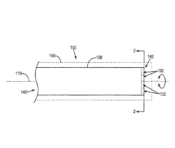
[0014] FIG. 1 is a plan view of an example of a device in accordance
with some
embodiments of the present invention;
[0015] FIG. 2 is a cross-sectional view of the device of FIG. 1 viewed
along line 2-
2;
[0016] FIG. 3 is a cross-sectional view of another configuration of the
device of
FIG. 1 viewed along line 2-2;
[0017] FIG. 4 is a pictorial representation of magnetic fields produced
by the
device of FIG. 1 in the presence of an external magnetic field, B;
[0018] FIG. 5 is a pictorial representation of an example of a sensitive
volume
adjacent the device of FIG. 1;
[0019] FIGS. 6A, 6B, and 6C illustrate a yaw angle, pitch angle, and
roll angle of a
device with respect to an external magnetic field, B;
[0020] FIG. 7A is a perspective view of an example of a device having
optical
encoding means for identifying an orientation of the device in accordance with
some
embodiments of the present invention;
[0021] FIG. 78 is a partial cross-section view of the device of FIG. 7A;
[0022] FIG. 7C is a cross-section view of another configuration of the
device of
FIG. 7A, in which a tracking system is incorporated into the device 7A;
[0023] FIG. 8A is a perspective view of an example of a device having a
radiofrequency receiver coil for receiving magnetic resonance signals and
optical
encoding means for identifying an orientation of the device in accordance with
some
embodiments of the present invention;
[0024] FIG. 8B is a partial cross-section view of the device of FIG. 8A;
[0025] FIG. 9A is an example of a measured one-dimensional projection of
a
device such as the one in FIG. 1 along an x-axis, in which the device is
oriented at a first
roll angle;
[0026] FIG. 98 is an example of a measured one-dimensional projection of
a
device such as the one in FIG. 1 along an x-axis, in which the device is
oriented at a
second roll angle that is different than the one corresponding to FIG. 9A;
[0027] FIG. 10 is a pulse sequence diagram of an example of a pulse
sequence
that may be used to measure the orientation of a device such as the one in
FIG. 1;
[0028] FIG. 11 is a block diagram of an example of a magnetic resonance
imaging
-4-
CA 02833620 2013-10-18
WO 2012/142715
PCT/CA2012/050258
("MRI") system for imaging with the device of the present invention; and
[0029] FIG. 12 is a flowchart setting forth the steps of an example of a
method for
imaging a volume-of-interest during an imaging application, such as an
interventional
or endoscopic procedure, using the provided device and an MRI system.
DETAILED DESCRIPTION OF THE INVENTION
[0030] When exposed to an external magnetic field, such as the main
magnetic
field, B0, of an MRI system, paramagnetic materials become magnetized such
that their
magnetization is oriented in the same direction as the external magnetic
field.
Conversely, diamagnetic materials become magnetized such that their
magnetization is
oriented opposite the direction of the external magnetic field. This
magnetization of
paramagnetic and diamagnetic materials results in magnetic field
perturbations. As an
example, for a spherical object composed of a material with magnetic
susceptibility, C,
placed in the main magnetic field, B0, of an MRI system will produce a
magnetic field
perturbation, D , as follows:
DB2 ,lle Boa' 2z2 x2
(1);
3 (x2 + z2 )5/2
[0031] where a is the radius of the spherical object and C is the volume
susceptibility of the spherical object.
[0032] It is an aspect of the present invention that objects with different
magnetic susceptibilities can be arranged in certain configurations such that
specific
magnetic field perturbations will be generated when the objects are positioned
within
an external magnetic field. The inhomogeneous magnetic fields resulting from
these
magnetic field perturbations may then be used to spatially encode nuclear
magnetic
resonance signals; thus, when objects of different magnetic susceptibilities
are so
arranged, they may be referred to as spatial-encoding elements. By way of
example,
these spatial-encoding elements are incorporated into a device to allow the
spatial
localization of nuclear magnetic resonance signals generated in a volume-of-
interest
adjacent the device. The generated field perturbations will depend on the
angle of such
a device with respect to the external magnetic field in which it is
positioned. An
example of the magnetic fields produced by such spatial-encoding elements is
illustrated in FIG. 4.
-5-
CA 02833620 2013-10-18
WO 2012/142715
PCT/CA2012/050258
[0033] The
aforementioned magnetic field perturbations will change as the
device is rotated about a roll axis by a roll angle, q. Thus, by way of
example, the device
may be rotated through 360 degrees in a series of angular steps. At each
angular step,
spins in the volume-of-interest adjacent the device may be excited and a
resulting
nuclear magnetic resonance signal acquired. The device is rotated again by the
angular
step and more signals are acquired. By rotating the device through 360
degrees,
sufficient information can be acquired to reconstruct an image of a volume-of-
interest
adjacent the device.
[0034] The signal
value at each time point is the summation of all small signals
from a continuous medium:
s(q,ti)= fif C(q,x,y,z)r (x,y,z)e-lw("')"dxdy dz = skl
x y z
(2);
[0035] where r
(x,y,z) is the spin density of the medium; C(q,x,y,z) is a
time-independent scaling coefficient that incorporates flip-angle variations
that may
result from the excitation profile of the RF excitation pulse, the coil
sensitivity profile,
and so on; and w(qox,y,z) is the three-dimensional angular frequency
distribution
due to the magnetic field perturbations generated by the spatial-encoding
elements.
[0036] The linear
relationship between signal and magnetization provides the a
reconstruction process that is equivalent to finding a decoding matrix, F,
that
produces a magnetization vector with elements Fn, where n = 1,2,..., N for N
voxels
in the three-dimensional reconstruction grid. The reconstruction process using
the
decoding matrix, F, for voxel r is given by the following equation:
=EEF (IA n,qoti)sid (3).
k 1
[0037] Because the
spatial-encoding fields generated by the spatial-encoding
elements are nonlinear and non-bilective, the for a reconstructed voxel may be
contaminated by signals from neighboring voxels. The spatial distribution of
the signal
that contributes to a particular reconstructed voxel, F, can be characterized
by a
spatial response function ("SRF") of the voxel:
-6-
CA 02833620 2013-10-18
WO 2012/142715
PCT/CA2012/050258
SRF,(x,y,z)=IEF
(4)
k 1
[0038] where
exp(¨iw(qox,y,z)ti ) is the phase of the magnetization for roll
angle qk and time point t1. Practically, the SRF may be viewed as a linear
combination
of these phase maps.
[0039] As an
example, the reconstruction technique may be viewed as computing
the rows of the decoding matrix, Fap for n = , N , such
that the SRF for each voxel
is well localized to that voxel. Each row of the decoding matrix may then be
used to
compute the intensity of one voxel of the final reconstructed image using Eqn,
(3). The
rows of the decoding matrix may be calculated in one of at least two ways.
[0040] One approach
for calculating the coefficients of the decoding matrix
involves generating an approximation of an encoding matrix, E, from
discretized phase
evolution maps. An example of such an encoding matrix, E, is as follows:
C(r1)e-hvl (r )11 C(r2) e-"(')' = = - C(rN)e-i''(')I`
C (r, )e-A1")'2 C(r2)e-1"*2)1' === C(rm)e-iivi(rN)"
=
= =
E = C(r1)e' C(r2)e-iw1(1-2)'` = = = C(r1v)e-iw,(r,,)f,,
(5);
)e-jw2(n)' C(r.,)e-i'{")" = = - C(rN)e-`42(')'
=
= =
C(1-2 )e-' (1'2)11' = = = C(rN )e-iwK(rN )tt..
[0041] where rõ is
the locations of a group of N spatial points that are to be
reconstructed, which can be expressed as the following:
=
r2 (x2,y2,z2)
[0042] (6).
rN
[0043] The encoding
matrix, E , provided in Eqn. (5) assumes that there are N
discrete voxels in the reconstructed image, that signals were acquired for K
different
-7-
CA 02833620 2013-10-18
WO 2012/142715
PCT/CA2012/050258
roll angles, and that signals were recorded at L discrete sample times ending
at
Elements of the encoding matrix may be calculated using a discrete
representation of
magnetic field perturbations surrounding the spatial-encoding elements. By way
of
example, the elements of the encoding matrix may be calculated for a two-
dimensional
plane of voxels located two millimeters in front of the spatial-encoding
elements. it will
be appreciated by those skilled in the art that this approach can be extended
to three
spatial dimensions, and that it is important to have elements in the encoding
matrix that
correspond to all of the spatial locations that contribute to the signal.
[0044] If the N
points, VA,, adequately cover the volume that contributes signal
to s(qk,t1), then a good numerical approximation, i(q4,1-1), of this signal
can be given
by the following equation:
[0045]kl = EkinFn (7).
[0046] The signal
values, gm, at each time point, t,, for each roll angle, qk , are
calculated by multiplying one row of the encoding matrix
e¨iwk (r2A C (q õ )e- (r'Of/
C(q,,r2) = = = (8);
Jixiv
by the magnetization density vectors, F. In reality, the signal values at each
time point
will be a summation of all small signals from a continuous medium, as shown in
Eqn.
(2); thus, Eqn. (7) is a discretization of the continuous signal equation.
[0047] Because
spatial encoding is a linear process, the reconstructed image
voxel values, arranged as a vector in F, can be computed by solving a system
of over-
determined linear equations:
:57= EF (9);
[0048] or equivalently by computing the decoding matrix:
17" = (10).
[0049] If the
spatial-encoding magnetic fields are linear, Eqns. (9) and (10) could
be solved by performing a Fourier transform on the acquired signals. However,
because
of the nonlinear nature of the spatial-encoding magnetic fields, there is no
one-to-one
relationship between the acquired signals and spatial frequencies. Therefore,
a k-space
representation of the acquired signals will not be applicable in this
technique. The
encoding matrix, E, of Eqn. (5) is usually ill-conditioned, so taking the
pseudo-inverse
-8-
CA 02833620 2013-10-18
WO 2012/142715
PCT/CA2012/050258
of the encoding matrix to compute the decoding matrix, F, will typically
result in a poor
image reconstruction. To improve upon this result, a cascade regularization
technique
may be used to reduce the ill-posed nature of the encoding matrix. In
addition, to
improve the accuracy of the image reconstruction process, truncated singular
value
decomposition ("TSVD") and Tikhonov regularization may be used.
[0050] The
reconstruction technique described above is considered a forward-
problem solution for the spatial-encoding technique provided by the present
invention.
As described earlier, there is signal contamination in a reconstructed voxel
that
originates from other spatial locations. In the forward-problem solution
described
above, each element of the encoding matrix is an average of the evolved phase
over the
corresponding voxel, and this assumption will fail when a sharp gradient in
the phase
exists within a voxel. This inhomogeneous voxel then becomes a source of
signal
contamination for the other voxels and is itself reconstructed with attenuated
signal.
Therefore, it is advantageous to develop other reconstruction methods that can
yield
well-localized SRFs for all voxels.
[0051] In another
approach for calculating the decoding matrix coefficients, an
optimization technique can be used to solve for the decoding matrix
coefficients in one
row of the decoding matrix. As an example, this optimization may be done to
minimize
the least-square-error between an ideal SRF and a calculated SRF for a voxel,
Fn . It will
be appreciated by those skilled in the art that other optimization functions
may be used.
For example, the 1-norm could be minimized in a sparse domain. A least-
squares
minimization will have the following form:
{ ( S
arg min I RF
. r
oõf(x, .Y,z)¨EF ( oqk,tr)e
kl -ill (qk ,x ,y,z)t iN.,2
)} (11).
[0052] If a good
global minimum is found, the SRF from the resulting optimized
reconstruction coefficients will be fairly localized and signal contaminations
from other
spatial locations will be minimized. Global minima are only found when the
target
(ideal) SRF is realizable, given the particular set of phase maps. Thus, it is
important to
choose the appropriate size, shape, and position of each target SRF,
corresponding to
each desired voxel.
[0053] Once all of
the decoding matrix coefficients for all voxels in the
reconstruction grid have been computed, Eqn. (3) can be used to reconstruct a
-9-
CA 02833620 2013-10-18
WO 2012/142715
PCT/CA2012/050258
magnetization density matrix of size N. An approach such as the one just
described
can be considered as an inverse-problem solution for the spatial-encoding
technique
provided by the present invention.
[0054] Having described the spatial-encoding and image reconstruction
processes, a discussion of various configurations of the spatial-encoding
elements and
their incorporation into different devices is now provided.
[0055] With initial reference to FIG. 1, a device 100 for acquiring image
data with
magnetic resonance imaging ("MRI") is provided. By way of example, the device
100
may be a medical device, such as an interventional device or an endoscope. In
addition,
the device 100 may form a part of a nuclear magnetic resonance probe
configured for oil
well logging, or the device 100 may be configured for other possible magnetic
resonance imaging or nuclear magnetic resonance applications. Generally, the
device
100 includes multiple spatial-encoding elements 102 that are coupled to a
support, such
as a shaft 108 of the device 100. The spatial-encoding elements 102 are
coupled to the
shaft 108 of the device 100 such that the spatial-encoding elements 102 may be
spaced
in a fixed arrangement that allows the spatial-encoding elements 102 to move
relative
to one another. For example, the shaft 108 may be rotated about an axis of
rotation,
such as a longitudinal or roll axis 110. The shaft 108 extends from a proximal
end 140
of the device 100 to a distal end 142 of the device 100 along the roll axis
110.
[0056] The spatial encoding elements 102 may be arranged such that they are
all
collinear, as illustrated in FIG. 2, or they may be arranged such they are not
all collinear
with each other. An example of a configuration of the device 100 in which the
spatial-
encoding elements 102 are not all collinear with each other is illustrated in
FIG. 3. In
this arrangement, the spatial encoding elements 102 are arranged as the
vertices of a
square. It will be appreciated that the spatial encoding elements 102 can be
arranged in
any number of spatial arrangements not illustrated here, and such spatial
arrangements
may differ depending on the number of spatial encoding elements 102 that arc
used.
For example, if six spatial encoding elements 102 are used, then it may be
advantageous
for the spatial encoding elements 102 to be arranged as the vertices of a
hexagon.
Preferably, the spatial-encoding elements 102 are coplanar; however, in some
configurations the spatial-encoding elements may not be entirely coplanar. As
shown in
FIGS. 2 and 3, the spatial-encoding elements 102 include both diamagnetic
elements
104 and paramagnetic elements 106. The spatial-encoding elements 102 may be
-10-
CA 02833620 2013-10-18
WO 2012/142715
PCT/CA2012/050258
shaped as spheres, spherical frustums, cones, conical frustums, circular
discs, or other
geometries. Examples of paramagnetic materials include titanium; however, it
will be
appreciated by those skilled in the art that other suitable paramagnetic
materials may
also be used. Examples of diamagnetic materials include graphite; however, it
will be
appreciated by those skilled in the art that other suitable diamagnetic
materials may
also be used, such as bismuth.
[0057] Intravoxel phase dispersions may be caused by inhomogeneous magnetic
fields generated by the spatial-encoding elements 102 in the out-of-plane
direction. The
magnetic field perturbations generated by the spatial-encoding elements 102
vary
significantly in the out-of-plane direction, which can result in signal loss
in voxels. Thus,
it may be advantageous to define a sensitive region of the device 100. One
example of
the sensitive region of the device 100 is illustrated in FIG. 5. In this
configuration, the
sensitive region is defined for spatial-encoding elements 102 having a
circular cross-
section. The sensitive region 550 of the device 100 extends from the surface
552 of the
device 100 to a distance of 1.5R along an out-of-plane direction 554 that is
normal to
the surface 552 of the device 100, and where R is the radius of each spatial-
encoding
element 102. Beyond a distance of 1.5R, there are no significant off-resonance
magnetic field perturbations, which create the basis for spatial encoding, for
different
roll-angle steps of the device. lntravoxel dephasing may be addressed by
considering
three-dimensional magnetic field perturbations of the spatial-encoding
elements 102
and by building encoding and decoding matrices for a three-dimensional
reconstruction
grid with voxel sizes smaller than 1.5R in the out-of-plane direction 554.
[0058] As a practical matter, when the spatial-encoding elements 102 are
arranged such that there is an inherent symmetry of the magnetic field
perturbations
produced by the spatial-encoding elements with respect to the center of the
arrangement, images reconstructed using such a device may include image
artifacts that
manifest as replication of magnetization densities. To mitigate these
artifacts, the
spatial-encoding elements may be arranged such that there is a disconnect in
the
inherent symmetry of the magnetic field perturbations generated by the spatial-
encoding elements. One example of such an arrangement is the purely collinear
arrangement illustrated in FIG. 2; however, it will be appreciated by those
skilled in the
art that other arrangements are possible.
[0059] Optionally, the device 100 may be surrounded by a bio-compatible
layer
-11-
CA 02833620 2013-10-18
WO 2012/142715
PCT/CA2012/050258
160 so that the magnetic materials, such as the spatial-encoding elements 102,
do not
come into direct contact with blood and tissue. In addition, the bio-
compatible layer
160 may be configured to provide a gap between the spatial-encoding elements
102 and
the exterior of the device 100 such that the local magnetic fields in the
volume-of-
interest are less steep than they would be without the gap.
[0060] By way of example, when the spatial-encoding elements 102 are
arranged
as illustrated in FIG. 3 and are placed in an external magnetic field, B ,
they will produce
magnetic fields 112, such as those illustrated in FIG. 4. In particular, the
magnetic fields
112 emanating from the paramagnetic elements 106 will be substantially aligned
with
the external magnetic field, B, whereas magnetic fields 112 emanating from the
diamagnetic elements 104 will be substantially aligned opposite the external
magnetic
field.
[0061] With the spatial-encoding elements 102 arranged as illustrated in
FIGS. 3
and 4, the magnetic fields 112 emanating from each of spatial-encoding
elements 102
will be spatially non-homogeneous. The resulting magnetic field disturbances
produced
by the spatial-encoding elements 102 will move around as the device 100 is
rotated
about the rotation axis 110. Operating the device 100 in this manner provides
a means
for spatially encoding signals originating from spins in a volume-of-interest
adjacent the
device 100. However, to reconstruct an image from signals spatially encoded in
this
manner requires knowledge about the orientation of the device 100 with respect
to the
external magnetic field (such as the main magnetic field, B0, of an MRI
system), as will
be described below in detail.
[0062] Referring now to FIGS. 6A-6C, the orientation of a device with
respect to
an external magnetic field, B, can be defined using a yaw, pitch, and roll
angle. The yaw
of a device 100 relative to an external magnetic field B aligned along the z-
axis is
illustrated in FIG. 6A. The yaw is measured by a yaw angle, y , which is a
measure of
rotation about the y-axis in the x-z plane. The pitch of a device 100 relative
to an
external magnetic field B aligned along the z-axis is illustrated in FIG. 6B.
The pitch is
measured by a pitch angle, W, which is a measure of rotation about the x-axis
in the y-z
plane. The roll of a device 100 relative to an external magnetic field B
aligned along
the z-axis is illustrated in FIG. 6C. The roll is measured by a roll angle, q,
which is a
measure of rotation about the z-axis in the x-y plane.
-12-
[00331 To measure the roll angle of the device 100, the device 100 may be
configured to
incorporate a rotation tracking system, as illustrated in FIGS. 7 A and 7B. An
example of
a rotation tracking system includes optical encoding of marks 712 that are
arranged on
the shaft 108 of the device 700. For example, the marks 712 may be markings
that are
etched into the surface of the shaft 108. The marks 712 may be optically read
using an
optical fiber 714, A sheath 716 may be provided to support the optical fiber
714 and to
keep the position of the optical fiber 714 fixed while the shaft 108 is free
to rotate within
the sheath 716. The optical signal carried by the optical fiber 714 conveys
information
about the roll angle at which the shaft 108 is oriented, as encoded by the
marks 712, and
this roll angle information is used during image reconstruction, as will be
described below
in detail.
[0( ,zti The yaw and pitch of the device can be measured in a number of
different ways.
By way of example, and referring to FIG. 7C, when the device 700 is positioned
in the
main magnetic field, BQ, of an MRI system, the device 700 may be configured to
include a
tracking system 718 capable of tracking and measuring the yaw and pitch of the
device
700. In general, such a tracking system 718 may be incorporated into the
interior of the
shaft 108 of the device 700, and may include tracking elements having
different magnetic
susceptibilities. For example, one or more tracking elements may be composed
of a
paramagnetic material, and one or more tracking elements may be composed of a
diamagnetic material. These tracking elements can be operated into an
arrangement such
that when they are positioned in the main magnetic field, Bo, of an MRI
system, they
produce a measureable local magnetic field. However, it is noted that the
tracking system
718 can be designed such that the local magnetic field generated by the
tracking system
718 do not interfere or affect the field perturbations produced by the spatial-
encoding
elements 102. When the tracking elements in the tracking system 718 are
arranged such
that a measureable local magnetic field is produced, the device 700 can be
imaged. The
measurable local magnetic field will affect the images of the device and from
this effect,
the orientation of the device 700 can be determined. More particularly, the
yaw and pitch
angles of the device 700 can be determined from these images. Examples of
tracking
systems of this nature is described in co-pending PCT Application No.
CA2O10/002041.
13
Date Recue/Date Received 2020-07-03
CA 02833620 2013-10-18
WO 2012/142715
PCT/CA2012/050258
[00651 In other
configurations of the device, such as the one illustrated in FIG. 8,
a receiver coil 820 may be coupled to the device 800. Such a configuration is
advantageous for applications in which high spatial and temporal resolution
are
desired, such as intravascular imaging applications. In
intravascular imaging
applications it is important that adequate signal-to-noise ratio ("SNR") be
achieved;
thus, to limit the volume of material that contributes noise to the acquired
signals, the
receiver coil 820 may be coupled to the sheath 716 of the device 800. The
receiver coil
820 may include, for example, a conductive wire wound around the sheath 716.
In such
a configuration, the windings of the receiver coil 820 can inductively couple
to the
tissue, thereby enabling direct signal reception. In the alternative, the
windings can be
designed to inductively couple to the shaft of the device 716, as described,
for example,
by K. Anderson, et al., in "Active Visualization of MR-Compatible Guidewires,"
Proc. Intl.
Soc. Mug. Reson. Med. 17, 2009; 2569. In the latter configuration, the shaft
716 of the
device 800 acts as both a spatial-encoding mechanism and a signal reception
mechanism. The use of small signal reception structures such as the receiver
coil 820
provides the added advantage of restricting the volume from which signal is
acquired,
which acts as a regularization of the image reconstruction method that is
described
below in detail. Although the amplitude of signals detected by the receiver
coil 820 will
decrease with distance from the device 800, the achievable image pixel size
increases
with distance from the device 800; thus, the larger pixel size offsets the
loss in signal
amplitude and provides for an increase in the total measurable signal
amplitude for
pixels located farther from the device 800.
[0066] As described
above, the disclosed spatial-encoding elements 102 produce
magnetic field perturbations when placed in an external magnetic field. These
magnetic
field perturbations alter the resonance frequency of spins that exists at
spatial locations
affected by the magnetic field perturbations. By way of example, the spatial-
encoding
elements 102 produce magnetic field perturbations that result in resonance
frequency
offsets between -2000 Hz and +2000 Hz for an external magnetic field with a
strength of
1.51. Therefore, a free-induction decay ("FM") occurring in a region affected
by the
magnetic field perturbations will contains resonance frequencies in the range
defined
by these resonance frequency offsets. By rotating the device 100 about its
roll axis 110,
the frequency content of those Fills that occur in a volume-of-interest
adjacent the
device 100 will remain the same. However, spins that do not experience the
magnetic
-14-
CA 02833620 2013-10-18
WO 2012/142715
PCT/CA2012/050258
field perturbations generated by the spatial-encoding elements 102 will
precess at their
Larmor frequency, wo = gB0, and their phases will contain no spatial
information.
Thus, exciting these spins will degrade the accuracy of the reconstructed
image. Off-
resonance excitation can be used to excite only those spins that are
experiencing
magnetic field perturbations due to the device 100 in order to restrict the
region from
which signals are acquired. As mentioned earlier, another approach to
restricting the
signal region is to incorporate RF receiver coils for signal acquisitions into
the device
100.
[0067] To perform off-resonance excitation, a radio frequency ("RF") pulse
having frequency content that matches the frequency offsets noted above is
used to
excite the desired spins. This RF excitation pulse may be, for example, a
composite RF
pulse with a binomial distribution designed using the Shinnar-LeRoux
algorithm. Spins
that are precessing with different Larmor frequencies will experience
different flip
angles due to the excitation profile of these off-resonance RF excitation
pulses, which
are frequency dependent. It is contemplated that this excitation profile will
lead to
encoding matrix rows that are modulated by the flip angle profile of the RF
excitation
pulse, which will improve the reconstruction process.
[0068] In conventional slice-selective RF excitation, the RF excitation
profile will
be similar to the Fourier transform of the RF pulse in presence of a slice-
selective
gradient field. With this new technique, the magnetic field perturbations will
assume
the functions of the slice-selective gradient; however, because of the
nonlinear nature of
these magnetic field perturbations, the RF excitation profile will not
resemble the
Fourier transform of the RF excitation pulse. Thus, the off-resonance RF
excitation
pulse described above will excite only those spins precessing at Larmor
frequencies
within the bandwidth of the proposed off-resonance RF excitation pulse.
[0069] As described above, the magnetic field perturbations generated by
the
spatial-encoding elements 102 are dependent on the orientation of the device
100 with
respect to the externally applied magnetic field. Therefore, it is desirable
to provide a
method for effectively measuring the orientation of the device 100 with
respect to the
external magnetic field. The measured yaw, pitch, and rotation angles can be
used when
calculating the encoding or decoding matrices to correct for any deviation of
the device
100 from the external magnetic field. It is noted, however, that image
reconstruction
will not be significantly affected when the device 100 deviates from its
alignment with
-15-
CA 02833620 2013-10-18
WO 2012/142715
PCT/CA2012/050258
the external magnetic field by a yaw angle, pitch angle, or both of plus or
minus fifteen
degrees. However, if the device 100 undergoes significant motion (e.g., inside
coronary
arteries), it may be necessary to measure the angle between the device 100 and
the
external magnetic field.
[0070] As the
device 100 rotates around its roll axis 110, a positive image
contrast in the plane perpendicular to the roll axis 110 will be changed as a
result of the
rotation of the magnetic field perturbations with the device 100. In addition
to using a
rotation tracking system incorporated into the device 100 as discussed above,
the roll
angle of the device 100 can be determined from two projection images of the
device 100
that are acquired with the device rotated to two different roll angles, q1 and
q2. It is
contemplated that there is a bijective relationship between the roll angle, q,
of the
device 100 and the length of the magnetic field perturbations at this angle.
The length
of the magnetic field perturbations can be calculated from the size of a
hyperintense
region present in a projection image of the device 100. The roll angle, q, of
the device
can be determined from these two projection images as follows:
ib
q2 = arecos ¨ (12);
\sa,
[0071] where a is
the length of the magnetic field perturbations at the first roll
angle and b is the length of the magnetic field perturbations at the second
roll angle. It
is possible to generalize this concept to measure both the yaw and pitch
angles of the
device 100 as well.
[0072] Without loss
of generality and by way of example, assume that the device
100 is initially oriented along the z-axis and is parallel to the external
magnetic field, B,
and that the device is rotated to a roll angle of q1. If the device 100 is
moved, the
following equation can be used to establish a relationship between the initial
position of
the device 100 and the new position of the device 100 using rotational angles:
_x1_
0
= Rx(qi)Ry(W)Rz(y) (13).
a
[0073] However, the
initial roll angle, q , is unknown and Eqn. (13) cannot be
solved for the yaw and the pitch angles, y and W, respectively. To resolve the
roll
-16-
CA 02833620 2013-10-18
WO 2012/142715
PCT/CA2012/050258
angle, the 100 device may be rotate by a known angle, such as ninety degrees,
about the
roll axis and the new position of the 100 device may be computed.
_ _
x2 0
y2 = Rx(q, + 900) Ry(W)Rz(y ) 0 (14);
a
""2, _ _ _
[0074] By combining
Eqn. (13) and Eqn. (14), the yaw, pitch, and roll angles can
be computed. A total of six measurements (x1,y1,z1,x2,y2,z2) are required to
establish the following sets of linear equations.
1 0 0
R1(q)=z 0 cosg ¨sing (15);
0 sing cosq
cosW 0 sinW-
Ry(W)= 0 1 0 (16);
¨sinW 0 cosW
cosy ¨siny 0
Rz(y)= silly cosy 0 (17).
0 0 1_
[0075] The angle
measurement technique may be used to locate the spatial
position and orientation of device 100 under MRI guidance; however such a
technique
may suffer from its long acquisition time. For example, the long acquisition
time may be
unacceptable in some applications, such as positioning an interventional
device in
coronary arteries where there is significant motion in a short period of time.
To address
this issue, one-dimensional positive contrast projections can be obtained
along each of
three principal axes and used both to estimate the coordinates of the device
and to
measure the length of the hyperintense region adjacent the device.
[0076] One-
dimensional projections are commonly used for positioning devices
under MRI guidance, and can significantly reduce the acquisition time needed
for
localization. By way of example, FIGS. 9A and 9B illustrate examples of
measured one-
dimensional projections of the device 100. FIG. 9A illustrates an example in
which the
device 100 is oriented at a roll angle of zero degrees with respect to the
external
magnetic field, and FIG. 9B illustrates an example in which the device 100 is
oriented at
-17-
CA 02833620 2013-10-18
WO 2012/142715
PCT/CA2012/050258
a roll angle of forty-five degrees with respect to the external magnetic
field. The width
of the hyperintense region adjacent the device 100 can be readily measured
from these
one-dimensional projections. For example, the width of this region at the
first roll
angle, q1 CI degrees, is a, and the width of this region at the second roll
angle,
q2 = 45 degrees, is b.
[0077] An example of a pulse sequence that may be used to obtain one-
dimensional projections of the device 100 is illustrated in FIG. 10, to which
reference is
now made. The pulse sequence generally includes the application of an RF
excitation
pulse followed by a dephasing gradient along the axis of interest. Readout of
the
resulting FID then starts at an echo time ("TE") after the dephasing gradient.
Thus, for
example, the pulse sequence may include the application of an RF excitation
pulse 1002
followed by the application of a dephasing gradient 1004 along the x-axis such
that an
FID is produced and sampled during a readout window 1006. This sampled FID
will
provide a one-dimensional projection of the device 100 along the x-axis. The
pulse
sequence may further include the application of an RF excitation pulse 1002
followed by
the application of a dephasing gradient 1008 along the y-axis such that an FID
is
produced and sampled during a readout window 1006. This sampled FID will
provide a
one-dimensional projection of the device 100 along the y-axis. The pulse
sequence may
still further include the application of an RF excitation pulse 1002 followed
by the
application of a dephasing gradient 1010 along the z-axis such that an FID is
produced
and sampled during a readout window 1006. This FID will provide a one-
dimensional
projection of the device 100 along the z-axis. The foregoing RF excitation
pulses 1002
and dephasing gradients 1004, 1008, 1010 may be repeated a second time after
the
device 100 has be moved to a different position such that a different set of
one-
dimensional projections are acquired. After the angle measurement portion of
the pulse
sequence has concluded, the imaging portion begins. Because spatial-encoding
is
provided by the device, the imaging portion of the pulse sequence does not
require the
application of magnetic field gradients using the MRI system. Thus, the
imaging portion
of the pulse sequence generally includes only an RF excitation pulse 1012.
[00781 Using the foregoing pulse sequence, the angle of the device 100 can
be
measured with respect to each principal axis before each data acquisition
step. As
described above, knowing the orientation of the device with respect to
external
-18-
CA 02833620 2013-10-18
WO 2012/142715
PCT/CA2012/050258
magnetic field can be advantageous to improve the accuracy of the image
reconstruction
techniques described herein. With this proposed technique, the angle of the
device
100, and therefore the angle of the spatial-encoding elements 102, can be
measured
respect to the external magnetic field before each angle-step of the device
100 in order
to perform corrections to the decoding matrix, F. It is noted that the roll
angle may be
more precisely measured using a rotation tracking system, such as the optical
system
described above. For practical reasons, it is contemplated that using such a
system
instead of measuring the roll angle with an MRI system will be preferable.
[0079] Referring
particularly now to FIG. 11, an example of an MRI system 1100
for use with the provided device is illustrated. The MRI system 1100 includes
a
workstation 1102 having a display 1104 and a keyboard 1106. The workstation
1102
includes a processor 1108, such as a commercially available programmable
machine
running a commercially available operating system. The workstation 1102
provides the
operator interface that enables scan prescriptions to be entered into the MRI
system
1100. The workstation 1102 is coupled to four servers: a pulse sequence server
1110; a
data acquisition server 1112; a data processing server 1114, and a data store
server
1116. The workstation 1102 and each server 1110, 1112, 1114 and 1116 are
connected
to communicate with each other.
[0080] The pulse
sequence server 1110 functions in response to instructions
downloaded from the workstation 1102 to operate a gradient system 1118 and a
radiofrequency ("RF") system 1120. Gradient waveforms necessary to perform the
prescribed scan are produced and applied to the gradient system 1118, which
excites
gradient coils in an assembly 1122 to produce the magnetic field gradients Gõ,
, and
G., used for position encoding MR signals. The gradient coil assembly 1122
forms part
of a magnet assembly 1124 that includes a polarizing magnet 1126 and a whole-
body
1117 coil 1128.
[0081] RF
excitation waveforms are applied to the RF coil 1128, or a separate
local coil (not shown in FIG. 11), by the RF system 1120 to perform the
prescribed
magnetic resonance pulse sequence. Responsive MR signals detected by the RF
coil
1128, or a separate local coil (not shown in FIG. 11), are received by the RF
system
1120, amplified, demodulated, filtered, and digitized under direction of
commands
produced by the pulse sequence server 1110. The RF system 1120 includes an RF
-19-
CA 02833620 2013-10-18
WO 2012/142715
PCT/CA2012/050258
transmitter for producing a wide variety of RF pulses used in MR pulse
sequences. The
RF transmitter is responsive to the scan prescription and direction from the
pulse
sequence server 1110 to produce RF pulses of the desired frequency, phase, and
pulse
amplitude waveform. The generated RF pulses may be applied to the whole body
RF
coil 1128 or to one or more local coils or coil arrays (not shown in FIG. 11).
[0082] The RF system 1120 also includes one or more RF receiver channels.
Each RF receiver channel includes an RF amplifier that amplifies the MR signal
received
by the coil 1128 to which it is connected, and a detector that detects and
digitizes the /
and Q quadrature components of the received MR signal. The magnitude of the
received MR signal may thus be determined at any sampled point by the square
root of
the sum of the squares of the / and Q components:
= V12 + Q2
(18);
[0083] and the phase of the received MR signal may also be determined:
= tan-1 (19).
[0084] The pulse sequence server 1110 also optionally receives patient data
from a physiological acquisition controller 1130. The controller 1130 receives
signals
from a number of different sensors connected to the patient, such as
electrocardiograph
("ECG") signals from electrodes, or respiratory signals from a bellows or
other
respiratory monitoring device. Such signals are typically used by the pulse
sequence
server 1110 to synchronize, or "gate," the performance of the scan with the
subject's
heart beat or respiration.
[0085] The pulse sequence server 1110 also connects to a scan room
interface
circuit 1132 that receives signals from various sensors associated with the
condition of
the patient and the magnet system. It is also through the scan room interface
circuit
1132 that a patient positioning system 1134 receives commands to move the
patient to
desired positions during the scan.
[0086] The digitized MR signal samples produced by the RF system 1120 are
received by the data acquisition server 1112. The data acquisition server 1112
operates
in response to instructions downloaded from the workstation 1102 to receive
the real-
time MR data and provide buffer storage, such that no data is lost by data
overrun. In
some scans, the data acquisition server 1112 does little more than pass the
acquired MR
-20-
CA 02833620 2013-10-18
WO 2012/142715
PCT/CA2012/050258
data to the data processor server 1114. However, in scans that require
information
derived from acquired MR data to control the further performance of the scan,
the data
acquisition server 1112 is programmed to produce such information and convey
it to
the pulse sequence server 1110. For example, during prescans, MR data is
acquired and
used to calibrate the pulse sequence performed by the pulse sequence server
1110.
Also, navigator signals may be acquired during a scan and used to adjust the
operating
parameters of the RF system 1120 or the gradient system 1118, or to control
the view
order in which k-space is sampled. The data acquisition server 1112 may also
be
employed to process MR signals used to detect the arrival of contrast agent in
a
magnetic resonance angiography ("MRA") scan. In all these examples, the data
acquisition server 1112 acquires MR data and processes it in real-time to
produce
information that is used to control the scan.
[0087] The data processing server 1114 receives MR data from the data
acquisition server 1112 and processes it in accordance with instructions
downloaded
from the workstation 1102. Such processing may include, for example: Fourier
transformation of raw k-space MR data to produce two or three-dimensional
images;
the application of filters to a reconstructed image; the performance of a
backprojection
image reconstruction of acquired MR data; the generation of functional MR
images; and
the calculation of motion or flow images.
[0088] Images reconstructed by the data processing server 1114 are conveyed
back to the workstation 1102 where they are stored. Real-time images are
stored in a
data base memory cache (not shown in FIG. 11), from which they may be output
to
operator display 1112 or a display 1136 that is located near the magnet
assembly 1124
for use by attending physicians. Batch mode images or selected real time
images are
stored in a host database on disc storage 1138. When such images have been
reconstructed and transferred to storage, the data processing server 1114
notifies the
data store server 1116 on the workstation 1102. The workstation 1102 may be
used by
an operator to archive the images, produce films, or send the images via a
network to
other facilities.
[0089] Having described the general structure of the provided device, and
various examples of configurations thereof, a description of a general
operation of the
device is now provided. Referring now to FIG. 12, a flowchart setting forth
the steps of
an example of a method for imaging a volume-of-interest during an imaging
application,
-21-
CA 02833620 2013-10-18
WO 2012/142715
PCT/CA2012/050258
such as an interventional or endoscopic procedure, using the provided device
and an
MRI system is illustrated. First, the device is provided to a volume-of-
interest ("VOI")
that is to be imaged, as indicated at step 1202. For example, the device is
provided to a
VOI in a patient undergoing an interventional or endoscopic procedure in the
presence
of the main magnetic field, Bc, of an MRI system. In the alternative, the
device may be
provided to a bore hole for NIvIR oil well logging applications. The device is
then
operated to be positioned in a first position, as indicated at step 1204. In
this first
position, the device produces a first spatial-encoding magnetic field. For
example, the
device may be rotated about an axis of rotation into a first rotational
orientation. The
MRI system is then operated to acquire image data from the VOI, as indicated
at step
1206. Given the proximity of the VOI to the device, the acquired image data is
spatially-
encoded by the spatial-encoding magnetic fields produced by the device.
[0090] Advantageously, the device is free to move in conjunction with
physiological motion, such as respiration, cardiac rhythm, and pulsatile flow.
As a
result, the spatial-encoding magnetic fields move along with the device and in
conjunction with physiological motions. Because of this result, the device
effectively
images the VOI in a frame of reference that moves along with present
physiological
motions. By imaging in this local frame of reference, motion-related errors in
the
acquired image data are mitigated, and substantially motion-artifact free
images can be
reconstructed from the image data.
[0091] A determination is made at decision block 1208 as to whether the
desired
amount of image data has been acquired. If not, then the device is positioned
in a
different position, as indicated at step 1210, and the MRI system operated
again to
acquire more image data. By way of example, a new roll, pitch, and yaw angle
of the
device may then be calculated in this new position. In each different
position, the device
provides a different spatial encoding to the acquired image data. By acquiring
a
plurality of image data sets, each with a different spatial encoding, one of
the image
reconstruction techniques presented above can be solved. Thus, after the
desired
amount of image data has been acquired, images of the VOI are reconstructed,
as
indicated at step 1212, By way of example, reconstruction coefficients based
on the
measured roll, pitch, and yaw of the device may be calculated and used in the
reconstruction process. It is noted that the image data acquisition and image
reconstruction processes can take place in real-time, so as to provide visual
feedback to
-22-
CA 02833620 2013-10-18
WO 2012/142715
PCT/CA2012/050258
a user, such as a medical practitioner performing an interventional or
endoscopic
procedure. For real-time display applications, pre-computed reconstruction
coefficients
can be used and matched to the measured roll, pitch, and yaw of the device
using a look-
up table.
[0092] The present invention has been described in terms of one or more
preferred embodiments, and it should be appreciated that many equivalents,
alternatives, variations, and modifications, aside from those expressly
stated, are
possible and within the scope of the invention.
-23-