Note: Descriptions are shown in the official language in which they were submitted.
81774929
QUANTIFICATION OF A MINORITY NUCLEIC ACID SPECIES
USING INHIBITORY OLIGONUCLEOTIDES
Related Patent Application
This patent application claims the benefit of U.S. Provisional Application No.
61/ 480,686 filed on
April 29, 2011, entitled QUANTIFICATION OF A MINORITY NUCLEIC ACID SPECIES,
naming
Anders Nygren as inventor, and designated by attorney docket no. SEQ-6031-PV.
Field
The technology relates in part to quantification of a minority nucleic acid
species. In some
embodiments, methods for determining the amount of cell-free fetal DNA in a
maternal sample are
provided.
Backaround
Cell-free DNA (CF-DNA) is composed of DNA fragments that originate from cell
death and circulate
in peripheral blood. High concentrations of CF-DNA can be indicative of
certain clinical conditions
such as cancer, trauma, burns, myocardial infarction, stroke, sepsis,
infection, and other illnesses.
Additionally, cell-free fetal DNA (CFF-DNA) can be detected in the maternal
bloodstream and used
for various noninvasive prenatal diagnostics.
The presence of fetal nucleic acid in maternal plasma allows for non-invasive
prenatal diagnosis
through the analysis of a maternal blood sample. For example, quantitative
abnormalities of fetal
DNA in maternal plasma can be associated with a number of pregnancy-associated
disorders,
including preeclampsia, preterm labor, antepartum hemorrhage, invasive
placentation, fetal Down
syndrome, and other fetal chromosomal aneuploidies. Hence, fetal nucleic acid
analysis in
maternal plasma is a useful mechanism for the monitoring of fetomaternal well-
being.
Early detection of pregnancy-related conditions, including complications
during pregnancy and
genetic defects of the fetus is important, as it allows early medical
intervention necessary for the
safety of both the mother and the fetus. Prenatal diagnosis traditionally has
been conducted using
cells isolated from the fetus through procedures such as chorionic villus
sampling (CVS) or
1
CA 2834218 2019-11-21
CA 02834218 2013-10-23
WO 2012/149339 PCT/US2012/035479
amniocentesis. However, these conventional methods are invasive and present an
appreciable
risk to both the mother and the fetus. The National Health Service currently
cites a miscarriage
rate of between 1 and 2 per cent following the invasive amniocentesis and
chorionic villus sampling
(CVS) tests. An alternative to these invasive approaches is the use of non-
invasive screening
techniques that utilize circulating OFF-DNA.
Summary
Provided in some embodiments is a method for determining the amount of a
minority nucleic acid
species in a sample which contains a minority species and a majority species,
the combination of
the minority species and the majority species comprising total nucleic acid in
the sample,
comprising (a) contacting under amplification conditions a nucleic acid sample
comprising the
minority nucleic acid species with: (i) a first set of amplification primers
that specifically amplify a
first region comprising a feature that (1) is present in the minority nucleic
acid species and is not
present in the majority nucleic acid species, or (2) is not present in the
minority nucleic acid
species and is present in the majority nucleic acid species, (ii) a second set
of amplification primers
that amplify a second region allowing for a determination of total nucleic
acid in the sample, where
the first region and the second region are different, and (iii) one or more
inhibitory oligonucleotides
that reduce the amplification of the second region, thereby generating
minority and total nucleic
acid amplification products, where the total nucleic acid amplification
products are reduced relative
to total amplification products that would be generated if no inhibitory
oligonucleotide was present,
(b) separating the minority and total nucleic acid amplification products,
thereby generating
separated minority and total nucleic acid amplification products, and (c)
determining the fraction of
the minority nucleic acid species in the sample relative to the total amount
of the nucleic acid in the
sample based on the amount of each of the separated minority and total nucleic
acid amplification
products.
In some cases, the feature that is present in the minority nucleic acid
species and not present in
the majority nucleic acid species is methylation. Sometimes the first region
is methylated and the
second region is unmethylated.
In some embodiments, the method further comprises contacting the nucleic acid
sample with one
or more restriction enzymes prior to (a). Sometimes the one or more
restriction enzymes are
methylation sensitive. In some cases, the restriction enzymes are Hhal and
Hpall.
2
CA 02834218 2013-10-23
WO 2012/149339 PCT/US2012/035479
In some embodiments, the method further comprises contacting under
amplification conditions the
nucleic acid sample with a third set of amplification primers that amplify a
third region allowing for a
determination of the presence or absence of fetal specific nucleic acid. In
some cases, the fetal
specific nucleic acid is Y chromosome nucleic acid.
In some embodiments, the method further comprises contacting under
amplification conditions the
nucleic acid sample with a fourth set of amplification primers that amplify a
fourth region allowing
for a determination of the amount of digested or undigested nucleic acid, as
an indicator of
digestion efficiency. Often, the first, second, third and fourth regions each
comprise one or more
genomic loci. In some cases, the genomic loci are the same length. In some
cases, the genomic
loci are about 50 base pairs to about 200 base pairs. In some cases, the
genomic loci are about
60 base pairs to about 80 base pairs. In some cases, the genomic loci are
about 70 base pairs.
In some embodiments, the first region comprises one or more loci that are
differentially methylated
between the minority and majority species. In some cases, first region
comprises loci within the
TBX3 and SOX14 genes. In some cases, the loci for the first region each
comprise independently
SEQ ID NO:29 and SEQ ID NO:30.
In some embodiments, the second region comprises one or more loci which do not
contain a
restriction site for a methylation-sensitive restriction enzyme. In some
cases, the second region
comprises loci within the POP5 and APOE genes. In some cases, the loci for the
second region
each comprise independently SEQ ID NO:31 and SEQ ID NO:32.
In some embodiments, the third region comprises one or more loci within
chromosome Y. In some
cases, the third region comprises a locus within the DDX3Y gene. In some
cases, the locus for the
third region comprises SEQ ID NO:34.
In some embodiments, the fourth region comprises one or more loci present in
every genome in
the sample and unmethylated in all species. In some cases, the fourth region
comprises loci within
the POP5 or LDHA genes. In some cases, the loci for the fourth region each
comprise
independently SEQ ID NO:35 and SEQ ID NO:36.
3
CA 02834218 2013-10-23
WO 2012/149339 PCT/US2012/035479
In some embodiments, the first and second sets of amplification primers each
comprise one or
more pairs of forward and reverse primers. In some embodiments, the third and
fourth sets of
amplification primers each comprise one or more pairs of forward and reverse
primers. In some
cases, the one or more amplification primer pairs further comprise a 5' tail.
Sometimes the 5' tail is
a distinct length for each amplification primer set. In some cases, the
amplification primers each
comprise independently SEQ ID NOs:1 to 8 and SEQ ID NOs:11 to 16.
In some embodiments, an inhibitory oligonucleotide of the one or more
inhibitory oligonucleotides
comprises a nucleotide sequence complementary to a nucleotide sequence in the
second region.
.. In some cases, the inhibitory oligonucleotide and a primer in the second
set of amplification
primers are complementary to the same nucleotide sequence in the second
region. In some
cases, the inhibitory oligonucleotide comprises one or more 3' mismatched
nucleotides. In some
cases, the inhibitory oligonucleotides each comprise independently SEQ ID
NO:17, SEQ ID NO:18,
SEQ ID NO:19, and SEQ ID NO:20.
In some embodiments, the method further comprises contacting under
amplification conditions the
nucleic acid sample with one or more first competitor oligonucleotides that
compete with the first
region for hybridization of primers of the first amplification primer set. In
some embodiments, the
method further comprises contacting under amplification conditions the nucleic
acid sample with
one or more second competitor oligonucleotides that compete with the second
region for
hybridization of primers of the second amplification primer set. In some
embodiments, the method
further comprises contacting under amplification conditions the nucleic acid
sample with one or
more third competitor oligonucleotides that compete with the third region for
hybridization of
primers of the third amplification primer set. In some embodiments, the method
further comprises
contacting under amplification conditions the nucleic acid sample with one or
more fourth
competitor oligonucleotides that compete with the fourth region for
hybridization of primers of the
fourth amplification primer set. In some cases, the competitor
oligonucleotides comprise a stuffer
sequence. In some cases, the stuffer sequence length is constant for one or
more of the first
competitor oligonucleotides, second competitor oligonucleotides, third
competitor oligonucleotides
and fourth competitor oligonucleotides. In some cases, the stuffer sequence
length is variable for
one or more of the first competitor oligonucleotides, second competitor
oligonucleotides, third
competitor oligonucleotides and fourth competitor oligonucleotides. At times,
the stuffer sequence
is from a non-human genome. Sometimes the stuffer sequence is from the PhiX
174 genome. In
some embodiments, the competitor oligonucleotide is about 100 to about 150
base pairs long. In
4
CA 02834218 2013-10-23
WO 2012/149339 PCT/US2012/035479
some cases, the competitor oligonucleotide is about 115 to about 120 base
pairs long. In some
cases, the first and second competitor oligonucleotide is about 115 base pairs
long. In some
cases, the third competitor oligonucleotide is about 118 base pairs long. In
some cases, the fourth
competitor oligonucleotide is about 120 base pairs long. In some embodiments,
the one or more
.. first competitor oligonucleotides each comprise independently SEQ ID NO:21
and SEQ ID NO:22.
In some embodiments, the one or more second competitor oligonucleotides each
comprise
independently SEQ ID NO:23 and SEQ ID NO:24. In some embodiments, the third
competitor
oligonucleotide comprises SEQ ID NO:26. In some embodiments, the one or more
fourth
competitor oligonucleotides each comprise independently SEQ ID NO:27 and SEQ
ID NO:28. In
some embodiments, the one or more competitor oligonucleotides comprise a
detectable label. In
some cases, the detectable label is a flourophore and sometimes the
fluorophore is different for
each competitor oligonucleotide. In some embodiments, a predetermined copy
number of each
competitor oligonucleotide is used. In some embodiments, the method further
comprises
determining the copy number of the minority nucleic acid species based on the
amount of
competitor oligonucleotide used. In some embodiments, the method further
comprises determining
the copy number of the majority nucleic acid species.
In some embodiments, the sample nucleic acid is extracellular nucleic acid. In
some
embodiments, the minority nucleic acid species is fetal DNA and the majority
nucleic acid species
is maternal DNA. In some cases, the nucleic acid sample is obtained from a
pregnant female
subject. In some cases, the subject is human. In some embodiments, the sample
nucleic acid is
from plasma. In some cases, the sample nucleic acid is from serum.
In some embodiments, the amplification is in a single reaction vessel.
Sometimes two or more of
.. the amplification products are different lengths. Often, the amplification
is by polymerase chain
reaction (PCR). In some embodiments, the method further comprises contacting
the amplification
products with an exonuclease prior to (b). In some cases, the separation of
amplification products
is based on length. Often, the separation is performed using electrophoresis.
In some cases, the
electrophoresis is capillary electrophoresis. In some embodiments, the method
further comprises
determining whether the nucleic acid sample is utilized for a sequencing
reaction. In some cases,
the sequencing reaction is a reversible terminator-based sequencing reaction.
In some
embodiments, the method further comprises determining whether sequencing
information obtained
for a nucleic acid sample is used for a diagnostic determination.
5
CA 02834218 2013-10-23
WO 2012/149339 PCT/US2012/035479
Also provided in some embodiments is a method for determining the amount of a
minority nucleic
acid species in a sample which contains a minority species and a majority
species, the
combination of the minority species and the majority species comprising total
nucleic acid in the
sample, comprising a method of determining the copy number of the minority
nucleic acid species,
comprising the steps of (a) contacting under amplification conditions a
nucleic acid sample
comprising the minority nucleic acid species with (i) a first set of
amplification primers that
specifically amplify a first region comprising a feature that (1) is present
in the minority nucleic acid
species and is not present in the majority nucleic acid species, or (2) is not
present in the minority
nucleic acid species and is present in the majority nucleic acid species, (ii)
a second set of
amplification primers that amplify a second region allowing for a
determination of the total nucleic
acid in the sample, where the first region and the second region are
different, (iii) one or more first
competitor oligonucleotides that compete with the first region for
hybridization of primers of the first
amplification primer set, and (iv) one or more second competitor
oligonucleotides that compete with
the second region for hybridization of primers of the second amplification
primer set, thereby
generating amplification products where two or more of the amplification
products are different
lengths; (b) separating the minority nucleic acid, total nucleic acid, and
competitor amplification
products, thereby generating separated minority nucleic acid, total nucleic
acid, and competitor
amplification products; and (c) determining the copy number of the minority
nucleic acid species in
the sample based on the separated amplification products.
In some cases, the feature that is present in the minority nucleic acid
species and not present in
the majority nucleic acid species is methylation. Sometimes the first region
is methylated and the
second region is unmethylated.
In some embodiments, the method further comprises contacting the nucleic acid
sample with one
or more restriction enzymes prior to (a). Sometimes the one or more
restriction enzymes are
methylation sensitive. In some cases, the restriction enzymes are Hhal and
Hpall.
In some embodiments, the method further comprises contacting under
amplification conditions the
nucleic acid sample with a third set of amplification primers that amplify a
third region allowing for a
determination of the presence or absence of fetal specific nucleic acid. In
some cases, the fetal
specific nucleic acid is Y chromosome nucleic acid.
6
CA 02834218 2013-10-23
WO 2012/149339 PCT/US2012/035479
In some embodiments, the method further comprises contacting under
amplification conditions the
nucleic acid sample with a fourth set of amplification primers that amplify a
fourth region allowing
for a determination of the amount of digested or undigested nucleic acid, as
an indicator of
digestion efficiency. Often, the first, second, third and fourth regions each
comprise one or more
genomic loci. In some cases, the genomic loci are the same length. In some
cases, the genomic
loci are about 50 base pairs to about 200 base pairs. In some cases, the
genomic loci are about
60 base pairs to about 80 base pairs. In some cases, the genomic loci are
about 70 base pairs.
In some embodiments, the first region comprises one or more loci that are
differentially methylated
between the minority and majority species. In some cases, first region
comprises loci within the
TBX3 and SOX14 genes. In some cases, the loci for the first region each
comprise independently
SEQ ID NO:29 and SEQ ID NO:30.
In some embodiments, the second region comprises one or more loci which do not
contain a
restriction site for a methylation-sensitive restriction enzyme. In some
cases, the second region
comprises loci within the POPS and APOE genes. In some cases, the loci for the
second region
each comprise independently SEQ ID NO:31 and SEQ ID NO:32.
In some embodiments, the third region comprises one or more loci within
chromosome Y. In some
.. cases, the third region comprises a locus within the DDX3Y gene. In some
cases, the locus for the
third region comprises SEQ ID NO:34.
In some embodiments, the fourth region comprises one or more loci present in
every genome in
the sample and unmethylated in all species. In some cases, the fourth region
comprises loci within
the POPS or LDHA genes. In some cases, the loci for the fourth region each
comprise
independently SEQ ID NO:35 and SEQ ID NO:36.
In some embodiments, the first and second sets of amplification primers each
comprise one or
more pairs of forward and reverse primers. In some embodiments, the third and
fourth sets of
amplification primers each comprise one or more pairs of forward and reverse
primers. In some
cases, the one or more amplification primer pairs further comprise a 5' tail.
Sometimes the 5' tail is
a distinct length for each amplification primer set. In some cases, the
amplification primers each
comprise independently SEQ ID NOs:1 to 8 and SEQ ID NOs:11 to 16.
7
CA 02834218 2013-10-23
WO 2012/149339 PCT/US2012/035479
In some embodiments, the method further comprises contacting under
amplification conditions the
nucleic acid sample with one or more inhibitory oligonucleotides that reduce
the amplification of the
second region. In some embodiments, an inhibitory oligonucleotide of the one
or more inhibitory
oligonucleotides comprises a nucleotide sequence complementary to a nucleotide
sequence in the
second region. In some cases, the inhibitory oligonucleotide and a primer in
the second set of
amplification primers are complementary to the same nucleotide sequence in the
second region.
In some cases, the inhibitory oligonucleotide comprises one or more 3'
mismatched nucleotides.
In some cases, the inhibitory oligonucleotides each comprise independently SEQ
ID NO:17, SEQ
ID NO:18, SEQ ID NO:19, and SEQ ID NO:20.
In some embodiments, the method further comprises determining the fraction of
the minority
nucleic acid species in the sample relative to the total amount of the nucleic
acid in the sample
based on the amount of each of the separated minority and total nucleic acid
amplification
products.
In some embodiments, the method further comprises contacting under
amplification conditions the
nucleic acid sample with one or more third competitor oligonucleotides that
compete with the third
region for hybridization of primers of the third amplification primer set. In
some embodiments, the
method further comprises contacting under amplification conditions the nucleic
acid sample with
one or more fourth competitor oligonucleotides that compete with the fourth
region for hybridization
of primers of the fourth amplification primer set. In some cases, the
competitor oligonucleotides
comprise a stuffer sequence. In some cases, the stuffer sequence length is
constant for one or
more of the first competitor oligonucleotides, second competitor
oligonucleotides, third competitor
oligonucleotides and fourth competitor oligonucleotides. In some cases, the
stuffer sequence
length is variable for one or more of the first competitor oligonucleotides,
second competitor
oligonucleotides, third competitor oligonucleotides and fourth competitor
oligonucleotides. At
times, the stuffer sequence is from a non-human genome. Sometimes the stuffer
sequence is from
the PhiX 174 genome. In some embodiments, the competitor oligonucleotide is
about 100 to about
150 base pairs long. In some cases, the competitor oligonucleotide is about
115 to about 120
base pairs long. In some cases, the first and second competitor
oligonucleotide is about 115 base
pairs long. In some cases, the third competitor oligonucleotide is about 118
base pairs long. In
some cases, the fourth competitor oligonucleotide is about 120 base pairs
long. In some
embodiments, the one or more first competitor oligonucleotides each comprise
independently SEQ
ID NO:21 and SEQ ID NO:22. In some embodiments, the one or more second
competitor
8
CA 02834218 2013-10-23
WO 2012/149339 PCT/US2012/035479
oligonucleotides each comprise independently SEQ ID NO:23 and SEQ ID NO:24. In
some
embodiments, the third competitor oligonucleotide comprises SEQ ID NO:26. In
some
embodiments, the one or more fourth competitor oligonucleotides each comprise
independently
SEQ ID NO:27 and SEQ ID NO:28. In some embodiments, the one or more competitor
__ oligonucleotides comprise a detectable label. In some cases, the detectable
label is a flourophore
and sometimes the fluorophore is different for each competitor
oligonucleotide. In some
embodiments, a predetermined copy number of each competitor oligonucleotide is
used. In some
cases, the copy number of the minority nucleic acid species is determined
based on the amount of
competitor oligonucleotide used. In some cases, the copy number of the
majority nucleic acid
__ species is determined.
In some embodiments, the sample nucleic acid is extracellular nucleic acid. In
some
embodiments, the minority nucleic acid species is fetal DNA and the majority
nucleic acid species
is maternal DNA. In some cases, the nucleic acid sample is obtained from a
pregnant female
subject. In some cases, the subject is human. In some embodiments, the sample
nucleic acid is
from plasma. In some cases, the sample nucleic acid is from serum.
In some embodiments, the amplification is in a single reaction vessel.
Sometimes two or more of
the amplification products are different lengths. Often, the amplification is
by polymerase chain
reaction (FOR). In some embodiments, the method further comprises contacting
the amplification
products with an exonuclease prior to (b). In some cases, the separation of
amplification products
is based on length. Often, the separation is performed using electrophoresis.
In some cases, the
electrophoresis is capillary electrophoresis. In some embodiments, the method
further comprises
determining whether the nucleic acid sample is utilized for a sequencing
reaction. In some cases,
__ the sequencing reaction is a reversible terminator-based sequencing
reaction. In some
embodiments, the method further comprises determining whether sequencing
information obtained
for a nucleic acid sample is used for a diagnostic determination.
Also provided in some embodiments is a method for determining the amount of a
minority nucleic
acid species in a sample which contains a minority species and a majority
species, the
combination of the minority species and the majority species comprising total
nucleic acid in the
sample, comprising: (a) contacting under amplification conditions a nucleic
acid sample comprising
the minority nucleic acid species with: (i) a first set of amplification
primers that specifically amplify
a first region comprising a feature that (1) is present in the minority
nucleic acid species and is not
9
CA 02834218 2013-10-23
WO 2012/149339 PCT/US2012/035479
present in the majority nucleic acid species, or (2) is not present in the
minority nucleic acid
species and is present in the majority nucleic acid species, (ii) a second set
of amplification primers
that amplify a second region allowing for a determination of total nucleic
acid in the sample, where
the first region and the second region are different, (iii) one or more
inhibitory oligonucleotides that
reduce the amplification of the second region, (iv) one or more first
competitor oligonucleotides that
compete with the first region for hybridization of primers of the first
amplification primer set, and (v)
one or more second competitor oligonucleotides that compete with the second
region for
hybridization of primers of the second amplification primer set, thereby
generating minority nucleic
acid, total nucleic acid and competitor amplification products, where two or
more of the
amplification products are different lengths and the total nucleic acid
amplification products are
reduced relative to total amplification products that would be generated if no
inhibitory
oligonucleotide was present, (b) separating the amplification products,
thereby generating
separated minority nucleic acid, total nucleic acid and competitor
amplification products, and (c)
determining the amount of the minority nucleic acid species in the sample
based on the separated
amplification products.
In some cases, the feature that is present in the minority nucleic acid
species and not present in
the majority nucleic acid species is methylation. Sometimes the first region
is methylated and the
second region is unmethylated.
In some embodiments, the method further comprises contacting the nucleic acid
sample with one
or more restriction enzymes prior to (a). Sometimes the one or more
restriction enzymes are
methylation sensitive. In some cases, the restriction enzymes are Hhal and
Hpall.
In some embodiments, the method further comprises contacting under
amplification conditions the
nucleic acid sample with a third set of amplification primers that amplify a
third region allowing for a
determination of the presence or absence of fetal specific nucleic acid. In
some cases, the fetal
specific nucleic acid is Y chromosome nucleic acid.
In some embodiments, the method further comprises contacting under
amplification conditions the
nucleic acid sample with a fourth set of amplification primers that amplify a
fourth region allowing
for a determination of the amount of digested or undigested nucleic acid, as
an indicator of
digestion efficiency. Often, the first, second, third and fourth regions each
comprise one or more
genomic loci. In some cases, the genomic loci are the same length. In some
cases, the genomic
CA 02834218 2013-10-23
WO 2012/149339 PCT/US2012/035479
loci are about 50 base pairs to about 200 base pairs. In some cases, the
genomic loci are about
60 base pairs to about 80 base pairs. In some cases, the genomic loci are
about 70 base pairs.
In some embodiments, the first region comprises one or more loci that are
differentially methylated
between the minority and majority species. In some cases, first region
comprises loci within the
TBX3 and SOX14 genes. In some cases, the loci for the first region each
comprise independently
SEQ ID NO:29 and SEQ ID NO:30.
In some embodiments, the second region comprises one or more loci which do not
contain a
restriction site for a methylation-sensitive restriction enzyme. In some
cases, the second region
comprises loci within the POP5 and APOE genes. In some cases, the loci for the
second region
each comprise independently SEQ ID NO:31 and SEQ ID NO:32.
In some embodiments, the third region comprises one or more loci within
chromosome Y. In some
cases, the third region comprises a locus within the DDX3Y gene. In some
cases, the locus for the
third region comprises SEQ ID NO:34.
In some embodiments, the fourth region comprises one or more loci present in
every genome in
the sample and unmethylated in all species. In some cases, the fourth region
comprises loci within
the POP5 or LDHA genes. In some cases, the loci for the fourth region each
comprise
independently SEQ ID NO:35 and SEQ ID NO:36.
In some embodiments, the first and second sets of amplification primers each
comprise one or
more pairs of forward and reverse primers. In some embodiments, the third and
fourth sets of
amplification primers each comprise one or more pairs of forward and reverse
primers. In some
cases, the one or more amplification primer pairs further comprise a 5' tail.
Sometimes the 5' tail is
a distinct length for each amplification primer set. In some cases, the
amplification primers each
comprise independently SEQ ID NOs:1 to 8 and SEQ ID NOs:11 to 16.
In some embodiments, an inhibitory oligonucleotide of the one or more
inhibitory oligonucleotides
comprises a nucleotide sequence complementary to a nucleotide sequence in the
second region.
In some cases, the inhibitory oligonucleotide and a primer in the second set
of amplification
primers are complementary to the same nucleotide sequence in the second
region. In some
cases, the inhibitory oligonucleotide comprises one or more 3' mismatched
nucleotides. In some
11
CA 02834218 2013-10-23
WO 2012/149339 PCT/US2012/035479
cases, the inhibitory oligonucleotides each comprise independently SEQ ID
NO:17, SEQ ID NO:18,
SEQ ID NO:19, and SEQ ID NO:20.
In some embodiments, the amount of the minority nucleic acid determined is the
fraction of the
minority nucleic acid species in the sample relative to the total amount of
the nucleic acid in the
sample based on the amount of each of the separated minority and total nucleic
acid amplification
products.
In some embodiments, the method further comprises contacting under
amplification conditions the
nucleic acid sample with one or more third competitor oligonucleotides that
compete with the third
region for hybridization of primers of the third amplification primer set. In
some embodiments, the
method further comprises contacting under amplification conditions the nucleic
acid sample with
one or more fourth competitor oligonucleotides that compete with the fourth
region for hybridization
of primers of the fourth amplification primer set. In some cases, the
competitor oligonucleotides
comprise a stuffer sequence. In some cases, the stuffer sequence length is
constant for one or
more of the first competitor oligonucleotides, second competitor
oligonucleotides, third competitor
oligonucleotides and fourth competitor oligonucleotides. In some cases, the
stuffer sequence
length is variable for one or more of the first competitor oligonucleotides,
second competitor
oligonucleotides, third competitor oligonucleotides and fourth competitor
oligonucleotides. At
times, the stuffer sequence is from a non-human genome. Sometimes the stuffer
sequence is from
the PhiX 174 genome. In some embodiments, the competitor oligonucleotide is
about 100 to about
150 base pairs long. In some cases, the competitor oligonucleotide is about
115 to about 120
base pairs long. In some cases, the first and second competitor
oligonucleotide is about 115 base
pairs long. In some cases, the third competitor oligonucleotide is about 118
base pairs long. In
some cases, the fourth competitor oligonucleotide is about 120 base pairs
long. In some
embodiments, the one or more first competitor oligonucleotides each comprise
independently SEQ
ID NO:21 and SEQ ID NO:22. In some embodiments, the one or more second
competitor
oligonucleotides each comprise independently SEQ ID NO:23 and SEQ ID NO:24. In
some
embodiments, the third competitor oligonucleotide comprises SEQ ID NO:26. In
some
embodiments, the one or more fourth competitor oligonucleotides each comprise
independently
SEQ ID NO:27 and SEQ ID NO:28. In some embodiments, the one or more competitor
oligonucleotides comprise a detectable label. In some cases, the detectable
label is a flourophore
and sometimes the fluorophore is different for each competitor
oligonucleotide. In some
embodiments, a predetermined copy number of each competitor oligonucleotide is
used. In some
12
CA 02834218 2013-10-23
WO 2012/149339 PCT/US2012/035479
cases, the amount of the minority nucleic acid determined is the copy number
of the minority
nucleic acid species based on the amount of competitor oligonucleotide used.
In some cases, the
copy number of the majority nucleic acid species is determined.
In some embodiments, the sample nucleic acid is extracellular nucleic acid. In
some
embodiments, the minority nucleic acid species is fetal DNA and the majority
nucleic acid species
is maternal DNA. In some cases, the nucleic acid sample is obtained from a
pregnant female
subject. In some cases, the subject is human. In some embodiments, the sample
nucleic acid is
from plasma. In some cases, the sample nucleic acid is from serum.
In some embodiments, the amplification is in a single reaction vessel.
Sometimes two or more of
the amplification products are different lengths. Often, the amplification is
by polymerase chain
reaction (PCR). In some embodiments, the method further comprises contacting
the amplification
products with an exonuclease prior to (b). In some cases, the separation of
amplification products
is based on length. Often, the separation is performed using electrophoresis.
In some cases, the
electrophoresis is capillary electrophoresis. In some embodiments, the method
further comprises
determining whether the nucleic acid sample is utilized for a sequencing
reaction. In some cases,
the sequencing reaction is a reversible terminator-based sequencing reaction.
In some
embodiments, the method further comprises determining whether sequencing
information obtained
for a nucleic acid sample is used for a diagnostic determination.
Also provided in some embodiments is a method for determining the amount of
fetal nucleic acid in
a sample, which contains fetal nucleic acid and maternal nucleic acid, the
combination of the fetal
species and the maternal species comprising total nucleic acid in the sample,
comprising (a)
contacting under amplification conditions a nucleic acid sample comprising
fetal nucleic acid with:
(i) a first set of amplification primers that specifically amplify a first
region comprising a feature that
(1) is present in the fetal nucleic acid and is not present in the maternal
nucleic acid, or (2) is not
present in the fetal nucleic acid and is present in the maternal nucleic acid,
(ii) a second set of
amplification primers that amplify a second region allowing for a
determination of the total nucleic
acid in the sample, (iii) one or more inhibitory oligonucleotides that reduce
the amplification of the
second region, (iv) a third set of amplification primers that amplify a third
region allowing for a
determination of the presence or absence of Y chromosome nucleic acid, (v) a
fourth set of
amplification primers that amplify a fourth region allowing for a
determination of the amount of
digested or undigested nucleic acid, as an indicator of digestion efficiency,
where the first, second,
13
CA 02834218 2013-10-23
WO 2012/149339 PCT/US2012/035479
third and fourth regions are different, (vi) one or more first competitor
oligonucleotides that
compete with the first region for hybridization of primers of the first
amplification primer set, (vii)
one or more second competitor oligonucleotides that compete with the second
region for
hybridization of primers of the second amplification primer set, (viii) one or
more third competitor
oligonucleotides that compete with the third region for hybridization of
primers of the third
amplification primer set, and (ix) one or more fourth competitor
oligonucleotides that compete with
the fourth region for hybridization of primers of the fourth amplification
primer set, thereby
generating fetal nucleic acid, total nucleic acid, Y chromosome nucleic acid,
digestion efficiency
indicator and competitor amplification products, where two or more of the
amplification products
are different lengths and the total nucleic acid amplification products are
reduced relative to total
amplification products that would be generated if no inhibitory
oligonucleotide was present (b)
separating the amplification products, thereby generating separated fetal
nucleic acid, total nucleic
acid, Y chromosome nucleic acid, digestion efficiency indicator, and
competitor amplification
products, and (c) determining the amount of the fetal nucleic acid in the
sample based on the
separated amplification products.
In some embodiments, the feature that is present in the fetal nucleic acid and
not present in the
maternal nucleic acid is methylation. Sometimes the first region is methylated
and the second
region is unmethylated. In some embodiments, the method further comprises
contacting the
nucleic acid sample with one or more restriction enzymes prior to (a). In some
cases, the one or
more restriction enzymes are methylation sensitive. Sometimes the restriction
enzymes are Hhal
and Hpall.
In some embodiments, the first, second, third and fourth regions each comprise
one or more
genomic loci. In some cases, the genomic loci are the same length. In some
cases, the genomic
loci are about 50 base pairs to about 200 base pairs. In some cases, the
genomic loci are about
60 base pairs to about 80 base pairs. In some cases, the genomic loci are
about 70 base pairs.
In some embodiments, the first region comprises one or more loci that are
differentially methylated
between the fetal and maternal species. In some cases, first region comprises
loci within the TBX3
and SOX14 genes. In some cases, the loci for the first region each comprise
independently SEQ
ID NO:29 and SEQ ID NO:30.
14
CA 02834218 2013-10-23
WO 2012/149339 PCT/US2012/035479
In some embodiments, the second region comprises one or more loci which do not
contain a
restriction site for a methylation-sensitive restriction enzyme. In some
cases, the second region
comprises loci within the POP5 and APOE genes. In some cases, the loci for the
second region
each comprise independently SEQ ID NO:31 and SEQ ID NO:32.
In some embodiments, the third region comprises one or more loci within
chromosome Y. In some
cases, the third region comprises a locus within the DDX3Y gene. In some
cases, the locus for the
third region comprises SEQ ID NO:34.
In some embodiments, the fourth region comprises one or more loci present in
every genome in
the sample and unmethylated in fetal and maternal nucleic acid. In some cases,
the fourth region
comprises loci within the POP5 or LDHA genes. In some cases, the loci for the
fourth region each
comprise independently SEQ ID NO:35 and SEQ ID NO:36.
In some embodiments, the first and second sets of amplification primers each
comprise one or
more pairs of forward and reverse primers. In some embodiments, the third and
fourth sets of
amplification primers each comprise one or more pairs of forward and reverse
primers. In some
cases, the one or more amplification primer pairs further comprise a 5' tail.
Sometimes the 5' tail is
a distinct length for each amplification primer set. In some cases, the
amplification primers each
comprise independently SEQ ID NOs:1 to 8 and SEQ ID NOs:11 to 16.
In some embodiments, an inhibitory oligonucleotide of the one or more
inhibitory oligonucleotides
comprises a nucleotide sequence complementary to a nucleotide sequence in the
second region.
In some cases, the inhibitory oligonucleotide and a primer in the second set
of amplification
primers are complementary to the same nucleotide sequence in the second
region. In some
cases, the inhibitory oligonucleotide comprises one or more 3' mismatched
nucleotides. In some
cases, the inhibitory oligonucleotides each comprise independently SEQ ID
NO:17, SEQ ID NO:18,
SEQ ID NO:19, and SEQ ID NO:20.
In some embodiments, the amount of the fetal nucleic acid determined is the
fraction of the fetal
nucleic acid in the sample relative to the total amount of nucleic acid in the
sample based on the
amount of each of the separated fetal and total nucleic acid amplification
products.
CA 02834218 2013-10-23
WO 2012/149339 PCT/US2012/035479
In some embodiments, the competitor oligonucleotides comprise a stuffer
sequence. In some
cases, the stuffer sequence length is constant for one or more of the first
competitor
oligonucleotides, second competitor oligonucleotides, third competitor
oligonucleotides and fourth
competitor oligonucleotides. In some cases, the stuffer sequence length is
variable for one or
.. more of the first competitor oligonucleotides, second competitor
oligonucleotides, third competitor
oligonucleotides and fourth competitor oligonucleotides. At times, the stuffer
sequence is from a
non-human genome. Sometimes the stuffer sequence is from the PhiX 174 genome.
In some
embodiments, the competitor oligonucleotide is about 100 to about 150 base
pairs long. In some
cases, the competitor oligonucleotide is about 115 to about 120 base pairs
long. In some cases,
the first and second competitor oligonucleotide is about 115 base pairs long.
In some cases, the
third competitor oligonucleotide is about 118 base pairs long. In some cases,
the fourth competitor
oligonucleotide is about 120 base pairs long. In some embodiments, the one or
more first
competitor oligonucleotides each comprise independently SEQ ID NO:21 and SEQ
ID NO:22. In
some embodiments, the one or more second competitor oligonucleotides each
comprise
.. independently SEQ ID NO:23 and SEQ ID NO:24. In some embodiments, the third
competitor
oligonucleotide comprises SEQ ID NO:26. In some embodiments, the one or more
fourth
competitor oligonucleotides each comprise independently SEQ ID NO:27 and SEQ
ID NO:28. In
some embodiments, the one or more competitor oligonucleotides comprise a
detectable label. In
some cases, the detectable label is a flourophore and sometimes the
fluorophore is different for
each competitor oligonucleotide. In some embodiments, a predetermined copy
number of each
competitor oligonucleotide is used. In some cases, the amount of the fetal
nucleic acid determined
is the copy number of the fetal nucleic acid based on the amount of competitor
oligonucleotide
used. In some cases, the copy number of the majority nucleic acid species is
determined.
.. In some embodiments, the sample nucleic acid is extracellular nucleic acid.
In some cases, the
nucleic acid sample is obtained from a pregnant female subject. In some cases,
the subject is
human. In some embodiments, the sample nucleic acid is from plasma. In some
cases, the
sample nucleic acid is from serum.
.. In some embodiments, the amplification is in a single reaction vessel.
Sometimes two or more of
the amplification products are different lengths. Often, the amplification is
by polymerase chain
reaction (PCR). In some embodiments, the method further comprises contacting
the amplification
products with an exonuclease prior to (b). In some cases, the separation of
amplification products
is based on length. Often, the separation is performed using electrophoresis.
In some cases, the
16
CA 02834218 2013-10-23
WO 2012/149339 PCT/US2012/035479
electrophoresis is capillary electrophoresis. In some embodiments, the method
further comprises
determining whether the nucleic acid sample is utilized for a sequencing
reaction. In some cases,
the sequencing reaction is a reversible terminator-based sequencing reaction.
In some
embodiments, the method further comprises determining whether sequencing
information obtained
for a nucleic acid sample is used for a diagnostic determination.
Also provided in some embodiments is a composition comprising a mixture of two
or more
amplified target nucleic acids distinguishable by length, where each amplicon
comprises a first
sequence identical to a target nucleic acid and one or more second sequences
of variable length
that are not identical to a target nucleic acid, where the target nucleic
acids each comprise
independently (a) a first region comprising a feature that (i) is present in a
minority nucleic acid
species and is not present in a majority nucleic acid species, or (ii) is not
present in the minority
nucleic acid species and is present in the majority nucleic acid species, and
(b) a second region
allowing for a determination of total nucleic acid in the sample, where the
first and second regions
are different.
In some embodiments, the first region and the second region are differentially
methylated. In some
cases, the target nucleic acid further comprises a third region allowing for a
determination of the
presence or absence of Y chromosome nucleic acid. In some cases, the target
nucleic acid further
comprises a fourth region allowing for a determination of the amount of
digested or undigested
nucleic acid, as an indicator of digestion efficiency.
In some embodiments, the target nucleic acid comprises one or more independent
genomic DNA
target sequences. In some cases, the genomic DNA target sequences are the same
length. In
some cases, the genomic DNA target sequences each comprise independently SEQ
ID NOs:29 to
32 and SEQ ID NOs:34 to 36. Sometimes the target nucleic acid further
comprises one or more
independent competitor oligonucleotides. In some cases, the one or more
competitor
oligonucleotides comprise a stuffer sequence. In some cases, the competitor
oligonucleotides
each comprise independently SEQ ID NOs:21 to 24 and SEQ ID NOs:26 to 28.
Also provided in some embodiments is a kit for determining the amount of a
minority nucleic acid
species in a sample which contains a minority species and a majority species,
the combination of
the minority species and the majority species comprising total nucleic acid in
the sample,
comprising: (a) a first set of amplification primers that specifically amplify
a first region comprising a
17
CA 02834218 2013-10-23
WO 2012/149339 PCT/US2012/035479
feature that (1) is present in the minority nucleic acid species and is not
present in the majority
nucleic acid species, or (2) is not present in the minority nucleic acid
species and is present in the
majority nucleic acid species, (b) a second set of amplification primers that
amplify a second region
allowing for a determination of total nucleic acid in the sample, where the
first region and the
second region are different, and (c) one or more inhibitory oligonucleotides
that reduce the
amplification of the second region. In some embodiments, the kit further
comprises a third set of
amplification primers that amplify a third region allowing for a determination
of the presence or
absence of Y chromosome nucleic acid. In some embodiments, the kit further
comprises a fourth
set of amplification primers that amplify a fourth region allowing for a
determination of the amount
of digested or undigested nucleic acid, as an indicator of digestion
efficiency. In some
embodiments, the kit further comprises one or more first competitor
oligonucleotides that compete
with the first region for hybridization of primers of the first amplification
primer set. In some
embodiments, the kit further comprises one or more second competitor
oligonucleotides that
compete with the second region for hybridization of primers of the second
amplification primer set.
In some embodiments, the kit further comprises one or more third competitor
oligonucleotides that
compete with the third region for hybridization of primers of the third
amplification primer set. In
some embodiments, the kit further comprises one or more fourth competitor
oligonucleotides that
compete with the fourth region for hybridization of primers of the fourth
amplification primer set. In
some embodiments, the kit further comprises one or more methylation sensitive
restriction
enzymes.
In some embodiments, the kit further comprises instructions or a location for
carrying out a method
for determining the amount of a minority nucleic acid species in a sample
which contains a minority
species and a majority species, the combination of the minority species and
the majority species
comprising total nucleic acid in the sample, comprising (a) contacting under
amplification
conditions a nucleic acid sample comprising a minority nucleic acid species
with (i) a first set of
amplification primers that specifically amplify a first region comprising a
feature that (1) is present
in the minority nucleic acid species and is not present in the majority
nucleic acid species, or (2) is
not present in the minority nucleic acid species and is present in the
majority nucleic acid species,
(ii) a second set of amplification primers that amplify a second region
allowing for a determination
of total nucleic acid in the sample, where the first region and the second
region are different, and
(iii) one or more inhibitory oligonucleotides that reduce the amplification of
the second region,
thereby generating minority and total nucleic acid amplification products,
where the total nucleic
acid amplification products are reduced relative to total amplification
products that would be
18
CA 02834218 2013-10-23
WO 2012/149339 PCT/US2012/035479
generated if no inhibitory oligonucleotide was present, (b) separating the
amplification products,
thereby generating separated minority and total nucleic acid amplification
products, and (c)
determining the fraction of the minority nucleic acid species in the sample
relative to the total
amount of the nucleic acid in the sample based on the amount of each of the
separated minority
and total nucleic acid amplification products.
In some embodiments, the inhibitory oligonucleotide comprises one or more 3'
mismatched
nucleotides.
In some embodiments, the method further comprises contacting under
amplification conditions the
nucleic acid with third set of amplification primers that amplify a third
region allowing for a
determination of the presence or absence of Y chromosome nucleic acid. In some
embodiments,
the method further comprises contacting under amplification conditions the
nucleic acid with a
fourth set of amplification primers that amplify a fourth region allowing for
a determination of the
amount of digested or undigested nucleic acid, as an indicator of digestion
efficiency. In some
embodiments, the method further comprises contacting under amplification
conditions the nucleic
acid with one or more first competitor oligonucleotides that compete with the
first region for
hybridization of primers of the first amplification primer set. In some
embodiments, the method
further comprises contacting under amplification conditions the nucleic acid
with one or more
second competitor oligonucleotides that compete with the second region for
hybridization of
primers of the second amplification primer set. In some embodiments, the
method further
comprises contacting under amplification conditions the nucleic acid with one
or more third
competitor oligonucleotides that compete with the third region for
hybridization of primers of the
third amplification primer set. In some embodiments, the method further
comprises contacting
under amplification conditions the nucleic acid with one or more fourth
competitor oligonucleotides
that compete with the fourth region for hybridization of primers of the fourth
amplification primer
set.
In some embodiments, a predetermined copy number of each competitor
oligonucleotide is used.
In some cases, the amount of the minority nucleic acid determined is the copy
number of the
minority nucleic acid species based on the amount of competitor
oligonucleotide used. In some
embodiments, the minority nucleic acid species is fetal DNA and the majority
nucleic acid species
is maternal DNA. In some embodiments, the first region is methylated and the
second region is
unmethylated.
19
81774929
The present disclosure includes:
- a method for determining the amount of a minority nucleic acid
species in a
sample comprising: (a) contacting under amplification conditions a nucleic
acid
sample comprising a minority species and a majority species, the combination
of the
minority species and the majority species comprising total nucleic acid in the
sample,
with: (i) a first set of amplification primers that specifically amplify a
first region in
sample nucleic acid comprising a feature that (1) is present in the minority
nucleic
acid species and is not present in the majority nucleic acid species, or (2)
is not
present in the minority nucleic acid species and is present in the majority
nucleic acid =
species, (ii) a second set of amplification primers that amplify a second
region in the
sample nucleic acid allowing for a determination of total nucleic acid in the
sample,
wherein the first region and the second region are different, and (iii) one or
more
inhibitory oligonucleotides, each comprising a nucleotide sequence
complementary to
a nucleotide sequence in the second region, wherein the one or more inhibitory
oligonucleotides (1) hybridize to the second region but are not extended and
(2)
reduce the amplification of the second region, thereby generating minority
nucleic
acid amplification products and total nucleic acid amplification products,
wherein the
amount of the second set of amplification primers relative to the amount of
the one or
more inhibitory oligonucleotides comprises a ratio whereby the total nucleic
acid
amplification products are reduced relative to total amplification products
that would
be generated if no inhibitory oligonucleotide was present, and whereby the
total
nucleic acid amplification products are amplified to a similar degree as the
minority
amplification products; (b) separating the minority nucleic acid amplification
products
and total nucleic acid amplification products, thereby generating separated
minority
nucleic acid amplification products and total nucleic acid amplification
products; and
(c) determining the fraction of the minority nucleic acid species in the
sample based
on the amount of each of the separated minority nucleic acid amplification
products
and total nucleic acid amplification products;
19a
CA 2834218 2019-11-21
81774929
- a method for determining the copy number of a minority nucleic acid
species in
a sample comprising: (a) contacting under amplification conditions a nucleic
acid
sample comprising a minority species and a majority species, the combination
of the
minority species and the majority species comprising total nucleic acid in the
sample,
with: (i) a first set of amplification primers that specifically amplify a
first region in
sample nucleic acid comprising a feature that (1) is present in the minority
nucleic
acid species and is not present in the majority nucleic acid species, or (2)
is not
present in the minority nucleic acid species and is present in the majority
nucleic acid
species, (ii) a second set of amplification primers that amplify a second
region in the
.. sample nucleic acid allowing for a determination of the total nucleic acid
in the
sample, wherein the first region and the second region are different, (iii)
one or more
inhibitory oligonucleotides, each comprising a nucleotide sequence
complementary to
a nucleotide sequence in the second region, wherein the one or more inhibitory
oligonucleotides (1) hybridize to the second region but are not extended and
(2)
reduce the amplification of the second region; thereby generating minority
nucleic
acid amplification products and total nucleic amplification products, wherein
the
amount of the second set of amplification primers relative to the amount of
the one or
more inhibitory oligonucleotides comprises a ratio whereby the total nucleic
acid
amplification products are reduced relative to the total amplification
products that
.. would be generated if no inhibitory oligonucleotide was present, and
whereby the
total nucleic acid amplfication products are amplified to a similar degree as
the
minority nucleic acid amplifcation products; (iv) one or more first competitor
oligonucleotides at a predetermined amount or copy number that compete with
the
first region for hybridization of primers of the first amplification primer
set, and (v) one
or more second competitor oligonucleotides at a predetermined amount or copy
number that compete with the second region for hybridization of primers of the
second amplification primer set, thereby generating minority amplification
products,
total amplification products, first competitor amplification products, and
second
competitor amplification products, wherein each of the minority, total, first
competitor
and second competitor amplification products are different lengths; (b)
separating the
minority nucleic acid amplification products, total nucleic acid amplification
products,
19b
CA 2834218 2019-11-21
81774929
and competitor amplification products, thereby generating separated minority
nucleic
acid amplification products, total nucleic acid amplification products, and
competitor
amplification products; and (c) determining the copy number of the minority
nucleic
acid species in the sample based on the amount or copy number of each
competitor
.. oligonucleotide and based on the amounts of each of the separated
amplification
products;
- a method for determining the amount of a minority nucleic acid
species in a
sample comprising: (a) contacting under amplification conditions a nucleic
acid
sample comprising a minority species and a majority species, the combination
of the
minority species and the majority species comprising total nucleic acid in the
sample,
with: (i) a first set of amplification primers that specifically amplify a
first region in
sample nucleic acid comprising a feature that (1) is present in the minority
nucleic
acid species and is not present in the majority nucleic acid species, or (2)
is not
present in the minority nucleic acid species and is present in the majority
nucleic acid
.. species, (ii) a second set of amplification primers that amplify a second
region in the
sample nucleic acid allowing for a determination of total nucleic acid in the
sample,
wherein the first region and the second region are different, (iii) one or
more inhibitory
oligonucleotides, each comprising a nucleotide sequence complementary to a
nucleotide sequence in the second region, wherein the one or more inhibitory
.. oligonucleotides (1) hybridize to the second region but are not extended
and (2)
reduce the amplification of the second region; thereby generating minority
nucleic
acid amplification products and total nucleic amplification products, wherein
the
amount of the second set of amplification primers relative to the amount of
the one or
more inhibitory oligonucleotides comprises a ratio whereby the total nucleic
acid
amplification products are reduced relative to the total amplification
products that
would be generated if no inhibitory oligonucleotide was present, and whereby
the
total nucleic acid amplfication products are amplified to a similar degree as
the
minority nucleic acid amplifcation products; (iv) one or more first competitor
oligonucleotides at a predetermined amount or copy number that compete with
the
first region for hybridization of primers of the first amplification primer
set, and
19c
CA 2834218 2019-11-21
81774929
(v) one or more second competitor oligonucleotides at a predetermined amount
or
copy number that compete with the second region for hybridization of primers
of the
second amplification primer set, thereby generating minority nucleic acid
amplification
products, total nucleic acid amplification products and competitor
amplification
products, wherein each of the minority, total, first competitor and second
competitor
amplification products are different lengths and the total nucleic acid
amplification
products are reduced relative to total amplification products that would be
generated
if no inhibitory oligonucleotide was present; (b) separating the amplification
products,
thereby generating separated minority nucleic acid amplification products,
total
nucleic acid amplification products, and first and second competitor
amplification
products; and (c) determining the amount of the minority nucleic acid species
in the
sample based on the amount of each of the separated amplification products;
- a method for determining the amount of fetal nucleic acid in a sample
comprising: (a) contacting under amplification conditions a nucleic acid
sample
comprising fetal nucleic acid and maternal nucleic acid, the combination of
the fetal
species and the maternal species comprising total nucleic acid in the sample,
with: (i)
a first set of amplification primers that specifically amplify a first region
in sample
nucleic acid having a feature that (1) is present in the fetal nucleic acid
and is not
present in the maternal nucleic acid, or (2) is not present in the fetal
nucleic acid and
is present in the maternal nucleic acid, (ii) a second set of amplification
primers that
amplify a second region in the sample nucleic acid allowing for a
determination of the
total nucleic acid in the sample, (iii) one or more inhibitory
oligonucleotides each
comprising a nucleotide sequence complementary to a nucleotide sequence in the
second region, wherein the one or more inhibitory oligonucleotides (1)
hybridize to
the second region but are not extended and (2) reduce the amplification of the
second region, thereby generating fetal nucleic acid amplification products
and total
nucleic acid amplification products, wherein the amount of the second set of
amplification primers relative to the amount of the one or more inhibitory
oligonucleotides comprises a ratio whereby the total nucleic acid
amplification
19d
CA 2834218 2019-11-21
81774929
products are reduced relative to total amplification products that would be
generated
if no inhibitory oligonucleotide was present, and whereby the total nucleic
acid
amplification products are amplified to a similar degree as the fetal
amplification
products; (iv) a third set of amplification primers that amplify a third
region in the
sample nucleic acid allowing for a determination of the presence or absence of
Y
chromosome nucleic acid, (v) a fourth set of amplification primers that
amplify a
fourth region in the sample nucleic acid allowing for a determination of the
amount of
digested or undigested nucleic acid, as an indicator of digestion efficiency,
wherein
the first, second, third and fourth regions are different, (vi) one or more
first
competitor oligonucleotides at a predetermined amount or copy number that
compete
with the first region for hybridization of primers of the first amplification
primer set,
(vii) one or more second competitor oligonucleotides at a predetermined amount
or
copy number that compete with the second region for hybridization of primers
of the
second amplification primer set, (viii) one or more third competitor
oligonucleotides at
a predetermined amount or copy number that compete with the third region for
hybridization of primers of the third amplification primer set, and (ix) one
or more
fourth competitor oligonucleotides at a predetermined amount or copy number
that
compete with the fourth region for hybridization of primers of the fourth
amplification
primer set, thereby generating fetal nucleic acid amplification products,
total nucleic
acid amplification products, Y chromosome nucleic acid amplification products,
digestion efficiency indicator amplification products, and first, second and
third
competitor amplification products, wherein each of the fetal nucleic acid
amplification
products, total nucleic acid amplification products, Y chromosome nucleic acid
amplification products, digestion efficiency indicator amplification products,
and first,
second and third competitor amplification products are different lengths and
the total
nucleic acid amplification products are reduced relative to total
amplification products
that would be generated if no inhibitory oligonucleotide was present; (b)
separating
the amplification products, thereby generating separated fetal nucleic acid
amplification products, total nucleic acid amplification products, Y
chromosome
.. nucleic acid amplification products, digestion efficiency indicator
amplification
products, and first, second and third competitor amplification products; and
(c)
19e
CA 2834218 2019-11-21
81774929
determining the amount of the fetal nucleic acid in the sample based on the
amount
or copy number of each competitor oligonucleotide and based on the amounts of
each of the separated amplification products;
- a composition comprising a mixture of two or more amplified target
nucleic acids
distinguishable by length, wherein each amplicon comprises a first sequence
identical
to a target nucleic acid and one or more second sequences of variable length
that are
not identical to a target nucleic acid, wherein the target nucleic acids each
comprise
independently: (a) a first region comprising a feature that (i) is present in
a minority
nucleic acid and is not present in a majority nucleic acid species, or (ii) is
not present
in a minority nucleic acid species and is present in a majority nucleic acid
species,
and (b) a second region allowing for a determination of total nucleic acid in
the
sample, wherein the first and second regions are different; and (c) one or
more
inhibitory oligonucleotides, each comprising a nucleotide sequence
complementary to
a nucleotide sequence in the second region, wherein the one or more inhibitory
oligonucleotides (1) hybridize to the second region but are not extended and
(2)
reduce the amplification of the second region, thereby generating minority
nucleic
acid amplification products and total nucleic acid amplification products,
wherein the
amount of the second set of amplification primers relative to the amount of
the one or
more inhibitory oligonucleotides comprises a ratio whereby the total nucleic
acid
amplification products are reduced relative to total amplification products
that would
be generated if no inhibitory oligonucleotide was present, and whereby the
total
nucleic acid amplification products are amplified to a similar degree as the
minority
amplification products; and
- a kit for determining the amount of a minority nucleic acid species
in a sample
which contains a minority species and a majority species, the combination of
the
minority species and the majority species comprising total nucleic acid in the
sample,
comprising: (a) a first set of amplification primers that specifically amplify
a first region
in sample nucleic acid comprising a feature that (i) is present in the
minority nucleic
acid species and is not present in the majority nucleic acid species, or (ii)
is not
19f
CA 2834218 2019-11-21
81774929
present in the minority nucleic acid species and is present in the majority
nucleic acid
species, (b) a second set of amplification primers that amplify a second
region in the
sample nucleic acid allowing for a determination of total nucleic acid in the
sample,
wherein the first region and the second region are different, and (c) one or
more
inhibitory oligonucleotides each comprising a nucleotide sequence
complementary to
a nucleotide sequence in the second region, wherein the one or more inhibitory
oligonucleotides (1) hybridize to the second region but are not extended and
(2)
reduce the amplification of the second region, thereby generating minority
nucleic
acid amplification products and total nucleic acid amplification products,
wherein the
amount of the second set of amplification primers relative to the amount of
the one or
more inhibitory oligonucleotides comprises a ratio whereby the total nucleic
acid
amplification products are reduced relative to total amplification products
that would
be generated if no inhibitory oligonucleotide was present, and whereby the
total
nucleic acid amplification products are amplified to a similar degree as the
minority
amplification products.
19g
CA 2834218 2019-11-21
CA 02834218 2013-10-23
WO 2012/149339 PCT/US2012/035479
Certain embodiments are described further in the following description,
examples, claims and
drawings.
Brief Description of the Drawings
The drawings illustrate embodiments of the technology and are not limiting.
For clarity and ease of
illustration, the drawings are not made to scale and, in some instances,
various aspects may be
shown exaggerated or enlarged to facilitate an understanding of particular
embodiments.
Figure 1 illustrates an example amplification scheme for the genomic DNA
target sequences and
competitors. The multiplex assay includes four different genomic DNA target
sequences (each for
a particular region) and four corresponding competitors: Methylation (1 and
5), Total DNA (2 and
6), Chromosome Y (3 and 7) and Digestion control (4 and 8). Multiplex PCR is
performed using
marker specific tailed primers and competitors containing stuffer sequences.
The PCR products
are separated using electrophoresis.
Figure 2 illustrates an example of an amplification scheme where a plurality
of amplicons is
generated for each region. Accurate quantification is obtained using several
amplicons per region
and stacking each set on top of each other. Specifically, the genomic DNA
target sequences for
each region and their corresponding competitors are outlined in the scheme as
follows: Methylation
(1a,1b and 5a, 5b), Total DNA (2a, 2b and 6a, 6b), Chromosome Y (3a, 3b and
7a, 7b) and
Digestion control (4a, 4b and 8a, 8b). Each electropherogram peak, numbered 1
through 8, is
generated from multiple independent targets or competitors.
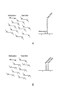
Figure 3A and Figure 3B illustrate an example targeted inhibitory FOR scheme.
To reduce
endogenous levels of the genomic DNA target sequences for total DNA, a
specific ratio of
inhibitory oligonucleotides are included. These inhibitory oligonucleotides
reduce the efficiency of
the total DNA FOR and can be titrated so that the products reach the level of
the genomic DNA
target sequences for methylation. Figure 3A illustrates a FOR assay where no
inhibitory
oligonucleotides are used. Figure 3B illustrates a FOR assay where inhibitory
oligonucleotides are
used to reduce the signal for total DNA in the electropherogram.
CA 02834218 2013-10-23
WO 2012/149339 PCT/US2012/035479
Figure 4A and Figure 4B illustrate a comparison between two assays used for
fetal DNA
quantification. In Figure 4A, the DNA quantification assay provided herein is
made up of three
distinct steps and allows for high throughput by a single operator. The entire
procedure can be
performed within 5 hours and the entire procedure including DNA extraction can
be performed in a
single day. Reaction mixtures are typically added in volumes greater than 10
microliters, which
minimizes technical and sampling variability. The only apparatuses needed are
a thermocycler
and an automated electophoresis instrument. As shown in Figure 4B, the other
DNA quantification
assay is made up of eight steps, six of which occur post PCR, and include a
proteinase K step
(ProtK), shrimp alkaline phosphatase step (SAP), single base extension step
(TYPEPLEX), water
and resin step (W & R), dispensing onto chip step (D), and mass
spectrophotometry
(MASSARRAY). This assay includes a MALDI-TOF based approach for quantification
which
requires special instrumentation as well as highly skilled operators able to
diagnose problems
occurring during any step of the post FOR procedure. Due to the many steps
involved the
complete reaction in Figure 4B cannot be performed on a single day.
Figure 5A and Figure 5B show visualization of the fetal DNA quantification
assay (FQA) amplicons
using capillary electrophoresis. Genomic DNA samples made up of 80%
nonmethylated DNA
isolated from PBMCs and 20% placental DNA at various dilutions were used. In
Figure 5A, female
placental DNA was used. In Figure 5B, male placental DNA was used. The
arrowhead in Figure
5B points to a peak generated by a 93 bp amplification product which
corresponds to Y
chromosome DNA. This peak is absent in Figure 5A (female placental DNA).
Figure 6A and Figure 6B show the effect of targeted inhibitory PCR. Two
parallel reactions were
performed using no inhibitors (Figure 6A) or inhibitors in a ratio of 2:1 to
the assays specific for
total DNA markers (Figure 6B). A significant reduction was observed for the
targeted total marker
(DNA template and competitive oligonucleotide) while no change was observed
for the non-
targeted assays.
Figure 7A and Figure 7B show examples of targeted inhibitory FOR using
different ratios of
.. inhibitor versus FOR primer. Parallel reactions were performed using two
different inhibitor/PCR
primer ratios. In Figure 7A, a ratio of 0.4 micromolar inhibitor/0.6
micromolar FOR primer was
used. In Figure 7B, a ratio of 0.6 micromolar inhibitor/0.4 micromolar PCR
primer was used. While
the intensity of the total markers was severely reduced with increased
addition of inhibitors, no
change was seen in the unaffected assays targeting methylation and Chromosome
Y markers.
21
CA 02834218 2013-10-23
WO 2012/149339 PCT/US2012/035479
Figure 8 shows the identification of primer dimer formation. Multiplex
inhibitory PCR was
performed using different combinations of FOR primers, inhibitor and
competitor oligonucleotides.
Different combinations of PCR primers and oligonucleotides were analyzed for
primer dimer
formation. By using the competitor as the only template for the FOR, four
fragments at 115 bp,
126bp, 141bp and 156bp were expected. Two template independent products were
identified: 1) a
70bp product generated from the interaction between the POPS forward inhibitor
with the UTY
reverse primer and 2) a 60 bp product from the APOE forward inhibitor with the
UTY forward
primer. P=POP, A=APOE, i=Forward PCR primer only, j= Reverse PCR primer only,
ij=Both
Primers present.
Figure 9A, Figure 9B, Figure 9C and Figure 9D show copy number quantification.
DNA samples
isolated from the blood of non-pregnant women mixed with different amounts of
male placental
DNA (0, 40, 80, 120, 160, 200, 240 and 280 copies). Each dilution was analyzed
in six parallel
reactions. Copy numbers were calculated using the ratio of each DNA/Competitor
peak. Figure
9A presents a strip chart showing the calculated placental copy numbers using
either the
methylation or chromosome Y specific markers. Figure 9B presents a strip chart
showing the
calculated total copy numbers. Each dilution contained a constant total number
of genomes.
Figure 90 shows a correlation between methylation markers and chromosome Y.
The copy
numbers of placental DNA spiked into maternal non-methylated DNA in varying
amounts was
calculated by using the ratios obtained from the methylation assays and the Y-
chromosome
markers compared to the respective competitors. The model system showed high
correlation
between the methylation-based quantification and chromosome Y¨specific
sequences (rho=0.93
(Pearson correlation)). Figure 9D shows a Q-Q plot comparing the calculated
placental copy
numbers using the methylation or chromosome Y markers.
Figure 10 shows the detection of CpG methylated DNA. A model system was
developed to
simulate degraded and circulating cell free DNA samples isolated from plasma.
These samples
contained approximately 2000 genomic copies where the bulk was DNA isolated
from maternal
PBMC to which different amounts of either male CpG methylated DNA were spiked.
The samples
were spiked with 0, 40, 80, 120, 160, 200, 240 or 280 placental copies
generating samples with a
placental fraction ranging from 0 to 14%. The arrowhead points to a peak
generated by a 93 bp
amplification product which corresponds to Y chromosome DNA.
22
CA 02834218 2013-10-23
WO 2012/149339 PCT/US2012/035479
Figure 11A shows a comparison between male and female pregnancies. A box plot
of the fetal
fraction of male versus female DNA samples obtained in DNA samples isolated
from 96 pregnant
women is presented. The upper and lower whiskers represent the 5th and 95th
percentiles. The
upper, middle, and lower bars represent the 25th, 50th, 75th percentiles. No
significant difference
was observed between male (n=36) and female (n=60) samples for the methylation
markers (p-
value greater than 0.05. Figure 11B presents a paired correlation between the
calculated fetal
copy numbers obtained using the methylation markers versus the Y-chromosome
markers for the
male samples. The given values indicated minimal difference between the two
different
measurements, thus validating the accuracy and stability of the method. (p =
0.9, Pearson
correlation).
Figure 12 shows a comparison between three consecutive FQA runs using
capillary
electrophoresis. Paired correlation between the calculated fetal fractions
obtained using the mean
of the methylation markers versus the mean from the Y-chromosome markers for
the male
samples is shown. The given values indicated minimal difference between the
three different
measurements, thus validating the accuracy and stability of the method.
Figure 13A and Figure 13B shows the results of post FOR treatment with
Exonuclease I. The FQA
assay was performed in duplicate and analyzed using capillary electrophoresis.
Figure 13A shows
assay results with no exonuclease treatment. The unspecific remaining FOR
primers are circled.
Figure 13B shows assay results from a sample treated post FOR with Exonuclease
I. The
unspecific peaks were no longer present in the electropherogram indicating
that the single
stranded PCR primers were removed by the exonuclease treatment and no double
stranded primer
dimers were formed.
Figure 14A and Figure14B present box plots showing the minority fraction based
SOX14 and TBX3
methylation markers in mixed DNA samples containing 0 to 10% minority species.
The boxes
represent the fractions obtained from 8 replicates. The upper and lower
whiskers represent the 5th
and 95th percentiles. The upper, middle, and lower bars represent the 25th,
50th, 75th percentiles.
Figure 14A shows samples containing a total of 1500 copies per reaction.
Figure 14B shows
samples containing a total of 3000 copies per reaction.
Figure 15A and Figure 15B present box plots showing calculated copy numbers
from the minority
fraction based on SOX14 and TBX3 methylation markers in mixed DNA samples
containing 0 to
23
CA 02834218 2013-10-23
WO 2012/149339 PCT/US2012/035479
10% minority species. The boxes represent the fractions obtained from 8
replicates. The upper
and lower whiskers represent the 5th and 95th percentiles. The upper, middle,
and lower bars
represent the 25th, 50th, 75th percentiles. Figure 15A show samples containing
a total of 1500
copies per reaction. Figure 15B show samples containing a total of 3000 copies
per reaction.
Figure 16 show a correlation plots for the minority species copy numbers
obtained using either the
MASSARRAY or capillary electrophoresis method. Each sample was analyzed in
duplicate for
each method. All correlation coefficients were above 0.9 using Pearson
correlation indicating very
good correlation between both methods and replicates. A paired T.test
generated p-values
indicating no significant different between the methods.
Figure 17 shows correlation plots for the minority species copy numbers
obtained using either the
MASSARRAY or capillary electrophoresis method. Each sample was analyzed in
duplicate for
each method. A paired T.test generated p-values above 0.05 indicating no
significant different
between the methods.
Detailed Description
Provided herein are methods for determining the amount of a minority nucleic
acid species in a
sample, methods for determining the amount of fetal nucleic acid in a sample,
kits for carrying out
such methods, and mixtures of amplified nucleic acids distinguishable by
length generated by such
methods. Quantification of a species of nucleic acid in a sample where a
limited number of copies
is present can be a challenge. In some cases, it is desirable to determine the
exact copy number
of a minority nucleic acid species. Often, nucleic acids are amplified and
separated according to
length to facilitate detection and quantification. Such techniques can be
adapted to high-
throughput screening methods. However, in cases where a minority nucleic acid
is co-amplified
with majority nucleic acid, the resulting analysis of amplification products
can be dominated by the
presence of the majority nucleic acid species. Under these circumstances, the
analytical window is
reduced and quantification of the minority nucleic acid is compromised.
Accurate quantification
and copy number determination of a minority nucleic acid species in a sample
is carried out by an
assay that can co-amplify nucleic acids present at relatively high and low
starting concentrations.
Such assays are provided by the compositions and methods described herein.
24
CA 02834218 2013-10-23
WO 2012/149339 PCT/US2012/035479
Nucleic Acids
Provided herein are methods for nucleic acid quantification. The terms
"nucleic acid", "nucleic acid
molecule" and "polynucleotide" may be used interchangeably throughout the
disclosure. The terms
refer to nucleic acids of any composition from, such as deoxyribonucleic acid
(DNA, e.g.,
complementary DNA (cDNA), genomic DNA (gDNA) and the like), ribonucleic acid
(RNA, e.g.,
message RNA (mRNA), short inhibitory RNA (siRNA), ribosomal RNA (rRNA),
transfer RNA
(tRNA), microRNA, RNA highly expressed by the fetus or placenta, and the
like), and/or DNA or
RNA analogs (e.g., containing base analogs, sugar analogs and/or a non-native
backbone and the
like), RNA/DNA hybrids and polyamide nucleic acids (PNAs), all of which can be
in single- or
double-stranded form, and unless otherwise limited, can encompass known
analogs of natural
nucleotides that can function in a similar manner as naturally occurring
nucleotides. A nucleic acid
can be in any form useful for conducting processes herein (e.g., linear,
circular, supercoiled,
single-stranded, double-stranded and the like). A nucleic acid may be, or may
be from, a plasmid,
phage, autonomously replicating sequence (ARS), centromere, artificial
chromosome,
chromosome, or other nucleic acid able to replicate or be replicated in vitro
or in a host cell, a cell,
a cell nucleus or cytoplasm of a cell in certain embodiments. A nucleic acid
in some embodiments
can be from a single chromosome (e.g., a nucleic acid sample may be from one
chromosome of a
sample obtained from a diploid organism). The term also may include, as
equivalents, derivatives,
variants and analogs of RNA or DNA synthesized from nucleotide analogs, single-
stranded
("sense" or "antisense", "plus" strand or "minus" strand, "forward" reading
frame or "reverse"
reading frame) and double-stranded polynucleotides. Deoxyribonucleotides
include
deoxyadenosine, deoxycytidine, deoxyguanosine and deoxythymidine. For RNA, the
base
cytosine is replaced with uracil. A nucleic acid may be prepared using a
nucleic acid obtained from
a subject.
Extracellular Nucleic Acid
Nucleic acid can be extracellular nucleic acid in certain embodiments. The
terms "extracellular
nucleic acid" or "cell free nucleic acid" or "circulating cell free nucleic
acid" as used herein refer to
nucleic acid isolated from a source having substantially no cells (e.g., no
detectable cells; may
contain cellular elements or cellular remnants). Examples of acellular sources
for extracellular
nucleic acid are blood plasma, blood serum and urine. Without being limited by
theory,
extracellular nucleic acid may be a product of cell apoptosis (e.g.
extracellular nucleic acid from
CA 02834218 2013-10-23
WO 2012/149339 PCT/US2012/035479
post-apoptotic placental cells), cell necrosis and/or cell breakdown, which
provides basis for
extracellular nucleic acid often having a series of lengths across a large
spectrum (e.g., a "ladder").
Extracellular nucleic acid can include different nucleic acid species, and
therefore is referred to
herein as "heterogeneous" in certain embodiments. For example, blood serum or
plasma from a
person having cancer can include nucleic acid from cancer cells and nucleic
acid from non-cancer
cells. In another example, blood serum or plasma from a pregnant female can
include maternal
nucleic acid and fetal nucleic acid. In some instances, fetal nucleic acid
sometimes is about 1% to
about 40% of the overall nucleic acid (e.g., about 1, 2, 3, 4, 5, 6, 7, 8, 9,
10, 11, 12, 13, 14, 15, 16,
17, 18, 19, 20, 21, 22, 23, 24, 25, 26, 27, 28, 29, 30, 31, 32, 33, 34, 35,
36, 37, 38, 39 or 40% of
the nucleic acid is fetal nucleic acid). In some embodiments, the majority of
fetal nucleic acid in
nucleic acid is of a length of about 500 base pairs or less (e.g., about 80,
85, 90, 91, 92, 93, 94, 95,
96, 97, 98, 99 or 100% of fetal nucleic acid is of a length of about 500 base
pairs or less).
Cellular Nucleic Acid
Nucleic acid can be cellular nucleic acid in certain embodiments. The term
"cellular nucleic acid"
as used herein refers to nucleic acid isolated from a source having intact
cells. Non-limiting
examples of sources for cellular nucleic acid are blood cells, tissue cells,
organ cells, tumor cells,
hair cells, skin cells, and bone cells.
In some embodiments, nucleic acid is from peripheral blood mononuclear cells
(PBMC). A PBMC
is any blood cell having a round nucleus, such as, for example, lymphocytes,
monocytes or
macrophages. These cells can be extracted from whole blood, for example, using
ficoll, a
hydrophilic polysaccharide that separates layers of blood, with PBMCs forming
a buffy coat under
a layer of plasma. Additionally, PBMCs can be extracted from whole blood using
a hypotonic lysis
which will preferentially lyse red blood cells and leave PBMCs intact.
In some embodiments, nucleic acid is from placental cells. The placenta is an
organ that connects
the developing fetus to the uterine wall to allow nutrient uptake, waste
elimination, and gas
exchange via the mother's blood supply. The placenta develops from the same
sperm and egg
cells that form the fetus, and functions as a fetomaternal organ with two
components, the fetal part
(Chorion frondosum), and the maternal part (Decidua basalis). In some
embodiments, nucleic acid
26
CA 02834218 2013-10-23
WO 2012/149339 PCT/US2012/035479
is obtained from the fetal part of the placenta. In some embodiments, nucleic
acid is obtained from
the maternal part of the placenta.
Nucleic Acid Quantification
Provided herein are methods for nucleic acid quantification. In some
embodiments, the amount of
a minority nucleic acid species is determined relative to the amount of total
nucleic acid. In some
embodiments, the copy number for a minority nucleic acid species is
determined.
Minority vs. Majority Species
As used herein, it is not intended that the terms "minority" or "majority" be
rigidly defined in any
respect. In one aspect, a nucleic acid that is considered "minority", for
example, can have an
abundance of at least about 0.1% of the total nucleic acid in a sample to less
than 50% of the total
nucleic acid in a sample. In some embodiments, a minority nucleic acid can
have an abundance of
at least about 1% of the total nucleic acid in a sample to about 40% of the
total nucleic acid in a
sample. In some embodiments, a minority nucleic acid can have an abundance of
at least about
2% of the total nucleic acid in a sample to about 30% of the total nucleic
acid in a sample. In some
embodiments, a minority nucleic acid can have an abundance of at least about
3% of the total
nucleic acid in a sample to about 25% of the total nucleic acid in a sample.
For example, a
minority nucleic acid can have an abundance of about 1%, 2%, 3%, 4%, 5%, 6%,
7%, 8%, 9%,
10 /0, 110/0, 12 /0, 13'3/0, 1 4 /0, 15 /0, 160/0, 17/o, -180/0, 19`)/0, 20%,
21%, 22 /0, 23%, 24%, 25'3/0, 26%,
27%, 28%, 29% or 30% of the total nucleic acid in a sample. In some
embodiments, the minority
nucleic acid is extracellular DNA. In some embodiments, the minority nucleic
acid is extracellular
fetal DNA.
In another aspect, a nucleic acid that is considered "majority", for example,
can have an
abundance greater than 50% of the total nucleic acid in a sample to about
99.9% of the total
nucleic acid in a sample. In some embodiments, a majority nucleic acid can
have an abundance of
at least about 60% of the total nucleic acid in a sample to about 99% of the
total nucleic acid in a
sample. In some embodiments, a majority nucleic acid can have an abundance of
at least about
70% of the total nucleic acid in a sample to about 98% of the total nucleic
acid in a sample. In
some embodiments, a majority nucleic acid can have an abundance of at least
about 75% of the
total nucleic acid in a sample to about 97% of the total nucleic acid in a
sample. For example, a
27
CA 02834218 2013-10-23
WO 2012/149339 PCT/US2012/035479
majority nucleic acid can have an abundance of at least about 70%, 71%, 72%,
73%, 74%, 75%,
76%, 77%, 78%, 79%, 80%, 81%, 82%, 83%, 84%, 85%, 86%, 87%, 88%, 89%, 90%,
91%, 92%,
93%, 94%, 95%, 96%, 97%, 98% or 99% of the total nucleic acid in a sample. In
some
embodiments, the majority nucleic acid is extracellular DNA. In some
embodiments, the majority
nucleic acid is extracellular maternal DNA.
Competitor Oligonucleotides
In some embodiments of the methods provided herein, one or more competitor
oligonucleotides
are used to achieve quantification of a minority nucleic acid species. As used
herein, a "competitor
oligonucleotide" or "competitive oligonucleotide" or "competitor" is a nucleic
acid polymer that
competes with a target nucleotide sequence for hybridization of amplification
primers. Often, the
competitor has the same nucleotide sequence as the target nucleotide sequence.
In some cases,
the competitor optionally has an additional length of nucleotide sequence that
is different from the
target nucleotide sequence. Often, the additional length of nucleotide
sequence is from a different
genome than the target nucleotide sequence or is a synthetic sequence. In some
embodiments, a
known amount, or copy number, of competitor is used. In some embodiments, two
or more
competitors are used. In some cases, the two or more competitors possess
similar characteristics
(e.g. length, detectable label). In some cases, the two or more competitors
possess different
characteristics (e.g. length, detectable label). In some embodiments, one or
more competitors are
used for a particular region. In some cases, the competitor possesses a
characteristic that is
unique for each set of competitors for a given region. Often, competitors for
different regions
possess different characteristics.
A competitor oligonucleotide may be composed of naturally occurring and/or non-
naturally
occurring nucleotides (e.g., labeled nucleotides), or a mixture thereof.
Competitor oligonucleotides
suitable for use with embodiments described herein, may be synthesized and
labeled using known
techniques. Competitor oligonucleotides may be chemically synthesized
according to any suitable
method known, for example, the solid phase phosphoramidite triester method
first described by
Beaucage and Caruthers, Tetrahedron Letts., 22:1859-1862, 1981, using an
automated
synthesizer, as described in Needham-VanDevanter et al., Nucleic Acids Res.
12:6159-6168,
1984. Purification of competitor oligonucleotides can be effected by any
suitable method known,
for example, native acrylamide gel electrophoresis or by anion-exchange high-
performance liquid
28
CA 02834218 2013-10-23
WO 2012/149339 PCT/US2012/035479
chromatography (HPLC), for example, as described in Pearson and Regnier, J.
Chrom., 255:137-
149, 1983.
Competitor Length
In some embodiments, multiple competitors are used within a multiplex
amplification assay. In
some cases, the competitors used for a particular region are the same length.
In some
embodiments, the competitors used for different regions are different lengths.
Competitors can be,
for example, at least about 30 base pairs in length to about 500 base pairs in
length. In some
embodiments, competitors can be at least about 50 base pairs in length to
about 200 base pairs in
length. In some embodiments, competitors can be at least about 100 base pairs
in length to about
150 base pairs in length. In some embodiments, competitors can be at least
about 115 base pairs
in length to about 125 base pairs in length. For example, a competitor can be
about 115, 116, 117,
118, 119, 120, 121, 122, 123, 124, or 125 base pairs in length. Non-limiting
examples of
competitor oligonucleotides that can be used with the methods provided herein
are set forth in
SEQ ID NOs:21-28.
In some embodiments, the competitor possesses a stuffer sequence. A stuffer
sequence is a
sequence of nucleotides that, in general, does not share any sequence
similarity with the target
nucleotide sequence. Often a stuffer is added to the competitor to
distinguish, by length, the
amplified products for the competitor from the amplified products for the
target nucleotide
sequence. Stuffer sequences can be included anywhere within the competitor
oligonucleotide. In
some embodiments, the stuffer sequence is included at one or more positions
within the competitor
oligonucleotide that are (i) downstream of the nucleotide sequence
complementary to the forward
primer and (ii) upstream of the nucleotide sequence complementary to the
reverse primer. In some
cases, the stuffer sequence is a contiguous sequence. In some cases, the
stuffer sequence exists
as two or more fragments within a competitor oligonucleotide. For the methods
provided herein,
the term "sequence length" when used in reference to a stuffer sequence means
a contiguous
length of nucleotide sequence or a sum of nucleotide sequence fragment
lengths. In some
embodiments, a stuffer sequence is from a genome that is different from the
target genome. For
example, if the target genome is human, then the stuffer sequence will often
be selected from a
non-human genome. For embodiments where the target nucleotide sequence is from
the human
genome, a competitor stuffer sequence can be from any non-human genome known
in the art,
such as, for example, non-mammalian animal genomes, plant genomes, fungal
genomes, bacterial
29
CA 02834218 2013-10-23
WO 2012/149339 PCT/US2012/035479
genomes, or viral genomes. In some embodiments, the stuffer sequence is from
the PhiX 174
genome. In some embodiments, the stuffer sequence is not from genomic DNA and
sometimes is
synthetic.
In some embodiments, the competitor possesses a stuffer sequence whose length
(or sum of
fragment lengths) is unique, or constant, for each set of competitors for a
given region. Often,
competitors for different regions possess stuffer sequences of different, or
variable, lengths (or sum
of fragment lengths). As used herein, the term "constant length" refers to a
length of a sequence of
nucleotides that is the same for one or more nucleotide sequences, such as,
for example, a stuffer
sequence. As used herein, the term "variable length" refers to a length of a
sequence of
nucleotides that is different for one or more nucleotide sequences, such as,
for example, a stuffer
sequence. Competitor stuffer sequences can be of any length suitable for the
methods provided
herein. For example, competitor stuffer sequences can be at least about 1 base
pair in length to
about 100 base pairs in length. In some embodiments, the stuffer sequence is
at least about 15
base pairs in length to about 55 base pairs in length. In some embodiments the
stuffer sequence
can be composed of several shorter stuffer sequences. This design can be
utilized if the amplicon
is prone to form secondary structures during PCR. For example, the stuffer
sequence can be 15,
16, 17, 18, 19, 20, 21, 22, 23, 24, 25, 26, 27, 28, 29, 30, 31, 32, 33, 34,
35, 36, 37, 38, 39, 40, 41,
42, 43, 44, 45, 46, 47, 48, 49, 50, 51, 52, 53, 54, or 55 base pairs in
length. Examples of
competitor design using single and multiple stuffer sequences are provided
herein and shown in
Table 3 of Example 1.
Amplified competitors can optionally be further distinguishable from one
another through the use of
tailed amplification primers containing additional non-hybridizing nucleotide
sequences of varying
length. These are described in further detail below.
Labeled Competitors
In some embodiments, the competitor oligonucleotide can be detected by
detecting a detectable
label, molecule or entity or "signal-generating moiety" (e.g., a fluorophore,
radioisotope,
colorimetric agent, particle, enzyme and the like). The term "signal-
generating" as used herein
refers to any atom or molecule that can provide a detectable or quantifiable
effect, and that can be
attached to a nucleic acid. In certain embodiments, a detectable label
generates a unique light
CA 02834218 2013-10-23
WO 2012/149339 PCT/US2012/035479
signal, a fluorescent signal, a luminescent signal, an electrical property, a
chemical property, a
magnetic property and the like.
In certain embodiments, the oligonucleotide can be modified to include a
detectable label using
any method known to one of skill in the art. The label may be incorporated as
part of the
synthesis, or added on prior to using the primer in any of the processes
described herein.
Incorporation of label may be performed either in liquid phase or on solid
phase. In some
embodiments the detectable label may be useful for detection of targets. In
some embodiments
the detectable label may be useful for the quantification target nucleic acids
(e.g., determining copy
number of a particular sequence or species of nucleic acid). Any detectable
label suitable for
detection of an interaction or biological activity in a system can be
appropriately selected and
utilized by the artisan. Examples of detectable labels are fluorescent labels
or tags such as
fluorescein, rhodamine, and others (e.g., Anantha, et al., Biochemistry (1998)
37:2709 2714; and
Qu & Chaires, Methods Enzymol. (2000) 321:353 369); radioactive isotopes
(e.g., 1251, 1311, 35S,
.. 31P, 32P, 33P, 14C, 3H, 7Be, 28Mg, 57Co, 65Zn, 67Cu, 68Ge, 82Sr, 83Rb,
95Tc, 96Tc, 103Pd,
109Cd, and 127Xe); light scattering labels (e.g., U.S. Patent No. 6,214,560,
and commercially
available from Gen icon Sciences Corporation, CA); chemiluminescent labels and
enzyme
substrates (e.g., dioxetanes and acridinium esters), enzymic or protein labels
(e.g., green
fluorescence protein (GFP) or color variant thereof, luciferase, peroxidase);
other chromogenic
labels or dyes (e.g., cyanine), and other cofactors or biomolecules such as
digoxigenin,
streptavidin, biotin (e.g., members of a binding pair such as biotin and
avidin for example), affinity
capture moieties and the like. Additional detectable labels include, but are
not limited to,
nucleotides (labeled or unlabelled), compomers, sugars, peptides, proteins,
antibodies, chemical
compounds, conducting polymers, binding moieties such as biotin, mass tags,
colorimetric agents,
light emitting agents, radioactive tags, charge tags (electrical or magnetic
charge), volatile tags and
hydrophobic tags, biomolecules (e.g., members of a binding pair
antibody/antigen,
antibody/antibody, antibody/antibody fragment, antibody/antibody receptor,
antibody/protein A or
protein G, hapten/anti-hapten, biotin/avidin, biotin/streptavidin, folic
acid/folate binding protein,
vitamin B12/intrinsic factor, chemical reactive group/complementary chemical
reactive group (e.g.,
sulfhydryl/maleimide, sulfhydryl/haloacetyl derivative, amine/isotriocyanate,
amine(succinimidyl
ester, and amine/sulfonyl halides) and the like. In some embodiments a probe
may contain a
signal-generating moiety that hybridizes to a target and alters the passage of
the target nucleic
acid through a nanopore, and can generate a signal when released from the
nucleic acid when it
passes through the nanopore (e.g., alters the speed or time through a pore of
known size). In
31
CA 02834218 2013-10-23
WO 2012/149339 PCT/US2012/035479
some embodiments a primer may be labeled with an affinity capture moiety. Also
included in
detectable labels are those labels useful for mass modification for detection
with mass
spectrometry (e.g., matrix-assisted laser desorption ionization (MALDI) mass
spectrometry and
electrospray (ES) mass spectrometry).
In some embodiments, the detectable label is a fluorophore. A fluorophore is a
functional group in
a molecule which can absorb energy of a specific wavelength and re-emit energy
at a different (but
equally specific) wavelength. The amount and wavelength of the emitted energy
depend on both
the fluorophore and the chemical environment of the fluorophore. Any
fluorophore known in the art
can be used in conjunction with the methods provided herein and include, for
example, fluorescein
isothiocyanate (FITC), Xanthene derivatives (e.g. fluorescein, rhodamine
(TRITC), Oregon green,
eosin, Texas red, Cal Fluor), Cyanine derivatives (e.g. cyanine,
indocarbocyanine,
oxacarbocyanine, thiacarbocyanine, merocyanine, Quasar), Naphthalene
derivatives (e.g. dansyl
and prodan derivatives), Coumarin derivatives, oxadiazole derivatives (e.g.
pyridyloxazole,
nitrobenzoxadiazole, benzoxadiazole), Pyrene derivatives (e.g. cascade blue),
Oxazine derivatives
(e.g. Nile red, Nile blue, cresyl violet, oxazine 170), Acridine derivatives
(e.g. proflavin, acridine
orange, acridine yellow), Arylmethine derivatives (e.g. auramine, crystal
violet, malachite green),
Tetrapyrrole derivatives (e.g. porphin, phtalocyanine, bilirubin), CF DYE
(Biotium), BODIPY
(Invitrogen), ALEXA FLUOR (Invitrogen), DYLIGHT FLUOR (Thermo Scientific,
Pierce), ATTO and
TRACEY (Sigma Aldrich), FLUOPROBES (Interchim), and MEGASTOKES DYES (Dyomics).
In
some embodiments, the competitor possesses a fluorophore that is unique for
each set of
competitors for a given region. Often, competitors for different regions
possess different
fluorophores.
Features for Distinguishing a Minority Nucleic Acid from a Majority Nucleic
Acid
In some of the embodiments of the methods provided herein, a minority nucleic
acid possesses a
feature that is present in the minority nucleic acid and is not present in the
majority nucleic acid. In
some of the embodiments of the methods provided herein, a minority nucleic
acid possesses a
feature that is not present in the minority nucleic acid and is present in the
majority nucleic acid.
The feature that is present or not present in the minority nucleic acid can be
any feature that can
distinguish the minority nucleic acid from the majority nucleic acid such as,
for example, a
sequence paralog or sequence variation (e.g. single nucleotide polymorphism
(SNP), addition,
insertion, deletion) or a particular epigenetic state. The term "epigenetic
state" or ''epigenetic
32
CA 02834218 2013-10-23
WO 2012/149339 PCT/US2012/035479
status" as used herein refers to any structural feature at a molecular level
of a nucleic acid (e.g.,
DNA or RNA) other than the primary nucleotide sequence. For instance, the
epigenetic state of a
genomic DNA may include its secondary or tertiary structure determined or
influenced by, for
example, its methylation pattern or its association with cellular or
extracellular proteins.
Methylation
In some embodiments, a feature that distinguishes a minority nucleic acid from
a majority nucleic
acid is methylation state. The terms "methylation state", "methylation
profile", or "methylation
status," as used herein to describe the state of methylation of a genomic
sequence, refer to the
characteristics of a DNA segment at a particular genomic locus relevant to
methylation. Such
characteristics include, but are not limited to, whether any of the cytosine
(C) residues within a
DNA sequence are methylated, location of methylated C residue(s), percentage
of methylated C at
any particular stretch of residues, and allelic differences in methylation due
to, e.g., difference in
the origin of the alleles. The terms above also refer to the relative or
absolute concentration of
methylated C or unmethylated C at any particular stretch of residues in a
biological sample. For
example, if the cytosine (C) residue(s) within a DNA sequence are methylated
it can be referred to
as "hypermethylated"; whereas if the cytosine (C) residue(s) within a DNA
sequence are not
methylated it may be referred to as "hypomethylated". Likewise, if the
cytosine (C) residue(s)
within a DNA sequence (e.g., fetal nucleic acid) are methylated as compared to
another sequence
from a different region or from a different individual (e.g., relative to
maternal nucleic acid), that
sequence is considered hypermethylated compared to the other sequence.
Alternatively, if the
cytosine (C) residue(s) within a DNA sequence are not methylated as compared
to another
sequence from a different region or from a different individual (e.g., the
mother), that sequence is
considered hypomethylated compared to the other sequence. These sequences are
said to be
"differentially methylated", and more specifically, when the methylation
status differs between
mother and fetus, the sequences are considered "differentially methylated
maternal and fetal
nucleic acid". Methods and examples of differentially methylated sites in
fetal nucleic acid are
described in, for example, PCT Publication No. W02010/033639.
As used herein, a "methylated nucleotide" or a "methylated nucleotide base"
refers to the presence
of a methyl moiety on a nucleotide base, where the methyl moiety is not
present in a recognized
typical nucleotide base. For example, cytosine does not contain a methyl
moiety on its pyrimidine
ring, but 5-methylcytosine contains a methyl moiety at position 5 of its
pyrimidine ring. Therefore,
33
CA 02834218 2013-10-23
WO 2012/149339 PCT/US2012/035479
cytosine is not a methylated nucleotide and 5-methylcytosine is a methylated
nucleotide. In
another example, thymine contains a methyl moiety at position 5 of its
pyrimidine ring, however, for
purposes herein, thymine is not considered a methylated nucleotide when
present in DNA since
thymine is a typical nucleotide base of DNA. Typical nucleoside bases for DNA
are thymine,
adenine, cytosine and guanine. Typical bases for RNA are uracil, adenine,
cytosine and guanine.
Correspondingly a "methylation site" is the location in the target gene
nucleic acid region where
methylation has, or has the possibility of occurring. For example a location
containing CpG is a
methylation site where the cytosine may or may not be methylated. Such
methylation sites can be
susceptible to methylation either by natural occurring events in vivo or by an
event instituted to
chemically methylate the nucleotide in vitro.
A "nucleic acid comprising one or more CpG sites" or a "CpG-containing genomic
sequence" as
used herein refers to a segment of DNA sequence at a defined location in the
genome of an
individual such as a human fetus or a pregnant woman. Typically, a "CpG-
containing genomic
sequence" is at least 15 nucleotides in length and contains at least one
cytosine. Often, it can be
at least 30, 50, 80, 100, 150, 200, 250, or 300 nucleotides in length and
contains at least 2, 5, 10,
15, 20, 25, or 30 cytosines. For any one ''CpG-containing genomic sequence" at
a given location,
e.g., within a region centering on a given genetic locus, nucleotide sequence
variations may exist
from individual to individual and from allele to allele even for the same
individual. Typically, such a
region centering on a defined genetic locus (e.g., a CpG island) contains the
locus as well as
upstream and/or downstream sequences. Each of the upstream or downstream
sequence
(counting from the 5' or 3' boundary of the genetic locus, respectively) can
be as long as 10 kb, in
other cases may be as long as 5 kb, 2 kb, 1 kb, 500 bp, 200 bp, or 100 bp.
Furthermore, a "CpG-
containing genomic sequence" may encompass a nucleotide sequence transcribed
or not
transcribed for protein production, and the nucleotide sequence can be an
inter-gene sequence,
intra-gene sequence, protein-coding sequence, a non protein-coding sequence
(such as a
transcription promoter), or a combination thereof. A "CpG island" as used
herein describes a
segment of DNA sequence that possesses a functionally or structurally deviated
CpG density. A
CpG island can typically be, for example, at least 400 nucleotides in length,
have a greater than
50% GC content, and an OCF/ECF ratio greater than 0.6. In some cases a CpG
island can be
characterized as being at least 200 nucleotides in length, having a greater
than 50% GC content,
and an OCF/ECF ratio greater than 0.6.
34
CA 02834218 2013-10-23
WO 2012/149339 PCT/US2012/035479
In some embodiments, nucleic acid may be exposed to a process that modifies
certain nucleotides
in the nucleic acid before providing nucleic acid for a method described
herein. A process that
selectively modifies nucleic acid based upon the methylation state of
nucleotides therein can be
applied to a nucleic acid sample, for example. Methods for modifying a nucleic
acid molecule in a
manner that reflects the methylation pattern of the nucleic acid molecule are
known in the art, as
exemplified in U.S. Pat. No. 5,786,146 and U.S. patent publications
20030180779 and
20030082600. For example, non-methylated cytosine nucleotides in a nucleic
acid can be
converted to uracil by bisulfite treatment, which does not modify methylated
cytosine.
Nucleic Acid Cleavage
In some embodiments of the methods provided herein, the nucleic acid is
exposed to one or more
cleavage agents. The term "cleavage agent" as used herein refers to an agent,
sometimes a
chemical or an enzyme that can cleave a nucleic acid at one or more specific
or non-specific sites.
Specific cleavage agents often cleave specifically according to a particular
nucleotide sequence at
a particular site. In some cases, the nucleic acid is exposed to one or more
cleavage agents prior
to amplification. In some cases, the nucleic acid is exposed to one or more
cleavage agents
following amplification. In some cases, the nucleic acid is exposed to one or
more cleavage
agents prior to amplification and following amplification.
Examples of enzymatic cleavage agents include without limitation endonucleases
(e.g., DNase
(e.g., DNase I, II); RNase (e.g., RNase E, F, H, P); CLEAVASE enzyme; TAQ DNA
polymerase; E.
coli DNA polymerase I and eukaryotic structure-specific endonucleases; murine
FEN-1
endonucleases; type I, ll or III restriction endonucleases (i.e. restriction
enzymes) such as Acc I,
Acil, Afl III, Alu I, Alw44 I, Apa I, Asn I, Ava I, Ava II, BamH I, Ban II,
Bc1 I, Bgl I. Bgl II, Bln I, Bsm I,
BssH II, BstE II, BstUI, Cfo I, Cla I, Dde I, Dpn I, Dra I, EcIX I, EcoR I,
EcoR I, EcoR II, EcoR V,
Hae II, Hae II, Hhal, Hind II, Hind III, Hpa I, Hpa II, Kpn I, Ksp I, MaeII,
McrBC, Mlu I, MluN I, Msp I,
Nci I, Nco I, Nde I, Nde II, Nhe I, Not I, Nru I, Nsi I, Pst I, Pvu I, Pvu II,
Rsa I, Sac I, Sail, Sau3A I,
Sca I, ScrF I, Sfi I, Sma I, Spe I, Sph I, Ssp I, Stu I, Sty I, Swa I, Taq I,
Xba I, Xho I; glycosylases
(e.g., uracil-DNA glycolsylase (UDG), 3-methyladenine DNA glycosylase, 3-
methyladenine DNA
glycosylase II, pyrimidine hydrate-DNA glycosylase, FaPy-DNA glycosylase,
thymine mismatch-
DNA glycosylase, hypoxanthine- DNA glycosylase, 5-Hydroxymethyluracil DNA
glycosylase
(HmUDG), 5-Hydroxymethylcytosine DNA glycosylase, or 1,N6-etheno-adenine DNA
glycosylase);
CA 02834218 2013-10-23
WO 2012/149339 PCT/US2012/035479
exonucleases (e.g., exonuclease I, exonuclease II, exonuclease III,
exonuclease IV, exonuclease
V, exonuclease VI, exonuclease VII, exonuclease VIII); ribozymes, and
DNAzymes.
Methylation-Sensitive Restriction Enzyme Digestion
In some embodiments of the methods provided herein, the nucleic acid is
treated with one or more
methylation-sensitive restriction enzymes. As used herein, "methylation
sensitive restriction
enzymes" or "methyl-sensitive enzymes" are restriction enzymes that
preferentially or substantially
cleave or digest at their DNA recognition sequence if it is non-methylated.
Thus, an unmethylated
DNA sample treated with a methylation-sensitive restriction enzyme will be
digested into smaller
fragments, whereas a methylated or hypermethylated DNA sample would remain
substantially
undigested. Conversely, there are examples of methyl-sensitive enzymes that
cleave at their DNA
recognition sequence only if it is methylated. Examples of enzymes that digest
only methylated
DNA include, but are not limited to, Dpnl, which cuts at a recognition
sequence GATC, and McrBC,
which cuts DNA containing modified cytosines (New England BioLabse, Inc,
Beverly, Mass.).
Methyl-sensitive enzymes that digest unmethylated DNA suitable for use in the
methods provided
herein include, but are not limited to, Hpall, Hhal, Mael I, BstUl and Acil.
In some embodiments,
combinations of two or more methyl-sensitive enzymes that digest only
unmethylated DNA can be
used. In some embodiments, Hpall, which cuts only the unmethylated sequence
CCGG, is used.
In some embodiments, Hhal, which cuts only the unmethylated sequence GCGC, is
used. Both
enzymes are available from New England BioLabs , Inc (Beverly, Mass.).
Cleavage methods and procedures for selected restriction enzymes for cutting
DNA at specific
sites are well known to the skilled artisan. For example, many suppliers of
restriction enzymes
provide information on conditions and types of DNA sequences cut by specific
restriction enzymes,
including New England Bio Labs, Pro-Mega Biochems, Boehringer-Mannheim, and
the like.
Sambrook et al. (See Sambrook et al., Molecular Biology: A laboratory
Approach, Cold Spring
Harbor, N.Y. 1989) provide a general description of methods for using
restriction enzymes and
other enzymes. Enzymes often are used under conditions that will enable
cleavage of the DNA
with about 95%-100% efficiency, preferably with about 98%-100% efficiency.
36
CA 02834218 2013-10-23
WO 2012/149339 PCT/US2012/035479
Exonuclease Treatment
In some embodiments of the methods provided herein, the nucleic acid is
treated with an
exonuclease. Exonucleases are enzymes that work by cleaving nucleotides one at
a time from the
end of a polynucleotide chain through a hydrolyzing reaction that breaks
phosphodiester bonds at
either the 3' or the 5' end. Any exonuclease known in the art can be used with
the methods
provided herein and include, for example, 5' to 3' exonucleases (e.g.
exonuclease II), 3' to 5'
exonucleases (e.g. exonuclease l), and poly(A)-specific 3' to 5' exonucleases.
In some
embodiments, the nucleic acid is optionally treated with an exonuclease to
remove any
contaminating nucleic acids such as, for example, single stranded PCR primers.
Typically, this
step is performed after completion of nucleic acid amplification. In some
embodiments, a single
strand specific exonuclease is used. In some embodiments, exonuclease I is
used.
Genomic DNA Target Sequences
In some embodiments of the methods provided herein, one or more nucleic acid
species, and
sometimes one or more nucleotide sequence species, are targeted for
amplification and
quantification. In some embodiments, the targeted nucleic acids are genomic
DNA sequences.
Certain genomic DNA target sequences are used, for example, because they can
allow for the
determination of a particular feature for a given assay. Genomic DNA target
sequences can be
referred to herein as markers for a given assay. In some embodiments, more
than one genomic
DNA target sequence or marker can allow for the determination of a particular
feature for a given
assay. Such genomic DNA target sequences are considered to be of a particular
"region". As
used herein, a "region" is not intended to be limited to a description of a
genomic location, such as
a particular chromosome, stretch of chromosomal DNA or genetic locus. Rather,
the term "region"
is used herein to identify a collection of one or more genomic DNA target
sequences or markers
that can be indicative of a particular assay. Such assays can include, but are
not limited to, assays
for the detection and quantification of a minority nucleic acid species,
assays for the detection and
quantification of a majority nucleic acid, assays for the detection and
quantification of total DNA,
assays for the detection and quantification of methylated DNA, assays for the
detection and
quantification of fetal specific nucleic acid (e.g. chromosome Y DNA), and
assays for the detection
and quantification of digested and/or undigested DNA, as an indicator of
digestion efficiency. In
some embodiments, the genomic DNA target sequence is described as being within
a particular
genomic locus. As used herein, a genomic locus can include any or a
combination of open reading
37
CA 02834218 2013-10-23
WO 2012/149339 PCT/US2012/035479
frame DNA, non-transcribed DNA, intronic sequences, extronic sequences,
promoter sequences,
enhancer sequences, flanking sequences, or any sequences considered by one of
skill in the art to
be associated with a given genomic locus.
Assays for the Determination of Methylated DNA
In some embodiments of the methods provided herein, one or more genomic DNA
target
sequences are used that can allow for the determination of methylated DNA.
Generally, genomic
DNA target sequences used for the determination of methylated DNA are
differentially methylated
in the minority and majority species, and thus, differentially digested
according to the methods
provided herein for methylation-sensitive restriction enzymes. In some cases,
the genomic DNA
target sequence is a single copy gene. In some cases, the genomic DNA target
sequence is not
located on chromosome 13. In some cases, the genomic DNA target sequence is
not located on
chromosome 18. In some cases, the genomic DNA target sequence is not located
on
chromosome 21. In some cases, the genomic DNA target sequence is not located
on
chromosome X. In some cases, the genomic DNA target sequence is not located on
chromosome
Y. In some cases, the genomic DNA target sequence is typically methylated in
one DNA species
such as, for example, placental DNA (i.e. at least about 50% or greater
methylation). In some
cases, the genomic DNA target sequence is minimally methylated in another DNA
species such as,
for example, maternal DNA (i.e. less than about 1% methylation). In some
cases, the genomic
DNA target sequence does not contain any known single nucleotide polymorphisms
(SNPs) within
the FOR primer hybridization sequences. In some cases, the genomic DNA target
sequence does
not contain any known mutations within the FOR primer hybridization sequences.
In some cases,
the genomic DNA target sequence does not contain any known insertion or
deletions within the
FOR primer hybridization sequences. In some cases, the melting temperature of
the FOR primers
that can hybridize to a genomic DNA target sequence is not below 65 C. In
some cases, the
melting temperature of the PCR primers that can hybridize to a genomic DNA
target sequence is
not above 75 C. In some cases, the genomic DNA target sequence contains at
least two
restriction sites within the amplified region. In some cases, the restriction
site sequence is GCGC.
In some cases, the restriction site sequence is CCGG. In some cases, the
genomic DNA target
sequence contains a combination of the restriction site sequences GCGC and
CCGG within the
amplified region. In some embodiments, the genomic DNA target sequence length
is about 50
base pairs to about 200 base pairs. In some cases, the genomic DNA target
sequence length is
70 base pairs. In some cases, the genomic DNA target sequence does not possess
any negative
38
CA 02834218 2013-10-23
WO 2012/149339 PCT/US2012/035479
AG values for secondary structure of the complete amplicon prediction using
mfold (M. Zuker,
Mfold web server for nucleic acid folding and hybridization prediction.
Nucleic Acids Res. 31(13),
3406-15, (2003)). In some embodiments, the genomic DNA target sequence used
for the
determination of methylated DNA is within the TBX3 locus. An example of a TBX3
genomic target
sequence is set forth in SEQ ID NO:29. In some embodiments, the genomic DNA
target sequence
used for the determination of methylated DNA is within the 50X14 locus. An
example of a SOX14
genomic target is set forth in SEQ ID NO:30. Additional genomic targets that
can be used for the
determination of methylated DNA in conjunction with the methods provided
herein are presented in
Table 14 and Table 15 in Example 10.
Assays for the Determination of Total DNA
In some embodiments of the methods provided herein, one or more genomic DNA
target
sequences are used that can allow for the determination of total DNA.
Generally, genomic DNA
target sequences used for the determination of total DNA are present in every
genome copy (e.g.
is present in fetal DNA and maternal DNA, cancer DNA and normal DNA, pathogen
DNA and host
DNA). In some cases, the genomic DNA target sequence is a single copy gene. In
some cases,
the genomic DNA target sequence is not located on chromosome 13. In some
cases, the genomic
DNA target sequence is not located on chromosome 18. In some cases, the
genomic DNA target
sequence is not located on chromosome 21. In some cases, the genomic DNA
target sequence is
not located on chromosome X. In some cases, the genomic DNA target sequence is
not located
on chromosome Y. In some cases, the genomic DNA target sequence does not
contain any
known single nucleotide polymorphisms (SNPs) within the FOR primer
hybridization sequences. In
some cases, the genomic DNA target sequence does not contain any known
mutations within the
FOR primer hybridization sequences. In some cases, the genomic DNA target
sequence does not
contain any known insertion or deletions within the PCR primer hybridization
sequences. In some
cases, the melting temperature of the PCR primers that can hybridize to a
genomic DNA target
sequence is not below 65 C. In some cases, the melting temperature of the FOR
primers that can
hybridize to a genomic DNA target sequence is not above 75 C. In some cases,
the genomic
DNA target sequence does not contain the restriction site GCGC within the
amplified region. In
some cases, the genomic DNA target sequence does not contain the restriction
site CCGG within
the amplified region. In some embodiments, the genomic DNA target sequence
length is about 50
base pairs to about 200 base pairs. In some cases, the genomic DNA target
sequence length is
70 base pairs. In some cases, the genomic DNA target sequence does not possess
any negative
39
CA 02834218 2013-10-23
WO 2012/149339 PCT/US2012/035479
AG values for secondary structure of the complete amplicon prediction using
mfold (M. Zuker,
Mfold web server for nucleic acid folding and hybridization prediction.
Nucleic Acids Res. 31(13),
3406-15, (2003)). In some embodiments, the genomic DNA target sequence used
for the
determination of total DNA is within the POP5 locus. An example of a POP5
genomic target
sequence is set forth in SEQ ID NO:31. In some embodiments, the genomic DNA
target sequence
used for the determination of total DNA is within the APOE locus. An example
of an APOE
genomic target is set forth in SEQ ID NO:32.
Assays for the Determination of Fetal DNA
In some embodiments of the methods provided herein, one or more genomic DNA
target
sequences are used that can allow for the determination of fetal DNA. In some
embodiments,
genomic DNA target sequences used for the determination of fetal DNA are
specific to the Y
chromosome. In some cases, the genomic DNA target sequence is a single copy
gene. In some
cases, the genomic DNA target sequence does not contain any known single
nucleotide
polymorphisms (SNPs) within the PCR primer hybridization sequences. In some
cases, the
genomic DNA target sequence does not contain any known mutations within the
FOR primer
hybridization sequences. In some cases, the genomic DNA target sequence does
not contain any
known insertion or deletions within the PCR primer hybridization sequences. In
some cases, the
.. melting temperature of the FOR primers that can hybridize to a genomic DNA
target sequence is
not below 65 C. In some cases, the melting temperature of the FOR primers
that can hybridize to
a genomic DNA target sequence is not above 75 C. In some cases, the genomic
DNA target
sequence does not contain the restriction site GCGC within the amplified
region. In some cases,
the genomic DNA target sequence does not contain the restriction site CCGG
within the amplified
region. In some embodiments, the genomic DNA target sequence length is about
50 base pairs to
about 200 base pairs. In some cases, the genomic DNA target sequence length is
70 base pairs.
In some cases, the genomic DNA target sequence does not possess any negative
AG values for
secondary structure of the complete amplicon prediction using mfold (M. Zuker,
Mfold web server
for nucleic acid folding and hybridization prediction. Nucleic Acids Res.
31(13), 3406-15, (2003)).
In some embodiments, the genomic DNA target sequence used for the
determination of fetal DNA
is within the UTY locus. An example of a UTY genomic target sequence is set
forth in SEQ ID
NO:33. In some embodiments, the genomic DNA target sequence used for the
determination of
fetal DNA is within the DDX3Y locus. An example of a DDX3Y genomic target is
set forth in SEQ
ID NO:34.
CA 02834218 2013-10-23
WO 2012/149339 PCT/US2012/035479
Assays for the Determination of Digested and/or Undigested DNA
In some embodiments of the methods provided herein, one or more genomic DNA
target
sequences are used that can allow for the determination of the amount of
digested or undigested
nucleic acid, as an indicator of digestion efficiency. Such genomic DNA target
sequences are
present in every genome in the sample (e.g. majority and minority species
genomes). Generally,
genomic DNA target sequences used for the determination of digested or
undigested DNA contain
at least one restriction site present in a genomic DNA target sequence used in
another assay.
Thus, the genomic DNA target sequences used for the determination of digested
or undigested
DNA serve as controls for assays that include differential digestion.
Generally, the genomic DNA
target sequence is unmethylated in all nucleic acid species tested (e.g.
unmethylated in both
majority and minority species genomes). In some cases, the genomic DNA target
sequence
contains at least one restriction site GCGC within the amplified region. In
some cases, the
genomic DNA target sequence contains at least one restriction site CCGG within
the amplified
region. In some cases, the genomic DNA target sequence contains exactly one
restriction site
GCGC within the amplified region and exactly one restriction site CCGG within
the amplified
region. In some cases, the genomic DNA target sequence is a single copy gene.
In some cases,
the genomic DNA target sequence is not located on chromosome 13. In some
cases, the genomic
DNA target sequence is not located on chromosome 18. In some cases, the
genomic DNA target
sequence is not located on chromosome 21. In some cases, the genomic DNA
target sequence is
not located on chromosome X. In some cases, the genomic DNA target sequence is
not located
on chromosome Y. In some cases, the genomic DNA target sequence does not
contain any
known single nucleotide polymorphisms (SNPs) within the FOR primer
hybridization sequences. In
some cases, the genomic DNA target sequence does not contain any known
mutations within the
PCR primer hybridization sequences. In some cases, the genomic DNA target
sequence does not
contain any known insertion or deletions within the PCR primer hybridization
sequences. In some
cases, the melting temperature of the FOR primers that can hybridize to a
genomic DNA target
sequence is not below 65 C. In some cases, the melting temperature of the PCR
primers that can
hybridize to a genomic DNA target sequence is not above 75 C. In some
embodiments, the
genomic DNA target sequence length is about 50 base pairs to about 200 base
pairs. In some
cases, the genomic DNA target sequence length is 70 base pairs. In some cases,
the genomic
DNA target sequence does not possess any negative AG values for secondary
structure of the
complete amplicon prediction using mfold (M. Zuker, Mfold web server for
nucleic acid folding and
41
CA 02834218 2013-10-23
WO 2012/149339 PCT/US2012/035479
hybridization prediction. Nucleic Acids Res. 31(13), 3406-15, (2003)). In some
embodiments, the
genomic DNA target sequence used for the determination of digested or
undigested DNA is within
the POP5 locus. An example of a POPS genomic target sequence is set forth in
SEQ ID NO:35.
In some embodiments, the genomic DNA target sequence used for the
determination of digested
or undigested DNA is within the LDHA locus. An example of an LDHA genomic
target is set forth
in SEQ ID NO:36.
Amplification
In some embodiments of the methods provided herein, nucleic acid species are
amplified using a
suitable amplification process. It can be desirable to amplify nucleotide
sequence species
particularly if one or more of the nucleic acid species exist at low copy
number. An amplification
product (amplicon) of a particular nucleic acid species is referred to herein
as an "amplified nucleic
acid species." Nucleic acid amplification typically involves enzymatic
synthesis of nucleic acid
amplicons (copies), which contain a sequence complementary to a nucleotide
sequence species
being amplified. Amplifying a nucleic acid species and detecting the amplicons
synthesized can
improve the sensitivity of an assay, since fewer target sequences are needed
at the beginning of
the assay, and can facilitate detection and quantification of a nucleic acid
species.
The terms "amplify", "amplification", "amplification reaction", or
"amplifying" refer to any in vitro
processes for multiplying the copies of a target sequence of nucleic acid.
Amplification sometimes
refers to an "exponential" increase in target nucleic acid. However,
"amplifying" as used herein can
also refer to linear increases in the numbers of a select target sequence of
nucleic acid, but is
different than a one-time, single primer extension step. In some embodiments a
limited
amplification reaction, also known as pre-amplification, can be performed. Pre-
amplification is a
method in which a limited amount of amplification occurs due to a small number
of cycles, for
example 10 cycles, being performed. Pre-amplification can allow some
amplification, but stops
amplification prior to the exponential phase, and typically produces about 500
copies of the desired
nucleotide sequence(s). Use of pre-amplification may also limit inaccuracies
associated with
depleted reactants in standard FOR reactions, for example, and also may reduce
amplification
biases due to nucleotide sequence or species abundance of the target. In some
embodiments a
one-time primer extension may be used may be performed as a prelude to linear
or exponential
amplification.
42
CA 02834218 2013-10-23
WO 2012/149339 PCT/US2012/035479
Any suitable amplification technique can be utilized. Amplification of
polynucleotides include, but
are not limited to, polymerase chain reaction (PCR); ligation amplification
(or ligase chain reaction
(LCR)); amplification methods based on the use of Q-beta replicase or template-
dependent
polymerase (see US Patent Publication Number U520050287592); helicase-
dependant isothermal
amplification (Vincent et al., "Helicase-dependent isothermal DNA
amplification". EMBO reports 5
(8): 795-800 (2004)); strand displacement amplification (SDA); thermophilic
SDA nucleic acid
sequence based amplification (3SR or NASBA) and transcription-associated
amplification (TAA).
Non-limiting examples of PCR amplification methods include standard PCR, AFLP-
PCR, Allele-
specific PCR, Alu-PCR, Asymmetric PCR, Colony PCR, Hot start PCR, Inverse PCR
(IPCR), In
situ PCR (ISH), lntersequence-specific PCR (ISSR-PCR), Long PCR, Multiplex
PCR, Nested PCR,
Quantitative PCR, Reverse Transcriptase PCR (RT-PCR), Real Time PCR, Single
cell PCR, Solid
phase PCR, combinations thereof, and the like. Reagents and hardware for
conducting PCR are
commercially available.
A generalized description of an amplification process is presented herein.
Primers and target
nucleic acid are contacted, and complementary sequences anneal to one another,
for example.
Primers can anneal to a target nucleic acid, at or near (e.g., adjacent to,
abutting, and the like) a
sequence of interest. The terms "near" or "adjacent to" when referring to a
nucleotide sequence of
interest refers to a distance or region between the end of the primer and the
nucleotide or
nucleotides of interest. As used herein adjacent is in the range of about 5
nucleotides to about 500
nucleotides (e.g., about 5 nucleotides away from nucleotide of interest, about
10, about 20, about
30, about 40, about 50, about 60, about 70, about 80, about 90, about 100,
about 150, about 200,
about 250, about 300, abut 350, about 400, about 450 or about 500 nucleotides
from a nucleotide
of interest). In some embodiments, the primers in a set hybridize within about
10 to 30 nucleotides
from a nucleic acid sequence of interest and produce amplified products. In
some embodiments,
the primers hybridize within the nucleic acid sequence of interest.
A reaction mixture, containing components necessary for enzymatic
functionality, is added to the
primer-target nucleic acid hybrid, and amplification can occur under suitable
conditions.
Components of an amplification reaction may include, but are not limited to,
e.g., primers (e.g.,
individual primers, primer pairs, primer sets and the like) a polynucleotide
template (e.g., target
nucleic acid), polymerase, nucleotides, dNTPs and the like. In some
embodiments, non-naturally
occurring nucleotides or nucleotide analogs, such as analogs containing a
detectable label (e.g.,
fluorescent or colorimetric label), may be used for example. Polymerases can
be selected by a
43
81774929
person of ordinary skill and include polymerases for thermocycle amplification
(e.g., Taq DNA
Polymerase; QBioTM Taq DNA Polymerase (recombinant truncated form of Taq DNA
Polymerase
lacking 5'-3'exo activity); SurePrime", Polymerase (chemically modified Taq
DNA polymerase for
"hot start" PCR); ArrowTM Tag DNA Polymerase (high sensitivity and long
template amplification))
and polymerases for thermostable amplification (e.g., RNA polymerase for
transcription-mediated
amplification (TMA) described at World Wide Web URL "gen-probe.com/pdfs/tma
whiteppr.pdf").
Other enzyme components can be added, such as reverse transcriptase for
transcription mediated
amplification (TMA) reactions, for example.
FOR conditions can be dependent upon primer sequences, target abundance, and
the desired
amount of amplification, and therefore, one of skill in the art may choose
from a number of PCR
protocols available (see, e.g., U.S. Pat. Nos. 4,683,195 and 4,683,202; and
FOR Protocols: A
Guide to Methods and Applications, Innis et al., eds, 1990. Digital PCR is
also known to those of
skill in the art; see, e.g., US Patent Application Publication Number
20070202525, filed February 2,
2007). PCR is typically carried out as an automated process with a
thermostable
enzyme. In this process, the temperature of the reaction mixture is cycled
through a denaturing step, a primer-annealing step, and an extension reaction
step
automatically. Some PCR protocols also include an activation step and a final
extension step.
Machines specifically adapted for this purpose are commercially available. A
non-limiting example
of a PCR protocol that may be suitable for embodiments described herein is,
treating the sample at
95 PC for 5 minutes; repeating thirty-five cycles of 95 PC for 45 seconds and
68 2C for 30 seconds;
and then treating the sample at 72 C for 3 minutes. A completed PCR reaction
can optionally be
kept at 4 C until further action is desired. Multiple cycles frequently are
performed using a
commercially available thermal cycler. Suitable isothermal amplification
processes known and
selected by the person of ordinary skill in the art also may be applied, in
certain embodiments.
In some embodiments, an amplification product may include naturally occurring
nucleotides, non-
naturally occurring nucleotides, nucleotide analogs and the like and
combinations of the foregoing.
An amplification product often has a nucleotide sequence that is identical to
or substantially
identical to a sample nucleic acid nucleotide sequence or complement thereof.
A "substantially
identical" nucleotide sequence in an amplification product will generally have
a high degree of
sequence identity to the nucleotide sequence species being amplified or
complement thereof (e.g.,
about 75%, 76%, 77%, 78%, 79%, 80%, 81%, 82%, 83%, 84%, 85%, 86%, 87%, 88%,
89%, 90%,
91%, 92%, 93%, 94%, 95%, 96%, 97%, 98%, 99% or greater than 99% sequence
identity), and
44
CA 2834218 2018-10-23
CA 02834218 2013-10-23
WO 2012/149339 PCT/US2012/035479
variations sometimes are a result of infidelity of the polymerase used for
extension and/or
amplification, or additional nucleotide sequence(s) added to the primers used
for amplification.
In certain embodiments, nucleic acid amplification can generate additional
nucleic acid species of
different or substantially similar nucleic acid sequence. In certain
embodiments described herein,
contaminating or additional nucleic acid species, such as, for example, the
competitor
oligonucleotides provided herein, which may contain sequences substantially
complementary to, or
may be substantially identical to, the sequence of interest, can be useful for
sequence
quantification, with the proviso that the level of contaminating or additional
sequences remains
constant and therefore can be a reliable marker whose level can be
substantially reproduced.
Additional considerations that may affect sequence amplification
reproducibility are; PCR
conditions (number of cycles, volume of reactions, melting temperature
difference between primers
pairs, and the like), concentration of target nucleic acid in sample (e.g.
fetal nucleic acid in
maternal nucleic acid background, viral nucleic acid in host background), the
number of
chromosomes on which the nucleotide species of interest resides (e.g.,
paralogous sequence),
variations in quality of prepared sample, and the like. The terms
"substantially reproduced" or
"substantially reproducible" as used herein refer to a result (e.g.,
quantifiable amount of nucleic
acid) that under substantially similar conditions would occur in substantially
the same way about
75% of the time or greater, about 80%, about 85%, about 90%, about 95%, or
about 99% of the
time or greater.
Each amplified nucleic acid species generally is amplified under conditions
that amplify that
species at a substantially reproducible level. In this case, the term
"substantially reproducible
level" as used herein refers to consistency of amplification levels for a
particular amplified nucleic
acid species per unit nucleic acid (e.g., per unit nucleic acid that contains
the particular nucleotide
sequence species amplified). A substantially reproducible level varies by
about 1% or less in
certain embodiments, after factoring the amount of nucleic acid giving rise to
a particular
amplification nucleic acid species (e.g., normalized for the amount of nucleic
acid). In some
embodiments, a substantially reproducible level varies by 10%, 5%, 4%, 3%, 2%,
1.5%, 1%, 0.5%,
0.1%, 0.05%, 0.01%, 0.005% or 0.001% after factoring the amount of nucleic
acid giving rise to a
particular amplification nucleic acid species. Alternatively, substantially
reproducible means that
any two or more measurements of an amplification level are within a particular
coefficient of
variation ("CV") from a given mean. Such CV may be 20% or less, sometimes 10%
or less and at
CA 02834218 2013-10-23
WO 2012/149339 PCT/US2012/035479
times 5% or less. The two or more measurements of an amplification level may
be determined
between two or more reactions and/or two or more of the same sample types.
In some embodiments where a target nucleic acid is RNA, prior to the
amplification step, a DNA
.. copy (cDNA) of the RNA transcript of interest may be synthesized. A cDNA
can be synthesized by
reverse transcription, which can be carried out as a separate step, or in a
homogeneous reverse
transcription-polymerase chain reaction (RT-PCR), a modification of the
polymerase chain reaction
for amplifying RNA. Methods suitable for PCR amplification of ribonucleic
acids are described by
Romero and Rotbart in Diagnostic Molecular Biology: Principles and
Applications pp. 401-406;
Persing et al., eds., Mayo Foundation, Rochester, Minn., 1993; Egger et al.,
J. Clin. Microbiol.
33:1442-1447, 1995; and U.S. Pat. No. 5,075,212. Branched-DNA technology may
be used to
amplify the signal of RNA markers in certain samples, such as maternal blood.
For a review of
branched-DNA (bDNA) signal amplification for direct quantification of nucleic
acid sequences in
clinical samples, see Nolte, Adv. Olin. Chem. 33:201-235, 1998.
Amplification also can be accomplished using digital PCR, in certain
embodiments (see e.g.
Kalinina et al., "Nanoliter scale FOR with TaqMan detection." Nucleic Acids
Research. 25; 1999-
2004, (1997); Vogelstein and Kinzler (Digital PCR. Proc Natl Acad Sci USA. 96;
9236-41, (1999);
PCT Patent Publication No. W005023091A2; US Patent Publication No. US
20070202525).
.. Digital FOR takes advantage of nucleic acid (DNA, cDNA or RNA)
amplification on a single
molecule level, and offers a highly sensitive method for quantifying low copy
number nucleic acid.
Systems for digital amplification and analysis of nucleic acids are available
(e.g., Fluidigm
Corporation).
Primers
Primers useful for detection, amplification, quantification, sequencing and
analysis of nucleic acid
are provided. The term "primer" as used herein refers to a nucleic acid that
includes a nucleotide
sequence capable of hybridizing or annealing to a target nucleic acid, at or
near (e.g., adjacent to)
a specific region of interest. Primers can allow for specific determination of
a target nucleic acid
nucleotide sequence or detection of the target nucleic acid (e.g., presence or
absence of a
sequence or copy number of a sequence), or feature thereof, for example. A
primer may be
naturally occurring or synthetic. The term "specific" or "specificity", as
used herein, refers to the
binding or hybridization of one molecule to another molecule, such as a primer
for a target
46
CA 02834218 2013-10-23
WO 2012/149339 PCT/US2012/035479
polynucleotide. That is, "specific" or "specificity" refers to the
recognition, contact, and formation of
a stable complex between two molecules, as compared to substantially less
recognition, contact,
or complex formation of either of those two molecules with other molecules. As
used herein, the
term "anneal" refers to the formation of a stable complex between two
molecules. The terms
"primer", "oligo", or "oligonucleotide" may be used interchangeably throughout
the document, when
referring to primers.
A primer nucleic acid can be designed and synthesized using suitable
processes, and may be of
any length suitable for hybridizing to a nucleotide sequence of interest
(e.g., where the nucleic acid
is in liquid phase or bound to a solid support) and performing analysis
processes described herein.
Primers may be designed based upon a target nucleotide sequence. A primer in
some
embodiments may be about 10 to about 100 nucleotides, about 10 to about 70
nucleotides, about
10 to about 50 nucleotides, about 15 to about 30 nucleotides, or about 5, 6,
7, 8, 9, 10, 11, 12, 13,
14, 15, 16, 17, 18, 19, 20, 25, 30, 35, 40, 45, 50, 55, 60, 65, 70, 75, 80,
85, 90, 95 or 100
nucleotides in length. A primer may be composed of naturally occurring and/or
non-naturally
occurring nucleotides (e.g., labeled nucleotides), or a mixture thereof.
Primers suitable for use with
embodiments described herein, may be synthesized and labeled using known
techniques. Primers
may be chemically synthesized according to the solid phase phosphoramidite
triester method first
described by Beaucage and Caruthers, Tetrahedron Letts., 22:1859-1862, 1981,
using an
automated synthesizer, as described in Needham-VanDevanter et al., Nucleic
Acids Res. 12:6159-
6168, 1984. Purification of primers can be effected by native acrylamide gel
electrophoresis or by
anion-exchange high-performance liquid chromatography (HPLC), for example, as
described in
Pearson and Regnier, J. Chrom., 255:137-149, 1983.
All or a portion of a primer nucleic acid sequence (naturally occurring or
synthetic) may be
substantially complementary to a target nucleic acid, in some embodiments. As
referred to herein,
"substantially complementary" with respect to sequences refers to nucleotide
sequences that will
hybridize with each other. The stringency of the hybridization conditions can
be altered to tolerate
varying amounts of sequence mismatch. Included are target and primer sequences
that are 55%
or more, 56% or more, 57% or more, 58% or more, 59% or more, 60% or more, 61%
or more, 62%
or more, 63% or more, 64% or more, 65% or more, 66% or more, 67% or more, 68%
or more, 69%
or more, 70% or more, 71% or more, 72% or more, 73% or more, 74% or more, 75%
or more, 76%
or more, 77% or more, 78% or more, 79% or more, 80% or more, 81% or more, 82%
or more, 83%
or more, 84% or more, 85% or more, 86% or more, 87% or more, 88% or more, 89%
or more, 90%
47
CA 02834218 2013-10-23
WO 2012/149339 PCT/US2012/035479
or more, 91% or more, 92% or more, 93% or more, 94% or more, 95% or more, 96%
or more, 97%
or more, 98% or more or 99% or more complementary to each other.
Primers that are substantially complimentary to a target nucleic acid sequence
are also
substantially identical to the compliment of the target nucleic acid sequence.
That is, primers are
substantially identical to the anti-sense strand of the nucleic acid. As
referred to herein,
"substantially identical" with respect to sequences refers to nucleotide
sequences that are 55% or
more, 56% or more, 57% or more, 58% or more, 59% or more, 60% or more, 61% or
more, 62% or
more, 63% or more, 64% or more, 65% or more, 66% or more, 67% or more, 68% or
more, 69% or
more, 70% or more, 71% or more, 72% or more, 73% or more, 74% or more, 75% or
more, 76% or
more, 77% or more, 78% or more, 79% or more, 80% or more, 81% or more, 82% or
more, 83% or
more, 84% or more, 85% or more, 86% or more, 87% or more, 88% or more, 89% or
more, 90% or
more, 91% or more, 92% or more, 93% or more, 94% or more, 95% or more, 96% or
more, 97% or
more, 98% or more or 99% or more identical to each other. One test for
determining whether two
nucleotide sequences are substantially identical is to determine the percent
of identical nucleotide
sequences shared.
Primer sequences and length may affect hybridization to target nucleic acid
sequences.
Depending on the degree of mismatch between the primer and target nucleic
acid, low, medium or
.. high stringency conditions may be used to effect primer/target annealing.
As used herein, the term
"stringent conditions" refers to conditions for hybridization and washing.
Methods for hybridization
reaction temperature condition optimization are known to those of skill in the
art, and may be found
in Current Protocols in Molecular Biology, John Wiley & Sons, N.Y. , 6.3.1-
6.3.6 (1989). Aqueous
and non-aqueous methods are described in that reference and either can be
used. Non-limiting
examples of stringent hybridization conditions are hybridization in 6X sodium
chloride/sodium
citrate (SSC) at about 45 C, followed by one or more washes in 0.2X SSC, 0.1%
SDS at 50 C.
Another example of stringent hybridization conditions are hybridization in 6X
sodium
chloride/sodium citrate (SSC) at about 45 C, followed by one or more washes in
0.2X SSC, 0.1%
SDS at 55 C. A further example of stringent hybridization conditions is
hybridization in 6X sodium
chloride/sodium citrate (SSC) at about 45 C, followed by one or more washes in
0.2X SSC, 0.1%
SDS at 60 C. Often, stringent hybridization conditions are hybridization in 6X
sodium
chloride/sodium citrate (SSC) at about 45 C, followed by one or more washes in
0.2X SSC, 0.1%
SDS at 652C. More often, stringency conditions are 0.5M sodium phosphate, 7%
SDS at 65 C,
followed by one or more washes at 0.2X SSC, 1% SDS at 65 C. Stringent
hybridization
48
CA 02834218 2013-10-23
WO 2012/149339 PCT/US2012/035479
temperatures can also be altered (i.e. lowered) with the addition of certain
organic solvents,
formamide for example. Organic solvents, like formamide, reduce the thermal
stability of double-
stranded polynucleotides, so that hybridization can be performed at lower
temperatures, while still
maintaining stringent conditions and extending the useful life of nucleic
acids that may be heat
labile. Features of primers can be applied to probes and oligonucleotides,
such as, for example,
the competitive and inhibitory oligonucleotides provided herein.
As used herein, the phrase "hybridizing" or grammatical variations thereof,
refers to binding of a
first nucleic acid molecule to a second nucleic acid molecule under low,
medium or high stringency
conditions, or under nucleic acid synthesis conditions. Hybridizing can
include instances where a
first nucleic acid molecule binds to a second nucleic acid molecule, where the
first and second
nucleic acid molecules are complementary. As used herein, "specifically
hybridizes" refers to
preferential hybridization under nucleic acid synthesis conditions of a
primer, to a nucleic acid
molecule having a sequence complementary to the primer compared to
hybridization to a nucleic
acid molecule not having a complementary sequence. For example, specific
hybridization includes
the hybridization of a primer to a target nucleic acid sequence that is
complementary to the primer.
In some embodiments primers can include a nucleotide subsequence that may be
complementary
to a solid phase nucleic acid primer hybridization sequence or substantially
complementary to a
solid phase nucleic acid primer hybridization sequence (e.g., about 75%, 76%,
77%, 78%, 79%,
80%, 81%, 82%, 83%, 84%, 85%, 86%, 87%, 88%, 89%, 90%, 91%, 92%, 93%, 94%,
95%, 96%,
97%, 98%, 99% or greater than 99% identical to the primer hybridization
sequence complement
when aligned). A primer may contain a nucleotide subsequence not complementary
to or not
substantially complementary to a solid phase nucleic acid primer hybridization
sequence (e.g., at
the 3' or 5' end of the nucleotide subsequence in the primer complementary to
or substantially
complementary to the solid phase primer hybridization sequence).
A primer, in certain embodiments, may contain a modification such as one or
more inosines, abasic
sites, locked nucleic acids, minor groove binders, duplex stabilizers (e.g.,
acridine, spermidine), Tm
.. modifiers or any modifier that changes the binding properties of the
primers or probes. A primer, in
certain embodiments, may contain a detectable molecule or entity (e.g., a
fluorophore,
radioisotope, colorimetric agent, particle, enzyme and the like, as described
above for labeled
competitor oligonucleotides).
49
CA 02834218 2013-10-23
WO 2012/149339 PCT/US2012/035479
A primer also may refer to a polynucleotide sequence that hybridizes to a
subsequence of a target
nucleic acid or another primer and facilitates the detection of a primer, a
target nucleic acid or both,
as with molecular beacons, for example. The term "molecular beacon" as used
herein refers to
detectable molecule, where the detectable property of the molecule is
detectable only under
certain specific conditions, thereby enabling it to function as a specific and
informative signal. Non-
limiting examples of detectable properties are, optical properties, electrical
properties, magnetic
properties, chemical properties and time or speed through an opening of known
size.
In some embodiments, the primers are complementary to genomic DNA target
sequences. In
some cases, the forward and reverse primers hybridize to the 5' and 3' ends of
the genomic DNA
target sequences. In some embodiments, primers that hybridize to the genomic
DNA target
sequences also hybridize to competitor oligonucleotides that were designed to
compete with
corresponding genomic DNA target sequences for binding of the primers. In some
cases, the
primers hybridize or anneal to the genomic DNA target sequences and the
corresponding
competitor oligonucleotides with the same or similar hybridization
efficiencies. In some cases the
hybridization efficiencies are different. The ratio between genomic DNA target
amplicons and
competitor amplicons can be measured during the reaction. For example if the
ratio is 1:1 at 28
cycles but 2:1 at 35, this could indicate that during the end of the
amplification reaction the primers
for one target (i.e. genomic DNA target or competitor) are either reannealing
faster than the other,
or the denaturation is less effective than the other.
In some embodiments primers are used in sets. As used herein, an amplification
primer set is one
or more pairs of forward and reverse primers for a given region. Thus, for
example, primers that
amplify genomic targets for region 1 (i.e. targets la and 1 b) are considered
a primer set. Primers
that amplify genomic targets for region 2 (i.e. targets 2a and 2b) are
considered a different primer
set. In some embodiments, the primer sets that amplify targets within a
particular region also
amplify the corresponding competitor oligonucleotide(s). A plurality of primer
pairs may constitute
a primer set in certain embodiments (e.g., about 2, 3, 4, 5, 6, 7, 8, 9, 10,
15, 20, 25, 30, 35, 40, 45,
50, 55, 60, 65, 70, 75, 80, 85, 90, 95 or 100 pairs). In some embodiments a
plurality of primer
sets, each set comprising pair(s) of primers, may be used.
CA 02834218 2013-10-23
WO 2012/149339 PCT/US2012/035479
Tailed Primers
In some embodiments, the primers are composed of a hybridization sequence
(i.e. nucleotides that
hybridize or anneal to a target) and a non-hybridizing sequence (i.e.
nucleotides that do not
hybridize or anneal to a target). In some embodiments, the non-hybridizing
sequence is located at
the 5' end of the primer. Primers that contain a 5' non-hybridizing sequence
are referred to herein
as "tailed primers" or "primers with 5' tails". 5' tails can possess any of
the features described
herein for primers and can be any length. For example, 5' tails can be about 1
to about 100
nucleotides in length. In some embodiments, a 5' tail can be about 3 to about
20 nucleotides in
length. For example, a5' tail can be 1, 2, 3, 4, 5, 6, 7, 8, 9, 10, 11, 12,
13, 14, 15, 16, 17, 18, 19,
or 20 nucleotides in length. In some embodiments, the forward and reverse
primers of a primer
pair each contain a 5' tail of the same length. In some embodiments, the
forward and reverse
primers of a primer pair each contain a 5' tail that is a different length. In
some embodiments, only
the forward or the reverse primer contains a 5' tail. In some embodiments, one
or more primers do
not contain a 5' tail. In some embodiments one or more primers have a 5' tail
and one or more
primers do not have a 5' tail.
In some embodiments of the methods provided herein, pairs of forward and
reverse primers can
each contain a 5' tail of varying length. Such primers can add a specific
length to a given
amplification product. As used herein, "overall added length" refers to the
combined length of the
forward primer 5' tail and the reverse primer 5' tail for a given primer pair.
In some embodiments,
primers that amplify a genomic DNA target sequence and a corresponding
competitor
oligonucleotide each contain a 5' tail of the same overall added length (see
e.g. Figure 1). In some
embodiments, where more than one genomic DNA target sequence is amplified for
a particular
region, the primers that amplify the genomic DNA target sequences and
corresponding competitor
oligonucleotides each contain a 5' tail of the same overall added length. For
example, if genomic
target la and genomic target lb are in the same region, primers that amplify
genomic targets la
and lb (and the corresponding competitor oligonucleotides) will each contain a
5' tail of the same
overall added length (see e.g. Figure 2). In some embodiments, where more than
one genomic
DNA target sequence is amplified for a particular region and more than one
region is assayed, the
primers that amplify the genomic DNA target sequences and competitor
oligonucleotides within a
region each contain a 5' tail of the same overall added length and among
regions each contain a 5'
tail of different overall added lengths. For example, if genomic target la and
genomic target lb are
in region 1 and genomic target 2a and genomic target 2b are in region 2,
primers that amplify
51
CA 02834218 2013-10-23
WO 2012/149339 PCT/US2012/035479
genomic targets (and the corresponding competitor oligonucleotides) in region
1 will each contain a
tail of a given overall added length that is different from the 5' tail
overall added length for
primers in region 2 (see e.g. Figure 2). Examples of primers that can be used
with the methods
provided herein are set forth in SEQ ID NOs:1-16.
5
Inhibitory PCR
In some embodiments of the methods provided herein, inhibitory PCR is
performed. In some
cases, enrichment of a minority nucleic acid species in a polynucleotide
sample is desired. Often,
this enrichment involves selectively reducing the amplification of a majority
nucleic acid species.
This approach provides advantages over simply increasing the amount of
starting material used in
an amplification reaction. First, this approach eliminates the potential for
non-specific cross
hybridization of abundant messages to the primers. Second, it results in an
increase of the relative
abundance of the moderate and low abundance messages. This means, for example,
that for a
given amount of material used in an amplification reaction or other
application, each of the
remaining sequences (e.g. a minority nucleic acid species) is present in a
higher proportion and
will therefore be more easily detected, quantified and/or isolated. Third, it
allows for quantitative
analysis of the amplification products by a separation-based method, such as,
for example,
electrophoresis, by generating different amplification products in amounts
that are more
proportional.
These methods combine the polymerization and/or amplification of desired
species (i.e., a minority
nucleic acid species) and the suppression or reduction of polymerization
and/or amplification of
non-desired species (i.e., at least one majority nucleic acid species) in a
single reaction, and thus
simplifies the enrichment process. By combining these steps into a single
step, loss and/or
degradation of sample, especially low abundance or rare species in a sample,
is minimized. The
methods provided herein often may not require large amounts of starting
material, and thus, find
particular use in the analysis of samples where the amount of starting
material is limited. The
methods provided herein can be applied to any situation where a low-abundance
polynucleotide is
in a sample of polynucleotides, where more abundant polynucleotides prevent or
hinder the
detection or isolation of the low-abundance species. This sequence-specific
suppression or
reduction of high-abundance species, and consequent enrichment of low-
abundance species,
permits the detection, isolation and/or analysis of the low-abundance
polynucleotides that were
previously too low in concentration to be readily detected or isolated prior
to the enrichment.
52
CA 02834218 2013-10-23
WO 2012/149339 PCT/US2012/035479
The methods described herein provide, in some embodiments, for the enrichment
of one or more
minority nucleic acid species in a sample. These methods enrich a sample for a
minority nucleic
acid species by exposing the polynucleotides in a sample to conditions for
enzymatic
polymerization, and simultaneously suppressing the polymerization of at least
one majority nucleic
species in the sample. The inhibition of polymerization of at least one
majority nucleic acid species
results in the relative enrichment of other less abundant species (i.e. a
minority nucleic acid) in the
sample. The methods provided herein, in some embodiments, utilize sequence-
specific non-
extendable oligonucleotides, for example, that can preferentially block the
polymerization of at
least one majority nucleic acid species in a pool of nucleic acid, and thus,
increase the relative
proportion of one or more minority nucleic acid species. In some embodiments,
the minority
nucleic acid species is fetal nucleic acid and the majority nucleic acid is
maternal nucleic acid. In
some embodiments, the minority nucleic acid species is nucleic acid from
cancer cells and the
majority nucleic acid is nucleic acid from normal cells. In some embodiments,
the minority nucleic
acid species is nucleic acid from a pathogen (e.g. virus, bacteria, fungus)
and the majority nucleic
acid is host nucleic acid.
Methods for the enrichment of a minority nucleic acid species in a sample,
include, for example,
inhibitory PCR methods. Such methods can involve the use of, for example, one
or more inhibitory
primers, in some embodiments. As used herein, the terms "inhibitory
oligonucleotide", "inhibitor",
"inhibitory primer", "non-extendable oligonucleotide", or "blocking/blocked
primer" refer to
oligonucleotides designed to reduce the amplification of a nucleic acid
species. As used herein,
the term "reduce" or "reduction" when used in reference to amplification means
to generate an
amount of amplification product that is less than an amount of amplification
product generated by a
reaction where no inhibitor is used. A reduction in amplification can be to
any degree, such as for
example, about 1% reduction to about 100% reduction. For example, the
reduction in amplification
can be about 1%, 5%, 10%, 20%, 30%, 40%, 50%, 60%, 70%, 80%, 90%, 95%, or
100%.
In some embodiments, one or more inhibitory oligonucleotides are added to the
sample prior to
initiating a polymerase amplification reaction. The inhibitory
oligonucleotides anneal to their target
sequence and create a duplex that selectively suppresses the amplification of
the target
polynucleotide by blocking the progression or initiation of a polymerase
enzyme (i.e. primer
extension). Inhibitory oligonucleotides can be designed and produced according
to methods
known in the art and/or methods for primer design and production provided
herein. Inhibitory
53
CA 02834218 2013-10-23
WO 2012/149339 PCT/US2012/035479
oligonucleotides can possess any of the features described herein for primers
and can be any
length. In some embodiments, an inhibitory oligonucleotide contains a sequence
that hybridizes to
a particular nucleic acid species. In some cases the inhibitory
oligonucleotide can hybridize to a
particular genomic DNA target sequence. In some cases the inhibitory
oligonucleotide can
hybridize to a particular competitor oligonucleotide. Often the same
inhibitory oligonucleotide can
hybridize to both a genomic DNA target sequence and its corresponding
competitor
oligonucleotide. It is not intended that the site of duplex formation between
the inhibitory
oligonucleotide and target nucleic acid be particularly limited. In some
embodiments, a site of
duplex formation is more proximal to the site of polymerase initiation. In
some embodiments, a site
of duplex formation is more distal from the site of polymerase initiation. In
some cases, the site of
duplex formation overlaps or encompasses the polymerase start site.
In some embodiments, the amount of inhibitory oligonucleotide used is
determined based on the
degree of amplification reduction desired for a particular nucleic acid
target. For example, a small
reduction in amplification would require fewer copies of an inhibitory
oligonucleotide than a large
reduction in amplification. Amplification of a particular nucleic acid target
can be reduced by at
least about 1% to about 100%. For example, amplification of a particular
nucleic acid target, such
as, for example, a marker for total nucleic acid, can be reduced by about 1%,
5%, 10%, 20%, 30%,
40%, 50%, 60%, 70%, 80%, 90%, 95%, or 100%. The amount of inhibitor used in an
amplification
assay can be determined as follows. For each amplicon, a predetermined amount
of amplification
primers is used, such as, for example, 0.2 micromoles FOR primers per
reaction. For amplicons
where inhibitors are used the total concentration (PCR primer+ inhibitor)
remains the same (e.g.
0.2 micromoles) but the ratio of each pair is varied. To find a suitable ratio
of FOR primer/inhibitor
a DNA model system is generated which can include, for example, 90% major
species and 10%
minor species. Several parallel reactions can be performed where the ratio of
FOR primer/inhibitor
is varied, such as, for example, 9/1, 8/2, 7/3, 6/4, 5/5, 4/6, etc
primer/inhibitor. The ratio where the
minority species amplicons generate a product of similar degree of
amplification as the majority
species is an optimal ratio.
In some embodiments, an inhibitory oligonucleotide pair is used. An inhibitory
oligonucleotide pair
includes a forward and a reverse inhibitory oligonucleotide. As used herein, a
forward inhibitory
oligonucleotide is an inhibitory oligonucleotide that can inhibit nucleotide
extension of the sense
strand of a nucleic acid. As used herein, a reverse inhibitory primer is an
inhibitory oligonucleotide
that can inhibit nucleotide extension of the antisense strand of a nucleic
acid. In some
54
CA 02834218 2013-10-23
WO 2012/149339 PCT/US2012/035479
embodiments, a forward inhibitory oligonucleotide is used. In some
embodiments, a reverse
inhibitory oligonucleotide is used. In some embodiments inhibitory
oligonucleotides are used in
sets. As used herein, an inhibitory oligonucleotide set is one or more pairs
of forward and reverse
inhibitory oligonucleotides for a given region. Thus, for example, inhibitory
oligonucleotides that
inhibit amplification of genomic targets for region 1 (i.e. targets la and 1b)
are considered an
inhibitory oligonucleotide set. Primers that inhibit amplification of genomic
targets for region 2 (i.e.
targets 2a and 2b) are considered a different inhibitory oligonucleotide set.
In some embodiments,
the inhibitory oligonucleotide sets that inhibit amplification of targets
within a particular region can
also inhibit amplification of the corresponding competitor oligonucleotide(s).
A plurality of inhibitory
oligonucleotide pairs may constitute a inhibitory oligonucleotide set in
certain embodiments (e.g.,
about 2, 3, 4, 5, 6, 7, 8, 9, 10, 15, 20, 25, 30, 35, 40,45, 50, 55, 60, 65,
70, 75, 80, 85, 90, 95 or
100 pairs). In some embodiments a plurality of inhibitory oligonucleotide
sets, each set comprising
pair(s) of inhibitory oligonucleotides, may be used.
Inhibitory oligonucleotides can inhibit the amplification of a nucleic acid
species through
mechanisms known in the art for inhibitory oligonucleotides and through
mechanisms provided
herein. It is not intended that the chemical structure of the inhibitory
oligonucleotide be particularly
limited, except where the inhibitory oligonucleotide retains the ability to
hybridize to a
complementary target in a sequence-specific manner. Any type of inhibitory
oligonucleotides can
be used in the methods provided herein and include, but are not limited to,
any oligonucleotide
that: (i) is lacking a hydroxyl group on the 3' position of the ribose sugar
in the 3' terminal
nucleotide; (ii) has a modification to a sugar, nucleobase, or internucleotide
linkage at or near the
3' terminal nucleotide that blocks polymerase activity, e.g., 2'-0-methyl;
(iii) does not utilize a ribose
sugar phosphodiester backbone in their oligmeric structure (e.g. peptide
nucleic acids (PNAs));
and/or (iv) possesses one or more mismatched nucleotides at the 3' end.
Examples of inhibitory
oligonucleotides include, but are not limited to, locked nucleic acids (LNAs;
see, WO 98/22489;
WO 98/39352; and WO 99/14226), 2'-0-alkyl oligonucleotides (e.g., 2'-0-methyl
modified
oligonucleotides; see Majlessi et al., Nucleic Acids Research, 26(9):2224-2229
[1998]), 3' modified
oligodeoxyribonucleotides, N3'-P5' phosphoramidate (NP) oligomers, MOB-
oligonucleotides (minor
groove binder-linked oligos), phosphorothioate (PS) oligomers, C1-04
alkylphosphonate oligomers
(e.g., methyl phosphonate (MP) oligomers), phosphoramidates, beta-
phosphodiester
oligonucleotides, and alpha-phosphodiester oligonucleotides, and 3' mismatched
oligonucleotides.
CA 02834218 2013-10-23
WO 2012/149339 PCT/US2012/035479
In some cases, inhibitory oligonucleotides can be formed by using terminator
nucleotides.
Terminator nucleotides are nucleotides that are capable of being enzymatically
incorporated onto a
3' terminus of a polynucleotide through the action of a polymerase enzyme, but
cannot be further
extended. Thus, a terminator nucleotide can be enzymatically incorporated, but
is not
enzymatically extendable. Examples of terminator nucleotides include 2,3-
dideoxyribonucleotides
(ddNTP), 2'-deoxy, 3'-fluoro nucleotide 54riphosphates, and labeled forms
thereof.
In some embodiments, one or more inhibitory oligonucleotides can be used in
amplification assays
in combination with competitor oligonucleotides, such as, for example, the
competitor
oligonucleotides provided herein. In some embodiments, one or more inhibitory
oligonucleotides
are used in amplification assays where competitor oligonucleotides are not
included. In some
embodiments, amplifications assays include competitor oligonucleotides and do
not include
inhibitory oligonucleotides.
In some embodiments, one or more inhibitory oligonucleotides are used that
contain one or more
mismatched nucleotides at the 3' end. Such mismatched nucleotides do not
hybridize to the target
nucleic acid sequence, and thus, prevent nucleotide extension initiation
and/or progression. An
inhibitory oligonucleotide can contain any number of mismatched nucleotides at
the 3' end. In
some embodiments, an inhibitory oligonucleotide can have about 1 to about 20
mismatched
nucleotides. For example, an inhibitory oligonucleotide can have 1, 2, 3, 4,
5, 6, 7, 8, 9, 10, 15, or
20 mismatched nucleotides. Examples of inhibitory oligonucleotides that can be
used with the
methods provided herein are set forth in SEQ ID NOs:17-20.
Multiplex Amplification
In some embodiments, multiplex amplification processes may be used to amplify
target nucleic
acids, such that multiple amplicons are simultaneously amplified in a single,
homogenous reaction.
As used herein "multiplex amplification" refers to a variant of FOR where
simultaneous
amplification of multiple targets of interest in one reaction vessel may be
accomplished by using
more than one pair of primers (e.g., more than one primer set). In some
embodiments multiplex
amplification may be combined with another amplification (e.g., FOR) method
(e.g., nested FOR or
hot start PCR, for example) to increase amplification specificity and
reproducibility. In certain
embodiments multiplex amplification may be done in replicates, for example, to
reduce the
variance introduced by amplification. Design methods for multiplexed assays
can include primer
56
CA 02834218 2013-10-23
WO 2012/149339 PCT/US2012/035479
and oligonucleotide design methods and reaction design methods. For primer and
oligonucleotide
design in multiplexed assays, the same general guidelines for primer design
applies for uniplexed
reactions, such as avoiding false priming and primer dimers, although more
primers are involved
for multiplex reactions. In some embodiments, multiplex amplification can be
useful for quantitative
assays.
In some embodiments, one or more nucleic acid targets are amplified using a
multiplex
amplification process. In some embodiments, 10, 20, 50, 100, 200, 500, 1000 or
more nucleic acid
targets are amplified using a multiplex amplification process. In some
embodiments, 2, 3, 4, 5, 6,
7, 8, 9, 10, 11, 12, 13, 14, 15, 16, 17, 18, 19, or 20 nucleic acid targets
are amplified using a
multiplex amplification process. In some cases, the nucleic acid target is one
or more genomic
DNA sequences. In some cases, the nucleic acid target is one or more
oligonucleotides, such as,
for example, the competitor oligonucleotides provided herein. In some
embodiments, the genomic
DNA sequences are coamplified with their corresponding competitor
oligonucleotides. Often, the
primer pairs used for amplification of a genomic DNA target sequence are the
same for the
amplification of the corresponding competitor oligonucleotide.
Amp/icons
Each amplified nucleic acid species independently is about 10 to about 1000
base pairs in length in
some embodiments. In certain embodiments, an amplified nucleic acid species is
about 20 to
about 500 base pairs in length. In certain embodiments, an amplified nucleic
acid species is about
to about 250 base pairs in length, sometimes is about 50 to about 200 base
pairs in length and
sometimes is about 65 base pairs in length to about 160 base pairs in length.
Thus, in some
25 embodiments, the length of each of the amplified nucleic acid species
products independently is
about 65, 66, 67, 68, 69, 70, 71, 72, 73, 74, 75, 76, 77, 78, 79, 80, 81, 82,
83, 84, 85, 86, 87, 88,
89, 90, 91, 92, 93, 94, 95, 96, 97, 98, 99, 100, 101, 102, 103, 104, 105, 106,
107, 108, 109, 110,
111, 112, 113, 114, 115, 116, 117, 118, 119, 120, 121, 122, 123, 124, 125,
126, 127, 128, 129,
130, 131, 132, 133, 134, 135, 136, 137, 138, 139, 140, 141, 142, 143, 144,
145, 146, 147, 148,
30 149, 150, 151, 152, 153, 154, 155, 156, 157, 158, 159, or 160 base pairs
(bp) in length.
In certain embodiments, one or more amplified nucleic acid species (i.e.
amplicons) in an assay
are of identical length, and sometimes the amplified nucleic acid species
(i.e. amplicons) are of a
different length. For example, one amplified nucleic acid species may be
longer than one or more
57
CA 02834218 2013-10-23
WO 2012/149339 PCT/US2012/035479
other amplified nucleic acid species by about 1 to about 100 nucleotides
(e.g., about 2, 3, 4, 5, 6,
7, 8, 9, 10, 11, 12, 13, 14, 15, 16, 17, 18, 19, 20, 30, 40, 50, 60, 70, 80 or
90 nucleotides longer).
In certain embodiments, the target DNA sequences are of identical length, and
sometimes the
target DNA sequences are of a different length. In some embodiments, where one
or more
genomic DNA target sequences are amplified for a particular region, the
genomic DNA target
sequence lengths can be the same and the amplicon lengths can be the same. In
such cases, the
amplicon may be the same length as the target or may be longer due to, for
example, added length
from tailed primers. For example, genomic DNA targets la and lb are the same
length and
amplicons la and lb are identical to each other in length, but not necessarily
identical to the target
length (i.e. if tailed primers were used for the amplification, for example).
In such cases, amplicons
of identical length will be detected as a single signal, such as, for example,
a single
electropherogram peak. In some embodiments, a single electropherogram peak can
be the
culmination of multiple independent amplified targets. For example, a single
electropherogram
peak can be generated by 1, 2, 3, 4, 5, 6, 7, 8, 9, 10 or more independent
amplified targets. In
some embodiments, where two or more genomic DNA target sequences are amplified
for two or
more different regions, the genomic DNA target sequence lengths can be the
same and the
amplicon lengths can be different. For example, genomic DNA targets la and 2a
are for different
regions and are the same length; and amplicons la and 2a are different
lengths. In such cases,
differential amplicon length may be achieved, for example, through the use of
primers with 5' tails
.. of different lengths. In such cases, amplicons of different lengths will be
detected as multiple
signals, such as, for example, two or more electropherogram peaks. Thus, in a
multiplex
amplification assay, such as a multiplex amplification assay provided herein,
where a plurality of
genomic DNA target sequences are amplified, the genomic target sequences can
be all of the
same length while the amplicon lengths can vary such that each amplicon length
is indicative of
one or more amplification products for a particular region.
In some embodiments of the methods provided herein, competitor
oligonucleotides are co-
amplified with genomic target DNA sequences. In certain embodiments, amplified
competitor
oligonucleotides (i.e. competitor amplicons) are of identical length, and
sometimes the amplified
.. competitor oligonucleotides (i.e. competitor amplicons) are of a different
length. For example, one
amplified competitor oligonucleotide may be longer than one or more other
amplified competitor
oligonucleotides by about 1 to about 100 nucleotides (e.g., about 2, 3, 4, 5,
6, 7, 8, 9, 10, 11, 12,
13, 14, 15, 16, 17, 18, 19, 20, 30, 40, 50, 60, 70, 80 or 90 nucleotides
longer). In certain
embodiments, the competitor oligonucleotides are of identical length, and
sometimes the
58
CA 02834218 2013-10-23
WO 2012/149339 PCT/US2012/035479
competitor oligonucleotides are of a different length. In some embodiments,
where one or more
competitor oligonucleotides are amplified that correspond to genomic DNA
target sequence(s) for a
particular region, the competitor oligonucleotide lengths can be the same and
the competitor
amplicon lengths can be the same. In such cases, the competitor amplicon may
be the same
length as the competitor oligonucleotides or may be longer due to, for
example, added length from
tailed primers. For example, competitor oligonucleotides Xa and Xb are the
same length and
competitor amplicons Xa and Xb are identical to each other in length, but not
necessarily identical
to the competitor oligonucleotide length (i.e. if tailed primers were used for
the amplification, for
example). In some embodiments, where two or more competitor oligonucleotides
are amplified
that correspond to genomic DNA target sequences for two or more different
regions, the competitor
oligonucleotide lengths can be the same or different and the competitor
amplicon lengths can be
different. For example, competitor oligonucleotides Xa, Ya and Za correspond
to genomic DNA
target sequences for different regions. Competitor oligonucleotides Xa and Ya
are the same length
and competitor Za is a different length; and competitor amplicons Xa, Ya and
Za are each different
lengths. In such cases, differential competitor amplicon length may be
achieved, for example,
through the use of primers with 5' tails of different lengths. Thus, in a
multiplex amplification assay,
such as a multiplex amplification assay provided herein, where a plurality of
competitor
oligonucleotides are amplified, the competitor oligonucleotides can be all of
the same or different
lengths while the competitor amplicon lengths can vary such that each
competitor amplicon length
is indicative of one or more competitor amplification products that correspond
to genomic DNA
targets for a particular region. In a multiplex amplification assay, for
example, where a plurality of
genomic DNA target sequences are co-amplified with a plurality of
corresponding competitor
oligonucleotides, the genomic DNA target sequences can all be of the same
length, and the
competitor oligonucleotides can be of the same or different lengths, such that
the amplicons
generated (through the use of tailed primers of varying length, for example)
are distinct for each
region assayed. In such cases, each region would be represented by two
amplicon lengths (i.e.
one amplicon length for the genomic DNA target(s) for that region and another
amplicon length for
the corresponding competitor oligonucleotide(s)).
Detection of Amplification Products
Nucleotide sequence species, or amplified nucleic acid species, or detectable
products prepared
from the foregoing, can be detected by a suitable detection process. Non-
limiting examples of
methods of detection, quantification, sequencing and the like include mass
detection of mass
59
CA 02834218 2013-10-23
WO 2012/149339 PCT/US2012/035479
modified amplicons (e.g., matrix-assisted laser desorption ionization (MALDI)
mass spectrometry
and electrospray (ES) mass spectrometry), a primer extension method (e.g.,
iPLEXTM; Sequenom,
Inc.), direct DNA sequencing, Molecular Inversion Probe (MIP) technology from
Affymetrix, restriction fragment length polymorphism (RFLP analysis), allele
specific
oligonucleotide (ASO) analysis, methylation-specific FOR (MSPCR),
pyrosequencing analysis,
acycloprime analysis, Reverse dot blot, GeneChip microarrays, Dynamic allele-
specific
hybridization (DASH), Peptide nucleic acid (PNA) and locked nucleic acids
(LNA) probes, TaqMan,
Molecular Beacons, Intercalating dye, FRET primers, AlphaScreen, SNPstream,
genetic bit
analysis (GBA), Multiplex minisequencing, SNaPshot, GOOD assay, Microarray
miniseq, arrayed
primer extension (APEX), Microarray primer extension, Tag arrays, Coded
microspheres,
Template-directed incorporation (TD I), fluorescence polarization,
Colorimetric oligonucleotide
ligation assay (OLA), Sequence-coded OLA, Microarray ligation, Ligase chain
reaction, Padlock
probes, Invader assay, hybridization using at least one probe, hybridization
using at least one
fluorescently labeled probe, cloning and sequencing, electrophoresis, the use
of hybridization
probes and quantitative real time polymerase chain reaction (QRT-PCR), digital
PCR, nanopore
sequencing, chips and combinations thereof. The detection and quantification
of alleles or
paralogs can be carried out using the "closed-tube" methods described in U.S.
Patent Application
11/950,395, which was filed December 4, 2007. In some embodiments the amount
of each
amplified nucleic acid species is determined by mass spectrometry, primer
extension, sequencing
.. (e.g., any suitable method, for example nanopore or pyrosequencing),
Quantitative FOR (Q-PCR
or QRT-PCR), digital FOR, combinations thereof, and the like.
Electrophoresis
In some embodiments of the methods provided herein, amplified nucleic acid
sequences can be
detected using electrophoresis. Any electrophoresis method known in the art,
whereby amplified
nucleic acids are separated by size, can be used in conjunction with the
methods provided herein,
which include, but are not limited to, standard electrophoretic techniques and
specialized
electrophoretic techniques, such as, for example capillary electrophoresis.
Examples of methods
for detection and quantification of target nucleic acid sequences using
standard electrophoretic
techniques can be found in the art. A non-limiting example is presented
herein. After running an
amplified nucleic acid sample in an agarose or polyacrylamide gel, the gel may
be labeled (e.g.,
stained) with ethidium bromide (see, Sambrook and Russell, Molecular Cloning:
A Laboratory
Manual 3d ed., 2001). The presence of a band of the same size as the standard
control is an
CA 02834218 2013-10-23
WO 2012/149339 PCT/US2012/035479
indication of the presence of a target nucleic acid sequence, the amount of
which may then be
compared to the control based on the intensity of the band, thus detecting and
quantifying the
target sequence of interest. In certain embodiments, competitor
oligonucleotides described herein
can be used to detect the presence of the target sequence of interest. The
competitor
oligonucleotides can also be used to indicate the amount of the target nucleic
acid molecules in
comparison to the standard control, based on the intensity of signal imparted
by the competitor
oligonucleotides.
In some embodiments, capillary electrophoresis is used to separate and
quantify amplified nucleic
acids. Capillary electrophoresis (CE) encompasses a family of related
separation techniques that
use narrow-bore fused-silica capillaries to separate a complex array of large
and small molecules,
such as, for example, nucleic acids of varying length. High electric field
strengths can be used to
separate nucleic acid molecules based on differences in charge, size and
hydrophobicity. Sample
introduction is accomplished by immersing the end of the capillary into a
sample vial and applying
pressure, vacuum or voltage. Depending on the types of capillary and
electrolytes used, the
technology of CE can be segmented into several separation techniques, any of
which can be
adapted to the methods provided herein. Examples of these are provided below.
Capillary Zone Electrophoresis (CZE), also known as free-solution CE (FSCE),
is the simplest form
of CE. The separation mechanism is based on differences in the charge-to-mass
ratio of the
analytes. Fundamental to CZE are homogeneity of the buffer solution and
constant field strength
throughout the length of the capillary. The separation relies principally on
the pH controlled
dissociation of acidic groups on the solute or the protonation of basic
functions on the solute.
Capillary Gel Electrophoresis (CGE) is the adaptation of traditional gel
electrophoresis into the
capillary using polymers in solution to create a molecular sieve also known as
replaceable physical
gel. This allows analytes having similar charge-to-mass ratios to be resolved
by size. This
technique is commonly employed in SDS-Gel molecular weight analysis of
proteins and the sizing
of applications of DNA sequencing and genotyping.
Capillary Isoelectric Focusing (CIEF) allows amphoteric molecules, such as
proteins, to be
separated by electrophoresis in a pH gradient generated between the cathode
and anode. A
solute will migrate to a point where its net charge is zero. At the solutes
isoelectric point (pi),
migration stops and the sample is focused into a tight zone. In CIEF, once a
solute has focused at
its pl, the zone is mobilized past the detector by either pressure or chemical
means. This
61
CA 02834218 2013-10-23
WO 2012/149339 PCT/US2012/035479
technique is commonly employed in protein characterization as a mechanism to
determine a
protein's isoelectric point.
Isotachophoresis (ITP) is a focusing technique based on the migration of the
sample components
between leading and terminating electrolytes. Solutes having mobilities
intermediate to those of
the leading and terminating electrolytes stack into sharp, focused zones.
Electrokinetic Chromatography (EKC) is a family of electrophoresis techniques
named after
electrokinetic phenomena, which include electroosmosis, electrophoresis and
chromatography. A
key example of this is seen with cyclodextrin-mediated EKC. Here the
differential interaction of
enantiomers with the cyclodextrins allows for the separation of chiral
compounds.
Micellar Electrokinetic Capillary Chromatography (MECC OR MEKC) is a mode of
electrokinetic
chromatography in which surfactants are added to the buffer solution at
concentrations that form
micelles. The separation principle of MEKC is based on a differential
partition between the micelle
and the solvent. This principle can be employed with charged or neutral
solutes and may involve
stationary or mobile micelles. MEKC has great utility in separating mixtures
that contain both ionic
and neutral species.
Micro Emulsion Electrokinetic Chromatography (MEEKC) is a CE technique in
which solutes
partition with moving oil droplets in buffer. The microemulsion droplets are
usually formed by
sonicating immicible heptane or octane with water. SDS is added at relatively
high concentrations
to stabilize the emulsion. This allows the separation of both aqueous and
water-insoluble
compounds.
Non-Aqueous Capillary Electrophoresis (NACE) involves the separation of
analytes in a medium
composed of organic solvents. The viscosity and dielectric constants of
organic solvents affect
both sample ion mobility and the level of electroosmotic flow. The use of non-
aqueous medium
allows additional selectivity options in methods development and is also
valuable for the separation
of water-insoluble compounds.
Capillary Electrochromatography (CEC) is a hybrid separation method that
couples the high
separation efficiency of CZE with HPLC and uses an electric field rather than
hydraulic pressure to
propel the mobile phase through a packed bed. Because there is minimal
backpressure, it is
62
CA 02834218 2013-10-23
WO 2012/149339 PCT/US2012/035479
possible to use small-diameter packings and achieve very high efficiencies.
Its most useful
application appears to be in the form of on-line analyte concentration that
can be used to
concentrate a given sample prior to separation by CZE.
Any device, instrument or machine capable of performing capillary
electrophoresis can be used in
conjunction with the methods provided herein. In general, a capillary
electrophoresis system's
main components are a sample vial, source and destination vials, a capillary,
electrodes, a high-
voltage power supply, a detector, and a data output and handling device. The
source vial,
destination vial and capillary are filled with an electrolyte such as an
aqueous buffer solution. To
introduce the sample, the capillary inlet is placed into a vial containing the
sample and then
returned to the source vial (sample is introduced into the capillary via
capillary action, pressure, or
siphoning). The migration of the analytes (i.e. nucleic acids) is then
initiated by an electric field that
is applied between the source and destination vials and is supplied to the
electrodes by the high-
voltage power supply. Ions, positive or negative, are pulled through the
capillary in the same
direction by electroosmotic flow. The analytes (i.e. nucleic acids) separate
as they migrate due to
their electrophoretic mobility and are detected near the outlet end of the
capillary. The output of
the detector is sent to a data output and handling device such as an
integrator or computer. The
data is then displayed as an electropherogram, which can report detector
response as a function of
time. Separated nucleic acids can appear as peaks with different migration
times in an
electropherogram.
Separation by capillary electrophoresis can be detected by several detection
devices. The majority
of commercial systems use UV or UV-Vis absorbance as their primary mode of
detection. In these
systems, a section of the capillary itself is used as the detection cell. The
use of on-tube detection
enables detection of separated analytes with no loss of resolution. In
general, capillaries used in
capillary electrophoresis can be coated with a polymer for increased
stability. The portion of the
capillary used for UV detection is often optically transparent. The path
length of the detection cell
in capillary electrophoresis (- 50 micrometers) is far less than that of a
traditional UV cell (- 1 cm).
According to the Beer-Lambert law, the sensitivity of the detector is
proportional to the path length
of the cell. To improve the sensitivity, the path length can be increased,
though this can result in a
loss of resolution. The capillary tube itself can be expanded at the detection
point, creating a
"bubble cell" with a longer path length or additional tubing can be added at
the detection point.
Both of these methods, however, may decrease the resolution of the separation.
63
CA 02834218 2013-10-23
WO 2012/149339 PCT/US2012/035479
Fluorescence detection can also be used in capillary electrophoresis for
samples that naturally
fluoresce or are chemically modified to contain fluorescent tags, such as, for
example, labeled
nucleic acids provided herein. This mode of detection offers high sensitivity
and improved
selectivity for these samples. The method requires that the light beam be
focused on the capillary.
Laser-induced fluorescence can be been used in CE systems with detection
limits as low as 10-18
to 10-21 mol. The sensitivity of the technique is attributed to the high
intensity of the incident light
and the ability to accurately focus the light on the capillary.
Several capillary electrophoresis machines are known in the art and can be
used in conjunction
with the methods provided herein. These include, but are not limited to,
CALIPER LAB CHIP GX
(Caliper Life Sciences, Mountain View, CA), P/ACE 2000 Series (Beckman
Coulter, Brea, CA), HP
G1600A CE (Hewlett-Packard, Palo Alto, CA), AGILENT 7100 CE (Agilent
Technologies, Santa
Clara, CA), and ABI PRISM Genetic Analyzer (Applied Biosystems, Carlsbad, CA).
Nucleic Acid Quantification
In some embodiments of the methods provided herein, the method for
quantification of a minority
nucleic acid in a sample comprises the steps of (i) restriction digest, (ii)
amplification, and (iii)
separation. Each of these steps is described in detail herein. In some
embodiments, the method
further comprises an exonuclease step after amplification. In some embodiments
the methods
provided herein, the method for quantification of a minority nucleic acid does
not include a
protease (e.g. proteinase K) step after amplification. In some embodiments,
the method does not
include a dephosphorylation (e.g. shrimp alkaline phosphatase) step after
amplification. In some
embodiments, the method does not include a single base extension step after
amplification. In
some embodiments, the method does not include a salt elimination (e.g. water
and resin) step after
amplification. In some embodiments, the method does not include a
crystallization step after
amplification. In some embodiments, the method does not include mass
spectrophotometry. In
some embodiments of the methods provided herein, the method for quantification
of a minority
nucleic acid in a sample consists of the steps of (i) restriction digest, (ii)
amplification, and (iii)
separation. In some embodiments of the methods provided herein, the method for
quantification of
a minority nucleic acid in a sample consists of the steps of (i) restriction
digest, (ii) amplification, (iii)
exonuclease, and (iv) separation. In some embodiments, the method for
quantification of a
minority nucleic acid in a sample can be performed in a single day. In some
embodiments, the
64
CA 02834218 2013-10-23
WO 2012/149339 PCT/US2012/035479
method can be performed in about 4 hours to about 12 hours. For example, the
method can be
performed in about 4, 5, 6, 7, 8, 9, 10, 11 or 12 hours.
In some of the embodiments of the methods provided herein, the amount of a
minority nucleic acid
species in a sample is determined. Often, the amount of a minority nucleic
acid species is
determined based on the separated amplification products described above. The
term "amount" as
used herein with respect to amplified nucleic acids refers to any suitable
measurement, including,
but not limited to, absolute amount (e.g. copy number), relative amount (e.g.
fraction or ratio),
weight (e.g., grams), and concentration (e.g., grams per unit volume (e.g.,
milliliter); molar units).
Fraction Determination
In some embodiments, a fraction or ratio can be determined for the amount of
one amplified
nucleic acid relative to the amount of another amplified nucleic acid. In some
embodiments, the
fraction of a minority nucleic acid species in a sample relative to the total
amount of the nucleic
acid in the sample is determined based on the amount of each of the separated
minority and
(adjusted) total nucleic acid amplification products. To calculate the
fraction of a minority nucleic
acid species in a sample relative to the total amount of the nucleic acid in
the sample, the following
equation can be applied:
The fraction minority nucleic acid = (Concentration of the minority nucleic
acid) / [(Concentration of
total nucleic acid) x k)], where k is the damping coefficient by which the
majority nucleic acid
amplification product is modulated. In some cases, the damping coefficient (k)
can be determined
experimentally based on the ratio of total primers to inhibitor used in the
amplification reaction
described above. For example, to experimentally determine the damping
coefficient, identical
samples can be analyzed using 1) no inhibitors and 2) set amounts of PCR
primer/inhibitor ratios.
After PCR is performed, the concentration of amplification products can be
calculated. The ratio
difference between the products obtained with inhibitors compared to the
products obtained
without inhibitors is the damping coefficient.
The concentration of total nucleic acid multiplied by the damping coefficient
(k) provides the
adjusted amount of amplified total nucleic acid. In some cases, the
concentrations of the total and
minority species are obtained in a readout generated by the separation device
(e.g. capillary
electrophoresis device).
CA 02834218 2013-10-23
WO 2012/149339 PCT/US2012/035479
In some embodiments, the amount of an amplified minority nucleic acid is about
equal to the
adjusted amount of amplified total nucleic acid (i.e., amounts of amplified
nucleic acids are about
1:1). In some embodiments, the amount of an amplified minority nucleic acid is
about one half the
adjusted amount of amplified total nucleic acid (i.e., amounts of amplified
nucleic acids are about
1:2). In some embodiments, the amount of an amplified minority nucleic acid is
about one third the
amount of adjusted amplified total nucleic acid (i.e., amounts of amplified
nucleic acids are about
1:3). In some embodiments, the amount of an amplified minority nucleic acid is
about one fourth
the amount of adjusted amplified total nucleic acid (i.e., amounts of
amplified nucleic acids are
about 1:4). In some embodiments, the amount of an amplified minority nucleic
acid is about one
tenth the adjusted amount of amplified total nucleic acid (i.e., amounts of
amplified nucleic acids
are about 1:10). In some embodiments, the amount an amplified minority nucleic
acid is about one
one hundredths the adjusted amount of amplified total nucleic acid (i.e.,
amounts of amplified
nucleic acids are about 1:100).
Copy number Determination using Competitors
In some embodiments, the absolute amount (e.g. copy number) of a minority
nucleic acid species
is determined. Often, the copy number of a minority nucleic acid species is
determined based on
the amount of competitor oligonucleotide used. In some embodiments, the copy
number of a
majority nucleic acid species is determined. To calculate the copy number of a
minority nucleic
acid species in a sample, the following equation can be applied:
Copy number (minority nucleic acid species) = [(Concentration of the minority
nucleic acid)
/(Concentration of the minority competitor)] x C, where C is the number of
competitor
oligonucleotides added into the reaction. In some cases, the concentrations of
the minority nucleic
acid and minority competitor are obtained in a readout generated by the
separation device (e.g.
capillary electrophoresis device).
Sequencing
In some embodiments, amplification products generated from the methods
provided herein may be
subject to sequence analysis. In some embodiments, a determination is made
whether a particular
nucleic acid sample can be used for sequencing analysis based on the
quantitative data obtained
66
CA 02834218 2013-10-23
WO 2012/149339 PCT/US2012/035479
for a sample using the methods provided herein. In some embodiments, a
determination is made
whether the sequencing information obtained for a nucleic acid can be used for
one or more
diagnostic determinations based on the quantitative data obtained for a sample
using the methods
provided herein. Such determinations can be made based on the amount of a
nucleic acid species
(e.g. minority nucleic acid, fetal nucleic acid, nucleic acid from cancer
cells, pathogen nucleic acid)
detected for a given sample. In some cases, a determination can be based on a
threshold amount
determined by the practitioner for a given nucleic acid species. In some
embodiments, the
threshold amount can be at least about 1% to about 40% of the total nucleic
acid in a sample. For
example, the threshold amount can be at least about 1, 2, 3, 4, 5, 6, 7, 8, 9,
10, 11, 12, 13, 14, 15,
16, 17, 18, 19, 20, 25, 30, 35, or 40% of total nucleic acid in a sample.
The term "sequence analysis" as used herein refers to determining a nucleotide
sequence of an
amplification product. The entire sequence or a partial sequence of an
amplification product can
be determined, and the determined nucleotide sequence is referred to herein as
a "read." For
example, linear amplification products may be analyzed directly without
further amplification in
some embodiments (e.g., by using single-molecule sequencing methodology
(described in greater
detail hereafter)). In certain embodiments, linear amplification products may
be subject to further
amplification and then analyzed (e.g., using sequencing by ligation or
pyrosequencing
methodology (described in greater detail hereafter)). Reads may be subject to
different types of
sequence analysis. Any suitable sequencing method can be utilized to detect,
and determine the
amount of, nucleotide sequence species, amplified nucleic acid species, or
detectable products
generated from the foregoing. Examples of certain sequencing methods are
described hereafter.
In some embodiments, one nucleic acid sample from one individual is sequenced.
In certain
embodiments, nucleic acid samples from two or more samples, where each sample
is from one
individual or two or more individuals, are pooled and the pool is sequenced.
In the latter
embodiments, a nucleic acid sample from each sample is identified by one or
more unique
identification tags. For a pooled sample sequencing run, each pool can contain
a suitable number
of samples, such as, for example 2 samples (i.e. 2-plex), 3 samples (i.e. 3-
plex), 4 samples (i.e. 4-
plex), 5 samples (i.e.5-plex), or more.
In certain embodiments, a fraction of a nucleic acid pool that is sequenced in
a run is further sub-
selected prior to sequencing. In certain embodiments, hybridization-based
techniques (e.g., using
oligonucleotide arrays) can be used to first sub-select for nucleic acid
sequences from certain
67
CA 02834218 2013-10-23
WO 2012/149339 PCT/US2012/035479
chromosomes (e.g. a potentially aneuploid chromosome and other chromosome(s)
not involved in
the aneuploidy tested). In some embodiments, nucleic acid can be fractionated
by size (e.g., by
gel electrophoresis, size exclusion chromatography or by microfluidics-based
approach) and in
certain instances, fetal nucleic acid can be enriched by selecting for nucleic
acid having a lower
molecular weight (e.g., less than 300 base pairs, less than 200 base pairs,
less than 150 base
pairs, less than 100 base pairs). In some embodiments, fetal nucleic acid can
be enriched by
suppressing maternal background nucleic acid, such as by the addition of
formaldehyde. In some
embodiments, a portion or subset of a pre-selected pool of nucleic acids is
sequenced randomly.
The terms "sequence analysis apparatus" and "sequence analysis component(s)"
used herein refer
to apparatus, and one or more components used in conjunction with such
apparatus, that can be
used by a person of ordinary skill to determine a nucleotide sequence from
amplification products
resulting from processes described herein (e.g., linear and/or exponential
amplification products).
Examples of sequencing platforms include, without limitation, the 454 platform
(Roche) (Margulies,
M. et al. 2005 Nature 437, 376-380), IIlumina Genomic Analyzer (or Solexa
platform) or SOLID
System (Applied Biosystems) or the Helicos True Single Molecule DNA sequencing
technology
(Harris TD et at. 2008 Science, 320, 106-109), the single molecule, real-time
(SMRTTM)
technology of Pacific Biosciences, and nanopore sequencing (Soni GV and MeIler
A. 2007 Clin
Chem 53: 1996-2001). Such platforms allow sequencing of many nucleic acid
molecules isolated
from a specimen at high orders of multiplexing in a parallel manner (Dear
Brief Funct Genomic
Proteomic 2003; 1: 397-416). Each of these platforms allow sequencing of
clonally expanded or
non-amplified single molecules of nucleic acid fragments. Certain platforms
involve, for example,
(i) sequencing by ligation of dye-modified probes (including cyclic ligation
and cleavage), (ii)
pyrosequencing, and (iii) single-molecule sequencing. Nucleotide sequence
species, amplification
nucleic acid species and detectable products generated there from can be
considered a "study
nucleic acid" for purposes of analyzing a nucleotide sequence by such sequence
analysis
platforms. A massively parallel sequencing process often produces many short
nucleotide
sequences that sometimes are referred to as "reads." Reads can be generated
from one end of
nucleic acid fragments ("single-end reads"), and sometimes are generated from
both ends of
nucleic acids ("double-end reads").
In some embodiments, single-end sequencing is performed. Such sequencing can
be performed
using an IIlumina Genome Analyzer (IIlumina, San Diego, CA), for example. The
IIlumina Genome
Analyzer sequences clonally-expanded single DNA molecules captured on a solid
surface termed
68
CA 02834218 2013-10-23
WO 2012/149339 PCT/US2012/035479
a flow cell. Each flow cell has eight lanes for the sequencing of eight
individual specimens or pools
of specimens. Each lane is capable of generating about 200 Mb of sequence
which is only a
fraction of the 3 billion base pairs of sequences in the human genome. Each
genomic DNA or
plasma DNA sample is sequenced using one lane of a flow cell. The short
sequence tags
generated are aligned to a reference genome sequence and the chromosomal
origin is noted. The
total number of individual sequenced tags aligned to each chromosome are
tabulated and
compared with the relative size of each chromosome as expected from the
reference genome.
In some embodiments, reversible terminator-based sequencing is performed.
Reversible
terminator-based sequencing can detect single bases as they are incorporated
into growing DNA
strands. A fluorescently-labeled terminator is imaged as each dNTP is added
and then cleaved to
allow incorporation of the next base. Since all four reversible terminator-
bound dNTPs are present
during each sequencing cycle, natural competition minimizes incorporation
bias. Base calls can be
made directly from signal intensity measurements during each cycle, which can
reduce raw error
rates compared to other technologies. The end result is highly accurate base-
by-base sequencing
that can eliminate sequence-context specific errors. Such sequencing can be
performed using any
machine designed to perform a reversible terminator-based sequencing reaction,
such as the
IIlumina HISEQ 2000 Genome Analyzer (IIlumina, San Diego, CA), for example.
Using the HISEQ
2000 Genome Analyzer, flow cells are loaded on a vacuum-controlled loading
dock. Pre-
configured reagents sufficient for up to 200 cycles drop into racks in the
machine's chiller
compartment. The HISEQ 200 Genome Analyzer can be operated in a single or duel
flow cell
mode. Independently-operable flow cells can allow applications requiring
different read lengths to
run simultaneously.
In some embodiments, sequencing by ligation is performed, which is a method
that relies on the
sensitivity of DNA ligase to base-pairing mismatch. DNA ligase joins together
ends of DNA that
are correctly base paired. Combining the ability of DNA ligase to join
together only correctly base
paired DNA ends, with mixed pools of fluorescently labeled oligonucleotides or
primers, enables
sequence determination by fluorescence detection. Longer sequence reads may be
obtained by
including primers containing cleavable linkages that can be cleaved after
label identification.
Cleavage at the linker removes the label and regenerates the 5' phosphate on
the end of the
ligated primer, preparing the primer for another round of ligation. In some
embodiments primers
may be labeled with more than one fluorescent label (e.g., 1 fluorescent
label, 2, 3, or 4 fluorescent
labels).
69
CA 02834218 2013-10-23
WO 2012/149339 PCT/US2012/035479
An example of a system that can be used by a person of ordinary skill based on
sequencing by
ligation generally involves the following steps. Clonal bead populations can
be prepared in
emulsion microreactors containing study nucleic acid ("template"),
amplification reaction
components, beads and primers. After amplification, templates are denatured
and bead
enrichment is performed to separate beads with extended templates from
undesired beads (e.g.,
beads with no extended templates). The template on the selected beads
undergoes a 3'
modification to allow covalent bonding to the slide, and modified beads can be
deposited onto a
glass slide. Deposition chambers offer the ability to segment a slide into
one, four or eight
chambers during the bead loading process. For sequence analysis, primers
hybridize to the
adapter sequence. A set of four color dye-labeled probes competes for ligation
to the sequencing
primer. Specificity of probe ligation is achieved by interrogating every 4th
and 5th base during the
ligation series. Five to seven rounds of ligation, detection and cleavage
record the color at every
5th position with the number of rounds determined by the type of library used.
Following each
round of ligation, a new complimentary primer offset by one base in the 5'
direction is laid down for
another series of ligations. Primer reset and ligation rounds (5-7 ligation
cycles per round) are
repeated sequentially five times to generate 25-35 base pairs of sequence for
a single tag. With
mate-paired sequencing, this process is repeated for a second tag. Such a
system can be used to
exponentially amplify amplification products generated by a process described
herein, e.g., by
ligating a heterologous nucleic acid to the first amplification product
generated by a process
described herein and performing emulsion amplification using the same or a
different solid support
originally used to generate the first amplification product. Such a system
also may be used to
analyze amplification products directly generated by a process described
herein by bypassing an
exponential amplification process and directly sorting the solid supports
described herein on the
glass slide.
In some embodiments, pyrosequencing is used, which is a nucleic acid
sequencing method based
on sequencing by synthesis, and relies on detection of a pyrophosphate
released on nucleotide
incorporation. Generally, sequencing by synthesis involves synthesizing, one
nucleotide at a time,
a DNA strand complimentary to the strand whose sequence is being sought. Study
nucleic acids
may be immobilized to a solid support, hybridized with a sequencing primer,
incubated with DNA
polymerase, ATP sulfurylase, lucif erase, apyrase, adenosine 5' phosphsulf ate
and luciferin.
Nucleotide solutions are sequentially added and removed. Correct incorporation
of a nucleotide
releases a pyrophosphate, which interacts with ATP sulfurylase and produces
ATP in the presence
CA 02834218 2013-10-23
WO 2012/149339 PCT/US2012/035479
of adenosine 5' phosphsulf ate, fueling the luciferin reaction, which produces
a chemiluminescent
signal allowing sequence determination.
An example of a system that can be used by a person of ordinary skill based on
pyrosequencing
generally involves the following steps: ligating an adaptor nucleic acid to a
study nucleic acid and
hybridizing the study nucleic acid to a bead; amplifying a nucleotide sequence
in the study nucleic
acid in an emulsion; sorting beads using a picoliter multiwell solid support;
and sequencing
amplified nucleotide sequences by pyrosequencing methodology (e.g., Nakano et
al., "Single-
molecule PCR using water-in-oil emulsion;" Journal of Biotechnology 102: 117-
124 (2003)). Such
a system can be used to exponentially amplify amplification products generated
by a process
described herein, e.g., by ligating a heterologous nucleic acid to the first
amplification product
generated by a process described herein.
Certain single-molecule sequencing embodiments are based on the principal of
sequencing by
synthesis, and utilize single-pair Fluorescence Resonance Energy Transfer
(single pair FRET) as a
mechanism by which photons are emitted as a result of successful nucleotide
incorporation. The
emitted photons often are detected using intensified or high sensitivity
cooled charge-couple-
devices in conjunction with total internal reflection microscopy (TIRM).
Photons are only emitted
when the introduced reaction solution contains the correct nucleotide for
incorporation into the
growing nucleic acid chain that is synthesized as a result of the sequencing
process. In FRET
based single-molecule sequencing, energy is transferred between two
fluorescent dyes,
sometimes polymethine cyanine dyes Cy3 and Cy5, through long-range dipole
interactions. The
donor is excited at its specific excitation wavelength and the excited state
energy is transferred,
non-radiatively to the acceptor dye, which in turn becomes excited. The
acceptor dye eventually
returns to the ground state by radiative emission of a photon. The two dyes
used in the energy
transfer process represent the "single pair", in single pair FRET. Cy3 often
is used as the donor
fluorophore and often is incorporated as the first labeled nucleotide. Cy5
often is used as the
acceptor fluorophore and is used as the nucleotide label for successive
nucleotide additions after
incorporation of a first Cy3 labeled nucleotide. The fluorophores generally
are within 10
nanometers of each for energy transfer to occur successfully.
An example of a system that can be used based on single-molecule sequencing
generally involves
hybridizing a primer to a study nucleic acid to generate a complex;
associating the complex with a
solid phase; iteratively extending the primer by a nucleotide tagged with a
fluorescent molecule;
71
CA 02834218 2013-10-23
WO 2012/149339 PCT/US2012/035479
and capturing an image of fluorescence resonance energy transfer signals after
each iteration
(e.g., U.S. Patent No. 7,169,314; Braslaysky et al., PNAS 100(7): 3960-3964
(2003)). Such a
system can be used to directly sequence amplification products generated by
processes described
herein. In some embodiments the released linear amplification product can be
hybridized to a
primer that contains sequences complementary to immobilized capture sequences
present on a
solid support, a bead or glass slide for example. Hybridization of the primer--
released linear
amplification product complexes with the immobilized capture sequences,
immobilizes released
linear amplification products to solid supports for single pair FRET based
sequencing by synthesis.
The primer often is fluorescent, so that an initial reference image of the
surface of the slide with
immobilized nucleic acids can be generated. The initial reference image is
useful for determining
locations at which true nucleotide incorporation is occurring. Fluorescence
signals detected in
array locations not initially identified in the "primer only" reference image
are discarded as non-
specific fluorescence. Following immobilization of the primer--released linear
amplification product
complexes, the bound nucleic acids often are sequenced in parallel by the
iterative steps of, a)
polymerase extension in the presence of one fluorescently labeled nucleotide,
b) detection of
fluorescence using appropriate microscopy, TIRM for example, c) removal of
fluorescent
nucleotide, and d) return to step a with a different fluorescently labeled
nucleotide.
In some embodiments, nucleotide sequencing may be by solid phase single
nucleotide sequencing
methods and processes. Solid phase single nucleotide sequencing methods
involve contacting
sample nucleic acid and solid support under conditions in which a single
molecule of sample
nucleic acid hybridizes to a single molecule of a solid support. Such
conditions can include
providing the solid support molecules and a single molecule of sample nucleic
acid in a
"microreactor." Such conditions also can include providing a mixture in which
the sample nucleic
acid molecule can hybridize to solid phase nucleic acid on the solid support.
Single nucleotide
sequencing methods useful in the embodiments described herein are described in
United States
Provisional Patent Application Serial Number 61/021,871 filed January 17,
2008.
In certain embodiments, nanopore sequencing detection methods include (a)
contacting a nucleic
acid for sequencing ("base nucleic acid," e.g., linked probe molecule) with
sequence-specific
detectors, under conditions in which the detectors specifically hybridize to
substantially
complementary subsequences of the base nucleic acid; (b) detecting signals
from the detectors
and (c) determining the sequence of the base nucleic acid according to the
signals detected. In
certain embodiments, the detectors hybridized to the base nucleic acid are
disassociated from the
72
CA 02834218 2013-10-23
WO 2012/149339 PCT/US2012/035479
base nucleic acid (e.g., sequentially dissociated) when the detectors
interfere with a nanopore
structure as the base nucleic acid passes through a pore, and the detectors
disassociated from the
base sequence are detected. In some embodiments, a detector disassociated from
a base nucleic
acid emits a detectable signal, and the detector hybridized to the base
nucleic acid emits a
different detectable signal or no detectable signal. In certain embodiments,
nucleotides in a
nucleic acid (e.g., linked probe molecule) are substituted with specific
nucleotide sequences
corresponding to specific nucleotides ("nucleotide representatives"), thereby
giving rise to an
expanded nucleic acid (e.g., U.S. Patent No. 6,723,513), and the detectors
hybridize to the
nucleotide representatives in the expanded nucleic acid, which serves as a
base nucleic acid. In
such embodiments, nucleotide representatives may be arranged in a binary or
higher order
arrangement (e.g., Soni and MeIler, Clinical Chemistry 53(11): 1996-2001
(2007)). In some
embodiments, a nucleic acid is not expanded, does not give rise to an expanded
nucleic acid, and
directly serves a base nucleic acid (e.g., a linked probe molecule serves as a
non-expanded base
nucleic acid), and detectors are directly contacted with the base nucleic
acid. For example, a first
detector may hybridize to a first subsequence and a second detector may
hybridize to a second
subsequence, where the first detector and second detector each have detectable
labels that can
be distinguished from one another, and where the signals from the first
detector and second
detector can be distinguished from one another when the detectors are
disassociated from the
base nucleic acid. In certain embodiments, detectors include a region that
hybridizes to the base
nucleic acid (e.g., two regions), which can be about 3 to about 100
nucleotides in length (e.g.,
about 4, 5, 6, 7, 8, 9, 1 0, 11, 12, 13, 14, 15, 16, 17, 18, 19, 20, 25, 30,
35, 40, 50, 55, 60, 65, 70,
75, 80, 85, 90, or 95 nucleotides in length). A detector also may include one
or more regions of
nucleotides that do not hybridize to the base nucleic acid. In some
embodiments, a detector is a
molecular beacon. A detector often comprises one or more detectable labels
independently
selected from those described herein. Each detectable label can be detected by
any convenient
detection process capable of detecting a signal generated by each label (e.g.,
magnetic, electric,
chemical, optical and the like). For example, a CD camera can be used to
detect signals from one
or more distinguishable quantum dots linked to a detector.
In certain sequence analysis embodiments, reads may be used to construct a
larger nucleotide
sequence, which can be facilitated by identifying overlapping sequences in
different reads and by
using identification sequences in the reads. Such sequence analysis methods
and software for
constructing larger sequences from reads are known in the art (e.g., Venter et
al., Science 291:
1304-1351 (2001)). Specific reads, partial nucleotide sequence constructs, and
full nucleotide
73
CA 02834218 2013-10-23
WO 2012/149339 PCT/US2012/035479
sequence constructs may be compared between nucleotide sequences within a
sample nucleic
acid (i.e., internal comparison) or may be compared with a reference sequence
(i.e., reference
comparison) in certain sequence analysis embodiments. Internal comparisons
sometimes are
performed in situations where a sample nucleic acid is prepared from multiple
samples or from a
single sample source that contains sequence variations. Reference comparisons
sometimes are
performed when a reference nucleotide sequence is known and an objective is to
determine
whether a sample nucleic acid contains a nucleotide sequence that is
substantially similar or the
same, or different, than a reference nucleotide sequence. Sequence analysis is
facilitated by
sequence analysis apparatus and components known in the art.
Diagnostic Determination
In some embodiments of the methods provided herein, a diagnostic determination
is made.
Diagnostic determination can be made for any condition where the detection,
quantification and/or
sequencing of a nucleic acid species can be indicative of that condition. In
some cases, the
presence or absence of a fetal chromosome abnormality (e.g. fetal aneuploidy)
is determined or
sex (i.e. gender) determination is performed. In some cases, the presence or
absence of a cell
proliferation disorder (e.g. cancer) or pathogen (e.g. virus, bacteria,
fungus) is determined.
In some cases, a diagnostic determination is made in conjunction with other
methods provided
herein, such as, for example, sequencing. In some cases, the methods provided
herein can be
used to determine whether sequencing information obtained for a nucleic acid
sample is used for a
diagnostic determination. Such determinations can be made based on the amount
of a nucleic
acid species (e.g. minority nucleic acid, fetal nucleic acid, nucleic acid
from cancer cells, pathogen
nucleic acid) detected for a given sample. In some cases, a determination can
be based on a
threshold amount determined by the practitioner for a given nucleic acid
species. In some
embodiments, the threshold amount can be at least about 1% to about 40% of the
total nucleic
acid in a sample. For example, the threshold amount can be at least about 1,
2, 3, 4, 5, 6, 7, 8, 9,
10, 11, 12, 13, 14, 15, 16, 17, 18, 19, 20, 25, 30, 35, or 40% of total
nucleic acid in a sample.
Chromosome Abnormalities
In some embodiments, the presence or absence of a fetal chromosome abnormality
is determined.
Chromosome abnormalities include, without limitation, a gain or loss of an
entire chromosome or a
74
CA 02834218 2013-10-23
WO 2012/149339 PCT/US2012/035479
region of a chromosome comprising one or more genes. Chromosome abnormalities
include
monosomies, trisomies, polysomies, loss of heterozygosity, deletions and/or
duplications of one or
more nucleotide sequences (e.g., one or more genes), including deletions and
duplications caused
by unbalanced translocations. The terms "aneuploidy" and "aneuploid" as used
herein refer to an
abnormal number of chromosomes in cells of an organism. As different organisms
have widely
varying chromosome complements, the term "aneuploidy" does not refer to a
particular number of
chromosomes, but rather to the situation in which the chromosome content
within a given cell or
cells of an organism is abnormal.
The term "monosomy" as used herein refers to lack of one chromosome of the
normal
complement. Partial monosomy can occur in unbalanced translocations or
deletions, in which only
a portion of the chromosome is present in a single copy (see deletion
(genetics)). Monosomy of
sex chromosomes (45, X) causes Turner syndrome.
The term "disomy" refers to the presence of two copies of a chromosome. For
organisms such as
humans that have two copies of each chromosome (those that are diploid or
"euploid"), it is the
normal condition. For organisms that normally have three or more copies of
each chromosome
(those that are triploid or above), disomy is an aneuploid chromosome
complement. In uniparental
disomy, both copies of a chromosome come from the same parent (with no
contribution from the
other parent).
The term "trisomy" refers to the presence of three copies, instead of the
normal two, of a particular
chromosome. The presence of an extra chromosome 21, which is found in Down
syndrome, is
called trisomy 21. Trisomy 18 and Trisomy 13 are the two other autosomal
trisomies recognized in
live-born humans. Trisomy of sex chromosomes can be seen in females (47, XXX)
or males (47,
)0(Y which is found in Klinefelter's syndrome; or 47,XYY).
The terms "tetrasomy" and "pentasomy" as used herein refer to the presence of
four or five copies
of a chromosome, respectively. Although rarely seen with autosomes, sex
chromosome tetrasomy
and pentasomy have been reported in humans, including XXXX, XXXY, XXYY, XYYY,
XXXXX,
XXXXY, XXXYY, XXYYY and XYYYY.
Chromosome abnormalities can be caused by a variety of mechanisms. Mechanisms
include, but
are not limited to (i) nondisjunction occurring as the result of a weakened
mitotic checkpoint, (ii)
CA 02834218 2013-10-23
WO 2012/149339 PCT/US2012/035479
inactive mitotic checkpoints causing non-disjunction at multiple chromosomes,
(iii) merotelic
attachment occurring when one kinetochore is attached to both mitotic spindle
poles, (iv) a
multipolar spindle forming when more than two spindle poles form, (v) a
monopolar spindle forming
when only a single spindle pole forms, and (vi) a tetraploid intermediate
occurring as an end result
of the monopolar spindle mechanism.
The terms "partial monosomy" and "partial trisomy" as used herein refer to an
imbalance of genetic
material caused by loss or gain of part of a chromosome. A partial monosomy or
partial trisomy
can result from an unbalanced translocation, where an individual carries a
derivative chromosome
formed through the breakage and fusion of two different chromosomes. In this
situation, the
individual would have three copies of part of one chromosome (two normal
copies and the portion
that exists on the derivative chromosome) and only one copy of part of the
other chromosome
involved in the derivative chromosome.
The term "mosaicism" as used herein refers to aneuploidy in some cells, but
not all cells, of an
organism. Certain chromosome abnormalities can exist as mosaic and non-mosaic
chromosome
abnormalities. For example, certain trisomy 21 individuals have mosaic Down
syndrome and some
have non-mosaic Down syndrome. Different mechanisms can lead to mosaicism. For
example, (i)
an initial zygote may have three 21st chromosomes, which normally would result
in simple trisomy
21, but during the course of cell division one or more cell lines lost one of
the 21st chromosomes;
and (ii) an initial zygote may have two 21st chromosomes, but during the
course of cell division one
of the 21st chromosomes were duplicated. Somatic mosaicism most likely occurs
through
mechanisms distinct from those typically associated with genetic syndromes
involving complete or
mosaic aneuploidy. Somatic mosaicism has been identified in certain types of
cancers and in
neurons, for example. In certain instances, trisomy 12 has been identified in
chronic lymphocytic
leukemia (CLL) and trisomy 8 has been identified in acute myeloid leukemia
(AML). Also, genetic
syndromes in which an individual is predisposed to breakage of chromosomes
(chromosome
instability syndromes) are frequently associated with increased risk for
various types of cancer,
thus highlighting the role of somatic aneuploidy in carcinogenesis. Methods
and kits described
herein can identify presence or absence of non-mosaic and mosaic chromosome
abnormalities.
76
CA 02834218 2013-10-23
WO 2012/149339 PCT/US2012/035479
Following is a non-limiting list of chromosome abnormalities that can be
potentially identified by
methods and kits described herein.
Chromosome Abnormality Disease Association
X XO Turner's Syndrome
Y XXY Klinefelter syndrome
Y XYY Double Y syndrome
Y XXX Trisomy X syndrome
Y XXXX Four X syndrome
Y Xp21 deletion Duchenne's/Becker syndrome, congenital adrenal
hypoplasia, chronic granulomatus disease
Y Xp22 deletion steroid sulfatase deficiency
Y Xq26 deletion X-linked lymphproliferative disease
1 1p (somatic) neuroblastoma
monosomy trisomy
2 monosomy trisomy growth retardation, developmental and mental
delay, and
2q minor physical abnormalities
3 monosomy trisomy Non-Hodgkin's lymphoma
(somatic)
4 monosomy trsiomy Acute non lymphocytic leukemia (ANLL)
(somatic)
5p Cri du chat; Lejeune syndrome
5 5q myelodysplastic syndrome
(somatic) monosomy
trisomy
6 monosomy trisomy clear-cell sarcoma
(somatic)
7 7q11.23 deletion William's syndrome
7 monosomy trisomy monosomy 7 syndrome of childhood; somatic:
renal cortical
adenomas; myelodysplastic syndrome
8 8q24.1 deletion Langer-Giedon syndrome
8 monosomy trisomy myelodysplastic syndrome; Warkany syndrome;
somatic:
chronic myelogenous leukemia
9 monosomy 9p Alfi's syndrome
9 monosomy 9p partial Rethore syndrome
trisomy
9 trisomy complete trisomy 9 syndrome; mosaic trisomy 9
syndrome
77
CA 02834218 2013-10-23
WO 2012/149339 PCT/US2012/035479
Chromosome Abnormality Disease Association
Monosomy trisomy ALL or ANLL
(somatic)
11 11p- Aniridia; Wilms tumor
11 11q- Jacobson Syndrome
11 monosomy (somatic) myeloid lineages affected (ANLL, MDS)
trisomy
12 monosomy trisomy CLL, Juvenile granulosa cell tumor (JGCT)
(somatic)
13 13q- 13q-syndrome; Orbeli syndrome
13 13q14 deletion retinoblastoma
13 monosomy trisomy Patau's syndrome
14 monosomy trisomy myeloid disorders (MDS, ANLL, atypical CML)
(somatic)
15q11-q13 deletion Prader-Willi, Angelman's syndrome
monosomy
15 trisomy (somatic) myeloid and lymphoid lineages affected, e.g.,
MDS, ANLL,
ALL, CLL)
16 16q13.3 deletion Rubenstein-Taybi
monosomy trisomy papillary renal cell carcinomas (malignant)
(somatic)
17 17p-(somatic) 17p syndrome in myeloid malignancies
17 17q11.2 deletion Smith-Magenis
17 17q13.3 Miller-Dieker
17 monosomy trisomy renal cortical adenomas
(somatic)
17 17p11.2-12 trisomy Charcot-Marie Tooth Syndrome type 1; HN PP
18 18p- 18p partial monosomy syndrome or Grouchy Lamy
Thieffry
syndrome
18 18q- Grouchy Lamy Salmon Landry Syndrome
18 monosomy trisomy Edwards Syndrome
19 monosomy trisomy
20p- trisomy 20p syndrome
20 20p11.2-12 deletion Alagille
20 20q- somatic: MDS, ANLL, polycythemia vera, chronic
neutrophilic leukemia
20 monosomy trisomy papillary renal cell carcinomas (malignant)
(somatic)
78
CA 02834218 2013-10-23
WO 2012/149339 PCT/US2012/035479
Chromosome Abnormality Disease Association
21 monosomy trisomy Down's syndrome
22 22q11.2 deletion DiGeorge's syndrome, velocardiofacial
syndrome,
conotruncal anomaly face syndrome, autosomal dominant
Opitz G/BBB syndrome, Caylor cardiofacial syndrome
22 monosomy tnsomy complete trisomy 22 syndrome
Preeclampsia
In some embodiments of the methods provided herein, the presence or absence of
preeclampsia is
determined. Preeclampsia is a condition in which hypertension arises in
pregnancy (i.e.
pregnancy-induced hypertension) and is associated with significant amounts of
protein in the urine.
In some cases, preeclampsia also is associated with elevated levels of
extracellular nucleic acid
and/or alterations in methylation patterns (see e.g. Kulkarni et al., (2011)
DNA Cell Biol. 30(2):79-
84; Hahn et al., (2011) Placenta 32 Suppl: S17-20). For example, a positive
correlation between
extracellular fetal-derived hypermethylated RASSF1A levels and the severity of
pre-eclampsia has
been observed (Zhao, et al., (2010) Pretat. Diagn. 30(8):778-82). In another
example, increased
DNA methylation was observed for the H19 gene in preeclamptic placentas
compared to normal
controls (Gao et al., (2011) Hypertens Res. Feb 17 (epub ahead of print)).
Preeclampsia is one of the leading causes of maternal and fetal/neonatal
mortality and morbidity
worldwide. Thus, widely applicable and affordable tests are needed to make an
early diagnosis
before the occurrence of the clinical symptoms. Circulating cell-free nucleic
acids in plasma and
serum are novel biomarkers with promising clinical applications in different
medical fields, including
prenatal diagnosis. Quantitative changes of cell-free fetal (cff)DNA in
maternal plasma as an
indicator for impending preeclampsia have been reported in different studies,
for example, using
real-time quantitative PCR for the male-specific SRY or DYS 14 loci. In cases
of early onset
preeclampsia, elevated levels may be seen in the first trimester. The
increased levels of cffDNA
before the onset of symptoms may be due to hypoxia/reoxygenation within the
intervillous space
leading to tissue oxidative stress and increased placental apoptosis and
necrosis. In addition to
the evidence for increased shedding of cffDNA into the maternal circulation,
there is also evidence
for reduced renal clearance of cffDNA in preeclampsia. As the amount of fetal
DNA is currently
determined by quantifying Y-chromosome specific sequences, alternative
approaches such as the
measurement of total cell-free DNA or the use of gender-independent fetal
epigenetic markers,
79
CA 02834218 2013-10-23
WO 2012/149339 PCT/US2012/035479
such as DNA methylation, offer an alternative. Cell-free RNA of placental
origin might be another
potentially useful biomarker for screening and diagnosis of preeclampsia in
clinical practice. Fetal
RNA is associated with subcellular placental particles that protect it from
degradation. Its levels
are ten-fold higher in pregnant women with preeclampsia compared to controls.
Cancer
In some embodiments, the presence or absence of a cell proliferation disorder
(e.g. cancer) is
determined. For example, levels of cell-free nucleic acid in serum can be
elevated in patients with
various types of cancer compared with healthy patients. Patients with
metastatic diseases, for
example, can sometimes have serum DNA levels approximately twice as high as
non-metastatic
patients. Non-limiting examples of cancer types that can be positively
correlated with elevated
levels of circulating DNA include, breast cancer, colorectal cancer,
gastrointestinal cancer,
hepatocellular cancer, lung cancer, melanoma, non-Hodgkin lymphoma, leukemia,
multiple
myeloma, bladder cancer, hepatoma, cervical cancer, esophageal cancer,
pancreatic cancer, and
prostate cancer. Various cancers can possess, and can sometimes release into
the bloodstream,
nucleic acids with characteristics that are distinguishable from nucleic acids
from healthy cells,
such as, for example, epigenetic state and/or sequence variations,
duplications and/or deletions.
Such characteristics can, for example, be specific to a particular type of
cancer. Thus, it is further
contemplated that the methods provided herein can be used to identify a
particular type of cancer.
Pathogens
In some embodiments, the presence or absence of a pathogenic condition is
determined. A
pathogenic condition can be caused by infection of a host by any pathogen
including, but not
limited to, bacteria, viruses or fungi. Since pathogens typically possess
nucleic acid (e.g. genomic
DNA, genomic RNA, mRNA) that can be distinguishable from the host nucleic
acid, the methods
provided herein can be used to diagnose the presence or absence of a pathogen.
Often,
pathogens possess nucleic acid with characteristics that are unique to a
particular pathogen such
as, for example, epigenetic state and/or sequence variations, duplications
and/or deletions. Thus,
it is further contemplated that the methods provided herein can be used to
identify a particular
pathogen or pathogen variant (e.g. strain).
CA 02834218 2013-10-23
WO 2012/149339 PCT/US2012/035479
Samples
Nucleic acid utilized in methods and kits described herein often is obtained
and isolated from a
subject. A subject can be any living or non-living source, including but not
limited to a human, an
animal, a plant, a bacterium, a fungus, a protist. Any human or animal can be
selected, including
but not limited, non-human, mammal, reptile, cattle, cat, dog, goat, swine,
pig, monkey, ape,
gorilla, bull, cow, bear, horse, sheep, poultry, mouse, rat, fish, dolphin,
whale, and shark.
Nucleic acid may be isolated from any type of fluid or tissue from a subject,
including, without
limitation, umbilical cord blood, chorionic villi, amniotic fluid,
cerbrospinal fluid, spinal fluid, lavage
fluid (e.g., bronchoalveolar, gastric, peritoneal, ductal, ear, athroscopic),
biopsy sample (e.g., from
pre-implantation embryo), celocentesis sample, fetal nucleated cells or fetal
cellular remnants,
washings of female reproductive tract, urine, feces, sputum, saliva, nasal
mucous, prostate fluid,
lavage, semen, lymphatic fluid, bile, tears, sweat, breast milk, breast fluid,
embryonic cells and fetal
cells (e.g. placental cells). In some embodiments, a biological sample may be
blood, and
sometimes plasma. As used herein, the term "blood" encompasses whole blood or
any fractions of
blood, such as serum and plasma as conventionally defined. Blood plasma refers
to the fraction of
whole blood resulting from centrifugation of blood treated with
anticoagulants. Blood serum refers
to the watery portion of fluid remaining after a blood sample has coagulated.
Fluid or tissue
samples often are collected in accordance with standard protocols hospitals or
clinics generally
follow. For blood, an appropriate amount of peripheral blood (e.g., between 3-
40 milliliters) often is
collected and can be stored according to standard procedures prior to further
preparation in such
embodiments. A fluid or tissue sample from which nucleic acid is extracted may
be acellular. In
some embodiments, a fluid or tissue sample may contain cellular elements or
cellular remnants. In
some embodiments fetal cells or cancer cells may comprise the sample.
The sample may be heterogeneous, by which is meant that more than one type of
nucleic acid
species is present in the sample. For example, heterogeneous nucleic acid can
include, but is not
limited to, (i) fetally derived and maternally derived nucleic acid, (ii)
cancer and non-cancer nucleic
acid, (iii) pathogen and host nucleic acid, and more generally, (iv) mutated
and wild-type nucleic
acid. A sample may be heterogeneous because more than one cell type is
present, such as a fetal
cell and a maternal cell, a cancer and non-cancer cell, or a pathogenic and
host cell. In some
embodiments, a minority nucleic acid species and a majority nucleic acid
species is present.
81
81774929
For prenatal applications of technology described herein, fluid or tissue
sample may be collected
from a female at a gestational age suitable for testing, or from a female who
is being tested for
possible pregnancy. Suitable gestational age may vary depending on the
prenatal test being
performed. In certain embodiments, a pregnant female subject sometimes is in
the first trimester of
pregnancy, at times in the second trimester of pregnancy, or sometimes in the
third trimester of
pregnancy. In certain embodiments, a fluid or tissue is collected from a
pregnant woman at 1-4, 4-
8, 8-12, 12-16, 16-20, 20-24, 24-28, 28-32, 32-36, 36-40, or 40-44 weeks of
fetal gestation, and
sometimes between 5-28 weeks of fetal gestation.
Nucleic Acid Isolation
Nucleic acid may be derived from one or more sources (e.g., cells, soil, etc.)
by methods known in
the art. Cell lysis procedures and reagents are commonly known in the art and
may generally be
performed by chemical, physical, or electrolytic lysis methods. For example,
chemical methods
generally employ lysing agents to disrupt the cells and extract the nucleic
acids from the cells,
followed by treatment with chaotropic salts. Physical methods such as
freeze/thaw followed by
grinding, the use of cell presses and the like are also useful. High salt
lysis procedures are also
commonly used. For example, an alkaline lysis procedure may be utilized. The
latter procedure
traditionally incorporates the use of phenol-chloroform solutions, and an
alternative phenol-
chloroform-free procedure involving three solutions can be utilized. In the
latter procedures,
solution 1 can contain 15mM Tris, pH 8.0; 10mM EDTA and 100 ug/ml Rnase A;
solution 2 can
contain 0.2N NaOH and 1% SDS; and solution 3 can contain 3M KOAc, pH 5.5.
These procedures
can be found in Current Protocols in Molecular Biology, John Wiley & Sons,
N.Y., 6.3.1-6.3.6
(1989),
Nucleic acid also may be isolated at a different time point as compared to
another nucleic acid,
where each of the samples are from the same or a different source. A nucleic
acid may be from a
nucleic acid library, such as a cDNA or RNA library, for example. A nucleic
acid may be a result of
nucleic acid purification or isolation and/or amplification of nucleic acid
molecules from the sample.
Nucleic acid provided for processes described herein may contain nucleic acid
from one sample or
from two or more samples (e.g., from 1 or more, 2 or more, 3 or more, 4 or
more, 5 or more, 6 or
more, 7 or more, 8 or more, 9 or more, 10 or more, 11 or more, 12 or more, 13
or more, 14 or
more, 15 or more, 16 or more, 17 or more, 18 or more, 19 or more, or 20 or
more samples).
82
CA 2834218 2018-10-23
CA 02834218 2013-10-23
WO 2012/149339 PCT/US2012/035479
Nucleic acid may be provided for conducting methods described herein without
processing of the
sample(s) containing the nucleic acid in certain embodiments. In some
embodiments, nucleic acid
is provided for conducting methods described herein after processing of the
sample(s) containing
the nucleic acid. For example, a nucleic acid may be extracted, isolated,
purified or amplified from
the sample(s). The term "isolated" as used herein refers to nucleic acid
removed from its original
environment (e.g., the natural environment if it is naturally occurring, or a
host cell if expressed
exogenously), and thus is altered by human intervention (e.g., "by the hand of
man") from its
original environment. An isolated nucleic acid generally is provided with
fewer non-nucleic acid
components (e.g., protein, lipid) than the amount of components present in a
source sample. A
.. composition comprising isolated nucleic acid can be substantially isolated
(e.g., about 90%, 91%,
92%, 93%, 94%, 95%, 96%, 97%, 98%, 99% or greater than 99% free of non-nucleic
acid
components). The term "purified" as used herein refers to nucleic acid
provided that contains
fewer nucleic acid species than in the sample source from which the nucleic
acid is derived. A
composition comprising nucleic acid may be substantially purified (e.g., about
90%, 91%, 92%,
93%, 94%, 95%, 96%, 97%, 98%, 99% or greater than 99% free of other nucleic
acid species).
The term "amplified" as used herein refers to subjecting nucleic acid of a
sample to a process that
linearly or exponentially generates amplicon nucleic acids having the same or
substantially the
same nucleotide sequence as the nucleotide sequence of the nucleic acid in the
sample, or portion
thereof.
Kits
Kits often are made up of one or more containers that contain one or more
components described
herein. A kit comprises one or more components in any number of separate
containers, packets,
tubes, vials, multiwell plates and the like, or components may be combined in
various combinations
in such containers. One or more of the following components, for example, may
be included in a
kit: (i) one or more enzymes for cleaving nucleic acid; (ii) one or more
amplification primer pairs for
amplifying a nucleotide sequence or nucleotide sequences for one or more
regions; (iii) one or
more inhibitory oligonucleotides; (iv) one or more competitor
oligonucleotides; (v) reagents and/or
equipment for amplifying nucleic acids; (vi) reagents and/or equipment for a
process for separating
amplified nucleic acids; (vii) software and/or a machine for analyzing signals
resulting from a
process for separating the amplified nucleic acids; (viii) information for
calculating the relative
amount and/or copy number of a nucleic acid, (ix) equipment for drawing blood;
(x) equipment for
generating cell-free blood; (xi) reagents for isolating nucleic acid (e.g.,
DNA, RNA) from plasma,
83
CA 02834218 2013-10-23
WO 2012/149339 PCT/US2012/035479
serum or urine; (xii) reagents for stabilizing serum, plasma, urine or nucleic
acid for shipment
and/or processing.
A kit sometimes is utilized in conjunction with a process, and can include
instructions for
performing one or more processes and/or a description of one or more
compositions. A kit may be
utilized to carry out a process described herein. Instructions and/or
descriptions may be in tangible
form (e.g., paper and the like) or electronic form (e.g., computer readable
file on a tangle medium
(e.g., compact disc) and the like) and may be included in a kit insert. A kit
also may include a
written description of an internet location that provides such instructions or
descriptions (e.g., a
URL for the World-Wide Web).
Thus, provided herein is a kit that comprises one or more amplification primer
pairs for amplifying a
nucleotide sequence or nucleotide sequences for one or more regions, one or
more inhibitory
oligonucleotides, and one or more competitor oligonucleotides. In some
embodiments, one or
more primers in the kit are selected from those described herein. In some
embodiments, one or
more inhibitory oligonucleotides in the kit are selected from those described
herein. In some
embodiments, one or more competitor oligonucleotides in the kit are selected
from those described
herein. The kit also comprises a conversion table, software, executable
instructions, and/or an
internet location that provides the foregoing, in certain embodiments, where a
conversion table,
software and/or executable instructions can be utilized to convert data
resulting from separation
and quantification of amplified nucleic acids into relative amounts or copy
number. A kit also may
comprise one or more enzymes for nucleic acid cleavage, in certain
embodiments. In some
embodiments, a kit comprises reagents and/or equipment for performing an
amplification reaction
(e.g., polymerase, nucleotides, buffer solution, thermocycler, oil for
generating an emulsion).
Examples
The examples set forth below illustrate certain embodiments and do not limit
the technology.
Example 1: Materials and Methods
The materials and methods set forth in this Example were used to perform
diagnostic assays
described in Examples 2-9, except where otherwise noted.
84
CA 02834218 2013-10-23
WO 2012/149339 PCT/US2012/035479
Fetal DNA Quantification Assay
A test for accurate quantification of circulating cell free fetal (CCFF) DNA
using methylation based
DNA discrimination was developed. The fetal DNA quantification assay (FQA)
described herein
was designed according to several criteria including: 1) the assay would be a
single well test to
enable multiple measurements from the same blood sample, while reserving a
majority of the
plasma derived DNA for further analytical assays, such as, for example, RhD,
Fetal sexing or
aneuploidy testing, 2) the assay would not consume more than 25% of the
available DNA obtained
from a 4 ml extraction, 3) the assay would be a multiplex assembly made up of
four different types
of assays for the detection and quantification of fetal methylated DNA, total
DNA, male DNA and
controls for the restriction digest reaction, 4) the multiplex would contain
several markers for each
of the four types of assays to minimize measurement variance, 5) a high
resolution, microfluidics
(i.e. capillary)-based electrophoretic nucleic acid separation system and/or a
reversible terminator-
based sequencing system could be used for the analysis, and 6) the assay would
be evaluated
with regards to reproducibility, accuracy and precision, and would be designed
to have equal or
better performance compared to other fetal DNA quantification assays
including, for example, the
MassARRAY based FQA. Particular details regarding the development of the fetal
DNA
quantification assay are described below.
Amp/icon Design
The assay provided herein for fetal DNA quantification was designed such that
the amplified DNA
sequences and competitive oligonucleotides for each marker could be
differentiated by amplicon
length. Specifically, sets of FOR amplicons were designed where each multiplex
set was made up
of four markers (i.e. genomic DNA target sequences): methylation, total DNA,
chromosome Y and
digestion controls. In this assay, all markers were made up of genomic DNA
regions of identical
length. The PCR primers, however, contained 5' non-hybridizing tails of
varying length, which
enabled separation of each amplicon using electrophoresis. To achieve exact
quantification of
DNA in this assay, competitor oligonucleotides of known amounts were co-
amplified. These
competitor oligonucleotides were not of identical length or sequence and
contained a stuffer
sequence to distinguish the competitor oligonucleotide amplicons from the
genomic DNA
amplicons (see e.g. Figure 1). The stuffer sequence was not specific to the
human genome and
was obtained from the PhiX 174 genome. Table 1 below presents the design
scheme for the
genomic DNA amplicons and the competitor oligonucleotide amplicons.
CA 02834218 2013-10-23
WO 2012/149339 PCT/US2012/035479
Table 1: Design of Amplicon Length
Target Length Primer Tail Amplicon Length
Methylated DNA 70 0+0 70
Total DNA 70 6+5 81
Chromosome Y 70 11+12 93
DNA
Digestion Control 70 18+18 106
DNA
Methylated 115 0+0 115
COMPETITOR
Total 115 6+5 126
COMPETITOR
Chromosome Y 118 11+12 141
COMPETITOR
Digestion Control 120 18+18 156
COMPETITOR
Compared to DNA derived from the cellular compartment of a blood draw, cell-
free DNA from
plasma can be several orders of magnitude less abundant. In particular, the
fetal fraction, which
generally constitutes about 3% to about 25% of all circulating cell-free DNA,
can be limited.
Because of the low copy numbers to be detected, redundant measurements were
made to
increase the confidence in the results. Fragments of the same length were
selected to avoid
unnecessary amplification bias. As described above, tailed primers of varying
length were used to
separate the different amplicons, which allowed for generation of unique
amplicon length. To
enable redundant measurements, the assay was designed such that each peak in
the
electropherogram was generated from several independent amplicons per marker
(see Figure 2).
Specifically, as shown in Figure 2, independent amplicons la and lb designed
to represent the
methylation marker, for example, together generated peak #1 in the
electropherogram. Using this
assay design, the necessity for multiple measurements was addressed despite
the limited
resolution of electrophoresis. As each peak in the electropherogram was made
up of several
independent amplicons, the technical as well as the biological variability of
the assay was reduced.
86
CA 02834218 2013-10-23
WO 2012/149339 PCT/US2012/035479
Genomic target selection
As described above, a fetal DNA quantification assay (FQA) provided herein
includes a multiplex
FOR reaction where four types of assays were carried out. These include, 1)
assays for the
detection and quantification of methylated placental (fetal) DNA, 2) assays
for the detection and
quantification of total DNA, 3) assays for the detection and quantification of
total copies of
Chromosome Y specific sequences, and 4) assays for the detection and
quantification of
undigested DNA. Each assay was manually designed according to the criteria set
forth below.
Assays for the detection and quantification of methylated placental DNA
The following criteria were observed while selecting the genomic loci targets,
FOR primers, and
competitor oligonucleotides for the detection and quantification of methylated
placental DNA
assays. 1) Each locus was a single copy gene, not located Chr. 13, 18, 21, X
or Y. 2) There was
minimal maternal methylation (<1%), and a placental methylation (>50%) for
each locus. 3) There
were no known SNPs, mutations or insertions/deletions located under the FOR
primer sequences.
4) At least two restriction sites ((GCGC CCGG), any combination) existed
within each locus. 5)
The genomic sequence length and final product was exactly 70 bp. 6) The
competitor length
would be 115 bp long generating a final length of 126bp using untailed FOR
primers. 7) The
melting temperature of the FOR primers was not below 6520 and not above 75 C.
8) There were
no negative AG values for secondary structure of the complete amplicon
prediction using mfold (M.
Zuker, Mfold web server for nucleic acid folding and hybridization prediction.
Nucleic Acids Res. 31
(13), 3406-15, (2003)).
Assays for the detection and quantification of total DNA
The following criteria were observed while selecting the genomic loci targets,
FOR primers, and
competitor oligonucleotides for the detection and quantification of total DNA.
1) Each locus was a
single copy gene lacking restriction sites (GCGC CCGG), not located Chr. 13,
18, 21, X or Y. 2)
The genomic sequence length was exactly 70 bp, but the final amplicon length
was 81 bp using
tailed primers (5+6 bp). 3) There were no known SNPs, mutations or
insertions/deletions located
under the FOR primer sequences. 4) The melting temperature of FOR primers was
not below
65 C and not above 75 C. 5) The competitor length would be 115 bp long
generating a final length
87
CA 02834218 2013-10-23
WO 2012/149339
PCT/US2012/035479
of 126 bp. 6) There were no negative AG values for secondary structure
prediction of the complete
amplicon or competitor using mfold (M. Zuker, Mfold web server for nucleic
acid folding and
hybridization prediction. Nucleic Acids Res. 31(13), 3406-15, (2003)).
Assays for the detection and quantification of total copies of Chromosome Y
specific
sequences
The following criteria were observed while selecting the genomic loci targets,
FOR primers, and
competitor oligonucleotides for the detection and quantification of total
copies of Chromosome Y
specific sequences. 1) Each locus was a single copy gene lacking restriction
sites (GCGC CCGG),
specific to chromosome Y. 2) The genomic sequence length was exactly 70 bp,
but final amplicon
length was 93 bp using tailed primers (11+12bp). 3) There were no known SNPs,
mutations or
insertions/deletions located under the FOR primer sequences. 4) The melting
temperature of the
FOR primers was not below 65 C and not above 75 C. 5) The competitor length
would bell 8 bp
long generating a final length of 141 bp. 6) There were no negative AG values
for secondary
structure of the complete amplicon prediction using mfold (M. Zuker, Mfold web
server for nucleic
acid folding and hybridization prediction. Nucleic Acids Res. 31(13), 3406-15,
(2003)).
Assays for the detection and quantification of undigested DNA
The following criteria were observed while selecting the genomic loci targets,
FOR primers, and
competitor oligonucleotides for the detection and quantification of undigested
DNA. 1) Each locus
was a single copy gene, known to be unmethylated in all tissues and not
located Chr. 13, 18, 21, X
or Y. 2). Each amplicon contained exactly one site for each restriction enzyme
i.e. (GCGC or
CCGG). 3) There were no known SNPs, mutations or insertions/deletions located
under the FOR
primer sequences. 4) The genomic sequence length was exactly 70 bp, but the
final amplicon
length was 106 bp using tailed primers (18+18 bp). 5) The melting temperature
of the FOR
primers was not below 65 C and not above 75 C. 6) The competitor length would
be 120 bp long
generating a final length of 156 bp. 7) There were no negative AG values for
secondary structure
of the complete amplicon prediction using mfold (M. Zuker, Mfold web server
for nucleic acid
folding and hybridization prediction. Nucleic Acids Res. 31 (13), 3406-15,
(2003)).
88
CA 02834218 2013-10-23
WO 2012/149339 PCT/US2012/035479
Table 2 below presents the genomic target loci selected for each of the four
assays described
above.
Table 2: Genomic Target Loci
Locus Assay Sequence SEQ Length
Type ID (bp)
NO
TBX3 Meth CTTTGTCTCTGCGTGCCCGGCGCGCCCCC 29 70
CTCCCGGTGGGTGATAAACCCACTCTGGC
GCCGGCCATGCG
SOX14 Meth CCACGGAATCCCGGCTCTGTGTGCGCCCA 30 70
GGTTCCGGGGCTTGGGCGTTGCCGGTTCT
CACACTAGGAAG
POP5 Total CCACCAGTTTAGACTGAACTGTGAACGTGT 31 70
CACCAATTGAAAATCAGTAGCCATACCACC
TCACTCCTAC
APOE Total GACAGTTTCTCCTTCCCCAGACTGGCCAAT 32 70
CACAGGCAGGAAGATGAAGGTTCTGTGGG
CTGCGTTGCTG
UTY Chr Y GATGCCGCCCTTCCCATCGCTCTCTTCCCC 33 70
TTCAAGCGTATCGCAACTGCAAAAACACCC
AGCACAGACA
DDX3Y Chr Y CCTTCTGCGGACCTGTTCTTTCACCTCCCT 34 70
AACCTGAAGATTGTATTCAAACCACCGTGG
ATCGCTCACG
DIGctrIl Control CACTCGTGCCCCTTCTTTCTTCCTCCGGCG 35 70
CCTGCCCCCTCCACATCCCGCCATCCTCC
CGGGTTCCCCT
DIGctrI2 Control GACAGGCCTTTGCAACAAGGATCACGGCC 36 70
GAAGCCACACCGTGCGCCTCCCTCCCGGT
TGGTTAACAGGC
Following the selection of genomic target loci, competitor oligonucleotides
were designed for the
fetal DNA quantification assay according to the methods described above. Table
3 below presents
the competitor oligonucleotides.
89
CA 02834218 2013-10-23
WO 2012/149339 PCT/US2012/035479
Table 3: Competitor Oligonucleotides
Competitor Amount Sequence SEQ Length
(copy ID (bp)
number) NO
TBX3 200
CTTTGTCTCTGCGTGCCCGGCAATTC 21 115
GGATGTTCGTCAAGGACGCGCCCCC
CTCCCGGTGGGTGATAAACCGATTAA
GTTCATCAAGTCTGATCCACTCTGGC
GCCGGCCATGCG
SOX14 200
CCACGGAATCCCGGCTCTGTGCAGTT 22 115
TTCTGGTCGTGTTCAACATGCGCCCA
GGTTCCGGGGCTTGGGCATGACTTC
GTGATAAAAGATTCGTTGCCGGTTCT
CACACTAGGAAG
POP5 2000
CCACCAGTTTAGACTGAACTGTGAAC 23 115
GCTTGGCTTCCATAAGCAGATGGGTG
TCACCAATTGAAAATCACTCTTAAGGA
TATTCGCGATGAGTAGCCATACCACC
TCACTCCTAC
APOE 2000
GACAGTTTCTCCTTCCCCAGACTGAC 24 115
TGCCTATGATGTTTATCCTGGCCAATC
ACAGGCAGGAAGATGAAGGTCTGATA
AAGGAAAGGATACTCGTTCTGTGGG
CTGCGTTGCTG
SRY N/A
GTGCAACTGGACAACAGGTIGTACAG 37 118
GGATGACTGTTTTATGATAATCCCAAT
GCTTTGCGTGACTATTTTCGTGATATT
GGGTACGAAAGCCACACACTCAAGAA
TGGAGCACCAGC
UTY 200
GATGCCGCCCTTCCCATCGCTCTCTT 25 118
CCCCTTCATCAGTATTTTACCAATGAC
CAAATCAAAGAAATGACTCGCAAGG
TTAGAGCGTATCGCAACTGCAAAAAC
ACCCAGCACAGACA
DDX3Y 200
CCTTCTGCGGACCTGTTCTTTCACCTC 26 118
CCTAACCTGAGCAGCGTTACCATGAT
GTTATTTCTTCATTTGGAGGTAAAACC
TCTTAAGATTGTATTCAAACCACCGTG
GATCGCTCACG
D 1GctrIl 200 CACTCGTGCCCCTTCTTTCTTCTTATG 27 120
TTCATCCCGTCAACATTCAACTCCGG
CGCCTGCCCCCTCCACATCCCGCATA
GCTTGCAAAATACGTGGCCTTACATC
CTCCCGGGTTCCCCT
CA 02834218 2013-10-23
WO 2012/149339 PCT/US2012/035479
Table 3: Competitor Oligonucleotides
Competitor Amount Sequence SEQ Length
(copy ID (bp)
number) NO
DIGctrI2 200
GACAGGCCTTTGCAACAAGGATCTAA 28 120
CCCTAATGAGCTTAATCAAGATACGG
CCGAAGCCACACCGTGCGCCTCAGG
AAACACTGACGTTCTTACTGACCCTC
CCGGTTGGTTAACAGGC
Table 3: List of competitor oligonucleotides used in the fetal DNA
quantification assay. Bold letters indicate
PhiX 174 genomic DNA stuffer sequences.
Targeted inhibitory PCR
To reliably monitor small changes in copy number of the target genes, the
signal strength and
ratios should not be influenced by the number of PCR cycles and large changes
in copy numbers
of a certain gene should not influence the signals of the other markers.
Because the number of
methylated sequences is only a fraction of the total sequences in the fetal
quantification assay
provided herein, the electropherogram would be dominated by the peaks
generated by the markers
for total DNA if traditional PCR were employed. This would reduce the
analytical window of the
assay and make quantification difficult. This problem was overcome by the use
of targeted
inhibitory FOR.
In this approach, a specific inhibitory oligonucleotide was introduced at a
specific ratio compared to
its corresponding PCR primer. The inhibitory oligonucleotide was designed to
be identical to the
PCR primer with an additional 5 bp non-hybridizing 3' end. The inhibitory
oligonucleotide was also
designed to have the same hybridization parameters as the corresponding PCR
primer, but due to
the 3' mismatching, amplification would be inhibited; thereby reducing the FOR
efficiency of the
targeted high copy number amplicons for total DNA (see Figure 3). The effect
of these inhibitory
oligonucleotides increased during the FOR assay as the real primers were
consumed while the
number of inhibitory oligonucleotides remained constant. The ratio was thus
optimized to a level
where the total DNA markers generated a peak of identical height as the fetal
specific marker
peak.
Table 4 below presents the FOR primers and inhibitory oligonucleotides
designed for the fetal DNA
quantification assay.
91
CA 02834218 2013-10-23
WO 2012/149339
PCT/US2012/035479
Table 4: PCR Primers and Inhibitory Oligonucleotides
Primer Type Sequence SEQ Length Tm
ID (bp) (2C)
NO
TBX3 FP Meth 5'-CTTTGTCTCTGCGTGCC 1 20 66.9
CGG-3'
TBX3 RP Meth 5'-CGCATGGCCGGCGCCA 2 20 73.1
GAGT-3'
SOX14 Fp Meth 5'-CCACGGAATCCCGGCTCTGT 3 20 67.7
SOX14 RP Meth 5'-CTTCCTAGTGTGAGAACC 4 24 66.2
GGCAAC-3'
POP5 FP Total 5'-TTGGACCACCAGTTTAGAC 5 30 64.4
TGAACTGTGAA-3'
POP5 RP Total 5'-AGTTGGGTAGGAGTGAGG 6 29 64.2
TGGTATGGCTA-3'
APOE FP Total 5'-TTGGAGACAGTTTCTCCTT 7 27 63.7
CCCCAGAC-3'
APOE RP Total 5'-AGTTGGCAGCAACGCAGC 8 24 65.5
CCACAG-3'
UTY FP Chr. Y 5'-TTTCGTGATATTGATGCCG 9 32 69.2
CCCTTCCCATCGC-3'
UTY RP Chr. Y 5'-TTTCGTGATATTGTCTGTG 10 35 67.6
CTGGGTGTTTTTGCAG-3'
DDX3Y FP Chr. Y 5'-TTTCGTGATATTCCAAGTTT 11 42 66.5
CAAAAAATCCTGAGTCCAC
AAT-3'
DDX3Y RP Chr. Y 5'-TTTCGTGATATGACTTACT 12 39 65.3
GCTCACTGAATTTTGGAGTC-3'
DIGctrI1 FP Control 5'-CTTCGATAAAAATGATTGCA 13 39 64.2
CTCGTGCCCCTTCTTTCTT-3'
DIGctrIl RP Control 5'-CTTCGATAAAAATGATTGAG 14 36 66.3
GGGAA000GGGAGGAT-3'
DIGctrI2 FP Control 5'-CTTCGATAAAAATGATTGGA 15 40 65.3
CAGGCCTTTGCAACAAGGAT
DIGctrI2 RP Control 5'-CTTCGATAAAAATGATTGGC 16 38 64.4
CTGTTAACCAACCGGGAG-3'
POP5 FP InHib 5'-TTGGACCACCAGTTTAGAC 17 35 64.4
Inhibitor TGAACTGTGAA TA CA C-3'
92
CA 02834218 2013-10-23
WO 2012/149339 PCT/US2012/035479
Table 4: PCR Primers and Inhibitory Oligonucleotides
Primer Type Sequence SEQ Length Tm
ID (bp) (2C)
NO
POP5 RP InHib 5'-TTGGGTAGGAGTGAGGTG 18 34 64.2
Inhibitor GTATGGCTATCTGC-3'
APOE FP InHib 5'-TTGGAGACAGTTTCTCCTT 19 32 63.7
Inhibitor CCCCAGACACTA T-3'
APOE RP InHib 5'-TTGGCAGCAACGCAGCCC 20 29 65.5
Inhibitor ACAG CAATG-3'
Table 4: List of FOR primers and inhibitory oligonucleotides used in the fetal
DNA quantification assay.
Bold letters indicate non-hybridizing 5' tails and bold italic letters
indicate non-hybridizing 3' inhibitor
sequences.
Outline of the reaction
The fetal DNA quantification assay provided herein involved three steps. Step
1: Digestion of the
DNA sample was performed using methylation sensitive restriction enzymes in
combination with
exonucleases to eliminate any residual single stranded DNA. When the reaction
was complete,
the enzymes were inhibited and the DNA was denatured using a heating step.
Step 2: Following
digestion, a PCR mixture was added containing all the reagents necessary for
amplification
including primers, inhibitory oligonucleotides, competitors and polymerase.
PCR was performed
using a two step cycling for approximately 35 cycles. By using a lower number
of cycles a
minimum of bias was introduced in the multiplex reaction yet enough products
were generated for
accurate analysis with a large signal over noise. In some cases, a post PCR
exonuclease step
was performed. Step 3: Electrophoresis was performed using a high resolution,
microfluidics (i.e.
capillary)-based electrophoretic nucleic acid separation system. In some
instances, each reaction
was sampled three times to eliminate variation introduced in the analysis
step. This three step
fetal quantification assay allowed for high-throughput and minimal operator
handling time, and
generated a significant reduction in workload compared to other DNA
quantification methods, such
as, for example, MALDI-TOF based FQA (see Figure 4 for a comparison of the two
methods).
93
CA 02834218 2013-10-23
WO 2012/149339 PCT/US2012/035479
Assay Biochemistry and Protocols
Restriction Digest Reaction
Samples for analysis, whether from the model system or from plasma, were first
subjected to DNA
digestion using methylation sensitive restriction enzymes with a total
reaction volume of 20
microliters, which included 10 microliters of reagents and 10 microliters of
sample. Given the dilute
nature of the circulating cell free (CCF) DNA, the digestion reaction and PCR
were performed in
96-well plate format. This was chosen due to the low concentration of CCF DNA
in plasma which
is typically between 1000 and 2000 genomic copies per microliter, or 0.15 -
0.30 ng/microliter,
which requires more volume of sample to meet a minimum practical target value
outlined by the
reagent manufacture of -5 ng per reaction.
The digestion mixture was mixed as described in Table 5 below and distributed
into a 96 well non-
skirted plate and centrifuged. After centrifugation 10 microliters of DNA
sample was added to each
well and mixed by repeated pipetting. DNA digestion, enzyme inactivation and
DNA denaturation
was performed according to the parameters shown in Table 6.
Table 5: Restriction Digest Reagents
Reagent Supplier Final Volume
Concentration (microliters)
[20u1]
Water N/A N/A 2.2
10x PCR Buffer Sequenom 1.0x 2.0
(contains 20
mM MgCl2)
MgCl2 (20 mM) Sequenom 3.5 mM 1.6
Hhal (20 U/ NEB 0.5 U/microliter 0.5
microliter)
Hpall (10 U/ NEB 0.5 U/microliter 1.0
microliter)
Exonuclease I NEB 0.5 U/microliter 0.5
Total Volume = 10.0 microliters
94
CA 02834218 2013-10-23
WO 2012/149339 PCT/US2012/035479
Table 6: Restriction Digest Reaction Protocol
Temperature Time Cycles Comments
41 C 30 minutes 1 Digestion step
98 C 10 minutes 1 Inactivation of RE/
Denaturation of
DNA
4 C forever 1 Store reaction
Polymerase Chain Reaction (PCR)
Amplification of non-digested DNA targets and the competitors was performed in
the digestion
plate following the addition of 20 microliters of PCR mixture. The FOR
reagents and thermal cycler
profile are each presented below in Table 7 and Table 8, respectively.
Table 7: PCR Reagent Protocol
Reagent Supplier Final Concentration Volume
(microliters)
Water N/A N/A 4.8
10x PCR Buffer Sequenom 1.0x 2
(contains 20 mM
MgCl2)
MgCl2 (20 mM) Sequenom 3.5 mM 1.6
dNTPs Mix (10 Roche 500 micromolar 0.8
mM each)
Primer Mix IDT 0.1*/0.06**/0.04*** 5.0
micromolar
5 U/microliter Sequenom 5 U/rxn 0.8
Fast Start
Competitor mix IDT 2000/200 copies 0.25
(Dilution 5)
Total Volume = 20.0 microliters
*Methylation, Chromosome Y and Digestion Controls
**Total Markers
¨*Total Marker Inhibitors
CA 02834218 2013-10-23
WO 2012/149339 PCT/US2012/035479
Table 8: PCR Thermal Cycler Profile
Temperature Time Cycles Comments
95 C 5 minutes 1 Fast Start Activation
95 C 45 seconds 1 35 Denaturation
63 C 30 seconds Annealing/Extension
72 C 3 minutes 1 Final Extension
4 C forever 1 Store reaction
Post-PCR
Remnants of PCR primers were removed using Exonuclease I by addition of 5
microliters of
Exonuclease I mixture directly into the PCR reaction tube. The purpose of this
step was to prevent
unspecific peaks generated from the primers and/or oligonucleotides to
interfere with the
electropherogram analysis. The exonuclease reagents and reaction protocol are
each presented
below in Table 9 and Table 10, respectively.
Table 9: Exonuclease Reagent Protocol
Reagent Supplier Final Concentration Volume
[5u1] (microliters)
Water N/A N/A 4.0
10x PCR Buffer Sequenom 1.0x 0.5
(contains 20 mM
MgCl2)
MgCl2 (20 mM) Sequenom 3.5 mM 0.4
Exonuclease I NEB 0.4 U/microliter 0.1
Total Volume = 5.0 microliters
Table 10: Exonuclease Reaction Protocol
Temperature Time Cycles Comments
41 C 20 minutes 1 Digestion step
4 C forever 1 Store reaction
96
CA 02834218 2013-10-23
WO 2012/149339 PCT/US2012/035479
Example 2: Detection of multiple amplicons using capillary electrophoresis
To determine the feasibility of quantification using a high resolution,
microfluidics-based
electrophoretic nucleic acid separation system (i.e. capillary
electrophoresis), dilutions of genomic
DNA were mixed in a ratio of 80% maternal nonmethylated DNA isolated from
PBMCs and 20% of
either male or female placental (fetal) DNA. Each sample was diluted to
contain approximately
3000, 2000, 1000, 500, 250 or 0 total genomic copies with corresponding 600,
400, 200, 100, 50
and 0 methylated copies per reaction (see Figure 5).
Using this assay assembly, the different amplicons were separated. No signal
was detected for
the Chromosome Y specific markers in female samples. The observed amplicon
length was
approximately 10 bp longer than the expected length using capillary
electrophoresis. Possible
explanations for this could be polyadenylation and/or a bulkier fragment due
to dye binding. As the
observation applied to all amplicons in the assay, there was no disruption in
amplicon separation.
Example 3: Targeted inhibitory PCF?
In this example, the effect of targeted inhibitory PCR was assayed. The
results are shown in
Figure 6A and Figure 6B. Two parallel reactions were performed using no
inhibitors (Figure 6A) or
inhibitors in a ratio of 2:1 for the assays specific for total DNA markers
(Figure 6B). A significant
reduction in amplification signal was observed for the targeted total marker
(DNA template and
competitive oligonucleotide) while no change was observed for the non-targeted
assays.
In another assay, targeted inhibitory PCR was performed using different ratios
of inhibitor versus
PCR primer. Parallel reactions were performed using two different
inhibitor:PCR primer ratios. In
Figure 7A, a ratio of 0.4 micromolar inhibitor:0.6 micromolar PCR primer was
used. In Figure 7B, a
ratio of 0.6 micromolar inhibitor:0.4 micromolar PCR primer was used. While
the intensity of the
total markers was severely reduced with increased addition of inhibitors, no
change was seen in
the unaffected assays targeting methylation and Chromosome Y markers.
97
CA 02834218 2013-10-23
WO 2012/149339 PCT/US2012/035479
Example 4: Identification of PCR primer dimer formation
Successful quantification of CCFF DNA depends on factors such as, for example,
specificity and
efficiency. Nonspecific products and inefficient primer association with
target, for example, can
affect the quality of the data. PCR primer design where the chosen primers are
specific for the
desired target sequence, bind at positions that avoid secondary structure, and
minimize the
occurrence of primer-dimer formation can lead to a successful CCFF DNA
quantification assay.
Appropriate reaction conditions can also improve the efficiency of
amplification. Specificity and
efficiency also can be addressed through, for example, optimization of the
detection method,
magnesium concentration, annealing temperature, enzyme concentration, PCR
product length, etc.
To ensure sensitive and specific quantification of fetal DNA as well as robust
and efficient
amplification for both DNA and competitor oligonucleotides, a set of
experiments were performed
to identify potential primer-dimer formation. In this setup, the primer dimer
potential of all FOR
primers used was tested. The experiments included amplification with
competitor oligonucleotides,
inhibitor oligonucleotides, and forward primers and reverse primers only.
Details for each
experiment are presented below in Table 11, Table 12, and Table 13 below. By
using the
competitor as the only template for the PCR, four fragments at 115 bp, 126bp,
141bp and 156bp
were expected. As shown in Figure 8, two template independent products were
identified. The
FOR primers specific to the UTY gene located on chromosome Y were identified
to interact with
the inhibitor oligonucleotides of the total markers. Specifically, the POP5
forward inhibitor
interacted with the UTY reverse primer (generating a 70 bp product), and the
APOE forward
inhibitor interacted with the UTY forward primer (generating a 60 bp product),
but only in the
absence of the APOE reverse inhibitor. Each occurrence of primer dimer
formation is circled in
Figure 8. Because of this primer dimer formation, the UTY FOR primers were
removed from the
fetal DNA quantification assay. Following the removal of UTY PCR primers,
there was no sign of
any primer dimer formation in the electropherogram.
98
CA 02834218 2013-10-23
WO 2012/149339 PCT/US2012/035479
TABLE 11: Primer Dimer Formation (Figure 8, left panel)
1 2 3 4 5 6 7 8 9 10 11
12
DNA _ _ _ _ _ _ _ _ _ _ _
Master mix _F + + + + + + + +
UTY + + + + + + - - - _ _ _
Competitor
UTY + + + + + + + + + + + +
Forward
FOR primer
UTY + + + + + + + + +
Reverse
FOR primer
Pop5 + _ _ _ + _ _ _ + _ +
Forward
inhibitor
(Pi)
Pop5 + _ _ _ _+ _ _ _ _ + +
Reverse
inhibitor
(Pi)
ApoE _ + _ + _ + _ + _ _ _
Forward
inhibitor
(Aj)
ApoE _ _ + + _ _ + + _ _ _
Reverse
inhibitor
(Aj)
Primer + + _ _ + + _ _ + _ +
Dimer
Table 11: Master mix includes FOR primers (FP and RP) for UTY; (FP and RP) for
Tbx3, Sox14, Pop5,
ApoE, Dig1 and Dig2. DDX3Y primers were omitted.
10
99
CA 02834218 2013-10-23
WO 2012/149339 PCT/US2012/035479
TABLE 12: Primer Dimer Formation (Figure 8, middle panel)
1 2 3 4 5 6 7 8 9 10 11
12
DNA _ _ _ _ _ _ _ _ _ _ _
Master mix + + + + + + + + + + + +
UTY + + + + + + - - - _ _ _
Competitor
UTY _ _ _ _ _ _ _ _ _ _ _
Forward
FOR primer
UTY + + + + + + + + + + + +
Reverse
FOR primer
Pop5 _ _ _ + _ + _ _ _ + _ +
Forward
inhibitor
Pop5 _ _ _ _ + + _ _ _ _ + +
Reverse
inhibitor
ApoE + _ + _ _ + _ + _ _ _
Forward
inhibitor
ApoE _ + + _ _ _ + + _ _ _
Reverse
inhibitor
Primer _ _ _ + _ + _ _ _ + _ +
Dimer
Table 12: Master mix includes FOR primers (RP) for UTY; (FP and RP) for Tbx3,
Sox14, Pop5, ApoE, Dig1
and Dig2. DDX3Y primers were omitted.
10
100
CA 02834218 2013-10-23
WO 2012/149339 PCT/US2012/035479
TABLE 13: Primer Dimer Formation (Figure 8, right panel)
1 2 3 4 5 6 7 8 9 10 11
12
DNA _ _ _ _ _ _ _ _ _
Master mix + + + + + + + + + + + +
UTY + + + + + + _ _ _ - _
Competitor
UTY + + + + + + + + + + + +
Forward
FOR primer
UTY _ _ _ _ _ _ _ _ _
Reverse
FOR primer
Pop5 _ _ _ + + _ _ _ + +
Forward
inhibitor
Pop5 _ _ _ _ + + _ _ _ _ + +
Reverse
inhibitor
ApoE + _ + _ + _ + _ _
Forward
inhibitor
ApoE _ + + _ _ + + _ _
Reverse
inhibitor
Primer + _ _ _ + _ _ _ _
Dimer
Table 13: Master mix includes FOR primers (FP) for UTY; (FP and RP) for Tbx3,
Sox14, Pop5, ApoE, Dig1
and Dig2. DDX3Y primers were omitted.
.. Example 5: Genomic model system
To determine the sensitivity and accuracy of the method, a model system was
developed to
simulate circulating cell free DNA samples isolated from plasma. The samples
contained
approximately 2000 genomic copies where the bulk was DNA isolated from
maternal PBMC, and
were spiked with different amounts of either male or female placental DNA. The
samples were
spiked with 0, 40, 80, 120, 160, 200, 240 or 280 placental copies generating
samples with a
placental fraction ranging from 0 to 14%. Each dilution was analyzed in six
parallel reactions.
101
CA 02834218 2013-10-23
WO 2012/149339 PCT/US2012/035479
Copy numbers were calculated using the ratio of each DNA marker/competitor
peak. Figure 9A
presents a strip chart showing the calculated placental copy numbers using
either the methylation
or chromosome Y specific markers. The results indicate a correlation between
placental fraction in
the samples and the calculated copy number for a given peak for both
methylation and Y specific
markers. Figure 9B presents a strip chart showing the calculated copy numbers
for total DNA.
Each dilution contained a constant total number of genomes, which was
reflected in the calculated
copy number values. As shown in Figure 9C, a correlation between calculated
copy numbers
based on methylation markers and chromosome Y markers was observed. The copy
numbers of
placental DNA spiked into maternal non-methylated DNA in varying amounts was
calculated by
using the ratios obtained from the methylation assays and the Y-chromosome
markers compared
to the respective competitors. The model system showed high correlation
between the
methylation-based quantification and chromosome Y¨specific sequences (rho=0.93
(Pearson
correlation)). In Figure 9D, a Q-Q plot comparing the calculated placental
copy numbers using the
methylation or chromosome Y markers is presented. A Q-Q plot ("Q" stands for
quantile) is a
probability plot, which is a graphical method for comparing two probability
distributions by plotting
their quantiles against each other. If the two distributions being compared
are similar, the points in
the Q-Q plot will approximately lie on the line y = x. If the distributions
are linearly related, the
points in the Q-Q plot will approximately lie on a line, but not necessarily
on the line y = x. As
shown in Figure 9D, the two distributions lie on the y = x line, and thus were
similar.
Example 6: CpG methylated model system
To determine the sensitivity and accuracy of the digestion control, a model
system was developed
to simulate degraded and circulating cell free DNA samples isolated from
plasma. These samples
contained approximately 2000 genomic copies where the bulk was DNA isolated
from maternal
PBMC, and were spiked with different amounts of male CpG methylated DNA. The
samples were
spiked with 0, 40, 80, 120, 160, 200, 240 or 280 placental copies generating
samples with a
placental fraction ranging from 0 to 14%. As shown in Figure 10, a distinct
peak was seen at
approximately 110 bp in all samples where CpG methylated DNA was spiked into
the reaction. As
all DNA was methylated in the spiked in DNA, the restriction was incomplete
and the peak
generated could be quantified. The peak heights obtained for the digestion
control showed
linearity with the methylation peak and the chromosome Y peak thereby
validating the digestion
controls.
102
CA 02834218 2013-10-23
WO 2012/149339 PCT/US2012/035479
Example 7: Plasma-derived DNA
To investigate the sensitivity and accuracy of the methods for fetal DNA
quantification in clinical
samples, 96 plasma samples obtained from pregnant women were analyzed. The DNA
samples
which were obtained from 4 ml extractions were eluted in 55 microliters and
used in four parallel
reactions: two reactions for Capillary Based FQA and two reactions using a
MassARRAY based
FQA. The results obtained with the capillary electrophoresis-based FQA
provided herein were
compared to the MASSARRAY based FQAs. Paired correlation between the
calculated copy
numbers and minority fractions obtained using the mean of the methylation
markers and total DNA
were calculated. The given values indicated minimal difference between the two
different
measurements, thus validating the accuracy and stability of the method (see
Figures 16 and 17).
The data was analyzed as shown in Figure 11 and Figure 12. As shown in Figure
11A, a
comparison was made between male and female pregnancies. A box plot of the
fetal fraction of
male versus female DNA samples (of the 96 DNA samples) was generated. The
upper and lower
whiskers represent the 5th and 95th percentiles. The upper, middle, and lower
bars represent the
25th, 50th, 75th percentiles. No significant difference was observed between
male (n=36) and
female (n=60) samples for the methylation markers (p-value > 0.05). Figure 11B
presents a paired
correlation between the calculated fetal copy numbers obtained using the
methylation markers
versus the Y-chromosome markers for the male samples. The given values
indicated minimal
difference between the two different measurements, thus validating the
accuracy and stability of
the method. (p = 0.9, Pearson correlation).
Additional analysis of the data above included a comparison of three
consecutive capillary
electrophoresis runs. The results are presented in Figure 12. Paired
correlation between the
calculated fetal fractions obtained using the mean of the methylation markers
versus the mean
from the Y-chromosome markers for the male samples is shown. The given values
indicated
minimal difference between the three different measurements, thus validating
the accuracy and
stability of the method.
Example 8: Post PCR optimization
In this example, electrophoresis was used to visualize and quantify the
multiple amplicons
generated in the PCR and identify any nonspecific peaks in the
electropherogram. Since the
103
CA 02834218 2013-10-23
WO 2012/149339 PCT/US2012/035479
specificity of the assay is dependent on the PCR primers, novel structures
such as primer-dimer
formation and non-specific amplification of either genomic DNA present in the
reaction or the
competitor oligonucleotide can lead to noise in the electropherogram. A clean
electropherogram
containing only the expected peaks can provide a more reassuring impression
than an
electropherogram containing unspecific peaks.
As shown in Figure 13A, nonspecific peaks corresponding to the FOR primers
were detected in the
electropherogram (circled in Figure 13A). To remove all remaining single
stranded FOR primers
the post-PCR sample was treated with 2.5 U Exonuclease I for 15 minutes at 41
C. The assay
was performed in duplicate. As shown in Figure 13B, the exonuclease removed
all PCR primers.
Furthermore, because the annealing/elongation step of the PCR is performed at
68 C the
specificity of the PCR primers is excellent and no primer dimers were formed.
These structures, if
they were present, would be readily identified in the electropherogram because
they are double
stranded and would escape the single strand specific exonuclease.
Example 9: Calculation of fraction and copy number of a minority nucleic acid
To demonstrate a determination of minority nucleic acid fractions using the
capillary-based FQA,
mixed samples with predetermined ratios of minority to majority nucleic acid
were used. Minority
fractions based SOX14 and TBX3 methylation markers in mixed DNA samples
containing 0 to 10%
minority species were determined. Each sample contained either 1500 or 3000
total copies
(minority and majority) in the following ratios: 0%:100%, 1%:99%, 2%:98%,
3%:97%, 4%:96%,
5%:95%, 6%:94%, 7%:93%, 8%:92%, 9%:91%, and 10%:90% minority to majority. To
calculate
the fraction of the minority nucleic acid in the sample relative to the total
amount of the nucleic acid
in the sample, the following equation was applied:
The fraction of minority nucleic acid = (Concentration of the minority nucleic
acid) / [(Concentration
of total nucleic acid) x k)], where k is the damping coefficient by which the
majority nucleic acid
amplification product is modulated. K was determined experimentally based on
the ratio of total
primers to inhibitor used in the amplification reaction. The concentrations of
the total and minority
nucleic acid species were obtained in a readout generated by capillary
electrophoresis. Each
sample was analyzed using the capillary-based FQA assay in 8 replicates and
the results are
shown in Figure 14A (1500 total copies) and Figure 14B (3000 total copies).
The expected
minority fractions correlated well with the observed minority fractions for
each type of sample.
104
CA 02834218 2013-10-23
WO 2012/149339 PCT/US2012/035479
To demonstrate a determination of minority nucleic acid copy number using the
capillary-based
FQA, mixed samples with predetermined copy numbers of minority nucleic acid
were used.
Minority copy numbers based SOX14 and TBX3 methylation markers in DNA samples
containing 0
.. to 10% minority species (i.e. 0 to 150 minority species copies in Figure
15A and 0 to 300 minority
species copies in Figure 15B) were determined. Each sample contained either
1500 or 3000 total
copies (minority and majority) in the following ratios: 0%:100%, 1%:99%,
2%:98%, 3%:97%,
4%:96%, 5%:95%, 6%:94%, 7%:93%, 8%:92%, 9%:91%,
and 10%:90% minority to majority. To
calculate the copy number of the minority nucleic acid in the sample, the
following equation was
applied:
Copy number (minority nucleic acid species) = [(Concentration of the minority
nucleic acid)
/(Concentration of the minority competitor)] x C, where C is the number of
competitor
oligonucleotides added to the amplification reaction. The concentrations of
the minority nucleic
acid and minority competitor were obtained in a readout generated by capillary
electrophoresis.
Each sample was analyzed using the capillary-based FQA assay in 8 replicates
and the results are
shown in Figure 15A (1500 total copies) and Figure 15B (3000 total copies).
The expected
minority copy numbers correlated well with the observed minority copy numbers
for each type of
sample.
Example 10: Differentially Methylated Genomic Regions
In this example, additional genomic DNA targets for the determination of
methylated or
unmethylated nucleic acid are presented. The regions listed in Table 14 (non-
chromosome 21
regions) and Table 15 (chromosome 21 regions) below exhibit different levels
of DNA methylation
in a significant portion of the examined CpG dinucleotides within the defined
region and are thus
described as being differentially methylated regions (DMR) when comparing DNA
derived from
placenta tissue and peripheral blood mononuclear cells (PBMC). Differential
DNA methylation of
CpG sites was determined using a paired T Test with those sites considered
differentially
methylated if the p-value when comparing placental tissue to PBMC is p<0.05.
The definition of
each column is listed below.
105
CA 02834218 2013-10-23
WO 2012/149339 PCT/US2012/035479
Region Name: Each region is named by the gene(s) residing within the area
defined or nearby.
Regions where no gene name is listed but rather only contain a locus have no
refseq genes in near
proximity.
Gene Region: For those regions contained either in close proximity to or
within a gene, the gene
region further explains the relationship of this region to the nearby gene.
Chrom: The chromosome on which the DMR is located using the hg18 build of the
UCSC genome
browser (World Wide Web URL genome.ucsc.edu).
Start: The starting position of the DMR as designated by the hg18 build of the
UCSC genome
browser World Wide Web URL genome.ucsc.edu).
End: The ending position of the DMR as designated by the hg18 build of the
UCSC genome
browser (World Wide Web URL genome.ucsc.edu).
Microarray Analysis: Describes whether this region was also/initially
determined to be differentially
methylated by microarray analysis. The methylated fraction of ten paired
placenta and PBMC
samples was isolated using the MBD-Fc protein. The two tissue fractions were
then labeled with
either ALEXA FLUOR 555¨aha¨dCTP (PBMC) or ALEXA FLUOR 647¨aha¨dCTP (placental)
using the BioPrime Total Genomic Labeling SystemTM and hybridized to Agilent
CpG Island
microarrays. Many regions examined in these studies were not contained on the
initial microarray.
EPITYPER 8 Samples: Describes whether this region was analyzed and determined
to be
differentially methylated in eight paired samples of placenta and peripheral
blood mononuclear
cells (PBMC) using EPITYPER technology. Regions that were chosen for
examination were based
on multiple criteria. First, regions were selected based on data from the
Microarray Analysis.
Secondly, a comprehensive examination of all CpG islands located on chromosome
21 was
undertaken. Finally, selected regions on chromosome 21 which had lower CpG
frequency than
those located in CpG islands were examined.
EPITYPER 73 Samples: Describes whether this region was subsequently analyzed
using
EPITYPER technology in a sample cohort consisting of 73 paired samples of
placenta and PBMC.
All regions selected for analysis in this second sample cohort were selected
on the basis of the
106
CA 02834218 2013-10-23
WO 2012/149339 PCT/US2012/035479
results from the experimentation described in the EPITYPER 8 column. All
regions in this
additional cohort exhibited a methylation profile similar to that determined
in the EPITYPER 8
Samples analysis.
Previously Validated EPITYPER: Describes whether this region or a portion of
this region was
validated using EPITYPER during previous experimentation.
Relative Methylation Placenta to Maternal: Describes the direction of
differential methylation.
Regions labeled as "hypermethylation" are more methylated within the
designated region in
placenta samples relative to PBMC and "hypomethylation" are more methylated
within the
designated region in PBMC samples.
Region Length (bp): Length of the DMR using the hg18 build of the UCSC genome
browser (World
Wide Web URL genome.ucsc.edu).
107
0
TABLE 14: Differentially Methylated Genomic Regions (non-Chromosome 21)
No
,-,
t.)
,--
=W=
EPI- EPI-
Previously Relative .co
c.,.)
Micro- TYPER TYPER Validated Methylation
Region c...)
Gene array 8
73 EPI- Placenta to Length
Region Name Region Chrom Start End Analysis Samples
Samples TYPER Maternal (bp)
TFAP2E lntron chr1 35815000 35816200
YES YES NO NO Hypermethylation 1200
LRRC8D Intron/Exon chr1 90081350 90082250
YES YES NO NO Hypermethylation 900
TBX15 Promoter chr1 119333500
119333700 YES YES NO NO Hypermethylation
200 o
>
0
C1orf51 Upstream chr1 148520900 148521300 YES YES NO
NO Hypermethylation 400 iv
co
(,)
.1.=
chr1:179553900-
iv
I-,
H
0 179554600 lntergenic chr1 179553900 179554600 YES YES NO
NO Hypermethylation 700 co
oo
iv
0
ZFP36L2 Exon chr2 43304900 43305100
YES YES NO NO Hypermethylation 200 1¨
w
1-.
SIX2 Downstream chr2 45081000 45086000
YES YES NO YES Hypermethylation 5000
0
mi
(....)
chr2:137238500-
137240000 lntergenic chr2 137238500 137240000 YES YES NO
NO Hypermethylation 1500
MAP1 D Intron/Exon chr2
172652800 172653600 YES YES NO NO Hypermethylation 800
WNT6 lntron chr2 219444250
219444290 YES YES NO NO Hypermethylation 40
,-;
INPP5D Promoter chr2 233633200
233633700 YES YES YES NO Hypermethylation 500
n
ct
chr2:241211100-
241211600 Intergenic chr2 241211100 241211600 YES YES YES
NO Hypermethylation 500
1--L
l=J
WNT5A lntron chr3 55492550 55492850
YES YES NO NO Hypermethylation 300 CE5
Gli
=P
chr3:138971600-
--4
v:
138972200 lntergenic chr3 138971600 138972200 YES YES YES
YES Hypermethylation 600
TABLE 14: Differentially Methylated Genomic Regions (non-Chromosome 21)
0
No
EPI- EPI-
Previously Relative r.)
Micro- TYPER TYPER Validated Methylation
Region 1--,
=W=
Gene array 8
73 EPI- Placenta to Length .co
c.,.)
Region Name Region Chrom Start End Analysis Samples
Samples TYPER Maternal (bp) c...)
ZIC4 lntron chr3 148598200
148599000 YES YES NO NO Hypermethylation 800
FGF12 Intron/Exon chr3 193608500 193610500 YES YES NO
NO Hypermethylation 2000
GP5 Exon chr3 195598400
195599200 YES YES NO NO Hypermethylation 800
MSX1 Upstream chr4 4910550 4911100
YES YES NO NO Hypermethylation 550 o
>
0
N)
NKX3-2 lntron/Exon chr4 13152500 13154500
YES YES NO NO Hypermethylation 2000
co
(,)
.1.=
1-, chr4:111752000-
iv
H
0
CO
111753000 lntergenic chr4 111752000 111753000 YES YES YES
NO Hypermethylation 1000
iv
0
SFRP2 Promoter chr4 154928800
154930100 YES YES NO NO Hypermethylation
1300 1-
w
i
1-.
chr4:174664300-
0
174664800 I ntergenic chr4
174664300 174664800 YES YES NO NO Hypermethylation
500 (....)
chr4:174676300-
174676800 lntergenic chr4 174676300 174676800 YES YES NO
NO Hypermethylation 500
SORBS2 , lntron chr4 186796900
186797500 YES YES NO NO Hypermethylation , 600
chr5:42986900-
42988200 lntergenic chr5 42986900 42988200
YES YES NO NO Hypermethylation 1300
n
chr5:72712000-
ct
72714100 lntergenic chr5 72712000 72714100
YES YES NO NO Hypermethylation 2100
chr5:72767550-
1--L
l=J
72767800 lntergenic chr5 72767550 72767800
YES YES NO NO Hypermethylation 250 CE5
Gli
=P
N R2F1 lntron/Exon chr5 92955000
92955250 YES YES NO NO Hypermethylation 250 -
-4
v:
PCDHGA1 lntron chr5 140850500
140852500 YES YES YES NO Hypermethylation 2000
TABLE 14: Differentially Methylated Genomic Regions (non-Chromosome 21)
0
No
EPI- EPI-
Previously Relative r.)
Micro- TYPER TYPER Validated Methylation
Region 1--,
=W=
Gene array 8
73 EPI- Placenta to Length .co
c.,.)
Region Name Region Chrom Start End Analysis Samples
Samples TYPER Maternal (bp) ca
chr6:10489100-
10490200 lntergenic chr6 10489100 10490200
YES YES YES NO Hypermethylation 1100
FOXP4 lntron chr6 41636200 41637000
YES YES NO YES Hypermethylation 800
chr7:19118400-
19118700 lntergenic chr7 19118400 19118700
YES YES NO NO Hypermethylation 300
o
>
chr7:27258000-
0
27258400 lntergenic chr7 27258000 27258400
YES YES NO NO Hypermethylation 400 iv
co
(,)
.1.=
1¨, TBX20 Upstream chr7 35267500 35268300
YES YES NO NO Hypermethylation 800 iv
H
0
AGBL3 Promoter chr7 134321300
134322300 YES YES NO NO
Hypermethylation 1000 iv
0
1¨
w
i
XPO7 Downstream chr8 21924000 21924300
YES YES NO NO Hypermethylation 300
0
mi
chr8:41543400-
Lo
41544000 lntergenic chr8 41543400 41544000
YES YES NO NO Hypermethylation 600
GDF6 Exon chr8 97225400 97227100
YES YES NO NO Hypermethylation 1700
OSR2 Intron/Exon chr8 100029000 100031000 YES YES YES
YES Hypermethylation 2000
GLIS3 lntron/Exon chr9 4288000 4290000
YES YES NO YES Hypermethylation 2000
n
NOTCH1 lntron chr9 138547600
138548400 YES YES YES NO Hypermethylation 800
ct
EGFL7 Upstream chr9 138672350 138672850 YES YES NO
NO Hypermethylation 500 1--L
l=J
C,4
CELF2 lntron/Exon chr10 11246700 11247900 YES YES NO
NO Hypermethylation 1200 Gli
=P
--4
V:
HHEX lntron chr10 94441000
94441800 YES YES NO NO Hypermethylation 800
TABLE 14: Differentially Methylated Genomic Regions (non-Chromosome 21)
0
No
EPI- EPI-
Previously Relative r.)
Micro- TYPER TYPER Validated Methylation
Region 1--L
=W=
Gene array 8 73
EPI- Placenta to Length .co
c.,.)
Region Name Region Chrom Start End Analysis Samples
Samples TYPER Maternal (bp) ca
DOCK1/FAM196A Intron/Exon chr10 128883000 128883500 YES YES
NO NO Hypermethylation .. 500
PAX6 Intron chr11 31782400 31783500 YES YES NO
NO Hypermethylation 1100
FERMT3 Intron/Exon chr11 63731200 63731700 YES YES YES
NO Hypermethylation 500
PKNOX2 Intron chr11 124541200 124541800 YES YES NO
NO Hypermethylation 600 o
>
0
N)
KIRREL3 Intron chr11 126375150 126375300 YES YES NO
NO Hypermethylation 150 co
(,)
.1.=
iv
1-, BCAT1 Intron chr12 24946700 24947600 YES YES NO
NO Hypermethylation 900 H
I-L
CO
I--,
N
HOXC13 Intron/Exon chr12 52625000 52625600 YES YES NO
NO Hypermethylation 600 0
1-
w
i
1-.
TBX5 Promoter chr12 113330500 113332000 YES YES
NO NO Hypermethylation 1500 0
-
Lo
TBX3 Upstream chr12 113609000 113609500 YES YES NO
YES Hypermethylation 500
chr12:113622100-
113623000 lntergenic chr12 113622100 113623000 YES YES YES
NO Hypermethylation 900
chr12:113657800-
113658300 lntergenic chr12 113657800 113658300 YES YES NO
NO Hypermethylation 500
n
THEM233 Promoter chr12 118515500 118517500 YES YES
NO YES .. Hypermethylation 2000
ct
NCOR2 Intron/Exon chr12 123516200 123516800 YES YES
YES NO Hypermethylation 600 1--L
l=J
C,4
THEM132C Intron chr12 127416300 127416700 YES YES NO
NO Hypermethylation 400 Gli
=P
--1
V:
PTGDR Promoter chr14 51804000 51805200 YES YES NO
NO Hypermethylation 1200
TABLE 14: Differentially Methylated Genomic Regions (non-Chromosome 21)
0
No
EPI- EPI-
Previously Relative r.)
Micro- TYPER TYPER Validated Methylation
Region 1--L
=W=
Gene array 8
73 EPI- Placenta to Length .co
c.,.)
Region Name Region Chrom Start End Analysis Samples
Samples TYPER Maternal (bp) c...)
ISL2 Intron/Exon ch r15 74420000
74422000 YES YES NO YES Hypermethylation 2000
chr15:87750000-
87751000 lntergenic chr15 87750000 87751000 YES YES NO
NO Hypermethylation 1000
chr15:87753000-
87754100 lntergenic chr15 87753000 87754100 YES YES NO
NO Hypermethylation 1100
o
>
NR2F2 Upstream ch r15 94666000
94667500 YES YES YES NO Hypermethylation 1500
0
iv
co
chr16:11234300-
(,)
.1.=
1-, 11234900 lntergenic chr16 11234300 11234900 YES YES NO
NO Hypermethylation 600 iv
H
I-L
CO
l=-)
SPN Exon chr16 29582800 29583500 YES YES YES
NO Hypermethylation 700 iv
0
1-
w
chr16:85469900-
i
1-.
85470200 lntergenic ch r16 85469900
85470200 YES YES NO NO Hypermethylation 300
0
mi
, .
. .
(....)
SLFN11 Promoter chr17 30725100 30725600 YES YES NO
NO Hypermethylation 500
DLX4 Upstream ch r17 45396800
45397800 YES YES NO YES Hypermethylation 1000
SLC38A10
(MGC15523) lntron chr17 76873800 76874300 YES YES YES
NO Hypermethylation 500
,-;
S1PR4 Exon chr19 3129900 3131100
YES YES YES YES Hypermethylation 1200
n
ct
MAP2K2 lntron chr19 4059700 4060300
YES YES YES NO Hypermethylation 600
1--L
UHRF1 lntron chr19 4867300 4867800
YES YES YES NO Hypermethylation 500 l=J
C,4
DEDD2 Exon ch r19 47395300 47395900 YES
YES YES NO Hypermethylation 600 =P
--1
V:
CDC42EP1 Exon chr22 36292300 36292800 YES YES YES
NO Hypermethylation 500
0
No
TABLE 15: Differentially Methylated Genomic Regions (Chromosome 21)
,-,
t.)
,--
=W=
.00
EPI- EPI-
Previously Relative
ca
Micro- TYPER TYPER Validated Methylation
Region
Gene array 8 73 EPI-
Placenta to Length
Region Name Region Chrom Start End Analysis Samples Samples
TYPER Maternal -- (bp)
chr21:9906600-
9906800 Intergenic chr21 9906600 9906800 NO YES NO NO
Hypomethylation 200
chr21:9907000-
9907400 Intergenic chr21 9907000 9907400 NO YES NO NO
Hypomethylation 400 o
>
chr21:9917800-
0
9918450 Intergenic chr21 9917800 9918450 NO YES NO NO
Hypomethylation 650 iv
co
(,)
.1.=
1¨, TPTE Promoter chr21 10010000 10015000 NO YES NO NO
Hypomethylation 5000 iv
H
1-L
CO
c...)
chr21:13974500-
iv
0
13976000 Intergenic chr21 13974500 13976000 NO YES NO NO
Hypomethylation 1500 1¨
w
i
chr21:13989500-
0
mi
13992000 Intergenic chr21 13989500 13992000 NO YES NO NO
Hypomethylation 2500
Lo
chr21:13998500-
14000100 Intergenic chr21 13998500 14000100 NO YES NO NO
Hypomethylation 1600
chr21:14017000-
14018500 Intergenic chr21 14017000 14018500 NO YES NO NO
Hypomethylation 1500
chr21:14056400-
l-d
14058100 Intergenic chr21 14056400 14058100 NO YES NO NO
Hypomethylation 1700 (")
chr21:14070250-
ct
14070550 Intergenic chr21 14070250 14070550 NO YES NO NO
Hypomethylation 300
chr21:14119800-
14120400 Intergenic chr21 14119800 14120400 NO YES NO NO
Hypomethylation 600 l-cl-5
Gli
=P
chr21:14304800-
--4
v:
14306100 Intergenic chr21 14304800 14306100 NO YES NO NO
Hypomethylation 1300
TABLE 15: Differentially Methylated Genomic Regions (Chromosome 21)
0
EPI- EPI-
Previously Relative No
o
Micro- TYPER TYPER Validated Methylation
Region
r.)
Gene array 8 73 EPI-
Placenta to Length 1--L
=W=
Region Name Region Chrom Start End Analysis Samples Samples
TYPER Maternal (bp) .co
c.,.)
ca
chr21:15649340-
15649450 lntergenic chr21 15649340 15649450 NO YES YES NO
Hypermethylation 110
C21orf34 Intron chr21 16881500 16883000 NO YES NO NO
Hypomethylation 1500
BTG3 Intron chr21 17905300 17905500 NO YES NO NO
Hypomethylation 200
CHODL Promoter chr21 18539000 18539800 NO YES YES
NO Hypermethylation 800 o
>
NCAM2 Upstream chr21 21291500 21292100 NO YES NO NO
Hypermethylation 600 0
iv
co
(,)
chr21:23574000-
.1.=
iv
1-, 23574600 lntergenic chr21 23574000 23574600 NO YES NO NO
Hypomethylation 600 H
I-L
CO
=F
chr21:24366920-
iv
o
24367060 lntergenic chr21 24366920 24367060 NO YES NO NO
Hypomethylation 140 1-
w
i
1-.
chr21:25656000-
o
mi
25656900 lntergenic chr21 25656000 25656900 NO YES NO NO
Hypomethylation 900
Lo
MIR155HG Promoter chr21 25855800 25857200 NO YES YES
NO Hypermethylation 1400
CYYR1 Intron chr21 26830750 26830950 NO YES NO NO
Hypomethylation 200
chr21:26938800-
26939200 lntergenic chr21 26938800 26939200 NO YES NO NO
Hypomethylation 400
(")
GRIK1 Intron chr21 30176500 30176750 NO YES NO NO
Hypomethylation 250
ct
chr21:30741350-
30741600 lntergenic chr21 30741350 30741600 NO YES NO NO
Hypermethylation 250 1--L
l=J
C,4
TIAM1 Intron chr21 31426800 31427300 NO YES YES NO
Hypermethylation 500 Gli
=P
--1
V:
TIAM1 Intron chr21 31475300 31475450 NO YES NO NO
Hypermethylation 150
TABLE 15: Differentially Methylated Genomic Regions (Chromosome 21)
0
EPI- EPI-
Previously Relative No
o
Micro- TYPER TYPER Validated Methylation
Region
r.)
Gene array 8 73 EPI-
Placenta to Length 1--L
=W=
Region Name Region Chrom Start End Analysis Samples Samples
TYPER Maternal (bp) .co
c.,.)
ca
TIAM1 lntron chr21 31621050 31621350 NO YES YES NO
Hypermethylation 300
SOD1 lntron chr21 31955000 31955300 NO YES NO NO
Hypomethylation 300
HUNK lntron/Exon chr21 32268700 32269100 NO YES YES
NO Hypermethylation 400
chr21:33272200-
33273300 lntergenic chr21 33272200 33273300 NO YES NO NO
Hypomethylation 1100 o
>
OLIG2 Promoter chr21 33314000 33324000 YES YES NO
YES Hypermethylation 10000 0
iv
co
(,)
.1.=
iv
I-,
H
1-L OLIG2 Downstream chr21 33328000 33328500 YES YES NO NO
Hypomethylation 500 co
un
iv
0
RUNX1 lntron chr21 35185000 35186000 NO YES NO NO
Hypomethylation 1000 1-
w
i
1-.
RUNX1 lntron chr21 35320300 35320400 NO YES NO NO
Hypermethylation 100 0
mi
Lo
RUNX1 lntron chr21 35321200 35321600 NO YES NO NO
Hypermethylation 400
RUNX1 lntron/Exon chr21 35340000 35345000 NO YES YES
NO Hypermethylation 5000
chr21:35499200-
35499700 lntergenic chr21 35499200 35499700 NO YES YES NO
Hypermethylation 500
,-;
chr21:35822800-
n
35823500 lntergenic chr21 35822800 35823500 NO YES YES NO
Hypermethylation 700
ct
CBR1 Promoter chr21 36364000 36364500 NO YES NO NO
Hypermethylation 500 1--L
l=J
DOPEY2 Downstream chr21 36589000 36590500 NO YES NO NO
Hypomethylation 1500 Gli
=P
--1
V:
S IM2 Promoter chr21 36988000 37005000
YES YES YES YES Hypermethylation 17000
TABLE 15: Differentially Methylated Genomic Regions (Chromosome 21)
0
EPI- EPI-
Previously Relative "
o
Micro- TYPER TYPER Validated Methylation
Region
r.)
Gene array 8 73 EPI-
Placenta to Length 1--L
=W=
Region Name Region Chrom Start End Analysis Samples Samples
TYPER Maternal (bp) .co
c.,.)
ca
HLCS lntron chr21 37274000 37275500 YES YES YES NO
Hypermethylation 1500
DSCR6 Upstream chr21 37300200 37300400 YES YES NO
YES Hypermethylation 200
DSCR3 lntron chr21 37551000 37553000 YES YES YES NO
Hypermethylation 2000
chr21:37841100-
37841800 lntergenic chr21 37841100 37841800 NO YES YES NO
Hypermethylation 700 o
>
ERG lntron chr21 38791400 38792000 NO YES YES NO
Hypermethylation 600 0
iv
co
(,)
chr21:39278700-
.1.=
iv
1¨, 39279800 lntergenic chr21 39278700 39279800 NO YES YES NO
Hypermethylation 1100 H
I-L
CO
CT
N
021orf129 Exon chr21 42006000 42006250 NO YES YES NO
Hypermethylation 250 0
1¨
w
i
C2CD2 lntron chr21 42188900 42189500 NO YES YES NO
Hypermethylation 600
0
mi
Lo
UMODL1 Upstream chr21 42355500 42357500 NO YES YES
NO Hypermethylation 2000
UMODL1 fC21orf128 lntron chr21 42399200 42399900 NO YES NO NO
Hypomethylation 700
ABCG1 lntron chr21 42528400 42528600 YES YES NO NO
Hypomethylation 200
chr21:42598300-
42599600 lntergenic chr21 42598300 42599600 YES YES NO NO
Hypomethylation 1300 (")
chr21:42910000-
ct
42911000 Intergenic chr21 42910000 42911000 NO YES NO NO
Hypomethylation 1000
1--L
l=J
PDE9A Upstream chr21 42945500 42946000 NO YES NO NO
Hypomethylation 500 CE5
Gli
=P
PDE9A lntron chr21 42961400 42962700 NO YES NO NO
Hypomethylation 1300 --4
v:
PDE9A lntron chr21 42977400 42977600 NO YES NO NO
Hypermethylation 200
TABLE 15: Differentially Methylated Genomic Regions (Chromosome 21)
0
EPI- EPI-
Previously Relative "
o
Micro- TYPER TYPER Validated Methylation
Region
r.)
Gene array 8 73 EPI-
Placenta to Length 1--L
=W=
Region Name Region Chrom Start End Analysis Samples Samples
TYPER Maternal (bp) .co
c.,.)
c...)
PDE9A lntron/Exon chr21 42978200 42979800 YES YES NO
NO Hypomethylation 1600
PDE9A lntron chr21 43039800 43040200 NO YES YES NO
Hypermethylation 400
chr21:43130800-
43131500 Intergenic chr21 43130800 43131500
NO YES NO , NO , Hypomethylation , 700
U2AF1 lntron chr21 43395500 43395800 NO YES NO NO
Hypermethylation 300 o
>
U2AF1 lntron chr21 43398000 43398450 NO YES YES NO
Hypermethylation 450 0
iv
co
(,)
chr21:43446600-
.1.=
iv
1¨, 43447600 Intergenic chr21 43446600 43447600 NO YES NO NO
Hypomethylation 1000 H
I-L
CO
--1
IV
CRYAA lntron/Exon chr21 43463000 43466100 NO YES NO NO
Hypomethylation 3100 0
1¨
w
i
chr21:43545000-
43546000 Intergenic chr21 43545000 43546000 YES YES NO NO
Hypomethylation 1000 0
mi
(....)
chr21:43606000-
43606500 Intergenic chr21 43606000 43606500 NO YES NO NO
Hypomethylation 500
chr21:43643000-
43644300 Intergenic chr21 43643000 43644300 YES YES YES
YES Hypermethylation 1300
021orf125 Upstream chr21 43689100 43689300 NO YES NO NO
Hypermethylation 200
(")
021orf125 Downstream chr21 43700700 43701700 NO YES NO NO
Hypermethylation 1000 ct
1--L
HSF2BP lntron/Exon chr21 43902500 43903800 YES YES NO
NO Hypomethylation 1300 l=J
C,4
Gli
AGPAT3 Intron chr21 44161100 44161400 NO YES YES NO
Hypermethylation 300 =P
--1
V:
chr21:44446500-
44447500 Intergenic chr21 44446500 44447500 NO YES NO NO
Hypomethylation 1000
TABLE 15: Differentially Methylated Genomic Regions (Chromosome 21)
0
EPI- EPI-
Previously Relative No
o
Micro- TYPER TYPER Validated Methylation
Region
r.)
Gene array 8 73 EPI-
Placenta to Length 1--L
=W=
Region Name Region Chrom Start End Analysis Samples Samples
TYPER Maternal (bp) .co
c.,.)
ca
TRPM2 lntron chr21 44614500 44615000 NO YES NO NO
Hypomethylation 500
021orf29 lntron chr21 44750400 44751000 NO YES NO NO
Hypomethylation 600
0210rf29 lntron chr21 44950000 44955000 NO YES YES NO
Hypermethylation 5000
ITGB2 Intron/Exon chr21 45145500 45146100 NO YES NO NO
Hypomethylation 600
0
>
0
POFUT2 Downstream chr21 45501000 45503000 NO YES NO NO
Hypomethylation 2000 iv
co
(,)
.1.=
chr21:45571500-
iv
I-,
H
1-L 45573700 lntergenic chr21 45571500 45573700 NO YES NO NO
Hypomethylation 2200 co
oo
iv
chr21:45609000-
0
1-
45610600 lntergenic chr21 45609000 45610600 NO YES NO NO
Hypomethylation 1600 w
i
1-.
0
mi
COL18A1 lntron chr21 45670000 45677000 YES YES NO YES
Hypomethylation 7000
Lo
COL18A1 Intron/Exon chr21 45700500 45702000 NO YES NO NO
Hypomethylation 1500
COL18A1 Intron/Exon chr21 45753000 45755000 YES YES NO
YES Hypomethylation 2000
chr21:45885000-
45887000 lntergenic chr21 45885000 45887000 NO YES NO NO
Hypomethylation 2000
,-;
(")
PCBP3 lntron chr21 46111000 46114000 NO YES NO NO
Hypomethylation 3000
ct
PC8P3 Intron/Exon chr21 46142000 46144500 NO YES NO NO
Hypomethylation 2500 1--L
l=J
COL6A1 Intron/Exon chr21 46227000 46233000 NO YES NO NO
Hypomethylation 6000 CE5
Gli
=P
--1
COL6A1 Intron/Exon chr21 46245000 46252000 NO YES NO NO
Hypomethylation 7000 v:
chr21:46280500- lntergenic chr21 46280500 46283000 NO YES NO NO
Hypomethylation 2500
TABLE 15: Differentially Methylated Genomic Regions (Chromosome 21)
0
EPI- EPI-
Previously Relative "
o
Micro- TYPER TYPER Validated Methylation
Region
r.)
Gene array 8 73 EPI-
Placenta to Length 1--L
=W=
Region Name Region Chrom Start End Analysis Samples Samples
TYPER Maternal (bp) .co
c.,.)
ca
46283000
COL6A2 lntron chr21 46343500 46344200 NO YES NO NO
Hypomethylation 700
COL6A2 lntron/Exon chr21 46368000 46378000 NO YES NO NO
Hypomethylation 10000
021orf56 Intron/Exon chr21 46426700 46427500 NO YES NO NO
Hypomethylation 800
0
>
0210rf57 lntron chr21 46541568 46541861 NO YES NO NO
Hypermethylation 293 0
iv
co
(,)
021orf57 Exon chr21 46541872 46542346 NO YES NO NO
Hypermethylation 474 .1.=
iv
I..L
CO
N
C21orf57 Downstream chr21 46542319 46542665 NO YES NO NO
Hypermethylation 346 0
1¨
w
i
C21orf58 Intron chr21 46546914 46547404 NO YES NO NO
Hypomethylation 490
0
ivi
Lo
PRMT2 Downstream chr21 46911000 46913000 YES YES NO
YES Hypermethylation 2000
ITGB2 lntron chr21 45170700 45171100 NO YES YES NO
Hypermethylation 400
,-;
(")
ct
1--L
l=J
Gli
=P
--4
V:
Example 11: Examples of Sequences
0
Provided hereafter are non-limiting examples of certain nucleotide and amino
acid sequences.
TABLE 16: Examples of Sequences
(.04
Name Type SEQ ID NO Sequence
TBX3 FP NA 1 CTTTGTCTCTGCGTGCCCGG
TBX3 RP NA 2 CGCATGGCCGGCGCCAGAGT
SOX14 FP NA 3 CCACGGAATCCCGGCTCTGT
SOX14 RP NA 4 CTTCCTAGTGTGAGAACCGGCAAC
0
POP5 FP NA 5 TTGGACCACCAGTTTAGACTGAACTGTGAA
POP5 RP NA 6 AGTTGGGTAGGAGTGAGGTGGTATGGCTA
CO
APOE FP NA 7 TTGGAGACAGTTTCTCCTTCCCCAGAC
APOE RP NA 8 AGTTGGCAGCAACGCAGCCCACAG
0
UTY FP NA 9 TTTCGTGATATTGATGCCGCCCTTCCCATCGC
UTY RP NA 10 TTTCGTGATATTGTCTGTGCTGGGTGTTTTTGCAG
DDX3Y FP NA 11 TTTCGTGATATTCCAAGTTTCAAAAAATCCTGAGTCCACAAT
DDX3Y RP NA 12 TTTCGTGATATGACTTACTGCTCACTGAATTTTGGAGTC
DIGctrI1 FP NA 13 CTTCGATAAAAATGATTGCACTCGTGCCCCTTCTTTCTT
DIGctrI1 RP NA 14 CTTCGATAAAAATGATTGAGGGGAACCCGGGAGGAT
DIGctrI2 FP NA 15 CTTCGATAAAAATGATTGGACAGGCCTTTGCAACAAGGAT
l=J
lo4
DIGctrI2 RP NA 16 CTTCGATAAAAATGATTGGCCTGTTAACCAACCGGGAG
4=
PO PSFPInhibitor NA 17 TTGGACCACCAGTTTAGACTGAACTGTGAATACAC
TABLE 16: Examples of Sequences
Name Type SEQ ID NO Sequence
0
POP5 RP Inhibitor NA 18 TTGGGTAGGAGTGAGGTGGTATGGCTATCTGC
APOE FP Inhibitor NA 19 TTGGAGACAGTTTCTCCTTCCCCAGACACTAT
Go4
APOE RP Inhibitor NA 20 TTGGCAGCAACGCAGCCCACAGCAATG
TBX3 Competitor NA 21
CTTTGTCTCTGCGTGCCCGGCAATTCGGATGTTCGTCAAGGACGCGCCCCCCT
CCCGGTGGGTGATAAACCGATTAAGTTCATCAAGTCTGATCCACTCTGGCGCC
GGCCATGCG
SOX14 Competitor NA 22
CCACGGAATCCCGGCTCTGTGCAGTTTTCTGGTCGTGTTCAACATGCGCCCAG
GTTCCGGGGCTTGGGCATGACTTCGTGATAAAAGATTCGTTGCCGGTTCTCAC
ACTAGGAAG
0
P0 P5 Competitor NA 23
CCACCAGTTTAGACTGAACTGTGAACGCTTGGCTTCCATAAGCAGATGGGTGT
CACCAATTGAAAATCACTCTTAAGGATATTCGCGATGAGTAGCCATACCACCTC
ACTCCTAC
APOE Competitor NA 24
GACAGTTTCTCCTTCCCCAGACTGACTGCCTATGATGTTTATCCTGGCCAATCA
CAGGCAGGAAGATGAAGGTCTGATAAAGGAAAGGATACTCGTTCTGTGGGCTG
CGTTGCTG
0
1.)
UTY Competitor NA 25
GATGCCGCCCTTCCCATCGCTCTCTTCCCCTTCATCAGTATTTTACCAATGACC
AAATCAAAGAAATGACTCGCAAGGTTAGAGCGTATCGCAACTGCAAAAACACCC
AGCACAGACA
DDX3Y Cornpetitor NA 26
CCTTCTGCGGACCTGTTCTTTCACCTCCCTAACCTGAGCAGCGTTACCATGATG
TTATTTCTTCATTTGGAGGTAAAACCTCTTAAGATTGTATTCAAACCACCGTGGA
TCGCTCACG
DIGctr11 Cornpetitor NA 27
CACTCGTGCCCCTTCITTCTICTTATGTTCATCCCGTCAACATTCAACTCCGGC
GCCTGCCCCCTCCACATCCCGCATAGCTTGCAAAATACGTGGCCTTACATCCT
CCCGGGTTCCCCT
L=4
DIGctr12 Cornpetitor NA 28
GACAGGCCTTTGCAACAAGGATCTAACCCTAATGAGCTTAATCAAGATACGGCC
GAAGCCACACCGTGCGCCTCAGGAAACACTGACGTTCTTACTGACCCTCCCGG
TTGGTTAACAGGC
TABLE 16: Examples of Sequences
Name Type SEQ ID NO Sequence
0
N
TBX3 genomic NA 29
CTTTGTCTCTGCGTGCCCGGCGCGCCCCCCTCCCGGTGGGTGATAAACCCACT 1=
target CTGGCGCCGGCCATGCG
"
4.
SOX14 genomic NA 30
CCACGGAATCCCGGCTCTGTGTGCGCCCAGGTTCCGGGGCTTGGGCGTTGCC
t.4
w
target GGTTCTCACACTAGGAAG
P0 P5 genomic NA 31
CCACCAGTTTAGACTGAACTGTGAACGTGTCACCAATTGAAAATCAGTAGCCAT
target ACCACCTCACTCCTAC
APOE genomic NA 32
GACAGTTICTCCTTCCCCAGACTGGCCAATCACAGGCAGGAAGATGAAGGITC
target TGTGGGCTGCGTTGCTG
UTY genomic target NA 33
GATGCCGCCCTTCCCATCGCTCTCTTCCCCTTCAAGCGTATCGCAACTGCAAAA
c-)
>
ACACCCAGCACAGACA
0
1.,
0
(,)
DDX3Y genomic NA 34
CCTTCTGCGGACCTGTTCTTTCACCTCCCTAACCTGAAGATTGTATTCAAACCA
.L.
1.,
target CCGTGGATCGCTCACG
H
CO
IV
DIGctrI1 genomic NA 35
CACTCGTGCCCCTTCTTTCTTCCTCCGGCGCCTGCCCCCTCCACATCCCGCCA
1-,
target (POP5) TCCTCCCGGGTTCCCCT
w
1
,-,
0
i
DIGctr12 genomic NA 36
GACAGGCCTTTGCAACAAGGATCACGGCCGAAGCCACACCGTGCGCCTCCCT
N)
target (LDHA) CCCGGTTGGTTAACAGGC
SRY Cornpetitor NA 37
GTGCAACTGGACAACAGGTTGTACAGGGATGACTGITTTATGATAATCCCAATG
CTTTGCGTGACTATITTCGTGATATTGGGTACGAAAGCCACACACTCAAGAATG
GAGCACCAGC
SRY genomic target NA 38
GTGCAACTGGACAACAGGTTGTACAGGGATGACTCGAAAGCCACACACTCAAG .o
n
AATGGAGCACCAGC
l=J
-0'
r.o4
CA
4-
-,1
CA 02834218 2013-10-23
WO 2012/149339
PCT/US2012/035479
Example 12: Examples of Embodiments
Al. A method for determining the amount of a minority nucleic acid
species in a sample
comprising:
(a) contacting under amplification conditions a nucleic acid sample comprising
a minority
species and a majority species, the combination of the minority species and
the majority species
comprising total nucleic acid in the sample, with:
(i) a first set of amplification primers that specifically amplify a first
region in sample
nucleic acid comprising a feature that (1) is present in the minority nucleic
acid species and
is not present in the majority nucleic acid species, or (2) is not present in
the minority
nucleic acid species and is present in the majority nucleic acid species,
(ii) a second set of amplification primers that amplify a second region in the
sample
nucleic acid allowing for a determination of total nucleic acid in the sample,
wherein the first
region and the second region are different, and
(iii) one or more inhibitory oligonucleotides that reduce the amplification of
the
second region, thereby generating minority nucleic acid amplification products
and total
nucleic acid amplification products, wherein the total nucleic acid
amplification products are
reduced relative to total amplification products that would be generated if no
inhibitory
oligonucleotide was present;
(b) separating the minority nucleic acid amplification products and total
nucleic acid
amplification products, thereby generating separated minority nucleic acid
amplification products
and total nucleic acid amplification products; and
(c) determining the fraction of the minority nucleic acid species in the
sample based on the
amount of each of the separated minority nucleic acid amplification products
and total nucleic acid
amplification products.
A1.1 The method of embodiment Al or A1.1, wherein the fraction of the minority
nucleic acid
species in the sample is relative to the total amount of the nucleic acid in
the sample.
A2. The method of embodiment Al, wherein the feature that is present in the
minority nucleic
acid species and not present in the majority nucleic acid species is
methylation.
A3. The method of embodiment A2, wherein the first region is methylated.
123
CA 02834218 2013-10-23
WO 2012/149339 PCT/US2012/035479
A4. The method of embodiment A3, wherein the second region is unmethylated.
A5. The method of any one of embodiments Al to A4, further comprising
contacting the nucleic
acid sample with one or more restriction enzymes prior to (a).
A6. The method of embodiment A5, wherein the one or more restriction
enzymes are
methylation sensitive.
A7. The method of embodiment A6, wherein the restriction enzymes are Hhal
and Hpall.
A8. The method of any one of embodiments Al to A7, further comprising
contacting under
amplification conditions the nucleic acid sample with a third set of
amplification primers that amplify
a third region in the sample nucleic acid allowing for a determination of the
presence or absence of
fetal specific nucleic acid.
A9. The method of embodiment A8, wherein the fetal specific nucleic acid is
Y chromosome
nucleic acid.
A10. The method of any one of embodiments Al to A9, further comprising
contacting under
amplification conditions the nucleic acid sample with a fourth set of
amplification primers that
amplify a fourth region in the sample nucleic acid allowing for a
determination of the amount of
digested or undigested nucleic acid, as an indicator of digestion efficiency.
A11. The method of embodiment A10, wherein the first, second, third and fourth
regions each
comprise one or more genomic loci.
Al2. The method of embodiment Al 1, wherein the genomic loci are the same
length.
A13. The method of embodiment Al2, wherein the genomic loci are about 50 base
pairs to about
200 base pairs.
A14. The method of embodiment A13, wherein the genomic loci are about 60 base
pairs to about
80 base pairs.
124
CA 02834218 2013-10-23
WO 2012/149339 PCT/US2012/035479
A15. The method of embodiment A14, wherein the genomic loci are about 70 base
pairs.
A16. The method of embodiment Al, wherein the first region comprises one or
more loci that are
differentially methylated between the minority and majority species.
A17. The method of embodiment Al, wherein the first region comprises loci
within the TBX3 and
SOX14 genes.
A18. The method of embodiment A17, wherein the loci for the first region each
comprise
independently SEQ ID NO:29 and SEQ ID NO:30.
A19. The method of embodiment Al, wherein the second region comprises one or
more loci
which do not contain a restriction site for a methylation-sensitive
restriction enzyme.
A20. The method of embodiment Al, wherein the second region comprises loci
within the POP5
and APOE genes.
A21. The method of embodiment A20, wherein the loci for the second region each
comprise
independently SEQ ID NO:31 and SEQ ID NO:32.
A22. The method of embodiment A9, wherein the third region comprises one or
more loci within
chromosome Y.
A23. The method of embodiment A9, wherein the third region comprises a locus
within the
DDX3Y gene.
A24. The method of embodiment A23, wherein the locus for the third region
comprises SEQ ID
NO:34.
A25. The method of embodiment A10, wherein the fourth region comprises one or
more loci
present in every genome in the sample and unmethylated in all species.
A26. The method of embodiment A10, wherein the fourth region comprises loci
within the POP5
or LDHA genes.
125
CA 02834218 2013-10-23
WO 2012/149339 PCT/US2012/035479
A27. The method of embodiment A26, wherein the loci for the fourth region each
comprise
independently SEQ ID NO:35 and SEQ ID NO:36.
A28. The method of embodiment Al, wherein the first and second sets of
amplification primers
each comprise one or more pairs of forward and reverse primers.
A29. The method of embodiment A10, wherein the third and fourth sets of
amplification primers
each comprise one or more pairs of forward and reverse primers.
A30. The method of embodiment A28 or A29, wherein one or more amplification
primer pairs
further comprise a 5' tail.
A31. The method of embodiment A30, wherein the 5' tail is a distinct length
for each amplification
primer set.
A32. The method of any one of embodiments A28 to A31, wherein the
amplification primers each
comprise independently SEQ ID NOs:1 to 8 and SEQ ID NOs:11 to 16.
A33. The method of embodiment Al, wherein an inhibitory oligonucleotide of the
one or more
inhibitory oligonucleotides comprises a nucleotide sequence complementary to a
nucleotide
sequence in the second region.
A34. The method of embodiment A33, wherein the inhibitory oligonucleotide and
a primer in the
second set of amplification primers are complementary to the same nucleotide
sequence in the
second region.
A35. The method of embodiment A33 or A34, wherein the inhibitory
oligonucleotide comprises
one or more 3' mismatched nucleotides.
A36. The method of embodiment A35, wherein the inhibitory oligonucleotides
each comprise
independently SEQ ID NO:17, SEQ ID NO:18, SEQ ID NO:19, and SEQ ID NO:20.
126
CA 02834218 2013-10-23
WO 2012/149339 PCT/US2012/035479
A37. The method of any one of embodiments Al to A36, further comprising
contacting under
amplification conditions the nucleic acid sample with one or more first
competitor oligonucleotides
that compete with the first region for hybridization of primers of the first
amplification primer set.
A38. The method of any one of embodiments Al to A37, further comprising
contacting under
amplification conditions the nucleic acid sample with one or more second
competitor
oligonucleotides that compete with the second region for hybridization of
primers of the second
amplification primer set.
A39. The method of any one of embodiments A8 to A38, further comprising
contacting under
amplification conditions the nucleic acid sample with one or more third
competitor oligonucleotides
that compete with the third region for hybridization of primers of the third
amplification primer set.
A40. The method of any one of embodiments A10 to A39, further comprising
contacting under
amplification conditions the nucleic acid sample with one or more fourth
competitor
oligonucleotides that compete with the fourth region for hybridization of
primers of the fourth
amplification primer set.
A41. The method of any one of embodiments A37 to A40, wherein the competitor
oligonucleotides comprise a stuffer sequence.
A42. The method of embodiment A41, wherein the stuffer sequence length is
constant for one or
more of the first competitor oligonucleotides, second competitor
oligonucleotides, third competitor
oligonucleotides and fourth competitor oligonucleotides.
A43. The method of embodiment A41, wherein the stuffer sequence length is
variable for one or
more of the first competitor oligonucleotides, second competitor
oligonucleotides, third competitor
oligonucleotides and fourth competitor oligonucleotides.
A44. The method of embodiment A42 or A43, wherein the stuffer sequence is from
a non-human
genome.
A45. The method of embodiment A44, wherein the stuffer sequence is from the
PhiX 174
genome.
127
CA 02834218 2013-10-23
WO 2012/149339 PCT/US2012/035479
A46. The method of any one of embodiments A37 to A45, wherein the competitor
oligonucleotide
is about 100 to about 150 base pairs long.
A47. The method of embodiment A46, wherein the competitor oligonucleotide is
about 115 to
about 120 base pairs long.
A48. The method of embodiment A47, wherein the first and second competitor
oligonucleotide is
about 115 base pairs long.
A49. The method of embodiment A47, wherein the third competitor
oligonucleotide is about 118
base pairs long.
A50. The method of embodiment A47, wherein the fourth competitor
oligonucleotide is about 120
base pairs long.
A51. The method of embodiment A48, wherein the one or more first competitor
oligonucleotides
each comprise independently SEQ ID NO:21 and SEQ ID NO:22.
A52. The method of embodiment A48, wherein the one or more second competitor
oligonucleotides each comprise independently SEQ ID NO:23 and SEQ ID NO:24.
A53. The method of embodiment A49, wherein the third competitor
oligonucleotide comprises
SEQ ID NO:26.
A54. The method of embodiment A50, wherein the one or more fourth competitor
oligonucleotides each comprise independently SEQ ID NO:27 and SEQ ID NO:28.
A55. The method of any one of embodiments A37 to A54, wherein one or more
competitor
oligonucleotides comprise a detectable label.
A56. The method of embodiment A55, wherein the detectable label is a
flourophore.
128
CA 02834218 2013-10-23
WO 2012/149339 PCT/US2012/035479
A57. The method of embodiment A56, wherein the fluorophore is different for
each competitor
oligonucleotide.
A58. The method of any one of embodiments A37 to A57, wherein a predetermined
copy number
of each competitor oligonucleotide is used.
A59. The method of embodiment A58, further comprising determining the copy
number of the
minority nucleic acid species based on the amount of competitor
oligonucleotide used.
A60. The method of embodiment A59, further comprising determining the copy
number of the
majority nucleic acid species.
A61. The method of any one of embodiments Al to A60, wherein the sample
nucleic acid is
extracellular nucleic acid.
A62. The method of any one embodiments Al to A61, wherein the minority nucleic
acid species
is fetal DNA.
A63. The method of any one of embodiments Al to A62, wherein the majority
nucleic acid
species is maternal DNA.
A64. The method of any one of embodiments Al to A63, wherein the nucleic acid
sample is
obtained from a pregnant female subject.
A65. The method of embodiment A64, wherein the subject is human.
A66. The method of any one of embodiments Al to A65, wherein the sample
nucleic acid is from
plasma.
A67. The method of any one of embodiments Al to A65, wherein the sample
nucleic acid is from
serum.
A68. The method of any one of embodiments Al to A67, wherein the amplification
is in a single
reaction vessel.
129
CA 02834218 2013-10-23
WO 2012/149339 PCT/US2012/035479
A69. The method of any one of embodiments Al to A68, wherein two or more of
the amplification
products are different lengths.
A70. The method of any one of embodiments Al to A69, wherein the amplification
is by
polymerase chain reaction (POP).
A71. The method of any one of embodiments Al to A70, further comprising
contacting the
amplification products with an exonuclease prior to (b).
A72. The method of any one of embodiments Al to A71, wherein the separation of
amplification
products is based on length.
A73. The method of any one of embodiments Al to A72, wherein the separation is
performed
using electrophoresis.
A74. The method of embodiment A73, wherein the electrophoresis is capillary
electrophoresis.
A75. The method of any of embodiments Al to A74, further comprising
determining whether the
nucleic acid sample is utilized for a sequencing reaction.
A76. The method of embodiment A75, wherein the sequencing reaction is a
reversible
terminator-based sequencing reaction.
A77. The method of any of embodiments Al to A76, further comprising
determining whether
sequencing information obtained for a nucleic acid sample is used for a
diagnostic determination.
Bl. A method for determining the copy number of a minority nucleic acid
species in a sample
comprising:
(a) contacting under amplification conditions a nucleic acid sample comprising
a minority
species and a majority species, the combination of the minority species and
the majority species
comprising total nucleic acid in the sample, with:
(i) a first set of amplification primers that specifically amplify a first
region in sample
nucleic acid comprising a feature that (1) is present in the minority nucleic
acid species and
130
CA 02834218 2013-10-23
WO 2012/149339 PCT/US2012/035479
is not present in the majority nucleic acid species, or (2) is not present in
the minority
nucleic acid species and is present in the majority nucleic acid species,
(ii) a second set of amplification primers that amplify a second region in the
sample
nucleic acid allowing for a determination of the total nucleic acid in the
sample, wherein the
first region and the second region are different,
(iii) one or more first competitor oligonucleotides that compete with the
first region
for hybridization of primers of the first amplification primer set, and
(iv) one or more second competitor oligonucleotides that compete with the
second
region for hybridization of primers of the second amplification primer set,
thereby generating
amplification products, wherein two or more of the amplification products are
different
lengths;
(b) separating the minority nucleic acid amplification products, total nucleic
acid
amplification products, and competitor amplification products, thereby
generating separated
minority nucleic acid amplification products, total nucleic acid amplification
products, and
.. competitor amplification products; and
(c) determining the copy number of the minority nucleic acid species in the
sample based
on the separated amplification products.
B2. The method of embodiment Bl, wherein the feature that is present in the
minority nucleic
acid species and not present in the majority nucleic acid species is
methylation.
B3. The method of embodiment B2, wherein the first region is methylated.
B4. The method of embodiment B3, wherein the second region is unmethylated.
B5. The method of any one of embodiments B1 to B4, further comprising
contacting the nucleic
acid sample with one or more restriction enzymes prior to (a).
B6. The method of embodiment B5, wherein the one or more restriction
enzymes are
methylation sensitive.
B7. The method of embodiment B6, wherein the restriction enzymes are Hhal
and Hpall.
131
CA 02834218 2013-10-23
WO 2012/149339 PCT/US2012/035479
B8. The method of any one of embodiments B1 to B7, further comprising
contacting under
amplification conditions the nucleic acid sample with a third set of
amplification primers that amplify
a third region in the sample nucleic acid allowing for a determination of the
presence or absence of
fetal specific nucleic acid.
B9. The method of embodiment B8, wherein the fetal specific nucleic acid is
Y chromosome
nucleic acid.
B10. The method of any one of embodiments B1 to B9, further comprising
contacting under
amplification conditions the nucleic acid sample with a fourth set of
amplification primers that
amplify a fourth region in the sample nucleic acid allowing for a
determination of the amount of
digested or undigested nucleic acid, as an indicator of digestion efficiency.
B11. The method of embodiment B10, wherein the first, second, third and fourth
regions each
comprise one or more genomic loci.
B12. The method of embodiment B11, wherein the genomic loci are the same
length.
B13. The method of embodiment B12, wherein the genomic loci are about 50 base
pairs to about
200 base pairs.
B14. The method of embodiment B13, wherein the genomic loci are about 60 base
pairs to about
80 base pairs.
B15. The method of embodiment B14, wherein the genomic loci are about 70 base
pairs.
B16. The method of embodiment B1, wherein the first region comprises one or
more loci that are
differentially methylated between the minority and majority species.
B17. The method of embodiment B1, wherein the first region comprises loci
within the TBX3 and
SOX14 genes.
B18. The method of embodiment B17, wherein the loci for the first region each
comprise
independently SEQ ID NO:29 and SEQ ID NO:30.
132
CA 02834218 2013-10-23
WO 2012/149339 PCT/US2012/035479
B19. The method of embodiment B1, wherein the second region comprises one or
more loci
which do not contain a restriction site for a methylation-sensitive
restriction enzyme.
B20. The method of embodiment Bl, wherein the second region comprises loci
within the POP5
and APOE genes.
B21. The method of embodiment B20, wherein the loci for the second region each
comprise
independently SEQ ID NO:31 and SEQ ID NO:32.
B22. The method of embodiment B9, wherein the third region comprises one or
more loci within
chromosome Y.
B23. The method of embodiment B9, wherein the third region comprises a locus
within the
DDX3Y gene.
B24. The method of embodiment B23, wherein the locus for the third region
comprises SEQ ID
NO:34.
B25. The method of embodiment B10, wherein the fourth region comprises one or
more loci
present in every genome in the sample and unmethylated in all species.
B26. The method of embodiment B10, wherein the fourth region comprises loci
within the POP5
or LDHA genes.
B27. The method of embodiment B26, wherein the loci for the fourth region each
comprise
independently SEQ ID NO:35 and SEQ ID NO:36.
B28. The method of embodiment Bl, wherein the first and second sets of
amplification primers
.. each comprise one or more pairs of forward and reverse primers.
B29. The method of embodiment B10, wherein the third and fourth sets of
amplification primers
each comprise one or more pairs of forward and reverse primers.
133
CA 02834218 2013-10-23
WO 2012/149339 PCT/US2012/035479
B30. The method of embodiment B28 or B29, wherein one or more amplification
primer pairs
further comprise a 5' tail.
B31. The method of embodiment B30, wherein the 5' tail is a distinct length
for each amplification
primer set.
B32. The method of any one of embodiments B28 to B31, wherein the
amplification primers each
comprise independently SEQ ID NOs:1 to 8 and SEQ ID NOs:11 to 16.
B33. The method of any one of embodiments B1 to B32, further comprising
contacting under
amplification conditions the nucleic acid sample with one or more inhibitory
oligonucleotides that
reduce the amplification of the second region.
B34. The method of embodiment B33, wherein an inhibitory oligonucleotide of
the one or more
inhibitory oligonucleotides comprises a nucleotide sequence complementary to a
nucleotide
sequence in the second region.
B35. The method of embodiment B34, wherein the inhibitory oligonucleotide and
a primer in the
second set of amplification primers are complementary to the same nucleotide
sequence in the
second region.
B36. The method of embodiment B34 or B35, wherein the inhibitory
oligonucleotide comprises
one or more 3' mismatched nucleotides.
B37. The method of embodiment B36, wherein the inhibitory oligonucleotides
each comprise
independently SEQ ID NO:17, SEQ ID NO:18, SEQ ID NO:19, and SEQ ID NO:20.
B38. The method of any one of embodiments B1 to B37, further comprising
determining the
fraction of the minority nucleic acid species in the sample based on the
amount of each of the
separated minority and total nucleic acid amplification products.
B38.1 The method of embodiment B38, wherein the fraction of the minority
nucleic acid species in
the sample is relative to the total amount of the nucleic acid in the sample.
134
CA 02834218 2013-10-23
WO 2012/149339 PCT/US2012/035479
B39. The method of any one of embodiments B8 to B38.1, further comprising
contacting under
amplification conditions the nucleic acid sample with one or more third
competitor oligonucleotides
that compete with the third region for hybridization of primers of the third
amplification primer set.
B40. The method of any one of embodiments B10 to B39, further comprising
contacting under
amplification conditions the nucleic acid sample with one or more fourth
competitor
oligonucleotides that compete with the fourth region for hybridization of
primers of the fourth
amplification primer set.
B41. The method of any one of embodiments B1 to B40, wherein the competitor
oligonucleotides
comprise a stuffer sequence.
B42. The method of embodiment B41, wherein the stuffer sequence length is
constant for one or
more of the first competitor oligonucleotides, second competitor
oligonucleotides, third competitor
oligonucleotides and fourth competitor oligonucleotides.
B43. The method of embodiment B41, wherein the stuffer sequence length is
variable for one or
more of the first competitor oligonucleotides, second competitor
oligonucleotides, third competitor
oligonucleotides and fourth competitor oligonucleotides.
B44. The method of embodiments B42 or B43, wherein the stuffer sequence is
from a non-
human genome.
B45. The method of embodiment B44, wherein the stuffer sequence is from the
PhiX 174
genome.
B46. The method of any one of embodiments B1 to B45, wherein the competitor
oligonucleotide
is about 100 to about 150 base pairs long.
B47. The method of embodiment B46, wherein the competitor oligonucleotide is
about 115 to
about 120 base pairs long.
135
CA 02834218 2013-10-23
WO 2012/149339 PCT/US2012/035479
B48. The method of embodiment B47, wherein the first and second competitor
oligonucleotide is
about 115 base pairs long.
B49. The method of embodiment B47, wherein the third competitor
oligonucleotide is about 118
base pairs long.
B50. The method of embodiment B47, wherein the fourth competitor
oligonucleotide is about 120
base pairs long.
B51. The method of embodiment B48, wherein the one or more first competitor
oligonucleotides
each comprise independently SEQ ID NO:21 and SEQ ID NO:22.
B52. The method of embodiment B48, wherein the one or more second competitor
oligonucleotides each comprise independently SEQ ID NO:23 and SEQ ID NO:24.
B53. The method of embodiment B49, wherein the third competitor
oligonucleotide comprises
SEQ ID NO:26.
B54. The method of embodiment B50, wherein the one or more fourth competitor
oligonucleotides each comprise independently SEQ ID NO:27 and SEQ ID NO:28.
B55. The method of any one of embodiments B1 to B54, wherein one or more
competitor
oligonucleotides comprise a detectable label.
B56. The method of embodiment B55, wherein the detectable label is a
flourophore.
B57. The method of embodiment B56, wherein the fluorophore is different for
each competitor
oligonucleotide.
B58. The method of any one of embodiments B1 to B57, wherein a predetermined
copy number
of each competitor oligonucleotide is used.
B59. The method of embodiment B58, wherein the copy number of the minority
nucleic acid
species is determined based on the amount of competitor oligonucleotide used.
136
CA 02834218 2013-10-23
WO 2012/149339 PCT/US2012/035479
B60. The method of embodiment B59, wherein the copy number of the majority
nucleic acid
species is determined.
B61. The method of any one of embodiments B1 to B60, wherein the sample
nucleic acid is
extracellular nucleic acid.
B62. The method of any one embodiments B1 to B61, wherein the minority nucleic
acid species
is fetal DNA.
B63. The method of any one of embodiments B1 to B62, wherein the majority
nucleic acid
species is maternal DNA.
B64. The method of any one of embodiments B1 to B63, wherein the nucleic acid
sample is
obtained from a pregnant female subject.
B65. The method of embodiment B64, wherein the subject is human.
B66. The method of any one of embodiments B1 to B65, wherein the sample
nucleic acid is from
plasma.
B67. The method of any one of embodiments B1 to B65, wherein the sample
nucleic acid is from
serum.
B68. The method of any one of embodiments B1 to B67, wherein the amplification
is in a single
reaction vessel.
B69. The method of any one of embodiments B1 to B68, wherein the amplification
is by
polymerase chain reaction (PCR).
B70. The method of any one of embodiments B1 to B69, further comprising
contacting the
amplification products with an exonuclease prior to (b).
137
CA 02834218 2013-10-23
WO 2012/149339
PCT/US2012/035479
B71. The method of any one of embodiments B1 to B70, wherein the separation of
amplification
products is based on length.
B72. The method of any one of embodiments B1 to B71, wherein the separation is
performed
using electrophoresis.
B73. The method of embodiment B72, wherein the electrophoresis is capillary
electrophoresis.
B74. The method of any of embodiments B1 to B73, further comprising
determining whether the
nucleic acid sample is utilized for a sequencing reaction.
B75. The method of embodiment B74, wherein the sequencing reaction is a
reversible
terminator-based sequencing reaction.
B76. The method of any of embodiments B1 to B75, further comprising
determining whether
sequencing information obtained for a nucleic acid sample is used for a
diagnostic determination.
Cl. A method for determining the amount of a minority nucleic acid
species in a sample
comprising:
(a) contacting under amplification conditions a nucleic acid sample comprising
a minority
species and a majority species, the combination of the minority species and
the majority species
comprising total nucleic acid in the sample, with:
(i) a first set of amplification primers that specifically amplify a first
region in sample
nucleic acid comprising a feature that (1) is present in the minority nucleic
acid species and
is not present in the majority nucleic acid species, or (2) is not present in
the minority
nucleic acid species and is present in the majority nucleic acid species,
(ii) a second set of amplification primers that amplify a second region in the
sample
nucleic acid allowing for a determination of total nucleic acid in the sample,
wherein the first
region and the second region are different,
(iii) one or more inhibitory oligonucleotides that reduce the amplification of
the
second region,
(iv) one or more first competitor oligonucleotides that compete with the first
region
for hybridization of primers of the first amplification primer set, and
138
CA 02834218 2013-10-23
WO 2012/149339 PCT/US2012/035479
(v) one or more second competitor oligonucleotides that compete with the
second
region for hybridization of primers of the second amplification primer set,
thereby generating
minority nucleic acid amplification products, total nucleic acid amplification
products and
competitor amplification products, wherein two or more of the amplification
products are
different lengths and the total nucleic acid amplification products are
reduced relative to
total amplification products that would be generated if no inhibitory
oligonucleotide was
present;
(b) separating the amplification products, thereby generating separated
minority nucleic
acid amplification products, total nucleic acid amplification products, and
competitor amplification
products; and
(c) determining the amount of the minority nucleic acid species in the sample
based on the
separated amplification products.
C2. The method of embodiment Cl, wherein the feature that is present in the
minority nucleic
acid species and not present in the majority nucleic acid species is
methylation.
C3. The method of embodiment C2, wherein the first region is methylated.
C4. The method of embodiment 03, wherein the second region is unmethylated.
C5. The method of any one of embodiments Cl to 04, further comprising
contacting the nucleic
acid sample with one or more restriction enzymes prior to (a).
C6. The method of embodiment 05, wherein the one or more restriction
enzymes are
methylation sensitive.
C7. The method of embodiment 06, wherein the restriction enzymes are Hhal
and Hpall.
C8. The method of any one of embodiments Cl to C7, further comprising
contacting under
amplification conditions the nucleic acid sample with a third set of
amplification primers that amplify
a third region in the sample nucleic acid allowing for a determination of the
presence or absence of
fetal specific nucleic acid.
139
CA 02834218 2013-10-23
WO 2012/149339 PCT/US2012/035479
C9. The method of embodiment 08, wherein the fetal specific nucleic acid
is Y chromosome
nucleic acid.
010. The method of any one of embodiments Cl to 09, further comprising
contacting under
amplification conditions the nucleic acid sample with a fourth set of
amplification primers that
amplify a fourth region in the sample nucleic acid allowing for a
determination of the amount of
digested or undigested nucleic acid, as an indicator of digestion efficiency.
C11. The method of embodiment 010, wherein the first, second, third and fourth
regions each
comprise one or more genomic loci.
C12. The method of embodiment C11, wherein the genomic loci are the same
length.
013. The method of embodiment 012, wherein the genomic loci are about 50 base
pairs to about
200 base pairs.
014. The method of embodiment 013, wherein the genomic loci are about 60 base
pairs to about
80 base pairs.
015. The method of embodiment 014, wherein the genomic loci are about 70 base
pairs.
016. The method of embodiment C1, wherein the first region comprises one or
more loci that are
differentially methylated between the minority and majority species.
017. The method of embodiment C1, wherein the first region comprises loci
within the TBX3 and
SOX14 genes.
018. The method of embodiment 017, wherein the loci for the first region each
comprise
independently SEQ ID NO:29 and SEQ ID NO:30.
019. The method of embodiment C1, wherein the second region comprises one or
more loci
which do not contain a restriction site for a methylation-sensitive
restriction enzyme.
140
CA 02834218 2013-10-23
WO 2012/149339 PCT/US2012/035479
C20. The method of embodiment Cl, wherein the second region comprises loci
within the POP5
and APOE genes.
021. The method of embodiment 020, wherein the loci for the second region each
comprise
independently SEQ ID NO:31 and SEQ ID NO:32.
022. The method of embodiment C9, wherein the third region comprises one or
more loci within
chromosome Y.
023. The method of embodiment 09, wherein the third region comprises a locus
within the
DDX3Y gene.
024. The method of embodiment 023, wherein the locus for the third region
comprises SEQ ID
NO:34.
025. The method of embodiment 010, wherein the fourth region comprises one or
more loci
present in every genome in the sample and unmethylated in all species.
026. The method of embodiment 010, wherein the fourth region comprises loci
within the POP5
or LDHA genes.
027. The method of embodiment 026, wherein the loci for the fourth region each
comprise
independently SEQ ID NO:35 and SEQ ID NO:36.
028. The method of embodiment Cl, wherein the first and second sets of
amplification primers
each comprise one or more pairs of forward and reverse primers.
029. The method of embodiment 010, wherein the third and fourth sets of
amplification primers
each comprise one or more pairs of forward and reverse primers.
030. The method of embodiment C28 or C29, wherein one or more amplification
primer pairs
further comprise a 5' tail.
141
CA 02834218 2013-10-23
WO 2012/149339 PCT/US2012/035479
C31. The method of embodiment 030, wherein the 5' tail is a distinct length
for each
amplification primer set.
032. The method of any one of embodiments 028 to 031, wherein the
amplification primers
each comprise independently SEQ ID NOs:1 to 8 and SEQ ID NOs:11 to 16.
C33. The method of embodiment Cl, wherein an inhibitory oligonucleotide of the
one or more
inhibitory oligonucleotides comprises a nucleotide sequence complementary to a
nucleotide
sequence in the second region.
034. The method of embodiment 033, wherein the inhibitory oligonucleotide and
a primer in the
second set of amplification primers are complementary to the same nucleotide
sequence in the
second region.
C35. The method of embodiment 033 or C34, wherein the inhibitory
oligonucleotide comprises
one or more 3' mismatched nucleotides.
036. The method of embodiment 035, wherein the inhibitory oligonucleotides
each comprise
independently SEQ ID NO:17, SEQ ID NO:18, SEQ ID NO:19, and SEQ ID NO:20.
037. The method of any one of embodiments Cl to 036, wherein the amount of the
minority
nucleic acid determined is the fraction of the minority nucleic acid species
in the sample based on
the amount of each of the separated minority and total nucleic acid
amplification products.
037.1 The method of embodiment 037, wherein the fraction of the minority
nucleic acid species in
the sample is relative to the total amount of the nucleic acid in the sample.
038. The method of any one of embodiments 08 to 037.1, further comprising
contacting under
amplification conditions the nucleic acid sample with one or more third
competitor oligonucleotides
that compete with the third region for hybridization of primers of the third
amplification primer set.
039. The method of any one of embodiments C10 to 038, further comprising
contacting under
amplification conditions the nucleic acid sample with one or more fourth
competitor
142
CA 02834218 2013-10-23
WO 2012/149339 PCT/US2012/035479
oligonucleotides that compete with the fourth region for hybridization of
primers of the fourth
amplification primer set.
040. The method of any one of embodiments Cl to 039, wherein the competitor
oligonucleotides
comprise a stutter sequence.
C41. The method of embodiment C40, wherein the stuffer sequence length is
constant for one or
more of the first competitor oligonucleotides, second competitor
oligonucleotides, third competitor
oligonucleotides and fourth competitor oligonucleotides.
C42. The method of embodiment 040, wherein the stuffer sequence length is
variable for one or
more of the first competitor oligonucleotides, second competitor
oligonucleotides, third competitor
oligonucleotides and fourth competitor oligonucleotides.
043. The method of embodiment 041 or C42, wherein the stuffer sequence is from
a non-human
genome.
044. The method of embodiment 043, wherein the stuffer sequence is from the
PhiX 174
genome.
045. The method of any one of embodiments Cl to 044, wherein the competitor
oligonucleotide
is about 100 to about 150 base pairs long.
C46. The method of embodiment 045, wherein the competitor oligonucleotide is
about 115 to
about 120 base pairs long.
047. The method of embodiment 046, wherein the first and second competitor
oligonucleotide is
about 115 base pairs long.
048. The method of embodiment 046, wherein the third competitor
oligonucleotide is about 118
base pairs long.
049. The method of embodiment C46, wherein the fourth competitor
oligonucleotide is about 120
base pairs long.
143
CA 02834218 2013-10-23
WO 2012/149339 PCT/US2012/035479
050. The method of embodiment 047, wherein the one or more first competitor
oligonucleotides
each comprise independently SEQ ID NO:21 and SEQ ID NO:22.
C51. The method of embodiment C47, wherein the one or more second competitor
oligonucleotides each comprise independently SEQ ID NO:23 and SEQ ID NO:24.
C52. The method of embodiment 048, wherein the third competitor
oligonucleotide comprises
SEQ ID NO:26.
C53. The method of embodiment 049, wherein the one or more fourth competitor
oligonucleotides each comprise independently SEQ ID NO:27 and SEQ ID NO:28.
054. The method of any one of embodiments Cl to 053, wherein one or more
competitor
.. oligonucleotides comprise a detectable label.
055. The method of embodiment C54, wherein the detectable label is a
flourophore.
056. The method of embodiment 055, wherein the fluorophore is different for
each competitor
oligonucleotide.
057. The method of any one of embodiments Cl to 056, wherein a predetermined
copy number
of each competitor oligonucleotide is used.
058. The method of embodiment 057, wherein the amount of the minority nucleic
acid
determined is the copy number of the minority nucleic acid species based on
the amount of
competitor oligonucleotide used.
059. The method of embodiment C58, further comprising determining the copy
number of the
majority nucleic acid species.
060. The method of any one of embodiments Cl to 059, wherein the sample
nucleic acid is
extracellular nucleic acid.
144
CA 02834218 2013-10-23
WO 2012/149339 PCT/US2012/035479
C61. The method of any one embodiments Cl to 060, wherein the minority nucleic
acid species
is fetal DNA.
062. The method of any one of embodiments Cl to 061, wherein the majority
nucleic acid
species is maternal DNA.
C63. The method of any one of embodiments Cl to 062, wherein the nucleic acid
sample is
obtained from a pregnant female subject.
064. The method of embodiment 063, wherein the subject is human.
C65. The method of any one of embodiments Cl to C64, wherein the sample
nucleic acid is from
plasma.
066. The method of any one of embodiments Cl to 064, wherein the sample
nucleic acid is from
serum.
067. The method of any one of embodiments Cl to 066, wherein the amplification
is in a single
reaction vessel.
068. The method of any one of embodiments 01 to 067, wherein the amplification
is by
polymerase chain reaction (PCR).
C69. The method of any one of embodiments Cl to 068, further comprising
contacting the
amplification products with an exonuclease prior to (b).
070. The method of any one of embodiments Cl to 069, wherein the separation of
amplification
products is based on length.
071. The method of any one of embodiments Cl to 070, wherein the separation is
performed
using electrophoresis.
C72. The method of embodiment 071, wherein the electrophoresis is capillary
electrophoresis.
145
CA 02834218 2013-10-23
WO 2012/149339 PCT/US2012/035479
C73. The method of any of embodiments Cl to 072, further comprising
determining whether the
nucleic acid sample is utilized for a sequencing reaction.
074. The method of embodiment 073, wherein the sequencing reaction is a
reversible
.. terminator-based sequencing reaction.
C75. The method of any of embodiments Cl to C74, further comprising
determining whether
sequencing information obtained for a nucleic acid sample is used for a
diagnostic determination.
Dl. A method for determining the amount of fetal nucleic acid in a sample
comprising:
(a) contacting under amplification conditions a nucleic acid sample comprising
fetal nucleic
acid and maternal nucleic acid, the combination of the fetal species and the
maternal species
comprising total nucleic acid in the sample, with:
(i) a first set of amplification primers that specifically amplify a first
region in sample
nucleic acid having a feature that (1) is present in the fetal nucleic acid
and is not present in
the maternal nucleic acid, or (2) is not present in the fetal nucleic acid and
is present in the
maternal nucleic acid,
(ii) a second set of amplification primers that amplify a second region in the
sample
nucleic acid allowing for a determination of the total nucleic acid in the
sample,
(iii) one or more inhibitory oligonucleotides that reduce the amplification of
the
second region,
(iv) a third set of amplification primers that amplify a third region in the
sample
nucleic acid allowing for a determination of the presence or absence of Y
chromosome
nucleic acid,
(v) a fourth set of amplification primers that amplify a fourth region in the
sample
nucleic acid allowing for a determination of the amount of digested or
undigested nucleic
acid, as an indicator of digestion efficiency, wherein the first, second,
third and fourth
regions are different,
(vi) one or more first competitor oligonucleotides that compete with the first
region
for hybridization of primers of the first amplification primer set,
(vii) one or more second competitor oligonucleotides that compete with the
second
region for hybridization of primers of the second amplification primer set,
(viii) one or more third competitor oligonucleotides that compete with the
third region
for hybridization of primers of the third amplification primer set, and
146
CA 02834218 2013-10-23
WO 2012/149339 PCT/US2012/035479
(ix) one or more fourth competitor oligonucleotides that compete with the
fourth
region for hybridization of primers of the fourth amplification primer set,
thereby generating
fetal nucleic acid amplification products, total nucleic acid amplification
products, Y
chromosome nucleic acid amplification products, digestion efficiency indicator
amplification
products, and competitor amplification products, wherein two or more of the
amplification
products are different lengths and the total nucleic acid amplification
products are reduced
relative to total amplification products that would be generated if no
inhibitory
oligonucleotide was present;
(b) separating the amplification products, thereby generating separated fetal
nucleic acid
amplification products, total nucleic acid amplification products, Y
chromosome nucleic acid
amplification products, digestion efficiency indicator amplification products,
and competitor
amplification products; and
(c) determining the amount of the fetal nucleic acid in the sample based on
the separated
amplification products.
02. The method of embodiment D1, wherein the feature that is present in
the fetal nucleic acid
and not present in the maternal nucleic acid is methylation.
D3. The method of embodiment D2, wherein the first region is methylated.
D4. The method of embodiment D3, wherein the second region is unmethylated.
05. The method of any one of embodiments D1 to 04, further comprising
contacting the nucleic
acid sample with one or more restriction enzymes prior to (a).
06. The method of embodiment 05, wherein the one or more restriction
enzymes are
methylation sensitive.
07. The method of embodiment 06, wherein the restriction enzymes are Hhal
and Hpall.
08. The method of any one of embodiments D1 to 07, wherein the first,
second, third and fourth
regions each comprise one or more genomic loci.
09. The method of embodiment 08, wherein the genomic loci are the same
length.
147
CA 02834218 2013-10-23
WO 2012/149339 PCT/US2012/035479
010. The method of embodiment 09, wherein the genomic loci are about 50 base
pairs to about
200 base pairs.
D11. The method of embodiment D10, wherein the genomic loci are about 60 base
pairs to about
80 base pairs.
012. The method of embodiment D11, wherein the genomic loci are about 70 base
pairs.
013. The method of embodiment D1, wherein the first region comprises one or
more loci that are
differentially methylated between the fetal and maternal nucleic acid.
014. The method of embodiment D1, wherein the first region comprises loci
within the TBX3 and
SOX14 genes.
015. The method of embodiment D14, wherein the loci for the first region each
comprise
independently SEQ ID NO:29 and SEQ ID NO:30.
016. The method of embodiment D1, wherein the second region comprises one or
more loci
which do not contain a restriction site for a methylation-sensitive
restriction enzyme.
017. The method of embodiment D1, wherein the second region comprises loci
within the POP5
and APOE genes.
018. The method of embodiment D17, wherein the loci for the second region each
comprise
independently SEQ ID NO:31 and SEQ ID NO:32.
019. The method of embodiment D1, wherein the third region comprises one or
more loci within
chromosome Y.
020. The method of embodiment D1, wherein the third region comprises a locus
within the
DDX3Y gene.
148
CA 02834218 2013-10-23
WO 2012/149339 PCT/US2012/035479
021. The method of embodiment D20, wherein the locus for the third region
comprises SEQ ID
NO:34.
022. The method of embodiment D1, wherein the fourth region comprises one or
more loci
present in every genome in the sample and unmethylated in fetal and maternal
nucleic acid.
023. The method of embodiment D1, wherein the fourth region comprises loci
within the POP5
or LDHA genes.
024. The method of embodiment 023, wherein the loci for the fourth region each
comprise
independently SEQ ID NO:35 and SEQ ID NO:36.
025. The method of embodiment D1, wherein the first and second sets of
amplification primers
each comprise one or more pairs of forward and reverse primers.
026. The method of embodiment D1, wherein the third and fourth sets of
amplification primers
each comprise one or more pairs of forward and reverse primers.
027. The method of embodiment D25 or 026, wherein one or more amplification
primer pairs
further comprise a 5' tail.
028. The method of embodiment 027, wherein the 5' tail is a distinct length
for each
amplification primer set.
029. The method of any one of embodiments 025 to D28, wherein the
amplification primers
each comprise independently SEQ ID NOs:1 to 8 and SEQ ID NOs:11 to 16.
030. The method of embodiment D1, wherein an inhibitory oligonucleotide of the
one or more
inhibitory oligonucleotides comprises a nucleotide sequence complementary to a
nucleotide
sequence in the second region.
031. The method of embodiment D30, wherein the inhibitory oligonucleotide and
a primer in the
second set of amplification primers are complementary to the same nucleotide
sequence in the
second region.
149
CA 02834218 2013-10-23
WO 2012/149339 PCT/US2012/035479
032. The method of embodiment 030 or D31, wherein the inhibitory
oligonucleotide comprises
one or more 3' mismatched nucleotides.
D33. The method of embodiment D32, wherein the inhibitory oligonucleotides
each comprise
independently SEQ ID NO:17, SEQ ID NO:18, SEQ ID NO:19, and SEQ ID NO:20.
034. The method of any one of embodiments D1 to 033, wherein the amount of the
fetal nucleic
acid determined is the fraction of the fetal nucleic acid in the sample based
on the amount of each
of the separated fetal and total nucleic acid amplification products.
034.1 The method of embodiment 034, wherein the fraction of fetal nucleic acid
is relative to the
total amount of nucleic acid in the sample.
035. The method of any one of embodiments D1 to 034.1, wherein the competitor
oligonucleotides comprise a stuffer sequence.
036. The method of embodiment D35, wherein the stuffer sequence length is
constant for one or
more of the first competitor oligonucleotides, second competitor
oligonucleotides, third competitor
oligonucleotides and fourth competitor oligonucleotides.
037. The method of embodiment D35, wherein the stuffer sequence length is
variable for one or
more of the first competitor oligonucleotides, second competitor
oligonucleotides, third competitor
oligonucleotides and fourth competitor oligonucleotides.
038. The method of embodiments 036 or 037, wherein the stuffer sequence is
from a non-
human genome.
039. The method of embodiment D38, wherein the stuffer sequence is from the
PhiX 174
genome.
040. The method of any one of embodiments D1 to 039, wherein the competitor
oligonucleotide
is about 100 to about 150 base pairs long.
150
CA 02834218 2013-10-23
WO 2012/149339 PCT/US2012/035479
041. The method of embodiment D40, wherein the competitor oligonucleotide is
about 115 to
about 120 base pairs long.
042. The method of embodiment D41, wherein the first and second competitor
oligonucleotide is
.. about 115 base pairs long.
043. The method of embodiment 041, wherein the third competitor
oligonucleotide is about 118
base pairs long.
.. 044. The method of embodiment 041, wherein the fourth competitor
oligonucleotide is about 120
base pairs long.
045. The method of embodiment D42, wherein the one or more first competitor
oligonucleotides
each comprise independently SEQ ID NO:21 and SEQ ID NO:22.
046. The method of embodiment D42, wherein the one or more second competitor
oligonucleotides each comprise independently SEQ ID NO:23 and SEQ ID NO:24.
047. The method of embodiment D43, wherein the third competitor
oligonucleotide comprises
SEQ ID NO:26.
048. The method of embodiment 044, wherein the one or more fourth competitor
oligonucleotides each comprise independently SEQ ID NO:26 and SEQ ID NO:27.
.. 049. The method of any one of embodiments D1 to 048, wherein one or more
competitor
oligonucleotides comprise a detectable label.
050. The method of embodiment D49, wherein the detectable label is a
flourophore.
.. 051. The method of embodiment 050, wherein the fluorophore is different for
each competitor
oligonucleotide.
D52. The method of any one of embodiments D1 to D51, wherein a predetermined
copy number
of each competitor oligonucleotide is used.
151
CA 02834218 2013-10-23
WO 2012/149339 PCT/US2012/035479
053. The method of embodiment D52, wherein the amount of the fetal nucleic
acid determined is
the copy number of the fetal nucleic acid based on the amount of competitor
oligonucleotide used.
D54. The method of embodiment D53, further comprising determining the copy
number of the
maternal nucleic acid.
055. The method of any one of embodiments D1 to 054, wherein the sample
nucleic acid is
extracellular nucleic acid.
056. The method of any one of embodiments D1 to 055, wherein the nucleic acid
sample is
obtained from a pregnant female subject.
057. The method of embodiment 056, wherein the subject is human.
058. The method of any one of embodiments 01 to 057, wherein the sample
nucleic acid is from
plasma.
059. The method of any one of embodiments D1 to 057, wherein the sample
nucleic acid is from
serum.
060. The method of any one of embodiments D1 to 059, wherein the amplification
is in a single
reaction vessel.
061. The method of any one of embodiments D1 to 060, wherein the amplification
is by
polymerase chain reaction (PCR).
062. The method of any one of embodiments D1 to 061, further comprising
contacting the
amplification products with an exonuclease prior to (b).
063. The method of any one of embodiments D1 to 062, wherein the separation of
amplification
products is based on length.
152
CA 02834218 2013-10-23
WO 2012/149339 PCT/US2012/035479
064. The method of any one of embodiments 01 to 063, wherein the separation is
performed
using electrophoresis.
065. The method of embodiment D64, wherein the electrophoresis is capillary
electrophoresis.
066. The method of any of embodiments D1 to D65, further comprising
determining whether the
nucleic acid sample is utilized for a sequencing reaction.
067. The method of embodiment 066, wherein the sequencing reaction is a
reversible
terminator-based sequencing reaction.
068. The method of any of embodiments D1 to D67, further comprising
determining whether
sequencing information obtained for a nucleic acid sample is used for a
diagnostic determination.
El. A composition comprising a mixture of two or more amplified target
nucleic acids
distinguishable by length, wherein each amplicon comprises a first sequence
identical to a target
nucleic acid and one or more second sequences of variable length that are not
identical to a target
nucleic acid, wherein the target nucleic acids each comprise independently:
(a) a first region comprising a feature that (i) is present in a minority
nucleic acid and is not
present in a majority nucleic acid species, or (ii) is not present in a
minority nucleic acid species
and is present in a majority nucleic acid species, and
(b) a second region allowing for a determination of total nucleic acid in the
sample, wherein
the first and second regions are different.
E2. The composition of embodiment El, wherein the first region and the
second region are
differentially methylated.
E3. The composition of embodiment El or E2, wherein the target nucleic acid
further comprises
a third region allowing for a determination of the presence or absence of Y
chromosome nucleic
acid.
E4. The composition of any one of embodiments El to E3, wherein the target
nucleic acid
further comprises a fourth region allowing for a determination of the amount
of digested or
undigested nucleic acid, as an indicator of digestion efficiency.
153
CA 02834218 2013-10-23
WO 2012/149339 PCT/US2012/035479
E5. The composition of any one of embodiments El to E4, wherein the
target nucleic acid
comprises one or more independent genomic DNA target sequences.
E6. The composition of embodiment E5, wherein the genomic DNA target
sequences are the
same length.
E7. The composition of embodiment E6, wherein the genomic DNA target
sequences each
comprise independently SEQ ID NOs:29 to 32 and SEQ ID NOs:34 to 36.
E8. The composition of any one of embodiments El to E7, wherein the target
nucleic acid
further comprises one or more independent competitor oligonucleotides.
E9. The composition of embodiment E8, wherein the one or more competitor
oligonucleotides
comprise a stuffer sequence.
El O. The composition of embodiment E9, wherein the competitor
oligonucleotides each
comprise independently SEQ ID NOs:21 to 24 and SEQ ID NOs:26 to 28.
Fl. A kit for determining the amount of a minority nucleic acid species in
a sample which
contains a minority species and a majority species, the combination of the
minority species and the
majority species comprising total nucleic acid in the sample, comprising:
(a) a first set of amplification primers that specifically amplify a first
region in sample nucleic
acid comprising a feature that (i) is present in the minority nucleic acid
species and is
not present in the majority nucleic acid species, or (ii) is not present in
the minority
nucleic acid species and is present in the majority nucleic acid species,
(b) a second set of amplification primers that amplify a second region in the
sample nucleic
acid allowing for a determination of total nucleic acid in the sample, wherein
the first
region and the second region are different, and
(c) one or more inhibitory oligonucleotides that reduce the amplification of
the second
region.
154
CA 02834218 2013-10-23
WO 2012/149339 PCT/US2012/035479
F2. The kit of embodiment Fl, further comprising a third set of
amplification primers that amplify
a third region in the sample nucleic acid allowing for a determination of the
presence or absence of
Y chromosome nucleic acid.
F3. The kit of embodiments Fl or F2, further comprising a fourth set of
amplification primers
that amplify a fourth region in the sample nucleic acid allowing for a
determination of the amount of
digested or undigested nucleic acid, as an indicator of digestion efficiency.
F4. The kit of any one of embodiments Fl to F3, further comprising one or
more first competitor
oligonucleotides that compete with the first region for hybridization of
primers of the first
amplification primer set.
F5. The kit of any one of embodiments Fl to F4, further comprising one or
more second
competitor oligonucleotides that compete with the second region for
hybridization of primers of the
second amplification primer set.
E6. The kit of any one of embodiments E2 to F5, further comprising one
or more third
competitor oligonucleotides that compete with the third region for
hybridization of primers of the
third amplification primer set.
F7. The kit of any one of embodiments F3 to F6, further comprising one
or more fourth
competitor oligonucleotides that compete with the fourth region for
hybridization of primers of the
fourth amplification primer set.
F8. The kit of any one of embodiments Fl to F7, further comprising one or
more methylation
sensitive restriction enzymes.
F9. The kit of any of embodiments Fl to F8, further comprising
instructions or a location for
carrying out a method for determining the amount of a minority nucleic acid
species in a sample
comprising:
(a) contacting under amplification conditions a nucleic acid sample comprising
a minority
species and a majority species, the combination of the minority species and
the majority species
comprising total nucleic acid in the sample, with:
155
CA 02834218 2013-10-23
WO 2012/149339 PCT/US2012/035479
(i) a first set of amplification primers that specifically amplify a first
region in sample
nucleic acid comprising a feature that (1) is present in the minority nucleic
acid species and
is not present in the majority nucleic acid species, or (2) is not present in
the minority
nucleic acid species and is present in the majority nucleic acid species,
(ii) a second set of amplification primers that amplify a second region in the
sample
nucleic acid allowing for a determination of total nucleic acid in the sample,
wherein the first
region and the second region are different, and
(iii) one or more inhibitory oligonucleotides that reduce the amplification of
the
second region, thereby generating minority nucleic acid amplification products
and total
nucleic acid amplification products, wherein the total nucleic acid
amplification products are
reduced relative to total amplification products that would be generated if no
inhibitory
oligonucleotide was present;
(b) separating the amplification products, thereby generating separated
minority
amplification products and total nucleic acid amplification products; and
(c) determining the fraction of the minority nucleic acid species in the
sample based on the
amount of each of the separated minority amplification products and total
nucleic acid amplification
products.
F9.1 The kit of embodiment F9, wherein the fraction of the minority
nucleic acid species in the
sample is relative to the total amount of the nucleic acid in the sample.
F10. The kit of any one of embodiments Fl to F9.1, wherein the inhibitory
oligonucleotide
comprises one or more 3' mismatched nucleotides.
F11. The kit of embodiment F9 or F10, wherein the method further comprises
contacting under
amplification conditions the nucleic acid with third set of amplification
primers that amplify a third
region in the sample nucleic acid allowing for a determination of the presence
or absence of Y
chromosome nucleic acid.
F12. The kit of any one of embodiments F9 to Fl 1, wherein the method further
comprises
contacting under amplification conditions the nucleic acid with a fourth set
of amplification primers
that amplify a fourth region in the sample nucleic acid allowing for a
determination of the amount of
digested or undigested nucleic acid, as an indicator of digestion efficiency.
156
CA 02834218 2013-10-23
WO 2012/149339 PCT/US2012/035479
F13. The kit of any one of embodiments F9 to F12, wherein the method further
comprises
contacting under amplification conditions the nucleic acid with one or more
first competitor
oligonucleotides that compete with the first region for hybridization of
primers of the first
amplification primer set.
F14. The kit of any one of embodiments F9 to F13, wherein the method further
comprises
contacting under amplification conditions the nucleic acid with one or more
second competitor
oligonucleotides that compete with the second region for hybridization of
primers of the second
amplification primer set.
F15. The kit of any one of embodiments F11 to F14, wherein the method further
comprises
contacting under amplification conditions the nucleic acid with one or more
third competitor
oligonucleotides that compete with the third region for hybridization of
primers of the third
amplification primer set.
F16. The kit of any one of embodiments F12 to F15, wherein the method further
comprises
contacting under amplification conditions the nucleic acid with one or more
fourth competitor
oligonucleotides that compete with the fourth region for hybridization of
primers of the fourth
amplification primer set.
F17. The kit of any one of embodiments F4 to F16, wherein a predetermined copy
number of
each competitor oligonucleotide is used.
F18. The kit of embodiment F17, wherein the amount of the minority nucleic
acid determined is
the copy number of the minority nucleic acid species based on the amount of
competitor
oligonucleotide used.
F19. The kit of any one embodiments Fl to F18, wherein the minority nucleic
acid species is
fetal DNA and the majority nucleic acid species is maternal DNA.
F20. The kit of any one of embodiments Fl to F19, wherein the first region is
methylated and the
second region is unmethylated.
157
81774929
Citation of the above patents, patent applications, publications and
documents is not an admission that any of the foregoing is pertinent prior
art, nor does it constitute
any admission as to the contents or date of these publications or documents.
Modifications may be made to the foregoing without departing from the basic
aspects of the
technology. Although the technology has been described in substantial detail
with reference to one
or more specific embodiments, those of ordinary skill in the art will
recognize that changes may be
made to the embodiments specifically disclosed in this application, yet these
modifications and
improvements are within the scope and spirit of the technology.
The technology illustratively described herein suitably may be practiced in
the absence of any
element(s) not specifically disclosed herein. Thus, for example, in each
instance herein any of the
terms "comprising," "consisting essentially of," and "consisting of" may be
replaced with either of
the other two terms. The terms and expressions which have been employed are
used as terms of
description and not of limitation, and use of such terms and expressions do
not exclude any
equivalents of the features shown and described or portions thereof, and
various modifications are
possible within the scope of the technology claimed. The term "a" or "an" can
refer to one of or a
plurality of the elements it modifies (e.g., "a reagent" can mean one or more
reagents) unless it is
contextually clear either one of the elements or more than one of the elements
is described. The
term "about" as used herein refers to a value within 10% of the underlying
parameter (i.e., plus or
minus 10%), and use of the term "about" at the beginning of a string of values
modifies each of the
values (i.e., "about 1, 2 and 3" refers to about 1, about 2 and about 3). For
example, a weight of
"about 100 grams" can include weights between 90 grams and 110 grams. Further,
when a listing
of values is described herein (e.g., about 50%, 60%, 70%, 80%, 85% or 86%) the
listing includes
all intermediate and fractional values thereof (e.g., 54%, 85.4%). Thus, it
should be understood
that although the present technology has been specifically disclosed by
representative
embodiments and optional features, modification and variation of the concepts
herein disclosed
may be resorted to by those skilled in the art, and such modifications and
variations are considered
within the scope of this technology.
Certain embodiments of the technology are set forth in the claim(s) that
follow(s).
158
CA 2834218 2018-10-23
81774929
SEQUENCE LISTING IN ELECTRONIC FORM
In accordance with Section 111(1) of the Patent Rules, this
description contains a sequence listing in electronic form in ASCII
text format (file; 52923-39 Seq 04-NOV-13 vl.txt).
A copy of the sequence listing in electronic form is available from
the Canadian Intellectual Property Office.
158a
CA 2834218 2019-11-21