Note: Descriptions are shown in the official language in which they were submitted.
CA 02834819 2013-10-31
WO 2012/152840
PCT/EP2012/058566
1
Description
GUIDE DEVICE FOR INTRAOCULAR INJECTION
Background
The present invention relates to a guide device for intraocular injection and
a system
comprising this guide device and an injection device.
A number of vision-threatening disorders or diseases of the eye need to
deliver a
medicament (e.g., pharmaceutical, proteins, antibodies, implantable devices,
etc.) to
a posterior segment of the eye by intraocular delivery (more specifically
intravitreal
delivery). One such technique for intraocular delivery is accomplished by
intraocular
injection directly into the vitreous body.
Conventionally, the eye lids are clamped open, and a physician measures an
appropriate distance from the cornea of the eye to identify the injection
site. Then, a
needle is inserted into the injection site and injects the medicament. After
that, the
needle is removed, and the injection site is closed with a pliers type tool,
until the
medicament is believed to have dissipated. By closing the injection site,
leakage of
the medicament out of the injection site is minimized. Document US
2010/0100054
Al discusses an injection system for intraocular injection.
The conventional method of administered the intraocular injection requires
several
devices and post-injection tools for properly administering the injection.
Hence, it is
an object of the present invention to provide a device for facilitating an
intraocular
injection.
Summary
In an exemplary embodiment, a guide device for intraocular injection comprises
a
base plate having an inner surface and an outer surface, a handle formed on
the
CA 02834819 2013-10-31
WO 2012/152840
PCT/EP2012/058566
2
outer surface, and a hole formed in the base plate and extending through the
inner
and outer surfaces. The inner surface is adapted to contact an eye ball. The
inner
surface may comprise a first convex portion adapted to cover a cornea of the
eye ball
and a second convex portion surrounding a periphery of the first portion and
adapted
to cover a portion of the eye ball surrounding the cornea.
In an exemplary embodiment, a first radius of curvature of the first convex
portion is
greater than a second radius of curvature of the second convex portion. The
hole
may be located at a predetermined distance from the periphery of the first
portion.
The handle may be an elongated rib extending substantially across a width of
the
base plate.
In an exemplary embodiment, the base plate comprises first opposing edges to
abut
respective eyelids of the eye ball. The first opposing edges may include
barriers
formed on the outer surface to abut the respective eyelids.
In an exemplary embodiment, the inner surface includes a medicament which may
be
at least one of an antimicrobial agent, an anti-infection agent, and an anti-
bacterial
agent.
In an exemplary embodiment, the base plate is rotatable relative to the eye
ball
between at least a first orientation and a second orientation.
These as well as other advantages of various aspects of the present invention
will
become apparent to those of ordinary skilled in the art by reading the
following
description, with appropriate reference to the accompanying drawings.
Brief Description of the Drawings
Exemplary embodiments are described herein with reference to the schematic
drawings in which:
CA 02834819 2013-10-31
WO 2012/152840
PCT/EP2012/058566
3
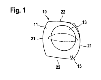
Fig. 1 illustrates an exemplary embodiment of a guide device
for
intraocular injection in a back view according to the present
invention;
Fig. 2 shows the embodiment of Fig. 1 in a first side view;
Fig. 3 shows the embodiment of Fig. 1 in a second side view;
and
Figures 4 to 8 illustrates an exemplary embodiment of a sequence for
using the
guide device of Figures 1 to 3 with an eye according to the
present invention.
Detailed Description
Figure 1 shows an exemplary embodiment of a guide device 10 for intraocular
injection according to the present invention. In an exemplary embodiment, the
guide
device 10 comprises a base plate 11 having two pairs of opposing edges, namely
first
opposing edges 21 and second opposing edges 22, and a convex form, such that
the
base plate 11 easily adapts to the outer surface of an eye ball 32. Those of
skill in
the art will understand that the base plate 11 may be square, rectangular,
circular,
ellipsoidal, star-shaped, or a variety of other shapes.
In an exemplary embodiment, the base plate 11 includes a contoured inner
surface
13 such that the guide device 10 may center itself over the cornea of the eye
ball 32.
For example, the inner surface 13 may include a first portion having a first
convex
shape (e.g., similar to that of a contact lens) and a second portion
surrounding a
periphery of the first portion and having a second convex shape to match that
of an
exposed portion of the eye ball 32 surrounding the cornea. When the guide
device
10 is placed on the eye ball 32, it may be positioned such that the cornea
fits at least
partially within the first portion. Abutment of the cornea to the first
portion may
prevent the guide device 10 from moving relative to the eye ball 32 during and
after
an injection procedure. In an exemplary embodiment, a radius of curvature of
the
first portion may be greater than a radius of curvature of the second portion,
such that
CA 02834819 2013-10-31
WO 2012/152840
PCT/EP2012/058566
4
the first portion accommodates the cornea and the second portion accommodates
the
eye ball 32.
In an exemplary embodiment, the base plate 11 further comprises a through hole
15
for aligning a needle of an injection device. The hole 15 may be formed at a
predetermined distance from the periphery of the first portion. The
predetermined
distance may be determined as a function of a desired injection site. For
example, to
ensure that the injection site is not aligning with the cornea or the lens
(unless that is
the desired target), the hole 15 may be formed on a periphery of the base
plate 11. A
diameter of the hole 15 may be substantially equal to (or slightly greater
than) a
diameter of a needle used to administer the injection.
In an exemplary embodiment as shown in Figure 2, a handle 17 is formed on an
outer
surface 19 of the base plate 11. The handle 17 may allow a physician to rotate
the
guide device 10 relative to the eye ball 32. In the exemplary embodiment shown
in
Figure 2, the handle 17 is formed as an elongated rib extending across a width
of the
base plate 11. However, those of skill in the art will understand that the
handle 17
may be other shapes or sizes, and a height of the handle 17 may be optimized
for
gripping by hand or by a medical instrument (e.g., forceps). Other forms are
possible
as well, for example a knob or a handle.
Figures 4 to 8 show an exemplary embodiment of a method of using the guide
device
10 according to the present invention.
As shown in Figures 4 and 5, an eye 30 is shown in an initial position. At
first, eye
lids 34 of the eye 30 are separated, and the eye ball 32 may be cleaned or
otherwise
prepared for an injection.
As shown in Figure 6, the guide device 10 is placed in a first orientation in
contact
with the eye ball 32. In an exemplary embodiment, in the first orientation,
the handle
17 may be parallel to a sagittal plane, such that terminal ends of the handle
17 abut
the respective eye lids to maintain separation of the eye lids during the
injection
procedure.
CA 02834819 2013-10-31
WO 2012/152840
PCT/EP2012/058566
In another exemplary embodiment, the first and/or second opposing edges 21, 22
may be contoured in accordance with a shape of the eye. For example, one pair
of
the first and second opposing edges 21, 22 may have opposing concave and
convex
contours to abut opposing eye lids. In this manner, the guide device 10 may be
5 utilized to maintain separation of the eye lids during the injection. In
an exemplary
embodiment, barriers may be formed on the first and/or second opposing edges
21,
22 to abut the eye lids and provided positional stability to the guide device
10. In the
first orientation, the hole 15 may be aligned with the desired injection site,
and the
needle may pierce the eye ball 32 through the hole 15.
As shown in Figure 7, after the injection has been delivered and the needle
has been
withdrawn from the eye ball 32, the guide device 10 may be rotated into a
second
orientation using the handle 17 such that the injection site is covered by the
base
plate 11. In an exemplary embodiment, the guide device 10 may be rotated by
approximately 90 relative to the eye ball 32. After rotating the device 10,
the handle
17 may be parallel to a transverse plane, allowing the eye lids 34 to close
over the
guide device 10, as shown in Figure 8. Allowing the eye lids 34 to close after
the
procedure may be more comfortable for the patient.
After the guide device 10 has been placed in the second orientation, the
injection site
is covered by the base plate 11 (because the hole 15 is offset from the
injection site).
Thus, covering the injection site may prevent the medicament from leaking out
of the
eye ball 32.
Covering the injection site may also promote faster healing and prevent
infection. For
example, at least a portion (e.g., the second portion or part thereof) may be
coated
with a medicament (e.g., an antimicrobial agent, an anti-infection agent, an
anti-
bacterial agent, etc.) to promote healing of the injection site and/or prevent
infection.
Those of skill in the art will understand the modifications (additions and/or
removals)
of various components of the device and/or system and embodiment described
herein may be made without departing from the full scope and spirit of the
present
invention, which encompass such modifications and any and all equivalents
thereof.
CA 02834819 2013-10-31
WO 2012/152840
PCT/EP2012/058566
6
Reference signs
guide device
11 base plate
5 13 inner surface of the base plate 11
14 through hole
17 handle
19 outer surface of the base plate 11
21 first edge
10 22 second edge
30 eye
32 eye ball
34 eye lid