Note: Descriptions are shown in the official language in which they were submitted.
CA 02835616 2013-11-08
WO 2012/155003 PCT/US2012/037401
CRANIAL PLATING AND BUR HOLE COVER SYSTEM
CROSS REFERENCE TO RELATED APPLICATION(S)
[0001] This application claims the benefit of priority under 35 U.S.C.
119(e) of US
Serial No. 61/484,641 filed May 10, 2011 the entire content is incorporated
herein by
reference.
BACKGROUND OF THE INVENTION
FIELD OF THE INVENTION
[0002] The invention relates generally to cranial closure improvements and
more
specifically to devices and methods used to improve cranial healing and
reconstruction and
the decrease in patient discomfort and/or palpable or visible deformities
often present after a
craniotomy.
BACKGROUND INFORMATION
[0003] Craniotomy is a common operation in the United States. It is performed
for a
variety of indications, including head trauma, aneurysm repair, and tumor
removal, among
others. Most craniotomies are performed by drilling one or more bur holes in
the skull down
to the level of the dura covering the brain and connecting them with a routing
bit on a high-
speed drill. The bit pulverizes a tract of bone typically two or more
millimeters wide. The
space left between the bone edges is called the kerf. At the time of closure,
the bone flap is
replaced with plates and screws, a specialized compressible closure device,
wires, or
sutures. All of these present methods leave a gap (shown in FIG. 1) which is
either centered
(FIG. la) or eccentric (FIG. lb). Current cranial plates, bur hole covers and
screw systems
are mainly if not entirely affixed on the outer surface of the cranium
resulting in palpable
and often visible protrusions which provide discomfort if in a place where the
scalp moves
against the protrusions regularly. Because many craniotomies are performed
below the
hairline, this often results in gross external deformity. Even for
craniotomies located off of
the forehead, the palpable or visible deformity (particularly for patients who
do not have
covering hair) and/or accompanying discomfort from the rubbing is often
distressing to the
patient.
CA 02835616 2013-11-08
WO 2012/155003
PCT/US2012/037401
2
100041 Unfortunately, a suitable device for assisting cranial
reconstruction and
decreasing cranial deformities and discomfort has not yet been described.
Thus, a need
exists for methods and devices capable of assisting the surgeon with improved
clinical and
procedural outcomes when performing craniotomies.
SUMMARY OF THE INVENTION
[0005] The present disclosure generally comprises a cranial closure system,
devices and
methods of use, tooling and kits for improving cranial closure outcomes by
using a device
including a cranial plate or a bur hole cover with a minimized cranial surface
profile. The
devices are substantially inset within the kerf and/or bur hole and thus
reduces protrusions
on the cranial surface and thus provides a better clinical outcome for the
patient. Additional
embodiments further reduce cranial protrusions by partially or fully insetting
the device in
relation to the outer surface of the cranium.
[0006] An embodiment of the present invention features a cranial plating
device which
comprises two attachment wings separated by a keel, wherein the two attachment
wings are
on opposing sides and ends of the keel. When in use one attachment wing
attaches to the
cranial flap, and the other attaches to the surrounding cranium. A plurality
of the embodied
cranial plates may be used alone, or with other known cranial plates to secure
the cranial
flap back to the cranium. Additional embodiments include various keel
attachment wing
orientations, such as perpendicular or angled, and keel shapes which are
straight, curved or
bent. Additional embodiments include the keel being solid or perforated to
allow or
facilitate bone growth therethrough.
[0007] Another embodiment of the present invention features a bur hole cover
device
which comprises three attachment wings separated connected to arms which
extend radially
from a center and wherein the general shape of the bur hole cover is circular,
wherein the
three attachment wings are spaced relatively equidistant around the circle.
When in use, if
one attachment wing attaches to the cranial flap, then the other two arms
attach to the
surrounding cranium and vice versa. A bur hole cover may be used alone in
cases where
only a bur hole access was needed for surgical access, or one or more bur hole
covers may
CA 02835616 2013-11-08
WO 2012/155003 PCT/US2012/037401
3
be used with a plurality of the embodied cranial plates or with other known
cranial plates to
secure the cranial flap back to the cranium. Additional embodiments include
various arm
attachment wing orientations, which are rigid enough to provide the structural
support
necessary yet are somewhat pliable to allow the user to place in non-uniform
environments.
The embodied arms extend radially from a bur hole cover center and may have
various
snaking, zigzag or looping shapes which create cover area surface but allow
the arm to be
flexed a little or somewhat pliable at the attachment wings. Additional
embodiments
include the bur hole cover being solid or perforated to allow or facilitate
further cranial
access or bone growth.
100081 Yet another embodiment of the present invention features a specialized
tooling
apparatus to create a more precise impression for the placement of the
attachment wings of
an embodied cranial plating and/or bur hole cover device. The tooling
apparatus is made to
attach to standard neurosurgical tooling. And a preferred tooling apparatus
includes a drill
bit attachment and guard mechanism. The drill bit attachment comprises a
shaft, cutting
head and cylindrical tip for creating a pilot hole. The guard attachment fits
over the shaft of
the drill bit and the top attaches to the drill the guard prevents the drill
from drilling past a
certain preset depth. Additionally, the guard comprises protruding posts or an
orientation
ridge which fit against the bone edge so the cylindrical tip makes the pilot
hole a standard
distance from the edge of the craniotomy and the cutting head removes a more
precise
section of bone near the kerf.
[0010] Additional embodiments feature methods of using the embodied cranial
plating
device, one embodied method places the keel portion of the plating device
within the kerf
and affixes the attachment wings of the device on the outer cranial surface
without making
an impression in the bone first. An additional embodiment uses readily
available
neurosurgery tools and with a free-hand technique a perforator or bur hole
creator is used to
cut away a section of bone to create an impression to place the wings of the
cranial plate
into the impression and to reduce the cranial profile of the plate on the
outer surface of the
cranium. Another additional embodiment utilizes an embodied precision tooling
apparatus
to create precise impressions in depth and orientation to the kerf, and
affixation pilot holes
CA 02835616 2013-11-08
WO 2012/155003
PCT/US2012/037401
4
to secure an embodied cranial plating device to the cranium resulting in a
reduced cranial
profile that may allow the entire plating system to be fully inset into the
cranium and thus
avoiding or minimizing the protrusions typically associated with cranial plate
devices.
[0011] An additional embodiment features a kit for treating a cranial gap
associated with a
craniotomy in a subject comprising: a bur hole cover, and/or a plurality of
cranial plates, a
specialized drill bit, and guidance apparatus; and instructions for use.
BRIEF DESCRIPTION OF THE DRAWINGS
[0012] Figure 1 is a top plan view of a cranium after a craniotomy and
consists of Figure 1 a
which shows a centered bone flap and Figure lb which shows an eccentric bone
flap.
[0013] Figure 2 is a side front perspective view of embodiments of the present
invention
and consists of Figure 2a which shows an embodiment wherein the front and back
surface
of the keel is solid; Figure 2b which shows an embodiment wherein the front
and back
surface of the keel is perforated with circular apertures from front to back;
and Figure 2c
which shows an embodiment wherein the front and back surface of the keel is
opened with
rectangular apertures from front to back.
[0014] Figure 3 is a top plan view of embodiments of the present invention and
consists of
Figure 3a which shows an embodiment wherein the keel is straight in relation
to the wings;
Figure 3b which shows an embodiment wherein the keel is angled in relation to
the wings;
Figure 3c which shows an embodiment wherein the keel is curved in relation to
the wings;
and Figure 3d which shows an embodiment wherein the device shown in Figure 3c
as a
right handed configuration is shown in Figure 3d as a left-handed
configuration.
[0015] Figure 4 Figure 4a shows the cranial flap with the embodied plates
attached. Figure
4b shows the cranial flap placed back into the cranial opening and being
resecurecl, and
Figure 4c shows the completed cranial closure with embodied cranial plates.
[0016] Figure 5 is a side plan view of a drill-bit tooling embodiment of the
present
invention and consists of Figure 5a and Figure 5b which respectively, show the
front side
and bottom side of a drill-bit contemplated.
CA 02835616 2013-11-08
WO 2012/155003
PCT/US2012/037401
[0017] Figure 6 is a side plan view of a drill-bit guard tooling embodiment of
the present
invention and consists of Figure 6a and Figure 6b which respectively, show the
front side
and bottom side of a drill-bit guard contemplated; and Figure 6c and Figure 6d
which
respectively, show the front side and bottom side of an alternative drill-bit
guard
contemplated.
[0018] Figure 7 is a side plan view of a drill-bit with guard tooling
embodiment of the
present invention and consists of Figure 7a and Figure 7b which respectively,
show the
front side and bottom side of a drill-bit with guard tooling embodiment
contemplated; and
Figure 7c and Figure 7d which respectively, show the front and bottom surfaces
of an
alternative drill-bit and guard tooling embodiment contemplated.
[0019] Figure 8 is a top perspective view showing the use of a drill-bit with
guard tooling
embodiment to create a recessed fixation point on the cranial flap so that the
wings of an
embodied cranial plate can be nested and secured and consists of Figures 8a,
8b, and 8c
which represent a tooling embodiment used on the cranial flap, before cutting
(Figure 8a)
during cutting (Figure 8b) and after cutting (Figure 8c).
[0020] Figure 9 is a side plan view showing the use of a drill-bit with guard
tooling
embodiment to create a recessed fixation point on the cranium or cranial flap
so that the
wings of an embodied cranial plate can be nested and secured and consists of
Figures 9a,
9b, 9c and 9d which represent a tooling embodiment used on the cranium
surface, before
cutting (Figure 9a) during orientation and engagement of pilot hole bit
(Figure 9b) when the
cutting tool reaches the depth preset by the guide (Figure 9c) and after
cutting (Figure 9d).
[0021] Figure 10 is a side perspective view of the cranium and embodied
cranial plates
after attachment and consists of Figure 10a which shows a cranial plate
secured to the
cranium like standard plates (thus embedding only the keel within the kerf and
the wings
are protruding on the cranial surface, Figure 10b which shows the plate
partially embedded
as if the embedding process was created free-hand with a perforator only; and
Figure 10c
shows the plate embedded in a zero-profile or fully embedded relationship with
the cranium
CA 02835616 2013-11-08
WO 2012/155003
PCT/US2012/037401
6
as if the embedding process was created with the precision of the embodied
tooling
components.
[0022] Figure 11 consists of Figures 11A-11L each of which is a perspective
view of an
embodied cranial plate device.
[0023] Figure 12 consists of Figures 12A-E each of which is a top plan view of
an
embodied bur hole cover device.
[0024] Figure 13 consists of Figures 13A-C wherein Figure 13A shows the
cranial flap
with embodied cranial plates and an embodied bur hole cover attached, Figure
13B shows
the cranial flap placed back into the cranial opening and being resecured with
embodied
cranial plates and bur hole covers, and Figure 13C shows the completed cranial
closure with
embodied cranial plates and bur hole covers.
[0025] Figure 14 is a side perspective view of the cranium and an embodied bur
hole cover
after attachment.
DETAILED DESCRIPTION OF THE INVENTION
[0026] A craniotomy is a procedure that is frequently performed for the
treatment of
neurosurgical conditions and diseases. A craniotomy involves the placement of
one or more
bur holes (full-thickness holes placed in the skull through to the level of
the dura) which are
connected with the use of a cutting instrument. This cutting instrument can be
manual (e.g.
a handheld Gigli saw that cuts using a wire blade) or, more commonly, a high-
speed drill
with a router attachment (craniotome). At the end of the procedure the bone is
usually
replaced. When it is replaced, the fixation devices used reside above the
cranium surface
resulting in deformity of the contour of the skull and distress and/or
discomfort to the
patient.
[0027] The devices and methods contemplated in the present invention are based
on
providing a neurosurgeon with an effective, rapidly deployable, product with a
reduced
CA 02835616 2013-11-08
WO 2012/155003
PCT/US2012/037401
7
cranial profile when closing the cranial flap and will partially fill the gap
(kerf) made by a
craniotomy.
100281 Standard craniotome router bits for cutting the human skull that are
commercially
available include those made by Medtronic Midas Rex, Anspach, Aesculap,
Stryker,
Codman, and others. Virtually all leave a channel-shaped trough or gap through
the bone
whose height is the thickness of bone, length is the perimeter of the desired
craniotomy, and
the width is 2 2 mm. A pediatric bit may leave a gap that is 1.5 mm+/1 mm.
Given that at
the time of closure the gap may be all positioned to one side or the other,
the gap may be 2-
4 nun+1-2 mm. The distinct shape and dimensions of the cranial plate embodied
allows it to
conform to the dimensions of the kerf, even where the kerf varies in width.
Embodiments in Use
100291 A craniotomy is performed for a neurosurgical procedure as follows: The
patient's
head is positioned and a line is marked in the scalp. The skin is incised with
a scalpel and
the scalp is held out of the way with a retractor. The bone is exposed by
removing the
overlying periosteal layer. A high-speed drill is used to drill a small hole
through the bone
down to the level of the dura, for example, an 8 mm round hole, shaped like a
cylinder. A
craniotome drill, which is a side-cutting bit with a footplate guard, is used
to cut out a flap
of bone. This flap can be of any shape or size. The bone removed by the action
of the side
cutting bur is typically powdered by the bit and is washed away. The gap that
is left is
called the kerf. The bone flap is elevated off the dura and set aside. The
intracranial portion
of the procedure is then completed. At the time of closure the bone flap is
resecured to the
surrounding bone using plates and screws, a clamping device, wire, or suture,
or some
equivalent method. The secured bone flap will have around it a surrounding
gap, the kerf,
which is usually left unfilled. The scalp is closed over the bone, the skin is
closed with
sutures or staples, and the procedure is completed.
[0030] The kerf is a concentric defect in the bone at the time it is created.
When the bone
flap 20 is replaced, the bone 20 may be replaced in centered fashion (see FIG.
la), with a
kerf 10 of uniform width, or eccentric (see FIG. 1b), with the bone 20 pushed
to one side,
creating a minimal gap 10 on one side and a wider gap on the other. Placement
of the flap
CA 02835616 2013-11-08
WO 2012/155003 PCT/US2012/037401
8
eccentrically has advantages in that the presence of bone-to-bone contact on
at least one
cranial surface 30 will allow the blood supply of the cranium 30 to contact
the flap, keeping
the bone flap alive and promote fusion of the bone flap 20 to the surrounding
bone 30.
When the flap 20 is placed eccentrically, the kerf 10 will be tapered at its
ends and widest at
the middle when viewed from above. The use of a cranial plate that comprises a
keel section
which resides within the kerf (as shown in Figures 2-4) which has at least one
of the
following features, thin keel material, that is bendable or flexible and is
straight, curved or
angled from the extending wings, which are placed on opposite sides of the
keel and allows
the use of the same cranial plate to be used in tight and wide kerfs by simply
adjusting the
orientation of the device.
100311 Additionally, based on the orientation and features of the embodied
cranial plating
devices it is possible that less total cranial plates may be necessary to
resecure the cranial
flap after a craniotomy.
Cranial Plate Designs
[00321 Generally, Figures 2, 3 and 11 display exemplifications of embodied
plates which
may feature a straight or curved keel with a perpendicularly oriented wing on
either side
with each wing facing opposite the other. The wings are designed to
accommodate screws
to fasten into bone. The plate may or may not have perforations along the
length of the keel
to allow bone and growth. Additionally the keel may be straight, angled
slightly, or curved
slightly, and/or the keel may be capable of being bent or adjusted a little
bit at time of use.
The device may have lateral flex or bend incorporated into the device to
assist filling the
curved spaces associated with the kerf; or the device may have vertical flex
or angle
components incorporated into the device to assist the user when dealing with
the external
curvature of the cranium. Additionally the keel portion which resides in the
kerf may be
coated with various medicaments and/or healing agents. The keel may be
perforated or
solid.
CA 02835616 2013-11-08
WO 2012/155003 PCT/US2012/037401
9
[0033] As demonstrated in more detail in Figure 2, embodied cranial plates
comprise of a
keel portion attached to two fixation wings, wherein the fixation wings are on
opposite sides
and opposite ends of each other. Each of the embodied cranial plates shown in
Figure 2 are
shown in a side front perspective view. In the embodied cranial plate 101
shown in Figure
2a the keel 111 is solid, thus no holes or perforations are visible on the
front keel surface
119 or the back keel surface 121 and each fixation wing 131 is attached to the
top keel
surface 115 at a wing-keel attachment point 123, additionally the keel 111
includes a
bottom surface 117 which resides within a kerf 20 (not shown) and is
positioned closest to
the dura or the brain (not shown) when in use. Each fixation wing 131 has a
top surface
133 and a bottom surface 135 and a fixation aperture 139 which extends from
the top
surface 133 to the bottom surface 135. Typically the fixation aperture 139 is
tapered and
thus has a wider diameter at the top surface 133 than the bottom surface 135.
This tapered
fixation aperture 139 is designed to match and closely fit a tapered head
fixation device 141
such as a screw (not shown).
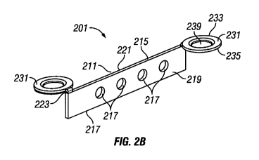
[0034] In the embodied cranial plate 201 shown in Figure 2b the front keel
surface 219 and
back keel surface 221 of the keel 211 is perforated with circular apertures
225 and each
fixation wing 231 is attached to the top keel surface 215 at a wing-keel
attachment point
223, additionally the keel 211 includes a bottom surface 217 which resides
within a kerf 20
and is positioned closest to the dura or the brain (not shown) when in use.
Each fixation
wing 231 has a top surface 233 and a bottom surface 235 and a fixation
aperture 239 which
extends from the top surface 233 to the bottom surface 235. Typically the
fixation aperture
239 is tapered and thus has a wider diameter at the top surface 233 than the
bottom surface
235. This tapered fixation aperture 239 is designed to match and closely fit a
tapered head
fixation device 241 such as a screw (not shown).
[0035] In the embodied cranial plate 301 shown in Figure 2c the front keel
surface 319 and
back keel surface 321 of the keel 311 is opened with rectangular apertures 325
and each
fixation wing 331 is attached to the top keel surface 315 at a wing-keel
attachment point
323, additionally the keel 311 includes a bottom surface 317 which resides
within a kerf 20
and is positioned closest to the dura or the brain (not shown) when in use.
Each fixation
CA 02835616 2013-11-08
WO 2012/155003 PCT/US2012/037401
wing 331 has a top surface 333 and a bottom surface 335 and a fixation
aperture 339 which
extends from the top surface 333 to the bottom surface 335. Typically the
fixation aperture
339 is tapered and thus has a wider diameter at the top surface 333 than the
bottom surface
335. This tapered fixation aperture 339 is designed to match and closely fit a
tapered head
fixation device 341 such as a screw (not shown).
[0036] As generally discussed above, the keel portion of the cranial plates
embodied may
have different properties such as being straight or curved, and different
orientations in
relation to the attachment wings in that the keel may be attached
perpendicular to the
fixation wings or at an offset angle, additionally each configuration may be
created in either
a left-handed or right-handed orientation. Figure 3 is a top plan view of
exemplary
embodiments of the present invention wherein the orientation and shape of the
keel is
shown in various designs contemplated in the present invention. In the
embodied cranial
plate 401 shown in Figure 3a the keel 411 is not curved and is straight in
relation to each
fixation wing 431 and each fixation wing 431 is attached to the top keel
surface 415 at a
wing-keel attachment point 423, wherein the orientation of the keel 411 to
each fixation
wing 431 is perpendicular.
[0037] In the embodied cranial plate 501 shown in Figure 3b the keel 511 is
not curved but
is angled in relation to each fixation wing 531 and each fixation wing 531 is
attached to the
top keel surface 515 at a wing-keel attachment point 523, wherein the
orientation of the keel
511 to each fixation wing 531 is at an offset angle.
[0038] In the embodied cranial plates 601 and 701 shown in Figure 3c and the d
respectively, the keel 611, 711 is curved or bowed slightly in relation to
each fixation wing
631, 731 and each fixation wing 631, 731 is attached to the top keel surface
615, 715 at a
wing-keel attachment point 623, 723,wherein the orientation of the keel 611,
711 to each
fixation wing 631, 731 may be perpendicular (as shown), or at an offset angle.
Additionally,
the difference between cranial plate 601 in Figure 3c and cranial plate 701 in
Figure 3d is
that cranial plate 601 is in a right handed configuration as shown and cranial
plate 701 is in
a left-handed configuration as shown.
CA 02835616 2013-11-08
WO 2012/155003 PCT/US2012/037401
11
[0039] Figure 11 comprises of cranial plate designs 11A-11L which further
demonstrate a
sampling of versatile cranial plates contemplated in the present invention. In
the embodied
cranial plate 801 shown in Figure 11A the keel 811 is straight in relation to
each fixation
wing 831 and each fixation wing 831 is attached to the top keel surface 815 at
a wing-keel
attachment point 823, and the keel 811 has a shortened depth as measured by
the distant
from the top keel surface 815 to the bottom keel surface 817 this embodiment
may be useful
in places where the cranium thickness is less or compromised.
[0040] In the embodied cranial plate 901 shown in Figure 11B the keel 911 is
straight in
relation to each fixation wing 931 and each fixation wing 931 is attached to
the top keel
surface 915 at a wing-keel attachment point 923, which is thick to provide
greater structural
integrity to the device.
[0041] In the embodied cranial plate 1001 shown in Figure 11C the keel 1011 is
straight in
relation to each fixation wing 1031 and each fixation wing 1031 is attached to
the top keel
surface 1015 at a wing-keel attachment point 1023, and the keel 1011 has a
shortened depth
shelf created by a larger wing-keel attachment point 1023 which enables the
plate to fit
better in certain kerf arrangements. The embodied cranial plate 1111 shown in
Figure 11D
is identical to that shown in 11C with the exception that the larger wing-keel
attachment
point 1123 is angled downward so that the wing-keel attachment point 1123 is
at less than a
90 degree angle in relation to the keel 1111. The angle created is an example
of a vertical
flex or angle which assists the user when dealing with the external contours
of a cranium.
[0042] In the embodied cranial plate 1201 shown in Figure 11E the keel 1211 is
angled in a
very slight s-formation in relation to each fixation wing 1231 and each
fixation wing 1231
is attached to the top keel surface 1215 at a wing-keel attachment point 1223,
and the keel
has a shallow depth similar to plate 801. In Figure 11F the cranial plate 1301
has a keel
1311 that is angled in an s-formation like 1201 but in this case the angle is
much more
pronounced and the keel 1311 is not a shallow depth keel like 1211.
[0043] In the embodied cranial plate 1401 shown in Figure 11G the keel 1411 is
straight in
relation to each fixation wing 1431 and each fixation wing 1431 is attached to
the top keel
CA 02835616 2013-11-08
WO 2012/155003
PCT/US2012/037401
12
surface 1415 at a wing-keel attachment point 1423, and the keel 1411 circular
chain design
and apertures therethrough which may facilitate bone growth. Figure 1111 shows
an
embodied cranial plate 1501 with a similar circular keel design 1511 as shown
in Figure
11G with the exception that the keel 1511 does not have apertures.
Additionally each
fixation wing 1531 is attached to the top keel surface 1515 at a wing-keel
attachment point
1523 but the wing-keel attachment point 1523 is angled or dog-eared down.
[0044] In the embodied cranial plate 1601 shown in Figure 111 the keel 1611 is
angled in a
c-curve formation in relation to each fixation wing 1631. In the embodied
cranial plate
1701 shown in Figure 11J the keel 1711 is straight in relation to each
fixation wing 1731
but the keel has a tapered or rounded bottom surface 1717. In the embodied
cranial plate
1801 shown in Figure 11K the keel 1811 is angled in a z or zigzag formation in
relation to
each fixation wing 1831. In the embodied cranial plate 1901 shown in Figure
11L the keel
1911 is straight in relation to each fixation wing 1931 but the length of the
keel 1911 and
the subsequent distance between each fixation wing 1931 is short which enables
the plate
1901 to fit better in certain kerf arrangements.
[0045] A demonstration of the application of an embodied cranial plating
device 101 into a
kerf 10 where the cranial bone flap 20 is centered compared to the outlying
cranium 30 is
shown in Figure 4. Figure 4a demonstrates how one fixation wing 131 of a
cranial plate
101 is first placed into a cranial flap wing recess 50 which has been cut away
to provide a
specific placement of the fixation wing 131 and is adhered to the cranial bone
flap 20, with
fixation screws 141. Figure 4b demonstrates the step wherein the cranial flap
20 is oriented
back within the cranium 10, using the matching sides of one or more bur holes
40 to
provide proper orientation and the keel portion 111 of each device 101 is
placed within the
kerf 30, and the other fixation wing 131 is placed into the cranium wing
recess 60 which
has been cut away to provide a specific placement the fixation wing 131 and is
adhered to
the cranium 10 with fixation screws 141. Figure 4c demonstrates how the
finished
procedure appears, wherein a plurality of cranial plates 101 are positioned
such that the keel
111 of each plate resides within the kerf 30, and that the fixation wings 131
for each plate
101 are recessed within cranial flap wing recesses 50 or cranium wing recesses
60 such that
CA 02835616 2013-11-08
WO 2012/155003 PCT/US2012/037401
13
one wing 131 from each plate 101 is recessed 50 and attached to the cranial
flap 20 and the
other wing 131 from each plate 101 is recessed 60 and attached to the cranium
10 with
fixation screws 141.
[0046] Exemplary dimensions of embodied cranial plating devices contemplated
in the
present invention are as follows: The wings are generally circular but could
be a semi-
circular, oval or trapezoidal shape in alternative embodiments. The diameter
of the wings
are usually the same but could be different in certain embodiments for
example, where it is
advantageous to have a smaller fixation wing on the cranial plate then on the
cranium. The
diameter of the wings may be from about 2 mm to about 8 mm, with a preferred
diameter
from about 3 mm to about 5 mm with a diameter of about 4 mm being most
preferred. The
depth of the wings is from about 0.1 to about 2.0 mm, with a preferred depth
of about 0.2 to
0.8mm and most preferred about 0.5 to 0.6 mm. The affixation aperture within
the wing
may be tapered or untapered. Although tapered is preferred and used in
conjunction with
matching tapered affixation screws. The top width of the affixation aperture
is about 1.5 to
4 mm, with a preferred top width of 1.7 to 3.0 mm and most preferred about 2
mm. The
bottom width of the affixation aperture is about 0.1 to about 2.0 mm, with a
preferred width
of about 0.2 to about 1.5 mm, and a most preferred width of about 1.0 mm. The
affixation
screws preferred are size 2-8 mm affixation screws with size 3-4 mm affixation
screw most
preferred. The size of the affixation screws are chosen by the neurosurgeon
based on the
thickness of the bone at the point of use. The keel portion of an embodied
cranial plate may
have a depth (length from top of keel portion which interfaces with the
fixation wings
located on the outer cranial side to the bottom of the keel located towards
the dura or brain)
ranging from 1-10 mm, with a preferred depth from 2-7 mm and the most
preferred depth of
about 4 mm. The keel length contemplated may range from about 6-20 mm, with a
preferred length of 8-15 mm, and the most preferred length of 10-12 mm. The
contemplated
keel widths may be the same from top to bottom when in rectangular form or
will have a
greater top width than bottom width when the keel is tapered in width. The
contemplated
keel widths for either tapered or rectangular embodiments range from a top
width of 0.1 to
about 2 mm with a preferred width of 0.2 to about 1.0 mm and a most preferred
top width of
about 0.5 mm for untapered widths and 0.6 mm for tapered widths. The bottom
width
CA 02835616 2013-11-08
WO 2012/155003 PCT/US2012/037401
14
ranges from of 0.1 mm to 2.0 mm with a preferred width of 0.3 mm to 0.6 mm and
a most
preferred bottom width of 0.4 mm to 0.5 mm. This matches the contemplated kerf
widths of
about 1-5 mm in an adult and 1-4 mm in pediatric procedures.
[0047] Additional embodiments of the present disclosure include a method for
improving
the clinical outcome of a craniotomy comprising: reducing the protrusions
associated with
current cranial plating systems and reducing the indentations or gaps left in
the bone
following a craniotomy; wherein said indentations or gaps are filled with the
keel portion of
an embodied cranial plating device.
[0048] Additional embodiments of the cranial plating device can include:
[0049] Embodiments where the keel may be coated or provide a scaffold for
attachment
and/or distribution of analgesics, antibiotics, antibacterial agents, or
antiseptic agents in
order to prevent or reduce patient discomfort and/or bone flap infection.
[0050] Embodiments where the keel may be coated or provide a scaffold for
attachment
and/or distribution of biological growth factors in order to promote bone
growth and
ingrowth, such as via osteogenesis, osteoconduction, and/or osteoinduction.
[0051] Embodiments where the keel can serve as a scaffold to hold a paste,
gel, or other
moldable or pourable liquid for the purpose of hardening the bone into a solid
matrix to
create a hard surface or a watertight seal.
Tooling Embodiment Designs
[0052] The tooling system may comprise of a high-powered drill and an
impression device
which consists of a cylindrical or disc-like bur (drill-bit) and a surrounding
guide, the drill-
bit is meant to be applied end on into the bone to make a semicircular
depression in the
bone comprising a 3/5 to almost one whole circle that has a central point to
allow the bit to
bite bone at a precise spot this will also serve to create a pilot hole for an
fixation screw
when the plate is applied. The surrounding guide orients the drill-bit into
the proper
positioning in relation to the bone edge and also may control and/or limit the
depth in which
the drill-bit may cut into the bone.
CA 02835616 2013-11-08
WO 2012/155003 PCT/US2012/037401
[0053] Embodiments of contemplated features of the tooling system are shown in
Figures
5-7. Figure 5 is a side plan view of a drill-bit tooling embodiment 161 of the
present
invention and consists of Figure 5a and Figure 5b which respectively, show the
front side
and bottom side of a drill-bit contemplated in the present disclosure. Figure
5a shows an
embodied cutting drill-bit 161 which comprises a shaft portion 163 which has
an upper end
165 which attaches to the drill or perforator tool 155 (not shown) and a lower
end 167
which attaches to a cutting bit 171, at the upper surface 175 of the cutting
bit 171. The
cutting bit 171 also has a bottom cutting surface 177 which is used cut away
the bone and
make the recesses for the wings of the cranial plates. Attached to the bottom
cutting surface
177 is a pilot hole bit 179 which drills a pilot hole 70 (not shown) which
orients and directs
the proper placement of a cranial plate 101, when the fixation screws 141 (not
shown) are
screwed into a pilot hole 70 (not shown). Figure 5b shows the bottom cutting
surface 177
of the cutting bit 171 and shows the placement of the pilot hole bit 179 in
the center of the
bottom cutting surface 177.
100541 Figure 6 is a side plan view of a drill-bit guard tooling embodiment
181 of the
present invention and consists of Figure 6a and Figure 6b which respectively,
show the side
and bottom surface of a drill-bit guard contemplated; and Figure 6c and Figure
6d which
respectively, show the side and bottom surface of an alternative drill-bit
guard 281
contemplated. Figure 6a shows the side view of a guard 181 contemplated, the
guard 181
has an upper end 185 which interfaces with the drill or perforator tool 155
(not shown), and
controls the depth that a cutting drill-bit 161 can cut by having the upper
surface 185 fixed
or stopped at a certain drill depth, and the bottom surface 187 which
interfaces with the
bone to be cut does not allow the cutting bit 171 to drill deeper than where
the bottom
surface 187 of the guide 181 surrounds the cutting bit 171. Additionally the
guide has two
protruding orientation posts 191 which extend from the bottom surface 187 of
the guide
181. Figure 6b further shows the bottom surface 187 of the embodied guide 181
and the
relation of the orientation posts 191 on the bottom surface 187. Additionally
there is a drill-
bit clearance space 189 that is an aperture so that a drill-bit 161 and
particularly the cutting
bit portion 171 can spin freely within the clearance space 189 without
impedance from the
guide 181.
CA 02835616 2013-11-08
WO 2012/155003 PCT/US2012/037401
16
[0055] Figure 6c shows the side view of a guard 281 contemplated, the guard
281 has an
upper end 285 which interfaces with the drill or perforator tool 155 (not
shown), and
controls the depth that a cutting drill-bit 161 can cut by having the upper
surface 285 fixed
or stopped at a certain drill depth, and the bottom surface 287 which
interfaces with the
bone to be cut does not allow the cutting bit 171 to drill deeper than where
the bottom
surface 287 of the guide 281 surrounds the cutting bit 171. Additionally the
guide has an
orientation ridge 291 which extends along the bottom surface 287 of the guide
281 from
about the seven o-clock to about the 11 o-clock position. Figure 6d further
shows the
bottom surface 287 of the embodied guide 281 and the relation of the
orientation ridge 291
along the bottom surface. Additionally there is a drill-bit clearance space
289 that is an
aperture so that a drill-bit 161 and particularly the cutting bit portion 171
can spin freely
within the clearance space 289 without impedance from the guide 281.
[0056] Figure 7 is a side plan view of a complete specialized cranial plate
tooling apparatus
255 with a drill-bit 161 and a guard tooling embodiment 281 of the present
invention and
consists of Figure 7a and Figure 7b which respectively, show the side and
bottom surface of
the complete apparatus 255 and shows a drill-bit 161 nested within the drill-
bit clearance
space 289 with-in the guard tooling embodiment 281; Figure 7b shows the bottom
surface
287 of the embodied guide 281 and the relation of the orientation ridge 291
along the
bottom surface 287 and in relation to the bottom cutting surface of the bit
177 and the pilot
hole cutting bit 179.
[0057] Figure 7 further includes Figure 7c and Figure 7d which respectively,
show the side
and bottom surface of an alternative complete apparatus 155 and Figure 7c
shows a drill-bit
161 nested within the drill-bit clearance space 189 with-in the guard tooling
embodiment
181; Figure 7d shows the bottom surface 187 of the embodied guide 181 and the
relation of
the orientation posts 191 along the bottom surface 187 and in relation to the
bottom cutting
surface of the bit 177 and the pilot hole cutting bit 179.
[0058] Exemplary dimensions of an embodied cranial plate tooling apparatus
contemplated
in the present invention are as follows: The bit portion comprises a head with
a diameter of
about 2 to about 8 mm, with a preferred diameter from 3-7 mm and the most
preferred
CA 02835616 2013-11-08
WO 2012/155003
PCT/US2012/037401
17
diameter of about 4 mm. The bit may have a depth of about 0.3 to about 2 mm,
but the
placement of the guard at a specific depth will restrict the usable depth of
the bit to the
designated depth for creating the impression in the cranium. This depth ranges
from about
0.2 to about 1.0 mm, with a preferred depth of about 0.3 to about 0.8 mm and a
most
preferred depth of about 0.6 mm. Additionally, the bit comprises a pilot hole
bit which may
be about 0.5 mm to about 2mm in diameter and preferred at 1 mm in diameter and
0.5 to 3.0
mm deep, with a preferred depth of about 2 mm. The guard portion of an
embodied tooling
apparatus has a diameter slightly larger than the diameter of the head of the
bit, so that the
head can spin unobstructed within the guard. The guard orientation posts may
by about 1 to
6 mm long, with a preferred depth of 3 mm.
[0059] Figures 8-10 provide visual exemplifications of how the cranial plates
and tooling
systems embodied are used to produce a lower profile cranial plate when
securing the
cranial flap back to the cranium. Figure 8 comprising Figures 8a, 8b, and 8c
show a top
perspective view showing the temporal use of a complete tooling apparatus 155
to create a
recessed fixation point 50 and fixation screw pilot hole 70 on the cranial
flap 30. Figure 8a
shows the cranium 10, cranial flap 20 and the kerf 30 awaiting the placement
of a cranial
plate 101 following surgery. Figure 8b shows the use of tooling apparatus 155
on the
cranial flap 20 while drilling a recessed fixation point 50 and a fixation
screw pilot hole
which are visible in Figure 8c.
[0060] Figure 9 is a side plan view showing the use of a complete tooling
apparatus 155
including a drill-bit 161 with guard 181 tooling embodiment to create a
recessed fixation
point on the cranium 60 or cranial flap 50 so that the wings 131 of an
embodied cranial
plate 101 can be nested and secured and consists of Figures 9a, 9b, 9c and 9d
which
represent the time sequence of the methods when using a tooling apparatus 155
on the
cranium surface. Figure 9a shows the use of the tooling apparatus 155 when
preparing to
orient the tool 155 to the cranium 10, the user lines the orientation posts
191 up with the
bone end 52 before cutting. Figure 9b shows the tooling apparatus 155
beginning
engagement with the cranium bone 10, the orientation posts 191 are flush
against the bone
end 52 and the pilot hole bit 179 is just starting to drill into the cranium
10. Figure 9c
CA 02835616 2013-11-08
WO 2012/155003 PCT/US2012/037401
18
shows the time when the cutting tool 155 reaches the depth preset by the guide
181 the
bottom surface 177 of the guide 181 rests on the cranium 10 and does not allow
the
apparatus 155 to continue drilling deeper. Figure 9c shows the result after
using the tooling
apparatus 155 a cranium recess 60, and fixation screw pilot hole 70 are placed
in a uniform
position from the bone edge 52.
[0061] Figure 10 is a side perspective view of the cranium and embodied
cranial plates
after attachment and consists of Figure 10a which shows a cranial plate 101
secured to the
cranium 10 like standard plates. The exploded view portion demonstrates the
orientation of
the keel 111 within the kerf 30 but the protrusion of the wings 131 when
attached to the
cranium 10 and flap 20 is evident when seen from this side view. Figure 10b
shows the
plate 101 partially embedded in recesses created free-hand with a perforator
only. The
exploded view portion, shown from the side view, demonstrates the orientation
of the keel
111 within the kerf 30 but the protrusion of the wings 131 when attached to
the cranium 10
and flap 20 is still evident even though the protrusions of the wings 131 is
not as
pronounced because a uniform and accurate recess is very difficult to achieve
free-hand.
Figure 10 shows the plate 101 embedded in a zero-profile or fully embedded
relationship
with the cranium as if the embedding process was created with the precision of
the
embodied tooling components. The exploded side-view portion shows how the wing
is
embedded in the cranium 10 at a recess point 60, because of the zero-profile
of being fully
embedded it is not possible to see the cranial flap 20 recess point 50 and
fixed wing 131
because the entire cranial plate device 101 resides even with or slightly
below the external
surface of the cranium.
[0062] Bur Hole Cover Designs
[0063] Generally, Figure 12 displays 5 exemplifications (Figures 12A-E) of
embodied bur
hole covers which may feature various arrangements of three arms extending
from a center
cover portion with attachment wings near the distal end of the arms. The wings
are designed
to accommodate screws to fasten into bone. The bur hole cover may or may not
have
perforations along in the center portion or arms to allow brain access or to
allow for bone
and future growth. Additionally, the arms are meant to cover as much surface
area as
CA 02835616 2013-11-08
WO 2012/155003
PCT/US2012/037401
19
possible through a combination of any of serpentine, winding, zigzagging, or
looping,
arrangements in relation to the center portion. The arms must also be thin
enough to allow
them to be somewhat pliable by the user so that they can make adjustments of
the wing
placement when attaching the covers to the cranium or a cranial flap. The wing
dimensions
and attachment methods and tooling may include the same as that described
above for the
cranial plates and the wing components and properties. The cover may be used
in addition
with cranial plates during a procedure with a cranial flap, or the cover may
be used alone
when only a bur hole is drilled for the procedure.
[0064] As demonstrated in more detail in Figure 12, embodied bur hole covers
comprise of
a center cover portion with three arms extending therefrom to which are
attached three
fixation wings, wherein the fixation wings dispersed around the perimeter of
the
substantially circular device. Additional embodiments feature the fixation
wings about
equidistant from each other. Each of the embodied bur hole covers shown in
Figure 12 are
shown in a top plan view. In the embodied bur hole cover 2001 shown in Figure
12A the
center cover portion 2024 is solid, thus no holes or perforations are visible
on the surface.
Each arm 2022 starts from the center cover portion 2024 and extends radially
outward.
Each arm 2022 further includes a each fixation wing 2031 in this embodiment
the fixation
wing 2031 is located at the distal end of the arm 2022. Each fixation wing
2031 has a top
surface 2033 and a bottom surface 2035 and a fixation aperture 2039 which
extends from
the top surface 2033 to the bottom surface 2035. Typically the fixation
aperture 2039 is
tapered and thus has a wider diameter at the top surface 2033 than the bottom
surface 2035.
This tapered fixation aperture 2039 is designed to match and closely fit a
tapered head
fixation device 2041 such as a screw (not shown).
[0065] In the embodied bur hole cover 2101 shown in Figure 12B the center
cover portion
2124 is solid and relatively large in that it covers more surface area. Each
arm 2122 starts
from the center cover portion 2124 and ex-tends radially outward. Each arm
2122 further
includes a each fixation wing 2131 in this embodiment the fixation wing 2031
is located
closer to the center cover portion so that although near the distal end of the
arm 2022 it is
not located at the very distal end. Embodiments contemplated allow for the
fixation wing
CA 02835616 2013-11-08
WO 2012/155003 PCT/US2012/037401
2131 to be located at various points along the arm 2122 as long as some flex
or give is
available to the user when securing the fixation wings 2131 to the cranial
flap or cranium.
[0066] In the embodied bur hole covers shown in Figures 12C and 12D the covers
2201 and
2301 respectively each have thinner arms 2222, 2322 than those shown in
Figures 12A and
B and therefore the arms 2222 and 2322 require more bends and switchbacks to
create
enough surface area to substantially cover the bur hole.
[0067] In the embodied bur hole cover shown in Figure 12E the cover 2401
includes in
addition to the center cover portion 2424 arms 2422 and wings 2431, a
additional surface
plates 2426 which are provided to further cover a bur hole 40 (not shown).
[0068] A demonstration of the application of an embodied bur hole cover 2301
into a bur
hole 40 where the cranial bone flap 20 is centered compared to the outlying
cranium 30 is
shown in Figure 13 which is the same as Figure 4 previously described for the
cranial plate
devices but now includes the addition of a bur hole cover embodiment. Figure
13A
demonstrates how one fixation wing 2331 of a bur hole cover 2301 is first
placed into a
cranial flap wing recess 50 which has been cut away to provide a specific
placement of the
fixation wing 2331 and is adhered to the cranial bone flap 20, with fixation
screws 2341.
Figure 13B demonstrates the step wherein the cranial flap 20 is oriented back
within the
cranium 10, using the matching sides of one or more bur holes 40 to provide
proper
orientation and the bur hole cover 2301 is placed within the bur hole 40, and
the other two
fixation wings 2331 are placed into the cranium wing recesses 60 which have
been cut away
to provide a specific placement for the fixation wings 2331 and are adhered to
the cranium
10 with fixation screws 2341. Figure 13C demonstrates how the finished
procedure
appears, wherein a plurality of cranial plates 101 are positioned such that
the keel 111 of
each plate resides within the kerf 30, and that the fixation wings 131 for
each plate 101 are
recessed within cranial flap wing recesses 50 or cranium wing recesses 60 such
that one
wing 131 from each plate 101 is recessed 50 and attached to the cranial flap
20 and the
other wing 131 from each plate 101 is recessed 60 and attached to the cranium
10 with
fixation screws 141 and additionally the bur hole cover 2301 substantially
covers the bur
CA 02835616 2013-11-08
WO 2012/155003 PCT/US2012/037401
21
hole 40 and that the fixation wings 2331 for the bur hole cover 2301 are
recessed within
cranial flap wing recesses 50 or cranium wing recesses 60 such that one wing
2331 from the
bur hole cover 2301 is recessed 50 and attached to the cranial flap 20 and the
other two
wings 2331 are recessed 60 and attached to the cranium 10 with fixation screws
2341.
[0069] An additional demonstration of the application of an embodied bur hole
cover 2301
into a bur hole 40 is shown in Figure 14. In this application a bur hole 40
was drilled for
surgical access which did not require a further cutting away of bone or the
creation of a
cranial flap 20. A bur hole cover 2301 is into the bur hole 40, the proposed
placement of
the attachment wings 2331 is marked on the cranium 30 with an awl and three
cranium
wing recesses 60 are cut away to provide specific placements for each of the
three fixation
wings 2331 which are adhered to the cranium 30, with fixation screws 2341.
[0070] The following examples are intended to illustrate but not limit the
invention.
Example 1
[0071] Cranial Plating Device Design Variations
[0072] The kerf cranial closure device contemplated will feature many of the
following
properties which may optimize cranial closure performance:
[0073] 1) The design is intended to specifically close the bony defect made in
the skull by
any of the common commercially available craniotomes, known as the kerf;
[0074] 2) The keel portion of the cranial plate shall reside within the kerf;
[0075] 3) The cranial plate fixation wings shall be on opposing sides of each
other in
relation to the keel;
[0076] 4) The cranial plate fixation wings shall have a tapered fixation
aperture which
allows for the placement of a tapered fixation screw to reside flush within
the cranial plate
wing and allows for the thread of the fixation screw to secure into the
cranium and flap;
[0077] 5) The keel portion should be flexible or bendable, and/or manufactured
with
specific angles or curves to allow for the variability for closing various
kerf configurations;
[0078] 6) The keel portion may be solid or perforated;
CA 02835616 2013-11-08
WO 2012/155003
PCT/US2012/037401
22
[0079] 7) The material contemplated is titanium but additional material such
as other alloys
of titanium, aluminum, stainless steel, tungsten, brass, cobalt, or copper;
also nonmetallic
materials such as poly (LOlactic acid), plastics such as polyethetketone
(PEEK), or
ceramics.
Example 2
[0080] Tooling embodiments
[0081] The tooling system may comprise of an impression device which is a high-
powered
drill which consists of the cylindrical or disc-like bur (drill-bit) and the
surrounding guide,
the drill-bit is meant to be applied end on into the bone to make semicircular
depression in
the bone comprising a 3/5 to almost one whole circle that has a central point
to allow the bit
to bite bone at a precise spot this will also serve to create a pilot hole for
screw when the
plate is applied.
[0082] Another embodiment of the tooling system comprises the use of a guide
the guide
covers all the drill-bit except the last 0.4 to 0.7 mm the disc section the
guide will prevent
the drill-bit from cutting too deep when the guide tip rests on the bone, the
impression made
is at the proper depth to receive the plate. The guide has a two orientation
posts or an
orientation ridge, that are rested at the edge of the bone to assure that the
impressions made
at the edge of the bone are at the proper distance to allow plating.
Example 3
[0083] Exemplary Method of Using an Embodied Cranial Plating System.
Cranial Defect
[0084] In the creation of a craniotomy, the bone is opened from its external
surface to the
level of the dura by placement of one or more bur holes, made either freehand
with a high-
speed drill or with a cranial perforator. The bur holes are connected with a
high speed drill
router (craniotome footplate attachment), which creates a trough in the bone,
known as the
kerf.
Closure of Cranium
CA 02835616 2013-11-08
WO 2012/155003 PCT/US2012/037401
23
[0085] At the conclusion of the intracranial part of the operation, the free
bone flap is
typically secured to the surrounding cranium with a fixation device in which
pilot holes are
drilled in the cranial flap and a plurality of fixation devices are attached
to the cranial flap
with screws inserted into the pilot holes, then the cranial flap is placed
back into the cranial
opening, and positions for attachment of the fixation devices is measured and
marked with
an awl, and a pilot hole is drilled into the cranium and the fixation devices
are attached to
the cranium with screws and the entire fixation device resides above the
cranium surface.
The typical fixation devices consist of titanium plates and screws (various
manufacturers,
e.g. Medtronic, Integra, Codtnan, Innovasis, Aesculap, W. Lorenz, etc. . . . )
or a disk/post
device (Rapid Flap, CranioFix, others) which are all secured on the outer
surface of the
cranium and cranial plate.
Application of a reduced Profile Cranial Plating System
[0086] Example 3a - Application of a reduced Profile Cranial Plating System
Using Current
Techniques
[0087] A reduced profile cranial plating system may be created merely by
securing an
embodied cranial plate device. Because the keel portion (a substantial portion
of the entire
device) resides within the kerf, there is much less plating device protruding
over the surface
of the cranium. Only the two affixation wings protrude. The cranial plating
system in this
application is applied just like any of the other standard fixation plates.
One or more cranial
plates are attached to the cranial flap at one affixation wing, the flap is
placed back within
the cranium space and the cranium is marked where the second affixation wing
should be
secured, a pilot hole is drilled and the plates are attached to the cranium.
[0088] Example 3b - Application of inset cranial plate, using freehand
perforator or drill to
partially or fully inset wings of cranial plate.
[0089] A surgical drill is used to sculpt the surface of the bone to
correspond to the shape of
the wing using a round or straight bit. The plate is then inset into the
trough cut by the drill
into the bone flap and affixed with screws. Corresponding troughs are cut into
the edges of
the cranium to accommodate the wings, and the wings are then affixed to the
cranium with
screws.
CA 02835616 2013-11-08
WO 2012/155003
PCT/US2012/037401
24
[0090] Example 3c - Application of inset cranial plate, using specialized
tooling to inset
wings of cranial plate with a higher level of precision.
[0091] Features a process of impressing then plating comprising the following
steps: 1. A
special drill bit makes an inset 3/4 to 5/6 impression into the cranial flap
at 3-4 or more
fixation points 2. The resulting depression and bone thickness is equal to or
slightly greater
than the plate. 3. The cranial flap is plated at each inset point with
fixation screws. 4. When
placed into the kerf the plate has some spring or tension to it. 5. The plate
is lined up with
the cranium and corresponding holes are marked with an awl. 6. The bone flap
is removed
while matching insets are made in the cranium with the specialized drill tool.
7.
The bone flap is inserted and plated into place.
Example 4
[0092] Bur Hole Cover Device Design Variations
[0093] The bur hole cover device contemplated will feature many of the
following
properties which may optimize cranial closure performance:
[0094] 1) The design is intended to specifically close the bony defect made in
the skull by
any of the common commercially available bur hole cutters;
[0095] 2) The center portion and arms of the bur hole cover shall comprise
enough surface
area to substantially cover the bur hole;
[0096] 3) The bur hole cover fixation wings shall be spaced substantially
equidistant from
each other around a perimeter of the device;
[0097] 4) The bur hole cover fixation wings shall be located at the distal
ends of the arms
which extend from the center portion;
[0098] 5) The bur hole cover device is a unitary body, and the device is
secured to the
cranium or cranial flap with independent fixation sources selected from
screws, tacks,
rivets, or wires;
[0099] 6) The bur hole cover device may comprise a center cover portion chosen
from a
substantially solid surface, or a perforated surface;
CA 02835616 2013-11-08
WO 2012/155003
PCT/US2012/037401
[00100] 7) The arms of the bur hole cover device may be capable of being
flexed or bent
so as to enable an alignment with each wing and a placement hole drilled into
the cranium
or cranial flap;
[00101] 8) The arms of the bur hole cover device may zigzag, loop or
serpentine in order
to increase surface area coverage for the bur hole, and allow for the arms to
be flexed into a
position so that the attached wings may be secured to the cranium or cranial
plate without
requiring precise placement of placement holes
[00102] 9) The bur hole cover fixation wings shall have a tapered fixation
aperture which
allows for the placement of a tapered fixation screw to reside flush within
the bur hole
cover wing and allows for the thread of the fixation screw to secure into the
cranium and
flap;
[00103] 10) The material contemplated is titanium but additional material such
as other
alloys of titanium, aluminum, stainless steel, tungsten, brass, cobalt, or
copper; also
nonmetallic materials such as poly (LOlactic acid), plastics such as
polyethetketone (PEEK),
or ceramics.
[00104] Application of a reduced Profile Bur Hole Cover
[00105] Example 4a - Application of an inset bur hole cover, for covering a
bur hole
using specialized tooling to inset wings of bur hole cover with a higher level
of precision.
[00106] Features a process of impressing then plating comprising the following
steps: 1.
The 3 fixation wings are lined up with the cranium and corresponding holes are
marked
with an awl. 2. A special drill bit makes an inset 3/4 to 5/6 impression into
the cranium at 3
fixation points 3. The resulting depression and bone thickness is equal to or
slightly greater
than the bur hole cover. 4. When placed into the bur hole the arms of the bur
hole cover has
some flexibility to it to allow the user to manipulate the fixation wings into
the predrilled
fixation points. 5. The arms of the bur hole cover are secured at each inset
point with
fixation screws.
[00107] Example 4b - Application of an inset bur hole cover, using specialized
tooling to
inset wings of bur hole cover with a higher level of precision.
CA 02835616 2013-11-08
WO 2012/155003 PCT/US2012/037401
26
[00108] This procedure is typically done in concert with Example 3c above and
features a
process of impressing then plating comprising the following steps: 1. A
special drill bit
makes an inset 3/4 to 5/6 impression into the cranial flap at 1 or 2 fixation
points for each bur
hole cover 2. The resulting depression and bone thickness is equal to or
slightly greater than
the bur hole cover. 3. The cranial flap is secured at each inset point with
fixation screws. 4.
When placed into the bur hole the arms of the bur hole cover has some
flexibility to it. 5.
The other 1 or 2 fixation wings are lined up with the cranium and
corresponding holes are
marked with an awl. 6. The bone flap is removed while insets are made in the
cranium with
the specialized drill tool. 7. Additional cranial plates or bur holeThe bone
flap is inserted
and secured into place.
[00109] Although the invention has been described with reference to the above
example,
it will be understood that modifications and variations are encompassed within
the spirit and
scope of the invention. Accordingly, the invention is limited only by the
following claims.