Note: Descriptions are shown in the official language in which they were submitted.
NUCLEIC ACID EXTRACTION FROM HETEROGENEOUS
BIOLOGICAL MATERIALS
RELATED APPLICATIONS
[001] This application claims benefit to U.S. Provisional Application No.
61/485,112, filed May
11, 2012.
FIELD OF INVENTION
[002] The present invention relates to the general field of nucleic acid
analysis, particularly the
procurement and analysis of high quality nucleic acids from a sample of
heterogeneous biological
materials.
BACKGROUND
[003] Increasing knowledge of the genetic and epigenetic changes occurring
in cancer
cells provides an opportunity to detect, characterize, and monitor tumors by
analyzing tumor-
related nucleic acid sequences and profiles. Cancer-related biomarkcrs
include, e.g., specific
mutations in gene sequences (Cortez and Calin, 2009; Diehl et al., 2008;
Network, 2008;
Parsons et al., 2008), up- and down-regulation of rriRNA and miRNA expression
(Cortcz and
Calin, 2009; Itadani et al., 2008; Novakova et al., 2009), mRNA splicing
variations, changes in
DNA methylation patterns (Cadieux et al., 2006; Kristensen and Hansen, 2009),
amplification
and deletion of gcnomic regions (Cowell and Lo, 2009), and aberrant expression
of repeated
DNA sequences (Ting et al., 2011). Various molecular diagnostic assays such as
mutational
analysis, methylation status of gcnomic DNA, and gene expression analysis may
detect these
biomarkers and provide valuable information for doctors, clinicians and
researchers. These
tests so far utilize cancer cells derived from surgically removed tumor tissue
or from tissue
obtained by biopsy.
[004] However, the ability to perform these tests using a bodily fluid is
oftentimes more
desirable than using a patient tissue sample. A less invasive approach using a
bodily fluid
1
CA 2835641 2018-07-03
CA 02835641 2013-11-08
WO 2012/155014 PCMJS2012/037443
sample has wide ranging implications in terms of patient welfare, the ability
to conduct
longitudinal disease monitoring, and the ability to obtain expression profiles
even when tissue
cells are not easily accessible, e.g., in ovarian or brain cancer patients.
[005] The present invention is directed to methods and systems for
extracting high
quality nucleic acid from a biological sample, preferably a fluid sample, and
the resulting
nucleic acid extractions. The subject methods, systems and extractions may be
used in support
of patient diagnostics, prognostics, thcranostics, monitoring, predictive
medicine, personalized
medicine, integrated medicine, pharmacodiagnostics and diagnostic/prescription
partnering
(companion diagnostics).
SUMMARY
[006] In general terms, the present invention is a new method of extracting
nucleic acid
from a biological sample utilizing principles of extraction enhancement and
affinity exclusion
to reduce heterogeneity in a sample containing a heterogeneous collection of
nucleic acid-
containing materials. A number of variations are possible, each of which is
described below.
[007] In all aspects of the invention as described herein, nucleic acid-
containing materials
refer to cells, microvesicles, RNA-protein complexes, and other nucleic acid-
containing
particles naturally found in biological samples. Examples of cells containing
nucleic acids of
special interest include, but are not limited to, circulating tumor cells and
other cells that have
undergone or are undergoing disease-related transformation, or other cells
that contain genomic
evidence of the physical status or health of an organism. Examples of
microvesicles include,
but are not limited to, exosomes, membrane vesicles, shedding microvesicles,
microparticles,
nanovesicles, apoptotic bodies, nanoparticles and membrane vesicles, and will
collectively be
referred to throughout this specification as "microvesicles" unless otherwise
expressly denoted.
Nucleic acid-containing materials may originate from, for example, a
particular cell, organ or
tissue of the body, or bodily fluid. For example, nucleic acid-containing
materials can be
detected or isolated from urine. Alternatively, a nucleic acid-containing
material may originate
from, for example, a tumor, hyperplastic growth, nodule, neoplasm, cyst, or
mass. Nucleic
acid-containing materials carry surface molecules, such as antigens,
biomarkers, receptors, that
may be used to identify, detect, isolate, enrich, or sort nucleic acid-
containing materials from a
specific donor cell type, tissue or organ of the body, or bodily fluid.
Individual species of
CA 02835641 2013-11-08
WO 2012/155014 PCMJS2012/037443
nucleic acid-containing materials may co-purify during extraction methods, as
described herein.
For example, circulating tumor cells may co-purify with microvesicles.
[008] A "heterogeneous collection of nucleic acid-containing materials," as
used herein,
is a mixture of any of the foregoing species of nucleic acid-containing
materials, e.g., cells, any
species of microvesicle, RNA-protein complexes, and any other species of
nucleic acid-
containing particles, or any combination thereof For example, a heterogeneous
collection of
nucleic acid-containing materials of the present invention includes cells or
microvesicles, or
both. In one aspect, a heterogeneous collection of nucleic acid-containing
materials of the
present invention is circulating tumor cells and microvesicles. In some
embodiments, the
mixture will comprise one or more cells in addition to any or all of the other
species of nucleic
acid-containing materials.
[009] In one aspect, the invention is a method of extracting nucleic acid
from a biological
sample, comprising the steps of: obtaining a biological sample; performing a
sample pre-
processing step on the biological sample to obtain a fraction comprising a
heterogeneous
collection of nucleic acid-containing materials; performing an extraction
enhancement
operation; and extracting nucleic acid from the resulting materials. There is
no specified order
to the performance of the sample pre-processing step and the extraction
enhancement
operation, and indeed, the two may be performed simultaneously. Preferably,
this method will
result in a nucleic acid extraction that meets one or more of the quality
standards described
below in terms of the quantitative ratio of 18S rRNA to 28S rRNA, or nucleic
acid yield. The
heterogeneous collection of nucleic acid-containing materials includes, but is
not limited to, a
mixture of nucleic acid-containing materials, which include, but are not
limited to, cells or
microvesicles, or both.
[010] In another aspect, the invention is a method of extracting nucleic
acid from a
biological sample, comprising the steps of: obtaining a biological sample;
performing a sample
pre-processing step on the biological sample to obtain a fraction comprising a
heterogeneous
collection of nucleic acid-containing materials; performing an affinity
exclusion operation on
the heterogeneous collection of nucleic acid-containing materials; and
extracting nucleic acid
from the resulting materials. Preferably, this method will result in a nucleic
acid extraction that
meets one or more of the quality standards described below in terms of the
quantitative ratio of
18S rRNA to 28S rRNA, or nucleic acid yield. The heterogeneous collection of
nucleic acid-
3
CA 02835641 2013-11-08
WO 2012/155014 PCMJS2012/037443
containing materials includes, but is not limited to, a mixture of nucleic
acid-containing
materials, which include, but are not limited to, cells or microvesicles or
both.
[011] In yet another aspect, the invention is a method of extracting
nucleic acid from a
biological sample, comprising the steps of: obtaining a biological sample;
performing a sample
pre-processing step on the biological sample to obtain a fraction comprising a
heterogeneous
collection of nucleic acid-containing materials; performing an extraction
enhancement
operation; performing an affinity exclusion operation on the resulting
materials; and extracting
nucleic acid from the remaining materials. There is no specified order to the
performance of the
sample pre-processing step and the extraction enhancement operation, and
indeed, the two may
be performed simultaneously. The affinity exclusion operation is performed at
any time after
the pre-processing step. Preferably, this method will result in a nucleic acid
extraction that
meets one or more of the quality standards described below in terms of the
quantitative ratio of
18S rRNA to 28S rRNA, or nucleic acid yield. The heterogeneous collection of
nucleic acid-
containing materials includes, but is not limited to, a mixture of nucleic
acid-containing
materials, which include, but are not limited to, cells or microvesicles, or
both.
[012] In a further aspect, the invention is a nucleic acid extraction from
a heterogeneous
collection of nucleic acid-containing materials obtained from a eukaryotic
biological sample,
wherein 18S rRNA and 28S rRNA arc detectable in the extraction. Preferably,
the quantitative
ratio of 18S rRNA to 28S rRNA detectable in the nucleic acid extractions is
within the range of
approximately 1:1 to approximately 1:2; and is preferably approximately 1:2.
Nucleic acid
extractions of this nature are obtainable using any of the above-described
methods.
[013] In a further aspect, the invention is a nucleic acid extraction from
a heterogeneous
collection of nucleic acid-containing materials obtained from a bodily fluid
sample with a
protein concentration of less than 10 mWml, such as urine, where the nucleic
acid extraction
has a nucleic acid yield of great than or equal to 50 pg/ml from 20 ml of
biological sample.
Nucleic acid extractions of this nature are obtainable using any of the above-
described
methods.
[014] In a still further aspect, the invention is a nucleic acid extraction
from a
heterogeneous collection of nucleic acid-containing materials obtained from a
bodily fluid
sample with a protein concentration of greater than 10 mg/ml, such as serum or
plasma,
wherein the nucleic acid extraction has a nucleic acid yield of greater than
or equal to 50 pg/ml
4
CA 02835641 2013-11-08
WO 2012/155014 PCMJS2012/037443
from 1 ml of biological sample. The heterogeneous collection of nucleic acid-
containing
materials includes, but is not limited to, a mixture of nucleic-acid
containing materials, which
include, but are not limited to, cells or mierovesicles. Nucleic acid
extractions of this nature are
obtained by using any of the above-described methods.
[015] In yet another aspect, nucleic acid profiles are obtained by
analyzing the nucleic
acid extractions resulting from any of the foregoing methods.
[016] In a further aspect, the invention is a kit for extracting nucleic
acids from biological
samples or heterogeneous nucleic acid-containing collection. Embodiments,
variations, and
examples of which are described below. The heterogeneous collection of nucleic
acid-
containing materials includes, but is not limited to, a mixture of nucleic-
acid containing
materials, which include, but are not limited to, cells or microvesicles, or
both.
[017] All of the foregoing embodiments may include a sample pre-processing
step which
includes techniques for separating nucleic acid-containing materials from a
biological sample.
For example, methods of centrifugation, filtration concentration, and/or anion
exchange and/or
gel permeation chromatography can be used.
[018] All of the foregoing embodiments may include an extraction
enhancement
operation step to remove or mitigate adverse factors that prevent high quality
nucleic acid
extraction from a biological sample. Extraction enhancement agents may
include, but arc not
limited to, RNase inhibitor, protease, reducing agent, decoy substrate (e.g.,
synthetic RNA),
soluble receptor, small interfering RNA, RNA binding molecule (e.g., anti-RNA
antibody,
chaperone protein, RNase inhibitory protein), or RNase denaturing substance
(e.g., high
osmolarity solution detergent), or any combination of the foregoing agents.
[019] All of the foregoing embodiments may include an affinity exclusion
operation, as
described below, for reducing the heterogeneity of the fraction of nucleic
acid-containing
materials obtained from the preprocessing step. For example, the affinity
exclusion operation
may remove nucleic acid-containing materials that are not of interest. The
depletion may be
complete or partial. For example, in some instances a depletion of 50% of the
undesirable
materials would be sufficient to achieve a high quality nucleic acid
extraction.
[020] All of the foregoing embodiments may include an affinity enrichment
operation, as
described below, wherein affinity selection methods are used to enrich for
nucleic acid-
containing materials of a certain type or originating from a particular cell,
tissue or organ of the
CA 02835641 2013-11-08
WO 2012/155014 PCMJS2012/037443
body. For example, nucleic acid-containing materials from specific donor cells
can be detected,
selected, or enriched by the specific surface molecules known to be present.
[021] In a further aspect, the invention provides a use for any of the
nucleic acid
extraction methods disclosed herein in any of a variety of known methods and
techniques for
analyzing nucleic acids in support of patient diagnostics, prognostics,
theranostics, monitoring,
predictive medicine, personalized medicine, integrated medicine,
pharmacodiagnostics and
diagnostic/prescription partnering (companion diagnostics). For example, the
nucleic acid
obtained from the practice of the extraction method is analyzed for the
presence or absence of a
genetic aberration associated with a disease or medical condition.
[022] In any of the aspects of the present invention, a nucleic acid is,
for example, DNA
or RNA. The RNA can be, for example, coding RNA, e.g. messenger RNA which may
encode
proteins, or non-coding RNA (ncRNA), e.g., ribosomal RNA, transfer RNA,
microRNA, and
other non-coding transcripts that may originate from genomic DNA. Non-coding
RNA
transcripts may include, but are not limited to, transcripts that are
transcribed from satellite
repeats and transposons, which may be DNA transposons or retrotransposons. The
DNA can
be, for example, single stranded DNA, e.g. cDNA that is reverse transcribed
from RNA or
generated from DNA replication; double-stranded DNA; genomic DNA; non-coding
DNA
(ncDNA), e.g. satellite repeats, transposons, or retrotransposons; or any
fragment or
combination thereof.
[023] In any of the aspects of the present invention, the biological sample
can be any
sample from an organism, for example, a mammal, and in particular, a human.
Preferably, the
biological sample is a bodily fluid such as urine, blood, serum or plasma, and
may also include
sputum, spinal fluid, pleural fluid, nipple aspirates, lymph fluid, fluid of
the respiratory,
intestinal, and genitourinary tracts, tear fluid, saliva, breast milk, fluid
from the lymphatic
system, semen, cerebrospinal fluid, intraorgan system fluid, ascitic fluid,
tumor cyst fluid,
amniotic fluid and combinations thereof.
[024] In any of the aspects of the present invention, a biological sample
may come from a
subject. Examples of subjects include, but are not limited to, all animals
shown to or expected
to have nucleic acid-containing materials. In particular embodiments, the
subject is a mammal,
a human or nonhuman primate, a dog, a cat, a horse, a cow, other farm animals,
or a rodent
(e.g. mouse, rat, guinea pig, etc.).
6
CA 02835641 2013-11-08
WO 2012/155014 PCMJS2012/037443
[025] Other features and advantages of the invention will be apparent from
and are
encompassed by the following detailed description and claims.
BRIEF DESCRIPTION OF THE DRAWINGS
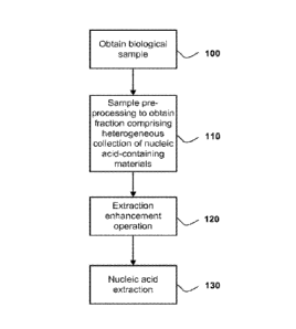
[026] FIGURE 1 is a flow chart depicting a first aspect of the present
invention directed
to a new method of nucleic acid extraction from a biological sample.
[027] FIGURE 2 is a flow chart depicting a second aspect of the present
invention
directed to a new method of nucleic acid extraction from a biological sample.
[028] FIGURE 3 is a flow chart depicting a third aspect of the present
invention directed
to a new method of nucleic acid extraction from a biological sample.
DETAILED DESCRIPTION
Nucleic Acid-containing Materials and Heterogeneous Collections Thereof
[029] Nucleic acid-containing biological materials are often used as
starting materials for
nucleic acid extraction and analysis. Cells are an example of a nucleic acid-
containing
biological material. Examples of cells containing nucleic acids of special
interest include, but
are not limited to, circulating tumor cells and other cells that have
undergone or are undergoing
disease-related transformation, or other cells that contain genomic evidence
of the physical
status or health of an organism. In addition, nucleic acids can be found in
smaller materials
ranging in size from about 10 nm in diameter to about 10000 nm in diameter.
For example,
"exosomes" have diameters of approximately 30 to 200 nm, with shedding
microvesicles and
apoptotic bodies often described as larger (Orozco and Lewis, 2010). Exosomes,
shedding
microvesicles, microparticles, nanovesicles, apoptotic bodies, nanoparticles
and membrane
vesicles co-isolate using various techniques and will, therefore, collectively
be referred to
throughout this specification as "microvesicles" unless otherwise expressly
denoted. Other
nucleic acid-containing materials, such as RNA-protein complexes, may co-
isolate with cells
and microvesicles using the various methods and techniques described herein.
Accordingly, the
generic term "nucleic acid -containing materials" will be used herein to refer
to cells,
microvesicles, RNA-protein complexes, and other nucleic acid containing
particles naturally
found in biological samples.
7
CA 02835641 2013-11-08
WO 2012/155014 PCMJS2012/037443
[030] A "heterogeneous collection of nucleic acid -containing materials,"
as used herein,
is a mixture of any of the foregoing species of nucleic acid-containing
materials, e.g., cells, any
species of microvesicle, RNA-protein complexes, and any other species of
nucleic acid-
containing particles. Preferably, the mixture will comprise one or more cells
in addition to any
or all of the other species of nucleic acid-containing materials.
[031] Nucleic acid-containing materials may originate from particular
cells, tissues or
organs of the body, or bodily fluids. In particular, nucleic acid-containing
materials may be
isolated from urine, plasma, or serum. In some embodiments, nucleic acid-
containing materials
may originate from a tumor, hyperplastic growth, nodule, neoplasm, cyst, or
mass. Nucleic
acid-containing materials often carry surface molecules such as antigens,
biomarkers, or
receptors from their donor cells. These surface molecules may be used to
detect, identify,
isolate, sort, and/or enrich nucleic acid-containing materials from a specific
donor cell type (Al-
Nedawi et al., 2008; Taylor and Gercel-Taylor, 2008). In this way, nucleic
acid-containing
materials originating from distinct cell populations can be analyzed for their
nucleic acid
content. For example, tumor (malignant and non-malignant) nucleic acid-
containing materials
carry tumor-associated surface antigen and may be detected, isolated, or
enriched via these
specific tumor-associated surface antigens.
Nucleic Acid Extraction Methods
[032] In a first embodiment, the invention is a method of extracting
nucleic acid from a
biological sample, comprising the steps of: obtaining a biological sample;
performing a sample
pre-processing step on the biological sample to obtain a fraction comprising a
heterogeneous
collection of nucleic acid-containing materials (preferably said heterogeneous
collection
comprises cells in addition to other nucleic acid-containing materials);
performing an extraction
enhancement operation; and extracting nucleic acid from the resulting
materials. There is no
specified order to the performance of the sample pre-processing step and the
extraction
enhancement operation, and indeed, the two may be performed simultaneously.
Preferably, this
method will result in a nucleic acid extraction that meets one or more of the
quality standards
described below in terms of the quantitative ratio of 18S rRNA to 28S rRNA, or
nucleic acid
yield.
CA 02835641 2013-11-08
WO 2012/155014 PCMJS2012/037443
[033] One variation of this first embodiment is shown in FIG. 1, wherein
the method
comprises the steps of obtaining a biological sample (100), pre-processing the
sample to obtain
a fraction comprising a heterogeneous collection of nucleic acid-containing
materials (110),
performing an extraction enhancement operation on the fraction (120), and
extracting nucleic
acid from the fraction (130).
[034] In variations of this first embodiment, the extraction enhancement
operation is
performed prior to the sample pre-processing, or the pre-processing and
extraction
enhancement operations are performed simultaneously.
[035] In further variations, there may be an additional step of removing
nucleic acids that
are not located inside the cells or microvesicles that may be part of the
heterogeneous
collection of nucleic acid-containing materials. Methods of removing nucleic
acids are well
known in the art. For example, an enzyme digestion step may be performed at
any point in the
process, e.g., prior to sample pre-processing, prior to performance of the
enhancement
extraction operation, or prior to nucleic acid extraction. Such enzymes may be
a type of
ribonuclease that catalyzes the enzymatic digestion of ribonucleic acids or a
type of
deoxyribonuclease that catalyzes the enzymatic digestion of deoxyribonucleic
acids.
[036] The biological sample can be any sample from an organism, for
example, a
mammal, and in particular, a human. Preferably, the biological sample is a
bodily fluid such as
urine, blood, serum or plasma, and may also include sputum, spinal fluid,
pleural fluid, nipple
aspirates, lymph fluid, fluid of the respiratory, intestinal, and
genitourinary tracts, tear fluid,
saliva, breast milk, fluid from the lymphatic system, semen, cerebrospinal
fluid, intraorgan
system fluid, ascitic fluid, tumor cyst fluid, amniotic fluid and combinations
thereof.
[037] A biological sample may sometimes come from a subject. The term
"subject" is
intended to include all animals shown to or expected to have nucleic acid-
containing materials.
In particular embodiments, the subject is a mammal, a human or nonhuman
primate, a dog, a
cat, a horse, a cow, other farm animals, or a rodent (e.g. mouse, rat, guinea
pig, etc.). The terms
"subject," "individual" and "patient" are used interchangeably herein and have
the same
meaning.
[038] The sample pre-processing step provides certain advantages not
present in nucleic
acid extraction methods of the prior art that do not employ a pre-processing
step to obtain from
the sample a fraction comprising a heterogeneous collection of nucleic acid-
containing
9
materials. For example, the methods of the present invention, employing as
they all do, a pre-
processing step, (1) tend to produce significantly higher yields of extracted
nucleic acid with
higher integrity; (2) provide advantages associated with scalability, e.g.,
when used in support
of an assay to detect nucleic acids expressed in a subject at low levels, the
sensitivity of the
assay can be increased by isolating, in the pre-processing step, more nucleic
acid-containing
materials from a larger volume of sample fluid; (3) purer nucleic acids in
that protein and
lipids, debris from dead cells, and other potential contaminants and PCR
inhibitors can be
excluded from the nucleic acid-containing materials isolated in the
preprocessing step; and (4)
more choices in nucleic acid extraction tools and techniques as the fraction
comprising nucleic
acid-containing materials that results from the pre-processing step is
typically of much smaller
volume than the starting sample volume, making it possible to extract nucleic
acids from the
fraction using small volume tools and techniques such as small volume column
filters.
[039] The sample pre-processing step may be any of several known techniques
for
separating nucleic acid-containing materials from a biological sample. For
example, a method
of isolating circulating tumor cells is described in a paper by Stott et al.
(Stott et al., 2010), a
method of differential centrifugation is described in a paper by Raposo et al.
(Raposo ct al.,
1996), a paper by Skog et. al.(Skog et al., 2008) and a paper by Nilsson et
al.(Nilsson ct al.,
2009). Methods of anion exchange and/or gel permeation chromatography are
described in US
Patent Nos. 6,899,863 and 6,812,023. Methods of sucrose density gradients or
organelle
electrophoresis are described in U.S. Patent No. 7,198,923. A method of
magnetic activated
cell sorting (MACS) is described in a paper by Taylor and Gcrccl-Taylor
(Taylor and Gucci-
Taylor, 2008). Methods of filtration concentration are described in a paper by
Cheruvanky ct al.
(Cheruvanky et al., 2007) and in PCT Publication No. W02011/009104 (Russo et
al.). Further,
microvesicles can be identified and isolated from bodily fluid of a subject by
a newly
developed microchip technology that uses a unique microfluidic platform to
efficiently and
selectively separate tumor-derived microvcsicics (Chen ct al., 2010).
[040] The purpose of the extraction enhancement step is to remove or
mitigate adverse
factors that prevent high quality nucleic acid extraction from a biological
sample. In some
biological samples, factors such as excessive circulating DNA may affect the
quality of nucleic
acid extraction from such samples and contaminate DNA extracted from within
nucleic acid-
CA 2835641 2018-07-03
CA 02835641 2013-11-08
WO 2012/155014 PCMJS2012/037443
containing materials. In other samples, factors such as excessive levels of
endogenous RNase
may affect the quality of nucleic acid extraction from such samples. Many
agents and methods
may be used to remove these adverse factors. These methods and agents are
referred to
collectively herein as an "extraction enhancement operation."
[041] In some instances, the extraction enhancement operation may involve
the addition
of nucleic acid extraction enhancement agents to the biological sample or
various derivatives of
the sample at any given stage of the process. For the purpose of removing
adverse factors such
as endogenous RNase, extraction enhancement agents may include, but are not
limited to, a
commercially available RNase inhibitor such as Superase-In (Ambion Inc.),
RNaseIN
(Promega Corp.), or other agents that function in a similar fashion; a
protease; a reducing
agent; a decoy substrate such as a synthetic RNA; a soluble receptor that can
bind RNase; a
small interfering RNA (siRNA); an RNA binding molecule, such as an anti-RNA
antibody, or a
chaperone protein; an RNase denaturing substance, such as a high osmolarity
solution, a
detergent, or a combination thereof. These enhancement agents may exert their
functions in
various ways, for example, but not limited to, through inhibiting RNase
activity (e.g., RNase
inhibitors), through a ubiquitous degradation of proteins (e.g., proteases),
or through a
chaperone protein (e.g., a RNA-binding protein) that binds and protects RNA.
In all instances,
such extraction enhancement agents remove or mitigate some or all of the
adverse factors in the
biological sample that would otherwise prevent or interfere with the high
quality extraction of
nucleic acids from the sample.
[042] In other instances, the extraction enhancement operation may involve
the
performance of one or more process steps. Such processes include extensive or
substantially
thorough washing of nucleic acid-containing components of the fraction or
sample; size
separation of RNases from the biological sample; denaturation of proteins in
the biological
sample by various techniques including, but not limited to, generating a
particular pH
condition, a temperature condition, (e.g., the maintenance of a decreasing or
lower
temperature), freeze/thaw cycles, and combinations thereof.
[043] Thus, the extraction enhancement operation is comprised of: (a) the
addition of one
or more enhancement agents to the biological sample; or (b) the performance of
one or more
enhancement steps prior to nucleic acid extraction; or (c) a combination of
enhancement agents
and enhancement steps. The enhancement agents may include: (i) RNase
inhibitor; (ii)
11
protease; (iii) reducing agent; (iv) decoy substrate, such as synthetic RNA;
(v) soluble receptor;
(vi) small interfering RNA; (vii) RNA binding molecule, such as anti-RNA
antibody,
chaperone protein, or an RNase inhibitory protein; and (ix) RNase denaturing
substance, such
as high osmolarity solution or detergent. The extraction enhancement steps may
include: (x)
washing; (xi) size-separating RNasc from the sample; (xii) effecting RNase
denaturation
through a physical change, such as by decreasing temperature, or executing a
freeze/thaw cycle.
[044] In variations in which the extraction enhancement operation involves
the addition
of an RNase inhibitor, the RNase inhibitor may be added to the biological
sample or to the
fraction comprising a heterogeneous collection of nucleic acid-containing
materials prior to
extracting nucleic acid. Preferably the RNasc inhibitor has a concentration of
greater than 0.027
AU (1X) for a sample equal to or more than 1 pl; alternatively, greater than
or equal to 0.135
AU (5X) for a sample equal to or more than 1 1; alternatively, greater than
or equal to 0.27
AU (10X) for a sample equal to or more than 1 1; alternatively, greater than
or equal to 0.675
AU (25X) for a sample equal to or more than 1 pl; and alternatively, greater
than or equal to
1.35 AU (50X) for a sample equal to or more than wherein the lx protease
concentration refers
to an enzymatic condition wherein 0.027 AU or more protease is used to treat
microvesicics
isolated from 1 1 or more bodily fluid; the 5X protease concentration refers
to an enzymatic
condition wherein 0.135 AU or more protease is used to treat microvesicles
isolated from 1 IA
or more bodily fluid; the 10X protease concentration refers to an enzymatic
condition wherein
0.27 AU or more protease is used to treat microvesicles isolated from 1 I or
more bodily fluid;
the 25X protease concentration refers to an enzymatic condition wherein 0.675
AU or more
protease is used to treat microvesicles isolated from 1 ul or more bodily
fluid; the 50X protease
concentration refers to an enzymatic condition wherein 1.35 AU or more
protease is used to
treat microvesicles isolated from or more bodily fluid. Preferably, the RNase
inhibitor is a
protease.
[045] The nucleic acid extraction step may be performed using procedures
that arc well-
known in the art. Persons of skill will select a particular extraction
procedure as appropriate for
the particular biological sample. Examples of extraction procedures arc
provided in patent
publications WO/2009/100029 and WO/2011/009104.
12
CA 2835641 2018-07-03
In some instances, with some techniques, it may also be possible to analyze
the nucleic
acid without first extracting it from the nucleic acid-containing materials.
[046] In a second embodiment, the invention is a method of extracting
nucleic acid from
a biological sample, comprising the steps of: obtaining a biological sample;
performing a
sample pre-processing step on the biological sample to obtain a fraction
comprising a
heterogeneous collection of nucleic acid-containing materials; performing an
affinity exclusion
operation on the heterogeneous collection of nucleic acid-containing
materials; and extracting
nucleic acid from the resulting materials. The biological sample, pre-
processing step, and
nucleic acid extraction step are all as described above in relation to the
first embodiment.
Preferably, this method will result in a nucleic acid extraction that meets
one or more of the
quality standards described below in terms of the quantitative ratio of 18S
rRNA to 28S rRNA,
or nucleic acid yield.
[047] One variation of this second embodiment is shown in FIG. 2, wherein
the method
comprises the steps of obtaining a biological sample (200), pre-processing the
sample to obtain
a fraction comprising a heterogeneous collection of nucleic acid-containing
materials (210),
performing an affinity exclusion operation (220), and extracting nucleic acids
from thc affinity
reduced fraction (230).
[048] The affinity exclusion operation is a novel means for reducing the
heterogeneity of
the fraction of nucleic acid-containing materials obtained from the
preprocessing step. Instead
of using affinity selection techniques to enrich for nucleic-acid containing
materials of interest,
in the affinity exclusion operation, affinity techniques arc used to remove
nucleic-acid
containing materials that are not of interest (e.g., nucleic acid containing
materials originating
from a cell type that is not of interest in a biomarker assay to be performed
on the extracted
nucleic acid). For example, using the methods and techniques described herein,
epithelial cells,
erythrocytes, leukocytes, neutrophils, lymphocytes, monocytes, basophils,
thrombocytes,
fibroblasts, and mescnchymal cells may be eliminated from the sample prior to
execution of the
nucleic acid extraction step. The depletion may be complete or partial. For
example, in some
instances a depiction of 50% of the undesirable materials would be sufficient
to achieve a high
quality nucleic acid extraction.
[049] Because nucleic acid-containing materials often carry surface
molecules such as
antigens from their donor cells, surface molecules may be used to identify and
deplete nucleic
13
CA 2835641 2018-07-03
CA 02835641 2013-11-08
WO 2012/155014
PCMJS2012/037443
acid-containing materials originating from a specific donor cell type. In one
example, the
surface molecule used in the affinity exclusion operation is a molecule
specific to cell type,
e.g., but not limited to, any of the cell-type markers listed in Table 1.
Alternatively, depending
upon assay design, the surface molecule used in the affinity exclusion
operation may be a
surface molecule listed in Table 2 if nucleic acid-containing materials
originating from a
specific tumor cell type are to be excluded in the assay.
Table 1: Examples of Cell-Type Specific Markers.
Cell types and Markers References
I. For positive selection:
A. Epithelial cell markers:
CD51 (Siegel et al., 2009)
Cytokeratin 8 (Punnoose et al., 2010)
Cytokeratin 18 (Punnoose et al., 2010)
Cytokeratin 19 (Punnoose et al., 2010)
E-cadherin (CD324, Cadherin-1) (Punnoose et al., 2010)
EpCAM (ESA; Epithelial cell adhesion (Shmelkov et al., 2008)
molecule; CD326)
Mucin 1 (EMA, Epithelial membrane antigen; (Matthews et al., 1988)
CA15-3; CD227)
ZO-1 (Siegel et al., 2009)
H. For negative selection from urine samples
A. Erythrocyte (RBC) markers:
AEI (Band 3) (Ding et al., 2004)
BGP1 (Lewis et al., 1988)
CD47 (Oldenborg et al., 2000)
Globin (Min-Oo et al., 2004)
Glycophorin A (GPA) (Shan et al., 1998; Telen and Chasis,
1990)
Rh polypeptides and Rh glycoprotein (Agre et al., 1990; Avent et al., 1996)
TER119 (Jiang et al., 2005; Kobayashi et al.,
2004)
Transferrin receptor (CD71) (Min-Oo et al., 2004; Tao et al., 2000)
B. Leukocyte (WBC) markers:
Beta2 Leukocyte Integrins (CD11/CD18) (Flaherty et al., 1997)
CD45RAi CD45RB; CD45R0 (Bembridge et al., 1993; Lai et al., 1991;
Masuoka et al., 1992)
CD166 (ALCAM, activated leukocyte cell (Lunter et al., 2005)
adhesion molecule)
HLA (human leukocyte antigen) (Guerini et al., 2006)
LAM-1 (leukocyte adhesion molecule-1) (Kansas et al., 1991)
L-selectin (Tu et al., 2002; Venturi et al., 2003)
14
CA 02835641 2013-11-08
WO 2012/155014
PCMJS2012/037443
Cell types and Markers References
LSP1 (leukocyte-specific protein-1) (Hannigan et al., 2001; Marafioti et
al., 2004)
Ly-9 (de la Fuente et al., 2001)
M6 (leukocyte activation antigen) (Kasinrerk et al., 1992)
III. For negative selection from blood
samples
A. Same as II A and IT B
B. Neutrophil markers:
31D8 (Gallin et al., 1986; Spiekermann et al.,
1996)
CD1 lb¨ also a monocyte marker (De Clerck et al., 1995)
CD15
CD18 (De Clerck et al., 1995)
CD45
CD64 (Matsui et al., 2006)
Gelatinase (Borregaard et al., 1995)
Mac-1
C. Lymphocyte markers:
T-cells: CD3, CD5, T cell receptor (TCR) (Berrington et al., 2005)
B-cells: MHC class II, CD19, CD21 (Berrington et al., 2005)
NK-cells: CD16, CD56, NKp46, NKp44 (Berrington et al., 2005)
D. Monocyte/Macrophase markers:
1251-WVH-1 (Fayle et al., 1985)
CD1 lb ¨ also a neutrophil marker (Fink et al., 2003)
CD14 (Jonas etal., 1990; Ruppert et al., 1991)
FcRI and FcRII (Clement et al., 1985)
HLA-DR
Ki-Mlp (Rudolph et al., 1997)
p-selectin
E. Basophil markers:
2D7 (Agis et al., 2006b; Kepley et al., 1995)
Basogranulin (BB1) (Agis et al., 2006a)
Bsp-1 (Valent et al., 1990)
CCR-3 (eotaxin receptor) (Ducrest et al., 2005)
CD203-c (E-NPP3) (Sainte-Laudy and Belon, 2006)
CDw-17 (lactosylceramide) (Yokohama et al., 2002)
CD88 (Yokohama et al., 2002)
F. Thrombocyte (platelet) marker:
CD36 (Thibert et al., 1995)
G. Dendritic cell marker:
CD83
CD11c
CD1a
CA 02835641 2013-11-08
WO 2012/155014 PCMJS2012/037443
Cell types and Markers References
H. Endothelial cells
CD31
IV. Other type markers
A. Fibroblast marker:
Fibroblast-specific protein 1 (FSP1) (Nishitani et al., 2005; Strutz et
al., 1995)
MAb AS02
Thy. 1
B. Mesenchymal marker:
CD29 (Siegel et al., 2009)
N-cadherin (Li et al., 2011)
Vimentin (Punnoose et al., 2010)
C. Glioblastoma cells marker:
EGFRvIII protein (Al-Nedawi et al., 2008)
PDGFR
IL13Ra2
CD133
chondroitin proteoglycan sulfate
3'-isoLM1
3'6'-isoLD1
GPNMB
MRP3
podoplanin
D. HERV particle marker
HERV env
[050] In variations of this second embodiment, the method may additionally
comprise an
extraction enhancement operation, as described above in relation to the first
embodiment. The
extraction enhancement operation may be performed at any time in the process
prior to the final
nucleic acid extraction step.
[051] In further variations, there may be an additional step of removing
nucleic acids that
are not located inside the cells or microvesicles that may be part of the
heterogeneous
collection of nucleic acid-containing materials. Methods of removing nucleic
acids are well
known in the art. For example, an enzyme digestion step may be performed at
any point in the
process. Such enzymes may be a type of ribonuclease that catalyzes the
enzymatic digestion of
ribonucleic acids or a type of deoxyribonuclease that catalyzes the enzymatic
digestion of
deoxyribonucleic acids.
16
CA 02835641 2013-11-08
WO 2012/155014 PCMJS2012/037443
[052] In a third embodiment, the invention is a method of extracting
nucleic acid from a
biological sample, comprising the steps of: obtaining a biological sample;
performing a sample
pre-processing step on the biological sample to obtain a fraction comprising a
heterogeneous
collection of nucleic acid-containing materials; performing an extraction
enhancement
operation; performing an affinity exclusion operation; and extracting nucleic
acid from the
resulting materials. The biological sample, pre-processing step, extraction
enhancement
operation, affinity exclusion operation, and nucleic acid extraction step are
all as described
above in relation to the first and second embodiments.
[053] In this embodiment, the sample pre-processing step must occur before
the affinity
exclusion operation, but the extraction enhancement operation may occur at any
time prior to
the nucleic acid extraction step.
[054] Preferably, this embodiment too will result in a nucleic acid
extraction that meets
one or more of the quality standards described below in terms of the
quantitative ratio of 18S
rRNA to 28S rRNA, or nucleic acid yield.
[055] One variation of the method described in this embodiment is shown in
FIG. 3,
wherein the method comprises the steps of obtaining a biological sample (300),
pre-processing
the sample to obtain a fraction comprising a heterogeneous collection of
nucleic acid-
containing materials (310), performing an affinity exclusion operation (320),
performing an
extraction enhancement operation (330), and extracting nucleic acids.
[056] As with the first and second embodiments, this third embodiment may
further
comprise an additional step of removing nucleic acids that are not located
inside the cells or
microvesicles that may be part of the heterogeneous collection of nucleic acid-
containing
materials. Methods of removing nucleic acids are well known in the art. For
example, an
enzyme digestion step may be performed at any point in the process, e.g.,
prior to sample
preprocessing, prior to performance of the enhancement extraction operation,
or prior to nucleic
acid extraction. Such enzymes may be a type of ribonuclease that catalyzes the
enzymatic
digestion of ribonucleic acids or a type of deoxyribonuclease that catalyzes
the enzymatic
digestion of deoxyribonucleic acids.
17
CA 02835641 2013-11-08
WO 2012/155014 PCMJS2012/037443
Affinity enrichment.
[057] All of the foregoing embodiments and variations of the nucleic acid
extraction
methods described above may further comprise an affinity enrichment operation,
wherein
affinity selection methods are used to enrich for nucleic acid-containing
materials of a certain
type or originating from a particular cell, tissue or organ of the body, e.g.,
lung, pancreas,
stomach, intestine, bladder, kidney, ovary, testis, skin, colorectal, breast,
prostate, brain,
esophagus, liver, placenta, or fetus cells.
[058] Because the nucleic acid-containing materials often carry surface
molecules such as
antigens from their donor cells, surface molecules may be used to identify,
isolate and/or enrich
for nucleic acid-containing materials from a specific donor cell type (Al-
Nedawi et al., 2008;
Taylor and Gercel-Taylor, 2008). In this way, nucleic acid-containing
materials originating
from distinct cell populations can be analyzed for their nucleic acid content.
For example,
tumor (malignant and non-malignant) nucleic acid-containing materials carry
tumor-associated
surface antigens and may be detected, isolated, or enriched via these specific
tumor-associated
surface antigens.
[059] In one example, the surface antigen is epithelial-cell-adhesion-
molecule (EpCAM),
which is specific to nucleic acid-containing materials from carcinomas of
lung, colorectal,
breast, prostate, head and neck, and hepatic origin, but not of hematological
cell origin (Balzar
et al., 1999; Went et al., 2004).
[060] In another example, the surface antigen is CD24, which is a
glycoprotein specific to
urine nucleic acid-containing materials (Keller et al., 2007).
[061] In yet another example, the surface antigen is selected from a group
of molecules
such as CD70, carcinoembryonic antigen (CEA), EGFR, EGFRvIII and other
variants, Fas
ligand, TRAIL, transferrin receptor, p38.5, p97 and HSP72. Additionally, tumor
specific
nucleic acid-containing materials may be characterized by the lack of surface
markers, such as
CD80 and CD86.
[062] In further examples, the surface antigens are any one of the tumor
markers, listed in
Table 2. The surface antigens in Table 2 may be used to perform an affinity
enrichment
operation so that nucleic acid-containing materials from a specific tumor cell
type are enriched.
Alternatively, depending upon the assay design, the surface antigen in the
affinity enrichment
operation may be any of the surface markers listed in the foregoing Table 1.
18
CA 02835641 2013-11-08
WO 2012/155014 PCMJS2012/037443
Table 2 Examples of Tumor Biomarkers
BIOMARKER NAME(S) COMBINATION CANCER TYPE REFERENCE
ABCB1 MDR1; P- Acute myeloid (Young, 2007)
glycoprotein 1; leukemia
ATP-binding (AML), Pancreas
cassette sub- Ovary (Fong and
family B member Kakar, 2010)
1
ABCB5 ATP-binding Melanoma (Schatton et
cassette sub- al., 2008)
family B member
ABCG2 CDw338; BCRP; Breast (Kim et al.,
ATP-binding Ovary 2002) (Fong
cassette subfamily and Kakar,
G member 2 2010)
AFP Alpha-fctoprotcin Hepatocellular (Baig ct al.,
2009)
ALDH1 Aldehyde ALDH1+/CD44+/ Breast (Ginestier et
dchydrogenasc 1 CD24-ilin- al., 2007)
ALDH1 Aldehyde Hematopoietic (Matsui et al.,
dehydrogenase 1 Lung 2004) (Jiang et
al., 2009)
APOE Apolipoprotein E, Ovary (Chen et al.,
apo E 2005)
BIRC5 Survivin; Lung (Falleni et al.,
baculoviral 2003)
inhibitor of
apoptosis repeat-
containing 5
CD15 leuMl; 3-fucosyl- Breast, (Ball, 1995)
N-acetyl- colorectal,
lactosaminc leukemia, lung
CD20 B-lymphocyte B-cell (Coiffier,
antigen 20 lymphoma, 2007)
leukemia
CD24 HSA; heat stable CD24+/CD44+ / Pancreas (Li
et al., 2007)
antigen CD24 EpCAM+
CD24 HSA; heat stable Colon, (Lim and Oh,
antigen CD24 gallbladder, 2005; Sagiv et
ovary, pancreas, al., 2006)
stomach
CD34 CD34 molecule; CD34+/CD10- Leukemia (Cox et al.,
19
CA 02835641 2013-11-08
WO 2012/155014 PCMJS2012/037443
Hematopoietic CD34+/CD38- AML 2004) (Kojima
progenitor cell and Kitamura,
antigen CD34 1999)
CD44 CD44 molecule CD44+/CD24- Breast (Al-Hajj et al.,
(Indian blood /low Breast 2003)
group) CD44+/CD24- Gliomas (Al-Hajj et al.,
/low/lin- 2003)
CD44+/CD24- AML (Galli et al.,
Prostate 2004; Hemmati
CD44+/CD24- Breast et al., 2003;
CD44+/CD24- Ignatova et at.,
CD44+/CD24 Colon 2002; Lee et
low! EpCAM+ Ovary al., 2006;
CD44+/EpCA M+ Bladder Singh et al.,
CD44+/MYD8 8+ Bladder 2003; Singh et
CD44+/CD117 +1 al., 2004;
CD133+ Uchida et al.,
CD44+/K5+/K20- 2000; Yuan et
CD44+/CD44v 6+ al., 2004)
/EMA- (Bonnet and
Dick, 1997;
Ishikawa et al.,
2007; Lapidot
al., 1994)
(Hurt et al.,
2008)
(Fillmore and
Kuperwasser,
2008)
(Boman and
Huang, 2008)
(Alvero et al.,
2009)
(Fong and
Kakar, 2010)
(Chan et al.,
2009) (Yang
and Chang,
2008)
CD44 CD44 molecule AML Head and (Jin et al.,
(Indian blood neck 2006) (Prince
group) et al., 2007)
CD47 MER6; TAP; Bladder (Chan et al.,
immuno globulin- 2009)
like
transmembrane
CA 02835641 2013-11-08
WO 2012/155014 PCMJS2012/037443
integrin-
associated protein
CD90 Thy-1, thymocyte CD90+/CD44+ Liver (Yang et al.,
differentiation 2008)
antigen 1
CD96 CD96; Tactile; T- Leukemia (Hosen et al.,
cell activation 2007)
increased late
expression
CD133 PROM1, CD133+/ABCG2 Melanoma (Monzani et
prominin-1 al., 2007)
Colon (Dallas et at.,
CD133+/CD44 + 2009)
CD133 PROM1, Brain (Bao etal.,
pro minin-1 Colon 2006a;
Hepatocellular Hemmati et al.,
Lung 2003; Liu et
Ovary al., 2006;
Pancreas Singh et al.,
Prostate 2003; Singh et
Skin al., 2004;
Taylor et al.,
2005;
Zeppernick et
al., 2008)
(O'Brien et at.,
2007; Ricci-
Vitiani et at.,
2007; Todaro
et al., 2007)
(Smith et al.,
2008)
(Eramo et al.,
2008)
(Fernandina et
al., 2008)
(Hermann et
al., 2007; Li et
al., 2007)
(Collins et at.,
2005)
(Monzani et
al., 2007)
CD142 Tissue factor; Breast, (Zwieker et al.,
platelet tissue colorectal, lung, 2009)
factor; factor III; pancreas
21
CA 02835641 2013-11-08
WO 2012/155014 PCMJS2012/037443
thrombokinase
CD147 EMMPRIN; Prostate (Zhong et al.,
extracellular 2011)
matrix
metalloproteinase
inducer; basigin
CD326 CD326; Flotillin Breast, colon, (Naundorf et
GI, ovary al., 2002)
Prostate (Oberneder et
al., 2006)
CEA Carcinoembryoni Colon (Thomas et al.,
c antigen 2009)
CLDN3 Claudin 3 Ovary (Hough et al.,
2001; Rangel
et al., 2003)
CLDN4 Claudin 4 Ovary (Hough et al.,
2001; Rangel
et al., 2003)
CLDN7 Claudin 7 Ovary (Hough et al.,
2001)
CTSB Cathepsin B Glioma (Strojnik et al.,
2007)
CXCL1 GRO-alpha; Bladder (Kawanishi et
Chemokine (C-X- al., 2008)
C motif) ligand 1
CXCR4 Chemokine Colon (Ottaiano et al.,
receptor type 4 Gliomas 2005)
Melanoma (Dirks, 2001;
Prostate Liu et at.,
2006;
Salmaggi et al.,
2006)
(Alsayed et
al., 2007)
(Sun et al.,
2005)
EpCAM ESA; Epithelial EpCAM+/CD45- Breast, (Allard et al.,
cell adhesion colorectal, 2004)
molecule; CD326 prostate
EpCAM ESA; Epithelial Colon, prostate (Ammons et
cell adhesion al., 2003; Godl
molecule; CD326 et al., 2007;
Oberneder et
al., 2006)
EGFR1 erbB-1; HER1; Anal (Walker et al.,
22
CA 02835641 2013-11-08
WO 2012/155014 PCMJS2012/037443
Epidermal growth Breast 2009)
factor receptor 1 Glioblastoma (Neve et al.,
Lung 2006)
(Heimberger et
al., 2005)
(Jackman ct
al., 2009;
Punnoose et
al., 2010)
EGFRvIll Mutant EGFR GBM (Pelloski et al.,
2007)
FOLH I Folate hydrolase Prostate (Chang et al.,
1; PSM; PSMA, 1999; Ross ct
Prostate specific al., 2003)
membrane antigen
FOLR1 Folate receptor Ovary (Kalli et al.,
alpha 2008)
GDIa ganglioside Ovary (Prinetti et al.,
2010)
GFAP Glial fibrillary Glioblastoma (Hill et al.,
acidic protein 2003)
GYPA Glycophorin A; Leukemia (Andersson ct
CD235a al., 1979)
HER2 erbB-2; neu; Breast (Korkaya et al.,
Human epidermal Uterus 2008)
growth factor (Santin et al.,
receptor 2 2008)
HLA-G Human leukocyte Ovary (Shcu and Shih
antigen-G le, 2007)
HPN Hepsin; Prostate (Dhanasekaran
TMPRS S I et al., 2001)
KLK2 Kallikrein 2 Prostate (Magklara et
al., 1999;
Partin et al.,
1999;
Rittenhouse et
al., 1998)
KLK3 PSA; Kallikrcin Prostate (Rittenhouse et
3; prostate al., 1998)
specific antigen
KLK5 Kallikrcin 5 Ovary (Youscf et al.,
2003a; Yousef
et al., 2003b)
KLK6 Kallikrein 6 Ovary (Yousef et al.,
2003b)
23
CA 02835641 2013-11-08
WO 2012/155014 PCMJS2012/037443
KLK7 Kallikrein 7 Ovary (Yousef et al.,
2003b)
KLK8 Kallikrein 8 Ovary (Hoffman et
al., 2002;
Yousef et al.,
2003b)
KLK10 Kallikrein 10 Ovary (Luo etal.,
2001; Yousef
et al., 2003b)
KLK11 Kallikrein 11 Ovary (Yousef et al.,
2003b)
KLK14 Kallikrein 14 Breast (Borgono et
Ovary al., 2003)
(Borgono et
al., 2003;
Yousef et al.,
2003b)
Keratane sulfates Papillary thyroid (Magro et al.,
carcinoma 2003)
L 1 CAM CD171; Li cell Gliomas (Bao et al.,
adhesion 2008)
molecule
LMP1 EBV latent Lymphoblastom (Flanagan et
membrane protein a al., 2003)
1
MET c-Met; HGE1{; Breast (Neve et al.,
hepatocyte growth 2006)
factor receptor
MSLN Mesothelin Mesothelioma (Chang and
Ovary Pastan, 1996)
Pancreas (Chang and
Pastan, 1996;
Lu et al., 2004)
(Agarwal et al.,
2008)
MUC 1 Mucin 1; CD227 Breast (McGuckin et
Colon al., 1995;
Taylor-
Papadimitriou
et al., 1999)
(Niv, 2008)
MUC4 Mucin 4 Ovary (Shih Ie and
Davidson,
2009)
MUC16 Mucin 16; CA Ovary (Yin ct al.,
24
CA 02835641 2013-11-08
WO 2012/155014 PCMJS2012/037443
125 ovarian 2002; Yin and
cancer antigen Lloyd, 2001)
OPN BSP-1; BNSP; Ovary (Rosen et al.,
Osteopontin; bone 2005; Visintin
sialoprotein I et al., 2008)
PCA-3 DD3; Prostate Prostate (Laxman et al.,
cancer antigen 3 2008)
PNCAM Polysialic acid or Prolactinoma (Gurlek et al.,
polysialylated Neuroendocrine 2007)
NCAM (a Small-cell lung (Figarella-
posttranslational carcinoma Branger et al.,
modification of 1990; Jin et al
NCAM, neural 1991)
cell adhesion (Komminoth et
molecule) al., 1991)
PTK7 Protein tyrosine T-cell acute (Shangguan et
kinase 7 lymphoblastic al., 2008)
leukemia
TMPRSS2:ER Transmembrane Prostate (Hessels et al.,
protease, serine 2 2007; Laxman
: Ets related gene et al., 2008)
VEGF Vascular Gliomas (Bao et al
endothelial 2006b)
growth factor
[063] One of skill in the art will appreciate that the surface markers
described in Tables 1
and 2 may be used interchangeably for an affinity exclusion operation or an
affinity enrichment
operation depending on the objectives of a given assay and nucleic acid
extraction method
practiced according to the teachings of this disclosure. For example, on the
one hand, the
surface markers for fibroblasts may be used to exclude fibroblast-derived
nucleic acid-
containing materials when a procedure for evaluating glioblastoma biomarkers
is performed.
On the other hand, the surface markers for fibroblasts may be used to enrich
fibroblast-derived
nucleic acid-containing materials when a procedure for evaluating
fibroblastoma is performed.
[064] An affinity procedure for depletion or enrichment of nucleic acid-
containing
materials from a specific cell type may be accomplished, for example, by using
antibodies,
aptamers, aptamer analogs or molecularly imprinted polymers specific for a
desired surface
antigen (hereinafter "affinity agent(s)"). In one embodiment, the surface
antigen is specific for
a cancer type. In another embodiment, the surface antigen is specific for a
cell type which is not
necessarily cancerous.
[065] One example of a method of nucleic acid-containing material
separation based on
cell surface antigen is provided in U.S. Patent No. 7,198,923. There CD81
antibody was used
to enrich CD81 antigen-containing exosomes to prepare HCV RNA from a blood
sample.
[066] Another example is described in, e.g., U.S. Patent Nos. 5,840,867 and
5,582,981,
WO/2003/050290 and a publication by Johnson et al. (Johnson et al., 2008).
There, aptamers
and their analogs that specifically bind surface molecules were used as a
separation tool for
enriching cell type-specific nucleic acid-containing materials. In addition,
molecularly
imprinted polymers may also specifically recognize surface molecules as
described in, e.g., US
Patent Nos. 6,525,154, 7,332,553 and 7,384,589 and a publication by Bossi et
al. (Bossi et al.,
2007) and may also be a tool for retrieving and isolating cell type specific
nucleic acid
containing materials.
Quality standards for nucleic acid extractions.
[067] The nucleic acid extractions obtained by the novel methods described
herein are
characterized by high yield and high integrity, making the extracted nucleic
acids useful for
various applications in which high quality nucleic acid extractions are
required or preferred.
[068] As mentioned above, the performance of any of the various nucleic
acid extraction
methods according to the present invention preferably results in a nucleic
acid extraction that
meets one or more of the quality standards described below in terms of the
quantitative ratio of
18S rRNA to 28S rRNA, or nucleic acid yield.
[069] Preferably, the nucleic acid extraction methods of this invention
will result in a
nucleic acid extraction in which one can detect significant quantities of
ribosomal RNA
(rRNA), specifically 18S and 28S rRNA, preferably in a ratio of approximately
1:1 to
approximately 1:2; and more preferably, in a ratio of approximately 1:2.
[070] Further, the nucleic acid extraction methods of the present invention
will preferably
result in improved yields of extracted nucleic acid. For example, using the
methods described
herein, one may obtain a nucleic acid yield of greater than or equal to 50
pg/ml from a 20 ml
low protein biological sample such as urine. Alternatively, one may obtain a
nucleic acid yield
of greater than or equal to 50 pg/ml from 1 ml of a high protein biological
sample, such as
scrum or plasma.
26
CA 2835641 2018-07-03
[071] Thus, the novel nucleic acid extractions obtained by the methods
described herein
preferably meet one or more of the following quality standards: (1) the
detection of 18S and
28S rRNA, preferably in a ratio of approximately 1:1 to approximately 1:2; and
more
preferably, approximately 1:2; and/or (2) a nucleic acid yield of greater than
or equal to 50
pg/ml from a 20 ml low protein biological sample or a 1 ml high protein
biological sample.
[072] Use of the nucleic acid extraction methods, and resulting nucleic
acid extractions,
in nucleic acid analysis for research and clinical applications.
[073] The nucleic acid extraction methods of the present invention may be
used to
produce novel and improved nucleic acid extractions for various applications,
including but not
limited to analysis of nucleic acid for research (e.g., research in support of
the discovery of new
biomarkcrs or biomarker associations) or clinical analysis of nucleic acid in
aid of patient
diagnostics, prognostics, theranostics, monitoring, predictive medicine,
personalized medicine,
integrated medicine, pharmacodiagnostics and diagnostic/prescription
partnering (companion
diagnostics).
[074] In one embodiment, the extracted nucleic acids, including DNA and/or
RNA, are
analyzed directly without an amplification step. Direct analysis may be
performed with
different methods including, but not limited to, nanostring technology.
NanoString technology
enables identification and quantification of individual target molecules in a
biological sample
by attaching a color coded fluorescent reporter to each target molecule. This
approach is similar
to the concept of measuring inventory by scanning barcodes. Reporters can be
made with
hundreds or even thousands of different codes allowing for highly multiplexed
analysis. The
technology is described in a publication by Geiss et al. (Geiss et al., 2008).
[075] Tn another embodiment, it may be beneficial or otherwise desirable to
amplify the
nucleic acid prior to analyzing it. Methods of nucleic acid amplification are
commonly used
and generally known in the art, many examples of which are described herein.
If desired, the
amplification can be performed such that it is quantitative. Quantitative
amplification will
allow quantitative determination of relative amounts of the various nucleic
acids, to generate a
profile as described below.
[076] In one embodiment, the extracted nucleic acid is RNA. The RNA is then
preferably
reverse-transcribed into complementary DNA (cDNA) before further
amplification. Such
27
CA 2835641 2018-07-03
reverse transcription may be performed alone or in combination with an
amplification step. One
example of a method combining reverse transcription and amplification steps is
reverse
transcription polymerase chain reaction (RT-PCR), which may be further
modified to be
quantitative, e.g., quantitative RT-PCR as described in US Patent No.
5,639,606.
[077] Nucleic acid amplification methods include, without limitation,
polymerase chain
reaction (PCR) (US Patent No. 5,219,727) and its variants such as in situ
polymcrasc chain
reaction (US Patent No. 5,538,871), quantitative polymerase chain reaction (US
Patent No.
5,219,727), nested polymerase chain reaction (US Patent No. 5,556,773), self-
sustained
sequence replication and its variants (Guatelli et al., 1990), transcriptional
amplification system
and its variants (Kwoh et al., 1989), Qb Replicase and its variants (Miele et
al., 1983), cold-
PCR (Li et al., 2008), or any other nucleic acid amplification methods,
followed by the
detection of the amplified molecules using techniques well known to those of
skill in the art.
Especially useful are those detection schemes designed for the detection of
nucleic acid
molecules if such molecules are present in very low numbers. The foregoing
references are
[078] The analysis of nucleic acids present in the nucleic acid-containing
materials may
be quantitative and/or qualitative. For quantitative analysis, the amounts
(expression levels),
either relative or absolute, of specific nucleic acids of interest within the
nucleic acid-
containing materials are measured with methods known in the art (described
below). For
qualitative analysis, the species of specific nucleic acids of interest within
the nucleic acid-
containing materials, whether wild type or variants, arc identified with
methods known in the
art.
Nucleic acid profiles.
[079] The invention further includes a novel, high-quality profile of
nucleic acids from a
biological sample. Such profiles are generated by performing any of the
various embodiments
and variations of the nucleic acid extraction methods disclosed herein, and
analyzing the
resulting nucleic acid.
[080] A profile, as the term is used herein, refers to a collection of
characteristics, which
can be determined through the quantitative or qualitative analysis of one or
more biological
components or materials (such as nucleic acid) contained in a sample (such as
a nucleic acid
28
CA 2835641 2018-07-03
CA 02835641 2013-11-08
WO 2012/155014
PCMJS2012/037443
extraction obtained by any of the methods disclosed herein). A reference
profile is a profile
obtained from an independent subject or from the same subject at a different
time point.
[081] The nucleic acids of the profile can be RNA. RNA can be coding RNA,
e.g.,
messenger RNA which may encode proteins. RNA can also be non-coding RNA
(ncRNA),
e.g., ribosomal RNA, transfer RNA, microRNA, and other non-coding transcripts
that may
originate from genomic DNA. These non-coding RNA transcripts may include
transcripts that
arc transcribed from satellite repeats and transposons, which may be DNA
transposons or
retrotransposons.
[082] The nucleic acids can also be DNA. DNA can be single-stranded DNA,
e.g.,
cDNA, that is reverse transcribed from RNA. The DNA can also be single-
stranded DNA that
is generated during DNA replication. Genomic DNA replicates in the nucleus
while the cell is
dividing. Some of the replicated DNA may come off its template, be exported
out of nucleus,
and packaged in microvesicles. It is also possible for the DNA to be double-
stranded DNA. In
addition, the DNA can be non-coding DNA (ncDNA).
[083] High quality nucleic acid profiles are highly desirable for many
uses, such as for
research (e.g., research in support of the discovery of new biomarkers or
biomarker
associations) or clinical uses such as patient diagnostics, prognostics,
theranostics, monitoring,
predictive medicine, personalized medicine, integrated medicine,
pharmacodiagnostics and
diagnostic/prescription partnering (companion diagnostics). It is desirable in
that such profiles
are consistent between samples. Such consistency cannot be achieved without
high quality
nucleic acid extractions.
[084] In one embodiment, the nucleic acid profile includes one or more
genetic
aberrations, which is used herein to refer to nucleic acid amounts as well as
nucleic acid
variants. Preferably, the nucleic acid is endogenous to the subject. Genetic
aberrations include,
without limitation, over-expression of one or more genomic elements,
underexpression of one
or more genomic elements, alternative production of splice variants of one or
more genomic
elements, copy number variants (CNV) of one or more genomic elements (e.g. DNA
double
minutes) (Hahn, 1993), nucleic acid modifications (e.g., methylation,
acetylation and
phosphorylations), single nucleotide polymorphisms (SNPs), chromosomal
rearrangements
(e.g., inversions, deletions and duplications), and mutations (insertions,
deletions, duplications,
missense, nonsense, synonymous or any other nucleotide changes) of one or more
genomic
29
CA 02835641 2013-11-08
WO 2012/155014 PCMJS2012/037443
elements, which mutations, in many cases, ultimately affect the activity and
function of the
genome, lead to alternative transcriptional splice variants and/or changes of
gene expression
level.
[085] The nucleic acids in the nucleic acid-containing materials can be any
type of
nucleic acid, including but not limited to the examples provided herein. In
the category of
RNA, the nucleic acids can be coding RNA, e.g., messenger RNA which may encode
proteins;
non-coding RNA (ncRNA), e.g., ribosomal RNA, transfer RNA, microRNA, and other
non-
coding transcripts that may originate from genomic DNA. Non-coding RNA
transcripts may
include transcripts that are transcribed from satellite repeats and
transposons, which may be
DNA transposons or retrotransposons. In the category of DNA, the nucleic acids
can include
single-stranded DNA (ssDNA), e.g., cDNA, which is reverse transcribed from RNA
and
ssDNA that is generated during DNA replication; double-stranded DNA (dsDNA);
DNA that
codes for proteins (coding DNA); and DNA that does not code for proteins,
i.e., non-coding
DNA (ncDNA).
[086] The determination of such genetic aberrations can be performed by a
variety of
techniques known to the skilled practitioner. For example, expression levels
of nucleic acids,
alternative splicing variants, chromosome rearrangement and gene copy numbers
can be
determined by microarray analysis (US Patent Nos. 6,913,879, 7,364,848,
7,378,245, 6,893,837
and 6,004,755) and quantitative PCR. Particularly, copy number changes may be
detected with
the Illumina Infinium II whole genome genotyping assay or Agilent Human Genome
CGH
Microarray (Steemers et al., 2006). Nucleic acid modifications can be assayed
by methods
described in, e.g., US Patent No. 7,186,512 and patent publication
WO/2003/023065.
Particularly, methylation profiles may be determined by, e.g., the Illumina
DNA Methylation
OMA003 Cancer Panel. SNPs and mutations can be detected by hybridization with
allele-
specific probes, enzymatic mutation detection, chemical cleavage of mismatched
heteroduplex
(Cotton et al., 1988), ribonuclease cleavage of mismatched bases (Myers et
al., 1985), mass
spectrometry (US Patent Nos. 6,994,960, 7,074,563, and 7,198,893), nucleic
acid sequencing,
single strand conformation polymorphism (SSCP) (Orita et al., 1989),
denaturing gradient gel
electrophoresis (DGGE)(Fischer and Lerman, 1979a; Fischer and Lerman, 1979b),
temperature
gradient gel electrophoresis (TGGE) (Fischer and Lerman, 1979a; Fischer and
Lerman, 1979b),
restriction fragment length polymorphisms (RFLP) (Kan and Dozy, 1978a; Kan and
Dozy,
1978b), oligonueleotide ligation assay (OLA), allele-specific PCR (ASPCR) (US
Patent No.
5,639,611), ligation chain reaction (LCR) and its variants (Abravaya et al.,
1995; Landegren ct
al., 1988; Nakazawa et al., 1994), flow-cytometric heteroduplex analysis
(WO/2006/113590)
and combinations or modifications thereof. Notably, gene expression levels may
be determined
by the serial analysis of gene expression (SAGE) technique (Velculescu et al.,
1995). In
general, the methods for analyzing genetic aberrations are reported in
numerous publications,
not limited to those cited herein, and are available to skilled practitioners.
The appropriate
method of analysis will depend upon the specific goals of the analysis, the
condition/history of
the patient, and the specific cancer(s), diseases or other medical conditions
to be detected,
monitored or treated.
Kits for obtainin2 nucleic acids
[087] The present invention is also directed to a kit for obtaining nucleic
acids from
biological samples. The kit may comprise an affinity agent; an extraction
enhancement agent;
and a lysis buffer. In some embodiments, the affinity agent is capable of
binding to one or more
markers listed in Table 1 or Table 2.
[088] In some instances, the kit may further comprise instructions for
using the kit.
Instructions for using the kit may be put in the package with the other kit
components or in a
different location accessible to a kit user (e.g., on a website or webpage
accessible to the kit
purchaser). The content of the instructions may include, but is not limited
to, instructions for
how to use the affinity agent, how to perform an affinity exclusion operation,
how to
reconstitute reagents, how to do the nucleic acid enhancement, how to use the
lysis buffer, and
how to carry out the whole procedure of obtaining nucleic acids by using the
kit.
[089] In some embodiments of the kit, the extraction enhancement agent may
be RNase
inhibitor; protease; reducing agent; decoy substrate; soluble receptor; small
interfering RNA;
RNA binding molecule; RNase denaturing substance; or any combination of any of
the
foregoing.
[090] In some embodiments, affinity agent is suitable for performing an
exclusion
operation, and instructions included in or with the kit comprise instructions
for using the
affinity agent in an affinity exclusion operation. Kits of this nature may
further comprise a
31
CA 2835641 2018-07-03
CA 02835641 2013-11-08
WO 2012/155014 PCMJS2012/037443
second affinity agent, and instructions for using the second affinity agent in
an affinity
enrichment operation.
[091] In additional embodiments, the kit may further comprise DNase, RNase,
or both,
and instructions for their use. These reagents may be used to eliminate DNA or
RNA that is of
no interest in the intended assay, e.g., DNA or RNA that clings to the outside
of the nucleic
acid-containing materials in the extraction. The amount of DNase or RNase may
depend on the
source of the biological sample. In some samples, the amount of DNA or RNA of
no interest is
relatively high, and therefore, more DNase or RNase will need to be added in
the extraction
process.
[092] It should be understood that this invention is not limited to the
particular
methodologies, protocols and reagents, described herein, which may vary. The
terminology
used herein is for the purpose of describing particular embodiments only, and
is not intended to
limit the scope of the present invention, which is defined solely by the
claims.
[093] While the present invention has been disclosed with reference to
certain
embodiments, numerous modifications, alterations, and changes to the described
embodiments
are possible without departing from the sphere and scope of the present
invention, as defined in
the appended claims. Accordingly, it is intended that the present invention
not be limited to the
described embodiments, but that it has the full scope defined by the language
of the following
claims, and equivalents thereof
EXAMPLES
Example 1: Nucleic Acid Extraction with Extraction Enhancement Operation
[094] One variation of the invention is shown in Figure 1, where the method
comprises
the steps of obtaining a biological sample (100), pre-processing the sample to
obtain a fraction
comprising a heterogeneous collection of nucleic acid-containing materials
(110), performing
an extraction enhancement operation on the fraction (120), and extracting
nucleic acid from the
fraction (130).
Example 2: Nucleic Acid Extraction with Affinity Exclusion Operation
[095] One variation of the invention is shown in FIG. 2, where the method
comprises the
steps of obtaining a biological sample (200), pre-processing the sample to
obtain a fraction
32
CA 02835641 2013-11-08
WO 2012/155014 PCMJS2012/037443
comprising a heterogeneous collection of nucleic acid-containing materials
(210), performing
an affinity exclusion operation (220), and extracting nucleic acids from the
affinity reduced
fraction (230).
Example 3: Nucleic Acid Extraction with Extraction Enhancement Operation and
Affinity Exclusion Operation
[096] One variation of the invention is shown in FIG. 3, where the method
comprises the
steps of obtaining a biological sample (300), pre-processing the sample to
obtain a fraction
comprising a heterogeneous collection of nucleic acid-containing materials
(310), performing
an affinity exclusion operation (320), performing an extraction enhancement
operation (330),
and extracting nucleic acids.
Example 4: Nucleic Acid Extraction and Analysis from a Heterogeneous
Collection of
Nucleic Acid-containing Materials
[097] Heterogeneous collections of nucleic acid-containing materials can be
isolated from
a biological sample from a subject that has or is suspected to have cancer. A
urine sample is
collected from the subject. In the pre-processing step, a fraction containing
nucleic acid-
containing materials is enriched by centrifugation or filtration from the
urine. The resulting
fraction contains a heterogeoneous collection of nucleic acid-containing
materials, which
includes a mixture of microvesicles and cells in addition to other nucleic
acid-containing
materials. This fraction is then incubated with extraction enhancement agents,
such as RNase
inhibitors, to prevent or mitigate those factors that may prevent high quality
nucleic acid
extraction. Then, the fraction is subjected to an affinity enrichment
operation to enrich for the
potential circulating tumor cells and microvesicles of particular interest. A
surface antigen
carried by both the circulating tumor cells and microvesicles is used to
select for and purify
these particular nucleic acid-containing materials from the remaining mixture.
Nucleic acids
from the purified circulating tumor cells and microvesicles are extracted and
analyzed for the
presence, absence, or levels of genetic aberrations that are associated with
the presence or
absence of malignant cancer; or stage or grade of the tumor from which the
cells and
microvesicles may have originated from.
33
CA 02835641 2013-11-08
WO 2012/155014 PCMJS2012/037443
REFERENCES:
Abravaya, K., J.J. Carrino, S. Muldoon, and H.H. Lee. 1995. Detection of point
mutations
with a modified ligase chain reaction (Gap-LCR). Nucleic Acids Res. 23:675-82.
Agarwal, B., O.J. Ludwig, B.T. Collins, and C. Cortese. 2008. Immunostaining
as an adjunct
to cytology for diagnosis of pancreatic adenocarcinoma. Clin Gastroenterol
Hepatol.
6:1425-31.
Agis, H., M.T. Krauth, A. Bohm, I. Mosberger, L. Mullauer, 1. Simonitsch-
Klupp, A.F.
Walls, H.P. Horny, and P. Valent. 2006a. Identification of basogranulin (BB1)
as a
novel immunohistochemical marker of basophils in normal bone marrow and
patients
with myeloproliferative disorders. Am J Clin Pathol. 125:273-81.
Agis, H., M.T. Krauth, I. Mosberger, L. Mullauer, I. Simonitsch-Klupp, L.B.
Schwartz, D.
Printz, A. Bohm, G. Fritsch, H.P. Horny, and P. Valent. 2006b. Enumeration and
immunohistochemical characterisation of bone marrow basophils in
myeloproliferative disorders using the basophil specific monoclonal antibody
2D7. J
Clin Pathol. 59:396-402.
Agre, P., B.L. Smith, and S. Hartel-Schenk. 1990. Biochemistry of the
erythrocyte Rh
polypeptides: a review. Yale J Biol Med. 63:461-7.
Al-Hajj, M., M.S. Wicha, A. Benito-Hernandez, S.J. Morrison, and M.F. Clarke.
2003.
Prospective identification of tumorigenic breast cancer cells. Proc Nail Acta
Sci US
A. 100:3983-8.
Al-Nedawi, K., B. Meehan, J. Micallef, V. Lhotak, L. May, A. Guha, and J. Rak.
2008.
Intercellular transfer of the oncogenic receptor EGFRvIII by microvesicles
derived
from tumour cells. Nat Cell Biol. 10:619-24.
Allard, W.J., J. Matera, M.C. Miller, M. Repollet, M.C. Connelly, C. Rao, A.G.
Tibbe, J.W.
Uhr, and L.W. Terstappen. 2004. Tumor cells circulate in the peripheral blood
of all
major carcinomas but not in healthy subjects or patients with nonmalignant
diseases.
Clin Cancer Res. 10:6897-904.
Alsayed, Y., H. Ngo, J. Runnels, X. Leleu, U.K. Singha, C.M. Pitsillides, J.A.
Spencer, T.
Kimlinger, J.M. Ghobrial, X. Jia, G. Lu, M. Timm, A. Kumar, D. Cote, I.
Veilleux,
K.E. Hcdin, G.D. Roodman, T.E. Witzig, A.L. Kung, T. Hidcshima, K.C. Anderson,
C.P. Lin, and I.M. Ghobrial. 2007. Mechanisms of regulation of CXCR4/SDF-1
(CXCL12)-dependent migration and homing in multiple myeloma. Blood. 109:2708-
17.
Alvero, A.B., R. Chen, H.H. Fu, M Montagna, P.E. Schwartz, T. Rutherford, D.A.
Silasi,
K.D. Steffensen, M. Waldstrom, I. Visintin, and G. Mor. 2009. Molecular
phenotyping of human ovarian cancer stem cells unravels the mechanisms for
repair
and chemoresistance. Cell Cycle. 8:158-66.
Ammons, W.S., R.J. Bauer, A.H. Horwitz, Z.J. Chen, E. Bautista, H.H. Ruan, M.
Abramova,
K.R. Scott, and R.L. Dedrick. 2003. In vitro and in vivo pharmacology and
pharmacokinetics of a human engineered monoclonal antibody to epithelial cell
adhesion molecule. Neoplasia. 5:146-54.
Andersson, L.C., C.G. Gahmberg, L. Teerenhovi, and P. Vuopio. 1979.
Glycophorin A as a
cell surface marker of early erythroid differentiation in acute leukemia. MU
Cancer.
24:717-20.
Avent, N.D., W. Liu, K.M. Warner, W.J. Mawby, J.W. Jones, K. Ridgwell, and
M.J. Tanner.
34
CA 02835641 2013-11-08
WO 2012/155014
PCMJS2012/037443
1996. Immunochemical analysis of the human erythrocyte Rh polypeptides. J Biol
Chem. 271:14233-9.
Baig, J.A., J.M. Alam, S.R. Mahmood, M. Baig, R. Shaheen, I. Sultana, and A.
Wahecd.
2009. Hepatocellular carcinoma (HCC) and diagnostic significance of A-
fetoprotein
(AFP). J Ayub Med Coll Abbottabad. 21:72-5.
Ball, E.D. 1995. Introduction: workshop summary of the CD15 monoclonal
antibody panel
from the Fifth International Workshop on Leukocyte Antigens. Eur fMorphol.
33:95 -
100.
Balzar, M., M.J. Winter, C.J. de Boer, and S.V. Litvinov. 1999. The biology of
the 17-1A
antigen (Ep-CAM). J Mol Med. 77:699-712.
Bao, S., Q. Wu, Z. Li, S. Sathornsumetee, H. Wang, R.E. McLendon, A.B.
Hjelmeland, and
J.N. Rich. 2008. Targeting cancer stem cells through Li CAM suppresses glioma
growth. Cancer Res. 68:6043-8.
Bao, S., Q. Wu, R.E. McLendon, Y. Hao, Q. Shi, A.B. Hjelmeland, M.W. Dcwhirst,
D.D.
Bigner, and J.N. Rich. 2006a. Glioma stem cells promote radioresistance by
preferential activation of the DNA damage response. Nature. 444:756-60.
Bao, S., Q. Wu, S. Sathornsumetee, Y. Hao, Z. Li, A.B. Hjelmeland, Q. Shi,
R.E. McLendon,
D.D. Bigner, and J.N. Rich. 2006b. Stem cell-like glioma cells promote tumor
angiogenesis through vascular endothelial growth factor. Cancer Res. 66:7843-
8.
Bembridge, G.P., K.R. Parsons, P. Sopp, N.D. MacHugh, and C.J. Howard. 1993.
Comparison of monoclonal antibodies with potential specificity for restricted
isoforms of the leukocyte common antigen (CD45R). Vet linmunol Immunopathol.
39:129-36.
Berrington, J.E., D. Barge, A.C. Fenton, A.J. Cant, and G.P. Spickett. 2005.
Lymphocyte
subsets in term and significantly preterm UK infants in the first year of life
analysed
by single platform flow cytometry. Clin Exp Immunol. 140:289-92.
Boman, B.M., and E. Huang. 2008. Human colon cancer stem cells: a new paradigm
in
gastrointestinal oncology. J Clin Oncol. 26:2828-38.
Bonnet, D., and J.E. Dick. 1997. Human acute myeloid leukemia is organized as
a hierarchy
that originates from a primitive hematopoietic cell. Nat Med. 3:730-7.
Borgono, C.A., L. Grass, A. Soosaipillai, G.M. Yousef, C.D. Petraki, D.H.
Howarth, S.
Fracchioli, D. Katsaros, and E.P. Diamandis. 2003. Human kallikrein 14: a new
potential biomarker for ovarian and breast cancer. Cancer Res. 63:9032-41.
Borregaard, N., M. Sehested, B.S. Nielsen, H. Sengelov, and L. Kjeldsen. 1995.
Biosynthesis
of granule proteins in normal human bone marrow cells. Gelatinase is a marker
of
terminal ncutrophildiffcrcntiation. Blood. 85:812-7.
Bossi, A., F. Bonini, A.P. Turner, and S.A. Piletsky. 2007. Molecularly
imprinted polymers
for the recognition of proteins: the state of the art. Biosens Bioelectron.
22:1131-7.
Chan, K.S., I. Espinosa, M. Chao, D. Wong, L. Ailles, M. Diehn, H. Gill, J.
Presti, Jr., H.Y.
Chang, M. van de Rijn, L. Shortliffe, and I.L. Weissman. 2009. Identification,
molecular characterization, clinical prognosis, and therapeutic targeting of
human
bladder tumor-initiating cells. Proc Natl Acad Sci U SA. 106:14016-21.
Chang, K., and I. Pastan. 1996. Molecular cloning of mesothelin, a
differentiation antigen
present on mesothelium, mesotheliomas, and ovarian cancers. Proc Nat! Acad Sci
US
A. 93:136-40.
Chang, S.S., V.E. Reuter, W.D. Heston, N.H. Bander, L.S. Grauer, and P.B.
Gaudin. 1999.
CA 02835641 2013-11-08
WO 2012/155014 PCMJS2012/037443
Five different anti-prostate-specific membrane antigen (PSMA) antibodies
confirm
PSMA expression in tumor-associated neovasculature. Cancer Res. 59:3192-8.
Chen, C., J. Skog, C.H. Hsu, R.T. Lessard, L. Balaj, T. Wurdingcr, B.S.
Carter, X.O.
Breakefield, M. Toner, and D. Irimia. 2010. Microfluidic isolation and
transcriptome
analysis of serum microvesicles. Lab chip. 10:505-11.
Chen, Y.C., G. Pohl, T.L. Wang, P.J. Morin, B. Risberg, G.B. Kristensen, A.
Yu, B.
Davidson, and M. Shih Ie. 2005. Apolipoprotein E is required for cell
proliferation
and survival in ovarian cancer. Cancer Res. 65:331-7.
Cheruvanky, A., H. Zhou, T. Pisitkun, J.B. Kopp, M.A. Knepper, P.S. Yuen, and
R.A. Star.
2007. Rapid isolation of urinary exosomal biomarkers using a nanomembrane
ultrafiltration concentrator. Am J Physiol Renal Physiol. 292:F1657-61.
Clement, L.T., A.B. Tilden, and N.E. Dunlap. 1985. Analysis of the monocyte Fe
receptors
and antibody-mediated cellular interactions required for the induction of T
cell
proliferation by anti-T3 antibodies. J Immunol. 135:165-71.
Coiffier, B. 2007. Rituximab therapy in malignant lymphoma. Oncogene. 26:3603-
13.
Collins, A.T., P.A. Berry, C. Hyde, M.J. Stower, and N.J. Maitland. 2005.
Prospective
identification of tumorigenic prostate cancer stem cells. Cancer Res. 65:10946-
51.
Cotton, R.G., N.R. Rodrigues, and R.D. Campbell. 1988. Reactivity of cytosine
and thymine
in single-base-pair mismatches with hydroxylamine and osmium tetroxide and its
application to the study of mutations. Proc Natl Acad Sci USA. 85:4397-401.
Cowell, J.K., and K.C. Lo. 2009. Application of oligonucleotides arrays for
coincident
comparative genomic hybridization, ploidy status and loss of heterozygosity
studies in
human cancers. Methods Mol Biol. 556:47-65.
Cox, C.V., R.S. Evely, A. Oakhill, D.H. Pamphilon, N.J. Goulden, and A. Blair.
2004.
Characterization of acute lymphoblastic leukemia progenitor cells. Blood.
104:2919-25.
Dallas, N.A., L. Xia, F. Fan, M.J. Gray, P. Gaur, G. van Buren, 2nd, S.
Samuel, M.P. Kim,
S.J. Lim, and L.M. Ellis. 2009. Chemoresistant colorectal cancer cells, the
cancer
stem cell phenotype, and increased sensitivity to insulin-like growth factor-I
receptor
inhibition. Cancer Res. 69:1951-7.
De Clerck, L.S., C.M. De Gendt, C.H. Bridts, N. Van Osselaer, and W.J.
Stevens. 1995.
Expression of neutrophil activation markers and neutrophil adhesion to
chondrocytes
in rheumatoid arthritis patients: relationship with disease activity. Res
Immunol.
146:81-7.
de la Fuente, M.A., V. Tovar, N. Villamor, N. Zapater, P. Pizcueta, E. Campo,
J. Bosch, and
P. Engel. 2001. Molecular characterization and expression of a novel human
leukocyte cell-surface marker homologous to mouse Ly-9. Blood. 97:3513-20.
Dhanasekaran, S.M., T.R. Barrette, D. Ghosh, R. Shah, S. Varambally, K.
Kurachi, K.J.
Pienta, M.A. Rubin, and A.M. Chinnaiyan. 2001. Delineation of prognostic
biomarkers in prostate cancer. Nature. 412:822-6.
Ding, Y., W. Jiang, Y. Su, H. Zhou, and Z. Zhang. 2004. Expression and
purification of
recombinant cytoplasmic domain of human erythrocyte band 3 with hex ahistidine
tag
or chitin-binding tag in Escherichia coli. Protein Expr Purif. 34:167-75.
Dirks, P.B. 2001. Glioma migration: clues from the biology of neural
progenitor cells and
embryonic CNS cell migration. J Neurooncol. 53:203-12.
Ducrest, S., F. Meier, C. Tschopp, R. Pavlovic, and C.A. Dahinden. 2005.
Flowcytometric
analysis of basophil counts in human blood and inaccuracy of hematology
analyzers.
36
CA 02835641 2013-11-08
WO 2012/155014
PCMJS2012/037443
Allergy. 60:1446-50.
Eramo, A., F. Lotti, G. Sette, E. Pilozzi, M. Biffoni, A. Di Virgilio, C.
Conticello, L. Ruco,
C. Peschlc, and R. Dc Maria. 2008. Identification and expansion of the
tumorigcnic
lung cancer stem cell population. Cell Death Differ. 15:504-14.
Falleni, M., C. Pellegrini, A. Marchetti, B. Oprandi, F. Buttitta, F. Barassi,
L. Santambrogio,
G. Coggi, and S. Bosari. 2003. Survivin gene expression in early-stage non-
small cell
lung cancer. .1 Pathol. 200:620-6.
Fayle, D.R., P.S. Sim, D.K. Irvine, and W.F. Doe. 1985. Isolation of plasma
membrane from
human blood monocytes. Subcellular fractionation and marker distribution. Eur
J
Biochem. 147:409-19.
Ferrandina, G., G. Bonanno, L. Pierelli, A. Perillo, A. Procoli, A. Mariotti,
M. Corallo, E.
Martinelli, S. Rutella, A. Paglia, G. Zannoni, S. Mancuso, and G. Scambia.
2008.
Expression of CD 133-1 and CD133-2 in ovarian cancer. Int J Gynecol Cancer.
18:506-14.
Figarclla-Brangcr, D.F., P.L. Durbcc, and G.N. Rougon. 1990. Differential
spectrum of
expression of neural cell adhesion molecule isoforms and Ll adhesion molecules
on
human neuroectodermal tumors. Cancer Res. 50:6364-70.
Fillmore, C.M., and C. Kuperwasser. 2008. Human breast cancer cell lines
contain stem-like
cells that self-renew, give rise to phenotypically diverse progeny and survive
chemotherapy. Breast Cancer Res. 10:R25.
Fink, R., M. Al-Obaidi, S. Grewal, M. Winter, and J. Pepper. 2003. Monocytc
activation
markers during cardiopulmonary bypass. Perfusion. 18:83-6.
Fischer, S.G., and L.S. Lerman. 1979a. Length-independent separation of DNA
restriction
fragments in two-dimensional gel electrophoresis. Cell. 16:191-200.
Fischer, S.G., and L.S. Lerman. 1979b. Two-dimensional electrophoretic
separation of
restriction enzyme fragments of DNA. Methods Enzytnol. 68:183-91.
Flaherty, S.F., D.T. Golenbock, F.H. Milham, and R.R. Ingalls. 1997. CD11/CD18
leukocyte
integrins: new signaling receptors for bacterial endotoxin. J Surg Res. 73:85-
9.
Flanagan, J., J. Middeldorp, and T. Sculley. 2003. Localization of the Epstein-
Barr virus
protein LMP 1 to exosomes. J Gen Virol. 84:1871-9.
Fong, M.Y., and S.S. Kakar. 2010. The role of cancer stem cells and the side
population in
epithelial ovarian cancer. Histol Histopathol. 25:113-20.
Galli, R., E. Binda, U. Orfanelli, B. Cipelletti, A. Gritti, S. De Vitis, R.
Fiocco, C. Foroni, F.
Dimeco, and A. Vescovi. 2004. Isolation and characterization of tumorigenic,
stemlike
neural precursors from human glioblastoma. Cancer Res. 64:7011-21.
Gallin, J.I., R.J. Jacobson, B.E. Scligmann, J.A. Metcalf, J.H. McKay, R.A.
Sachcr, and H.L.
Malech. 1986. A neutrophil membrane marker reveals two groups of chronic
myelogenous leukemia and its absence may be a marker of disease progression.
Blood. 68:343-6.
Geiss, G.K., R.E. Bumgarner, B. Birditt, T. Dahl, N. Dowidar, D.L. Dunaway,
H.P. Fell, S.
Ferree, R.D. George, T. Grogan, J.J. James, M. Maysuria, J.D. Mitton, P.
Oliveri, J.L.
Osborn, T. Peng, A.L. Ratcliffe, P.J. Webster, E.H. Davidson, and L. Hood.
2008.
Dircct multiplexed measurement of gene expression with color-coded probe
pairs. Nat
Biotechnol. 26:317-25.
Ginestier, C., M.H. Hur, E. Charafe-Jauffret, F. Monville, J. Dutcher, M.
Brown, J.
Jacquemier, P. Viens, C.C. Kleer, S. Liu, A. Schott, D. Hayes, D. Birnbaum,
M.S.
37
CA 02835641 2013-11-08
WO 2012/155014 PCMJS2012/037443
Wicha, and G. Dontu. 2007. ALDH1 is a marker of normal and malignant human
mammary stem cells and a predictor of poor clinical outcome. Cell Stein Cell.
1:555-
67.
God, S., R.J. Bauer, K. Desai, A. Bulgaru, T. Iqbal, B.K. Strachan, G. Kim, A.
Kaubisch,
G.F. Vanhove, G. Goldberg, and S. Mani. 2007. Pharmacokinetic and safety study
of
subcutaneously administered weekly ING-1, a human engineere monoclonal
antibody
targeting human EpCAM, in patients with advanced solid tumors. Ann
Oncol.18:1704-
7.
Guatelli, J.C., K.M. Whitfield, D.Y. Kwoh, K.J. Barringer, D.D. Richman, and
T.R.
Gingeras. 1990. Isothermal, in vitro amplification of nucleic acids by a
multienzyme
reaction modeled after retroviral replication. Proc Nall Acad Sci USA. 87:1874-
8.
Guerini, F.R., C. Agliardi, M. Zanzottera, S. Delbue, E. Pagani, C. Tinelli,
R. Boldorini, P.G.
Car, C. Veggiani, and P. Ferrante. 2006. Human leukocyte antigen distribution
analysis in North Italian brain Glioma patients: an association with HLA-
DRB1*14. J
Neurooncol. 77:213-7.
Gurlek, A., N. Karavitaki, 0. Ansorge, and J.A. Wass. 2007. What are the
markers of
aggressiveness in prolactinomas? Changes in cell biology, extracellular matrix
components, angiogenesis and genetics. Eur J Endocrinol. 156:143-53.
Hahn, P.J. 1993. Molecular biology of double-minute chromosomes. Bioessays .
15:477-84.
Hannigan, M., L. Zhan, Y. Ai, and C.K. Huang. 2001. Leukocyte-specific gene 1
protein
(LSP1) is involved in chemokine KC-activated cytoskeletal reorganization in
murinc
neutrophils in vitro. J Leukoc Biol. 69:497-504.
Heimberger, A.B., D. Suki, D. Yang, W. Shi, and K. Aldape. 2005. The natural
history of
EGFR and EGFRvIII in glioblastoma patients. J Trans" Med. 3:38.
Hemmati, H.D., I. Nakano, J.A. Lazareff, M. Masterman-Smith, D.H. Geschwind,
M.
Bronner-Fraser, and H.T. Kornblum. 2003. Cancerous stem cells can arise from
pediatric brain tumors. Proc Natl Acad Sci USA. 100:15178-83.
Hermann, P.C., S.L. Huber, T. Herrler, A. Aicher, J.W. Ellwart, M. Guba, C.J.
Bruns, and C.
Heeschen. 2007. Distinct populations of cancer stem cells determine tumor
growth
and metastatic activity in human pancreatic cancer. Cell Stem Cell. 1:313-23.
Hessels, D., F.P. Smit, G.W. Verhaegh, J.A. Witjes, E.B. Come!, and J.A.
Schalken. 2007.
Detection of TMPRSS2-ERG fusion transcripts and prostate cancer antigen 3 in
urinary
sediments may improve diagnosis of prostate cancer. Clin Cancer Res.13:5103-8.
Hill, C., S.B. Hunter, and D.J. Brat. 2003. Genetic markers in glioblastoma:
prognostic
significance and future therapeutic implications. Adv Anat Pathol. 10:212-7.
Hoffman, B.R., D. Katsaros, A. Scorilas, P. Diamandis, S. Fracchioli, 1.A.
Rigault de la
Longrais, T. Colgan, M. Puopolo, G. Giardina, M. Massobrio, and E.P.
Diamandis.
2002. Immunofluorometric quantitation and histochemical localisation of
kallikrein 6
protein in ovarian cancer tissue: a new independent unfavourable prognostic
biomarker. Br. J Cancer. 87:763-71.
Hosen, N., C.Y. Park, N. Tatsumi, Y. Oji, H. Sugiyama, M. Gramatzki, A.M.
Krensky, and
I.L. Weissman. 2007. CD96 is a leukemic stem cell-specific marker in human
acute
myeloid leukemia. Proc Natl Acad Sci USA. 104:11008-13.
Hough, C.D., K.R. Cho, A.B. Zonderman, D.R. Schwartz, and P.J. Morin. 2001.
Coordinately up-regulated genes in ovarian cancer. Cancer Res. 61:3869-76.
Hurt, E.M., B.T. Kawasaki, G.J. Klarmann, S.B. Thomas, and W.L. Farrar. 2008.
CD44+
38
CA 02835641 2013-11-08
WO 2012/155014
PCMJS2012/037443
CD24(-) prostate cells are early cancer progenitor/stem cells that provide a
model for
patients with poor prognosis. Br J Cancer. 98:756-65.
Ignatova, T.N., V.G. Kukckov, E.D. Laywell, O.N. Suslov, F.D. Vrionis, and
D.A. Stcindlcr.
2002. Human cortical glial tumors contain neural stem-like cells expressing
astroglial
and neuronal markers in vitro. Glia. 39:193-206.
Ishikawa, F., S. Yoshida, Y. Saito, A. Hijikata, H. Kitamura, S. Tanaka, R.
Nakamura, T.
Tanaka, H. Tomiyama, N. Saito, M. Fukata, T. Miyamoto, B. Lyons, K. Ohshima,
N.
Uchida, S. Taniguchi, 0. Ohara, K. Akashi, M. Harada, and L.D. Shultz. 2007.
Chemotherapy-resistant human AML stem cells home to and engraft within the
bonemarrow endosteal region. Nat Biotechnol. 25:1315-21.
Jackman, D.M., V.A. Miller, L.A. Cioffredi, B.Y. Yeap, P.A. Janne, G.J. Riely,
M.G. Ruiz,
G. Giaccone, L.V. Sequist, and B.E. Johnson. 2009. Impact of epidermal growth
factor receptor and KRAS mutations on clinical outcomes in previously
untreated
non-small cell lung cancer patients: results of an online tumor registry of
clinical
trials. Clin Cancer Res. 15:5267-73.
Jiang, F., Q. Qiu, A. Khanna, N.W. Todd, J. Deepak, L. Xing, H. Wang, Z. Liu,
Y. Su, S.A.
Stass, and R.L. Katz. 2009. Aldehyde dehydrogenase 1 is a tumor stem cell-
associated
marker in lung cancer. Alol Cancer Res. 7:330-8.
Jiang, J., B. Kong, B. Shen, L. Li, X. Yang, H. Hou, Q. Shi, D. Ma, and X. Ma.
2005. High
dose chemotherapy and transplantation of hematopoietic progenitors from murine
D3
embryonic stem cells. J Chemother. 17:302-8.
Jin, L., J.J. Hemperly, and R.V. Lloyd. 1991. Expression of neural cell
adhesion molecule in
normal and neoplastic human neuroendocrine tissues. Am JPathol. 138:961-9.
Jin, L., K.J. Hope, Q. Zhai, F. Smadja-Joffe, and J.E. Dick. 2006. Targeting
of CD44
eradicates human acute myeloid leukemic stem cells. Nat Med. 12:1167-74.
Johnson, S., D. Evans, S. Laurenson, D. Paul, A.G. Davies, P.K. Ferrigno, and
C. Walti.
2008. Surface-immobilized peptide aptamers as probe molecules for protein
detection.
Anal Chem. 80:978-83.
Jonas, L., C. Schutt, P. Neels, H. Walzel, and E. Siegl. 1990. Electron
microscopic study of
receptor mediated endocytosis of a monoclonal antibody (RoMo-1) against the
surface marker CD 14 of human monocytes. Acta Histochem Suppl. 39:339-44.
Kalli, K.R., A.L. Obcrg, G.L. Keeney, T.J. Christianson, P.S. Low, K.L.
Knutson, and L.C.
Hartmann. 2008. Folate receptor alpha as a tumor target in epithelial ovarian
cancer.
Gynecol Oncol. 108:619-26.
Kan, Y.W., and A.M. Dozy. 1978a. Antenatal diagnosis of sickle-cell anaemia by
D.N.A.
analysis of amniotic-fluid cells. Lancet. 2:910-2.
Kan, Y.W., and A.M. Dozy. 1978b. Polymorphism of DNA sequence adjacent to
human
beta-globin structural gene: relationship to sickle mutation. Proc Nat! Acad
Sci USA.
75:5631-5.
Kansas, G.S., 0. Spertini, L.M. Stoolman, and T.F. Tedder. 1991. Molecular
mapping of
functional domains of the leukocyte receptor for endothelium, LAM-1. J Cell
Biol.
114:351-8.
Kasinrcrk, W., E. Ficbiger, I. Stcfanova, T. Baumrukcr, W. Knapp, and H.
Stockingcr. 1992.
Human leukocyte activation antigen M6, a member of the lg superfamily, is the
species homologue of rat OX-47, mouse basigin, and chicken HT7 molecule. J
Immunol. 149:847-54.
39
CA 02835641 2013-11-08
WO 2012/155014
PCMJS2012/037443
Kawanishi, H., Y. Matsui, M. Ito, J. Watanabe, T. Takahashi, K. Nishizawa, H.
Nishiyama,
T. Kamoto, Y. Mikami, Y. Tanaka, G. Jung, H. Akiyama, H. Nobumasa, P.
Guilford,
A. Reeve, Y. Okuno, G. Tsujimoto, E. Nakamura, and 0. Ogawa. 2008. Secreted
CXCL1 is a potential mediator and marker of the tumor invasion of bladder
cancer.
Clin Cancer Res. 14:2579-87.
Keller, S., C. Rupp, A. Stoeck, S. Runz, M. Fogel, S. Lugert, H.D. Hager, M.S.
Abdel-Bakky,
P. Gutwein, and P. Altevogt. 2007. CD24 is a marker of exosomes secreted into
urine
and amniotic fluid. Kidney Int. 72:1095-102.
Kepley, C.L., S.S. Craig, and L.B. Schwartz. 1995. Identification and partial
characterization
of a unique marker for human basophils. J Immunol. 154:6548-55.
Kim, M., H. Turnquist, J. Jackson, M. Sgagias, Y. Yan, M. Gong, M. Dean, J.G.
Sharp, and
K. Cowan. 2002. The multidrug resistance transporter ABCG2 (breast cancer
resistance protein 1) effluxes Hoechst 33342 and is overexpressed in
hematopoietic
stem cells. Clin. Cancer Res. 8:22-8.
Kobayashi, D., S. Aizawa, T. Maeda, I. Tsuboi, H. Yabuuchi, J. Nezu, A. Tsuji,
and I. Tamai.
2004. Expression of organic cation transporter OCTN 1 in hematopoietic cells
during
erythroid differentiation. Exp Hematol. 32:1156-62.
Kojima, T., and T. Kitamura. 1999. A signal sequence trap based on a
constitutively active
cytokine receptor. Nat Biotechnol. 17:487-90.
Komminoth, P., J. Roth, P.M. Lackie, D. Bitter-Suermann, and P.U. Heitz. 1991.
Polysialic
acid of the neural cell adhesion molecule distinguishes small cell lung
carcinoma from
carcinoids. Am .IPathol. 139:297-304.
Korkaya, H., A. Paulson, F. Iovino, and M.S. Wicha. 2008. HER2 regulates the
mammary
stem/progenitor cell population driving tumorigenesis and invasion. Oncogene.
27:6120-30.
Kwoh, D.Y., G.R. Davis, K.M. Whitfield, H.L. Chappelle, L.J. DiMichele, and
T.R.
Gingeras. 1989. Transcription-based amplification system and detection of
amplified
human immunodeficiency virus type 1 with a bead-based sandwich hybridization
format. Proc Nati Acad Sci USA. 86:1173-7.
Lai, R., L. Visser, and S. Poppema. 1991. Tissue distribution of restricted
leukocyte common
antigens. A comprehensive study with protein- and carbohydrate-specific CD45R
antibodies. Lab Invest. 64:844-54.
Landegren, U., R. Kaiser, J. Sanders, and L. Hood. 1988. A ligase-mediated
gene detection
technique. Science. 241:1077-80.
Lapidot, T., C. Sirard, J. Vormoor, B. Murdoch, T. Hoang, J. Caceres-Cortes,
M. Minden, B.
Paterson, M.A. Caligiuri, and J.E. Dick. 1994. A cell initiating human acute
myeloid
leukaemia after transplantation into SCID mice. Nature. 367:645-8.
Laxman, B., D.S. Morris, J. Yu, J. Siddiqui, J. Cao, R. Mehra, R.J. Lonigro,
A. Tsodikov,
J.T. Wei, S.A. Tomlins, and A.M. Chinnaiyan. 2008. A first-generation
multiplex
biomarker analysis of urine for the early detection of prostate cancer. Cancer
Res.
68:645-9.
Lee, J., S. Kotliarova, Y. Kotliarov, A. Li, Q. Su, N.M. Donin, S. Pastorino,
B.W. Purow, N.
Christopher, W. Zhang, J.K. Park, and H.A. Fine. 2006. Tumor stem cells
derived
from glioblastomas cultured in bFGF and EGF more closely mirror the phenotype
and
genotype of primary tumors than do serum-cultured cell lines. Cancer Cell.
9:391-403.
Lewis, C.D., S.P. Clark, G. Felsenfeld, and H. Gould. 1988. An erythrocyte-
specific protein
CA 02835641 2013-11-08
WO 2012/155014
PCMJS2012/037443
that binds to the poly(dG) region of the chicken beta-globin gene promoter.
Genes
Dev. 2:863-73.
Li, B., Y.W. Zheng, Y. Sano, and H. Taniguchi. 2011. Evidence for mesenchymal-
epithelial
transition associated with mouse hepatic stem cell differentiation. PLoS One.
6:e17092.
Li, C., D.G. Hcidt, P. Dalcrba, C.F. Burant, L. Zhang, V. Adsay, M. Wicha,
M.F. Clarke, and
D.M. Simeone. 2007. Identification of pancreatic cancer stem cells. Cancer
Res.
67:1030-7.
Li, J., L. Wang, H. Mamon, M.H. Kulke, R. Berbeco, and G.M. Makrigiorgos.
2008.
Replacing PCR with COLD-PCR enriches variant DNA sequences and redefines the
sensitivity of genetic testing. Nat Med. 14:579-84.
Lim, S.C., and S.H. Oh. 2005. The role of CD24 in various human epithelial
neoplasias.
Pathol Res Pract. 201:479-86.
Liu, G., X. Yuan, Z. Zeng, P. Tunici, H. Ng, 1.R. Abdulkadir, L. Lu, D. Irvin,
K.L. Black, and
J.S. Yu. 2006. Analysis of gene expression and chemoresistance of CD133+
cancer
stem cells in glioblastoma. Mol Cancer. 5:67.
Lu, K.H., A.P. Patterson, L. Wang, R.T. Marquez, E.N. Atkinson, K.A. Baggerly,
L.R.
Ramoth, D.G. Rosen, J. Liu, I. Hellstrom, D. Smith, L. Hartmann, D. Fishman,
A.
Berchuck, R. Schmandt, R. Whitaker, D.M. Gershenson, G.B. Mills, and R.C.
Bast, Jr.
2004. Selection of potential markers for epithelial ovarian cancer with gene
expression
arrays and recursive descent partition analysis. Clin Cancer Res. 10:3291-300.
Lunter, P.C., J.W. van Kilsdonk, H. van Beek, I.M. Cornelissen, M. Bergers,
P.H. Willems,
G.N. van Muijcn, and G.W. Swart. 2005. Activated leukocyte cell adhesion
molecule
(ALCAM/CD166/MEMD), a novel actor in invasive growth, controls matrix
metalloproteinase activity. Cancer Res. 65:8801-8.
Luo, L.Y., D. Katsaros, A. Scorilas, S. Fracchioli, R. Piccinno, I.A. Rigault
de la Longrais,
D.J. Howarth, and E.P. Diamandis. 2001. Prognostic value of human kallikrein
10
expression in epithelial ovarian carcinoma. Clin Cancer Res. 7:2372-9.
Magklara, A., A. Scorilas, W.J. Catalona, and E.P. Diamandis. 1999. The
combination of
human glandular kallikrein and free prostate-specific antigen (PSA) enhances
discrimination between prostate cancer and benign prostatic hyperplasia in
patients
with moderately increased total PSA. Clin Chem. 45:1960-6.
Magro, G., D. Perissinotto, M. Schiappacassi, S. Goletz, A. Otto, E.C. Muller,
M. Bisceglia,
G. Brown, T. Ellis, S. Grasso, A. Colombatti, and R. Penis. 2003. Proteomic
and
postproteomic characterization of keratan sulfate-glycanated isoforms of
thyroglobulin and transferrin uniquely elaborated by papillary thyroid
carcinomas. Am
J Pathol. 163:183-96.
Marafioti, T., C. Mancini, S. Ascani, E. Sabattini, P.L. Zinzani, M. Pozzobon,
K. Pulford, B.
Falini, E.S. Jaffe, H.K. Muller-Hermelink, D.Y. Mason, and S.A. Pileri. 2004.
Leukocyte-specific phosphoprotein-1 and PU.1: two useful markers for
distinguishing
T-cell-rich B-cell lymphoma from lymphocyte-predominant Hodgkin's disease.
Haematologica. 89:957-64.
Masuoka, K., T. Toyosaki, Y. Tohya, J. Noriminc, C. Kai, and T. Mikami. 1992.
Monoclonal
antibodies to feline lymphocyte membranes recognize the leukocyte-common
antigen
(CD45R). J Vet Med Sci. 54:865-70.
Matsui, T., K. Ohsumi, N. Ozawa, K. Shimada, S. Sumitomo, K. Shimane, M.
Kawakami, H.
41
CA 02835641 2013-11-08
WO 2012/155014
PCMJS2012/037443
Nakayama, S. Sugii, Y. Ozawa, and S. Tohma. 2006. CD64 on neutrophils is a
sensitive and specific marker for detection of infection in patients with
rheumatoid
arthritis. J Rhatinatol. 33:2416-24.
Matsui, W., C.A. Huff, Q. Wang, M.T. Malehorn, J. Barber, Y. Tanhehco, B.D.
Smith, C.I.
Civin, and R.J. Jones. 2004. Characterization of clonogenic multiple myeloma
cells.
Blood. 103:2332-6.
Matthews, J.B., G.I. Mason, and R.M. Browne. 1988. Epithelial cell markers and
proliferating cells in odontogenic jaw cysts. J Pathol. 156:283-90.
Mattick, J.S. 2004. RNA regulation: anew genetics? Nat Rev Genet. 5:316-23.
McGuckin, M.A., M.D. Walsh, B.G. Hohn, B.G. Ward, and R.G. Wright. 1995.
Prognostic
significance of MUC1 epithelial mucin expression in breast cancer. Hum Pathol.
26:432-9.
Miele, E.A., D.R. Mills, and F.R. Kramer. 1983. Autocatalytic replication of a
recombinant
RNA. J Mol Biol. 171:281-95.
Min-Oo, G., A. Fortin, M.F. Tam, P. Gros, and M.M. Stevenson. 2004. Phenotypic
expression of pyruvate kinase deficiency and protection against malaria in a
mouse
model. Genes 1171711U11. 5:168-75.
Monzani, E., F. Facchetti, E. Galmozzi, E. Corsini, A. Benefti, C. Cavazzin,
A. Gritti, A.
Piccinini, D. Porro, M. Santinami, G. Invernici, E. Parati, G. Alessandri, and
C.A. La
Porta. 2007. Melanoma contains CD133 and ABCG2 positive cells with enhanced
tumourigcnic potential. Eur J Cancer. 43:935-46.
Myers, R.M., Z. Larin, and T. Maniatis. 1985. Detection of single base
substitutions by
ribonuclease cleavage at mismatches in RNA:DNA duplexes. Science. 230:1242-6.
Nakazawa, H., D. English, P.L. Randell, K. Nakazawa, N. Martel, B.K.
Armstrong, and H.
Yamasaki. 1994. UV and skin cancer: specific p53 gene mutation in normal skin
as a
biologically relevant exposure measurement. Proc. Nall Acad Sci USA. 91:360-4.
Naundorf, S., S. Preithner, P. Mayer, S. Lippold, A. Wolf, F. Hanakam, I.
Fichtner, P. Kufer,
T. Raum, G. Riethmuller, P.A. Baeuerle, and T. Dreier. 2002. In vitro and in
vivo
activity of MT201, a fully human monoclonal antibody for pancarcinoma
treatment.
Int J Cancer. 100:101-10.
Neve, R.M., K. Chin, J. Fridlyand, J. Yeh, F.L. Baehner, T. Fevr, L. Clark, N.
Bayani, J.P.
Coppc, F. Tong, T. Speed, P.T. Spellman, S. DeVries, A. Lapuk, N.J. Wang, W.L.
Kuo, J.L. Stilwell, D. Pinkcl, D.G. Albertson, F.M. Waldman, F. McCormick,
R.B.
Dickson, M.D. Johnson, M. Lippman, S. Ethier, A. Gazdar, and J.W. Gray. 2006.
A
collection of breast cancer cell lines for the study of functionally distinct
cancer
subtypes. Cancer Cell. 10:515-27.
Nilsson, J., J. Skog, A. Nordstrand, V. Baranov, L. Mincheva-Nilsson, X.O.
Breakefield, and
A. Widmark. 2009. Prostate cancer-derived urine exosomes: a novel approach to
biomarkers for prostate cancer. Br. J Cancer. 100:1603-7.
Nishitani, Y., M. Iwano, Y. Yamaguchi, K. Harada, K. Nakatani, Y. Akai, T.
Nishino, H.
Shiiki, M. Kanauchi, Y. Saito, and E.G. Neilson. 2005. Fibroblast-specific
protein 1 is
a specific prognostic marker for renal survival in patients with IgAN. Kidney
Int.
68:1078-85.
Niv, Y. 2008. MUC1 and colorectal cancer pathophysiology considerations. World
J
Gastroenterol. 14:2139-41.
O'Brien, C.A., A. Pollett, S. Gallinger, and J.E. Dick. 2007. A human colon
cancer cell
42
CA 02835641 2013-11-08
WO 2012/155014
PCMJS2012/037443
capable of initiating tumour growth in immunodeficient mice. Nature. 445:106-
10.
Oberneder, R., D. Weckermann, B. Ebner, C. Quadt, P. Kirchinger, T. Raum, M.
Locher, N.
Prang, P.A. Baeuerle, and E. Leo. 2006. A phase 1 study with adecatumumab, a
human antibody directed against epithelial cell adhesion molecule, in hormone
refractory prostate cancer patients. Eur J Cancer. 42:2530-8.
Oldenborg, P.A., A. Zheleznyak, Y.F. Fang, C.F. Lagenaur, H.D. Gresham, and
F.P.
Lindberg. 2000. Role of CD47 as a marker of self on red blood cells. Science.
288:2051-4.
Orita, M., H. Iwahana, H. Kanazawa, K. Hayashi, and T. Sekiya. 1989. Detection
of
polymorphisms of human DNA by gel electrophoresis as single-strand
conformation
polymorphisms. Proc Natl Acad Sci USA. 86:2766-70.
Orozco, A.F., and D.E. Lewis. 2010. Flow cytometric analysis of circulating
microparticles in
plasma. Cytometty A. 77:502-14.
Ottaiano, A., A. di Palma, M. Napolitano, C. Pisano, S. Pignata, F. Tatangelo,
G. Botti, A.M.
Acquaviva, G. Castello, P.A. Ascicrto, R.V. Iaffaioli, and S. Scala. 2005
Inhibitory
effects of anti-CXCR4 antibodies on human colon cancer cells. Cancer Immunol
Immunother. 54:781-91.
Partin, A.W., W.J. Catalona, J.A. Finlay, C. Darte, D.J. Tindall, C.V. Young,
G.G. Klee,
D.W. Chan, H.G. Rittenhouse, R.L. Wolfert, and D.L. Woodrum. 1999. Use of
human
glandular kallikrein 2 for the detection of prostate cancer: preliminary
analysis.
Urology. 54:839-45.
Pelloski, C.E., K.V. Ballman, A.F. Furth, L. Zhang, E. Lin, E.P. Sulman, K.
Bhat, J.M.
McDonald, W.K. Yung, H. Colman, S.Y. Woo, A.B. Heimberger, D. Suki, M.D.
Prados, S.M. Chang, F.G. Barker, 2nd, J.C. Buckner, C.D. James, and K. Aldape.
2007. Epidermal growth factor receptor variant III status defines clinically
distinct
subtypes of glioblastoma. J Clin Oncol. 25:2288-94.
Prince, M.E., R. Sivanandan, A. Kaczorowski, G.T. Wolf, M.J. Kaplan, P.
Dalerba, I.L.
Weissman, M.F. Clarke, and L.E. Ailles. 2007. Identification of a
subpopulation of
cells with cancer stem cell properties in head and neck squamous cell
carcinoma. Proc
Natl Acad Sci USA. 104:973-8.
Prinetti, A., M. Aureli, G. Illuzzi, S. Prioni, V. Nocco, F. Scandroglio, N.
Gagliano, G.
Trcdici, V. Rodriguez-Menendez, V. Chigorno, and S. Sonnino. 2010. GM3
synthase
overexpression results in reduced cell motility and in cavcolin-1 upregulation
in
human ovarian carcinoma cells. Glycobiology. 20:62-77.
Punnoose, E.A., S.K. Atwal, J.M. Spoerke, H. Savage, A. Pandita, R.F. Yeh, A.
Pirzkall,
B.M. Fine, L.C. Amler, D.S. Chcn, and M.R. Lackner. 2010. Molecular biomarker
analyses using circulating tumor cells. PLoS One. 5:c12517.
Rangel, L.B., R. Agarwal, T. D'Souza, E.S. Pizer, P.L. Alo, W.D. Lancaster, L.
Gregoire,
D.R. Schwartz, K.R. Cho, and P.J. Morin. 2003. Tight junction proteins claudin-
3 and
claudin-4 are frequently overexpressed in ovarian cancer but not in ovarian
cystadenomas. Clin Cancer Res. 9:2567-75.
Raposo, G., H.W. Nijman, W. Stoorvogel, R. Liejendekker, C.V. Harding, C.J.
Melief, and
H.J. Geuze. 1996. B lymphocytes secrete antigen-presenting vesicles. J Exp
Med.
183:1161-72.
Ricci-Vitiani, L., D.C. Lombardi, E. Pilozzi, M. Biffoni, M. Todaro, C.
Peschle, and R. De
Maria. 2007. Identification and expansion of human colon-cancer-initiating
cells.
43
CA 02835641 2013-11-08
WO 2012/155014
PCMJS2012/037443
Nature. 445:111-5.
Rittenhouse, H.G., J.A. Finlay, S.D. Mikolajczyk, and A.W. Partin. 1998. Human
Kallikrein
2 (hK2) and prostate-specific antigen (PSA): two closely related, but
distinct,
kallikreins in the prostate. Crit Rev Clin Lab Sci. 35:275-368.
Rosen, D.G., L. Wang, J.N. Atkinson, Y. Yu, K.H. Lu, E.P. Diamandis, I.
Hellstrom, S.C.
Mok, J. Liu, and R.C. Bast, Jr. 2005. Potential markers that complement
expression of
CA125 in epithelial ovarian cancer. Gynecol Oncol. 99:267-77.
Ross, J.S., C.E. Sheehan, H.A. Fisher, R.P. Kaufman, Jr., P. Kaur, K. Gray, I.
Webb, G.S.
Gray, R. Mosher, and B.V. Kallakury. 2003. Correlation of primary tumor
prostatespecific membrane antigen expression with disease recurrence in
prostate
cancer. ClinCancer Res. 9:6357-62.
Rudolph, P., B. Schubert, H.H. Wacker, R. Parwaresch, and C. Schubert. 1997.
Immunophenotyping of dermal spindle cell tumors: diagnostic value of mono cyte
marker Ki-Mlp and histogenetic considerations. Am J Surg Pathol. 21:791-800.
Ruppert, J., D. Friedrichs, H. Xu, and J.H. Peters. 1991. IL-4 decreases the
expression of the
monocyte differentiation marker CD14, paralleled by an increasing accessory
potency. Immunobiology. 182:449-64.
Sagiv, E., L. Memeo, A. Karin, D. Kazanov, J. Jacob-Hirsch, M. Mansukhani, G.
Rechavi, H.
Hibshoosh, and N. Arber. 2006. CD24 is a new oncogene, early at the multistep
process of colorectal cancer carcinogenesis. Gastroenterology. 131:630-9.
Sainte-Laudy, J., and P. Belon. 2006. Improvement of flow cytometric analysis
of basophil
activation inhibition by high histamine dilutions. A novel basophil specific
marker:
CD 203c. Homeopathy. 95:3-8.
Salmaggi, A., A. Boiardi, M. Gelati, A. Russo, C. Calatozzolo, E. Ciusani,
F.L. Sciacca, A.
Ottolina, E.A. Parati, C. La Porta, G. Alessandri, C. Man-as, D. Croci, and M.
De
Rossi. 2006. Glioblastoma-derived Mmorospheres identify a population of tumor
stem-like cells with angiogenic potential and enhanced multidrug resistance
phenotype. Glia. 54:850-60.
Santin, A.D., S. Bellone, J.J. Roman, J.K. McKenney, and S. Pecorelli. 2008.
Trastuzumab
treatment in patients with advanced or recurrent endometrial carcinoma
overexpressing HER2/neu. Int J Gynaecol Obstet. 102:128-31.
Schatton, T., G.F. Murphy, N.Y. Frank, K. Yamaura, A.M. Waaga-Gasser, M.
Gasser, Q.
Zhan, S. Jordan, L.M. Duncan, C. Weishaupt, R.C. Fuhlbrigge, T.S. Kupper, M.H.
Sayegh, and M.H. Frank. 2008. Identification of cells initiating human
melanomas.
Nature. 451:345-9.
Shan, B., T. Sugiura, and U. Yamashita. 1998. Five monoclonal antibodies
against
glycophorin A of human erythrocyte recognize glycoprotein of bovine
erythrocyte.
Hybridoma. 17:55-62.
Shangguan, D., Z. Cao, L. Meng, P. Mallikaratchy, K. Sefah, H. Wang, Y. Li,
and W. Tan.
2008. Cell-specific aptamer probes for membrane protein elucidation in cancer
cells-1
Proteome Res. 7:2133-9.
Sheu, J.J., and M. Shih Ie. 2007. Clinical and biological significance of HLA-
G expression in
ovarian cancer. Semin Cancer Biol. 17:436-43.
Shih Ie, M., and B. Davidson. 2009. Pathogenesis of ovarian cancer: clues from
selected
overexpressed genes. Future Oncol. 5:1641-57.
Shmelkov, S.V., J.M. Butler, A.T. Hooper, A. Hormigo, J. Kushner, T. Milde, R.
St Clair, M.
44
CA 02835641 2013-11-08
WO 2012/155014
PCMJS2012/037443
Baljevic, I. White, D.K. Jin, A. Chadbum, A.J. Murphy, D.M. Valenzuela, N.W.
Gale, G. Thurston, G.D. Yancopoulos, M. D'Angelica, N. Kemeny, D. Lyden, and
S.
Rafii. 2008. CD133 expression is not restricted to stem cells, and both CD133+
and
CD133- metastatic colon cancer cells initiate tumors. J Clin Invest. 118:2111-
20.
Siegel, N., A. Valli, C. Fuchs, M. Rosner, and M. Hengstschlager. 2009.
Induction of
mesenchymal/epithelial marker expression in human amniotic fluid stem cells.
Reprod Biotned Online. 19:838-46.
Singh, S.K., I.D. Clarke, M. Terasaki, V.E. Bonn, C. Hawkins, J. Squire, and
P.B. Dirks.
2003. Identification of a cancer stem cell in human brain tumors. Cancer Res.
63:5821-8.
Singh, S.K., C. Hawkins, I D Clarke, J.A. Squire, J. Bayani, T. Hide, R.M.
Henkelman, M.D.
Cusimano, and P.B. Dirks. 2004. Identification of human brain tumour
initiating cells.
Nature. 432:396-401.
Skog, J., T. Wurdinger, S. van Rijn, D.H. Mcijcr, L. Gainchc, M. Scna-Esteves,
W.T. Curry,
Jr., B.S. Carter, A.M. Krichevsky, and X.O. Breakefield. 2008. Glioblastoma
microvesicles transport RNA and proteins that promote tumour growth and
provide
diagnostic biomarkers. Nat Cell Biol. 10:1470-6.
Smith, L.M., A. Nesterova, M.C. Ryan, S. Duniho, M. Jonas, M. Anderson, R.F.
Zabinski,
M.K. Sutherland, H.P. Gerber, K.L. Van Orden, P.A. Moore, S.M. Ruben, and P.J.
Carter. 2008. CD133/prominin-1 is a potential therapeutic target for antibody-
drug
conjugates in hepatocellular and gastric cancers. Br J Cancer. 99:100-9.
Spiekermann, K., J. Roesler, J. Elsner, M.L. Lohmann-Matthes, K. Welte, H.
Malech, J.I.
Gallin, and A. Emmcndocrffcr. 1996. Identification of the antigen recognized
by the
monoclonal antibody 31D8. Exp Heniatol. 24:453-8.
Steemers, F.J., W. Chang, G. Lee, D.L. Barker, R. Shen, and K.L. Gunderson.
2006.
Wholegenome genotyping with the single-base extension assay. Nat Methods. 3:31-
3.
Stott, S.L., C.H. Hsu, D.I. Tsukrov, M. Yu, D.T. Miyamoto, B.A. Waltman, S.M.
Rothenberg, A.M. Shah, M.E. Smas, G.K. Korir, F.P. Floyd, Jr., A.J. Gilman,
J.B.
Lord, D. Winokur, S. Springer, D. Irimia, S. Nagrath, L.V. Sequist, R.J. Lee,
K.J.
Isselbacher, S. Maheswaran, D.A. Haber, and M. Toner. 2010. Isolation of
circulating
tumor cells using a microvortex-generating herringbone-chip. Prot Natl Acad
Sci US
A. 107:18392-7.
Strojnik, T., G.V. Rosland, P.O. Sakariassen, R. Kavalar, and T. Lah. 2007.
Neural stem cell
markers, nestin and musashi proteins, in the progression of human glioma:
correlation
of nestin with prognosis of patient survival. Surg Neurol. 68:133-43;
discussion 143-4.
Strutz, F., H. Okada, C.W. Lo, T. Danoff, R.L. Caronc, J.E. Tomaszcwski, and
E.G. Neilson.
1995. Identification and characterization of a fibroblast marker: FSP 1. J
Cell Biol.
130:393-405.
Sun, Y.X., A. Schneider, Y. Jung, J. Wang, J. Dai, K. Cook, N.I. Osman, A.J.
Koh-Paige, H.
Shim, K.J. Picnta, E.T. Keller, L.K. McCauley, and R.S. Taichman. 2005.
Skeletal
localization and neutralization of the SDF-1(CXCL12)/CXCR4 axis blocks
prostate
cancer metastasis and growth in osseous sites in vivo. J Bone Miner Res.
20:318-29.
Tao, D., Y. Shen, X. Fcng, and H. Chen. 2000. The application of CD71 and
Hocchst33258
to staining method for sorting fetal nucleated red blood cells in the
peripheral blood of
pregnant women. Zhonghua Yi Xue Vi Chuan Xue Za Zhi. 17:352-4.
Taylor-Papadimitriou, J., J. Burchell, D.W. Miles, and M. Dalziel. 1999. MUC1
and cancer.
CA 02835641 2013-11-08
WO 2012/155014
PCMJS2012/037443
Biochim Biophys Acta. 1455:301-13.
Taylor, D.D., and C. Gercel-Taylor. 2008. MicroRNA signatures of tumor-derived
exosomes
as diagnostic biomarkers of ovarian cancer. Gynecol Oncol. 110:13-21.
Taylor, M.D., H. Poppleton, C. Fuller, X. Su, Y. Liu, P. Jensen, S. Magdaleno,
J. Dalton, C.
Calabrese, J. Board, T. Macdonald, J. Rutka, A. Guha, A. Gajjar, T. Curran,
and R.J.
Gilbertson. 2005. Radial glia cells are candidate stem cells of ependymoma.
Cancer
Cell. 8:323-35.
Telen, M.J., and J.A. Chasis. 1990. Relationship of the human erythrocyte Wrb
antigen to an
interaction between glycophorin A and band 3. Blood. 76:842-8.
Thibert, V., S. Bellucci, M. Cristofari, E. Gluckman, and C. Legrand. 1995.
Increased
platelet CD36 constitutes a common marker in myeloproliferative disorders. Br
J
Haematol. 91:618-24.
Thomas, S.N., Z. Tong, K.J. Stebe, and K. Konstantopoulos. 2009.
Identification,
characterization and utilization of tumor cell sclectin ligands in the design
of colon
cancer diagnostics. Biorheology. 46:207-25.
Ting, D.T., D. Lipson, S. Paul, B.W. Brannigan, S. Akhavanfard, E.J. Coffman,
G. Contino,
V. Deshpande, A.J. lafrate, S. Letovsky, M.N. Rivera, N. Bardeesy, S.
Maheswaran,
and D.A. Haber. 2011. Aberrant overexpression of satellite repeats in
pancreatic and
other epithelial cancers. Science. 331:593-6.
Todaro, M., M.P. Alea, A.B. Di Stefano, P. Cammareri, L. Vermeulen, F. Iovino,
C. Tripodo,
A. Russo, G. Gulotta, J.P. Mcdcma, and G. Stassi. 2007. Colon cancer stem
cells
dictate tumor growth and resist cell death by production of interleukin-4.
Cell Stem
Cell. 1:389-402.
Tu, L., J.C. Poe, T. Kadono, G.M. Venturi, D.C. Bullard, T.F. Tedder, and D.A.
Steeber.
2002. A functional role for circulating mouse L-selectin in regulating
leukocyte/endothelial cell interactions in vivo. J Immunol. 169:2034-43.
Uchida, N., D.W. Buck, D. He, M.J. Rcitsma, M. Masck, T.V. Phan, A.S.
Tsukamoto, F.H.
Gage, and I.L. Weissman. 2000. Direct isolation of human central nervous
system
stem cells. Proc Natl Acad Sci USA. 97:14720-5.
Valent, P., 0. Majdic, D. Maurer, M. Bodger, M. Muhm, and P. Bettelheim. 1990.
Further
characterization of surface membrane structures expressed on human basophils
and
mast cells. Int Arch Allergy Appi Iminunol. 91:198-203.
Velculescu, V.E., L. Zhang, B. Vogelstein, and K.W. Kinzler. 1995. Serial
analysis of gene
expression. Science. 270:484-7.
Venturi, G.M., L. Tu, T. Kadono, Al. Khan, Y. Fujimoto, P. Oshel, C.B. Bock,
A.S. Miller,
R.M. Albrecht, P. Kubes, D.A. Steeber, and T.F. Tedder. 2003. Leukocyte
migration
is regulated by L-selectin endoproteolytic release. Immunity. 19:713-24.
Visintin, I., Z. Feng, G. Longton, D.C. Ward, A.B. Alvero, Y. Lai, J.
Tenthorey, A. Leiser, R.
Flores-Saaib, H. Yu, M. Azori, T. Rutherford, P.E. Schwartz, and G. Mor. 2008.
Diagnostic markers for early detection of ovarian cancer. Clin Cancer Res.
14:1065-72.
Walker, F., L. Abramowitz, D. Benabderrahmane, X. Duval, V. Descatoire, D.
Henin, T.
Lehy, and T. Aparicio. 2009. Growth factor receptor expression in anal
squamous
lesions: modifications associated with oncogcnic human papillomavirus and
human
immunodeficiency virus. Hum Pathol. 40:1517-27.
Went, P.T., A. Lugli, S. Meier, M. Bundi, M. Mirlacher, G. Sauter, and S.
Dirnhofer. 2004.
Frequent EpCam protein expression in human carcinomas. Hum Pathol. 35:122-8.
46
CA 02835641 2013-11-08
WO 2012/155014
PCMJS2012/037443
Yang, Y.M., and J.W. Chang. 2008. Bladder cancer initiating cells (BCICs) are
among
EMACD44v6+ subset: novel methods for isolating undetermined cancer stem
(initiating) cells. Cancer Invest. 26:725-33.
Yang, Z.F., D.W. Ho, M.N. Ng, C.K. Lau, W.C. Yu, P. Ngai, P.W. Chu, C.T. Lam,
R.T.
Poon, and S.T. Fan. 2008. Significance of CD90+ cancer stem cells in human
liver
cancer. Cancer Cell. 13:153-66.
Yin, B.W., A. Dnistrian, and K.O. Lloyd. 2002. Ovarian cancer antigen CA125 is
encoded by
the MUC16 mucin gene. In! J Cancer. 98:737-40.
Yin, B.W., and K.O. Lloyd. 2001. Molecular cloning of the CA125 ovarian cancer
antigen:
identification as a new mucin, MUC16. J Biol Chem. 276:27371-5.
Yokohama, A., N. Tsukamoto, N. Hatsumi, M. Suto, T. Akiba, H. Uchiumi, T.
Maehara, T.
Matsushima, M. Karasawa, H. Murakami, S. Shinonome, H. Saito, and Y. Nojima.
2002. Acute basophilic leukemia lacking basophil-specific antigens: the
importance of
cytokinc receptor expression in differential diagnosis. Int J 75:309-13.
Young, D. 2007. Patent W02007098571. Arius Research Inc.
Yousef, G.M., M.E. Polymeris, L. Grass, A. Soosaipillai, P.C. Chan, A.
Scorilas, C. Borgono,
N. Harbeck, B. Schmalfeldt, J. Dorn, M. Schmitt, and E.P. Diamandis. 2003a.
Human
kallikrein 5: a potential novel serum biomarker for breast and ovarian cancer.
Cancer
Res. 63:3958-65.
Yousef, G.M., M.E. Polymeris, G.M. Yacoub, A. Scorilas, A. Soosaipillai, C.
Popalis, S.
Fracchioli, D. Katsaros, and E.P. Diamandis. 2003b. Parallel overexpression of
seven
kallikrein genes in ovarian cancer. Cancer Res. 63:2223-7.
Yuan, X., J. Curtin, Y. Xiong, G. Liu, S. Waschsmann-Hogiu, D.L. Farkas, K.L.
Black, and
J.S. Yu. 2004. Isolation of cancer stem cells from adult glioblastoma
multiforme.
Oncogene. 23:9392-400.
Zeppemick, F., R. Ahmadi, B. Campos, C. Dictus, B.M. Helmke, N. Becker, P.
Lichter, A.
Unterberg, B. Radlwimmer, and C.C. Herold-Mende. 2008. Stem cell marker CD133
affects clinical outcome in glioma patients. Clin Cancer Res. 14:123-9.
Zhong, W.D., Y.X. Liang, S.X. Lin, L. Li, H.C. He, X.C. Bi, Z.D. Han, Q.S.
Dai, Y.K. Ye,
Q.B. Chen, Y.S. Wang, G.H. Zeng, G. Zhu, Z. Zhang, Z.N. Chen, and C.L. Wu.
2011.
Expression of CD 147 is associated with prostate cancer progression. Int J
Cancer.
Zwickcr, ii., H.A. Liebman, D. Neubcrg, R. Lacroix, K.A. Bauer, B.C. Furic,
and B. Furic.
2009. Tumor-derived tissue factor-bearing microparticles are associated with
venous
thromboembolic events in malignancy. Clin Cancer Res. 15:6830-40.
47