Note: Descriptions are shown in the official language in which they were submitted.
CA 02835769 2013-12-03
LASSO CATHETER WITH TIP ELECTRODE
FIELD OF THE INVENTION
The present invention relates generally to methods and
4 devices for invasive medical treatment, and specifically to
catheters.
BACKGROUND OF THE INVENTION
Ablation of myocardial tissue is well known as a
8 treatment for cardiac arrhythmias. In radio-
frequency (RF)
ablation, for example, a catheter is inserted into the heart
and brought into contact with tissue at a target location.
RF energy is then applied through an electrode on the
12 catheter in order to create a lesion for the purpose of
breaking arrhythmogenic current paths in the tissue.
Recently, circumferential ablation of the ostia of the
pulmonary veins has gained acceptance as a treatment for
16 atrial arrhythmias, and particularly for atrial fibrillation.
For example, U.S. Patent 6,064,902 describes a catheter for
ablating tissue on the inner wall of a blood vessel, such as
a pulmonary vein. The tip
portion of the catheter is
20 deflectable from a first, generally straight, configuration,
in which the proximal and distal sections are substantially
co-linear, to a second, J-shaped, configuration in which the
proximal and distal sections are generally parallel with a
24 separation therebetween substantially corresponding to the
inside diameter of the blood vessel. The distal end portion
of the catheter is rotated about the longitudinal axis of the
catheter to cause a circumferential displacement of proximal
28 and distal ablation electrodes on the catheter along the
inner wall of the pulmonary vein. In this way, the electrode
catheter may be used to ablate a number of circumferentially-
spaced sites on the inner wall of the pulmonary vein by
32 ablating one or two sites at each circumferential position.
1
U.S. Patent Application Publication 2005/0033135
describes a lasso for pulmonary vein mapping and ablation. A
catheter for circumferentially mapping a pulmonary vein (PV)
4 includes a curved section shaped to generally conform to the
shape of the interior surface of the PV. The curved section
comprises one or more sensing electrodes, and its proximal
end is joined at a fixed or generally known angle to a base
8 section of the catheter. Position sensors are fixed to the
curved section of the catheter and to the distal end of the
base section. The catheter is inserted into the heart, and
the curved section is positioned in contact with the wall of
12 the PV, while the base section remains within the left
atrium, typically positioned such that the joint with the
curved section is at the ostium of the vein. The information
generated by the three position sensors is used to calculate
16 the locations and orientations of the sensing electrodes,
which enables mapping of the surface of the PV. The sensing
electrodes may additionally perform ablation of selected
sites, or the catheter may further comprise ablation
20 elements.
U.S. Patent Application Publication 2010/0168548
describes a lasso catheter for use in a system for electrical
mapping of the heart. The catheter has an array of raised,
24 perforated electrodes, which are in fluid communication with
an irrigating lumen. There are position sensors on a distal
loop section and on a proximal base section of the catheter.
The electrodes are sensing electrodes that may be adapted for
28 pacing or ablation. The raised electrodes securely contact
cardiac tissue, forming electrical connections having little
resistance.
U.S. Patent Application Publication 2011/0160719
32 describes a
2
CA 2835769 2020-03-24
CA 02835769 2013-12-03
catheter with an arcuate end section. The end
section is
formed so as to define, when unconstrained, an arc oriented
obliquely relative to the axis of the catheter shaft and
4 having a center of curvature on the axis. One or more
electrodes are disposed at respective locations along the end
section. In some embodiments, these electrodes include a tip
electrode extending over the tip and a plurality of proximal
8 electrodes distributed along the end section. The end section
is configured so that when the unconstrained end section is
advanced axially against a tissue surface in the body, the
end section engages the tissue surface along the arc so that
12 the tip electrode and at least some of the proximal
electrodes contact the tissue surface simultaneously.
Optionally, the end section includes one or more joints,
which can be straightened and steered so as to bring the tip
16 electrode alone into contact with the tissue surface.
SUMMARY
Embodiments of the present invention that are described
hereinbelow provide invasive devices and methods for
20 contacting tissue within the body with enhanced ease and
versatility.
There is therefore provided, in accordance with an
embodiment of the present invention, medical apparatus, which
24 includes a sheath, adapted for insertion into a body of a
patient and having a lumen with a distal opening. A flexible
probe is adapted for insertion through the sheath and
includes an insertion shaft, having a distal end; an end
28 section, which is connected to the distal end of the
insertion shaft and includes a distal tip; a tip electrode
extending over the tip; and a plurality of proximal
electrodes distributed along the end section. The probe is
32 manipulable, within the sheath, between a retracted
configuration in which the end section is contained within
3
CA 02835769 2013-12-03
the lumen so that only the tip electrode protrudes through
the distal opening, and an extended configuration in which
the entire end section protrudes from the distal opening and
4 assumes an arcuate shape. An energy generator is configured
to apply electrical energy only to the tip electrode while
the probe is in the retracted configuration and to apply the
electrical energy to at least the proximal electrodes while
8 the probe is in the extended configuration.
In disclosed embodiments, the end section is resilient
and is formed so as to define, when unconstrained, the
arcuate shape. Typically, the end section is configured so
12 that when the unconstrained end section is advanced axially
against a tissue surface in the body, the end section engages
the tissue surface along the arc so that the tip electrode
and at least some of the proximal electrodes contact the
16 tissue surface simultaneously.
In some embodiments, the probe includes a transducer,
which is configured to provide a signal indicating that the
probe is in the retracted configuration. The transducer may
20 be one of multiple position transducers, which are disposed
along the end section and are configured to provide signals
indication of a position of the end section within the body.
Additionally or alternatively, the sheath includes a
24 positioning element in proximity to the distal opening,
wherein the energy generator is configured to apply the
electrical energy only to the tip electrode when the signal
provided by the transducer indicates that the transducer is
28 within a predetermined minimum distance of the positioning
element.
In a disclosed embodiment, the sheath and the probe are
adapted for insertion into a chamber of a heart within the
32 body, so as to bring the tip electrode and proximal
electrodes into contact with myocardial tissue in the
chamber, and application of the electrical energy through the
4
electrodes ablates the myocardial tissue with which the
electrodes are in contact.
There is also provided, in accordance with an embodiment
4 of the present invention, a method for treatment, which
includes inserting a sheath, having a lumen with a distal
opening, into body of a patient. A
flexible probe is
inserted through the sheath so that an end section, at a
8 distal end of the probe, protrudes from the distal opening
and assumes an arcuate shape. While
the end section
protrudes from the distal opening, the arcuate shape is
brought into contact with tissue in the body, and a plurality
12 of electrodes, distributed along the end section, are
actuated to apply electrical energy to the tissue. The
flexible probe may also be withdrawn into the sheath so that
the end section is contained within the lumen and only a tip
16 electrode, at a distal tip of the end section, protrudes
through the distal opening. While
the end section is
contained within the lumen, the tip electrode is brought into
contact with the tissue, and only the tip electrode is
20 actuated with the electrical energy.
There is additionally provided, in accordance with an
embodiment of the present invention, a medical probe, which
includes a flexible insertion shaft, having a distal end and
24 an end section, which is connected to the distal end of the
insertion shaft and comprises a distal tip. A perforated tip
electrode extends over the tip, and a plurality of perforated
proximal electrodes are distributed along the end section. A
28 first irrigation lumen within the insertion shaft is coupled
to convey an irrigation fluid to the tip electrode, while a
second irrigation lumen within the insertion shaft, separate
from the first irrigation lumen, is coupled to convey the
32 irrigation fluid to the proximal electrodes.
There is additionally provided, in accordance with an
embodiment of the invention, a medical apparatus that
CA 2835769 2020-03-24
includes a sheath, a flexible probe and an energy generator.
The flexible probe includes an insertion shaft, an end
section, a tip electrode, a plurality of proximal electrodes
4 and a position transducer. The
sheath is adapted for
insertion into a body of a patient and has a lumen with a
distal opening. The flexible probe is adapted for Insertion
through the sheath. The
insertion shaft has a distal end.
8 The end section is connected to the distal end of the
insertion shaft and includes a distal tip. The tip electrode
extends over the distal tip. The
plurality of proximal
electrodes are distributed along the end section. The
12 position transducer is configured to provide a signal. The
probe is manipulable, within the sheath, between a retracted
configuration in which the end section is contained within
the lumen so that only the tip electrode protrudes through
16 the distal opening, and an extended configuration in which
the entire end section protrudes from the distal opening and
assumes an arcuate shape. The sheath includes a positioning
element in proximity to the distal opening. The
energy
20 generator is configured to apply electrical energy only to
the tip electrode while the probe is in the retracted
configuration and to apply the electrical energy to at least
the plurality of proximal electrodes while the probe is in
24 the extended configuration. The
energy generator is
configured to apply the electrical energy only to the tip
electrode when the signal provided by the position transducer
indicates that the position transducer is within a
28 predetermined minimum distance of the positioning element.
5a
Date Recue/Date Received 2021-10-12
CA 02835769 2013-12-03
The present invention will be more fully understood from
the following detailed description of the embodiments
thereof, taken together with the drawings in which:
4 BRIEF DESCRIPTION OF THE DRAWING
Fig. 1 is a schematic pictorial illustration of a system
for ablation of tissue in the heart, in accordance with an
embodiment of the present invention;
8 Fig. 2 is a schematic sectional view of a heart showing
insertion of a catheter into the left atrium, in accordance
with an embodiment of the present invention;
Fig. 3 is a schematic side view of a catheter in a
12 curved configuration, in accordance with an embodiment of the
present invention;
Fig. 4 is a schematic side view of the catheter of Fig.
3 in a straight configuration, in accordance with another
16 embodiment of the present invention; and
Fig. 5 is a schematic side view of the distal end of a
catheter, in accordance with an embodiment of the present
invention.
20 DETAILED DESCRIPTION OF EMBODIMENTS
Embodiments of the present invention that are described
hereinbelow provide a flexible invasive probe, such as a
catheter, with an arcuate end section and a sheath, which be
24 used simply and conveniently to make contact with the surface
of an organ either along an arc or at individual points. The
operator chooses between the arcuate configuration and of the
end section and a straight configuration, for contacting
28 individual points, by advancing and retracting the probe
through the sheath.
In the disclosed embodiments, the end section comprises
a tip electrode and multiple proximal electrodes distributed
32 along the length of the end section, which can be actuated to
6
CA 02835769 2013-12-03
ablate tissue with which the electrodes are in contact. In
the straight configuration, only the tip electrode is
actuated, whereas in the arcuate configuration, all of the
4 electrodes may be actuated to create arcuate lesions in the
tissue. The disclosed combination of the catheter with the
sheath thus provides a simple, practical way in which a lasso
catheter can be used for single-point ablation, without
8 requiring substantial mechanical modification to existing
lasso catheter designs. The added point-ablation capability
of the lasso catheter obviates the need to remove the lasso
catheter and insert a different, straight catheter when point
12 ablation is needed.
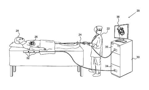
Fig. 1 is a schematic pictorial illustration of a system
20 for ablation of tissue in a heart 26 of a patient 28, in
accordance with an embodiment of the present invention. An
16 operator 22, such as a cardiologist, inserts a flexible
probe, such as a catheter 24, through the vascular system of
patient 28 so that the distal end of the catheter enters a
chamber of the patient's heart. Operator
22 advances the
20 catheter so that the end section of the catheter engages
endocardial tissue at a desired location or locations, as
shown in the figures that follow. Catheter 24 is connected
by a suitable connector (not shown) at its proximal end to a
24 console 30. The
console comprises an RF generator 36 for
applying RF energy through electrodes on the end section of
the catheter in order to ablate the tissue contacted by the
distal section. Alternatively or additionally, catheter 24
28 may be used for other diagnostic and/or therapeutic
functions, such as intracardiac electrical mapping or other
types of ablation therapy, including diagnostic and
therapeutic functions in organs other than the heart.
32 In the pictured embodiment, system 20 uses magnetic
position sensing to determine position coordinates of the end
section of the catheter inside heart 26. To determine the
7
position coordinates, a driver circuit 34 in console 30
drives field generators 32 to generate magnetic fields within
the body of patient 28.
Typically, field generators 32
4 comprise coils, which are placed below the patient's torso at
known positions external to the body. These coils generate
magnetic fields in a predefined working volume that contains
heart 26. One or more magnetic field sensors within the end
8 section of catheter 24 (as shown in Figs. 3 and 4) generate
electrical signals in response to these magnetic fields. The
console processes these signals in order to determine the
position (location and/or orientation) coordinates of the end
12 section of catheter 24, and possibly also the deformation of
the end section, as explained below. Console 30 may use the
coordinates in driving a display 38 to show the location and
status of the catheter. This method of position sensing and
16 processing is described in detail, for example, in PCT
International Publication WO 96/05768, and is implemented in
the CARTOTm system produced by Biosense Webster Inc. (Diamond
Bar, California).
20 Alternatively or additionally, system 20 may comprise an
automated mechanism (not shown) for maneuvering and operating
catheter 24 within the body of patient 28. Such mechanisms
are typically capable of controlling both the longitudinal
24 motion (advance/retract) and the rotation of catheter 24. In
such embodiments, console 30 generates a control input for
controlling the motion of the catheter based on the signals
provided by the position sensing system.
28 Although Fig. 1 shows a particular system configuration,
other system configurations may be used in alternative
embodiments of the present invention. For
example, the
methods described hereinbelow may be applied using position
32 transducers of other types, such as impedance-based or
ultrasonic position sensors. The term "position transducer"
8
CA 2835769 2020-03-24
CA 02835769 2013-12-03
as used herein refers to an element mounted on or in catheter
24 that causes console 30 to receive signals indicative of
the coordinates of the element. The position transducer may
4 thus comprise a receiver in the catheter, which generates a
position signal to the control unit based on energy received
by the transducer; or it may comprise a transmitter, emitting
energy that is sensed by a receiver external to the probe.
8 Furthermore, the methods described hereinbelow may similarly
be applied in mapping and measurement applications using not
only catheters, but also probes of other types, both in the
heart and in other body organs and regions.
12 Fig. 2 is a schematic sectional view of heart 26,
showing insertion of catheter 24 into the heart, in
accordance with an embodiment of the present invention. To
insert the catheter in the pictured embodiment, the operator
16 first passes a sheath 40 percutaneously through the vascular
system and into right atrium 44 of the heart through
ascending vena cava 42. The
sheath penetrates through
interatrial septum 48, typically via the fossa ovalis, into
20 left atrium 46. Alternatively, other approach paths may be
used. Catheter
24 is then inserted through the lumen of
sheath 40 until an end section 52 of the catheter passes out
of the distal opening at the end of the sheath into the left
24 atrium, as shown in the figure. The end section is formed so
as to define an arc when unconstrained, as is shown and
described in greater detail hereinbelow with reference to
Fig. 3. While end section 52 is passing through sheath 40,
28 however, the smaller inner diameter of the sheath holds the
end section straight and roughly parallel to the catheter
axis, as shown in Fig. 4.
Operator 22 aligns the longitudinal axis of sheath 40
32 (and of catheter 24) inside left atrium 46 with the axis of
one of pulmonary veins 50. The operator may carry out this
alignment using the position sensing methods described above,
9
CA 02835769 2013-12-03
along with a pre-acquired map or image of heart 26.
Alternatively or additionally, the alignment may be performed
under fluoroscopic or other means of visualization. The
4 operator advances end section 52 of the catheter toward the
target pulmonary vein so that the arc contacts the ostium,
and the end section either partly or fully surrounds the vein
(depending on the angle subtended by the arc). The operator
8 then rotates the catheter about its axis within the sheath so
that the end section traces an annular path around the
circumference of the vein. Meanwhile, the operator actuates
RF generator 36 to ablate the tissue along the path. After
12 completing this procedure around one pulmonary vein, the
operator may shift the sheath and catheter and repeat the
procedure around one or more of the other pulmonary veins.
After performing ablation along such an annular path,
16 operator 22 may assess the extent and quality of the ablation
by various means that are known in the art, such as
ultrasonic sensing, magnetic resonance imaging (MRI), or
measurement of local electrical properties of the tissue
20 (such as impedance and/or activation voltage). If the
operator discovers by such means (or in any other manner)
that a certain point or points have not been sufficiently
ablated, the operator may withdraw catheter 24 into sheath 40
24 until only the distal tip of end section 52 protrudes from
the sheath. This sort of configuration is shown in Fig. 4
and is described in greater detail with reference thereto.
In this latter configuration, the operator may advance the
28 sheath and catheter so that only the distal tip of the
catheter contacts the tissue at each point requiring further
ablation. While
the catheter contacts the tissue in this
manner, the tip electrode of the catheter may be energized by
32 RF generator 36 to ablate the tissue.
Fig. 3 is a schematic side view of the distal portion of
catheter 24, including end section 52 in its extended,
CA 02835769 2013-12-03
arcuate configuration, in accordance with an embodiment of
the present invention. The catheter comprises an insertion
shaft 54, which connects at its distal end to the base of end
4 section 52. Shaft 54 and end section 52 typically comprise
an outer shell made from a suitable flexible biocompatible
material, such as polyurethane, having a diameter around 2-3
mm, with internal wires and tubing as required. In one
8 embodiment, in which the catheter is designed for therapeutic
ablation, the size of the shaft is 7 Fr (about 2.3 mm
diameter), while the end section is of the same or slightly
larger size (such as 7.5 Fr). In other
embodiments, for
12 diagnostic measurements, the shaft is 7 Fr, while the end
section has a diameter between 1 and 2.5 mm.
End section 52 is formed as a complete or partial lasso,
i.e., as a preformed arcuate structure, which typically
16 subtends between 1800 and 3600. The radius of curvature of
end section 52, when unconstrained, is typically between 7.5
mm and 15 mm. Because the arc structure is resilient and,
possibly, slightly helical, when end section 52 is positioned
20 in the heart (against the ostium of a pulmonary vein, for
example), and insertion shaft 54 is advanced distally, the
end section will press against the heart tissue over the
entire length of the arc, thus facilitating good tissue
24 contact. The
arcuate and possibly helical shape of end
section 52 may be maintained, for example, by incorporating a
thin strut made from a shape memory material, such as Nitinol
(not shown in the figures), in the desired shape within the
28 end section. The
strut is made sufficiently flexible to
permit the end section to straighten during insertion and
withdrawal through sheath 40, but to resume its arcuate form
when it is unconstrained inside the heart chamber.
32 End section 52 comprises an array of electrodes along
its length, including, in this example, a tip electrode 60
extending over the distal tip of the end section and proximal
11
CA 02835769 2013-12-03
electrodes 58 distributed along the end section. Typically,
electrodes 58 have a width between 1 mm and 4 mm, and are
spaced between 1 mm and 10 mm apart. Electrodes 58 and 60
4 are connected to the connector at the proximal end of
catheter 24 by wires (not shown) running through the
catheter. Alternatively, other electrode configurations may
be used. For example, the end section may include smaller
8 "bump" electrodes, as described in the above-mentioned U.S.
Patent Application Publication 2010/0168548. In any of these
configurations, the electrodes may be used for sensing and/or
ablation. In order
to ablate an entire annulus around a
12 pulmonary vein, for example, catheter 24 may be rotated
("clocked") about its axis while applying RF electrical
energy to the electrodes, as noted above.
To provide local cooling and prevent adhesion during
16 ablation, electrodes 58 and 60 may have perforations for
irrigation.
(Perforations of this type are described and
shown, for example, in U.S. Patent Application Publication
2010/0168548.) The perforations are coupled to one or more
20 lumens in end section 52, which carries irrigation fluid from
shaft 54 to the electrodes and to the tissue surrounding
them. Details of an arrangement of electrodes and irrigation
lumens that may be used for this purpose are described
24 hereinbelow with reference to Fig. 5.
Catheter 24 may also include one or more position
transducers, such as positions sensors 62, 64 and 66. In
this embodiment, sensors 62, 64 and 66 comprise coils, which
28 output position signals in response to the magnetic fields of
field generators 32 (Fig. 1). For
example, sensor 66 may
comprise three coils, which give full location and
orientation information with regard to the base of end
32 section 52, while sensors 62 and 64 each comprise a single
coil, giving location and partial orientation information.
This sort of arrangement is described further in the above-
12
CA 02835769 2013-12-03
mentioned U.S. Patent Application Publication 2005/0033135.
It enables console 30 to track both the base location and the
deformation of end section 52, so that the operator can
4 verify that the end section is properly located and in good
contact with the tissue.
Alternatively, other types of
position transducers and sensing configurations may be used
in catheter 24 and system 20. Sheath 40 may also comprise
8 one or more position transducers, as shown in Fig. 4.
Fig. 4 is a schematic side view of the distal end of
catheter 24, showing end section 52 in its retracted,
straightened configuration, in accordance with an embodiment
12 of the present invention. Catheter
24 has been withdrawn
into sheath 40 so that only tip electrode 60 protrudes
distally, while proximal electrodes 58 are held inside the
sheath. With the catheter in this configuration, operator 22
16 may advance sheath 40 and catheter 24 together so that
electrode 60 contacts particular points on the endocardium
for purposes of electrical measurement and/or ablation. To
ablate tissue in this configuration, RF generator 36 applies
20 energy only to tip electrode 60, and proximal electrodes 58
are not actuated.
Optionally, sheath 40 may contain one or more position
transducers 70, as well. The
position signals provided by
24 transducer 70 in proximity to the distal opening of sheath
40, for example, can be used for either or both of two
purposes:
= To detect the location of the sheath within the body
28 relative to field generators 32, in order to assist
operator 22 in navigating the sheath to the desired
location; and
= To sense the disposition of end section 52 within the
32 sheath.
A processor in console 30 may thus determine that catheter 24
is properly deployed in the straightened configuration of
13
Fig. 4 when the position signals from transducers 62 and 70
indicate that they are located within a predetermined minimum
distance of one another. Alternatively or additionally, the
4 distance between transducers 62 and 70 may be determined by
transmitting a signal from one of these transducers and
receiving the signal at the other. RF generator 36 may be
controlled automatically so that only tip electrode 60 can be
8 energized as long as the remainder of end section 52 is
contained in sheath 40.
Alternatively or additionally, other positioning
elements may be used to sense the location of the distal tip
12 of catheter 24 relative to sheath 40. For
example, a
magnetic structure at the end of the sheath may be used for
this purpose, as described in U.S. Patent Application
13/467,158.
Similarly, other sorts of transducers in the
16 probe and/or sheath, such as proximity sensors, may be used
to ascertain the configuration of end section 52 relative to
sheath 40.
Fig. 5 is a schematic side view of the distal tip of end
20 section 52 of catheter 24, in accordance with an embodiment
of the present invention. In this embodiment, electrodes 58
and 60 have multiple perforations through which irrigation
fluid may be delivered to tissue with which the catheter is
24 in contact during ablation. Because tip electrode 60 may be
actuated individually (in the configuration shown in Fig. 4,
for example), separately from ring electrodes 58, it is
desirable that the tip electrode be irrigated separately from
28 the ring electrodes.
Thus, as shown in Fig. 5, tip electrode 60 is served by
a separate irrigation lumen 82, while ring electrodes 58 are
served by a common irrigation lumen 84. In the configuration
32 shown in Fig. 3, in which both the tip and ring electrodes
are actuated to ablate tissue, console 30 supplies irrigation
14
CA 2835769 2020-03-24
CA 02835769 2013-12-03
fluid to catheter 24 via both of lumens 82 and 84, so that
all electrodes are irrigated. On the
other hand, in the
configuration of Fig. 4, the console supplies irrigation
4 fluid only to lumen 82. This sort of differential irrigation
scheme may be applied, as well, to electrodes of other types
(such as the bump electrodes shown in the above-mentioned
U.S. Patent Application Publication 2010/0168548), and in
8 substantially any other type of irrigated ablation probe in
which multiple electrodes are actuated selectively.
Although the embodiments described above relate
specifically to catheters for use in certain intracardiac
12 procedures, probes made in accordance with the principles set
forth in this patent application may similarly be used in
diagnostic and therapeutic procedures of other types, both in
the heart and in other body organs. It will
thus be
16 appreciated that the embodiments described above are cited by
way of example, and that the present invention is not limited
to what has been particularly shown and described
hereinabove. Rather,
the scope of the present invention
20 includes both combinations and subcombinations of the various
features described hereinabove, as well as variations and
modifications thereof which would occur to persons skilled in
the art upon reading the foregoing description and which are
24 not disclosed in the prior art.