Note: Descriptions are shown in the official language in which they were submitted.
CA 02836973 2015-07-20
,
- 1 -
APPARATUS FOR EYE SURGERY
The present invention relates to laser-assisted operations on the human eye
that
include treatment of the eye by laser and further operation tasks to be
performed in a
sterile environment.
A laser-assisted cataract operation (i.e. the treatment of a cataract through
implanting of
an artificial lens in the eye) is an example of a form of operation in which,
after the use
of a laser, other operation devices are additionally used in order to complete
the
operation. The laser can be used to produce incisions, for example in order to
open the
anterior capsule region of the human eye (capsulorhexis) and to produce
lateral
incisions at the limbus edge so as to enable the human lens to be removed, an
artificial
lens to be inserted and the instruments required in this case to be
introduced. The laser
can also be used to prefragment the crystalline human lens, i.e. to divide it
into
segments, which can then be more easily liquified by phacoemulsification and
aspirated. For this purpose, the laser can use ultrashort-pulse, focussed
laser radiation,
the laser pulses giving rise to photodisruptions in the irradiated tissue as a
result of a
laser-induced dielectric breakdown. Concatenation of such photodisruptions
enables a
multiplicity of incision shapes to be produced intraocularly. The pulse
durations of the
laser pulses can be, for example, in the picosecond, femtosecond or attosecond
range,
but shorter or longer pulse durations are also conceivable within the scope of
the
invention, provided that they can ensure the desired photodisruptive effect.
Clearly, laser-assisted cataract operations are only one possible form of
operation
within the scope of the invention. In general, the invention is suitable for
any intraocular
operations with laser assistance wherein, after the laser treatment, there are
additionally
required further operation tasks that absolutely
CA 02836973 2013-11-21
WO 2013/000487 PCT/EP2011/003154
- 2 -
must be performed under sterile-room conditions in order to prevent germs from
entering open wounds of the eye.
In the case of a typical laser-assisted cataract operation, the laser
treatment of
the eye is performed first, in a first operation room, which is usually non-
sterile.
The patient lies on a treatment couch, the eye to be treated being immovably
coupled, in a manner known per se, by means of an adapter (patient interface),
to a focussing objective lens of the laser system used.
After completion of laser treatment, the patient is transferred by the medical
personnel to another bed and brought into a second, separate operation room,
in which conditions are sterile and in which the appliances and instruments
necessary for the extraction of the crystalline lens and for the implantation
of
the artificial lens are available. It is necessary in this case to transfer
the patient
to another bed, i.e. the patient must get up from the couch in the first
operation
room, walk into the second operation room and lie on another couch there.
Similarly, it is also necessary for the physician performing the treatment to
go
into the second operation room. Because conditions there are sterile, it is
necessary for the physician to undergo usual disinfection measures and change
his gloves and, if appropriate, also his clothes, before he may enter the
second
operation room.
This procedure (room change, transfer of patient to another bed, changing of
clothes and disinfection by medical personnel) is cumbersome and time-
consuming, and is also stressful for the patient, owing to the intermediate
interruption in the course of the operation. These disadvantages impede the
increasing advance of laser in cataract operations, although the advantages of
the use of a laser in such an operation are significant in comparison with
performance of an operation without laser.
CA 02836973 2016-02-05
,
- 3 -
It is therefore an object of embodiments of the invention to indicate a way in
which, in the case of laser-assisted intraocular eye operations that are to be
performed, at least partially, in a sterile environment, the course of the
treatment
can be shortened and inconvenience to the patient can be reduced.
Certain exemplary embodiments provide an apparatus for eye surgery,
comprising a stand having a stand base that is movable or realized for
mounting
on a wall or ceiling, and having a stand arm arrangement that is manually
adjustable, at least partially, relative to the stand base, an operation
microscope
attached to the stand arm arrangement, the operation microscope pivotable
about a horizontal pivot axis, a laser appliance, which provides pulsed,
focussed
laser radiation having radiation properties suited to the application of
incisions in
the human eye, the laser appliance having a laser source and a laser treatment
head that is attached to the stand arm arrangement and emits the laser
radiation,
the laser treatment head pivotable about an additional pivot joint that is
parallel to
the horizontal pivot axis, a flexible transmission fibre or a jointed beam
transport
arm being provided for the purpose of transporting the laser radiation to the
laser
treatment head, the laser treatment head being positioned or positionable in
an
observation beam path of the operation microscope and providing a passage for
an observation beam going along the observation beam path.
An eye-surgery apparatus designed in such a manner enables a laser-assisted
intraocular eye operation, for instance a cataract operation, to be performed
at
one operation location without a change of room. This avoids transfer of the
patient to another bed, shortens the duration of the operation and, since the
course of the operation is more convenient, because it is not interrupted,
allows
the expectation of better treatment results. The entire operation, including
the
laser treatment, can be performed in a sterile region of a single operation
room,
the operation room being easily cleaned and re-sterilized after completion of
the
CA 02836973 2013-11-21
WO 2013/000487 PCT/EP2011/003154
- 4 -
operation, owing to the stand being movable or mounted on a wall or ceiling.
If
required, sterile covers (e.g. cover films) can be provided, for example in
order
to cover particular parts (modules) of the eye-surgery apparatus, for instance
the microscope, the laser treatment head and/or a swivel arm of the stand.
Particularly in the case of use of such covers, the stand, with the components
fastened thereto, can remain in the operation room.
The invention allows a laser scalpel to be integrated into the usual device
system for a cataract operation or other intraocular eye operation. The
connection of the laser source generating the radiation to the laser treatment
head via a flexible transmission fibre or a jointed beam transport arm (mirror
jointed arm) enables the laser treatment head, with a patient adapter attached
thereto, to be moved, if required, into the conventional manual operation
space
of the physician. Through an observation passage in the laser treatment head,
the physician performing the treatment, or an assistant, can observe the eye
through the operation microscope for the purpose of performing the laser
treatment. The laser source itself, which comprises, for example, a fibre
laser or
other solid-state laser, can be disposed at a certain distance from the
sterile
working region, for instance in a semi-sterile region of the operation room,
but,
alternatively, it can also be disposed in the sterile region. Expediently, the
laser
treatment head remains coupled to the stand, only a patient adapter, which is
attached to the laser treatment head and via which the eye of the patient can
be
coupled to the laser treatment head, being detachable, such that it can be
exchanged between successive operations and replaced by a new, sterile patient
adapter.
In one design, it is conceivable for the laser treatment head to remain in the
observation beam path of the operation microscope, not only for the laser
treatment, but also in the case of subsequent operation tasks (e.g. extraction
of
CA 02836973 2013-11-21
WO 2013/000487 PCT/EP2011/003154
- 5 -
the human lens, implantation of an artificial lens), the observation passage
in
the laser treatment head affording the physician the necessary view of the eye
also in these subsequent operations tasks. For such a design, the operation
microscope and the laser treatment head are coupled or can be coupled to one
another relative to the stand base for the purpose of common positional
adjustment. After the laser treatment, the physician must then be able to
raise
the laser treatment head from the eye, to enable the patient adapter to be
removed. However, the laser treatment head can remain between the operation
microscope and the eye, it being possible to ensure, through appropriate
setting
of the stand, that there is sufficient space between the eye and the treatment
head, in order that the physician can perform the remaining tasks - observed
through the operation microscope - in an unimpeded manner.
In another design, it is conceivable for the laser treatment head to be moved
away out of the observation beam path of the operation microscope after the
laser treatment, i.e. not to remain between the operation microscope and the
eye during the subsequent operation tasks. For this purpose, the operation
microscope and the laser treatment head are positionally adjustable relative
to
one another, in such a way that the laser treatment head can be moved into and
out of the observation beam path of the operation microscope. The stand arm
arrangement in this case can have a first arm unit, to which the operation
microscope is attached, and have a second arm unit, to which the laser
treatment head is attached, the first and the second arm unit being adjustable
relative to one another and preferably independently of one another.
Such a design enables the laser treatment head to be swivelled or otherwise
moved into a non-use position, in which it does not interfere with the freedom
of
action of the physician working over the eye of the patient and looking
through
the operation microscope. Only if a laser treatment is to be performed on the
CA 02836973 2013-11-21
WO 2013/000487 PCT/EP2011/003154
- 6 -
eye, the physician can then move the laser treatment head under the operation
microscope.
Insofar as the laser treatment head and the operation microscope are
positionally adjustable relative to one another, it can be advantageous if the
laser treatment head is lockable relative to the operation microscope or can
otherwise be detachably coupled to the operation microscope once the laser
treatment head has been moved under the operation microscope. This enables
the laser treatment head to be fixed in position relative to the operation
microscope, which fixing in position can therefore also be important,
primarily, in
order that the operating physician can maintain a reliable view of what is
happening on and in the eye, through the observation passage provided in the
laser treatment head.
For sterile working conditions, at least the operation microscope, or in any
case
a part thereof, can be covered by a sterile cover during the operation. The
same applies to the laser treatment head, at least insofar as the latter is
also to
remain under the operation microscope, and therefore in the sterile working
region, during a subsequent open intervention on the eye. If, on the other
hand, the laser treatment head (without the operation microscope) can be
moved out of the working region of the operating physician, it is possible to
dispense with a sterile wrapping of the laser treatment head, in any case when
all laser tasks are performed before the intervention on the open eye.
The stand arm arrangement can provide at least one rotational degree of
freedom of movement or/and at least one translational degree of freedom of
movement for the operation microscope and the laser treatment head, relative
to the stand base in each case. In this case, it is possible to pass through
at
least a majority of the movement scope of the operation microscope and of the
CA 02836973 2013-11-21
WO 2013/000487 PCT/EP2011/003154
- 7 -
laser treatment head, relative to the stand base, by manual adjustment. If
required, a drive arrangement, for example an electric motor-operated drive
arrangement, which allows motor-operated adjustment, in particular for the
purpose of fine positioning of the operation microscope and/or of the laser
treatment head, can be provided additionally on the stand. However, the
adjustment range provided by such a drive arrangement is preferably small
relative to the available manual adjustment range.
According to one design, a method for performing an eye operation can
comprise the following steps:
- providing an adjustable stand in an operation room, there being attached
to
the stand an operation microscope and a laser treatment head that emits
pulsed, focussed laser radiation having radiation properties suited to the
application of incisions in the human eye,
- positioning a patient on a treatment couch in a sterile region of the
operation
room,
- setting the stand into a first position, in which the laser treatment
head is
positioned in an observation beam path of the operation microscope, and an
operating physician can observe, through the operation microscope and an
observation passage of the laser treatment head, an eye of the patient to be
operated upon,
- performing a laser treatment of the eye, by means of the laser radiation,
in
the first position of the stand,
- setting the stand into a second position, in which the laser treatment
head is
positioned outside the observation beam path of the operation microscope,
and the operating physician can observe, solely through the operation
microscope, the eye of the patient to be operated upon,
- performing further operation tasks on the eye, in the second position of
the
stand, without use of the laser radiation.
CA 02836973 2013-11-21
WO 2013/000487 PCT/EP2011/003154
- 8 -
The invention is explained further in the following with reference to the
appended drawings, wherein:
Figure 1 is a schematic representation of a first embodiment of an eye-surgery
apparatus for laser-assisted intraocular eye operations,
Figure 2 is a schematic representation of a second embodiment of an eye-
surgery apparatus for laser-assisted intraocular eye operations,
Figure 3 is a schematic representation of a third embodiment of an eye-surgery
apparatus for laser-assisted intraocular eye operations.
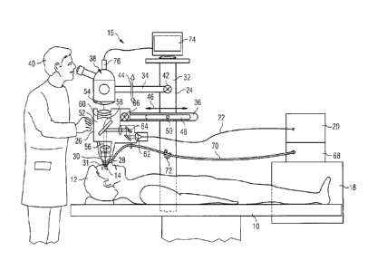
Reference is made first to Fig. 1. Set up at the operation station represented
therein is a patient bed (patient couch) 10, on which, in the representation
of
Fig. 1, there lies a patient 12, having an eye 14 to be treated, which is
represented merely schematically, and a laser system 16, which is suitable for
producing incisions in the tissue of the patient's eye 14 by photodisruption.
The
laser system 16 comprises a laser source 20, which is disposed on a supporting
frame 18 (for example, in the form of a shelf or table) and which contains
e.g. a
solid-state laser or a fibre laser and provides pulsed laser radiation. The
laser
radiation emitted by the laser source 20 is coupled into a flexible
transmission
fibre 22, via which the laser radiation is transmitted to a laser treatment
head
26, which is held on a stand 24 and from which the laser radiation is applied
to
the patient's eye 14. The laser radiation emitted by the laser treatment head
26
has radiation properties suited to producing photodisruptions in the tissue of
the
patient's eye 14. For example, the pulse durations of the applied laser pulses
are in the range of picoseconds or femtoseconds. In order to avoid excessively
high pulse intensities on the transmission fibre 22, the pulse durations of
the
CA 02836973 2013-11-21
WO 2013/000487 PCT/EP2011/003154
- 9 -
laser pulses coupled into transmission fibre 22 by the laser source 20 can be
greater than the pulse durations of the laser pulses applied to the eye 14.
For
this purpose, a pulse stretcher (not represented in greater detail), which
stretches the pulse durations of the laser pulses, for example to more than
one
picosecond, can be provided in the laser source 20. For the subsequent time
compression of the laser pulses to the required, shorter pulse durations of,
for
example, femtoseconds or picoseconds, the transmission fibre itself can have
corresponding compression properties, for which purpose, for example, a
photonic hollow core fibre can be used (frequently designated as a PCF fibre,
i.e.
"photonic crystal fibre"). Alternatively, it is possible to use a transmission
fibre
without, or at least without significant, compression properties, for instance
an
LMA fibre, i.e. a transmission fibre having a large mode area (LMA = large
mode
area). A suitable compression element, for instance a transmission grating or
a
crystal including a chirped Bragg grating (not represented in greater detail),
can
then be provided in the laser treatment head 26 for the purpose of pulse
compression.
Exchangeably attached to the laser treatment head 26 is a patient adapter
(applicator) 28, which constitutes a mechanical interface to the patient's eye
14
and allows referencing of the eye 14 in relation to the laser treatment head
26.
For this purpose, the adapter 28 has a contact element 30, which is
transparent
to laser radiation and through which the laser radiation is applied. On its
side
that faces towards the eye, the contact element 30 constitutes a contact
surface
against which the eye 14 is placed. In a manner known per se, the patient
adapter 28 can be realized for coupling to a suction ring 31 to be placed
beforehand on the eye 14.
In the exemplary case shown, the stand 24 is realized as a floor stand, which
is
preferably movable, and thus can be moved out of the operation room after an
CA 02836973 2013-11-21
WO 2013/000487 PCT/EP2011/003154
- 10 -
eye operation, to enable the operation room to be cleaned. Alternatively, the
stand 24 can be a wall stand or ceiling stand, which is fixedly mounted on a
wall
or on the ceiling of the operation room. In each case, the stand 24 has a
stand
base 32, which, in the exemplary case shown in Fig. 1, is realized
schematically
as an upright column and which, in the case of a floor stand, is realized with
rollers at its foot that can be locked if appropriate, or, in the case of a
wall stand
or ceiling stand, constitutes a support for mounting on the wall or ceiling.
Attached to this stand base 32, generally, is a stand arm arrangement, which
can be adjusted relative to the stand base in preferably a plurality of
degrees of
freedom (translationally and/or rotationally) and which, in the exemplary case
shown, comprises two arm units 34, 36 that can be adjusted separately from
one another. Attached to one of the arm units, in this case the arm unit 34,
there is an operation microscope 38, which offers an operating physician 40,
indicated schematically, an enlarged view of the operation region (the eye
14).
The laser treatment head 26, on the other hand, is attached to the other arm
unit (in this case, the arm unit 36). The arm units 34 - as in the simplified,
schematic representation of Fig. 1 - can each be individual arms that can be
adjusted pivotally or/and linearly in relation to the stand base 32. It is
understood, however, that each of the arm units 34 can be a multi-arm
structure composed of a plurality of arms, which are connected to one another
in a jointed manner or/and through linear motion guides.
In Fig. 1, merely for the purpose of illustration, the arm unit 34 that
carries the
operation microscope 38 is shown to be pivotable about a horizontal pivot axis
42, relative to the stand base 32 (according to a double arrow 44), while the
arm unit 36 that caries the laser treatment head 26 is adjustable in a
horizontal
direction, guided linearly in relation to the stand base 32 (as illustrated by
a
double arrow 46). In Fig. 1, the linear guidance of the arm unit 36 in
relation to
the stand base 32 is illustrated, in a purely schematic manner, by a peg and
CA 02836973 2013-11-21
WO 2013/000487 PCT/EP2011/003154
- 11 -
longitudinal slot arrangement, having a longitudinal slot 48 and a peg 50
guided
therein. It need not be especially emphasized that this is a representation
purely for the purpose of illustration, and that considerably more complex
motion mechanisms can be provided for the purpose of motional guidance of the
operation microscope 38 and of the laser treatment head 26 in a plurality of
degrees of freedom of movement in relation to the stand base 32.
A characteristic of the embodiment of Fig. 1, however, is that the laser
treatment head 26 can be moved, relative to the operation microscope 38,
between a position of use and a non-use position. The position of use is
represented in Fig. 1; in this position, the laser treatment head 26 is moved
over
the eye 14 to be treated, and can be docked onto the eye 14 through the use of
the patient adapter 28. The laser treatment head 26 in this case is located
between the eye 14 and the operation microscope 38. In order that the
operating physician 40 can nevertheless observe, through the operation
microscope 38, what is happening on the eye 14, the laser treatment head 26
constitutes an observation passage 52, which extends from an observation
window 54 (formed by suitable observation optics, for example), facing towards
the microscope 38, as far as the patient adapter 28, such that, when the laser
treatment head 26 is in the position of use, the observation beam path of the
operation microscope 38 extends through the observation passage of the laser
treatment head 26 as far as the eye 14.
In the non-use position, on the other hand, which is not represented in
greater
detail in the drawing, the laser treatment head 26 is moved out of the
observation beam path of the operation microscope 38, such that the operating
physician 40, when looking through the operation microscope 38, has a direct
view onto the eye 14. The laser treatment head 26 is then no longer located
under the operation microscope 38 and, in particular, is at such a distance
from
CA 02836973 2013-11-21
WO 2013/000487 PCT/EP2011/003154
- 12 -
the working region over the eye 14 that the operating physician 40 can perform
the remaining operation tasks on the eye 14 in an unimpeded manner.
When the laser treatment head 26 is in the position of use, the observation
beam path of the operation microscope 38 goes through various optical
elements, which are provided in the laser treatment head 26 for the purpose of
guiding or/and shaping the laser radiation. In particular, the observation
beam
path of the microscope 38 goes through a focussing optical system 56, for
example in the form of an F-Theta objective lens, and, in the exemplary case
shown, goes through a semi-transparent deflecting mirror 58. The optical
elements for guiding and shaping the laser radiation are matched to the
wavelength of the laser radiation used. For visible light, which reaches the
operation microscope 38 through the observation passage 52, optical
aberrations (for example, a chromatic dispersion) can therefore occur, to
compensate which a compensating optical system 60 can be provided, in the
laser treatment head 26.
Additionally accommodated in the laser treatment head 26 are a collimator lens
62 and a scanning arrangement, which is denoted in general by 64. The
collimator lens 62 serves to collimate the divergent radiation bundle leaving
the
transmission fibre 22. The scanning arrangement 64 serves to shift the focus
position of the focussed radiation bundle emerging from the laser treatment
head 26, both in the direction of beam propagation (usually designated as the
z
direction) and in a plane transverse to the z direction (usually designated as
the
x-y plane). For the purpose of transverse scanning (i.e. in the x-y
direction), the
scanning arrangement can comprise, for example, in a manner known per se, a
pair of galvanometrically controllable deflection mirrors, which can be tilted
about axes that are perpendicular to one another. For the purpose of
longitudinal scanning (i.e. in the z direction), on the other hand, the
scanning
CA 02836973 2013-11-21
WO 2013/000487 PCT/EP2011/003154
- 13 -
arrangement can have, for example, a lens that is positionally adjustable or
of
variable refractive power, or an adaptive mirror. For miniaturization it is
conceivable to provide, for example, as an alternative to a pair of
galvanometric
mirrors, an electrooptic crystal, by means of which a controlled x-y
deflection of
the focus position can likewise be achieved.
For the purpose of z displacement of the radiation focus it is also
conceivable,
alternatively, to realize the focussing optical system 56 so as to be
adjustable in
the direction of the radiation propagation (i.e. z direction).
Denoted at 66 in Fig. 1 is an additional pivot joint, which allows the laser
treatment head 26 to be pivoted about a pivot axis that is perpendicular to
the
arrow direction 46 (i.e., in the representation of Fig. 1, about a pivot axis
normal
to the plane of the page).
For the purpose of controlling the laser source 20, the scanning arrangement
64
and, if appropriate, the focussing optical system 56, the laser system 16
comprises a control unit 68, which can be set up together with the laser
source
20 on the supporting frame 18. For the purpose of transmitting electrical
control
signals from the control unit 68 to the laser treatment head 26, an electrical
connecting cable, not represented in Fig. 1, runs between the two components.
At the same time, the control unit 68 can include a pump arrangement, likewise
not represented in greater detail, but known per se, having at least one
vacuum
pump. The vacuum generated by this pump arrangement can be transported,
via a vacuum tube (or, if appropriate, a plurality of vacuum tubes) that can
be
connected to the control unit 68, to the suction ring 31, where the vacuum is
used to suck the suction ring 31 onto the eye 14 and, if appropriate, also to
suck
the patient adapter 28 onto the suction ring 31. The vacuum tube 70 can run
on the stand 24, through a guide 72, for instance through a guide clip 72,
which
CA 02836973 2013-11-21
WO 2013/000487 PCT/EP2011/003154
- 14 -
is indicated schematically. The control intelligence contained in the control
unit
68 also controls the pumping operation of the aforementioned pump
arrangement.
The frame 18 is realized, advantageously, such that it can be taken out of the
operation room with little effort. For this purpose, it can be realized as a
rolling
frame, or it can be fastened to the stand 24, such that it can be removed
together with the stand 24 from the operation room. For example, the frame 18
can be attached to the stand base 32. If a wall stand or ceiling stand is
used,
the frame 18, alternatively, can be mounted on the wall or ceiling of the
operation room, separately from the stand.
Further, in addition, a monitor 74 can be attached to the stand 24, for
example
to the stand base 32, on which monitor there can be visualized camera
recordings that are recorded by means of a microscope camera 76 attached to
the operation microscope 38. The physician 40 or his assisting personnel can
thus follow the operation on the monitor 74.
The laser appliance 16 shown in Fig. 1 enables the physician 40, the patient
12
and the assisting personnel to remain in their positions in the sterile region
of
the operation room during the entire cataract operation (or other laser-
assisted
intraocular operation). The operation need not be interrupted after the laser
treatment by means of the laser system 16. Instead, after use of the laser,
the
physician can continue working in an uninterrupted manner by means of an
ultrasound device (not represented in greater detail in Fig. 1) and the other
instruments required for extracting the crystalline lens and replacing it by
an
artificial lens and complete the surgery. After use of the laser, it is
necessary
only to move the laser treatment head 26 out of the working area of the
physician 40, through use of the degrees of freedom of movement offered by
CA 02836973 2013-11-21
WO 2013/000487 PCT/EP2011/003154
- 15 -
the stand arm unit 36. The patient 12 need not be transferred to another bed,
and the physician 40 need not leave the sterile region. Also, it is not
necessary
for the physician to change clothes. This saves a considerable amount of time.
It is understood that, even in the case of a design in which the laser
treatment
head 26 and the operation microscope 38 are positionally adjustable relative
to
one another (as represented in Fig. 1), the physician 40 can nevertheless
decide, after use of the laser, to continue the operation without moving the
laser
treatment head 26 back out of its position of use into the non-use position.
In
other words, the laser treatment head 26 can remain under the operation
microscope 38 even during the subsequent operation tasks. For this purpose, it
can be appropriate to first remove the patient adapter 28, in order thus to
create sufficient space under the laser treatment head 26 for the manual
operation tasks in hand.
The position of use of the laser treatment head 26 can be, for example, a
locking position, into which the arm unit 36 latches automatically when the
laser
treatment head 26 is moved into the position of use. If required, a motor-
assisted fine positioning of the laser treatment head 26, for example in the
vertical direction, can be possible in the position of use, in particular to
facilitate
docking of the patient adapter 28 to the suction ring 31 and to the eye 14.
For
this purpose, a suitable motor-operated drive means (not represented in
greater
detail), allowing a corresponding adjustment of the arm unit 36, can be
provided
on the stand 24.
On the other hand, the frame 18, with the control unit 68 and the laser source
20, can be disposed in a semi-sterile region of the operation room, at a
sufficient
distance from the sterile working region of the physician 40, and also remain
there during the entire operation.
CA 02836973 2013-11-21
WO 2013/000487 PCT/EP2011/003154
- 16 -
In the further Figures 2 and 3, components that are the same or have the same
function are denoted by the same references as in Fig. 1, but suffixed with a
lower-case letter. Unless otherwise stated in the following, for explanation
of
such components we refer to the preceding statements relating to Fig. 1.
The embodiment of Fig. 2 differs from that of Fig. 1, in essence, in the
provision
of a mirror jointed arm 78a for transporting the laser radiation from the
laser
source 20a to the laser treatment head 26a. The mirror jointed arm 78a offers
a
sufficient freedom of movement to allow the desired/required adjustability of
the
laser treatment head 26a relative to the stand 24a or/and of the arm unit 36a
carrying the laser treatment head 26a, in relation to the stand base 32a, and
not
to impede such adjustability.
It may be desirable for the eye-surgery apparatus to be equipped with a
diagnostic unit, in particular an imaging diagnostic unit, for example to
enable
the laser treatment of the patient's eye (for instance the capsulorhexis and
the
lens prefragmentation in the case of a laser-assisted cataract operation) to
be
performed in a precisely localized manner. For this purpose, the third
embodiment shown in Figure 3 is equipped with an OCT measuring appliance,
which comprises an OCT unit 80b disposed, together with the laser source 20b
and the control unit 68b, on the frame 18b. OCT stands for optical coherence
tomography. The OCT unit 80b can interferometrically overlay an emitted OCT
measurement radiation with an OCT reflected radiation reflected from the
patient's eye 14b and, from the thereby obtained interferometry data, generate
a two-dimensional or three-dimensional image of the tissue structures of the
eye
14b. The generated OCT image can be displayed, for example, on the monitor
74b. Alternatively, it is conceivable for the OCT unit 80b to be connected to
a
further monitor (not represented in greater detail), on which it can display
the
CA 02836973 2013-11-21
WO 2013/000487 PCT/EP2011/003154
- 17 -
OCT image. If required, such a monitor can also be integrated into the OCT
unit
80b.
In the embodiment shown in Figure 3, there is connected to the OCT unit 80b a
further transmission fibre 82b, which is separate from the transmission fibre
22b
and via which the OCT measurement radiation is transported from the OCT unit
80b to the laser treatment head 26b. In the laser treatment head 26b, the OCT
measurement radiation goes through the scanning arrangement 64b and the
focussing optical system 56b. It is coupled, via a collimator lens 84b and a
semi-transparent mirror 86b, into the radiation propagation path that is
provided, in the laser treatment head 26b, for the laser radiation transported
via
the transmission fibre 22b. The components of the OCT measurement radiation
reflected at the eye 14b (i.e. the OCT reflected radiation) is routed on the
same
path to the transmission fibre 82b and, via the latter, to the OCT unit 80b.
In departure from the exemplary case shown in Figure 3, it is conceivable for
one or both of the two transmission fibres 22b, 82b to be replaced by an
appropriately movable mirror jointed arm (analogous to the embodiment of
Figure 2). It is conceivable in this case, for example, to use a transmission
fibre
for one of the two radiation types (laser radiation, OCT measurement
radiation)
and, for the other radiation type, to use a mirror jointed arm for
transporting the
radiation to the laser treatment head 26b. Alternatively, it is conceivable to
use
two separate mirror jointed arms for transporting, respectively, one of the
two
radiation types.
In a further modification of Figure 3 it is conceivable to provide a common
transport path to the laser treatment head 26b for both radiation types,
either in
the form of a common transmission fibre or in the form of a common mirror
jointed arm. When provision is made for a common transport path for the laser
CA 02836973 2013-11-21
WO 2013/000487 PCT/EP2011/003154
- 18 -
radiation and the OCT measurement radiation (and also the OCT reflected
radiation) it may be provided that the laser radiation and the OCT measurement
radiation are not emitted simultaneously. If simultaneous operation of the
laser
source 20b and of the OCT unit 80b is required, it may be beneficial to use
separate transport media for the laser radiation and the OCT measurement
radiation. The wavelength of the laser radiation and the wavelength of the OCT
measurement radiation may be relatively close to one another, for example - to
give a number example that is not limiting in any way - 1030 nm for the laser
radiation and 1060 nm for the OCT measurement radiation. Alternatively, the
wavelengths of the laser radiation and the OCT measurement radiation may be
comparatively far apart from one another, for example 1030 nm for the laser
radiation and 800 nm for the OCT measurement radiation.
In respect of the generation of the OCT measurement radiation, use can be
made of a measurement radiation source that is separate from the laser source
20b and that, expediently, is integrated into the OCT unit 80b. It is also
conceivable, however, to generate the OCT measurement radiation by means of
the laser source 20b, such that, in this case, a single radiation source
suffices for
generation of both types of radiation.