Note: Descriptions are shown in the official language in which they were submitted.
CA 02837205 2013-11-22
WO 2012/162608 PCT/US2012/039574
AIMING DEVICE HAVING RADIO-OPAQUE MARKERS
CROSS-REFERENCE TO RELATED APPLICATIONS
[0001] This claims the benefit of U.S. Provisional Patent Application Serial
No.
61//489,930 filed on May 25, 2011 and U.S. Provisional Patent Application
Serial No.
61/501,868, filed June 28, 2011, the disclosure of each of which is hereby
incorporated by
reference as if set forth in its entirety herein.
TECHNICAL FIELD
[0002] The present disclosure relates to the fields of bone implants and bone
fixation
devices.
BACKGROUND
[0003] To stabilize fractured bones (including so-called long bones such as
femurs,
tibias, fibulas, humeri, radii, ulnas, metacarpals, metatarsals, and
phalanges), users have
employed intramedullary rods or nails to provide structural reinforcement to
the bone. Such
devices may be anchored, for instance in the medullary canal of the bone, by
way of screws
inserted through the bone (in a direction transverse to the major axis of the
nail) so as to engage
with locking holes in the nail.
[0004] Some bones, however, have a natural curvature, and intramedullary
devices
inserted into the medullary canal can deflect so as to conform to this
curvature. Accordingly
compensation for the curvature of the bone allows for accurate placement of
the fixation screws
when the screws are aimed and then inserted through the skin and bone to
engage with the
locking holes of the intramedullary device.
SUMMARY
[0005] In one embodiment, an aiming arm is configured to be aligned with an
implant.
The aiming arm includes a body that defines a plurality of apertures
configured to receive
respective fixation members. The aiming arm further includes at least a pair
of radio-opaque
markers carried by the body, wherein when a radiographic image is taken of the
aiming arm from
a first viewpoint, the radio-opaque markers define a first radiographic image
characteristic when
the aiming arm is not aligned with a source of the radiographic image, and the
radio-opaque
markers define a second radiographic image characteristic when the aiming arm
is aligned with
the source.
- 1 -
CA 02837205 2013-11-22
WO 2012/162608 PCT/US2012/039574
BRIEF DESCRIPTION OF THE DRAWINGS
[0006] The summary, as well as the following detailed description, is further
understood when read in conjunction with the appended drawings. For the
purpose of
illustrating the present disclosure, there are shown in the drawings exemplary
embodiments of
the disclosure; however, the disclosure is not limited to the specific
methods, compositions, and
devices disclosed. In addition, the drawings are not necessarily drawn to
scale. In the drawings:
[0007] Fig. lA is a top plan view of an aiming device constructed in
accordance with
one embodiment including a base and an aiming arm configured to be coupled to
the base;
[0008] Fig. 1B is a perspective view of the aiming device illustrated in Fig.
1B;
[0009] Fig. 2 is a perspective view of a bone fixation system including a
support frame,
an intramedullary nail, the aiming device illustrated in Fig. lA supported by
the support frame,
and a mask coupled to the aiming arm;
[0010] Fig. 3 is a perspective view of the bone fixation assembly illustrated
in Fig. 2,
showing the intramedullary nail in an inserted, deflected configuration and
showing the aiming
arm in a corresponding deflected configuration;
[0011] Fig. 4 is a perspective view of the bone fixation assembly illustrated
in Fig. 3,
showing the intramedullary nail in another deflected configuration and showing
the aiming arm
in a corresponding deflected configuration;
[0012] Fig. 5 is a perspective view of the implantation assembly as
illustrated in Fig.
10, showing the intramedullary nail implanted in a long bone;
[0013] Fig. 6A is a perspective view of the aiming arm illustrated in Fig. 1A;
[0014] Fig. 6B is a side elevation view of the aiming arm illustrated in Fig.
6A,
showing a plurality of radio-opaque alignment markers;
[0015] Fig. 6C is a perspective view of a portion of the aiming arm
illustrated in Fig.
6A, taken along line 6C;
[0016] Fig. 7A is a perspective view of a radiographic image of a portion of
the aiming
arm illustrated in Fig. 6C as taken from a radiographic image source, showing
the radio-opaque
alignment markers in a misaligned configuration, and further showing apertures
of the aiming
arm in a misaligned configuration;
[0017] Fig. 7B is a perspective view of a radiographic image of a portion of
the aiming
arm illustrated in Fig. 7A, but showing the radio-opaque alignment markers in
another
misaligned configuration;
- 2 -
CA 02837205 2013-11-22
WO 2012/162608 PCT/US2012/039574
[0018] Fig. 7C is a perspective view of a radiographic image of a portion of
the aiming
arm illustrated in Fig. 7B, showing the radio-opaque alignment markers in an
aligned
configuration;
[0019] Fig. 8A is a perspective view of a radiographic image of a portion of
the aiming
arm illustrated in Fig. 6C as taken from a radiographic image source, showing
the radio-opaque
alignment markers in the misaligned configuration shown in Fig. 7A, and
showing the apertures
of the aiming arm in an aligned configuration along the major axis of the
aiming arm;
[0020] Fig. 8B is a perspective view of a radiographic image of a portion of
the aiming
arm illustrated in Fig. 8A, but showing the radio-opaque alignment markers in
another
misaligned configuration shown in Fig. 7B;
[0021] Fig. 8C is a perspective view of a radiographic image of a portion of
the aiming
arm illustrated in Fig. 8A, but showing the radio-opaque alignment markers in
an aligned
configuration as shown in Fig. 7C, and further showing the apertures of the
aiming arm in the
aligned configuration along the major axis of the aiming arm;
[0022] Fig. 9A is a perspective view of the aiming arm including a plurality
of radio-
opaque markers in accordance with another embodiment;
[0023] Fig. 9B is a schematic view of a radiographic image of a portion of the
aiming
arm as illustrated in Fig. 9B, showing the radio-opaque markers in a
misaligned configuration;
and
[0024] Fig. 9C is a schematic view of the radiographic image illustrated in
Fig. 9B,
showing the radio-opaque markers in an aligned configuration.
[0025] Figs. 10A-10C are top plan views of implantation assemblies, but
showing the
aiming arm as including a radio-opaque material in accordance with various
embodiments;
[0026] Fig. 11 is an enlarged radiographic image of the implantation assembly
as
illustrated in Fig. 10A;
[0027] Fig. 12 is another radiographic image of the implantation assembly as
illustrated
in Fig. 11;
[0028] Fig. 13 is a radiographic image of an implantation assembly as
illustrated in Fig.
10A, but showing the aiming arm including a radio-opaque material in
accordance with another
embodiment;
[0029] Fig. 14 is another radiographic image of an implantation assembly as
illustrated
in Fig. 13;
[0030] Fig. 15 is another radiographic image of an implantation assembly as
illustrated
in Fig. 14;
- 3 -
CA 02837205 2013-11-22
WO 2012/162608 PCT/US2012/039574
[0031] Fig. 16 is an enlarged radiographic image of the implantation assembly
as
illustrated in Fig. 10B; and
[0032] Fig. 17 is an enlarged radiographic image of the implantation assembly
as
illustrated in Fig. 10C.
DETAILED DESCRIPTION
[0033] The present disclosure may be understood more readily by reference to
the
following detailed description taken in connection with the accompanying
figures and examples,
which form a part of this disclosure. It is to be understood that this
disclosure is not limited to
the specific devices, methods, applications, conditions or parameters
described and/or shown
herein, and that the terminology used herein is for the purpose of describing
particular
embodiments by way of example only and is not intended to be limiting of the
scope of the
present disclosure. Also, as used in the specification including the appended
claims, the singular
forms "a," "an," and "the" include the plural, and reference to a particular
numerical value
includes at least that particular value, unless the context clearly dictates
otherwise.
[0034] The term "plurality", as used herein, means more than one. When a range
of
values is expressed, another embodiment includes from the one particular value
and/or to the
other particular value. Similarly, when values are expressed as
approximations, by use of the
antecedent "about," it will be understood that the particular value forms
another embodiment.
All ranges are inclusive and combinable.
[0035] Referring initially to Figs. 1A-2, an aiming device 98 includes a base
100 and
an elongate aiming arm 106 that is supported by the base 100. In accordance
with one
embodiment, the aiming arm 106 can be movable with respect to the base 100.
The base 100 and
the aiming arm 106 can be arranged such that the base 100 is disposed proximal
with respect to
the aiming arm 106. The base 100 of the aiming device 98 is elongate along a
central axis 138
that extends along a longitudinal direction L. The base 100 may include a
spacer 101, for
instance at its proximal end, and a forked projection 102 that extends
distally from the spacer
101 and can thus define the distal end of the base 100. The forked projection
102 includes a pair
of legs 103 that are spaced along a lateral direction A that is substantially
perpendicular to the
longitudinal direction L. Thus, the aiming device 98 can include a length
along the longitudinal
direction L, a width along the lateral direction A, and a thickness along a
transverse direction T
that is substantially perpendicular to both the longitudinal direction L and
the lateral direction A.
[0036] The legs 103 can be spaced and arranged such that the projection 102
defines a
C-shape, a U-shape, a Y-shape, or any suitable alternative shape as desired,
such that the
- 4 -
CA 02837205 2013-11-22
WO 2012/162608 PCT/US2012/039574
projection 102 defines a gap 105 that is defined between the legs 103. The
aiming arm 106 can
include an arm body 117 that is elongate substantially along a respective
major axis 140 that
extends along a major direction M (see Fig. 2). The major direction M can be
coincident with
the longitudinal direction L or any alternative direction as desired. The
aiming arm 106 can
define a proximal portion 108 and an opposed distal portion 110 that is spaced
from the proximal
portion 108 along the major direction M. In accordance with the illustrated
embodiment, the
proximal portion 108 is disposed closer to the base 100 than the distal
portion 110. The distal
portion 110 can define a lateral width that can be different than, for
instance greater than as
shown in Fig. lA or less than, that of the proximal portion 108.
Alternatively, the lateral width
of the distal portion 110 can be the same as that of the proximal portion 108.
[0037] The lateral width of a portion of the aiming arm 106, such as the
proximal
portion 108, can be less than the lateral width of the gap 105, such that the
proximal portion 108
can be positioned in the gap 105 between the legs 103. Alternatively or
additionally, the distal
portion 110 of the aiming arm 106 may be positioned between the legs 103.
[0038] The aiming device 98 can further include an adjustment assembly 107
that is
configured to adjust at least one of a directional (e.g., lateral) position
and an angular orientation,
collectively referred to herein as a position, of the aiming arm 106 relative
to the base 100. For
instance, the adjustment assembly 107 can include a biasing member 109 that is
connected to and
between the base 100 and the aiming arm 106. In accordance with one
embodiment, the biasing
member 109 can be configured as a spring 104. The spring 104 can be configured
as a leaf
spring, as shown in Fig. 1A, a coil spring, or any suitable alternative spring
or alternatively
constructed biasing member as desired. The biasing member 109 is configured to
allow motion
of the aiming arm 106 in a particular direction, such as the lateral direction
A. The biasing
member 109 is disposed between the projections 102, so as to flexibly connect
the aiming arm
106 to the base 100. In accordance with the illustrated embodiment, the base
100 defines a slot
111 that extends transversely into or through the spacer 101. The slot 111 is
configured to
receive the biasing member 109 such that the biasing member 109 is secured in
the slot 111. The
biasing member 109 can define a plate 115 that can be supported by the base
100 in an
orientation that is substantially planar in the longitudinal and transverse
directions. Thus, the
biasing member 109 is supported by the base 100 so as to be laterally flexible
toward and away
from each of the legs 103 and less flexible or substantially rigid with
respect to the transverse
direction T. Likewise, the aiming arm 106 can define a slot 113 that extends
transversely into or
through the proximal portion 108. The slots 111 and 113 are configured to
receive opposed ends
of the biasing member 109. It should be appreciated that the biasing member
109 can be flexibly
- 5 -
CA 02837205 2013-11-22
WO 2012/162608 PCT/US2012/039574
connected between the base 100 and the aiming arm 106 in any suitable
alternative manner as
desired.
[0039] The aiming arm 106 can define an arm body 117 that defines at least one
aperture, such as a plurality of apertures 112 that can define at least a
first aperture 112 and a
second aperture 112. The apertures 112 can extend through the distal portion
110 along the
transverse direction T. The apertures 112 can be spaced along the major axis
140, and can be
defined by respective transverse central axes 119 that extend substantially
along the transverse
direction L and are aligned along the longitudinal direction L, and can
coincide with the major
axis 140, which can bisect at least a pair of, up to all of, the apertures
112. It should thus be
appreciated that the major axis 140 can be a central axis of the arm body 117,
or can be offset
from the central axis of the arm body 117, and can extend along the
longitudinal direction L or
along a direction offset from the longitudinal direction L as desired. As
illustrated in Fig. 5, the
central axes 119 of the apertures 112 are aligned along a first plane P1 that
can be defined by the
major axis 140 of the aiming arm 106 and the central axes of the apertures
112. Thus, the central
axes 119 extend along the first plane P1 and are spaced along the first plane
P1. The apertures
112 can be suitably configured to receive respective fixation members, such as
guiding sleeves,
screwdrivers or other aids to place complementary screws, bolts, pins, pegs,
or other locking
members. The locking members in turn engage with apertures (which may be
termed locking
holes) disposed in an intramedullary nail that has been installed in a bone,
as discussed and
described elsewhere herein in further detail.
[0040] The apertures 112 of the aiming arm 106 may be of a diameter suitable
for user
with the appropriate locking screw. Such diameters can be in the range of from
1 mm to 20 mm,
or from 2 mm to 18 mm, or from 5 mm to 15 mm, or even about 10 mm. The
apertures may be
spaced apart by regular increments, e.g., by 10 mm, 15 mm, 20 mm, or 25 mm.
The spacing of
the apertures 112 of the aiming arm 106 may depend, for instance, on the
spacing between the
apertures on the intramedullary nail being fixed, such that at least one such
as a plurality of the
apertures 112 can be selectively aligned with a corresponding at least one
such as a plurality of
apertures 164 of the intramedullary nail 162.
[0041] The adjustment assembly 107 further includes at least one knob such as
a pair of
knobs 116 and 120 and corresponding opposed movable members 114 and 118 that
are attached
to the knobs 116 and 120 and can define pusher members configured to engage
the aiming arm
106. For instance, the movable members 114 and 118 may be screws, bolts, and
the like. The
opposed movable members 114 and 118 can be supported by one of the legs 103
and laterally
aligned with each other, such that a lateral axis extends through both movable
members 114 and
- 6 -
CA 02837205 2013-11-22
WO 2012/162608 PCT/US2012/039574
118. It should be appreciated in accordance with an alternative embodiment
that the movable
members 114 and 118 can be laterally offset. The movable members 114 and 118
are configured
to translate laterally so as to bear against the aiming arm 106, such as the
proximal portion 108,
and bias the aiming arm 106 toward the opposed leg 103. Thus, the movable
members 114 can
define pusher members configured to bias the aiming arm 106, such as the
proximal portion 108
of the aiming arm 106, away from the respective leg 103 and at least one of
laterally offset and
angularly offset with respect to the base 100, such that the major axis 140 of
the aiming arm 106
and the longitudinal axis 138 of the base 100 are not aligned. Alternatively,
the movable
members 114 can be attached to the proximal portion so as to provide pull
members that can pull
the proximal portion 108 as they move laterally.
[0042] In accordance with the illustrated embodiment, the movable members 114
and
118 are threadedly supported by the legs 103 and rotatably coupled to the
respective knobs 116
and 120, such that rotation of the knobs 116 and 120 causes the movable
members 114 and 118
to correspondingly rotate. Accordingly, rotation of the respective knobs 116
and 120 in a first
direction causes the movable members 114 and 118 to extend deeper into the gap
105, while
rotation of the respective knobs 116 and 120 in an opposite second direction
causes the movable
members 114 and 118 to retract from the gap 105. Thus, each of the knobs can
be selectively
rotated (e.g., by turning) to adjust the position of the opposed movable
members 114 and 118,
which in turn adjusts the position of the aiming arm 106 and the deflection of
the biasing
member 109. For instance, the biasing member 109 can apply a force against the
aiming arm
106 as it travels in response to the biasing force applied against the aiming
arm 106 by the
movable members 114 and 118, thereby retaining the arm, and in particular the
proximal portion
108, against the movable members 114 and 118. The opposed movable members 114
and 118
may include an end 124, which can contact and apply a biasing force to the
aiming arm 106. The
opposed members 114 and 118 may alternatively or additionally be positioned to
adjustably
contact and apply a biasing force to the biasing member 109, such as the plate
115. Accordingly,
the opposed movable members 114 and 118 may deflect the biasing member 109
directly, which
in turn adjusts the position of the aiming arm 106. In both embodiments, it
should be
appreciated that the members 114 and 118 apply a biasing force to the aiming
arm 106, for
instance directly to the aiming arm 106 or indirectly via the biasing member
109.
[0043] The opposed movable members 114 and 118 can be suitably adjusted so as
to
secure the aiming arm 106 in a desired position, for instance by moving at
least one or both of
the members 114 and 118 so as to achieve a desired deflection of the biasing
member 109 or the
aiming arm 106.
- 7 -
CA 02837205 2013-11-22
WO 2012/162608 PCT/US2012/039574
[0044] The aiming arm 106 may be positioned in a number of ways. In one
embodiment, the user may retract both opposed members 114 and 118 and then
extend one of the
opposed members 114 and 118 into the gap 105 so as to operatively engage the
aiming arm 106
so as to apply a biasing force to the aiming arm 106 until the aiming arm 106
reaches the desired
position. The user may then bring the other of the opposed member 114 and 118
against the
aiming arm 106 so as to releasably lock the aiming arm 106 in the desired
position. The opposed
members 114 and 118 may have end caps 122 and 124, respectively, that are
configured to
contact the aiming arm 106. As shown in the illustrative figure, the opposed
members 114 and
118 may be screws, and can thus be referred to herein as set screws. One or
both of the caps 122
and 124 may be hardened. Alternatively, one or both of the caps 122 and 124
may be
deformable.
[0045] The adjustment assembly 107 may also include a guide member 126 that
guides
movement of the aiming arm 106. For instance, the guide member 126 can be
secured to either
or both of the opposed legs 103 and configured to restrict rotation of the
aiming arm 106. The
guide member 126 can be configured as a rod, a peg, or any suitable
alternative shape as desired.
The guide member 126 extends through a longitudinally elongate aperture 128
defined by in the
aiming arm 106. The guide member 126 can engage the hole 128 so as to allow
the aiming arm
106 to deflect laterally (e.g., right or left in the illustrated orientation
such that the axes 138 and
140 are angularly offset with respect to each other) and restrains the aiming
arm 106 from
deflecting into or outward from the plane of the figure. The guide member 126
permits
deflection of the elongate arm and spring 104 in the lateral direction A, but
limits or prevents
deflection in the transverse and longitudinal directions T and L.
[0046] The aiming device 98 can be configured such that the aiming arm 106 and
spring 104 are deflected to the left. To achieve this deflection, the opposed
right-hand member
114 may be tightened (not shown) so as to deflect the elongate arm and spring
104 to the left.
This deflection may also entail retracting the left-hand opposed member 118 so
as to permit the
desired amount of deflection of the spring 104. Once the spring 104 and aiming
arm 106 are
deflected, the left-hand opposed member 118 may be tightened so as to lock the
arm (and spring)
into place.
[0047] The aiming device 98 can further be configured such that the aiming arm
106
and spring 104 are deflected to the right. To achieve this deflection, the
opposed left-hand
member 114 may be tightened (not shown) so as to deflect the elongate arm and
spring 104 to
the right. This deflection may also entail retracting the right-hand opposed
member 118 so as to
permit the desired amount of deflection of the spring 104. Once the spring 104
and aiming arm
- 8 -
CA 02837205 2013-11-22
WO 2012/162608 PCT/US2012/039574
106 are deflected, the right-hand opposed member 118 may be tightened so as to
lock the arm
(and spring) into place.
[0048] Referring now to Figs. 2-5, a bone fixation system 629 can include the
aiming
device 98 and an implantation assembly 618 that, in turn, can include a
support frame 600 and an
intramedullary nail 602 that can be supported by the support frame 600,
implanted into a long
bone 608 such as a humerus, a femur, or the like, and subsequently detached
from the support
frame 600. The intramedullary nail 602 may include apertures 604 that receive
screws, rods,
pegs, or other fixation members to anchor the nail in the bone. The
intramedullary nail 602 may
be attached to the support frame 600 in such a manner that the intramedullary
nail 602 is
elongate along a direction that is substantially parallel to the major axis
140 of the aiming arm
106. The apertures 112 of the aiming arm 106 may be placed into alignment with
one or more
apertures 604 of the intramedullary nail 602 when the intramedullary nail 602
has been attached
to the support frame 600 and implanted into the bone 608, such that bone
anchors (e.g., a screw)
driven through select ones of the apertures 112 of the aiming arm 106 can
further be driven into
or through corresponding select ones of the apertures 604 of the
intramedullary nail 602.
Accordingly, screws, nails, pegs, and the like may be inserted through the
apertures 112 of the
aiming arm 106 and mate with the intramedullary nail 602, for instance at the
apertures 604. The
apertures 604 of the nail may be at least partially or fully unthreaded, or at
least partially or fully
threaded so as to engage a complementary thread on a fixation member (e.g., a
screw, peg) that
is inserted through an aperture 112 of the aiming arm 106. The aperture 604
may also be splined
so as to engage a complementary structure on a fixation member (e.g., a screw,
peg) inserted
through an aperture 112 of the aiming arm 106.
[0049] The aiming device 98 can also include a mask 142 that is configured to
be
coupled to the aiming arm 106, for instance at one or more of the apertures
112 of the aiming
arm 106 so as to identify one or more select ones of the apertures 112 that
are intended to receive
a fastener so as to couple the aiming arm 106 to the intramedullary nail 602.
The mask 142 may
be made of a material that differs (e.g., in color, in material) from the
material of the aiming arm
106. This in turn assists the user in identifying which particular apertures
112 in the aiming arm
106 have been identified to receive nails, screws, or other fixation members
that anchor a nail
into a patient. For example, the mask 142 may be made from a blue or red
material, which in
turn directs the user's attention to apertures 144 of the mask 142 that are
intended to be used for
a given procedure.
[0050] The mask 142 may also include a radio-opaque material, which material
allows
the user to locate the mask on a radiographic image. The radio-opaque material
of the mask 142
- 9 -
CA 02837205 2013-11-22
WO 2012/162608 PCT/US2012/039574
may be disposed about the edge or periphery of the mask 142 or at another
predetermined
location of the mask 142 to allow the user to locate the mask 142 on a
radiographic image. The
radio-opaque material may also be disposed around or near the apertures of the
mask so as to
facilitate the user's alignment of the apertures 144 relative to the apertures
or fixation holes of
the intramedullary rod or nail.
[0051] The mask 142 may also include one or more projections that allow the
user to
seat the mask onto the elongate arm. Such projections may engage the aiming
arm 106 and can
further enter into apertures 112. The mask 142 may include one or more
apertures 144 that are
aligned with select ones of the apertures 112 of the elongate arm so as to
identify those select
apertures 112 that are aligned with complementary apertures 604 of the
intramedullary nail 602.
Thus, a radiographic image can identify the mask 142 and the apertures 112
that are operatively
aligned with the complementary apertures 601 and 604 of the intramedullary
nail 602. The
aiming arm 106 defines at least one transverse guide path 150 such as a
plurality of transverse
guide paths 150 that are coincident with the respective central axes of the
one or more select
apertures 112. The guide paths 150 illustrate that the mask apertures 144 are
in alignment with
apertures 112 of the aiming arm 106, which paths are followed by fixation
members that are
inserted through the aiming arm 106 of the aiming assembly into the
intramedullary nail 602
when the aiming arm 106 and the intramedullary nail 602 are aligned. Thus, the
aiming arm 106
can define at least one aperture 112, and the mask 142 can define at least one
aperture 144 that
define respective central axes that are coincident with the guide path 150.
The aiming arm 106
can be position such that the guide path 150 can be coincident with a
respective at least one
central axis 604 of the intramedullary nail 602.
[0052] Referring now to Fig. 5 in particular, the intramedullary nail 602 is
shown
inserted into the long bone 608 in a deflected state, for instance angularly
offset from the
longitudinal direction L. Thus, the aiming arm 106 of the aiming device 98 can
be
correspondingly deflected such that the apertures 144 of the mask 142 and the
apertures 112 of
the aiming arm 106 are aligned with the apertures 604 of the intramedullary
nail 602, as
illustrated by the alignment paths 150 illustrate a trajectory of a fixation
member (e.g., a screw,
peg) that is inserted through apertures 112 of the elongate arm and apertures
144 of the mask 142
and into or through the apertures 604 of the intramedullary nail 602 to anchor
the intramedullary
nail 602 to the aiming arm 106.
[0053] Referring now to Figs. 6A-C, the aiming arm 106 can include at least
one radio-
opaque marker 160 such as a plurality of radio-opaque markers 160 that are
configured to
indicate whether a radiographic image source is at least partially aligned
with the aiming arm
- 10 -
CA 02837205 2013-11-22
WO 2012/162608 PCT/US2012/039574
106. For instance, the opaque markers 160 are configured to provide an
indication whether the
radiographic image source is aligned with the aiming arm 106 along the major
direction M, or
whether movement of the radiographic image source along a direction
substantially
perpendicular to the major direction M with respect to the aiming arm 106 will
bring the
radiographic image source in alignment with the aiming arm 106 along the major
direction M.
Once the radiographic image source is aligned with the aiming arm 106, a
determination can be
made as to whether the select apertures 112 of the aiming arm 106 are operably
aligned with the
corresponding apertures 604 of the intramedullary nail 602 when the
intramedullary nail 602 has
been implanted in the long bone 608. The radio-opaque markers 160 can be made
from any
suitable radio-opaque material, and can be in the form of pellets that are
injected into the aiming
arm 106, or any alternative structure suitable to be carried by the aiming arm
106 so as to define
an alignment guide that indicates whether the radiographic source is aligned
with the aiming arm
106.
[0054] For instance, the radiographic source can be said to be at least
partially aligned
with the aiming arm 106 when the radiographic beams of the radiographic image
source extend
along a direction that that intersects the central axes 119 of the select
apertures 112 of the aiming
arm 106, such that the radiographic source can define a view to the aiming arm
106 that is
directed substantially parallel to the central axis 119 of each of the select
apertures 112, and
further substantially parallel to the major axis 140 of the aiming arm 106.
When the
radiographic image source is aligned with the aiming arm 106, it can be
concluded whether the
select apertures 112 are aligned with the underlying apertures 604 of the
intramedullary nail 602
upon visual inspection of a radiographic image produced by the radiographic
source. The
resulting radiographic image can include at least a portion of the aiming arm
106 (including at
least one of the apertures 112) and at least a portion of the intramedullary
nail 602 (including at
least one of the apertures 604). For instance when the radiographic image that
illustrates that the
apertures 112 and 604 substantially coincide with each other when the
radiographic source is
aligned with the aiming arm 106, it can be concluded that the central axes 119
of the select
apertures 112 substantially coincide with the central axes the underlying
apertures 604.
Accordingly, a sleeve, fastener, or other fixation device driven through the
select apertures 112
along the respective central axes of the select apertures 112, for instance
along the guide path
150, will further extend through the underlying apertures 604 of the
intramedullary nail 602.
[0055] If, on the other hand, the radiographic source is not aligned with the
aiming arm
106 with respect to the axis, such that the radiographic source defines a view
to the aiming arm
106 that is angularly offset with respect to the central axis 119 of each of
the select apertures
-11-
CA 02837205 2013-11-22
WO 2012/162608 PCT/US2012/039574
112, the resulting radiographic image might illustrate that the apertures 112
and 604 substantially
coincide and are therefore aligned when in fact a sleeve, fastener, or other
fixation device that
extends through the apertures 112 along the respective central axes of the
apertures 112 toward
the intramedullary nail 602 will not, in fact, extend through the apertures
604 of the
intramedullary nail 602. Thus, it can be said that the radio-opaque markers
160 can be visible in
a radiographic image (for instance, during real-time radiography) so as to
assist in visual
alignment of the aiming arm 106, including at least one of the apertures 112,
with at least one
complementary aperture 604 on the intramedullary nail 602 or other fixation
member.
[0056] In accordance with one embodiment illustrated in Figs. 6B-C, the radio-
opaque
markers 160 are illustrated as elongate along a direction oblique with respect
to the transverse
direction T, and thus also oblique with respect to the guide paths 150 (see
Figs. 2-5) so as to
define a non-zero angle n with respect to an axis that extends substantially
parallel to the major
axis 140. The radio-opaque markers 160 can further be arranged in at least one
row 161a such as
a pair of rows 161a and 161b of radio-opaque markers 160 that are spaced along
the longitudinal
direction L. The rows 161a and 161b are positioned on opposite sides of the
apertures 112 along
a direction that extends substantially perpendicular to the major axis 140,
the direction defining a
select direction S. The apertures 112 can be equidistantly spaced from the
rows 161a and 161b
along the select direction S as desired. The radio-opaque markers 160 of each
of the rows 161a
and 161b can be aligned with the other radio-opaque markers 160 of the
respective row along a
direction that is substantially parallel to the major axis 140. Otherwise
stated, each of the radio-
opaque markers 160 of each of the first and second rows 161a and 161b can
define the same
distance to the major axis 140 (see Fig. 1A) of the aiming arm 106 along the
select direction S.
[0057] Thus, each of the radio-opaque markers 160 can be oriented such that
the
markers 160 are elongate along a direction that includes directional
components that includes the
major direction M and further includes the transverse direction T, but does
not include a
directional component along the select direction S, which can be oriented
substantially
perpendicular to the major axis 140 of the aiming arm 106 in accordance with
the illustrated
embodiment. As a result, when the radiographic image source produces an image
that illustrates
all of the radio-opaque markers 160 are in alignment, it can be concluded that
the radiographic
image source is aligned with the central axis of the apertures 122, at least
along the select
direction S.
[0058] Furthermore, in accordance with the embodiment illustrated in Figs. 6B-
C, the
radio-opaque markers 160 of each respective row 161a and 161b are
discontinuous and therefore
spaced from each other, for instance along the major direction M of the aiming
arm 106, which is
- 12 -
CA 02837205 2013-11-22
WO 2012/162608 PCT/US2012/039574
substantially perpendicular to the select direction S, though it should be
appreciated that the
aiming arm 106 can include radio-opaque markers 160 of each respective row
161a and 161b
that are continuous with each other along the major direction M, as described
in more detail
below.
[0059] With continuing reference to Fig. 6C, each radio-opaque marker 160 of
the first
row 161a is aligned along a second plane P2, and each radio-opaque marker of
the second row
161b is aligned along a third plane P3. Thus, each radio-opaque marker 160 of
the first row 161a
extends, or is elongate, along the second plane P2, and each radio-opaque
marker 160 of the first
row 161a is spaced along the second plane P2. Likewise, each radio-opaque
marker 160 of the
second row 161b extends, or is elongate, along the third plane P3, and each
radio-opaque marker
160 of the second row 161b is spaced along the third plane P3. The second and
third planes P2
and P3 are substantially parallel to the first plane P1 (Fig. 5), and can
further be coincident with
the first plane P1 if desired. It should be appreciated that the radio-opaque
markers 160 can be
aligned in a single plane that is parallel with the first plane P1.
Furthermore, the plane can be
coincident with the first plane P1 if desired.
[0060] As illustrated in Figs. 6B-C, at least a pair of the radio-opaque
markers 160 of
the first row 161a, and thus on the second plane P2, (such as adjacent radio-
opaque markers 160
of the second plane P2) define a location, which can be a point, such that the
respective locations
of the pair of radio-opaque markers 160 are offset from each other along an
axis 141. The axis
141 is illustrated as an axis that extends along the transverse direction T,
and thus substantially
perpendicular to the major direction M and the select direction S. The axis
114 is further
substantially parallel to the central axes 119 of the apertures 112 (and can
further be coincident
with the central axis 119, depending on the location of the second plane P2),
or substantially
perpendicular to the major axis 140 of the aiming arm 106. For instance, the
leading edge of a
first radio-opaque marker 160 is offset from the trailing edge of a second
adjacent radio-opaque
marker 160 along a first direction 141a on the axis 141, while the trailing
edge of the first radio-
opaque marker 160 is offset from the leading edge of the second adjacent radio-
opaque marker
160 along a second direction 141b on the axis 141 that is opposite the first
direction 141a.
[0061] Similarly, at least a pair of the radio-opaque markers 160 of the
second row
161b, and thus on the third plane P3 (such as adjacent radio-opaque markers
160 of the third
plane P3), define a location, which can be a point, such that the locations
are offset from each
other along the axis 141, which can be substantially parallel to the central
axes 119 or
substantially perpendicular to the major axis 140. For instance, the leading
edge of a first radio-
opaque marker 160 is offset from the trailing edge of a second adjacent radio-
opaque marker 160
- 13 -
CA 02837205 2013-11-22
WO 2012/162608 PCT/US2012/039574
along a first direction 141a on the axis 141, while the trailing edge of the
first radio-opaque
marker 160 is offset from the leading edge of the second adjacent radio-opaque
marker 160
along a second direction 141b on the axis 141 that is opposite the first
direction 141a. The first
direction 141a can be directed outward away from the underlying bone that
receives the
intramedullary nail 162 and the second direction can be direction inward
toward the underlying
bone.
[0062] Thus, a portion of one of the radio-opaque markers of the first row
161a or the
second plane P2 is offset with respect to a portion of another one (such as an
adjacent one) of the
radio-opaque markers of the first row 161a or the second plane P2 along the
axis 141, and a
portion of one of the radio-opaque markers of the second row 161b or the third
plane P3 is offset
with respect to a portion of another one (such as an adjacent one) of the
radio-opaque markers
160 of the second row 161b or the third plane P3 along the axis 141
[0063] Accordingly, referring now to Figs. 7A-8C, when a radiographic image
source is
offset from an aligned position with respect to the aiming arm 106 along the
select direction S
from (such that the radiographic image source is not oriented substantially
parallel to the central
axes 119), the radio-opaque markers present a nonlinear (for instance jagged)
line 170 along the
major direction M. The line 170 becomes increasingly jagged as the
radiographic image source
is increasingly angularly offset from the central axes of the select apertures
112 along the select
direction S, and becomes less jagged as the radiographic image source is
brought into alignment
with the central axis 119 of the select apertures 112, such that the angle
defined by the
radiographic image source and the central axis 119 decreases. Furthermore, the
radiographic
image source can be offset with respect to the apertures 112 along the major
direction M of the
aiming arm 106 (see Fig. 2).
[0064] Accordingly, when an initial radiographic image produces a jagged line
170
illustrated in Figs. 7A-B and 8A-B, the position of the radiographic image
source can by moved
toward or away from the aiming arm 106 along the select direction S. For
instance, movement
of the radiographic image source away from the aiming arm 106 can bring the
radiographic
image source further out of alignment with the central axes 119, while
movement of the
radiographic image source toward the aiming arm 106 can bring the radiographic
image source
into alignment with the central axes 119. If the jagged line 170 becomes more
jagged, the user
can conclude the that radiographic image source was moved away from alignment
with the
central axes 119, and the radiographic image source can then be moved in an
opposite direction
toward alignment with the central axes 119 until the radio-opaque markers
define a line 172,
which can be a linear line as illustrated in Fig. 7C. Thus, it should be
appreciated that the aiming
- 14 -
CA 02837205 2013-11-22
WO 2012/162608 PCT/US2012/039574
arm 106 can include at least a pair of radio-opaque markers 160 that define a
first radiographic
image characteristic with respect to each other when the radiographic image
source is not in a
desired alignment with respect to the aiming arm 106, and a second
radiographic image
characteristic with respect to each other when the radiographic image source
is in the desired
alignment with respect to the aiming arm 106. For instance, the radiographic
image
characteristic with respect to each other can be a distance between the
radiographic members, an
amount of overlap between the radiographic members, the shape of a line
defined by the
radiographic markers 160, or any suitable alternative radiographic image
characteristic with
respect to each other. Accordingly, at least a pair of the radio-opaque
markers 160 of each of the
respective rows 161a and 161b are aligned with respect to a first respective
location, and are
offset (for instance along the select direction S) with respect to a second
respective location that
is spaced (for instance along the select direction S) with respect to the
first respective location.
[0065] In accordance with another embodiment, the radiographic image of the
radio-
opaque markers, such as the jagged line 170, can indicate a desired direction
of movement of the
radiographic image source that brings the radiographic image source into
alignment with the
aiming arm 106. For instance, adjacent radio-opaque markers 160 along a given
row can be
stacked on top of each other and sloped, such that movement of the
radiographic image source
along the select direction as indicated by the slope of the radio-opaque
markers brings the
radiographic image source into alignment with the central axes 119 of the
apertures 112. It is
appreciated that even though the radiographic image source is aligned with the
radiographic
markers 160 along the select direction S, the radiographic image source can be
offset with
respect to the central axes 119 of the apertures 112 along the major direction
M, as illustrated in
Fig. 7C.
[0066] Similarly, it should be further appreciated that the radiographic image
source
can be aligned with the central axes 119 along the major direction M, but
offset from the central
axes 119 along the select direction S, as illustrated in Figs. 8A-B. Thus,
when the radio-opaque
markers are aligned with each other, such that the radiographic image source
is the aligned
position with respect to the aiming arm 106 along the select direction S, the
radiographic image
source can be offset with respect to, or aligned with, the central axes of the
apertures 112, for
instance along the major direction M. When the radiographic image source is
offset with respect
to the central axes of the apertures, the apertures 112 are shaped differently
than the shape that
appears from a view that is aligned with the central axes of the apertures
112. For instance, the
apertures 112 do not appear circular on the radiographic image when the
radiographic image
source is offset with respect to the central axes 119 of the apertures 112
along the major direction
- 15 -
CA 02837205 2013-11-22
WO 2012/162608 PCT/US2012/039574
M, for instance along the major axis 140. Furthermore, when the radiographic
image source is
offset along the major direction M, the underlying apertures 604 of the
intramedullary nail 602
(see Fig. 2) do not appear on the radiographic image to be aligned with the
central axes 119 of
the respective apertures 112. The radiographic image source can thus be
adjusted in one of two
directions relative to the aiming arm 106 along the major axis 140 (and thus
substantially
perpendicular to the select direction S) while maintaining the substantially
straight line 172 as
defined by the radio-opaque markers 160, which indicates that the radiographic
image source is
aligned with the axes 119 along the select direction S. In a first direction
along the major axis
140, the radiographic image of the apertures 112 deviate further from the
actual shape of the
apertures 112. In a second direction opposite the first direction along the
major axis 140, the
radiographic image of the apertures 112 more closely approximate the actual
shape of the
apertures 112 until the radiographic image source is aligned along both the
select direction S and
the major axis 140 with respect to the central axes 119 of the apertures 112,
in which case the
radiographic image of the apertures 112 is substantially the same as the
actual shape of the
apertures 112.
[0067] Once the radiographic image source is aligned with the aiming arm 106
along
the select direction S and the major direction M, as illustrated in Fig. 8C,
visual inspection of the
position of the intramedullary nail 602 relative to the aiming arm 106 can
determine whether the
aiming arm 106 and the intramedullary nail 602 are properly aligned, and in
particular whether
the central axes 119 of the apertures 112 are aligned with the respective
central axes of the select
apertures 604 of the intramedullary nail 602. If the central axes 119 of the
apertures 112 are
aligned with the respective central axes of the select apertures 604 of the
intramedullary nail 602,
a fastener member introduced through one of the select apertures 112 will also
extend through an
aligned one of the apertures 604 of the intramedullary nail 602. Thus, a
predetermined spatial
relationship between the at least one aperture 112 and the aiming arm 106
provides an indication
of whether the intramedullary nail 162 is aligned with the aiming arm 106. For
instance, if the
intramedullary nail 602 is substantially equidistantly spaced between, and
substantially parallel
to, the rows 161a and 161b of radio-opaque markers 160 of the aiming arm 106,
it can be
concluded that the apertures 112 of the aiming arm 106 and the apertures 604
of the
intramedullary nail 602 are operatively aligned along the guide path 150
(Figs. 2-5). If, on the
other hand, the intramedullary nail 602 is not substantially equidistantly
spaced from or not
substantially parallel to the radio-opaque markers 160 of the aiming arm 106
when the
radiographic image source is aligned with the aiming arm axes 119 of the
apertures 112, then the
- 16 -
CA 02837205 2013-11-22
WO 2012/162608 PCT/US2012/039574
aiming arm 106 can be deflected in the manner described above until the select
apertures 112 and
the apertures 604 are aligned.
[0068] In this regard, it should be appreciated that the aiming arm 106 can
include at
least one radio-opaque material configured in accordance with any desired
embodiment that has
a first configuration, such as a shape, when viewed from an undesired angle
(such as oblique to
the central axes of the apertures 112) and a second configuration, such as a
second shape, when
viewed from a desired angle (such as substantially parallel to the central
axes of the apertures
112). While the radiographic markers have been described in combination with
the aiming arm
106 that is attached to the base 100 of an aiming device 98, such that a
spring 104 is disposed at
an interface between the base portion and the aiming arm 106, it should be
appreciated that the
aiming arm 106 can be alternatively configured in any manner as desired. For
instance, the
aiming arm 106 can be attached to the base 100 in any manner desired, or can
be free from the
base 100 altogether. Furthermore, while the radio-opaque markers 160 have been
described in
combination with alignment of an aiming arm to an intramedullary nail, it
should be appreciated
that the radio-opaque markers can be positioned as described herein on any
suitable aiming
device that is to be aligned with any implant that has been implanted such
that visual alignment
becomes impractical.
[0069] Referring now to Figs. 9A-9C, the radio-opaque markers 160 of each of
the
rows 161a and 161b can be arranged as at least one first marker 160a, such as
a plurality of first
markers 160a, and at least one second marker 160b, such as a plurality of
second markers 160b.
The first and second pluralities of markers 160a and 160b are alternatingly
arranged along each
of the respective rows 161a and 161b. Each of the first plurality of markers
160a and 160b of the
respective first and second rows 161a and 161b can extend substantially
parallel to the major
direction M, or perpendicular to the central axes 119 of the apertures 112.
Alternatively, each of
the first and second markers 160a-b of each of the first and second rows 161a
and 161b can be
angularly offset with respect to both the major axis 140 and the central axes
119 of the apertures
112 as illustrated in Figs. 7-8. Alternatively still, some of the apertures of
each of the first and
second rows can extend substantially parallel to the major direction M, or
perpendicular to the
central axes 119 of the apertures 112, and some of the radio-opaque markers
160 of each of the
first and second rows 161a and 161b can be angularly offset with respect to
both the major axis
140 and the central axes 119 of the apertures 112.
[0070] Each of the first plurality of markers 160a are spaced from each of the
second
plurality of markers 160b along the transverse direction T, or substantially
parallel to the central
axis 119 of the apertures 112, which can be perpendicular with respect to the
major direction M.
- 17 -
CA 02837205 2013-11-22
WO 2012/162608 PCT/US2012/039574
For instance, as described above, each of the first and second markers 160a-b
of the first row
161a can be arranged in the second plane P2, and each of the first and second
markers 160a-b of
the second row 161b can be arranged in the third plane P3. Furthermore, as
described above
with respect to the radio-opaque markers as arranged in Figs. 6A-8C, at least
a pair of the radio-
opaque markers 160 of the second plane P2 (such as a first radio-opaque marker
160a and an
adjacent second radio-opaque marker 160b of the second plane P2) define a
location, which can
be a point on the respective markers, such that the locations are offset from
each other along an
axis 141, which is illustrated as a transverse axis, that is substantially
parallel to the central axes
119 of the apertures 112 (and can further be coincident with the central axis
119, depending on
the location of the second plane P2), or substantially perpendicular to the
major direction M.
Similarly, the radio-opaque markers 160 of at least a pair of the radio-opaque
markers 160 of the
third plane P3 (such as a first radio-opaque marker 160a and an adjacent
second radio-opaque
marker 160b of the third plane P3) define respective a location, such as a
point, whereby that the
locations are spaced offset each other along the axis 141 or substantially
perpendicular to the
major axis 140.
[0071] In accordance with the embodiment illustrated in Figs. 9A-C, an
entirety of one
of the radio-opaque markers of the first row 161a or the second plane P2 can
be offset with
respect to an entirety of another one (such as an adjacent one) of the radio-
opaque markers of the
first row 161a (or the second plane P2) along the axis 141. Accordingly, a
straight line
extending along the transverse direction T (or parallel to the axis 141) that
intersects one of the
first or second markers 160a-b of the first row 161a does not intersect any
other first or second
marker of the first row 161a. Furthermore, an entirety of one of the radio-
opaque markers of the
second row 161b or the third plane P3 can be offset with respect to an
entirety of another one
(such as an adjacent one) of the radio-opaque markers of the second row 161b
or the third plane
P3 along the axis 141. Accordingly, a straight line extending along the
transverse direction T (or
parallel to the axis 141) that intersects one of the first or second markers
160a, 160b of the
second row 161b does not intersect any other first or second marker of the of
the second row
161b. Accordingly, it can be said that at least a portion of a first radio-
opaque markers 160a of a
respective plane P1 or P2 can be offset with respect to at least a portion of
a second radio-opaque
marker of the respective plane along both the axis 141 and the major direction
M.
[0072] Accordingly, when a radiographic image source is positioned at a first
position,
for instance, aligned with at least one of the radio-opaque markers 160 along
a first direction,
which can be angularly offset with respect to the central axes 119, the
markers 160 of each row
161a and 161b can define the nonlinear or jagged line 170. When a radiographic
image source is
- 18 -
CA 02837205 2013-11-22
WO 2012/162608 PCT/US2012/039574
positioned at a first position, for instance, aligned with at least one of the
radio-opaque markers
160 along a first direction, which can be angularly offset with respect to the
central axes 119, the
markers 160 of each row 161a and 161b can define the linear straight line 172.
[0073] The elongate arm may 106 include a marker 160 or markers of a radio-
opaque
material. The radio-opaque material is suitably present in the distal portion
110 of the aiming
arm 106, and may be present near to the apertures 112. The radio-opaque
markers 160 can be
visible in a radiographic image (for instance, during real-time radiography)
so as to assist in
visual alignment of the aiming arm 106, including the apertures 112, with
complementary
apertures on an intramedullary nail or other fixation member while adjusting
the opposed
members 114 and 116 so as to correspondingly adjust the position the apertures
112 to place the
apertures 112 in register with the complementary apertures.
[0074] The radio-opaque markers 160 may be configured as a wire, a plate,
dots,
spheres, or any suitable alternatively constructed members. The radio-opaque
markers 160 may
be present at the edge or border of the aiming arm 106 or other predetermined
location so as to
allow the use to determine the position of the edge of the aiming arm 106
relative to the
intramedullary nail. The radio-opaque markers 160 may also be positioned so as
to delineate the
position(s) of one or more apertures 112 to allow the user to place the
apertures 112 in register
with complementary apertures or other features of an intramedullary nail or
other fixation
member. The radio-opaque markers 160 can be oriented substantially parallel to
the major axis
140.
[0075] Referring now to Figs. 10A-C, the aiming arm 106 can include at least
two
radio-opaque wires 163, such as four radio-opaque wires 163 that can be
arranged in pairs and
elongate along the major direction M. The wires 163 can define a dimension
that is substantially
equal in both the select direction S and the transverse direction T. At least
one wire 163, such as
a first pair 163a of the wires 163, can be disposed on a first side of the
apertures 112 with respect
to the select direction S, and a second at least one wire 163, such as a
second pair 163b of the
wires 163, can be disposed on a second side of the apertures 112 with respect
to the select
direction S opposite the first side. The pairs 163a-b of wires 163 can be
equidistantly spaced
from the major axis 140 as desired. Each of the wires 163 can be elongate and
continuous along
the major direction M. and can span at least a pair of the apertures 112, such
as a majority of the
apertures 112. First and second wires 163 of each of the pairs 163a-b of wires
can be spaced
along the transverse direction T, and spaced equidistantly from the major axis
140 along the
select direction S.
- 19 -
CA 02837205 2013-11-22
WO 2012/162608 PCT/US2012/039574
[0076] As illustrated in Fig. 10B, the aiming arm 106 includes radio-opaque
markers
160 that are present in the form of a pair of plates 165. The plates 165 can
define a dimension in
the select direction S and the transverse direction T, such that the dimension
in the transverse
direction T is greater than the dimension in the select direction S. A first
one of the plates 165
can be disposed on a first side of the apertures 112 with respect to the
select direction S, and a
second one of the plates 165 can be disposed on a second side of the apertures
112 with respect
to the select direction S opposite the first side. The plates 165 can be
equidistantly spaced from
the major axis 140 as desired. Each of the plates 165 can be elongate and
continuous along the
major direction M. and can span at least a pair of the apertures 112, such as
a majority of the
apertures 112.
[0077] As illustrated in Fig. 10C, the aiming arm 106 includes radio-opaque
markers
160 that are present in the form of a wire 163 and a plate 165 that can be
disposed on opposite
sides of the apertures 112 with respect to the select direction S, and can be
equidistantly spaced
from the apertures 112 along the select direction S. The radio-opaque markers
160, including the
wires 163 and the plates 165, can be in the form of radio-opaque paint,
inserts that are inserted
into the body of the aiming arm 106, or the like. It should be further
appreciated that the nail 602
can be made of a radio-opaque material or include radio-opaque markers. It
should be
appreciated that the radio-opaque markers 160 illustrated in Figs. 10A-C
positioned on opposite
sides of the apertures 112, and thus on opposite sides of the major axis 140,
are disposed in
respective second and third planes P2 and P3 that are positioned on opposite
sides with respect to
the first plane P1 defined by the major axis 140 of the aiming arm 106 and the
central axes of the
apertures 112, as illustrated in Figs. 5 and 6C. Further, it should be
appreciated that the wires
163 and plates 165 are elongate along a direction that includes a directional
component that
includes the major direction M, and the plates 165 can be further elongate
along a direction that
includes a directional component that includes the transverse direction T.
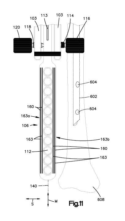
[0078] Referring now to Figs. 11-12, the aiming device 98 includes radio-
opaque
markers 160 present in the form of four wires 163 that can be arranged in two
pairs of wires
carried by the aiming arm 106 in the manner described above with respect to
Fig. 10A. An
intramedullary nail 602 that has been installed into a bone 608 is also
visible in the radiographic
image. The wires 163 of the radiographic image can be inspected to determine
whether each
wire 163 of a given pair 163a-b of wires is overlapped with respect to the
other wire 163 of the
given pair. If the wires 163 of each of the pairs 163a-b are not overlapped,
as illustrated in Figs.
11 and 12, then it can be concluded that the radiographic image is in a first
position that does not
reflect a desired view of the aiming arm 106 and nail 602, and alignment
between the aiming arm
- 20 -
CA 02837205 2013-11-22
WO 2012/162608 PCT/US2012/039574
106 and the nail 602 can not be reliably determined. Once the position of the
radiographic image
source has been corrected to a second position different than the first
position such that each wire
163 of the pairs 163a-b of wires overlap as illustrated in Fig. 13, it can be
concluded that the
second position of the radiographic image is a desired position that reflects
the desired view of
the aiming arm 106 and nail 602. For instance, the desired view can be along a
direction
substantially parallel to the central axes of the apertures 112 of the aiming
arm 106.
[0079] Once the desired view is established, visual inspection of the position
of the nail
602 relative to the arm can determine whether the aiming arm 106 and the nail
602 are properly
aligned. For instance, if the intramedullary nail 602 is substantially
equidistantly spaced
between, and substantially parallel to, the radio-opaque wires of the aiming
arm 106, it can be
concluded that the apertures 112 of the aiming arm 106 and the apertures 604
of the nail 602 are
operatively aligned as illustrated in Fig. 15. If, on the other hand, the nail
602 is not substantially
equidistantly spaced from and substantially parallel to the radio-opaque wires
of the aiming arm
106 as illustrated in Fig. 14, the aiming arm 106 can be deflected in the
manner described above
until the nail 602 and the aiming arm 106 are aligned as desired, such that
the apertures 112 and
604 substantially coincide. Screws or alternatively constructed bone anchors
can then be driven
through the apertures 144 of the mask 142 and the apertures 112 of the aiming
arm 106, and into
the corresponding aligned apertures 604 of the nail 602. Thus, the radio-
opaque markers 160 in
the form of wires 163 can define a first image characteristic with respect to
each other in the
form of less than an entire overlap of at least one of the wires with respect
to another of the
wires, wires of a pair of wires that are spaced along the transverse direction
T, which is
substantially perpendicular to the major direction M and the select direction
S, and a second
radiographic image characteristic with respect to each other, such as an
entire overlap of at least
one of the wires with respect to another of the wires, when the radiographic
image source is in
the desired alignment with respect to the aiming arm 106.
[0080] Referring to Fig. 16, the aiming device 98 can include radio-opaque
markers
160 in the form of two plates 165 that are carried by the aiming arm 106 at a
location on opposed
sides of the apertures 112 with respect to the select direction, as described
above with respect to
Fig. 10B. Thus, the radiographic image can be taken from a first position
whereby the imaged
thickness of the plates 165 along the select direction S has a first size,
which can be less than or
greater than the desired size so as to indicate that the first position is not
in a desired position.
The radiographic image can be moved to a second position different than the
first position until
the thickness of the plates along the select direction S has a second size
that is substantially equal
to a desired size. In one example, each of the plates 165 can be dimensioned
greater in the
- 21 -
CA 02837205 2013-11-22
WO 2012/162608 PCT/US2012/039574
transverse direction T than in the select direction S, such that the desired
imaged size of the
thickness of the plates 165 along the select direction S can be a minimal
thickness, equal to the
actual thickness of the plates 165 along the select direction S.
Alternatively, each of the plates
165 can be oriented in the body of the aiming arm 106 such that the plates 165
can be
dimensioned greater in the transverse direction S than in the transverse
direction T, such that the
desired imaged size of the thickness of the plates 165 along the select
direction S can be a
maximum thickness, equal to the actual thickness of the plates 165 along the
select direction S.
Once the imaged size of the thickness of each of the plates 165 is
substantially equal to the
desired imaged size, for instance minimized or maximized, the aiming arm 106
can be positioned
such that the nail 602 is positioned substantially equidistantly between and
substantially parallel
to the plates 165, such that the bone anchors can be driven through the
apertures 112 of the
aiming arm 106 into the apertures 604 of the nail 602. Thus, the radio-opaque
markers 160 in
the form of plates 165 can define a first image characteristic in the form of
a thickness (such as a
greater than a minimum thickness or less than a maximum thickness) of the
plates 165 along the
select direction S when the radiographic image source is in a first position
with respect to the
aiming arm 106, and a second radiographic image characteristic, such as a
minimum thickness or
a maximum thickness along the select direction S, when the radiographic image
source is in a
second desired position with respect to the aiming arm 106 that is different
than the first position
and in a desired alignment with respect to the aiming arm 106.
[0081] Referring now to Fig. 17, the aiming device 98 includes radio-opaque
markers
160 present as a wire 163 and a plate 165 that are carried by the aiming arm
106 at a location non
opposed sides of the apertures 112, as illustrated in Fig. 10C. Thus, the
radiographic image can
be taken from a firs position whereby the thickness of the plate has a size
that can be greater than
or less than desired as described above with respect to the plates 165 in Fig.
10B. The
radiographic image can be repositioned to a second position, whereby the
imaged size of the
thickness of the plate 165 along the select direction S is desired, such as a
minimized or
maximized thickness, and the aiming arm 106 can be positioned such that the
nail 602 is
positioned substantially equidistantly between and substantially parallel to
the wire 163 and the
plate 165, such that the bone anchors can be driven through the apertures 112
of the aiming arm
106 into the apertures 604 of the nail 602. Thus, it should be appreciated
that the aiming device
98 can include at least one radio-opaque material configured in accordance
with any desired
embodiment that has a first configuration, such as a shape, when viewed from
an undesired angle
(such as oblique to the central axes of the apertures 112) and a second
configuration, such as a
- 22 -
CA 02837205 2013-11-22
WO 2012/162608
PCT/US2012/039574
second shape, when viewed from a desired angle (such as substantially parallel
to the central
axes of the apertures 112).
[0082] The foregoing description is illustrative only and does not limit the
scope of the
present disclosure. Embodiments not described above may nonetheless be within
the scope of
the appended claims.
-23 -