Note: Descriptions are shown in the official language in which they were submitted.
CA 02837348 2013-11-26
IMMUNOMODULATORY AGENT, COMBINATIONS, USES THEREOF AND
IMMUNOTHERAPY METHOD FOR RECONTEXTUALISING, REPROGRAMMING AND
RESTORING IMMUNE SYSTEM IN REAL TIME
FIELD OF THE INVENTION
The present invention relates to immunogenic compositions for modulating the
immune
system comprising a therapeutically effective amount of two or more
immunoactive
antigenic agents presenting pathogen-associated molecular patterns (PAMPS)
and/or
danger associated molecular patterns (DAMPS) and one or more physiologically
acceptable carriers, excipients, diluents or solvents.
Preferably the compositions of the present invention comprise immunoactive
antigenic
agents presenting pathogen-associated molecular patterns (PAMPS) and/or danger
associated molecular patterns (DAMPS) selected from the group consisting of:
(A)
antigenic agents with molecular patterns associated with bacteria; (B)
antigenic agents with
molecular patterns associated with viruses; (C) antigenic agents with
molecular patterns
associated with fungi and yeasts; (D) antigenic agents with molecular patterns
associated
with protozoa; (E) antigenic agents with molecular patterns associated with
multicellular
parasites / or (F) antigenic agents with molecular patterns associated with
prions.
It is another aspect of the present invention the use of immunogenic
compositions in the
manufacture of medicaments for prevention and/or treatment of infectious
diseases,
autoimmune diseases, allergic diseases, inflammation, arthritis, inflammatory
diseases,
transplant rejection, diseases caused by vascular disorders, diseases caused
by
hemorrhagic or ischemic cardiovascular events, ischemia, infarction and
hemorrhage
leading to tissue destruction, cardiac, renal, respiratory or liver disease,
cancer, tumors and
malignant and benign lesions.
The immunogenic compositions of the invention are also directly used in the
prevention
and/or treatment of infectious diseases, autoimmune diseases, allergic
diseases,
inflammation, arthritis, inflammatory diseases, transplant rejection, diseases
caused by
vascular disorders, cardiovascular diseases caused by injury or bleeding
ischemic,
ischemia, infarction and hemorrhage leading to tissue destruction, cardiac,
renal,
1
CA 02837348 2013-11-26
respiratory or liver disease, cancer, tumors and malignant and benign lesions.
The present invention further relates to methods for inducing cell
regeneration, tissue
regeneration, organ regeneration and regeneration of organic systems such as
the
circulatory system, nervous system and endocrine system.
Finally, the present invention relates to methods for renewal of the immune
response in an
animal, particularly humans.
BACKGROUND OF THE INVENTION
Of the discovery of antibiotics and other drugs
From the pioneering discovery of antibiotics in the second half of the 20th
century, new
antibiotics, semi-synthetic antibiotics and new chemotherapeutics with
antimicrobial
activity, have been developed on a large scale against most intracellular and
extracellular
bacteria. These developments have changed the history of medicine, allowing it
to reach a
wide spectrum of healing, for the vast majority of bacterial infectious
diseases, which
racked humanity.
Thus, the discovery of antibiotics was a major milestone, a watershed, because
infection
could be addressed and healed, in a specific way, with a clear relationship of
cause and
effect, and measurable when established. This discovery greatly expanded the
ability of
healing in medicine, with enormous positive impact on human health and
lifespans. The
discovery of antibiotics in the evolution and treatment of disease profoundly
influenced the
research and thinking of researchers from the success achieved by this
experimental
model (Reeves G, Todd I. Lecture notes on immunology. 2nd ed: Blackwell
Scientific
Publications, 1991; Neto VA, Nicodemo AC, Lopes HV. Antibioticos na pratica
medica. 6th
ed: Sarvier, 2007; Murray PR, Rosenthal KS, Pfaller MA. Microbiologia Medica.
5th ed:
Mosby, 2006; Trabulsi LR, Alterthum F. Microbiologia. 5th ed: Atheneu Editora,
2008) .
Antibiotics were succeeded by the development and use of antifungal,
antiparasitic and
antiviral drugs. There was also the development of antineoplastic, cytostatic
and cytotoxic
drugs against malignant tumors, anti-inflammatory, anti-allergic and
immunosuppressive
2
CA 02837348 2013-11-26
drugs (anti-lymphocytes, neutralizing anti-leukocytes of the immune system) of
hormonal
and non-hormonal nature, with a huge range of applications, as in for
infectious diseases,
for inflammatory diseases of any origin, for autoimmune diseases, for genetic
diseases, for
vascular diseases, for allergic diseases, for trauma, for neoplastic diseases,
for hormonal
diseases, for degenerative diseases, among others.
Thus, the new drugs brought an enormous capacity for medical intervention,
with
numerous benefits, with definitive and partial cures, with the prolongation of
life in incurable
diseases, but with a huge morbidity due to side effects related to their lack
of specificity to
the pathophysiology of diseases treated.
Of the innate immunity
The innate immunity, in addition to preventing the entry of microorganisms and
preventing
their establishment, has another recently discovered vital function:
discrimination between
"self" and "not self" and the pattern recognition capability linked to the
alarm and the
command to start or inhibit an integrated immune response against an invading
microorganism or to arrest, repair or inhibit a condition of destruction or
self aggression to
the body, for example, in trauma, autoimmune diseases and allergic diseases,
among
others. This dual capability was previously erroneously attributed exclusively
to adaptive
immunity. The innate immunity, through its own receptors, recognizes invading
pathogenic
microorganisms, autologous or even allogeneic neoplastic cells, or allogeneic
or
heterologous transplants as "not self', identifying them as not belonging to
the organism.
From that moment, it triggers an alarm and a joint innate and adaptive immune
response to
eliminate them or suppress a response deleterious to the human or animal
organism
(Goldsby RA, Kindt TJ, Osborne B. lmunologia de kuby. 6 ed: ARTMED; 2008, 704
p;
Janeway C, Travers P, Walport M, Slhlomchik MJ. lmmunobiology five. 5 ed:
Garland Pub.;
2001. 732 p.; Voltarelli JC . Imunologia clinica ha pratica medica: atheneu
editora; 2009;
Janeway CA, Jr. , Medzhitov R. Innate immune recognition. Annual review of
immunology.
2002;20:197-216. Epub 2002/02/28; Matzinger P. The danger model: a renewed
sense of
self. Science. 2002;296 (5566) :301-5. Epub 2002/04/16; Steinman RM,
Banchereau J.
Taking dendritic cells into medicine. Nature. 2007; 449 (7161) : 419-26. Epub
2007/09/28.;
3
CA 02837348 2013-11-26
Beutler BA. TLRs and innate immunity. Blood. 2009; 113 (7): 1399-407. Epub
2008/09/02;
Moresco EM, LaVine D, Beutler B. Toll-like receptors. Current biology: CB.
2011 ; 21 ( 13
) : R488-93. Epub 2011/07/12).
The (default/standard/pattern?) recognition, of "not self', of an invasive
germ, of a
neoplastic cell or an altered or transplanted cell is performed by sentinel
cells, represented
by epithelial cells, mucosal cells, and the stromal cells, such as pericytes,
dendritic cells,
macrophages and fibroblasts, among others. These cells, strategically
distributed
throughout the body, have PRRs (Pattern Recognition Receptors) and DRRs
(Danger
Recognition Receptors) which are receptors respectively able to recognize a)
standard
identification molecules, characteristic of a wide range of microorganisms,
and b) certain
patterns for chemical and physical of said inert substances and changes to
metabolic
stress, such as release of free radicals and tissue chemical changes, caused
by ionizing
radiation or by chemical substances, among others.
The PRR does not discriminate one specific individual microorganism, but the
presence of
microorganisms other than the human body. Each PRR receiver may bind to
several
different pathogens, recognizing as PAMPs (Pathogen Associated Molecular
Patterns)
carbohydrates, lipids, peptides and nucleic acids from bacteria, viruses,
fungi or parasites
that are not found in the human or animal body.
The DRRs discriminate that there is tissue damage, a dangerous situation
caused by not
live or inert agents. The DRRs identify DAMPs (Danger Associated Molecular
Patterns)
associated with tissue damage by toxic substances, radiation, or trauma, which
cause
metabolic stress, release of free radicals and chemical changes in tissue,
recognized by
these receptors. (Janeway C, Travers P, alport M, Slhlomchik MJ. lmmunobiology
five. 5th
ed: Garland Pub.; 2001. 732 p.; Matzinger P. The danger model: a renewed sense
of self.
Science. 2002 ; 296 ( 5566) : 301-5. Epub 2002/04/16; Beutler BA. TLRs and
innate
immunity. Blood. 2009;113 (7) : 1399-407. Epub 2008/09/02; Moresco EM, LaVine
D,
Beutler B. Toll-like receptors. Current biology: CB. 2011;21 (13) :R488-93.
Epub
2011/07/12).
4
CA 02837348 2013-11-26
Thus, sentinel cells via their PRRs and their DRRs, have a role in the
breakdown of which
belongs ("self") and which is does not belong (not "self') and triggering
inflammation and
immune response, via recognition of PAMPs of invading pathogens and DAMPs
caused by
neoplastic cells and toxic substances or modifications due to trauma, leading
to a situation
of real danger to the human and animal organism.
Immediately, these activated sentinel cells give alarm signals, triggering the
innate immune
response through the NF-kB (Nuclear Factor-kB) signal translation system,
leading to the
secretion of pro-inflammatory cytokines and the IRF signal translation system,
that
produces Type I alpha and beta interferons. These cytokines, together, acting
on cells and
vessels, cause a local inflammatory process, initially to contain the invading
agent,
autologous (tumor cell), heterologous (microorganisms, prions, grafts and
transplants) or
allogeneic (grafts and transplants), or to repair danger situations. This
contention happens
through antibodies, pre-existing, opsonizing acute phase proteins and through
leukocytes
and macrophages, which engulf and start to destroy the extracellular and
intracellular
microorganisms respectively, or eliminating other etiologic agents of any
kind.
In gout, in silicosis, the chemical aggression, in foreign body granulomas, in
trauma, the
inflammatory process is formed to eliminate the offending agent if possible
and then induce
tissue healing and regeneration. When the offending agent is not eliminated
the
inflammation is perpetuated and causes an incurable or uncontrollable chronic
inflammatory disease, stable or progressive, which compromises the life or
health of
patients.
Interaction and integration of innate immunity with adaptive immunity
Simultaneously at the site of invasion, aggression and inflammation, the
innate immunity
sentinel cells with the APC role (Antigen Presenting Cells), such as dendritic
cells and
macrophages, phagocytosise and pinocytosise microorganisms or tumor cells, or
transplanted cells, among other aggressors and process their antigens. These
APC cells
pulsed by the antigens migrate to regional lymph nodes and activate them. The
APC cells
CA 02837348 2013-11-26
in reactive lymph nodes, activated and mature present the antigens to
lymphocytes,
secrete cytokines and thereby induce, coordinate, polarize, amplify and
maintain an
adaptive immune response specific to the invading germs, or neoplastic cells,
or to
transplanted cells, or other offending agent, allowing them to be fought and
eliminated,
where feasible and the consequent cure of the infection or inflammation and
repair and
regeneration or wound healing.
Thus, these immune mechanisms fight disease through the innate and adaptive
responses
in an integrated and synergistic way, performed by sentinels cells, APC
function sentinels,
and innate immunity effectors, cellular and molecular in conjunction with the
cellular and
molecular effectors of adaptive immunity that are respectively lymphocytes,
cytokines and
antibodies.
Thus, the interaction of the two immunities, innate and adaptive, in the
context of an
infection or immune response against an aggressor of any kind helps to combat
the
disease in an integrated and synergistic way. The integration of the two
initially occurs by
the action of the innate immunity cells with APC function, such as dendritic
cells and
macrophages, but mainly by the activity of dendritic cells, as they are the
ones that are
able to initiate an adaptive immune response against a primary infectious or
parasitic
agent, effectively protecting the body.
As noted macrophages also function as APC cells, but are more specialized and
involved
as part of the effector loop in phagocytosis and in the elimination of
microorganisms. B
lymphocytes, when mature, are also APC cells and its most well-known action is
the
presentation of antigens to the T lymphocytes, within the framework of
cooperation of both
lymphocytes to produce antibodies against T-dependent antigen, and the
secondary
antibody response. Macrophages, like other myeloid cells, are also involved in
suppressing
immune response in cancer and in incurable chronic infections. In these cases
its
performance is unfavorable to the defense of the organism because it
suppresses the
immune response and create tumor facilitation. A malignant tumor disease is
characterized
by causing an initial silent inflammation, imperceptible, and in the end it
becomes
extremely pro inflammatory and symptomatic through the TH17 profile
inflammatory tissue,
6
CA 02837348 2013-11-26
which usually leads the patient to prolonged suffering.
When co-stimulatory molecules are not expressed on the APC cell surface, by
the absence
of the alarm signal characterized by the lack of activation of PRRs by PAMPs
and DAMPs,
only the first signal occurs, given by the TCR. After the TCR binds with the
antigen, in the
absence of the second signal, the T lymphocyte becomes tolerant to the
specific antigen
shown and aborts the immune response.
On the other hand, the CD 40L molecule of activated T lymphocytes, when it
binds to the
CD40 molecule on the APC cells, significantly increases the expression of CD80
and CD86
molecules, increasing the current response, which thus occurs only when the
adaptive T
response is already engaged in defending the body. The third signal given by
cytokines
such as IL-1, is given only by the APC cells after the binding of co-
stimulatory molecules
and the emission of the second signal. The IL-1 secreted by the APC cells acts
on
lymphocyte cells and leads to the complete expression of the receptor for IL2
and to the
production of cytokines by virgin or memory lymphocytes engaged in response to
the
initiating clonal expansion.
Therefore the activation of innate immunity by pathogens is the key to
unleashing the
second and third signals and the occurrence of a potentially effective
immunity, through the
full activation of T lymphocytes engaged in the response. Without the
occurrence of the
second and third signal, the response is aborted and generates a tolerance
specific to the
antigen presented.
At the same time that the neutrophils, monocytes and macrophages initiate
combat to
bacteria and to other infectious agents by the linkage of PAMPs with PRRs on
antigen
presenting cells (APC), they activate dendritic cells and macrophages, local
and newly
arrived. These cells phagocytosise and pinocytosise bacteria and bacterial
antigens,
processing them and starting the maturation process. The activated and
maturing dendritic
cells now migrate to regional lymph nodes to present antigens and initiate
immune
response against the invading agent.
The mature antigen-pulsed APC cells, especially dendritic cells, in lymph
nodes,
7
CA 02837348 2013-11-26
collaborate with the T and B lymphocytes and initiate the adaptive response.
Dendritic cells
are the most potent cells for the presentation of antigens and the only APC
cells able to
activate a virgin CD4 T lymphocyte and to start a new immune response.
After a period of approximately seven days in the lymph node, the
collaboration between
blank CD4 lymphocytes, which become T CD4 Th2, with B lymphocytes and antigen
presenting dendritic cells, initiates the differentiation of specific
sensitized B lymphocytes.
These B cells, now activated, recognize bacterial antigens by surface
immunoglobulins,
after contact with these antigens, proliferate, mature, and differentiate into
plasma cells that
now secrete specific antibodies against this bacterium. Infections of all
types, bacterial,
viral, fungal and parasitic may, in general, in the acute phase, evolve to a
full cure with
regeneration and healing, or for a cure with sequelae. They can also develop
into an
incurable chronicity, with or without control of the disease, to chronicity
with healing, with or
without sequelae, or to death.
Polarization of the immune response
The immune profiles known and induced by dendritic cells by direct and
indirect contact
with the different cytokines and generated by T CD4 cells are of four types:
a) cellular Thl profile, which generates cellular immunity mediated by cells;
b) humoral Th2 profile, which generates humoral immunity mediated by
antibodies;
c) tissue or inflammatory Th17 profile, which generates inflammatory tissue
immunity, also
mediated by cells and cytokines, which induce an important inflammation for
the
elimination of certain pathogens, and
d) Treg/Trl profile, which suppresses the immune response and controls, by
inhibiting the
other three profiles described above, ensuring the return of the body
equilibrium state.
e) profiles not yet fully established, as the Tfh (follicular Helper) of the
humoral response,
the Th9 profile for certain parasites, or other profiles that may be
discovered.
8
CA 02837348 2013-11-26
Thus, the various profiles ensure the defense of the organism and the
elimination of
causative heterologous (infectious) agents invading and colonizing autologous
(neoplasia).
The last profile ensures the termination of the immune response, the balance,
the
regeneration, the safe return to normalcy and it prevents self-injury and
allergy and is
therefore vital to the health and preservation of the human species and
animal, as much as
the other profiles.
The phenomenon of polarization of the immune response is defined as the
predominance
of a certain immunological profile such as Thl or Th2 at the expense of other
profiles that
become secondary or null. This phenomenon happens according to the type of
attack
suffered by the body. That is, according to the type of infection, pathology,
and infection
stage or pathology stage, the different type of immune response will be
predominant, and it
may be a cellular, humoral, tissue, or immune-regulatory response, while other
types of
immune responses are inhibited, resulting in the phenomenon of polarization.
By definition, in polarization a profile is dominant, but other non-dominant
profiles are also
needed, and expressed in a complementary manner that will help eliminating the
disease.
For example, tuberculosis is the appearance of Th17 cells in the lung which
allows Thl cells
to settle and may lead to cure this infection in the lung parenchyma
(Stockinger, B. and
Veldhoen, M. Differentiation and function of Th17 T cells. Current Opinion in
Immunology,
19 (3), pp. 281-286. 2007). In viral infections, the CTL cells of Thl profile
destroy cells
infected by viruses, to eliminate the virus. However, antibodies are required
to prevent the
virus from infecting other healthy cells and thus preventing the spread of
infection. The
coordinated assembly of the two profiles is essential for the healing of
certain viral
infections. Certain intestinal infections by extracellular Gram-negative
bacilli require, for its
cure, in the final stage, besides the Th2 profile, the generation of a
supplementary Th17
profile capable of generating a strong inflammation, necessary to eliminate
this type of
bacteria.
In conclusion, due to the fact that the dendritic cells are the only
professional APC cells
capable to initiate a primary adaptive immune response and are the most potent
in
triggering a secondary specific immune response, in any profile, they are then
commanding
9
CA 02837348 2013-11-26
the interaction and integration of innate immunity with adaptive immunity to
produce an
effective immune response capable of curing a disease. Dendritic cells in
collaboration with
other APC and sentinel cells in contact with different aggressors in different
functional
states, in the inflammation sites, in the lymph nodes, in the spleen, in the
mucous
membranes, are able to lead, coordinate, polarize, and amplify the adaptive
immune
response, primary and secondary, e.g., specific for the peptides of invading
pathogens,
which in this case is the most appropriate for the removal of the ongoing
infection.
Therefore, dendritic cells and other APC cells are key cells of the innate
immune response,
since they evaluate the nature of the autologous and heterologous causative
agent, i.e.,
the type of pathogen or colonizing cells and aided by the sentinel cells, they
measure and
evaluate the size of the heterologous or autologous aggression, its extension,
its intensity
and aggressiveness, besides commanding the adaptive response with the profile
and the
intensity required for the elimination of the pathogen.
After differentiation, a re-differentiation can occur, induced by the
microenvironment and/or
the type of antigen or its presentation, in which a Thl or Th2 profile can be
exchanged for
an inflammatory profile or an immunosuppressant profile or vice versa. This
extreme
plasticity of the immune system to differentiate or re- differentiate in
either direction
indicates a strategic window for manipulation of the immune system, during
infection, when
the direction taken by the polarization is not the best one for curing the
infection process or
neoplasia.
As an illustrative example, we have what happens in a severe infection or
septicemia,
when the massive inflammation caused by the large number of microorganisms
which
touch the sentinel cells throughout the body, induces also a Th17 a profile,
which in turn
increases the inflammation more and therefore becomes detrimental, leading to
tissue
destruction, rather than inducing healing. In these cases the Th17 profile, by
tissue
destruction and the amplification of inflammation, is mainly responsible for
the generation
of clinical complications such as severe ARDS (acute respiratory distress
syndrome in
adults), lung shock, renal failure, or shock, that compromises healing.
CA 02837348 2013-11-26
The re-differentiation of polarization for the Thl or Th2 profiles, with the
inhibition of
massive inflammation, is the logical and strategic path for a designed or
prepared
immunotherapy to try to resolve this dramatic and deadly type of situation,
during a severe
infection or sepsis, which has a significant mortality and morbidity and for
which antibiotics
and other antimicrobials, in current patterns such as single mode, have
disappointing anti-
infective results. The same example applies to serious intra cellular
bacterial, fungal, viral
and parasitic infections, with extensive tissue destruction and massive
inflammation,
usually of poor prognosis.
The use of adjuvants to stimulate immune response
The human and animal organisms do not usually produce antibodies against
soluble
proteins, necessitating the use of so-called nonspecific or unrelated
adjuvants to obtain the
desired immune response. These adjuvants used since the dawn of immunology, in
immunizations and in vaccine applications, were and are made up of parts of
microorganisms, mineral oils and other substances that activate the innate
immunity, which
then gives the alarm and control necessary for the development of desired
immune
response to the protein or to the vaccine in question (GOLDSBY RA, KINDT TJ,
OSBORNE BA. IMUNOLOGIA DE KUBY. 6 ed: ARTMED; 2008. 704 p) ; (Janeway C,
Travers P, alport M, Slhlomchik MJ. Immunobiology five. 5 ed: Garland Pub.;
2001. 732 p.);
(VOLTARELLI JC . IMUNOLOGIA CLINICA NA PRATICA MEDICA: ATHENEU EDITORA;
2009); (Janeway CA, Jr., Medzhitov R. Innate immune recognition. Annual review
of
immunology. 2002;20:197-216. Epub 2002/02/28.); (Matzinger P. The danger
model: a
renewed sense of self. Science. 2002 ; 296 ( 5566) : 301-5. Epub 2002/04/16.):
(Steinman
RM, Banchereau J. Taking dendritic cells into medicine. Nature. 2007 ; 449 (
7161 ): 19-26.
Epub 2007/09/28.); (Beutler BA. TLRs and innate immunity. Blood. 2009 ; 113 (
7 ): 1399-
407. Epub 2008/09/02.); (Moresco EM, LaVine D, Beutler B. Toll-like receptors.
Current
biology: CB. 2011 ; 21 ( 13 ) : R488-93. Epub 2011/07/12).
It should be noted that the use of adjuvants for immunization, despite being
one of the
oldest features, and still current, highly used and essential for vaccinations
and for studies
of immunology, was considered only as a useful nonspecific effect. It was not
envisioned,
11
CA 02837348 2013-11-26
for more than a century, its role in the innate immunity in the discrimination
of what is "Self"
and not "Self' and its unique and fundamental capacity to the survival of the
human
species and animals: to give the alarm signal and the command to start or not
start, or
inhibit, an integrated, protective or healing, innate and adaptive, immune
response
(GOLDSBY RA, KINDT TJ, OSBORNE BA. IMUNOLOGIA DE KUBY. 6 ed: ARTMED;
2008. 704 p); (Janeway C, Travers P, Walport M, Slhlomchik MJ. Immunobiology
five. 5
ed: Garland Pub.; 2001. 732 p. ) ; (VOLTARELLI JC. IMUNOLOGIA CLINICA NA
PRATICA MEDICA: ATHENEU EDITORA; 2009); (Janeway CA, Jr., Medzhitov R. Innate
immune recognition. Annual review of immunology. 2002;20:197-216. Epub
2002/02/28.);
(Matzinger P. The danger model: a renewed sense of self. Science. 2002;296
(5566) : 301-
5. Epub 2002/04 /16.) : (Steinman RM, Banchereau J. Taking dendritic cells
into medicine.
Nature. 2007 ; 449 ( 7161 ): 419-26. Epub 2007/09/28.); (Beutler BA. TLRs and
innate
immunity. Blood. 2009 ; 113 ( 7 ): 1399-407. Epub 2008/09/02.); (Moresco EM,
LaVine D,
Beutler B. Toll-like receptors. Current biology : CB. 2011 ; 21 ( 13 ) : R488-
93. Epub
2011/07/12).
Today's anti-infective and anti-cancer treatments
A large number of medical materials, labor hours, medicines and hospital beds
could be
better used with a therapy that assessed, prioritized and optimized the
variables that affect
the displacement of the biological balance in favor of the patient and
modulated his or hers
immune system, decreasing its inefficiencies and allowing for a large number
of medical
discharges, in less time. The prior art has yet to provide alternatives to
perform an intended
repolarization of the immune system in real time, or time to change or reverse
its response
to a ongoing disease or illness, if possible to improve the quality of life,
or extend the life
span, or assist the process of combating the ongoing disease or illness on the
patient.
Bacterial, fungal, viral, parasitic and neoplastic resistance to antibiotic,
antifungal,
antiparasitic and antineoplastic medicines used in clinical practice is seen
as the main
obstacle to the cure of bacterial, fungal, viral, parasitic and tumor
diseases, and is
considered a serious health problem on a global scale. This problem is
circumvented by
using appropriate and rational use of antibiotic, antimicrobial and anticancer
medicines and
12
CA 02837348 2013-11-26
with the advent of new more potent drugs. However, sooner or later, resistance
is always
inevitably established, and yet a solution to this problem has not been found.
As antibiotics,
antifungal, antiviral, antiparasitic and antineoplastic agents are considered
as the only valid
and effective anti-infective, antiparasitic and antineoplastic treatment
modalities, the
prospect of future treatments is disturbing and dark, due to the phenomenon of
microbial
and tumor resistance.
Antibiotics, antifungals, antivirals, antiparasitics and antineoplastic agents
can be used at
any stage of the infectious bacterial, fungal, viral, parasitic and tumor
conditions. However,
antibiotics, antimicrobial and anticancer fail to cure most advanced,
pervasive and severe
bacterial, fungal, viral, parasitic, and cancer conditions that have, in
general, a very high
rate of mortality and morbidity.
Furthermore, the discovery of new drugs is directed to drugs that are capable
of eliminating
the causative agent and cure infection, infestation and neoplasia based on the
concept of a
single drug capable of curing infectious, parasitic, and neoplastic disease.
Treatment of neoplasms at the present time
Cytokines such as interleukin 2 and type I alpha and beta interferons, are
used for the
treatment of immunogenic tumors such as melanoma and hypernephroma. Cytokines
a
bone marrow colonies growth factors function are used to combat anemia,
leucopenia,
cytopenias of the elements in the peripheral blood, caused by disease or
treatment, with
good results. Type I Interferons are widely used to combat viral hepatitis B
and C, with
good results, and with less significant results for the treatment of multiple
sclerosis.
Transplantation of allogeneic and autologous bone marrow is used for the
treatment of
cancer. Passive immunotherapies with CTL CD8 dendritic cells, white blood
cells,
autologous or allogeneic, with or without cytokine, are used for the treatment
of certain
tumors, and the results are still not very significant or significant at all,
but limited to certain
exceptional pathologies, as virus-induced tumors that grow in immunosuppressed
transplant patients. In these cases, passive immunotherapy with specific T CTL
CD8 e
CD4 cells for the EBV virus, are usually successful and cure these exceptional
tumors that
13
CA 02837348 2013-11-26
only grow in immunosuppressed patients. However, it is noteworthy that these
techniques,
as well as other similar but less effective ones, did not develop agents or
sets of agents
capable of effectively immuno-modulate the immune system in order to start
reacting
against any invading pathogen (infection), or autologous colonizer (tumors)
that is present
in the body of the individual to be treated, or that can resolve dysautonomia
states in the
primary or secondary genetic immune systems, that lead to states of self-harm
by the
immune system, which should defend the body from aggression.
Examples include successful cancer treatment that uses an immunomodulatory
agent
containing molecular patterns recognizable by the PPRs, the use of the BCG
vaccine as
one of the rare established techniques, which are accepted and proven, that
use
immunomodulation as a means of treatment. Brake and colleagues described the
use of
BCG immunotherapy in patients with superficial bladder cancer, who were in
Stage TI
(Brake M, Loertzer H, Horsch R, Keller H (2000) . "Long-term results of
intravesical bacillus
Calmette-Guerin therapy for stage TI superficial bladder cancer". UROLOGY 55
(5): 673-
678). lmmunotherapy was applied in patients after a complete transurethral
resection of
bladder tumor by applying a second cycle of BCG in the case of recurrent
superficial
tumors. The conclusion was that immunotherapy with BCG after transurethral
resection of
bladder tumors represents a highly effective primary treatment for TI stage
bladder cancer,
with a 89% rate of tumor-free survival in all patients.
Following this line, Burger and colleagues have demonstrated a randomized
comparative
essay, in which patients with noninvasive bladder cancer of the muscle layer
made use of
BCG or cell therapy with autologous macrophages (BEXIDEM
(Burguer M, Thiounn N,
Denzinger S, Kondas J, Benoit G, et . al (2010). "The application of adjuvant
autologous
antravesical macrophage cell therapy VS . BCG in non-muscle invasive bladder
cancer: a
multicenter, randomized trial. Journal of Translation Medicine, 8:54. doi:
10.1186/1479-
5876-8-54). Compared with BCG, the incidence of adverse events was
significantly lower
in the treatment BEXIDEM (26% and 14%, respectively). However, the recurrence
rate of
tumor in patients treated with BEXIDEM was significantly higher than in
patients who
received BCG as adjuvant therapy.
14
CA 02837348 2013-11-26
Donald et al described the use of BCG as a form of immunotherapy in patients
with
melanoma (Donald L. Morton, M.D., Frederick R. Eilber, , M.D., E. Carmack
Holmes, M.D.,
John S. Hunt, M.D., et. al (1974). "BCG lmmunotherapy of Malignant Melanoma:
Summary
of a Seven-year Experience". Ann. Surg. , p: 635-641). Patients selected for
the study had
recurrent melanoma, known residual disease, or high risk of developing
recurrence. First,
direct injections were applied in malignant nodules of melanoma using 0.1-0.5
cc of BCG in
each intracutaneous and subcutaneous lesion. Patients who were in Stage II
were treated
with BCG immunotherapy alone or with BCG and allogeneic melanoma cells. BCG
was
administered alone or as an adjuvant mixed with tumor cells in patients in
stage III disease.
Patients with intradermal metastases who were treated with intratumoral
injections of BCG
were those who responded better to treatment, and three factors appeared to be
correlated
with response to BCG immunotherapy: the location of metastasis, the amount of
tumor
present and the immunocompetence of the patient. There was low antitumor
activity of
BCG in patients with bulky disease or visceral metastases. The result showed
that 31% of
patients with intradermal metastases were found free of disease recurrence for
a period of
up to 6 years after the start of immunotherapy.
The immunotherapy described by Grant et al consists of the use of BEC2
(idiotypic
antibody which mimics the GD3 ganglioside present on the surface of most
tumors of small
cell lung cancer) in combination with BCG (Grant SC, Kris MG, Houghton AN,
Chapman
PB (1999) . "Long Survival of Patients with Small Cell Lung Cancer after
Adjuvant
Treatment with the Anti-ldiotypic Antibody BEC2 Plus Bacillus Calmette-Gue".
Clinicai
Cancer Research, Vol. 5, 1319-1323). The applied dose in patients with lung
cancer was
2.5 mg for a period exceeding 10 weeks. Patients treated with immunotherapy
had a
significant increase in survival and survival free of recurrence of the
disease when
compared to a similar group of patients.
Popiela et al evaluated the use of BCG immunotherapy and chemotherapy with FAM
(5-
fluorouracil, adriamycin, mitomycin C) in patients with stage III or IV
gastric cancer, that
previously underwent curative gastrectomy for that cancer (Popiela T, Kulig J,
Czupryna A,
Sczepanik AM, Zembala M (2004). " Efficiency of adjuvant immunochemotherapy
following
CA 02837348 2013-11-26
curative resection in patientes with locally advanced gastric cancer". Gastric
cancer, 7:
240-245). Patients were randomly divided into 3 groups: BCG + FAM, FAM, and
control
(surgery only). The dose of BOG immunotherapy was administered at 2-4 viable
units per
dose. It was observed in general a 10 year survival rate of 47.1% in the
immunotherapy
group. Powles and colleagues reported a study in which patients with acute
myeloid
leukemia were treated with BOG and dead allogeneic tumor cells. The dose of
BOG was
estimated to be about 106 organisms (RL Powles, PJ Selby, DR Jones, JA Russel,
HG
Prentice, et. al (1977). "Maintenance of remission in acute myelogenous
leukemia by a
mixture of B. C.G. and irradiated leukemia cells". THE LANCET, 1107-1110).
Improvement
was observed in patients, who showed remission for a period, leading to the
conclusion
that the immunotherapy with a combination of leukemia tumor cells and BOG may
be
effective to prolong remission. Likewise Vuvan and et al described the use of
BOG
immunotherapy in patients with acute non-lymphocytic leukemia (H. Vuvan,
D.Fiere, M.
Doillon, C. Martin, B. Coiffier, et. al (1978). " BOG Therapy in Acute Non
Lymphoid
Leukaemias". Scand J Haematol 21, 40-46). A randomized study was conducted in
which
patients were divided into 2 groups: treated with chemotherapy alone and
treated with
chemotherapy and BOG, the BOG being administered during the interval of
chemotherapy
cycles at doses of 6 x 10⁸ viable units. The result showed that patients
receiving
immunotherapy had a higher survival rate as compared to the group receiving
only
chemotherapy. Furthermore, it was observed that BOG appeared to be more
effective in
patients older than 40 years.
Finally, Hsueh et al used a therapeutic vaccine consisting of melanoma cells
called
Canvaxin (Hsueh ED, Essner R, Foshag LJ, 01lila DW, Gammon G, et al (2002)
Prolonged Survival After Complete Resection of Disseminated Melanoma and
Active
lmmunotherapy with a Therapeutic Cancer Vaccine". Journal of Clinicai
Oncology, Vol 20,
n 23, pp 4549 - 4554). All patients were tested with PPD (purified protein
derivative) before
receiving therapy with the vaccine. For the first two treatments, the vaccine
was mixed with
BOG. In the first injection, BOG was applied in a dose from 2.7 to 10.8 x 10⁶
colony forming
units in PPD negative patients and half this dose in PPD positive patients.
There was a
prolongation of survival in patients who received, after surgery, active
immunotherapy with
16
CA 02837348 2013-11-26
Canvaxin.
The aforementioned studies with the use of BCG, although they are using an
immunostimulating agent separate from the causative agent of the disease or
disorder to
cause desirable effects in patients, whether or not in combination with other
medical
procedures and treatments as proposed in the present invention, are not
however taking
advantage of using multiple antigenic components that are associated with
distinct
pathogen molecular patterns, especially a combination that represents
intracellular and
extracellular bacteria, viruses, parasites, fungi and yeasts. The
aforementioned research
groups and studies only used BCG in a simple adjuvant function without taking
into
account the basis of the present invention that aim to activate memory or
blank cells, which
can be inactivated throughout various body tissues through a wide range of
pathogen
associated molecular patterns that can enable the largest possible number of
memory and
effector cells. By not presenting this combination of distinct antigenic
nonspecific agents
able to stimulate innate and specific immunity as described, many populations
of immune
memory cells will no longer be activated according to the arguments presented,
which will
not lead to a recontextualization, renewal and reprogramming of the immune
response,
that is as effective as presented herein.
Neither the state of the art describes the importance of immunization
protocols and of the
local and distal applications of immune-stimulatory agents, and how a lot of
applications in
different parts of the body are necessary, for in a programmed and intentional
way, cause
the PAMPS and DAMPS molecular patterns to reach the tissues that hold the APC
cells in
adequate quantity and quality to provoke optimal response and polarization.
Tanaka et al (Tanaka N., Gouchi A. Ohara T., Mannami T . , Konaga E.,
Fuchimoto S.,
Okamura S., Sato K., Orita K (1994). "Intratumoral injection of a
streptococcal preparation,
OK-432, before sugery for gastric cancer. A randomized Trial . Cooperative
Study Group of
Preoperative Intratumoral Immunotherapy for Cancer". Cancer, 74(12): 3097-
3103) and
Yasue and et al (Yasue M. , Murakami M., Nakazato H., Suchi T., Ota K (1981).
"A
Controlled Study of Maintenance Chemoimmunotherapy VS lmmunotherapy Alone
Immediately Following Palliative Gastrectomy and Induction Chemoimmunotherapy
for
17
CA 02837348 2013-11-26
Advanced Gastric Cancer". Cancer Chemother Prasmacol, 7: 5-10.) report the use
in
gastric cancer patients of an immunomodulatory agent prepared from attenuated
Streptococcus pyogenes called OK-432. Such an agent is able to activate the
immune
system and cause regional degeneration of the affected tissue in stomach
carcinomas.
Tanaka describes the preoperative use of 10KE of OK-432 injected
endoscopically, and
doses of 1KE to 5KE in intradermal injections in case of metastases in the
lymph nodes,
post-operation. Tanaka concluded that intratumoral injections of OK-432 may
have a
beneficial clinical effect in patients who are in Stage III gastric cancer,
because it seems to
improve survival in this subgroup of patients. Yoshida et al (Yoshida K.,
Sugiura T., Takifuji
N . , Kawahara M . , Matsui K. , et al (2007). "Randomized phase ll trial of
three intrapleural
therapy regimens for the management of malignant pleural effusion in
previously untreated
non-small cell lung cancer: JCOG 9515. Lunger Cancer, 58 : 362-368) evaluated
the
efficacy and toxicity of OK-432 (0.2 KE/kg, and the maximum dose 10KE/Kg) as
pleural
therapy in control of malignant pleural effusion in patients with non-small
cell lung cancer,
previously untreated. Apart from OK-432, bleomycin and cisplatin with
etoposide, were also
assessed as intrapleural therapy. It was concluded that the best intrapleural
therapy used
was the use of OK-432, because it was the one that had the best survival rate,
free of
disease, and the lowest rate of pleural recurrence.
Aftergut et al (Kent Aftergut, MD, Mary Curry, MD, Jack Cohen, DO (2005) .
"Candida
Antigen in the Treatment of Basal Cell Carcinoma". Dermatol Surg, 31: 16-18)
studied the
intralesional use of Candida antigens in the treatment of basal cell
carcinoma. The study
shows that 56% of patients had complete regression of tumor cells. The
antigens were
administered in doses of 0.1 mg via intradermal injection. Again, the present
invention is
distinguished by the use of a very elaborate and more complex combination of
antigenic
components, having the potential to achieve more favorable results when used
alone or in
combination with other therapies. The study described by Miles et al (Miles
DW, Tow[son
KE, Graham R, Reddish M, Longenecker BM, et al. (1996). "A randomised phase ll
study
of sialyl-Tn and DETOX-B adjuvant with or without cyclophosphamide
pretreatment of the
active specific immunotherapy of breast cancer". British Journal of Cancer,
74:1292-1296)
investigated the occurrence of improvement in the immune response caused by
the
18
CA 02837348 2013-11-26
association of sialyl-Tn-KLH with DETOX-B (containing in its composition
Mycobacterium
phlei cell wall skeleton) in patients with breast cancer, when subjected to a
pre-treatment
with low doses of cyclophosphamide. An emulsion of 0.5 ml, composed of STN-KLH
with
DETOX-B was used. As a result, it was observed that all the patients developed
IgM and
IgG against the sialyl-Tn, and patients who received a cyclophosphamide
pretreatment had
a significantly greater increase of IgM. Korec et al present a study in which
11 patients with
different tumor types and 3 patients with thrombotic thrombocytopenia purpura
associated
with mitomycin C, were treated with a extracorporeal plasma perfusion through
filters
containing Staphylococcus aureus immobilized protein A (Korec S, Smith FP,
Schein PS,
Phillips TM (1984) . "Clinicai experiences with extracorporeal immunoperfusion
of plasma
from cancer patients". J Biol Response Mod. 3(3): 330-5). As a result, there
was a modest
antitumor effect generated by immune-perfusion. In 10 properly treated
patients, there was
a measurable reduction of tumor (40% mass reduction of the original tumor).
Engelhardt et at (Engelhardt R, Mackensen A, Galanos C (1991) . "Phase I Trial
of
Intravenously Administered Endotoxin (Salmonella abortus equi) in Cancer
Patients".
CANCER . RESEARCH 51, 2524 - 2530) described an assay related to intravenous
endotoxin administration, prepared from Salmonella abortus equi
lipopolysaccharide
(essentially free of protein and nucleic acid). 24 patients aged 33 to 67
years were
selected, with 10 patients diagnosed with colorectal cancer, 5 with non-small
cell lung
cancer, 2 with carcinoma, 2 with pancreatic cancer, 2 with sarcoma, one with
gallbladder
cancer, 1 with cancer in the anus and 1 with cancer in the trachea. The
pancreatic cancer
patients received no prior treatment, while other patients had been treated
with radiation,
chemotherapy and/or surgery, these treatments being finalized four weeks
before the start
of the study treatment. The applied initial dose of endotoxin was 0.15 ng/kg,
and the
maximum tolerated dose is 4 ng/kg. The results showed two partial responses
and four
occurrences of disease stabilization in patients with colorectal cancer, and
as these
patients were in the group with the largest number of participants does not
necessarily
indicate that this type of tumor has more sensitivity to lipopolysaccharides
that other tumors
studied in the search. It was also verified disease stabilization for a period
in patients with
non-small cell lung cancer, renal cell cancer and tracheal cancer. Otto et al
describe the
19
CA 02837348 2013-11-26
phase ll of the study reported by Engelhardt. On this stage, 15 patients with
non-small cell
lung cancer, and 27 with colorectal cancer, received 4 injections of endotoxin
(4ng/kg
dose) and 1600 mg of ibuprofen orally every 2 weeks. The results showed
improvement in
3 patients with colorectal cancer, of which 2 patients had partial remission
of the tumor,
which was stabilized during 7 to 8 months, respectively, and one of them had
complete
tumor remission. A minimal antitumor effect was also observed in patients with
lung
cancer.
As we can observe in the examples of the prior art described by Aftergut (Kent
Aftergut,
MD, Mary Curry, MD, Jack Cohen, DO (2005) . "Candida Antigen in the Treatment
of Basal
Celi Carcinoma". Dermatol Surg, 31: 16-18) , Miles (Miles D , Towlson KE,
Graham R,
Reddish M, Longenecker BM, et al . (1996). "A randomised phase ll study of
sialyl-Tn and
DETOX-B adjuvant with or without cyclophosphamide pretreatment of the active
specific
immunotherapy of breast cancer". British Journal of Cancer, 74:1292-1296),
Korec (Korec
S, Smith FP, Schein PS, Phillips TM (1984) . "Clinicai experiences with
extracorporeal
immunoperfusion of plasma from cancer patients". J Biol Response Mod. 3(3):
330-5),
Engelhardt (Engelhardt R, Mackensen A, Galanos C (1991) . "Phase I Trial of
Intravenously Administered Endotoxin (Salmonella abortus equi) in Cancer
Patients".
CANCER RESEARCH 51, 2524 - 2530) e Otto (Otto F, Schmid P, Mackensen A, ehr U,
Seiz A, et. al (1996) . "Phase ll trial of intravenous endotoxin in patients
with colorectal and
non-small cell lung cancer". Eur J Cancer, 32A(10): 1712 - 8), only one
antigenic
component was used in each respective study.
William B. Coley was a pioneer in the research linking the use of
immunotherapy in cancer
patients (Edward F. McCarthy, MD. "The Toxins of Willian B. Coley and the
treatment of
bone and soft-tissue sarcomas". The Iowa Orthopaedic Journal, v. 26, p: 154-
157). In
studies carried out by Coley, it is described the successful use of
Streptococcus together
with Serratia marcescens (Coley Toxin) in the treatment of soft tissue
sarcoma, noting also
that such immunotherapy was not as effective in treating other cancers, such
as
melanomas and carcinomas. As these studies were conducted more than a century
ago
and have been relatively neglected by modern medicine (very focused on getting
a single
CA 02837348 2013-11-26
drug for diseases) its main concepts and possibilities have not been explored
and clarified.
Coley only used two bacterial components in its composition, and not did not
exploit the
utilization process and all possible modulations of the immune system as
described herein.
Hayashi et al were able to further advance the understanding of the importance
of the
immune system and also combined two antigenic components, but these concepts
have
not yet been explored in its entirety. In this work, Hayashi et al evaluated
the effect of the
importance of the lymph nodes in the treatment of patients with ovarian cancer
with cell
wall skeleton of Mycobacterium bovis associated with Bacillus Calmette-Guerin
(BCG-
CWS) ((Hayashi A, Nishida Y, Yoshii S, Kim SY, Uda H, Hamasaki T (2009) .
"Immunotherapy of ovarian cancer with cell wall skeleton of Mycobacterium
bovis Bacillus
Calmette-Guerin : Effect of lymphadenectomy" . Cancer Sei, vol.100, n 10, p:
1991-1995).
After surgical removal of tumors, patients received 2-200 pg intracutaneous
doses of BCG-
CWS. The vaccine was used in the study due to its potential to induce (IFN)-y
and
stimulate Langerhans cells (subsequently differentiated to dendritic cells) as
reported in
previous tests. The prognosis of patients after surgery without having
undergone
lymphadenectomy was considerably better than those who had it, which confirms
the
importance of the lymph nodes in obtaining immune responses against ovarian
cancer in
response to immunotherapy with BCG-CWS. Although two distinct antigenic
components
were used, nonspecific to the disease being treated, they originated in only
two bacteria,
not showing in its composition other pathogen-associated molecular patterns
such as those
found in viruses, parasites, fungi and yeasts.
According to the existing knowledge in the art, there is the vital role of the
immune system
to fight disease, but few technologies have been able to effectively stimulate
and immune-
modulate this system to better fight the disease when it is already
established.
Moreover, it is noteworthy that the healing of infections and neoplasms,
contrary to what is
preached and accepted nowadays, is always held by the immune system.
Antibiotics,
antimicrobial and anticancer drugs act primarily as an important facilitator
and often
essential for the healing of infections. In other words, antibiotics do not
achieve cure the
disease by themselves, but assist and facilitate the healing process carried
out by the
21
CA 02837348 2013-11-26
immune system. Antibiotics act in this sense, as a shifter of the biological
balance in favor
of the infected organism, to inhibit or kill, or destroy a portion of the
bacteria "in vivo",
through its specific action, allowing for faster and effective action of the
immune system.
However, there is no in vivo work demonstrating the elimination of
microorganisms by the
action of antimicrobials.
Under this new scientific assumption, it is necessary to develop
immunomodulatory agents,
immunogenic compositions and methods of treatment able to select agents that
allow the
induction of an innate immune response, in real-time, that will
recontextualize, reprogram,
and renew the immune system to a new specific adaptive response effective for
the
disease to be treated, through the proper presentation of pathogenic antigens
to APC cells,
which via memory and virgin cells of the immune system, will effectively
combat infectious
diseases and other diseases present in a given patient. That is, without the
need for the
generation and administration of a specific antigen for an established
disease, using the
respective mechanisms of the immune system, after its recontextualization,
reprogramming, renewal, optimally induced by immunomodulatory agents, with
immune
responses reaching the speed and effectiveness equivalent to immune responses
triggered
by repeated invasions of the same pathogen previously memorized by the immune
system.
That is, the new immunomodulatory agents, immunogenic compositions and methods
of
treatment would shift the balance of biological and antimicrobial chemotherapy
in all
malignancies, infections and infestations. This new therapeutic approach would
combine
the concurrent use of immunotherapy with traditional antibiotics, and in the
infectious
processes of any kind and in parasitic infections, increasing the chances of
cure, and which
can drastically reduce the morbidity and mortality from these diseases
compared with
therapies that take into account only the function of antimicrobial agents and
chemotherapy
alone.
OBJECTIVES OF THE INVENTION
It is an object of the present invention to provide immunogenic compositions
for modulating
the immune system comprising a therapeutically effective amount of two or more
22
CA 02837348 2013-11-26
immunoactive antigenic agents that present pathogen-associated molecular
patterns
(PAMPS) and/or danger associated molecular patterns (DAMPS), one or more
physiologically acceptable carriers, excipients, diluents or solvents.
In particular, it is an object of the present invention to provide immunogenic
compositions
for modulating the immune system which comprise antigenic agents that have
immune-
active pathogen-associated molecular patterns (PAMPS) and/or danger associated
molecular patterns (DAMPS) selected from the group consisting of: A) antigenic
agents
with molecular patterns associated with bacteria; (B) antigenic agents with
molecular
patterns associated with viruses; (C) antigenic agents with molecular patterns
associated
with fungi and yeasts; (D) antigenic agents with molecular patterns associated
with
protozoa; (E) antigenic agents with molecular patterns associated with
multicellular
parasites / or (F) antigenic agents with molecular patterns associated with
prions.
Another object of the invention is to provide the use of said immunological
compositions for
the manufacture of medicines for prevention and/or treatment of infectious
diseases,
autoimmune diseases, allergic diseases, inflammation, arthritis, inflammatory
diseases,
transplant rejection, diseases caused by vascular disturbances, diseases
caused by
hemorrhagic or ischemic cardiovascular events, ischemia, infarction and
hemorrhage
leading to tissue destruction, cardiac, renal, respiratory or liver disease,
cancer, tumors and
malignant and benign lesions.
The present invention also aims to provide methods for preventing or treating
infectious
diseases, autoimmune diseases, allergic diseases, inflammation, arthritis,
inflammatory
diseases, transplant rejection, diseases caused by vascular disturbances,
diseases caused
by hemorrhagic or ischemic cardiovascular events, ischemia, infarction and
hemorrhage
leading to tissue destruction, cardiac, renal, respiratory or liver disease,
cancer, tumors and
malignant and benign lesions., in animals, more particularly in humans.
The present invention also aims to provide methods to induce cellular repair,
tissue
regeneration, organ regeneration and regeneration of organic systems such as
the
circulatory system, nervous system and endocrine system.
23
CA 02837348 2013-11-26
Finally, the present invention aims to provide methods for the renewal of the
immune
response in an animal, particularly in humans.
DEFINITIONS
In the context of this patent application, abbreviations are used several
times, and their
definitions, according to their usage in this application, are summarized
below:
= BCG refers to attenuated Mycobacterium bovis, Bacille Calmette-Guerin;
= DAMPS refers to danger associated molecular patterns;
= DECA refers to the composition described in Example 1 of the present
patent application;
= GM-CSF refers to "Granulocyte macrophage colony-stimulating factor";
= IL12 refers to Interleukin-12;
= IL15 refers to Interleukin-15;
= IL2 refers to Interleukin-2;
= IL21 refers to Interleukin-21;
= IL4 refers to Interleukin-4;
= IL5 refers to Interleukin-5;
= IL7 refers to Interleukin-7;
= PAMPS refers to pathogen-associated molecular patterns.
= PFU: plaque forming units.
= PPD refers to purified protein derivative of M. tuberculosis;
= PPD refers to the fraction of the purified protein extract culture of
Koch's bacillus
("Purified Protein Derivative"). The PPD is the major antigen of Mycobacterium
tuberculosis;
= TDCI50 is a unit for quantification of viral particles and is the
infectious dose in 50% of
cells in a tissue culture;
= Koch's Tuberculin refers to inactivated Mycobacterium bovis lysate;
= Units Lf or "Limes flocculation units" is the international unit for
quantifying antigens in
24
CA 02837348 2013-11-26
toxoid vaccines accepted by the World Health Organization;
DESCRIPTION OF THE FIGURES
The following figures are part of this report and are included here to
illustrate certain
aspects of the invention. The object of the present invention may be better
understood by
reference to one or more of these figures in combination with the detailed
description of the
preferred embodiment presented here.
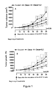
= Figure 1 shows the effect of treatment with DECA, DECA + IL-2 on tumor
growth in vivo.
Murine melanoma cells (B16F10) were inoculated on day zero (1 x 10⁶, 100
pL/animal),
subcutaneously (s.c.) on the back of C57616 male mice. The (A) tumor volume
(in mm3)
was measured every three days with the aid of a digital caliper. The (B)
calculated
percentage increase in the volume of each tumor obtained on the 7th day. The
results were
expressed as Mean Standard Error of Mean (SEM). * p <0.05 represents a
statistically
significant difference as compared to the control group (one-way ANOVA, post-
hoc:
Dunnett test). n = 9-10 animals.
= Figure 2 shows the effect of treatment with DECA, DECA + IL-2 on the
survival of animals
inoculated with murine melanoma cells. B16F10 cells were inoculated on day
zero (1 x 10⁶;
100 pL/animal) subcutaneously (s.c.) on the back of C57B16 male mice. The
graph
represents the mortality curve and the percentage represents the animals which
remained
alive at 30 days after tumor cell inoculation. n = 9-10 animals. * p, 0.05 (p
= 0.0361),
Statistical Analysis: Logrank Test - Chi square.
= Figure 3 shows the anatomopathological exams of volunteer "MBS". A. Pre-
immunotherapy treatment examination, the black arrow indicates the tumor
region and the
white arrow absence of inflammatory infiltrate. The region outlined in black
illustrates the
inhibition of the immune system by tumor detected by the absence of
inflammatory
infiltrate. B. Immunological post-treatment examination, where the complete
elimination of
the tumor can be seen, the white arrows indicate the dense inflammatory
infiltrate and the
area enclosed in black exemplifies areas of fibrosis and reparative changes
permeated by
inflammatory infiltrates. C. Recontextualization of the immune system by the
use of the
CA 02837348 2013-11-26
present invention, attested by the positive reaction to S-100 in countless
intra-epidermal
dendritic cells (indicated by arrows) and amid the dermal inflammatory
infiltrate extending
into the deep dermis without melanocytic cells or residual melanoma.
= Figure 4 shows the anatomopathological exams of volunteer "PPC". A. Pre-
immunotherapy treatment examination showing area of aggressive metastatic
melanoma
with some pigmented cells, and scarce and mild inflammatory peripheral
infiltrate indicated
by the arrow, confirming the inhibition of the immune system by tumor. B. Post
immune
therapy examination illustrating the disappearance of the tumor and
replacement by
intense and dense inflammatory infiltrate. C. Recontextualization of the
immune system by
treatment with the present invention, attested by the positive reaction to S-
100 in countless
intra-epidermal dendritic cells (indicated by arrows) and amid the dermal
inflammatory
infiltrate extending into the deep dermis without residual melanoma.
= Figure 5 shows Nuclear Magnetic Resonance Examinations (Al, A2 and A3 pre
immunological treatment in 30/07/2008) and CT scans (B1, B2, B3 after
treatment in
13/05/2009, Cl and C3 post treatment in 30/08/2011 and C2 after treatment in
13/04/2010)
of the patient R - M. Al carcinomatosis showing thickening of fat (arrow). A2.
Celiac trunk
lymph node cluster (arrow; largest measuring 3.7 cm). A3. Hepatogastric
ligament lymph
node cluster measuring 4 cm (arrow). B1 Disappearance of carcinomatosis, by
showing the
disappearance of the thickening of fat (arrow). B2. Reduction of the biggest
node (3.7 cm
to 1.4 cm) in the celiac trunk lymph node cluster (arrow). B3. Disappearance
of the
hepatogastric ligament lymph node cluster (arrow). Cl. Disappearance of the
carcinomatosis (arrow). C2. Reduction of the biggest node (1.4 cm to 1.1 cm)
in the celiac
lymph node cluster (arrow). C3. Confirmation of the disappearance of the
hepatogastric
ligament lymph node cluster (arrow). These data show a complete remission of
malignant
peritoneal carcinomatosis and lymphatic dissemination of gastric cancer with
the
combination of immunotherapy with the present invention associated to
palliative radio and
chemotherapy.
= Figure 6 shows CT scans examinations of the chest and abdomen of the
volunteer A-D.
A. Pre immunotherapy treatment exam held on 09/10/2006 identifying tumors in
the areas
26
CA 02837348 2013-11-26
indicated with circles. B. Post immune therapy exam in 11/12/2006 evidencing
the absence
of these tumors in the areas analyzed.
= Figure 7 shows tests of prostate specific antigen (PSA) serum levels in
patient O-S. The
first point refers to the residual value of the marker indicating the presence
of residual
neoplastic cells after non curative, which while being treated immunologically
became
undetectable (plotted as zero) in 4 weeks. This data strongly suggests that
the
immunotherapy treatment, provided it was the single drug therapy adopted
pending the
start of radiation therapy was effective in complete remission of the tumor
and locoregional
tumor eradication, since the current state of the art does not allow to
differentiate complete
eradication of the tumor mass in minimal residual disease.
DETAILED DESCRIPTION OF THE INVENTION
Description of the immunogenic compositions
The present invention relates to immunogenic compositions for modulating the
immune
system comprising a therapeutically effective amount of two or more antigenic
immunoactive agents presenting pathogen-associated molecular patterns (PAMPS)
and/or
danger associated molecular patterns (DAMPS) and one or more physiologically
acceptable carriers, excipients, diluents or solvents.
Preferably the compositions of the present invention comprise immunoactive
antigenic
agents presenting pathogen-associated molecular patterns (PAMPS) and/or danger
associated molecular patterns (DAMPS) selected from the group consisting of:
(A)
antigenic agents with molecular patterns associated with bacteria; (B)
antigenic agents with
molecular patterns associated with viruses; (C) antigenic agents with
molecular patterns
associated with fungi and yeasts; (D) antigenic agents with molecular patterns
associated
with protozoa; (E) antigenic agents with molecular patterns associated with
multicellular
parasites / or (F) antigenic agents with molecular patterns associated with
prions.
Still more preferably the compositions of this invention include pathogen-
associated
molecular patterns (PAMPS) and/or danger associated molecular patterns (DAMPS)
27
CA 02837348 2013-11-26
selected from among at least three categories (A), (B), (C), (D ), (E) and (F)
described
above.
Still more preferably the compositions of this invention include pathogen-
associated
molecular patterns (PAMPS) and/or danger associated molecular patterns (DAMPS)
selected from among at least four categories (A), (B), (C), (D), (E) and (F)
described
above.
Antigenic agents of the present invention can be selected from epitopes,
genetic materials,
lipids, polysaccharides and/or immunoactive proteins of the present invention
can be
obtained by purification from isolated fragments of material existing in
nature or fractions
derived from plant, animal or microbiological extracts, or produced by genetic
recombination, preferably derived from viral, fungal, parasitic or bacterial
prion strains.
Thus, the antigenic agents of the present invention with molecular patterns
associated with
bacteria of the present invention may be selected from, but not limited to
antigenic agents
with molecular patterns associated with bacteria of the genera Staphylococcus,
Streptococcus, Enterococcus, Corynebacterium, Bacillus, Listeria, Clostridium,
Mycobacterium, Actinomyces, Nocardia, Escherichia, Proteus, Klebsiella,
Serratia,
Enterobacter, Salmonella, Shigella, Pseudomonas, Burkholderia,
Stenotrophomonas,
Acinetobacter, Vibrio, Campylobacter, Helicobacter, Bacteroides, Neisseria,
Moraxella,
Haemophilus, Bordetella, BruceIla, Francisella, Pasteurella, Yersinia,
Legionella,
Gardnerella, Treponema, Leptospira, Borrelia, Mycoplasma, Rickettsial and
Chlamydia.
Antigenic agents with molecular patterns associated with virus of the present
invention may
be selected from, but not limited to antigenic agents with molecular patterns
associated
with virus families Adenoviridae, Arenaviridae, Bunyaviridae, Coronaviridae,
Filoviridae,
Flaviviridae, Hepadnaviridae, Deltavirus, Caliciviridae, Herpesviridae,
Orthomyxoviridae,
Papovaviridae, Paramyxoviridae, Parvoviridae, Picornaviridae, Poxyviridae,
Reoviridae,
Retroviridae, Rhabdoviridae and Togaviridae.
Antigenic agents with molecular patterns associated with fungi and yeasts of
the present
invention may be selected from, but not limited to antigenic agents with
molecular patterns
28
CA 02837348 2013-11-26
associated with fungi and yeasts of the genus Sporothrix, Aspergillus,
Blastomyces,
Candida, Coccidioides, Cryptococcus, Histoplasma and Pneumocystis.
Antigenic agents with molecular patterns associated with protozoa of the
present invention
may be selected from, but not limited to antigenic agents with molecular
patterns
associated with protozoa of the genera Cryptosporidium, Ciclospora, Entamoeba,
Naegleria, Giardia, Leishmania, Plasmodium, Toxoplasma, Trichomonas,
Trypanosoma,
microsporidia and lsospora.
Antigenic agents with molecular patterns associated with multicellular
parasites of the
present invention may be selected from, but not limited to antigenic agents
with molecular
patterns associated with multicellular parasites trematodes, cestodes and
nematodes.
The antigenic agents of the present invention comprise protein,
polysaccharide, lipid
molecules and/or composite synthetic molecules that mimic protein,
polysaccharide and/or
lipid molecules.
More specifically the agents of the invention comprise immunoactive antigenic
protein
molecules which have enzyme activity, for example kinases, phosphatases,
streptoquinases, estreptodornases and Deoxyribonucleases (e.g. dornases).
The immunogenic compositions for modulating the immune system of the present
invention
comprise from 0.001 to 500 micrograms per ml of each immunogenic agent.
Such immunogenic agents can be encapsulated in capsules, microparticles,
nanoparticles,
coated tablets, liposomes.
Specifically, the immunogenic compositions for modulating the immune system of
the
present invention comprise from 4 to 20 antigenic agents selected from the
group
consisting of antigens derived from agents: dornase, levedurin, oidiomycin,
PPD, prions,
streptoquinase, Streptococcus toxoid, diphtheria toxoid, Tetanus toxoid,
Koch's tuberculin,
inactivated lysate of Ascaris lumbricoides, Aspergillus spp., Aspergillus
flavus, Aspergillus
fumigatus, Aspergillus terreus, Candida spp., Candida albicans, Candida
glabrata, Candida
parapsilosis, Chlamydia spp., Chlamydia pneumoniae, Chlamydia psittaci,
Chlamydia
29
CA 02837348 2013-11-26
trachomatis, Cryptosporidium spp., Dermatophytes, Entamoeba hystolitica,
Enterobius
vermicularis, Enterococcus faecalis, Epidermophyton floccosum, Escherichia
coli, Giardia
lamblia, Haemophilus influenzae, Microsporum cannis, Mycobacterium spp. ,
Mycobacterium bovis, Mycobacterium leprae, Mycobacterium tuberculosis,
Neisseria
gonorrhoeae, human papilloma virus, Polio virus, Proteus spp., Proteus
mirabilis, Proteus
penerii, Proteus vulgaris, Salmonella spp. , Salmonella bongori, Salmonella
enterica,
Serratia spp. , Serratia liquefaciens, Serratia marcencens, Shigella spp.
Shigella flexneri,
Shigella sonnei, Staphylococcus spp. , Staphylococcus aureus, Staphylococcus
epidermidis, Strongyloides stercoralis, Streptococcus spp., Streptococcus
bovis,
Streptococcus viridans, Streptococcus equinus, Streptococcus pneumoniae,
Streptococcus
pyogenes, Toxoplasma gondii, Trichomonas vaginalis, trichophytin, Trichophyton
spp. ,
Trichophyton rubrum, Trichophyton tonsurans, Trichophyton mentagrophytes,
yellow fever
virus, hepatitis B virus, rubella virus, varicella zoster virus, variola
virus, mumps virus,
measles virus, herpes virus and vaccinia virus or synthetic analogues that
present
pathogen-associated molecular patterns (PAM PS) and/or danger-associated
molecular
patterns (DAMPS) associated with these antigenic agents.
A preferred immunogenic composition of the invention comprises inactivated
Mycobacterium bovis lysate, purified protein derivative of M. tuberculosis,
inactivated
Staphylococcus aureus lysate, inactivated Staphylococcus epidermidis lysate,
inactivated
Steptococcus pyogenes lysate, inactivated Streptococcus pneumonia lysate,
inactivated
Enterococcus faecalis lysate, Streptokinase/dornase, inactivated Candida
albicans lysate,
inactivated Candida glabrata lysate, inactivated Epidermophyton floccosum
lysate,
inactivated Microsporum cannis lysate, inactivated Trichophyton mentagrophytes
of the
interdigitale variety lysate, inactivated enteropathogenic Escherichia coli
lysate, inactivated
Salmonella bongori lysate, inactivated Salmonella enterica lysate and
inactivated
Salmonella subterranea lysate.
A preferred immunogenic composition of the invention comprising from 0.001 to
1 ng/ml of
inactivated Mycobacterium bovis lysate, 0.001 to 1 ng/ml of purified protein
derivative of M.
tuberculosis, 0.1 to 100 pg/m1 of inactivated Staphylococcus aureus lysate,
0.1 to 100
CA 02837348 2013-11-26
pg/ml of inactivated Staphylococcus epidermidis lysate; 0.1 to 100 pg/ml of
inactivated
Steptococcus pyogenes lysate; 0.1 to 100 pg/ml of inactivated Streptococcus
pneumonia
lysate; 0.1 to 100 pg/ml of inactivated Enterococcus faecalis lysate, 0.01 to
10 pg/ml of
streptokinase, 0.01 to 10 pg/ml of dornase; 0.1 to 100 pg/ml of inactivated
Candida
albicans lysate; 0.1 to 100 pg/ml of inactivated Candida glabrata lysate, 0.1
to 100 pg/ml of
inactivated Epidermophyton floccosum lysate; 0.1 to 100 pg/ml of inactivated
Microsporum
cannis lysate, 0.1 to 100 pg/ml of inactivated Trichophyton mentagrophytes of
the
interdigitale variety lysate; 0.1 to 100 pg/ml of inactivated enteropathogenic
Escherichia
coli lysate; 0.1 to 100 pg/ml inactivated Salmonella bongori lysate, 0.1 to
100 pg/ml
inactivated Salmonella enterica lysate and 0.1 to 100 pg/ml of inactivated
Salmonella
subterranea lysate.
Additionally, in order to raise, lower or polarize the immune response
depending of the goal
of the immunotherapy, the antigenic composition of the present invention may
comprise
cytokines and/or chemokines such as GM-CSF, IL4, IL5, IL7, IL12, IL15, IL21,
interferon
gamma, and most preferably IL2.
The compositions of the present invention can further comprise excipients,
such as
bactericides, bacteriostats, antioxidants, preservatives, buffers,
stabilizers, pH adjusters,
osmolarity adjusters, antifoaming agents and surfactants, and residual antigen
inactivating
or fractionation agents, growth medium components and solvents commonly used
in the
production of vaccines and immunotherapies.
The compositions of the present invention may be a solid, liquid or gel. As
used herein, the
use of the term "pharmaceutically acceptable carrier" means a non-toxic solid,
inert, semi-
solid liquid excipient, diluent, auxiliary formulation of any type, or simply
a sterile aqueous
solution such as saline. Some examples of materials which can serve as
pharmaceutically
acceptable carriers are sugars such as lactose, glucose and sucrose, starches
such as
corn starch and potato starch, cellulose and its derivatives such as sodium
carboxymethyl
cellulose, a ethyl cellulose and cellulose acetate, cyclodextrin; oils such as
peanut oil,
cottonseed oil, sunflower oil, sesame oil, olive oil, corn oil and soya bean
oil, glycols such
as propylene glycol, polyols, such as glycerol, sorbitol, mannitol and
polyethylene esters
31
CA 02837348 2013-11-26
such as ethyl laurate, ethyl oleate, agar, buffering agents such as aluminum
hydroxide and
magnesium hydroxide, alginic acid, pyrogen-free water, isotonic saline ,
Ringer's solution,
buffer solutions of ethyl alcohol and phosphate as well as other non-toxic
compatible
substances used in pharmaceutical formulations.
A variety of administration routes in animals or humans for the
immunotherapeutic
compositions and vaccines described herein are available. The particular
selected mode
will depend on the selected antigenic agents, the dosage required for
therapeutic efficacy
and patient to whom the composition is administered. The methods of the
present invention
can generally be practiced using any mode of administration biologically
acceptable, i.e.,
any means that produces effective levels of immune response without causing
clinically
adverse reactions. Such modes of administration include intradermal, oral,
rectal,
sublingual, topical, nasal, transdermal or parenteral administration. The term
"parenteral"
includes subcutaneous, intravenous, epidural, irrigation, intramuscular,
release pumps or
infusion. In particular, in this invention, oral, intradermal, parenteral,
subcutaneous,
intravenous, intramuscular, and, by the nasal mucosa and/or oral
administration are
preferred for administration of the compositions claimed herein.
For parenteral administration, the active ingredients may also be dissolved in
a
pharmaceutical carrier and administered as a solution, emulsion, including
micro-and nano-
emulsions or suspension. Examples of suitable carriers are water, saline,
dextrose
solutions, fructose solutions or oils of animal, vegetable or synthetic
origin. Other vehicles
may also contain other ingredients, for example, preservatives, suspending
agents,
solubilizing agents, buffers and the like.
Properties of the immunogenic compositions of the present invention
The immunogenic compositions of the present invention have an unexpected
effect on the
immune response. As can be seen in the Examples below, the immunogenic
compositions
of the present invention show an unexpected technical effect of causing an
immune
response that involves recontextualizing, renewal and reprogramming of the
immune
response in real time.
32
CA 02837348 2013-11-26
More specifically, the immunotherapeutic compositions of the present invention
are
capable of provoking a recontextualization of the operational action capacity
of the immune
system by changing the relationship of forces against the aggressors in its
favor, giving the
immune system a competitive advantage, which does not occur spontaneously in
the
evolution of disease. This recontextualization determines a consequent renewal
and
reprogramming of the established immune response or incipiently established,
or
erroneously established mistakenly attacking in a dysautonomical way the human
or
animal body, polarizing it into a primary or secondary, active or inhibitory,
more effective
appropriate immune response.
This effect occurs via stimulation, activation and joint action of certain
components of the
immune system, such as sentinel cells, antigen presenting sentinel cells, and
memory
lymphocytes. Specifically, the compositions of this invention properly
activate sentinel cells,
dendritic cells and other APC cells, generating the degree and intensity of
CD4 T cell
activation and the degree and intensity of the immune profile to fight the
infection,
infestation or neoplastic disease.
Accordingly, the immunomodulatory antigenic compositions of the present
invention, when
in larger or significant amounts trigger a specific active adaptive immune
response, desired
to combat bacterial, viral or parasitic infections, in combating neoplasms,
cancer and
tumors.
In addition, the treatment with the immunogenic compositions of the present
invention is
capable of stimulating the regenerative power of the immune system, providing
a
subsequent effect to the elimination of infectious disease and other diseases:
to recover
cells and tissues, by restoring organ function debilitated from trauma and
damage which
cause the loss of part of the organism.
Thus, the immunogenic compositions of the present invention are able to
mobilize the
immune system and lead to an increased regenerative power of the body, through
mobilization of stem cells or the activation of gene sets which allow the
regeneration of
cells and tissues and can even reconstruct organs and their functions, and can
reconstitute
33
CA 02837348 2013-11-26
organic systems such as the vascular system, the nervous system and the
endocrine
system, among others.
As can be seen in the Examples presented below, the immunogenic compositions
of the
present invention exhibit an unexpected technical effect of recontextualizing,
renewing, and
reprogramming the immune response in real time and consequently significant
cure rates
when compared to drugs and methodologies in the art.
In a first embodiment of the invention, a particular concentration of immuno-
modulator
agent(s) is used for preparing an immunotherapy pharmaceutical composition
capable of
inducing an innate immune response, which triggers a cascade of immune events,
including the activation of memory lymphocytes from the agent(s) inoculated by
human
intervention and the concomitant activation by antigens present in the
patient's own body,
resulting in a recontextualization, renewal and reprogramming of the ongoing
immune
response to a particular established disease (or still in the establishment
phase),
generating an adaptive response specific to this disease effectively, allowing
the pathogen
to be combated. As such, the administration of the composition containing the
agents of
the present invention repolarizes or improves the polarization of the immune
system in the
presence of a disease when the hitherto established polarization is
inadequate, by the
action of the etiologic agent or colonizer. The activities of the agents of
the present
invention affect the shape, time, accuracy and polarization of the immune
response,
preferably leading to an specific innate and/or adaptive response that it is
more effective to
combat the disease, leading to a better reaction of organism itself.
The present invention provides a way to combat these types of heterologous
(infections
and infestations) and autologous (neoplasms) attacks through the use of the
antigenic
combinations described. The present invention also provides for the
possibility of adding
traditional therapies to the agents of this invention, aiding the process of
elimination of the
etiological heterologous invading agents and of the colonizing autologous
cells, through the
real therapeutic potential of antimicrobial, anticancer and other drugs,
selective for the
pathogens and other infectious agents. This is made possible by the principle
of
displacement of the biological equilibrium in favor of the patient in
combination with a
34
CA 02837348 2013-11-26
correct polarization of the immune response as described herein.
When the immune stimulation follows a situation of immune response, after the
termination
of the disease mechanism or aggression, the continued activation of the immune
system
by antigens or immunomodulatory agents of the present invention leads, through
the
activation of stem cells, to the regeneration of tissues, organs and systems,
by
mechanisms not yet fully understood, but related to healing or restitutio ad
integrum
mechanisms observed in various medical situations.
The compositions of the present invention allow the recruiting of the maximum
number of
virgin and memory cells of the individual, producing more significant effects
than an
antibody increase as described in the prior art. The use of multiple antigenic
agents with
distinct enough PAMPS and DAMPS to simulate different types of attacks that
the
organism suffers and to which the organism has already immunologic memory of,
be it by
environmental exposure or vaccination programs, allows a wider recruitment of
memory
and virgin cells, enabling real-time recontextualization of the immune
response and thus
potentially and radically altering the type of immune response and disease or
illness
progression that affects the individual in a positive, and in several cases,
such amazing
way as compared to the prior art. Furthermore, the present invention, unlike
the prior art,
applies a greater amount of bacterial components, having representatives of
both
intracellular and extracellular bacteria in the composition, besides
components of viruses,
parasites, fungi and yeasts. Hayashi et al have not explored more diverse
compositions to
obtain a potentially greater effect. The application process of the antigenic
agents was also
different, since the present invention encompasses more areas of the body and
tissues that
have APC cells, and preferably looks for exposure on locations close to the
infection sites
and other distal applications to the disease sites (as is the case in
disorders or diseases
that manifest themselves in specific locations of the body). The compositions
of the present
invention, when applied according to the process of using the present
invention in one or,
usually, at various strategic of body regions drained by lymphoid territories
or primary
and/or secondary lymphoid organs, or even intralesional, are perceived by the
PRRs
(pathogen-associated pattern recognition receptors) off all sentinel cells of
the body.
CA 02837348 2013-11-26
In a first group of aggressive conditions or real danger, in which the immune
system is
being extemporaneously overwhelmed, paralyzed or overcome by bacteria, fungi,
viruses,
prions, parasites or other micro- or macro-organisms, uni- or multicellular,
(heterologous
aggression) or a benign or malignant neoplasm (autologous aggression), the
modification
of its preparation is given in the state of activation and mobilization of its
cellular and
molecular apparatus of the innate and adaptive immunity, which integrated are
able to
reverse the situation of competitive disadvantage in which the immune system
and the
body are found.
In a second group of aggressive conditions where the real danger comes from
the immune
system itself, i.e. when it's attacking the human or animal body, in an
autoimmune or
allergic disease, recontextualizing the immune system occurs as a preparation
to be able
to inhibit this detrimental action. The present invention induces the immune
system to
suppress its activation state and demobilize memory effector loops that
maintain the self
aggression. This effect is achieved by mobilizing the cellular and molecular
apparatus of
innate and adaptive immunity responsible for the suppression and regulation of
immune
response and a return to equilibrium known as homeostasis or normality.
In a third group of conditions where the immune system deals with the
aftermath of tissue,
organic or systemic attacks derived from multiple causes, heterologous or
autologous, or
even traumatic, the action of the immune system occurs in repairing the damage
caused by
these attacks. In this case, the preparation or mobilization of the immune
system is through
the mobilization of stem cells from the immune system itself or from other
cellular systems,
autologous, allogeneic or heterologous. Or even by activation of gene sets
present in the
patient's own cells.
Thus, the present invention employs immunomodulatory agents in amounts,
concentrations
and specific locations to recontextualize the immune system, activating and
redirecting the
mechanisms for tissue repair and regeneration, as occurs during cicatrization
and
regeneration of tissue, organ or system, leading to a "restitutio ad integrum"
or
reconstitution with scar. This repair is usually triggered at the end of an
immune response
process, after the healing a trauma, an infection, a tumor disease or an
autoimmune or
36
CA 02837348 2013-11-26
allergic reaction.
Use of the immunogenic compositions of the present invention.
Considering the properties of the immunogenic compositions of the present
invention, it
constitutes another aspect of the present invention using the immunogenic
compositions in
the manufacture of medicaments for the prevention and/or treatment of
infectious diseases,
autoimmune diseases, allergic diseases, inflammation, arthritis, inflammatory
diseases,
transplant rejections, diseases caused by vascular disorders, diseases caused
by
hemorrhagic or ischemic cardiovascular events, ischemia, infarction and
hemorrhage
leading to tissue destruction, cardiac, renal, respiratory or liver disease,
cancer, tumors and
malignant and benign lesions.
The immunogenic compositions of the present invention are also directly used
in the
prevention and/or treatment of infectious diseases, autoimmune diseases,
allergic
diseases, inflammation, arthritis, inflammatory diseases, transplant
rejection, diseases
caused by vascular disorders, diseases caused by hemorrhagic or ischemic
cardiovascular
events, ischemia, infarction and hemorrhage leading to tissue destruction,
cardiac, renal,
respiratory or liver disease, cancer, tumors and malignant and benign lesions.
These infectious diseases can be of viral, bacterial, fungal or parasitic
origin.
Diseases of viral origin prevented and/or treated by the immunogenic
compositions of the
present invention can be caused by the following viruses but not limited to:
HIV, hepatitis virus, herpes virus, rhabdovirus, rubella virus, smallpox
virus, poxvirus, and
Morbillivirus paramyxovirus.
Diseases of bacterial origin prevented and/or treated by the immunogenic
compositions of
the present invention may be caused by the following bacteria, but not limited
to,
Pneumococcus, Staphylococcus, Bacillus, Streptococcus, Meningococcus,
Gonococcus,
Escherichia, Klebsiella, Proteus, Pseudomonas, Salmonella, Shigella,
Haemophilus,
Yersinia, Listeria, Corynebacterium, Vibrio, Clostridia, Chlamydia,
Mycobacterium,
Treponema, and Helicobacter.
37
CA 02837348 2013-11-26
Fungal diseases prevented and/or treated by the immunogenic compositions of
the present
invention may be caused by the following fungi but not limited to: Candida,
Aspergillus,
Cryptococcus neoformans, and/or fungi that cause superficial and deep mycosis.
Diseases
caused by parasites are caused by the following parasites: Trypanosoma,
Schistosoma,
Leishmania, amoebas and tapeworm.
The immunogenic compositions of the present invention are also used in the
prevention
and/or treatment of erythematosus and located lupus, rheumatoid arthritis,
polyarteritis
nodosa, polymyositis and progressive dematomiosite, progressive systemic
sclerosis,
diffuse scleroderma, glomerulonephritis, myasthenia gravis, Sjogren's
syndrome,
Hashimoto's disease, Graves disease, adrenalitis, hypoparathyroidism,
pernicious anemia,
diabetes, multiple sclerosis, demyelinating diseases, uveitis, pemphigus,
pemphigoid
cirrhosis, ulcerative colitis, myocarditis, regional enteritis, respiratory
distress syndrome in
adults, and local manifestations of the reaction to drugs, atopic dermatitis,
infantile eczema,
contact dermatitis, psoriasis, lichen planus, allergic enteropathies,
bronchial asthma,
transplant rejection, post streptococcal diseases such as cardiac, renal and
articular
rheumatic fever manifestations and other related manifestations, multiple and
various
forms of cancers, such as carcinomas, adenocarcinomas, melanomas, sarcomas,
malignant astrocytomas, hepatomas, hypernephroma, lymphomas and melanomas,
among
others.
The immunotherapeutic compositions of the present invention are also useful in
the
treatment of cancer, autologous colonization by benign and malignant tumor
cells, in all
forms of cancer known as as carcinomas, adenomas, adenocarcinoma, hepatoma,
astrocytomas and other neoplasms of the central and peripheral nervous system,
melanomas, sarcomas, lymphomas and leukemias and all benign tumors.
The immunotherapeutic compositions of this invention may also be useful for
diseases
arising in a dysautonomia of the immune system (as already mentioned) such as
lupus
erythematosus; rheumatoid arthritis; polyarteritis nodosa, polymyositis and
dermatomyositis and progressive systemic sclerosis (diffuse scleroderma);
glomerulonephritis, myasthenia gravis, Sjogren's syndrome, Hashimoto's disease
38
CA 02837348 2013-11-26
(hypothyroidism), Graves disease (hyperthyroidism); adrenalites;
hypoparathyroidism,
pernicious anemia, diabetes, multiple sclerosis, and demineralizing co-related
or related
diseases; uveitis; pemphigus, pemphigoid cirrhosis; ulcerative colitis;
myocarditis; regional
enteritis, hepatitis and cirrhosis; adult respiratory distress syndrome, local
and systemic
manifestations of drug reactions, such as pharmacodermia, dermatitis, among
others.
Still in the field of dysautonomia diseases of the immune system, the present
invention also
provides immunotherapy treatments of arterial and venous vascular accidents,
in diseases
such as myocardial infarction, thromboembolic phenomena in the lung, brain and
digestive
system, or in any other area of the body where stroke or ischemia leads to
hemorrhage,
which results in necrosis or atrophy of these segments, such as, but not
limited to, in the
whole musculoskeletal system, in the whole central and peripheral nervous
system, that
lead to occlusion of the blood supply and results in heart attacks and brain
injuries. Thus,
the immunotherapy of the present invention provides an anti-inflammatory and
immune
enhancement that can lead to blockage of inflammatory processes important to
the
establishment of diseases such as metabolic syndrome, obesity, type 2
diabetes,
atherosclerosis, alcoholic fatty liver, non-alcoholic fatty liver,
hypertension, renal failure,
post thrombotic syndrome, post-thrombophlebitis and any other disease derived
from an
inflammatory action of the immune system.
In case of allergic, autoimmune and inflammatory diseases the immunotherapy of
the
present invention can be useful, but not limited to, for inflammation
associated with or
caused by allergic reactions of the skin, atopic eczema' in children; contact
dermatitis in
asthma, bronchial asthma, bronchiolitis and allergic bronchitis, allergic
rhinitis, allergic
enteritis; allergic enteropathy; inflammatory pseudo-tumor processes of
currently unknown
origin; psoriases (pseudo-inflammatory tumor); lichen planus, post-
streptococcal diseases;
heart, liver, lung, kidney, pancreatic islets transplant rejection and others;
hypersensitivity
or destructive immune responses against infectious agents, post-streptococcal
disease,
such as heart, kidney, myocarditis, pericarditis and rheumatic fever and
equivalent by other
etiologic agents, not limited by the forms of these manifestations . In the
case of
autoimmune and allergic diseases, concentrations and dosages are preferably
much lower,
39
CA 02837348 2013-11-26
acting on incomplete activation of immune cells, memory or not, which may
include, but not
limited to the aforementioned diseases.
The immunogenic compositions of the invention are also used to induce cell
regeneration,
tissue regeneration, organ regeneration and the regeneration of organic
systems such as
the circulatory system, nervous system and endocrine system.
Thus one embodiment of the invention is a method for inducing cellular repair,
tissue
regeneration, organ regeneration and regeneration of organic systems such as
the
circulatory system, nervous system and endocrine system in an animal
comprising
administering to the animal an effective amount of one or more immunogenic
compositions
of the present invention.
It is another embodiment of the present invention a method for the renewal of
the immune
response in an animal comprising the following steps:
a) administering systemically and/or locally to the animal a therapeutically
effective amount
of one or more immunogenic compositions as defined in any one of claims 1 to
21;
b) ensure contact of one or more immunogenic compositions, applied in step "a"
with
dendritic cells or other APC cells of the animal;
c) optionally administering prosthetic agents, such as vitamins in the site or
region in which
the disease is to be treated, in order to strengthen the metabolism and
therefore the
immune system of the animal, and
optionally administering medications or other specific treatments.
In one embodiment of the invention, the compositions of the present invention
are
administered once, in one area of the body or in different sites in order to
redirect the
immune system with the highest possible efficiency. The use of the immunogenic
compositions of the present invention for modulation of the immune system,
involving the
exposure of part or all of the system for recognition of antigens in the
immune system, such
as dendritic cells, macrophages and lymph nodes from different parts of the
body will
CA 02837348 2013-11-26
depend on the goal imposed by the illness being fought, and occurs
preferentially through
injections or use of guns, or delivery systems or controlled infusion or
pulsed cells with in
vitro antigens. The agent may be applied to only one location in the body or
in several tens
of locations in several forms: subcutaneous, muscular, intravenous, oral,
breathable
aerosol, cutaneous (dermal patches) in organs, the viscera, or specific
tissues, or in
different body cavities, which can vary in number from one to one hundred
(100)
applications in one to fifty (50) sessions.
The antigenic compositions of this invention may also be combined with other
drugs that
can weaken the reproduction, growth, or any other form of strengthening of the
disease's
causative agent, causing a shift of the equilibrium in favor of the biological
immune
defenses of the host, animal or human. Or still in concomitant treatment.
The antigenic compositions of this invention may also be combined with other
procedures
such as, but not limited to, antibiotics, chemotherapy, radiation therapy,
therapy with
antibodies and antisera, using hormones or other physiology modulating agents
(cytokines,
chemokines , neurohormones, peptides), treatment with antiviral agents, use of
herbal
medicines, vitamin supplementation, supplementation with other cofactors or
prosthetic
agents, transplantation of cells or tissues, methods of therapeutic or
prophylactic
vaccination (with or without cells and not limited to the type of vaccine
vehicles), gene
therapy, surgery or homeopathy, depending on the disease or illness being
fought related
to an improper or inefficient immune activity.
In particular, in order to raise, lower or polarize the immune response as the
goal of
immunotherapy, the antigenic compositions of this invention may be used in
conjunction
with therapy with cytokines and/or chemokines such as GM-CSF, IL4, IL5, IL7,
IL12, IL15,
IL21, interferon gamma, and most preferably IL-2.
Recontextualization, renewal and reprogramming of the immune response.
The recontextualizing of the immune system, as explained in the text of this
patent
application, is achieved by means of stimulation of the immune system by
antigens of
different pathogens not related to the pathology to be treated, for which the
human or
41
CA 02837348 2013-11-26
animal, preferably, already has an immunological memory of.
These varied and multiple antigens, in number greater than five, with multiple
PAMPs and
DAMPs, induce in the sentinel cells and in the APC cells, especially in
dendritic cells, an
intense activation allowing the mobilization of these memory CD4 lymphocytes
specific for
these antigens at the site of application.
These stimuli must be capable of causing an intense, strong and effective
secondary
specific immune response to these antigens at the site of application, in the
regional lymph
nodes, in the lymph nodes at a distance and a systemic mobilization of the
immune system
so that it can, in parallel, cause an effective response capable of
eradicating the specific
pathology in progress.
The innate and adaptive immune response caused intentionally by the
composition of the
present invention should encompass the full extent of the body area affected
by the
condition being treated and even exceed it if possible to be able to activate
the sentinel and
APC cells in the number and intensity that would be needed to properly address
the
aggression caused by the pathogenic disease to be treated, and activating and
triggering
the best specific adaptive response, effectively and properly sequentially
polarized, in order
to cure the condition being treated.
Thus the innate and adaptive response induced by the present invention will
geographically
overlap the condition being treated and by its intense and extensive
activation will correct
the inefficient activation, purposely limited by the action of the pathogen
that overcomes
the body's defenses, by preventing competition, its proper mobilization and
development of
an effective adaptive response according to its greatest genetic and
biological potential.
This ideal activation should also reverse the immunosuppression, the tolerance
and
escape mechanisms established by pathogens because it is known and proven that
an
unrelated strong and intense immune response, that fully covers the response
to be
corrected, through the activated cells and cytokines of the immune system,
will correct
these deficiency situations efficiently.
Effector cells and memories of specific antigens of the present invention,
activated and
42
CA 02837348 2013-11-26
generated at the site of application of the antigens, will, via the
bloodstream, enter the
already activated lymph nodes, which drain the region affected by the disease
and will
enable, in a strong and intense way, all the existing dendritic cells there.
This way, they will
lead to an activation of the entire lymph node, causing it to grow with
increased irrigation,
increasing its size and making it a reactional lymph node capable of provoking
an immune
response against weak antigens, which by themselves are not capable of causing
an
immune response. This adjuvant effect, well known and demonstrated
experimentally and
clinically, of the effector/memory T lymphocytes, will oppose the action of
the target
causative agent that is blocking the required activation of the lymph node for
the
development of an immune response that is necessary to combat the disease in
question.
- That, exclusively for the purpose and by the action of the present
invention, through its
potent antigenic composition, may occur that the sentinel cells and dendritic
cells and
macrophages of the immune response will be the same for unrelated antigens and
to the
pathological antigens, but from this action, will be intensely and properly
activated.
Dendritic cells strongly activated by multiple antigens, have a slow
metabolism and ideally
present all dominant and subdominant epitopes of the causative agent, by the
known
"helper" effect, mobilizing all possible and available T lymphocytes able to
specifically
recognize antigens of the autologous or heterologous pathogen, to be treated
and to react
against it.
That the areas of the inflammatory process and lymphatic territories are
exactly the same.
The inflamed area, through the anti-inflammatory action of specific memory
cells,
unrelated, mobilized by the present invention by their antigenic composition,
will block the
inflammasomes and exert an anti-inflammatory action that will correct the
pathological
inflammation responsible for the morbidity of the disease and which was caused
by its
etiological agent. For the memory effect it's important to note that this
known action of the
memory T cells is the major responsible for the fact that a second contact
with any
pathological agent, after an already established immunity, is asymptomatic,
without
causing a disease.
-That the lymphatic territories are exactly the same, only now intensely
activated and with
43
CA 02837348 2013-11-26
the necessary alarm signal, caused by the present invention, to cause any
immune
response, even for a weak antigen, similar to what occurs with dendritic cells
common to
this invention and to the autologous or heterologous etiological agent to be
fought.
Lymphokines and innate cells that command an effective secondary response will
be the
same and the T lymphocytes specific against the etiologic agent to be fought,
will "hitch a
ride" on this ideal microenvironment for holding an effective immune response.
That the dendritic cells activated by the present invention, can capture the
antigens of the
etiological agent to be fought at the site of the pathology and in the related
lymphatic
territories and be in contact with the pathogen specific TCD4 lymphocytes, in
a correctly
and ideally enabled lymphatic system. The role of the dendritic cells
activated and matured
with the TCD4 specific to the etiologic agent, occurs in a microenvironment
conducive to
conducting an immune response, with all the genetic and biological potential
of the host
organism's immune system.
These dendritic cells at the site of the pathology and at the lymph nodes will
properly
gauge the severity, extent, intensity and type of aggression, activating,
inducing,
coordinating, polarizing, leading and maintaining a new effective adaptive
immune
response, whose effector loop, with the collaboration of the cells and
effector molecules of
the intense and properly activated innate immunity may be able to eliminate
the causative
agent to be fought. So the answer is reprogrammed and lead back as noted
above,
reversing the biological balance in favor of the host, which until then was
under the yoke of
the offending autologous or heterologous agent.
This action may occur with or without the help of biological balance shifters
such as
antibiotics and anticancer drugs, capable to block, weaken or neutralize the
effects and
potential of the etiological agent, allowing the immune system to have a
chance to heal the
pathology that is the target of the treatment. Once triggered by any
etiological agent, the
immune system will only stop responding when the etiological agent is
eliminated or the
organism passes away, this way the invention will help avoid the latter
option, or it will
improve the patient's condition if there is a chronic disease that cannot be
cured.
44
CA 02837348 2013-11-26
Thus the action of the compositions of the present invention intentionally and
strategically
superimposed over the entire area under the action of the agent to be fought,
will
recontextualize the immune system by activating the PAMPs and DAMPs in the
sentinel
cells and common APCs and by the unrelated specific secondary adaptive immune
response. This intentionally induced immune response will efficiently activate
the whole
lymphatic territory and the organic territory affected by the etiological
agent. In the
recontextualized area and in the bulge, and within the context of a greater
immune
response, stronger, more intense and more extensive secondary anti-
inflammatory nature
of the target immune response will be, as described, reprogrammed and
efficiently
renewed within the scope of a greater chance for the host, now with a chance
of reversing
the biological balance in its favor.
Adequacy of the protocol to the pathophysiological characteristics of the
condition being
= treated:
a) the basis of immunotherapy against neoplasias.
The main characteristic feature of malignant neoplasms is the dominance of the
microenvironment as defined in the study of the present invention, which
differs from the
traditionally defined in the current state of the art, which is that of the
environment created
by the action of tumor cells with the cells of the organism by which such
action will make
them function in their favor. The microenvironment defined herein is the space
around a
single or a set of neoplastic cells, which by means of surface molecules
and/or other
molecules secreted by it totally dominate this environment to its advantage.
In this dominated space the connective tissue starts to nurture and sustain
these cells
through its structural elements and through new vessels destined to supply the
tumor cells
and their supporting tissue. Through surface molecules and substances and
enzymes
secreted by the tumor cells in this environment, they destroy the tissue from
which they
originated, and healthy tissue invaded by them, which become colonized and
replaced.
Surface and secreted molecules completely block the immune system,
inactivating and
immobilizing sentinel, APC and lymphocytic cells, inducing nonspecific and
specific
CA 02837348 2013-11-26
immunosuppression and inactivating the locoregional and distant lymph nodes.
Through
the domination of the microenvironment the tumor cells, through surface
molecules and
enzymes, enter the blood and lymphatic vessels, and colonize distant locations
away from
the local primary tumor and cause distant lymphatic and hematogenous
metastases.
Thus, the total domination of the microenvironment around a single cell makes
a tumor cell,
through its indiscriminate proliferation, to initially pathologically
subjugate space around
itself, its tissue of origin, the adjacent areas, the organ and finally
through metastases the
body as a whole. Similarly, the immune ignorance, immunosuppression and the
specific
and nonspecific induced tolerances are primarily in situ, and then local,
local-regional,
organic and finally completely dominating the systemic immune system of the
host body.
The dominance of the microenvironment is therefore the strategic, crucial and
determinant
effect produced by the genomic potential of a neoplastic cell, which leads one
tumor cell to
dominate the in situ, local, regional, organic and systemic space, colonizing
the host and
leading to death.
In short, an immunotherapy must necessarily break the dominance of the
established
tumor microenvironment and macroenvironment, and cover all the immunological
territories
dominated by the neoplasia. The immunotherapy treatment should also cover the
lymph
territories at a distance from the tumor, inducing a recontextualization,
renewal and
reprogramming of the immune system, from the outside to the inside the
affected area with
a strong inertia able to reverse completely, together with locoregional
treatment
(intratumoral and perilesional), the tumor dominance.
Immunoprophylaxis should be performed every 4 or 5 days, as it's the
physiological period
of the generation of suppressor cells that control the immune response.
Successive waves
of repeated antigenic stimulation in the meantime will indefinitely perennize
the immune
response, perpetuating the antigenic stimulus as it occurs with an infection
before its
chronicity phase and generation of an immune dysfunction. The failure to
generate
suppressor cells and the recontextualization prevents the domination of the
suppressor
cells by the tumor and its protection in opposition to the domination of the
environment.
46
CA 02837348 2013-11-26
The action of a neoplastic cell in the field of the microenvironment and of a
set of them in
the macro environment is carried out 24 hours a day and during the entire
period during
which the condition exists. Therefore, immunotherapy with the abovementioned
scope,
frequency and magnitude should be applied continuously as long as there are
still tumor
cells. It is interesting to mention that the traditional immunotherapy that
causes
discontinuous stimuli, like the protocols for immunization with inert antigens
(soluble or not)
or with attenuated agents do not find application in the pathophysiological
context induced
by tumors.
Any specific immune response can be amplified and efficiently enhanced by the
addition of
cytokines and/or chemokines, preferably exogenous IL-2 at a receptor
saturation level
which will produce the proliferation of immune cells that recognize the
antigen and,
therefore, have on its surface the complete expression of the interleukin 2
receptor.
Therefore, only the response of the antigens induced by the invention and
induced by the
causative agent (autologous or heterologous) will be amplified. In an
antitumor
immunotherapy in which there are only weak antigens, it should be supplemented
with IL2
in order to obtain an effective and robust immune response.
The foundations of immunotherapy against septicemia, sepsis and "septic shock"
Septicemia is defined as an extremely serious infection in which one or more
bacteria or
microorganisms, from their entry point, enter the bloodstream and start
circulating in large
numbers, getting established at distant points, colonizing tissues, organs,
and in the most
severe cases, can successively reach most of the body surface. Generally, when
the
microorganism load is too large, a large number of bacteria, with their toxic
and metabolic
products, with countless PAMPS and DAMPS, touching with all the also countless
PRRs
and RDPs of most of the body surface, while generating an extensive, intense
and violent
general inflammatory process, with the massive release of cytokines from the
translation of
all these signs.
The unfavorable evolution of septicemia leads to sepsis, through the massive
release of
proinflammatory cytokines such as TNFs, ILI, IL18, IL6 and others, causing an
47
CA 02837348 2013-11-26
inflammatory collapse with hemodynamic characteristic alterations, such as
hypotension,
rapid pulse, which may culminate in septic shock, usually irreversible.
Septicemia, sepsis
are serious infections with high morbidity and mortality. In these severe
infections the
immune system, in turn, with its compromised operability by weaknesses and
blockages
induced by bacteria, starts to act so as to eliminate the bacteria at any
cost, through the
inflammatory Th17 tissue profile, increasing inflammation disproportionately
and therefore
harming the organism.
In this inflammatory tissue profile, the effector loops of innate immunity,
controlled by the
TCD4 lymphocytes, cause tissue damage and sometimes massive destruction, that
compromise organs and tissues and that exacerbate infections, leading, for
example, to
respiratory failure, lung shock, and in ARDS (adult respiratory distress
syndrome), also
leading to renal failure and multiple organ failure.
Therefore, in septicemia, in sepsis and in septic shock there are two
variables that should
strategically be considered and should be the target of an immunotherapy, so
it is
successful. These two variables are the inflammation caused by the massive
spread of
countless bacteria in the whole body and its connection with the PRRs and DPPs
and the
polarization for the Th17 profile caused by the functional infeasibility of
the Thl and Th2
profiles. These variables are the cornerstone of severity, gravity, morbidity
and mortality of
these diseases.
Taking into account these two variables, for an immunotherapy to be effective
in these
infections, it should be applied to cover the entire body surface, including
the greatest
number of lymphatic territories to geographically overlap with the action of
the pathogen or
pathogens. It should also be applied to the injured areas and to the
perilesional region so
that together they can cause widespread recontextualization, that by its
action can recover
the integrity of the T loop and produce a wide, extensive and intensive, anti-
inflammatory
effect by effector/memory T cells generated within the application sites. It
should, in
parallel, through the recontextualization and reprogramming above described,
polarize the
TCD4 response of the Th17 inflammatory tissue profile for the humoral TH2 and
cell TH1
profiles, further decreasing the generalized inflammation.
48
CA 02837348 2013-11-26
The loop amplification by IL2 should be very low, just enough to specifically
amplify the
repolarization of the immune response of the inflammatory profile to the
immunity profile.
Thus, the recontextualizing and the reprogramming achieved by immunotherapy
using the
compositions of the present invention, by recovering immune cells through the
anti-
inflammatory action of non-related specific memory T lymphocytes, by the
repolarization of
the tissue inflammatory profile TH17 to elective and effective TH1 and TH2
immunity
profiles, will redirect the immune response. This immune response, renewed in
real time
during the infectious process, in conjunction with a biological balance
shifter, in the case of
the use of various antimicrobial agents, have a chance to reverse the
biological equilibrium
at the end of the curve in which is very favorable for the microorganism, to
be favorable to
the host and now have a chance of solution.
Adequacy of the protocol to the "status" of the immune system in the pathology
and in the
patient being treated.
In the case of cancer and septicemia, by the own pathophysiological
mechanisms, there is
a breach of the integrity and functionality of the T loop with an inadequate
polarization for a
suppressing TREG profile in cancer and for an inflammatory tissue Th17 profile
in
septicemia with a nearly complete inoperability of the immune system overcome
by
disease. In these cases, as in the examples cited herein, the
recontextualizing must reach
the whole body to reverse all immunosuppression, tolerance and immune
ignorance
induced by the pathology, as well as to restore all operational and functional
capacity of the
immune system to have a reprogrammed and renewed effective immune response.
Rationale of the therapeutic protocol
The therapeutic protocol of the present invention designed to be applied in
cases of cancer
and septicemia must:
-be applied in most strategic lymphatic regions of the body or infection. In
the cases
described herein, more than 10 lymphatic territories have been hit. It must be
applied within
the tumor, and infected and perilesional areas.
49
CA 02837348 2013-11-26
-the immunotherapy formulation must contain at least 5 antigens so it contains
PAMPs and
DAMPs so as to be able to recontextualize the immune system.
- the application area must overlap, cover, and overcome the whole
extension of regions
dominated by the tumor and infection.
-the antigenic stimuli must be repeated every 4 or 5 days in order to avoid
the generation of
suppressor cells capable of aborting the new desired immune response or to
suppress an
achieved repolarization.
-the treatment must be maintained in this manner until the elimination of the
last neoplastic
cell, or to the end of the infection, or to the healing of the wound, organ or
system.
-in practice, 1 to 3 ml of this immunotherapy must be applied to 10 or more
lymphatic
territories. This invention should be jointly applied in intra and extra
lesion or tumor areas
damaged by cancer or by infection.
In summary, the immunotherapy is "systemically" distributed in several (at
least ten)
lymphatic territories, pen- and intra-tumoral or lesion with a volume able to
disrupt and
destabilize the tumor from the domination of its micro and macro environment,
or cover the
area significantly affected by infection and inflammation, as well as to
restore the
microenvironment that is favorable to the immune response of the organism. It
will be
applied every 4 to 5 days with the use of low doses of exogenous interleukin-
2,
uninterruptedly during the period of duration of the disease. In the case of
septicemia,
sepsis, and septic shock as noted above this dose should be the lowest
possible.
EXAMPLES
To allow a better understanding of the invention and clearly demonstrate the
technical
progress achieved, the results of the various tests conducted with respect to
this invention
are shown below as examples.
In Example 1 several preferred immunogenic compositions of the present
invention are
described. In Examples 2 to 8 the properties, usage, and therapeutic methods
employing
CA 02837348 2013-11-26
the immunogenic compositions of the present invention are shown. In Examples 2
to 8 the
immunogenic composition described in Example 1, Composition 1 was used and
herein
referred to as DECA.
These Examples are presented for illustrative purposes only and should not be
regarded in
any way as limiting the scope and range of the invention.
Example 1: Immunogenic Compositions
In order to achieve the recontextualizing, renewal and reprogramming of the
immune
response in real time according to the innovative concepts described in the
present
invention, an expert skilled in the art can design different and distinct
compositions,
combinations or formulations of products, which fall within the scope of the
invention.
As described, for such compositions to meet the technical requirements for the
advantageous or unpublished results in combating a number of diseases and
illnesses,
they must have a high diversity of antigens from pathogens, so as to get the
maximum
synergistic effect in binding the PAMPs and DAMPs to their receptors and
allowing the
achievement of a high degree of activation of the innate immunity in the
sentinel cells (with
or without ATC function) thereby allowing the recontextualizing, renewal and
reprogramming of the immune response in real time.
Such compositions should preferably use antigenic agents for which most
people, because
of previous contact, would have memory clones of in their immune system
capable of
inducing a broad anti-inflammatory action in parallel to recontextualization.
For this,
antigenic agents should preferably be selected that:
= correspond to the most common infections contracted by the individual
from childhood to
maturity (when the animal or the human being acquires its "repertoire of
immunity").
= are used in immunization programs such as childhood vaccination programs
against
endemic and/or epidemic diseases.
= those from organisms of potentially pathogenic microflora, especially of
the
51
CA 02837348 2013-11-26
gastrointestinal tract, where the memory lymphocytes play an active dynamic
barrier
ensuring the survival of the individual.
= Ideally each of the antigenic agents should be present in a concentration
of 0.001 to 500
micrograms per mL.
In accordance with these concepts, several formulations have been developed,
using
antigenic agents in their already available, safe, and approved forms for use
in human
vaccination programs or allergic response tests and immunity assessment tests.
Therefore, we present the following several examples of compositions which
fall within the
scope of the present invention, without however the intention to limit it,
since the present
invention and its concepts allow for the design of immunogenic compositions
comprising a
very large number of combinations of antigenic agents.
Composition 1:
Component Concentration
Koch's Turberculin (inactivated Mycobacterium 0.004 ng/mL
bovis lysate).
PPD 0.004 g/mL
Inactivated Staphylococcus lysate (Staphylococcus 6.94 pg/mL
aureus and Staphylococcus epidermidis in equal
parts).
Inactivated Steptococcus lysate (Streptococcus 6.94 pg/ml
pyogenes, Streptococcus pneumoniae and
Enterococcus faecalis in equal parts).
Streptokinase derived from inactivated beta- 0.444 pg/mL
hemolytic Streptococcus lysate purification.
Dornase derived from inactivated beta-hemolytic 0.111 pg/mL
Streptococcus lysate purification.
Inactivated Candida lysate (Candida albicans and 6.94 pg/mL
Candida glabrata in equal parts).
52
CA 02837348 2013-11-26
Inactivated dermatophytes lysate 6.94 pg/mL
(Epidermophytonfloccosum, Microsporum cannis,
Trichophyton mentagrophytes of the interdigitale
variety in equal parts).
Inactivated enteropathogenic Escherichia coli 6.94 pg/mL
lysate (EPEC)
Inactivated Salmonella lysate (Salmonella bongori, 6.94 pg/mL
Salmonella enterica and Salmonella subterranea in
equal parts).
Sodium Chloride 7.5 mg/mL
Sodium phosphate dibasic heptahydrate 0.48 mg/mL
Potassium phosphate monobasic 0.06 mg/mL
Phenol 2.5 mg/mL
Water q.s.
Composition 2:
Component Concentration
Koch's Turberculin (inactivated Mycobacterium 0.004 ng/mL
bovis lysate).
PPD 0.004 g/mL
Inactivated Staphylococcus aureus lysate, 6.94 pg/mL
inactivated Staphylococcus epidermidis lysate in
equal parts.
Streptokinase derived from inactivated beta- 0.444 pg/mL
hemolytic Streptococcus lysate purification.
Dornase derived from inactivated beta-hemolytic 0.111 pg/mL
Streptococcus lysate purification.
Inactivated Candida albincans lysate, inactivated 6.94 pg/mL
Candida parapsilosis lysate, inactivated Candida
glabrata in equal parts.
53
CA 02837348 2013-11-26
Inactivated enteropathogenic (EPEC), "shiga-like" 6.94 pg/mL
toxin producer (STEC), enteroaggregative (EAEC),
enterotoxigenic (ETEC), enteroinvasive (EIEC) and
extraintestinal (ExPEC) Escherichia coil lysate in
equal parts.
Sodium Chloride 7.5 mg/mL
Sodium phosphate dibasic heptahydrate 0.48 mg/mL
Potassium phosphate monobasic 0.06 mg/mL
Phenol 2.5 mg/mL
Water q.s.
Composition 3:
Component Concentration
PPD 0.004 g/mL
Inactivated Streptococcus pyogenes lysate, 6.94 pg/mL
inactivated Streptococcus pneumonie lysate,
Enterococcus faecalis lysate in equal parts.
Inactivated Staphylococcus aureus lysate, 6.94 pg/mL
inactivated Staphylococcus epidermidis lysate in
equal parts.
Inactivated Candida albicans lysate, inactivated 6.94 pg/mL
Candida parapsilosis lysate, inactivated Candida
glabrata lysate in equal parts.
Sodium Chloride 7.5 mg/mL
Sodium phosphate dibasic heptahydrate 0.48 mg/mL
Potassium phosphate monobasic 0.06 mg/mL
Phenol 2.5 mg/mL
Water q.s.
54
CA 02837348 2013-11-26
Composition 4:
Component Concentration
Inactivated BCG lysate 50 mg/mL
Inactivated Staphylococcus aureus lysate, 6.94 pg/mL
inactivated Staphylococcus epidermidis lysate in
equal parts.
Inactivated Streptococcus agalactiae lysate, 6.94 pg/mL
inactivated Streptococcus mix (Streptococcus
pyogenes, Streptococcus pneumoniae and
Enterococcus faecalis) lysate in equal parts.
Inactivated Candida albincans lysate, inactivated 6.94 pg/mL
Candida parapsilosis lysate, inactivated Candida
glabrata in equal parts.
Sodium Chloride 7.5 mg/mL
Sodium phosphate dibasic heptahydrate 0.48 mg/mL
Potassium phosphate monobasic 0.06 mg/mL
Phenol 2.5 mg/mL
Water q.s.
Composition 5:
Component Concentration
PPD 0.004 g/mL
Inactivated Streptococcus pyogenes lysate, 6.94 pg/mL -
inactivated Streptococcus pneumonie lysate,
Enterococcus faecalis lysate in equal parts.
Inactivated Apergillus fumigatus, Apergillus flavus, 6.94 pg/mL
and Apergillus terreus lysate in equal parts.
Inactivated dermatophytes lysate 6.94 pg/mL
(Epidermophytonfloccosum, Microsporum cannis,
Trichophyton mentagrophytes of the interdigitale
CA 02837348 2013-11-26
variety in equal parts).
Sodium Chloride 7.5 mg/mL
Sodium phosphate dibasic heptahydrate 0.48 mg/mL
Potassium phosphate monobasic 0.06 mg/mL
Phenol 2.5 mg/mL
Water q.s.
Composition 6:
Component Concentration
Koch's Turberculin (inactivated Mycobacterium 0.004 ng/mL
bovis lysate).
Inactivated Streptococcus pyogenes lysate, 6.94 pg/mL
inactivated Streptococcus pneumonie lysate,
Enterococcus faecalis lysate in equal parts.
Inactivated Neisseria meningitides lysate. 6.94 pg/mL
Inactivated Apergillus fumigatus, Apergillus flavus, 6.94 pg/mL
and Apergillus terreus lysate in equal parts.
Sodium Chloride 7.5 mg/mL
Sodium phosphate dibasic heptahydrate 0.48 mg/mL
Potassium phosphate monobasic 0.06 mg/mL
Phenol 2.5 mg/mL
Water q.s.
Composition 7:
Component Concentration
Koch's Turberculin (inactivated Mycobacterium 0.004 ng/mL
56
CA 02837348 2013-11-26
bovis lysate).
Inactivated BCG lysate 50 mg/mL
Inactivated Staphylococcus aureus lysate, 6.94 pg/mL
inactivated Staphylococcus epidermidis lysate in
equal parts.
Inactivated Streptococcus pyogenes lysate, 6.94 pg/mL
inactivated Streptococcus pneumonie lysate,
Enterococcus faecalis lysate in equal parts.
Inactivated Candida albincans lysate, inactivated 6.94 pg/mL
Candida parapsilosis lysate, inactivated Candida
glabrata in equal parts.
Inactivated Streptococcus equinus, Streptococcus 6.94 pg/mL
bovis and Streptococcus viridans lysate in equal
parts.
Inactivated enteropathogenic (EPEC), "shiga-like" 6.94 pg/mL
toxin producer (STEC), enteroaggregative (EAEC),
enterotoxigenic (ETEC), enteroinvasive (EIEC) and
extraintestinal (ExPEC) Escherichia coli lysate in
equal parts.
Inactivated Salmonella typhi, Salmonella paratyphi 6.94 pg/mL
and Salmonella enterica lysate in equal parts.
Inactivated lysate of antigens of the measles virus 10,000
("Schwarz strain"). TDCI50/mL
Glycerol 500 mg/mL
Phenol 2.5 mg/mL
Water q.s.
Composition 8:
Component Concentration
Koch's Turberculin (inactivated Mycobacterium 0.004 ng/mL
bovis lysate).
57
CA 02837348 2013-11-26
PPD 0.004 g/mL
Inactivated Staphylococcus aureus lysate, 6.94 pg/mL
inactivated Staphylococcus epidermidis lysate in
equal parts.
Inactivated enteropathogenic (EPEC), "shiga-like" 6.94 pg/mL
toxin producer (STEC), enteroaggregative (EAEC),
enterotoxigenic (ETEC), enteroinvasive (EIEC) and
extraintestinal (ExPEC) Escherichia coil lysate in
equal parts.
Streptokinase derived from inactivated beta- 0.444 pg/mL
hemolytic Streptococcus lysate purification.
Dornase derived from inactivated beta-hemolytic 0.111 pg/mL
Streptococcus lysate purification.
Inactivated Streptococcus pyogenes lysate, 6.94 pg/mL
inactivated Streptococcus pneumonie lysate,
Enterococcus faecalis lysate in equal parts.
Inactivated Helicobacter pylori lysate. 6.94 pg/mL
Tetanus toxoid 50 units of
Lf/m L
Inactivated Candida albincans lysate, inactivated 6.94 pg/mL
Candida parapsilosis lysate, inactivated Candida
glabrata in equal parts.
Sodium Chloride 7.5 mg/mL
Sodium phosphate dibasic heptahydrate 0.48 mg/mL
Potassium phosphate monobasic 0.06 mg/mL
Phenol 2.5 mg/mL
Water q.s.
Composition 9:
Component Concentration
58
CA 02837348 2013-11-26
Inactivated BCG lysate 50 mg/mL
Inactivated Mycobacterium tuberculosis lysate 0.004 ng/mL
Inactivated Staphylococcus aureus lysate, 6.94 pg/mL
inactivated Staphylococcus epidermidis lysate in
equal parts.
Inactivated enteropathogenic (EPEC), "shiga-like" 6.94 pg/mL
toxin producer (STEC), enteroaggregative (EAEC),
enterotoxigenic (ETEC), enteroinvasive (EIEC) and
extraintestinal (ExPEC) Escherichia coli lysate in
equal parts.
Inactivated Haemophilus influenza lysate. 6.94 pg/mL
Inactivated Streptococcus agalactiae lysate, 6.94 pg/mL
inactivated Streptococcus mix (Streptococcus
pyogenes, Streptococcus pneumoniae and
Enterococcus faecalis) lysate in equal parts.
Inactivated Salmonella typhi, Salmonella paratyphi 6.94 pg/mL
and Salmonella enterica lysate in equal parts.
Inactivated Proteus mirabilis, Proteus vulgaris, and 6.94 pg/mL
Proteus penerii lysate in equal parts.
Inactivated lysate of antigens of the measles virus 10,000
("Schwarz strain"). TDCI50/mL
Inactivated Candida albincans lysate, inactivated 6.94 pg/mL
Candida parapsilosis lysate, inactivated Candida
glabrata in equal parts.
Glycerol 500 mg/mL
Phenol 2.5 mg/mL
Water q.s.
Composition 10:
Component Concentration
59
CA 02837348 2013-11-26
Inactivated Mycobacterium africanum lysate. 0.004 ng/mL
Koch's Turberculin (inactivated Mycobacterium 0.004 ng/mL
bovis lysate).
Inactivated enteropathogenic (EPEC), "shiga-like" 6.94 pg/mL
toxin producer (STEC), enteroaggregative (EAEC),
enterotoxigenic (ETEC), enteroinvasive (EIEC) and
extraintestinal (ExPEC) Escherichia coli lysate in
equal parts.
Inactivated Staphylococcus aureus lysate, 6.94 pg/mL
inactivated Staphylococcus epidermidis lysate in
equal parts.
Inactivated Epidermophyton floccosum, 6.94
pg/mL
Microsporum cannis, Trichophyton
mentagrophytes of the interdigitale variety lysate in
equal parts).
Inactivated Streptococcus pyogenes lysate, 6.94 pg/mL
inactivated Streptococcus pneumonie lysate,
Enterococcus faecalis lysate in equal parts.
Inactivated Acinetobacter baunnannii lysate. 6.94 pg/mL
Inactivated Helicobacter pylori lysate. 6.94 pg/mL
Inactivated lysate of antigens of the mumps virus 10,000
(Urabe AM9 strain) TDCI50/mL
Inactivated Polio virus lysate 40 UD of type
I antigens; 1.8
UD of type 2
antigens; 32
UD of type 3
antigens
Glycerol 500 mg/mL
Phenol 2.5 mg/mL
Water q.s.
Composition 11:
CA 02837348 2013-11-26
Component Concentration
Inactivated Mycobacterium leprae lysate 0.004 ng/mL
Koch's Turberculin (inactivated Mycobacterium 0.004 ng/mL
bovis lysate).
Inactivated Staphylococcus aureus lysate, 6.94 pg/mL
inactivated Staphylococcus epidermidis lysate in
equal parts.
Inactivated Candida albincans lysate, inactivated 6.94 pg/mL
Candida parapsilosis lysate, inactivated Candida
glabrata in equal parts.
Inactivated Streptococcus agalactiae lysate, 6.94 pg/mL
inactivated Streptococcus mix (Streptococcus
pyogenes, Streptococcus pneumoniae and
Enterococcus faecalis) lysate in equal parts.
Inactivated Streptococcus equin us, Streptococcus 6.94 pg/mL
bovis, and Streptococcus of the viridans group
lysate in equal parts.
Inactivated Haemophilus influenza lysate. 6.94 pg/mL
Inactivated Proteus mirabilis, Proteus vulgaris, and 6.94 pg/mL
Proteus penerii lysate in equal parts.
Antigens of the rubella virus (Wistar RA 27/3M 10,000
strain) TDCI50/mL
Inactivate antigen of the Varicella zoster virus 149 231
lysate PFU/mL
Glycerol 500 mg/mL
Phenol 2.5 mg/mL
Water q.s.
Composition 12:
Component Concentration
61
CA 02837348 2013-11-26
Inactivated Mycobacterium avium lysate 0.004 ng/mL
Inactivated Mycobacterium kansasii lysate 0.004 ng/mL
Inactivated Apergillus fumigatus, Apergillus flavus, 6.94 pg/mL
and Apergillus terreus lysate in equal parts.
Inactivated Neisseria gonorrhoeae lysate. 6.94 pg/mL
Inactivated Streptococcus equinus, Streptococcus 6.94 pg/mL
bovis, and Streptococcus of the viridans group
lysate in equal parts.
Inactivated Epidermophyton floccosum, 6.94 pg/mL
Microsporum cannis, Trichophyton
mentagrophytes of the interdigitale variety lysate in
equal parts).
Inactivated Chlanydia trachomatis, Chlamydia 6.94 pg/mL
psittaci, and Chamydia pneumoniae lysate in equal
parts.
Inactivated enteropathogenic (EPEC), "shiga-like" 6.94 pg/mL
toxin producer (STEC), enteroaggregative (EAEC),
enterotoxigenic (ETEC), enteroinvasive (EIEC) and
extraintestinal (ExPEC) Escherichia coli lysate in
equal parts.
Antigens of the rubella virus (Wistar RA 27/3M 10,000
strain) TDCI50/mL
Inactivated antigen of the Vaccinia (smallpox) virus 1 to 10 x 109
lysate PFU/mL
Glycerol 500 mg/mL
Phenol 2.5 mg/mL
Water q.s.
Composition 13:
Component Concentration
62
CA 02837348 2013-11-26
Inactivated Mycobacterium tuberculosis lysate 0.004 ng/mL
Inactivated Mycobacterium avium lysate 0.004 ng/mL
Inactivated Neisseria meningitides lysate 6.94 pg/mL
Diphtheria toxoid 67 units of
Lf/mL
Inactivated Streptococcus agalactiae lysate, 6.94 pg/mL
inactivated Streptococcus mix (Streptococcus
pyogenes, Streptococcus pneumoniae and
Enterococcus faecalis) lysate in equal parts.
Inactivated Candida albincans lysate, inactivated 6.94 pg/mL
Candida parapsilosis lysate, inactivated Candida
glabrata in equal parts.
Inactivated Helicobacter pylori lysate. 6.94 pg/mL
Inactivated Serratia marcencens e Serratia 6.94 pg/mL
liquefaciens lysate
Inactivated antigen of HSV-I and HSV-II lysate 149 231
PFU/mL
Inactivated antigen of the measles virus ("Schwarz 10,000
strain") lysate TDCI50/mL
Glycerol 500 mg/mL
Phenol 2.5 mg/mL
Water q.s.
Composition 14:
Component Concentration
Inactivated Mycobacterium africanum lysate 0.004 ng/mL
Inactivated Mycobacterium tuberculosis lysate 0.004 ng/mL
Inactivated Neisseria gonorrhoeae lysate 6.94 mg/mL
63
CA 02837348 2013-11-26
Inactivated Apergillus fumigatus, Apergillus flavus, 6.94 pg/mL
and Apergillus terreus lysate in equal parts.
Inactivated Neisseria meningitides lysate 6.94 pg/mL
Diphtheria toxoid 67 units of
Lf/mL
Inactivated Epidermophyton floccosum, 6.94
pg/mL
Microsporum cannis, Trichophyton
mentagrophytes of the interdigitale variety lysate in
equal parts).
Inactivated Shigella flexneri and Shigella sonnei 6.94 pg/mL
lysate in equal parts
Inactivated surface antigen of the hepatitis B (HBs 200 pg/mL
AG) virus lysate
Inactivated antigen of the measles virus ("Schwarz 10,000
strain") lysate TDCI50/mL
Glycerol 500 mg/mL
-Phenol 2.5 mg/mL
Water q.s.
Composition 15:
Component Concentration
PPD 0.004 ng/mL
Inactivated BCG lysate 50 mg/mL
Inactivated Streptococcus equinus, Streptococcus 6.94 pg/mL
bovis, and Streptococcus of the viridans group
lysate in equal parts.
Inactivated Staphylococcus aureus lysate, 6.94 pg/mL
inactivated Staphylococcus epidermidis lysate in
equal parts.
64
CA 02837348 2013-11-26
Tetanus toxoid 50 units of
Lf/mL
Diphtheria toxoid 67 units of
Lf/mL
Inactivated Acinetobacter baumannii lysate. 6.94 pg/mL
Inactivated enteropathogenic (EPEC), "shiga-like" 6.94 pg/mL
toxin producer (STEC), enteroaggregative (EAEC),
enterotoxigenic (ETEC), enteroinvasive (EIEC) and
extraintestinal (ExPEC) Escherichia coli lysate in
equal parts.
Inactivated Apergillus fumigatus, Apergillus flavus, 6.94 pg/mL
and Apergillus terreus lysate in equal parts.
Inactivated lysate of antigens of the mumps virus 10,000
(Urabe AM9 strain) TDCI50/mL
Glycerol 500 mg/mL
Phenol 2.5 mg/mL
Water q.s.
Composition 16:
Component Concentration
Koch's Turberculin (inactivated Mycobacterium 0.004 ng/mL
bovis lysate).
Inactivated Mycobacterium tuberculosis lysate 0.004 ng/mL
Inactivated Salmonella typhi, Salmonella paratyphi 6.94 pg/mL
and Salmonella enterica lysate in equal parts.
Inactivated Streptococcus pyogenes lysate, 6.94 pg/mL
inactivated Streptococcus pneumonie lysate,
Enterococcus faecalis lysate in equal parts.
Inactivated Epidermophyton floccosum, 6.94 pg/mL
Microsporum cannis, Trichophyton
mentagrophytes of the interdigitale variety lysate in
CA 02837348 2013-11-26
equal parts).
Bordetella pertussis toxoid 75 pg/mL
Inactivated Haemophilus influenza lysate. 6.94 pg/mL
Tetanus toxoid 50 units of
Lf/mL
Inactivated Polio virus lysate 40 UD of type
I antigens; 1.8
UD of type 2
antigens; 32
UD of type 3
antigens
Inactivated antigen of the Vaccinia (smallpox) virus 1 to 10 x 109
lysate PFU/mL
Glycerol 500 mg/mL
Phenol 2.5 mg/mL
Water q.s.
Composition 17:
Component Concentration
Koch's Turberculin (inactivated Mycobacterium 0.004 ng/mL
bovis lysate).
Inactivated BCG lysate 50 mg/mL
PPD 0.004 ng/mL
Inactivated Staphylococcus aureus lysate, 6.94 pg/mL
inactivated Staphylococcus epidermidis lysate in
equal parts.
Inactivated Streptococcus pyogenes lysate, 6.94 pg/mL
inactivated Streptococcus pneumonie lysate,
Enterococcus faecalis lysate in equal parts.
66
CA 02837348 2013-11-26
Inactivated Klebsiella oxytoca and Klebsiella 6.94 pg/mL
pneumonia lysate in equal parts
Inactivated Epidermophyton floccosum, 6.94 pg/mL
Microsporum cannis, Trichophyton
mentagrophytes of the interdigitale variety lysate in
equal parts).
Inactivated Streptococcus equinus, Streptococcus 6.94 pg/mL
bovis, and Streptococcus of the viridans group
lysate in equal parts.
Diphtheria toxoid 67 units of
Lf/mL
Inactivated enteropathogenic (EPEC), "shiga-like" 6.94 pg/mL
toxin producer (STEC), enteroaggregative (EAEC),
enterotoxigenic (ETEC), enteroinvasive (EIEC) and
extraintestinal (ExPEC) Escherichia coli lysate in
equal parts.
Inactivated Salmonella typhi, Salmonella paratyphi 6.94 pg/mL
and Salmonella enterica lysate in equal parts.
Bordetella pertussis toxoid 75 pg/mL
Inactivated Apergillus fumigatus, Apergillus flavus, 6.94 pg/mL
and Apergillus terreus lysate in equal parts.
Inactivated lysate of antigens of the measles virus 10,000
("Schwarz strain"). TDCI50/mL
Inactivated Candida albincans lysate, inactivated 6.94 pg/mL
Candida parapsilosis lysate, inactivated Candida
glabrata in equal parts.
Glycerol 500 mg/mL
Phenol 2.5 mg/mL
Water q.s.
Composition 18:
67
CA 02837348 2013-11-26
Component Concentration
PPD 0.004 ng/mL
Inactivated Mycobacterium tuberculosis lysate 0.004 ng/mL
Koch's Turberculin (inactivated Mycobacterium 0.004 ng/mL
bovis lysate).
Inactivated Staphylococcus aureus lysate, 6.94 pg/mL
inactivated Staphylococcus epidermidis lysate in
equal parts.
Inactivated Salmonella typhi, Salmonella paratyphi 6.94 pg/mL
and Salmonella enterica lysate in equal parts.
Inactivated Streptococcus pyogenes lysate, 6.94 pg/mL
inactivated Streptococcus pneumonie lysate,
Enterococcus faecalis lysate in equal parts.
Streptokinase derived from inactivated beta- 0.444 pg/mL
hemolytic Streptococcus lysate purification.
Dornase derived from inactivated beta-hemolytic 0.111 pg/mL
Streptococcus lysate purification.
Inactivated Klebsiella oxytoca and Klebsiella 6.94 pg/mL
pneumonia lysate in equal parts
Inactivated Streptococcus agalactiae lysate, 6.94 pg/mL
inactivated Streptococcus mix (Streptococcus
pyogenes, Streptococcus pneumoniae and
Enterococcus faecalis) lysate in equal parts.
Inactivated Helicobacter pylori lysate. 6.94 pg/mL
Tetanus toxoid 50 units of
Lf/mL
Inactivated enteropathogenic (EPEC), "shiga-like" 6.94 pg/mL
toxin producer (STEC), enteroaggregative (EAEC),
enterotoxigenic (ETEC), enteroinvasive (EIEC) and
extraintestinal (ExPEC) Escherichia coli lysate in
equal parts.
Inactivated Candida albicans lysate, inactivated 6.94 pg/mL
Candida parapsilosis lysate, inactivated Candida
68
CA 02837348 2013-11-26
glabrata lysate in equal parts.
Inactivated Apergillus fumigatus, Apergillus flavus, 6.94 pg/mL
and Apergillus terreus lysate in equal parts.
Inactivated YF-17D lysate 3,000,000
PFU/mL
Glycerol 500 mg/mL
Phenol 2.5 mg/mL
Water q.s.
Composition 19:
Component Concentration
Inactivated BCG lysate 50 mg/mL
Inactivated Mycobacterium tuberculosis lysate 0.004 ng/mL
Koch's Turberculin (inactivated Mycobacterium 0.004 ng/mL
bovis lysate).
Inactivated Staphylococcus aureus lysate, 6.94 pg/mL
inactivated Staphylococcus epidermidis lysate in
equal parts.
Inactivated Streptococcus pyogenes lysate, 6.94 pg/mL
inactivated Streptococcus pneumonie lysate,
Enterococcus faecalis lysate in equal parts.
Inactivated Serratia marcencens e Serratia 6.94 pg/mL
liquefaciens lysate
Inactivated Haemophilus influenza lysate. 6.94 pg/mL
Inactivated Streptococcus agalactiae lysate, 6.94 pg/mL
inactivated Streptococcus mix (Streptococcus
pyogenes, Streptococcus pneumoniae and
Enterococcus faecalis) lysate in equal parts.
Inactivated Klebsiella oxytoca and Klebsiella 6.94 pg/mL
69
CA 02837348 2013-11-26
pneumonia lysate in equal parts
Inactivated Epidermophyton floccosum, 6.94
pg/mL
Microsporum cannis, Trichophyton
mentagrophytes of the interdigitale variety lysate in
equal parts).
Inactivated Proteus mirabilis, Proteus vulgaris, and 6.94 pg/mL
Proteus penerii lysate in equal parts.
Inactivated Salmonella typhi, Salmonella paratyphi 6.94 pg/mL
and Salmonella enterica lysate in equal parts.
Inactivated lysate of antigens of the measles virus 10,000
("Schwarz strain"). TDCI50/mL
Inactivated Candida albicans lysate, inactivated 6.94 pg/mL
Candida parapsilosis lysate, inactivated Candida
glabrata lysate in equal parts.
Inactivated antigen of the Vaccinia (smallpox) virus 1 to 10 x 109
lysate PFU/mL
Glycerol 500 mg/mL
Phenol 2.5 mg/mL
Water q.s.
Composition 20:
Component Concentration
Inactivated Mycobacterium africanum lysate 0.004 ng/mL
Koch's Turberculin (inactivated Mycobacterium 0.004 ng/mL
bovis lysate).
Inactivated BCG lysate 50 mg/mL
Inactivated Apergillus fumigatus, Apergillus flavus, 6.94 pg/mL
and Apergillus terreus lysate in equal parts.
Inactivated Staphylococcus aureus lysate, 6.94 pg/mL
inactivated Staphylococcus epidermidis lysate in
CA 02837348 2013-11-26
equal parts.
Inactivated Neisseria meningitides lysate 6.94 pg/mL
Inactivated Streptococcus pyogenes lysate, 6.94 pg/mL
inactivated Streptococcus pneumonie lysate,
Enterococcus faecalis lysate in equal parts.
Inactivated enteropathogenic (EPEC), "shiga-like" 6.94 pg/mL
toxin producer (STEC), enteroaggregative (EAEC),
enterotoxigenic (ETEC), enteroinvasive (EIEC) and
extraintestinal (ExPEC) Escherichia coli lysate in
equal parts.
Inactivated Salmonella typhi, Salmonella paratyphi 6.94 pg/mL
and Salmonella enterica lysate in equal parts.
Inactivated Acinetobacter baumannii lysate. 6.94 pg/mL
Inactivated Helicobacter pylori lysate. 6.94 pg/mL
Inactivated Haemophilus influenza lysate. 6.94 pg/mL
Inactivated lysate of antigens of the mumps virus 50,000
(Urabe AM9 strain) TDCI50/mL
Inactivated Polio virus lysate 40 UD of type
I antigens; 1.8
UD of type 2
antigens; 32
UD of type 3
antigens
Inactivated Candida albicans lysate, inactivated 6.94 pg/mL
Candida parapsilosis lysate, inactivated Candida
glabrata lysate in equal parts.
Glycerol 500 mg/mL
Phenol 2.5 mg/mL
Water q.s.
Composition 21:
71
CA 02837348 2013-11-26
Component Concentration
Inactivated Mycobacterium leprae lysate 0.004 ng/mL
Koch's Turberculin (inactivated Mycobacterium 0.004 ng/mL
bovis lysate).
Inactivated Mycobacterium tuberculosis lysate 0.004 ng/mL
Inactivated Staphylococcus aureus lysate, 6.94 pg/mL
inactivated Staphylococcus epidermidis lysate in
equal parts.
Inactivated Epidermophyton floccosum, 6.94 pg/mL
Microsporum cannis, Trichophyton
mentagrophytes of the interdigitale variety lysate in
equal parts).
Diphtheria toxoid 67 units of
Lf/mL
Inactivated Streptococcus agalactiae lysate, 6.94 pg/mL
inactivated Streptococcus mix (Streptococcus
pyogenes, Streptococcus pneumoniae and
Enterococcus faecalis) lysate in equal parts.
Tetanus toxoid 50 units of
Lf/mL
Inactivated Neisseria meningitides lysate 6.94 pg/mL
Inactivated Haemophilus influenza lysate. 6.94 pg/mL
Inactivated Proteus mirabilis, Proteus vulgaris, and 6.94 pg/mL
Proteus penerii lysate in equal parts.
Inactivated Serratia marcencens e Serratia 6.94 pg/mL
liquefaciens lysate
Antigens of the rubella virus (Wistar RA 27/3M 10,000
strain) TDCI50/mL
Inactivate antigen of the Varicella zoster virus 149 231
lysate PFU/mL
Inactivated Apergillus fumigatus, Apergillus flavus, 6.94 pg/mL
72
CA 02837348 2013-11-26
and Apergillus terreus lysate in equal parts.
Glycerol 500 mg/mL
Phenol 2.5 mg/mL
Water q.s.
Composition 22:
Component Concentration
Inactivated Mycobacterium avium lysate 0.004 ng/mL
Inactivated Mycobacterium kansasii lysate 0.004 ng/mL
Koch's Turberculin (inactivated Mycobacterium 0.004 ng/mL
bovis lysate).
Inactivated Apergillus fumigatus, Apergillus flavus, 6.94 pg/mL
and Apergillus terreus lysate in equal parts.
Inactivated Neisseria gonorrhoeae lysate 6.94 mg/mL
Tetanus toxoid 50 units of
Lf/mL
Inactivated Streptococcus equinus, Streptococcus 6.94 pg/mL
bovis, and Streptococcus of the viridans group
lysate in equal parts.
Inactivated Candida albicans lysate, inactivated 6.94 pg/mL
Candida parapsilosis lysate, inactivated Candida
glabrata lysate in equal parts.
Inactivated Salmonella typhi, Salmonella paratyphi 6.94 pg/mL
and Salmonella enterica lysate in equal parts.
Inactivated Chlamydia trachomatis, Chlamydia 6.94 pg/mL
psittaci, and Chamydia pneumoniae lysate in equal
parts.
Inactivated enteropathogenic (EPEC), "shiga-like" 6.94 pg/mL
toxin producer (STEC), enteroaggregative (EAEC),
enterotoxigenic (ETEC), enteroinvasive (EIEC) and
73
CA 02837348 2013-11-26
extraintestinal (ExPEC) Escherichia coil lysate in
equal parts.
Inactivated Klebsiella oxytoca and Klebsiella 6.94 pg/mL
pneumonia lysate in equal parts
Antigens of the rubella virus (Wistar RA 27/3M 10,000
strain) TDCI50/mL
Inactivated antigen of the Vaccinia (smallpox) virus 1 to 10 x 109
lysate PFU/mL
Inactivated YF-17D lysate 3,000,000
PFU/mL
Glycerol 500 mg/mL
Phenol 2.5 mg/mL
Water q.s.
Composition 23:
Component Concentration
Inactivated Mycobacterium tuberculosis lysate 0.004 ng/mL
Inactivated Mycobacterium avium lysate 0.004 ng/mL
Koch's Turberculin (inactivated Mycobacterium 0.004 ng/mL
bovis lysate).
Inactivated Neisseria meningitides lysate 6.94 pg/mL
Diphtheria toxoid 67 units of
Lf/mL
Tetanus toxoid 50 units of
Lf/mL
Inactivated Streptococcus agalactiae lysate, 6.94 pg/mL
inactivated Streptococcus mix (Streptococcus
pyogenes, Streptococcus pneumoniae and
Enterococcus faecalis) lysate in equal parts.
74
CA 02837348 2013-11-26
Inactivated Candida albicans lysate, inactivated 6.94 pg/mL
Candida parapsilosis lysate, inactivated Candida
glabrata lysate in equal parts.
Inactivated Epidermophyton floccosum, 6.94 pg/mL
Microsporum cannis, Trichophyton
mentagrophytes of the interdigitale variety lysate in
equal parts).
Inactivated Helicobacter pylori lysate. 6.94 pg/mL
Inactivated Serratia marcencens e Serratia 6.94 pg/mL
liquefaciens lysate
Inactivated Salmonella typhi, Salmonella paratyphi 6.94 pg/mL
and Salmonella enterica lysate in equal parts.
Inactivated antigen of HSV-I and HSV-Il lysate 149 231
PFU/mL
Inactivated lysate of antigens of the measles virus 10,000
("Schwarz strain"). TDCI50/mL
Inactivated Apergillus fumigatus, Apergillus flavus, 6.94 pg/mL
and Apergillus terreus lysate in equal parts.
Glycerol 500 mg/mL
Phenol 2.5 mg/mL
Water q.s.
Composition 24:
Component Concentration
Inactivated Mycobacterium africanum lysate 0.004 ng/mL
Inactivated Mycobacterium tuberculosis lysate 0.004 ng/mL
PPD 0.004 ng/mL
Inactivated Neisseria gonorrhoeae lysate 6.94 mg/mL
CA 02837348 2013-11-26
Inactivated Candida albicans lysate, inactivated 6.94 pg/mL
Candida parapsilosis lysate, inactivated Candida
glabrata lysate in equal parts.
Inactivated Salmonella typhi, Salmonella paratyphi 6.94 pg/mL
and Salmonella enterica lysate in equal parts.
Inactivated Neisseria meningitides lysate 6.94 pg/mL
Diphtheria toxoid 67 units of
Lf/m L
Inactivated Streptococcus equinus, Streptococcus 6.94 pg/mL
bovis, and Streptococcus of the viridans group
lysate in equal parts.
Tetanus toxoid 50 units of
Lf/mL
Inactivated Shigella flexneri and Shigella sonnei 6.94 pg/mL
lysate in equal parts
Inactivated Proteus mirabilis, Proteus vulgaris, and 6.94 pg/mL
Proteus penerii lysate in equal parts.
Inactivated surface antigen of the hepatitis B (HBs 200 pg/mL
AG) virus lysate
Inactivated lysate of antigens of the measles virus 10,000
("Schwarz strain"). TDCI50/mL
Inactivated YF-17D lysate 3,000,000
PFU/mL
Glycerol 500 mg/mL
Phenol 2.5 mg/mL
Water q.s.
Composition 25:
Component Concentration
76
CA 02837348 2013-11-26
PPD 0.004 ng/mL
Inactivated BCG lysate 50 mg/mL
Koch's Turberculin (inactivated Mycobacterium 0.004 ng/mL
bovis lysate).
Inactivated Streptococcus pyogenes lysate, 6.94 pg/mL
inactivated Streptococcus pneumonie lysate,
Enterococcus faecalis lysate in equal parts.
Inactivated Staphylococcus aureus lysate, 6.94 pg/mL
inactivated Staphylococcus epidermidis lysate in
equal parts.
Diphtheria toxoid 67 units of
Lf/mL
Tetanus toxoid 50 units of
Lf/m L
Inactivated Salmonella typhi, Salmonella paratyphi 6.94 pg/mL
and Salmonella enterica lysate in equal parts.
Inactivated Epidermophyton floccosum, 6.94 pg/mL
Microsporum cannis, Trichophyton
mentagrophytes of the interdigitale variety lysate in
equal parts).
Inactivated Acinetobacter baumannii lysate. 6.94 pg/mL
Inactivated enteropathogenic (EPEC), "shiga-like" 6.94 pg/mL
toxin producer (STEC), enteroaggregative (EAEC),
enterotoxigenic (ETEC), enteroinvasive (EIEC) and
extraintestinal (ExPEC) Escherichia coli lysate in
equal parts.
Inactivated Candida albicans lysate, inactivated 6.94 pg/mL
Candida parapsilosis lysate, inactivated Candida
glabrata lysate in equal parts.
Inactivated Apergillus fumigatus, Apergillus flavus, 6.94 pg/mL
and Apergillus terreus lysate in equal parts.
Inactivated lysate of antigens of the mumps virus 50,000
(Urabe AM9 strain) TDCI50/mL
77
CA 02837348 2013-11-26
Inactivated antigen of the Vaccinia (smallpox) virus 1 to 10 x 109
lysate PFU/mL
Glycerol 500 mg/mL
Phenol 2.5 mg/mL
Water q.s.
Composition 26:
Component Concentration
Koch's Turberculin (inactivated Mycobacterium 0.004 ng/mL
bovis lysate).
Inactivated Mycobacterium tuberculosis lysate 0.004 ng/mL
Inactivated BCG lysate 50 mg/mL
Inactivated Apergillus fumigatus, Apergillus flavus, 6.94 pg/mL
and Apergillus terreus lysate in equal parts.
Inactivated Streptococcus pyogenes lysate, 6.94 pg/mL
inactivated Streptococcus pneu mon ie lysate,
Enterococcus faecalis lysate in equal parts.
Inactivated Chlamydia trachomatis, Chlamydia 6.94 pg/mL
psittaci, and Chamydia pneumoniae lysate in equal
parts.
Bordetella pertussis toxoid 75 pg/mL
Inactivated Haemophilus influenza lysate. 6.94 pg/mL
Inactivated Neisseria gonorrhoeae lysate 6.94 mg/mL
Tetanus toxoid 50 units of
Lf/mL
Inactivated Candida albicans lysate, inactivated 6.94 pg/mL
Candida parapsilosis lysate, inactivated Candida
glabrata lysate in equal parts.
78
CA 02837348 2013-11-26
Inactivated enteropathogenic (EPEC), "shiga-like" 6.94 pg/mL
toxin producer (STEC), enteroaggregative (EAEC),
enterotoxigenic (ETEC), enteroinvasive (EIEC) and
extraintestinal (ExPEC) Escherichia coli lysate in
equal parts.
Inactivated Polio virus lysate 40 UD of type
I antigens; 1.8
UD of type 2
antigens; 32
UD of type 3
antigens
Inactivated antigen of the Vaccinia (smallpox) virus 1 to 10 x 109
lysate PFU/mL
Inactivated YF-17D lysate 3,000,000
PFU/mL
Composition 27:
Component Concentration
Inactivated YF-17D lysate 3,000,000
PFU/mL
Koch's Turberculin (inactivated Mycobacterium 0.004 ng/mL
bovis lysate).
Inactivated BCG lysate 50 mg/mL
PPD 0.004 ng/mL
Inactivated Mycobacterium tuberculosis lysate 0.004 ng/mL
Inactivated Staphylococcus aureus lysate, 6.94 pg/mL
inactivated Staphylococcus epidermidis lysate in
equal parts.
Inactivated Streptococcus pyogenes lysate, 6.94 pg/mL
inactivated Streptococcus pneumonie lysate,
Enterococcus faecalis lysate in equal parts.
Inactivated Klebsiella oxytoca and Klebsiella 6.94 pg/mL
79
CA 02837348 2013-11-26
pneumonia lysate in equal parts
Inactivated Neisseria meningitides lysate 6.94 pg/mL
Inactivated Candida albicans lysate, inactivated 6.94 pg/mL
Candida parapsilosis lysate, inactivated Candida
glabrata lysate in equal parts.
Inactivated Streptococcus equinus, Streptococcus 6.94 pg/mL
bovis, and Streptococcus of the viridans group
lysate in equal parts.
Inactivated Epidermophyton floccosum, 6.94 pg/mL
Microsporum cannis, Trichophyton
mentagrophytes of the interdigitale variety lysate in
equal parts).
Inactivated Shigella flexneri and Shigella sonnei 6.94 pg/mL
lysate in equal parts
Inactivated enteropathogenic (EPEC), "shiga-like" 6.94 pg/mL
toxin producer (STEC), enteroaggregative (EAEC),
enterotoxigenic (ETEC), enteroinvasive (EIEC) and
extraintestinal (ExPEC) Escherichia coli lysate in
equal parts.
Inactivated Salmonella typhi, Salmonella paratyphi 6.94 pg/mL
and Salmonella enterica lysate in equal parts.
Bordetella pertussis toxoid 75 pg/mL
Inactivated antigen of the Vatcinia (smallpox) virus 1 to 10 x 109
lysate PFU/mL
Inactivated Apergillus fumigatus, Apergillus flavus, 6.94 pg/mL
and Apergillus terreus lysate. in equal parts.
Inactivated lysate of antigens of the measles virus 10,000
("Schwarz strain"). TDCI50/mL
Glycerol 500 mg/mL
Phenol 2.5 mg/mL
Water q.s.
CA 02837348 2013-11-26
Composition 28:
Component Concentration
Inactivated Mycobacterium tuberculosis lysate 0.004 ng/mL
Koch's Turberculin (inactivated Mycobacterium 0.004 ng/mL
bovis lysate).
Inactivated Mycobacterium avium lysate 0.004 ng/mL
Inactivated Staphylococcus aureus lysate, 6.94 pg/mL
inactivated Staphylococcus epidermidis lysate in
equal parts.
Inactivated Streptococcus pyogenes lysate, 6.94 pg/mL
inactivated Streptococcus pneumonie lysate,
Enterococcus faecalis lysate in equal parts.
Inactivated Epidermophyton floccosum, 6.94
pg/mL
Microsporum cannis, Trichophyton
mentagrophytes of the interdigitale variety lysate in
equal parts).
Inactivated Neisseria meningitides lysate 6.94 pg/mL
Streptokinase derived from inactivated beta- 0.444 pg/mL
hemolytic Streptococcus lysate purification.
Dornase derived from inactivated beta-hemolytic 0.111 pg/mL
Streptococcus lysate purification.
Inactivated Salmonella typhi, Salmonella paratyphi 6.94 pg/mL
and Salmonella enterica lysate in equal parts.
Inactivated Streptococcus agalactiae lysate, 6.94 pg/mL
inactivated Streptococcus mix (Streptococcus
pyogenes, Streptococcus pneumoniae and
Enterococcus faecalis) lysate in equal parts.
Inactivated Enterobacter aerogenes, Enterobacter 6.94 pg/mL
cloacae, and Enterobacter agglomerans group
lysate.
Inactivated Helicobacter pylori lysate. 6.94 pg/mL
81
CA 02837348 2013-11-26
Tetanus toxoid 50 units of
Lf/mL
Inactivated enteropathogenic (EPEC), "shiga-like" 6.94 pg/mL
toxin producer (STEC), enteroaggregative (EAEC),
enterotoxigenic (ETEC), enteroinvasive (EIEC) and
extraintestinal (ExPEC) Escherichia coli lysate in
equal parts.
Inactivated antigen of the Vaccinia (smallpox) virus 1 to 10 x 109
lysate PFU/mL
Inactivated Candida albicans lysate, inactivated 6.94 pg/mL
Candida parapsilosis lysate, inactivated Candida
glabrata lysate in equal parts.
Inactivated Apergillus fumigatus, Apergillus flavus, 6.94 pg/mL
and Apergillus terreus lysate in equal parts.
Inactivated YF-17D lysate 3,000,000
PFU/mL
Glycerol 500 mg/mL
Phenol 2.5 ring/mL
Water q.s.
Composition 29:
Component Concentration
Inactivated lysate of antigens of the mumps virus 50,000
(Urabe AM9 strain) TDCI50/mL
Inactivated BCG lysate 50 mg/mL
Inactivated Mycobacterium tuberculosis lysate 0.004 ng/mL
Koch's Turberculin (inactivated Mycobacterium 0.004 ng/mL
bovis lysate).
Inactivated Mycobacterium leprae lysate 0.004 ng/mL
82
CA 02837348 2013-11-26
Inactivated Staphylococcus aureus lysate, 6.94 pg/mL
inactivated Staphylococcus epidermidis lysate in
equal parts.
Inactivated Streptococcus equinus, Streptococcus 6.94 pg/mL
bovis, and Streptococcus of the viridans group
lysate in equal parts.
Inactivated Serratia marcencens and Serratia 6.94 pg/mL
liquefaciens lysate
Inactivated Epidermophyton floccosum, 6.94 pg/mL
Microsporum cannis, Trichophyton
mentagrophytes of the interdigitale variety lysate in
equal parts).
Inactivated Haemophilus influenza lysate. 6.94 pg/mL
Inactivated Streptococcus agalactiae lysate, 6.94 pg/mL
inactivated Streptococcus mix (Streptococcus
pyogenes, Streptococcus pneumoniae and
Enterococcus faecalis) lysate in equal parts.
Inactivated enteropathogenic (EPEC), "shiga-like" 6.94 pg/mL
toxin producer (STEC), enteroaggregative (EAEC),
enterotoxigenic (ETEC), enteroinvasive (EIEC) and
extraintestinal (ExPEC) Escherichia coli lysate in
equal parts.
Tetanus toxoid 50 units of
Lf/mL
Inactivated Proteus mirabilis, Proteus vulgaris, and 6.94 pg/mL
Proteus penerii lysate in equal parts.
Inactivated Salmonella typhi, Salmonella paratyphi 6.94 pg/mL
and Salmonella enterica lysate in equal parts.
Inactivated Apergillus fumigatus, Apergillus flavus, 6.94 pg/mL
and Apergillus terreus lysate in equal parts.
Inactivated lysate of antigens of the measles virus 10,000
("Schwarz strain"). TDCI50/mL
Inactivated Candida albicans lysate, inactivated 6.94 pg/mL
Candida parapsilosis lysate, inactivated Candida
83
CA 02837348 2013-11-26
glabrata lysate in equal parts.
Inactivated antigen of the Vaccinia (smallpox) virus 1 to 10 x 109
lysate PFU/mL
Glycerol 500 mg/mL
Phenol 2.5 mg/mL
Water q.s.
Composition 30:
Component Concentration
Inactivated Apergillus fumigatus, Apergillus flavus, 6.94 pg/mL
and Apergillus terreus lysate in equal parts.
Inactivated Mycobacterium africanum lysate 0.004 ng/mL
Koch's Turberculin (inactivated Mycobacterium 0.004 ng/mL
bovis lysate).
Inactivated BCG lysate 50 mg/m L
Inactivated Mycobacterium tuberculosis lysate 0.004 ng/mL
Inactivated Streptococcus equinus, Streptococcus 6.94 pg/mL
bovis, and Streptococcus of the viridans group
lysate in equal parts.
Inactivated Staphylococcus aureus lysate, 6.94 pg/mL
inactivated Staphylococcus epidermidis lysate in
equal parts.
Inactivated Neisseria meningitides lysate 6.94 pg/mL
Diphtheria toxoid 67 units of
Lf/mL
Inactivated enteropathogenic (EPEC), "shiga-like" 6.94 pg/mL
toxin producer (STEC), enteroaggregative (EAEC),
enterotoxigenic (ETEC), enteroinvasive (EIEC) and
extraintestinal (ExPEC) Escherichia coli lysate in
84
CA 02837348 2013-11-26
equal parts.
Inactivated Epidermophyton floccosum, 6.94 pg/mL
Microsporum cannis, Trichophyton
mentagrophytes of the interdigitale variety lysate in
equal parts).
Inactivated Salmonella typhi, Salmonella paratyphi 6.94 pg/mL
and Salmonella enterica lysate in equal parts.
Inactivated Acinetobacter baumannii lysate. 6.94 pg/mL
Inactivated Helicobacter pylori lysate. 6.94 pg/mL
Inactivated Haemophilus influenza lysate. 6.94 pg/mL
Inactivated YF-17D lysate 3,000,000
PFU/mL
Inactivated lysate of antigens of the mumps virus 50,000
(Urabe AM9 strain) TDCI50/mL
Inactivated Polio virus lysate 40 UD of type I
antigens; 1.8
UD of type 2
antigens; 32
UD of type 3
antigens
Inactivated Candida albicans lysate, inactivated 6.94 pg/mL
Candida parapsilosis lysate, inactivated Candida
glabrata lysate in equal parts.
Glycerol 500 mg/mL
Phenol 2.5 mg/mL
Water q.s.
Composition 31:
Component Concentration
Inactivated Salmonella 'typhi, Salmonella paratyphi 6.94 pg/mL
CA 02837348 2013-11-26
and Salmonella enterica lysate in equal parts.
Inactivated Mycobacterium leprae lysate 0.004 ng/mL
Koch's Turberculin (inactivated Mycobacterium 0.004 ng/mL
bovis lysate).
Inactivated Mycobacterium tuberculosis lysate 0.004 ng/mL
PPD 0.004 ng/mL
Inactivated Staphylococcus aureus lysate, 6.94 pg/mL
inactivated Staphylococcus epidermidis lysate in
equal parts.
Inactivated Streptococcus pyogenes lysate, 6.94 pg/mL
inactivated Streptococcus pneumonie lysate,
Enterococcus faecalis lysate in equal parts.
Diphtheria toxoid 67 units of
Lf/rnL
Inactivated Neisseria gonorrhoeae lysate 6.94 mg/mL
Inactivated Streptococcus agalactiae lysate, 6.94 pg/mL
inactivated Streptococcus mix (Streptococcus
pyogenes, Streptococcus pneumoniae and
Enterococcus faecalis) lysate in equal parts.
Inactivated Epidermophyton floccosum, 6.94 pg/mL
Microsporum cannis, Trichophyton
mentagrophytes of the interdigitale variety lysate in
equal parts).
Inactivated Neisseria meningitides lysate 6.94 pg/mL
Inactivated enteropathogenic (EPEC), "shiga-like" 6.94 pg/mL
toxin producer (STEC), enteroaggregative (EAEC),
enterotoxigenic (ETEC), enteroinvasive (EIEC) and
extraintestinal (ExPEC) Escherichia coli lysate in
equal parts.
Inactivated Haemophilus influenza lysate. 6.94 pg/mL
Inactivated Proteus mirabilis, Proteus vulgaris, and 6.94 pg/mL
Proteus penerii lysate in equal parts.
86
CA 02837348 2013-11-26
Inactivated Serratia marcencens e Serratia 6.94 pg/mL
liquefaciens lysate
Inactivated Candida albicans lysate, inactivated 6.94 pg/mL
Candida parapsilosis lysate, inactivated Candida
glabrata lysate in equal parts.
Antigens of the rubella virus (Wistar RA 27/3M 10,000
strain) TDCI50/mL
Inactivate antigen of the Varicella zoster virus 149 231
lysate PFU/mL
Inactivated Apergillus fumigatus, Apergillus flavus, 6.94 pg/mL
and Apergillus terreus lysate in equal parts.
Glycerol 500 mg/mL
Phenol 2.5 mg/mL
Water q.s.
Composition 32:
Component Concentration
Inactivated Candida albicans lysate, inactivated 6.94 pg/mL
Candida parapsilosis lysate, inactivated Candida
glabrata lysate in equal parts.
Inactivated Mycobacterium avium lysate 0.004 ng/mL
Inactivated Mycobacterium kansasii lysate 0.004 ng/mL
Koch's Turberculin (inactivated Mycobacterium 0.004 ng/mL
bovis lysate).
Inactivated BCG lysate 50 mg/mL
Inactivated Apergillus fumigatus, Apergillus flavus, 6.94 pg/mL
and Apergillus terreus lysate in equal parts.
Inactivated Neisseria gonorrhoeae lysate 6.94 mg/mL
87
CA 02837348 2013-11-26
Tetanus toxoid 50 units of
Lf/mL
Inactivated Streptococcus pyogenes lysate, 6.94 pg/mL
inactivated Streptococcus pneumonie lysate,
Enterococcus faecalis lysate in equal parts.
Inactivated Streptococcus equinus, Streptococcus 6.94 pg/mL
bovis, and Streptococcus of the viridans group
lysate in equal parts.
Inactivated Epidermophyton floccosum, 6.94 pg/mL
Microsporum cannis, Trichophyton
mentagrophytes of the interdigitale variety lysate in
equal parts).
Inactivated Salmonella typhi, Salmonella paratyphi 6.94 pg/mL
and Salmonella enterica lysate in equal parts.
Inactivated Helicobacter pylori lysate. 6.94 pg/mL
Inactivated Chlamydia trachomatis, Chlamydia 6.94 pg/mL
psittaci, and Chamydia pneumoniae lysate in equal
parts.
Inactivated enteropathogenic (EPEC), "shiga-like" 6.94 pg/mL
toxin producer (STEC), enteroaggregative (EAEC),
enterotoxigenic (ETEC), enteroinvasive (EIEC) and
extraintestinal (ExPEC) Escherichia coli lysate in
equal parts.
Inactivated Klebsiella oxytoca and Klebsiella 6.94 pg/mL
pneumonia lysate in equal parts
Antigens of the rubella virus (Wistar RA 27/3M 10,000
strain) TDCI50/mL
Inactivated antigen of the Vaccinia (smallpox) virus 1 to 10 x 109
lysate PFU/mL
Inactivated YF-17D lysate 3,000,000
PFU/mL
Glycerol 500 mg/mL
Phenol 2.5 mg/mL
88
CA 02837348 2013-11-26
Water q.s.
Composition 33:
Component Concentration
Inactivated enteropathogenic (EPEC), "shiga-like" 6.94 pg/mL
toxin producer (STEC), enteroaggregative (EAEC),
enterotoxigenic (ETEC), enteroinvasive (EIEC) and
extraintestinal (ExPEC) Escherichia coli lysate in
equal parts.
Inactivated Mycobacterium leprae lysate 0.004 ng/mL
Inactivated Mycobacterium avium lysate 0.004 ng/mL
Koch's Turberculin (inactivated Mycobacterium 0.004 ng/mL
bovis lysate).
Inactivated Mycobacterium tuberculosis lysate 0.004 ng/mL
Inactivated Neisseria meningitides lysate 6.94 pg/mL
Diphtheria toxoid 67 units of
Lf/mL
Tetanus toxoid 50 units of
Lf/mL
Inactivated Streptococcus pyogenes lysate, 6.94 pg/mL
inactivated Streptococcus pneumonie lysate,
Enterococcus faecalis lysate in equal parts.
Inactivated Apergillus fumigatus, Apergillus flavus, 6.94 pg/mL
and Apergillus terreus lysate in equal parts.
Inactivated Candida albicans lysate, inactivated 6.94 pg/mL
Candida parapsilosis lysate, inactivated Candida
glabrata lysate in equal parts.
Inactivated Shigella flexneri and Shigella sonnei 6.94 pg/mL
lysate in equal parts
Inactivated Helicobacter pylori lysate. 6.94 pg/mL
89
CA 02837348 2013-11-26
Inactivated Serratia marcencens e Serratia 6.94 pg/mL
liquefaciens lysate
Inactivated Salmonella typhi, Salmonella paratyphi 6.94 pg/mL
and Salmonella enterica lysate in equal parts.
Inactivated antigen of the Vaccinia (smallpox) virus 1 to 10 x 109
lysate PFU/mL
Inactivated antigen of HSV-I and HSV-Il lysate 149 231
PFU/mL
Inactivated lysate of antigens of the measles virus 10,000
("Schwarz strain"). TDCI50/mL
Glycerol 500 mg/mL
Phenol 2.5 mg/mL
Water q.s.
Composition 34:
Component Concentration
Inactivated Candida albicans lysate, inactivated 6.94 pg/mL
Candida parapsilosis lysate, inactivated Candida
glabrata lysate in equal parts.
Inactivated Mycobacterium africanum lysate 0.004 ng/mL
Inactivated Mycobacterium tuberculosis lysate 0.004 ng/mL
PPD 0.004 ng/mL
Inactivated BCG lysate 50 mg/mL
Tetanus toxoid 50 units of Lf/mL
Inactivated Streptococcus pyogenes lysate, 6.94 pg/mL
inactivated Streptococcus pneumonie lysate,
Enterococcus faecalis lysate in equal parts.
Inactivated Salmonella typhi, Salmonella paratyphi 6.94 pg/mL
CA 02837348 2013-11-26
and Salmonella enterica lysate in equal parts.
Inactivated enteropathogenic (EPEC), "shiga-like" 6.94 pg/mL
toxin producer (STEC), enteroaggregative (EAEC),
enterotoxigenic (ETEC), enteroinvasive (EIEC) and
extraintestinal (ExPEC) Escherichia coli lysate in
equal parts.
Inactivated Neisseria meningitides lysate 6.94 pg/mL
Diphtheria toxoid 67 units of Lf/mL
Inactivated Streptococcus equinus, Streptococcus 6.94 pg/mL
bovis, and Streptococcus of the viridans group
lysate in equal parts.
Inactivated Apergillus fumigatus, Apergillus flavus, 6.94 pg/mL
and Apergillus terreus lysate in equal parts.
Inactivated Shigella flexneri and Shigella sonnei 6.94 pg/mL
lysate in equal parts
Inactivated Proteus mirabilis, Proteus vulgaris, and 6.94 pg/mL
Proteus penerii lysate in equal parts.
Inactivated surface antigen of the hepatitis B (HBs 200 pg/mL
AG) virus lysate
Inactivated lysate of antigens of the measles virus 110,000
("Schwarz strain"). TDCI50/mL
Inactivated YF-17D lysate 3,000,000
PFU/mL
Glycerol 500 mg/mL
Phenol 2.5 mg/mL
Water q.s.
Composition 35:
Component Concentration
Inactivated Candida albicans lysate, inactivated 6.94 pg/mL
Candida parapsilosis lysate, inactivated Candida
91
CA 02837348 2013-11-26
glabrata lysate in equal parts.
PPD 0.004 ng/mL
Inactivated BCG lysate 50 mg/mL
Koch's Turberculin (inactivated Mycobacterium 0.004 ng/mL
bovis lysate).
Inactivated Mycobacterium tuberculosis lysate 0.004 ng/mL
Inactivated Streptococcus pyogenes lysate, 6.94 pg/mL
inactivated Streptococcus pneumonie lysate,
Enterococcus faecalis lysate in equal parts.
Inactivated Staphylococcus aureus lysate, 6.94 pg/mL
inactivated Staphylococcus epidermidis lysate in
equal parts.
Inactivated Epidermophyton floccosum, 6.94 pg/mL
Microsporum cannis, Trichophyton
mentagrophytes of the interdigitale variety lysate in
equal parts).
Inactivated Neisseria meningitides lysate 6.94 pg/mL
Tetanus toxoid 50 units of
Lf/mL
Diphtheria toxoid 67 units of
Lf/mL
Inactivated Streptococcus equinus, Streptococcus 6.94 pg/mL
bovis, and Streptococcus of the viridans group
lysate in equal parts.
Inactivated Serratia marcencens e Serratia 6.94 pg/mL
liquefaciens lysate
Inactivated Acinetobacter baumannii lysate. 6.94 pg/mL
Inactivated enteropathogenic (EPEC), "shiga-like" 6.94 pg/mL
toxin producer (STEC), enteroaggregative (EAEC),
enterotoxigenic (ETEC), enteroinvasive (EIEC) and
extraintestinal (ExPEC) Escherichia coil lysate in
equal parts.
92
CA 02837348 2013-11-26
Inactivated Salmonella typhi, Salmonella paratyphi 6.94 pg/mL
and Salmonella enterica lysate in equal parts.
Inactivated YF-17D lysate 3,000,000
PFU/mL
Inactivated Apergillus fumigatus, Apergillus flavus, 6.94 pg/mL
and Apergillus terreus lysate in equal parts.
Inactivated lysate of antigens of the mumps virus 50,000
(Urabe AM9 strain) TDCI50/mL
Inactivated antigen of the Vaccinia (smallpox) virus 1 to 10 x 109
lysate PFU/nr1L
Glycerol 500 mg/mL
Phenol 2.5 mg/mL
Water q.s.
Composition 36:
Component Concentration
Inactivated Apergillus fumigatus, Apergillus flavus, 6.94 pg/mL
and Apergillus terreus lysate in equal parts.
Koch's Turberculin (inactivated Mycobacterium 0.004 ng/mL
bovis lysate).
Inactivated Mycobacterium tuberculosis lysate 0.004 ng/mL
Inactivated BCG lysate 50 mg/mL
PPD (purified protein derivative) 0.004 ng/mL
Inactivated Streptococcus pyogenes lysate, 6.94 pg/mL
inactivated Streptococcus pneumonie lysate,
Enterococcus faecalis lysate in equal parts.
Inactivated Chlamydia trachomatis, Chlamydia 6.94 pg/mL
psittaci, and Chamydia pneumoniae lysate in equal
parts.
93
CA 02837348 2013-11-26
Inactivated Epidermophyton floccosum, 6.94 pg/mL
Microsporum cannis, Trichophyton
mentagrophytes of the interdigitale variety lysate in
equal parts).
Bordetella pertussis toxoid 75 pg/mL
Inactivated Haemophilus influenza lysate. 6.94 pg/mL
Streptokinase derived from inactivated beta- 0.444 pg/mL
hemolytic Streptococcus lysate purification.
Dornase derived from inactivated beta-hemolytic 0.111 pg/mL
Streptococcus lysate purification.
Inactivated Salmonella typhi, Salmonella paratyphi 6.94 pg/mL
and Salmonella enterica lysate in equal parts.
Tetanus toxoid 50 units of
Lf/mL
Inactivated surface antigen of the hepatitis B (HBs 200 pg/mL
AG) virus lysate
Inactivated enteropathogenic (EPEC), "shiga-like" 6.94 pg/mL
toxin producer (STEC), enteroaggregative (EAEC),
enterotoxigenic (ETEC), enteroinvasive (EIEC) and
extraintestinal (ExPEC) Escherichia coli lysate in
equal parts.
Inactivated Candida albicans lysate, inactivated 6.94 pg/mL
Candida parapsilosis lysate, inactivated Candida
glabrata lysate in equal parts.
Inactivated Polio virus lysate 40 UD of type I
antigens; 1.8
UD of type 2
antigens; 32
UD of type 3
antigens
Inactivated antigen of the Vaccinia (smallpox) virus 1 to 10 x 109
lysate PFU/mL
Inactivated YF-17D lysate 3,000,000
PFU/mL
94
CA 02837348 2013-11-26
Glycerol 500 mg/mL
Phenol 2.5 mg/mL
Water q.s.
When there are parasitic diseases, associated or to be fought, the
formulations will
preferentially contain antigenic agens of parasitic origin. In this case,
according to the
concept described in the present invention, the formulations should comprise
antigenic
agents originating from the most prevalent parasites for which the individuals
have more
memory cells, according to the geographic distribution and the local and
regional human
development (developed or non-developed countries). Such parameters are
determinant
for the occurrence of these parasites and the existence of corresponding
memory cells in
the immune system of the population of a given region.
Composition 37: Association of Composition 2 with:
Component Concentration
Inactivated Toxoplasma gondii lysate 400 pg/mL
Composition 38: Association of Composition 3 with:
Component Concentration
Inactivated Giardi lamblia lysate 400 pg/mL
Composition 39: Association of Composition 4 with:
Component Concentration
Inactivated Entamoeba histolytica lysate 400 pg/mL
Composition 40: Association of Composition 5 with:
CA 02837348 2013-11-26
Component Concentration
Inactivated Ascaris lumbricoides lysate 400 pg/mL
Composition 41: Association of Composition 6 with:
Component Concentration
Inactivated Enterobius vermicularis lysate 400 pg/mL
Composition 42: Association of Composition 7 with:
Component Concentration
Inactivated Entamoeba histolytica lysate 400 pg/mL
Inactivated Ascaris lumbricoides lysate 400 pg/mL
Composition 43: Association of Composition 8 with:
Component Concentration
Inactivated Giardi lamblia lysate 400 pg/mL
Inactivated Enterobius vermicularis lysate 400 pg/mL
Composition 44: Association of Composition 9 with:
Component Concentration
Inactivated Strongyloides stercoralis lysate 400 pg/mL
Inactivated Entamoeba histolytica lysate 400 pg/mL
Composition 45: Association of Composition 10 with:
Component Concentration
96
CA 02837348 2013-11-26
Inactivated Giardia lamblia lysate 400 pg/mL
Inactivated Ascaris lumbricoides lysate 400 pg/mL
Composition 46: Association of Composition 11 with:
Component Concentration
Inactivated Toxoplasma gondii lysate 400 pg/mL
Inactivated Entamoeba histolytica lysate 400 pg/mL
Composition 47: Association of Composition 12 with
Component Concentration
Inactivated Strongyloides stercoralis lysate 400 pg/mL
Inactivated Cryptosporidium spp. lysate 400 pg/mL
Composition 48: Association of Composition 13 with:
Component Concentration
Inactivated Ascaris lumbricoides lysate 400 pg/mL
Inactivated Toxoplasma gondii lysate 400 pg/mL
Composition 49: Association of Composition 14 with:
Component Concentration
Inactivated Entamoeba histolytica lysate 400 pg/mL
Inactivated Giardia lamblia lysate 400 pg/mL
Composition 50: Association of Composition 15 with:
97
CA 02837348 2013-11-26
Component Concentration
Inactivated Strongyloides stercoralis lysate 400 pg/mL
Inactivated Enterobius vermicularis lysate 400 pg/mL
Composition 51: Association of Composition 16 with:
Component Concentration
Inactivated Trichomonas vaginalis lysate 400 pg/mL
Inactivated Ascaris lumbricoides lysate 400 pg/mL
Composition 52: Association of Composition 17 with:
Component Concentration
Inactivated Entamoeba histolytica lysate 400 pg/mL
Inactivated Ascaris lumbricoides lysate 400 pg/mL
Inactivated Enterobius vermicularis lysate 400 pg/mL
Composition 53: Association of Composition 18 with:
Component Concentration
Inactivated Giardia lamblia lysate 400 pg/mL
Inactivated Enterobi us vermicularis lysate 400 pg/mL
Inactivated Toxoplasma gondii lysate 400 pg/mL
Composition 54: Association of Composition 19 with:
Component Concentration
Inactivated Strongyloides stercoralis lysate 400 pg/mL
98
CA 02837348 2013-11-26
Inactivated Entamoeba histolytica lysate 400 pg/mL
Inactivated Giardia lamblia lysate 400 pg/mL
Composition 55: Association of Composition 20 with:
Component Concentration
Inactivated Giardia lamblia lysate 400 pg/mL
Inactivated Ascaris lumbricoides lysate 400 pg/mL
Inactivated Strongyloides stercoralis lysate 400 pg/mL
Composition 56: Association of Composition 21 with:
Component Concentration
Inactivated Toxoplasma gondii lysate 400 pg/mL
Inactivated Entamoeba histolytica lysate 400 pg/mL
Inactivated Giardia lamblia lysate 400 pg/mL
Composition 57: Association of Composition 22 with:
Component Concentration
Inactivated Strongyloides stercoralis lysate 400 pg/mL
Inactivated Cryptosporidium spp. lysate 400 pg/mL
Inactivated Entamoeba histolytica lysate 400 pg/mL
Composition 58: Association of Composition 23 with:
Component Concentration
Inactivated Ascaris lumbricoides lysate 400 pg/mL
99
CA 02837348 2013-11-26
Inactivated Toxoplasma gondii lysate 400 pg/mL
Inactivated Enterobius vermicularis lysate 400 pg/mL
Composition 59: Association of Composition 24 with:
Component Concentration
Inactivated Entamoeba histolytica lysate 400 pg/mL
Inactivated Giardia lamblia lysate 400 pg/mL
Inactivated Ascaris lumbricoides lysate 400 pg/mL
Composition 60: Association of Composition 25 with:
Component Concentration
Inactivated Strongyloides stercoralis lysate 400 pg/mL
Inactivated Enterobius vermicularis lysate 400 pg/mL
Inactivated Entamoeba histolytica lysate 400 pg/mL
Composition 61: Association of Composition 26 with:
Component Concentration
Inactivated Trichomonas vaginalis lysate 400 pg/mL
Inactivated Ascaris lumbricoides lysate 400 pg/mL
Inactivated Giardia lamblia lysate 400 pg/mL
Composition 62: Association of Composition 27 with:
Component Concentration
Inactivated Entamoeba histolytica lysate 400 pg/mL
100
CA 02837348 2013-11-26
Inactivated Ascaris lumbricoides lysate 400 pg/mL
Inactivated Enterobius vermicularis lysate 400 pg/mL
Inactivated Cryptosporidium spp. lysate 400 pg/mL
Composition 63: Association of Composition 28 with:
Component Concentration
Inactivated Giardia lamblia lysate 400 pg/mL
Inactivated Enterobius vermicularis lysate 400 pg/mL
Inactivated Toxoplasma gondii lysate 400 pg/mL
Inactivated Ascaris lumbricoides lysate 400 pg/mL
Composition 64: Association of Composition 29 with:
Component Concentration
Inactivated Strongyloides stercoralis lysate 400 pg/mL
Inactivated Entamoeba histolytica lysate 400 pg/mL
Inactivated Giardia lamblia lysate 400 pg/mL
Inactivated Enterobius vermicularis lysate 400 pg/mL
Composition 65: Association of Composition 30 with:
Component Concentration
Inactivated Giardia lamblia lysate 400 pg/mL
Inactivated Ascaris lumbricoides lysate 400 pg/mL
Inactivated Strongyloides stercoralis lysate 400 pg/mL
Inactivated Entamoeba histolytica lysate 400 pg/mL
101
CA 02837348 2013-11-26
Composition 66: Association of Composition 31 with:
Component Concentration
Inactivated Toxoplasma gondii lysate 400 pg/mL
Inactivated Entamoeba histolytica lysate 400 pg/mL
Inactivated Giardia lamblia lysate 400 pg/mL
Inactivated Enterobius vermicularis lysate 400 pg/mL
Composition 67: Association of Composition 32 with:
Component Concentration
Inactivated Strongyloides stercoralis lysate 400 pg/mL
Inactivated Cryptosporidium spp. lysate 400 pg/mL
Inactivated Entamoeba histolytica lysate 400 pg/mL
Inactivated Ascaris lumbricoides lysate 400 pg/mL
Composition 68: Association of Composition 33 with:
Component Concentration
Inactivated Ascaris lumbricoides lysate 400 pg/mL
Inactivated Toxoplasma gondii lysate 400 pg/mL
Inactivated Enterobius vermicularis lysate 400 pg/mL
Inactivated Cryptosporidium spp. lysate 400 pg/mL
Composition 69: Association of Composition 34 with:
Component Concentration
102
CA 02837348 2013-11-26
Inactivated Entamoeba histolytica lysate 400 pg/mL
Inactivated Giardia lamblia lysate 400 pg/mL
Inactivated Ascaris lumbricoides lysate 400 pg/mL
Inactivated Trichomonas vaginalis lysate 400 pg/mL
Composition 70: Association of Composition 35 with:
Component Concentration
Inactivated Strongyloides stercoralis lysate 400 pg/mL
Inactivated Enterobius vermicularis lysate 400 pg/mL
Inactivated Entamoeba histolytica lysate 400 pg/mL
Inactivated Cryptosporidium spp. lysate 400 pg/mL
Composition 71: Association of Composition 36 with:
Component Concentration
Inactivated Trichomonas vaginalis lysate 400 pg/mL
Inactivated Ascaris lumbricoides lysate 400 pg/mL
Inactivated Giardia lamblia lysate 400 pg/mL
Inactivated Strongyloides stercoralis lysate 400 pg/mL
Exemplo 2: Experimental treatment model of melanoma on mice using the DECA
antigenic
composition
Animals
Specific Pathogen free (SPF) C57BL6 female mice were used (25 ¨ 35g, 8-12
weeks). The
animals were kept within a temperature and humidity controlled environment (22
2 C
103
CA 02837348 2013-11-26
and 60 ¨ 80%, respectively), with a 12-hour light/dark cycle, with free access
to water and
food up to the moment of the experiment.
Induction of Murine melanoma
Melanoma cells of the B16-F10 cell line were inoculated on day zero (1 x 10⁶
cells in 100
uL of culture medium per animal), subcutaneously (s.c.) in the back of the
C57BL/6 male
mice (Lee, Y.S., et al. Suppression of tumor growth by a new glycosaminoglycan
isolated
from the African giant snail Achatina fulica. European Journal of
Pharmacology, 465: 191-
198, 2003). The animals (n=8 per group, table 3) were treated from the 7th day
(and every
4 days afterwards) with excipient (control), DECA, or DECA+IL2, as shown on
the scheme
on table 1. The DECA+IL2 group received also daily injections of IL-2 (20,000
Ul, twice a
day, subcutaneously). The tumor volumes were evaluated with the assistance of
a digital
caliper and determined (mm3) according to the following formula: tumor volume
(mm3) =
width x length x 0.5 (Lee, Y.S., et al. Suppression of tumor growth by a new
glycosaminoglycan isolated from the African giant snail Achatina fulica.
European Journal
of Pharmacology, 465: 191- 198, 2003). The volume of the solid tumor mass was
evaluated every 3 days during the 28 day period after the injection of tumoral
cells. The
survival rate of the animals was evaluated for a period of 30 days after the
injection of the
tumoral cells.
Table 1. Treatment scheme
Start on the 7th day and subsequently every 4 days
Control GROUP (Excipient)
1st -1 Systemic saline - 24 intradermal injections of saline solution (NaCL
0.9% sterile) in pre-
determined points in the dorsal and ventral regions.
2' Intratumoral saline ¨ two injections (one 0.02mL injection at the center of
the lesion and
one 0.02mL injection at the base of the lesion)
3rd Perilesional saline (6 application points ¨ with the goal of circling the
tumor)
104
CA 02837348 2013-11-26
DECA GROUP
1st Systemic DECA - 24 intradermal injections of DECA solution (sterile) in
pre-determined
points in the dorsal and ventral regions. 2nd Intratumoral DECA ¨ two
injections (one
0.02mL injection at the center of the lesion and one 0.02mL injection at the
base of the
lesion). 31d Perilesional DECA (6 application points).
DECA + IL-2 GROUP
1st Systemic DECA - 24 intradermal injections of saline solution (NaCL 0.9%
sterile) in pre-
determined points in the dorsal and ventral regions.
2nd Intratumoral DECA ¨ two injections (one 0.02mL injection at the center of
the lesion and
one 0.02mL injection at the base of the lesion)
3rd Perilesional DECA (6 application points ¨ with the goal of circling the
tumor)
4111 Intratumoral IL-2 20,000 Ul (0.02mL injection at the center of the tumor)
5th Perilesional IL-2 20,000 Ul (1 application point close to the region
surrounded by the
DECA application
691 Intraperitonial IL-2 20,000 Ul
OBS.: Daily from the 7th day: 20 000 IU of intraperitoneal IL-2 (2x/day)
Results
The results demonstrated that 28 days after the inoculation of the tumoral
cells the tumor
volume reached its peak of 6,728.65 2,027.01 mm3 (mean SEM), with a 33,3%
survival
rate of the animals (3 of the 9 animals part of the study remained alive 30
days after the
inoculation with the B16F10 cells) (Figure 1). Despite the lack of a
significant statistic
difference, the group of animals that received the DECA treatment, on the 28th
day after the
start of the model presented a tumoral mass of inferior volume, when compared
to the
105
CA 02837348 2013-11-26
control group (3,524.87 871.01 mm3) and a survival rate of 50% (5 of the 10
animals part
of the study). It's important to mention that although it's not significant,
there was, on the
28th day, a 47.6% inhibition on the tumor volume (when compared with the
control group)
and that the lack of significance may be the result of the standard error of
the mean shown
by the control group. For the DECA+IL-2 group, the results showed that the
association
was capable of reducing the the tumor volume in a significant way from the
13th day (57%
inhibition) up to the 28th day, when an approximately 67% inhibition was
observed
(2,198.36 450.39 mm3) with a survival rate of 80% (8 of the 10 animals that
were part of
the study). Furthermore, the animals showed a good tolerance to the repeated
treatment
with IL-2. In clinical practice IL-2 is administered in a high dosage (600,000
¨ 720,000
Ul/Kg) and the toxic symptoms observed are comparable to the induction of a
controlled
state of septic shock (low blood pressure, low systemic vascular resistance,
liver and renal
toxicity, beside pulmonary edema) (Rosenberg SA, Yang YC, Topalian SL, et al.
Treatment
of 283 consecutive patients with metastatic melanoma or renal cell cancer
using high-dose
bolus interleukin-2. JAMA, 271: 907-913, 1994). The analysis presented on
figure 1B
corroborates the data of figura 1A, showing that the volume reduction is
related to the
reduction of the tumor growth rate (for the DECA+IL-2 group).
Overall, the results demonstrated that treatment with the DECA+IL-2
combination, besides
reducing the growth rate/tumor volume (Figure 1) increased the survival rate
of the animals
when compared with the control group (excipient) (Figure 2), suggesting it is
beneficial for
the treatment of melanoma.
Example 3: Treatment of metastatic malignant melanoma in the fourth recurrence
Patient data
Patient MBS, 46 years old, female.
Diagnosis
Metastatic malignant melanoma in the fourth recurrence, of Clark level III and
Breslow of
1.32 mm2 diagnosed on 16/05/2006.
106
CA 02837348 2013-11-26
Previous convencional treatment
a. First surgical oncologic treatment
Surgery was carried out on 01/06/2006, for the expansion of the margin at the
site of the
tumor with sentinel lymph node biopsy, which proved negative for malignancy.
Complementary immunohistochemical pathologic examination of the lymph node
showed
the presence of micrometastases, the greatest of 0.17 mm, confirming, a
posteriori, the
diagnosis of metastatic and immunogenic malignant melanoma, by the presence of
the
antigen MeIan A.
b. Second surgical oncologic treatment
On 20/02/2008 an extraction was performed of two superficial nodules suspected
of
recurrence in the left thigh, and the pathologic examination revealed a
diagnosis of
metastatic malignant melanoma. An extension of the surgical margin was then
performed,
with biopsy, of all the lesions operated on, on 09/04/2008.
c. Third surgical oncologic treatment
Eight months later (15/10/2008) there was a second recurrence in the skin of
the left thigh,
which showed metastatic malignant melanoma, with a lesion coincident with the
surgical
margin. Again, an enlargement of the surgical margin was held, and in
27/11/2008 a
pathologic examination revealed no remnants of tumor in the surgical margin.
d. Fourth surgical oncologic treatment
On 13/05/2010 a new lesion was was diagnosed in the gluteal region, and
surgically
removed on 19/05/2010 without a freezing test. The new specimen showed
metastatic
melanoma with compromised surgical margins, indicating the third recurrence of
the
disease.
e. Results of the fourth surgical oncologic treatment, DECA pre-
administration.
On 23/06/2010 a PET/CT exam was performed which showed that it was a tumoral
lesion,
107
CA 02837348 2013-11-26
proving the fourth tumor recurrence. The short time in which the fourth
recurrence formed
from a residual lesion showed the aggressive nature of the metastatic cells.
DECA pre-administration immunological evaluation
The immunological evaluation consisted in part of in vitro blood tests
(complete blood
count, lymphocyte phenotyping, immunoglobulin dosage, RAST test (allergy),
acute phase
protein electrophoresis and of autoimmunity testing) and in vivo (delayed
hypersensitivity
primary and secondary test).
The delayed hypersensitivity tests were performed with a secondary battery of
nine
antigens (administered aft 0.1 cc): 1) Koch's tuberculin 1:100,000; 2) PPD
20U1/mL; 3)
Staphylococcal toxin 1:100; 4) streptococcal toxin 1:100; 5)
streptokinase/Dornase 40/10
UDS/mL; 6) Oidiomycin 1:100; 7) trichophytin 1:100; 8) Escherichia coli 1:100;
9)
Salmonella spp. 1:100.
Tests for delayed primary hypersensitivity were performed using cutaneous DNCB
0.5%
and 2% patches.
The result of the immunological evaluation expressed a change in the acute
phase
proteins, with an increase in ESR, CRP, alpha-1-acid glycoprotein, showing a
systemic
inflammatory effect from the tumor growth, after surgery, according to blood
tests
performed on 12/06/ 2010.
The evaluation of primary hypersensitivity proved to be abolished. The
secondary systemic
delayed hypersensitivity showed a decrease of +/++ to +++++ for intracellular
antigens and
a normal ++/++++ to other antigens, at a distance from the tumor. In areas of
relapses all
antigens showed a much reduced response of 0/+ for intracellular antigens and
of +/+++ to
+++++ to other antigens. In the peritumoral region the reaction proved to be
virtually
abolished, with 0/0 for intracellular antigens and 0/+ to other antigens.
These results ,of the delayed secondary hypersensitivity, also showed a
significant
immunosuppression.
108
CA 02837348 2013-11-26
Treatment with DECA
Started on 26/06/2010 and ended on 04/08/2010 in the waiting period for the
release of the
health insurance arrangements for surgery. The immunotherapy treatment was
carried out
with the free and informed consent of the patient. The DECA immunotherapy was
carried
out as follows:
= Application of 1.8 cc of the antigenic composition divided into 2
applications of 0.9 ml near
the 10 major lymphatic territories.
= 3-4 cm distance margin between applications to facilitate the reading of
the evolution of
the treatment at an interval of 4 1 days.
= Administration of nine extra 1.8 cc perilesional sets, in two
applications of 0.9 cc per set,
bypassing the scars of the primary tumor's surgery, of the second and third
recurrence, as
well as the region of the fourth and fifth recurrence, also with an interval
of 4 1 days.
= Based on the evaluation of the second application, a joint intratumoral
application was
made with a volume equivalent to ten compositions of 1,8ml.
= Application of recombinant human interleukin-2 at low doses, at a
receptor saturation
level with a concentration of 1 to 2 million units per meter of body surface
located at 5 cm
from the lesion. For the patient, 1 million units were applied daily,
subcutaneously. In the
days of the antigen application, after the application, two extra doses of 1
million units were
given, one in the intraperilesional region and another in the intratumoral
area. On these
occasions these applications totaled 3 million units, still within the limits
of the
recommended low dose by body surface.
Thus, up to the time of surgery, 11 sessions of systemic and perilesional
immunotherapy
were applied, from 24/06/2010 to 02/08/2010, and also 5 concurrent
intratumoral
applications at a 4 1 day interval, or one day after the systemic and
perilesional
applications.
It is interesting to mention that the Doppler ultrasound examinations (on
19/07/2010 and
109
CA 02837348 2013-11-26
04/08/2010) suggest the transformation of the tumor into an inflammatory area
with no
angiogenesis.
Evaluation of the DECA immunotherapy treatment
On the fifth surgery on 05/08/2010, the frozen section exam showed no tumor in
the
treated area, which underwent only a conservative removal of the inflammatory
lesion.
Result of the DECA immunotherapy treatment
The postoperative pathological examination on 05/08/2010 showed the presence
of
palisading granuloma with central necrosis, skin with dense chronic
inflammatory infiltrate
involving the foreign body of the giant cell granuloma described above,
absence of residual
neoplasia and cancer-free surgical margins.
The immunohistochemical examination revealed the complete absence of tumor
cells from
the surgically removed tissue previously treated with DECA according to the
limits of
available diagnostic techniques (Figure 3).
After the first two applications of the protocol described above, the patient
recovered from
the observed immunosuppression, evidenced by the normalization and
hyperactivation of
all the application points of the immunotherapy, like a normal patient. These
results
demonstrate the recovery of the patient's T loop and of the whole TH1 profile
cell immunity
that was overwhelmed by the tumor. Concomitantly, the immunotherapy generated
an
inflammatory process involving the entire tumor, completely necrotizing it and
eliminating it
as shown by the ultrasound exams and proven by histological examination.
From 07/08/2010 to 30/11/2011 the patient was treated in the same systemic and
perilesional way, twice a week and with a recombinant human interleukin-2 dose
below the
receiver saturation level, with 600,000 units daily. Since then, the patient
receives a weekly
administration of antigens and a daily administration of interleukin 2. Thus,
the patient has
been tumor free for 18 months.
Conclusion of the case
110
CA 02837348 2013-11-26
The evaluated data and clinical outcome of patients, so far, strongly suggest
that the
immunotherapy with the immunogenic compositions of the present invention was
responsible for the elimination of the tumor.
Example 4: Fighting a malignant melanoma
Patient data
Patient PPC, 62 years old, male.
Diagnosis
Malignant melanoma of Clark level II and Breslow 1.2 mm2 diagnosed on
02/02/2011.
Previous treatments
In this case no prior treatment was performed because the DECA immunotherapy
was
performed before cancer surgery of the primary tumor after the application of
the term of
free and informed consent.
DECA pre-administration immunological evaluation
As there was no time for a prior immunological assessment because of the need
to have
the surgery in the shortest time possible, this evaluation was performed by
reading the
antigens applied during the DECA treatment.
Pre-oncological surgery DECA treatment
In the preoperative period (10/02/2011 to 17/02/2011) treatment of the patient
was started
on the following basis:
= Application, along the 10 major lymphatic territories, of 1.8 cc of
formulation 1 or DECA,
divided into 2 applications of 0.9 cc.
3-4 cm distance margin between applications to facilitate the reading of the
evolution of the
treatment at an interval of 4 1 days.
111
CA 02837348 2013-11-26
.. Administration of 2 extra 1.8 cc sets of DECA divided into two applications
0.9 cc for
each composition, bypassing the tumor melanoma on the first day of treatment.
= Intratumoral application of five DECA compositions of 1.8 cc each, with a
final volume of
9.0 cc.
= Application of low doses, at a receptor saturation level with a
concentration of 1 to 2
million units per m2 of body surface located at 5 cm from the lesion. For the
patient, 1
million units were applied daily, subcutaneously.
Thus, by the time of surgery, 2 sessions of systemical immunotherapy, 1
session of
perilesional immunotherapy, and 1 session of intratumoral immunotherapy were
applied,
these latter two being applied on the first day of treatment. A daily
application of
recombinant human interleukin-2 was associated to this treatment, in the
dosage and
manner described above.
Result of the pre-oncological surgery immunotherapy treatment with DECA
In this 8 day period of therapy, the patient responded well to the immune
treatment with
total regression of the malignant melanoma. The lesion in the tumor
transformed part
progressed with an intense local inflammatory process that necrosated and
disappeared
giving way to the inflammatory process described in surgical pathology. It is
necessary to
mention that the patient showed during this period: episodes of high and low
fevers and
intense inflammatory ipsilateral inguinal adenopathy.
Conventional surgical cancer treatment
A complete excision of the primary tumor was proposed, with a wide surgical
margin of
safety, with intrasurgical sentinel lymph node survey.
Conventional oncological surgery of the primary tumor
On 18/02/2011 the patient was operated on and a complete excision of the tumor
was
performed, with a wide margin of safety, and the survey of two satellites
nodes revealed
112
CA 02837348 2013-11-26
negative for malignancy. For this reason the ganglion draining was not
performed.
Results of the conventional oncological surgery of the primary tumor
Pathological examination confirmed complete tumor regression stating:
= on the skin: inflammatory changes with an ulceration area covered by a
fibrin-leukocyte
cap, presenting an exuberant granulation tissue at the base with mixed
inflammatory
infiltrate. This infiltration permeates and extends throughout the epithelium
at the edges of
this ulcer, also with multinucleated giant cells of a foreign body type. The
whitish domed
region described in the microscopy corresponds to a seborrheic keratosis of
the
papillomatous type with acanthosis, hyperkeratosis and papillomatosis of the
epidermis. All
the skin was subjected to a histological examination with no residual
melanocytic neoplasia
being found.
= in the sentinel lymph node I: extensive fibrosis of the hilar region and
subcapsular and
sinus histiocytosis, with no metastatic deposits being identified by
morphological
examination;
= in the sentinel lymph node II: histological findings similar to those
described in I, not
having metastatic deposits in the morphology.
On this date the immunohistochemical examination of sentinel nodes I and II
showed no
melanoma micrometastases.
The immunohistochemical examination of the primary tumor revealed complete
absence of
tumor cells on surgically removed tissue previously treated with DECA
according to the
limits of available diagnostic techniques (Figure 4).
Pre-oncologic surgery results of the DECA treatment of the primary tumor
These data produced by surgery within the context and limitations of
diagnostic techniques
available, showed surprising results by not detecting the primary tumor after
treatment with
DECA immunotherapy.
113
CA 02837348 2013-11-26
Post-oncologic surgery DECA treatment
With this complete tumor regression result the immune treatment was continued
on the
following bases:
= Application, along the 10 major lymphatic territories, of 1.8 cc of the
DECA composition
divided into 2 applications of 0.9 cc.
= 3-4 cm distance margin between applications to facilitate the reading of
the evolution of
the treatment at an interval of 4 1 days.
= Administration of two extra perilesional compositions of 1.8 cc each,
with two applications
of 0.9 cc per composition, bypassing the large surgical scar with no space
between them,
also with an interval of 4 1 days
= Daily application of human recombinant interleukin 2 in low doses, at a
receptor
saturation level with a concentration of 1 to 2 million units per of body
surface located at 5
cm from the surgical scar. 1 million units per m2 of body surface per
application were used
for the patient.
Post-oncological surgery results of the DECA treatment
The surgical area of the removal of the satellite nodes in the inguinal region
evolved with
the formation of a fluid collection, confirmed on 16/03/2011 by
ultrasonography that
showed: simple cystic formation with 6.0 x 5.2 x 3.1 cm, with blurring of
adjacent fat planes
and no abnormal vascularization or tumor type vascular alterations were
observed by color
Doppler.
This collection described above evolved with local inflammatory process,
reducing its size
and increasing the inflammatory adenopathy detected by ultrasonography on
28/03/2011.
No abnormal vascularization in this formation was detected by color Doppler.
Regarding
the exam on 16/03/2011 it is noted: 1) marked reduction of the formation that
previously
had a cystic aspect, suggesting significant reabsorption, organization and
favoring an
inflammatory/reactive hypothesis (post-surgical collection); it was also
observed in the
114
CA 02837348 2013-11-26
region of the left inguinal lymph nodes 2) increased size of the lymph nodes,
preserving a
vascularized hilum and a reactive aspect, situated medial and proximal to the
aforementioned formation measuring 1,6 x 0,8 cm and 2.4 x 1.7 cm.
The immunological treatment was continued until 31/07/2011, and the physical
examination revealed complete regression of the lesions and a transformation
of an
intense regional lymphadenopathy reaction into a residual regional
lymphadenopathy
reaction.
On the 5 and 8 July 2011, the repetition of the PET/CT and of the soft-tissue
"doppler"
color ultrasound, of the left leg and of the left inguinal region,
respectively, confirmed the
inflammatory nature and complete regression of the lesions, leaving only the
residual
reaction inflammatory adenopathy. There was also a regression of the diffuse
increase in
metabolic activity in the bone marrow of the axial and appendicular skeleton,
showing an
effect of bone marrow stimulation by DECA, in the renewal of the immune
response, which
demonstrates its ability to stimulate and regenerate tissues.
Discussion of the results of the DECA treatment, pre- and post- conventional
oncological
surgery
It is a case of malignant melanoma of approximately 1 cm, which underwent a
single
biopsy without surgical treatment. This tumor was the target of an
immunotherapy
treatment with a battery of 9 antigens associated with reduced doses of
recombinant
human interleukin-2 as described above. This treatment caused an intense
inflammatory
reaction involving the entire lesion, leading to necrosis and ulceration of
the whole tumor
area that disappeared within 8 days of the treatment.
After this period the patient underwent surgery and the pathological
examination confirmed
the replacement of tumor tissue with ulceration with a total absence of tumor
cells,
surrounded by intense inflammation with characteristics of foreign body
granuloma (Figure
4B).
Pathological examination of two sentinel lymph nodes proved the reactive
lymphoid
115
CA 02837348 2013-11-26
hyperplasia with an intense and subcapsular sinus histiocytosis, and extensive
fibrosis of
the hilar region, no metastatic deposits being identified. The
immunohistochemical
examination confirmed the finding confirming the absence of micrometastases in
these
lymph nodes.
The region from where the satellite lymph nodes were removed evolved with the
formation
of a fluid collection shrouded in an inflammatory process with increased
reactional
inflammatory locoregional lymphadenitis showing a good immune response. With
continued treatment, an intense inflammatory process surrounded this fluid
collection
causing its regression and absorption, accompanied by a non-tumoral
inflammatory
reaction of the satellite lymph nodes.
The ultrasonography Doppler exams and the PET-CT demonstrate the suggested the
nontumorous inflammatory aspect by proving the absence of a tumor mass. These
tests
show that the intense regional lymphatic reaction and increased bone marrow
activity
demonstrate a strong and effective anti-tumor immune response.
Conclusion of the case
The evaluated data, and the clinical outcome to date, strongly suggest that
the
immunotherapy using the compositions of the present invention, as the only
treatment used
in pre-cancer surgery of the primary tumor, was responsible for the observed
tumor
elimination in 8 days.
Example 5: Fighting an advanced microtubular gastric adenocarcinoma with
peritoneal
carcinomatosis and intra-abdominal lymphatic metastatic spread
Patient data
Patient R ¨ M, 72 years old, male.
Diagnosis
Advanced microtubular gastric adenocarcinoma with peritoneal carcinomatosis
and intra-
116
CA 02837348 2013-11-26
abdominal lymphatic metastatic spread.
Performed tests
a. Conventional upper gastrointestinal endoscopy and pathological
Upper gastrointestinal endoscopy on 12/06/2008 showed an advanced and
stenosing
gastric antrum neoplasm confirmed by pathological examination on 13/06/2008,
and the
biopsy showed:
b. Conventional imagiology
On 20/06/2008 a preoperative tomography abdomen and pelvis was done for
checking the
stage of the gastric cancer, with the conclusion that it was an advanced
gastric carcinoma
with peritoneal carcinomatosis by disseminating continuity and extensive
lymphatic nodes
in multiple areas measuring 4 cm the largest of them (Figure 5, Al - A3).
c. Postoperative immunological evaluation
The first consultation was held on 23/07/2008 after surgery, and conventional
tests and
immunological evaluation on 24/07/2008.
Traditional tests showed a mild microcytic anemia (Hb = 11.7 g/dL (NV = 13-18
g/dL, HT =
37.1% (NV = 40-54%) and VCM = 70 U3 (NV = 80 to 97 U3 ) and
hyperthrombocytosis
(755,000 (NV = 150,000 to 450,000/mm3)), lymphocytosis (9.100/mm3 (NV = 4,000
to
11,000/mm3), hyperglycemia (155 mg/dL (NV = up to 99 mg/dL), elevated ESR 110
mm /
h, elevated uric acid (7.3 mg/dL (NV = up to 7.0 mg/dL), elevated CRP (0.6
mg/dL VN
which is up to 0.5 mg/dL), high alpha-l-acid glycoprotein (141 mg/dL (NV = up
to 140
mg/di) and elevated amylase with 170 U/L (NV = 25 to 125 U/L).
The immunological evaluation was performed after surgery with the following in
vitro tests
(blood tests) and in vivo (primary and secondary hypersensitivity).
In vitro tests consisted of; dependent T immunoglobulin levels that appear
adjacent to the
maximum normal values (Ig A 324 (NV = 82-453), Ig G 1476 (NV = 751-1560), IgM
200
117
CA 02837348 2013-11-26
(NV = 46-304) and Ig E 61.89 (NRV = 100)), RAST negative to all tests, beta-2
microglobulin 2496 (NV = up to 2030) normal innmunophenotyping of CD3+ T
lymphocytes,
with normal CD4+ cells (43.3% (845/mm3) NV = 27-57% (560 to 2700/ mm3)),
decreased
CD8+ in absolute and relative values (242/ mm3 NV = 14-34% (330-1400 / mm3)
and a high
CD4+/CD8+ ratio (3.49 VN = 0.98 to 3.24).
In vivo tests:
= delayed primary hypersensitivity: tested with a cutaneous patch of 0.5%
and 2% DNCB.
= delayed secondary hypersensitivity.
The results showed:
= primary hypersensitivity proved to be abolished.
= systemic secondary delayed hypersensitivity showed decreased 0/+ in +++++
for
intracellular antigens and decreased +/++ to other antigens, at a distance
from the tumor.
In the pericicatricial area all antigens demonstrated an abolished the
reaction of 0/+ for
intracellular antigens and 0/+ in +++++ to other antigens.
These in vivo and in vitro tests, showed a significant immunosuppression of
the Th1 profile
cellular immunity, primary and secondary, local and systemic, which is
responsible for
antitumor immunity and elimination of tumor cells and tumor escape mechanism
by Th2,
with an antibody response rather than a cellular response. The primary
immunosuppression with loss of integrity of the T loop and without the
possibility of drafting
a new T response, coupled with the breakdown of cellular immunity profile TH1
responsible
for anti-tumor immunity and prevalence of the antibody escape response instead
of cell
response showed a compromised immune system, overwhelmed by the tumor without
chances of containing the disease by itself.
d. Diagnostic conclusion
Stenosing advanced microtubule gastric adenocarcinoma with peritoneal
carcinomatosis
118
CA 02837348 2013-11-26
by contiguous spread and extensive lymphatic metastatic spread in multiple
lymphatic
territories with the biggest one measuring 4 cm.
Treatment
e. Conventional surgical
The treatment (on 11/07/2008) was a partial gastrectomy with a B2 palliative
reconstruction
with parcial lymphadenectomy.
The pathology of the partial and palliative gastrectomy and lymphadenectomy of
11/07/2008 showed extensive remaining advanced neoplastic disease.
f. Conventional chemotherapy and radiotherapy
Because it was an advanced gastric carcinoma with peritoneal carcinomatosis
and intra-
abdominal lymphatic spread without possibility of cure by surgery and
chemotherapy,
radiotherapy was proposed, combined with non curative chemotherapy with 5-
fluorouracil
and taxotere in cycles of 21 days for control of the tumor mass and to improve
both the
quality of life and the survival chances of the patient. This chemotherapy was
conducted
from 14/08/2008 to 26/12/2008. The 25 radiotherapy sessions started on
10/10/2008 and
ended on 13/11/2008.
g. Treatment with DECA
For the above reasons it a combination of immunotherapy with the palliative
chemotherapy
was proposed to improve the patient's conditions and for possible beneficial
results of this
pharmacological association.
lmmunotherapy was performed one week (two applications of DECA) before the
start of
the chemotherapy and continued in the second and third weeks after the first
week and at
each 21 day cycle of chemotherapy. Thus, chemotherapy remained uninterrupted,
whereas
the immunetherapy was performed for a period of 2 weeks with a 1 week
interval.
DECA protocol was performed as follows:
119
CA 02837348 2013-11-26
= Application of 1.8 cc of the DECA composition in two applications of 0.9
cc to 10 of the
major lymphatic territories.
= 3-4 cm distance margin between applications to facilitate the reading of
the evolution of
the treatment at an interval of 4 1 days.
= From the avaliation of the 4th application, at which time all responses
normalized,
becoming hyperergic.
Application of recombinant human interleukin-2 at low doses, at a receptor
saturation level
with a concentration of 1 to 2 million units per m2 of the patient's body
surface at 600,000
units daily, applied near the surgical scar.
h. Results of the treatment
i. Conventional
The conventional treatment alone (surgery) was performed in palliative way to
solve the
patient's gastric obstruction.
ii. Treatment with DECA associated with chemotherapy
The delayed primary hypersensitivity tests of the patient normalized in one
month and the
delayed secondary hypersensitivity in two weeks showing a recovery of the T
loop cellular
response. In two weeks, the signs and symptoms of systemic inflammation and
infection
disappeared.
The patient was reassessed after six months of DECA treatment and associated
chemotherapy (started respectively on 06/08/2008 and 14/08/2008). After six
months
(09/02/2009) of immunological treatment and associated chemotherapy there was:
= a significant reduction of most of the adbdominal lymphadenomegaly;
= a significant reduction in the signs of carcinomatosis.
= a complete remission of immunosuppression with positivization, after 4
weeks of
120
CA 02837348 2013-11-26
treatment, of the secondary delayed hypersensitivity reading showing a
positive reaction
for 3+/4+ in 5+ to the 9 previously tolerated antigens. The primary delayed
hypersensitivity
previously abolished became positive, also after 1 month of treatment.
After 9 months (13/05/2009) of the above mentioned treatment there was:
= reduction of the lymphadenomegaly of the celiac trunk from 2.0-1.6 cm to
1.4 cm without
further lymphadenomegaly (Figure 5, B2 - B3).
= attenuation of the fibrocicatricial aspect showing disappearance of the
signs of
carcinomatosis (Figure 5, B1).
= unchanged left pleural effusion.
After 1 year and 2 months (03/10/2009) of the above mentioned treatment there
was:
= Significant reduction of the left pleural effusion;
= reduction of the celiac trunk from 1.4 cm to 1.3 cm without further
lymphadenomegaly
= Mitigation of the fibrous scarring change of the surgical cavity.
After 1 year and 8 months (13/04/2010) of the above mentioned treatment there
was:
= resolution of the left pleural effusion.
= unchanged lymphadenomegaly of the celiac trunk (Figure 5, 02).
After 1 year and 11 months (31/07/2010) of the above mentioned treatment there
was:
= reduction of the celiac trunk from 1.3 cm to 1.1 cm without further
lymphadenomegaly.
= Complete disappearance of liver nodules;
After two years and four months (18/02/2011) of the above mentioned treatment
there was:
= unchanged torax.
121
CA 02837348 2013-11-26
= Maintenance of the celiac trunk lymph nodes measuring 1.1 cm.
Conclusion of the case
Association of radio, chemo and immunotherapies conducted from August to
December
2008 brought: complete remission of immunosuppression and a significant
reduction of
both the carcinomatosis and of the lymphadenomegaly in the upper abdomen. The
liver
nodules and enlarged lymph nodes of the celiac trunk remained, the largest at
1.6 cm.
From this assessment, immunotherapy was instituted exclusively until February
2012 . As
a result of this treatment it can be observed: complete remission of the
suspicious liver
nodules, disappearance of signs of carcinomatosis and significant reduction of
the lymph
nodes from 2.0-1.6 cm to 1.1 cm.
These data strongly suggest that immunotherapy was effective as an adjunct to
radiotherapy and chemotherapy, and when applied alone was effective for the
induction
and maintenance of tumor remission after 3 years and 6 months of treatment
(Figure 5, Cl,
C3).
Example 6: Combat to a multiple inflammatory pseudotumor related to human
herpes virus
type VIII.
Patient data
Patient A-D, 40 years old, female.
Diagnosis
Multiple inflammatory pseudotumor related to human herpes virus type VIII.
Clinical history
a. Clinical Summary
In a consultation on 04/06/2006, presented a history of evening fever (between
37.5 to 37.8
C), headache, fatigue and dyspnea on mild exertion. At the clinical
examination the
122
CA 02837348 2013-11-26
patient was febrile, adynamic, somewhat prostrate, with sparse rhonchi in both
lungs and
significant hepatosplenomegaly.
b. Performed tests
Conventional blood tests
Laboratory tests on 05/10/2006 showed an infectious/inflammatory scenario: ESR
= 41 mm
(NV <= 10 mm), PCR = 3.83 mg / dL (N = <0,50 mg/mL), alpha-1-acid glycoprotein
= I, 66
mg / dL (N = 50 to 120 mg / dL) , hypocalcemia Ca 2 + = 7.4 mg / dL (N = 8.6
to 10.3 mg /
dL), mild thrombocytopenia with platelet count of 143.000/mm3 (NV = 150,000 to
450,000
mm3), proteinuria 0.66 g. On 05/10/2006 the serological survey was negative
for the
following etiologic agents: Toxoplasmosis, Dengue fever, brucellosis, HIV,
hepatitis by
virus: A, B and C; Paracoccus spp, Histoplasma spp., The direct PCR antigen
survey was
negative for Cryptococcus spp. and Histoplasma spp. Serology showed previous
infection
for Cytomegalovirus, EBV (mononucleosis) and Rubella. While the test for the
IgM herpes
virus was positive. This condition is related to herpes virus type VIII and
cross-reactivity
between this type with I and II suggests infection with human serotype VIII.
Conventional imagiology
A computerized tomography of the torax on 09/10/2006 revealed: multiple
bilateral
pulmonary nodules of up to 3.0 cm, an irregular area of tumoral aspect of 5.0
cm in the left
apex, right air bronchogram and a lump in the RML adhered to the pleura
(Figure 6A).
Tomography of the abdomen confirmed an important contemporary
hepatosplenomegaly
with multiple nodes across the root of the mesentery, liver nodules and
splenic nodules. It
was also found a scenario of maxillary sinusitis and edema and hypertrophy of
nasal
passages.
Conventional pathological
The pathology proved complex showing an inflammatory process with lots of
histiocytes.
The survey was sent to one lung specialist. The analysis of the
histopathologic diagnosis of
a rare disease: inflammatory pseudotumor.
123
CA 02837348 2013-11-26
Immunological evaluation
The immunological evaluation was held on 05/10/2006 with the following in
vitro tests
(blood tests) and in vivo tests (primary and secondary hypersensitivity).
The in vitro tests showed the following clinical situation: normal
immunoglobulin dosage (Ig
G, Ig A, Ig E), total complement and C3 and C4 according to normal standards,
immunophenotyping of total CD3+ T lymphocytes diminished in absolute numbers
(715/mm3 - normal minimum = 1035/ mm3) indicating T lymphopenia, with normal
CD4+
(54% (551/mm3) NV = 35-62% (535 to 2580/mm3)), decreased CD8+ in absolute
values
(163/mm3 NV = 17-43% (255 to 1720/mm3) and a high CD4+/ CD8+ ratio (3.4 NV =
0.9 to
2.6).
These results showed humoral immunity and of the normal complement system,
however,
non-reactive, i.e. not involved in the immunological response to the ongoing
infection.
Immunophenotyping demonstrated an ongoing T lymphopenia and T response due to
the
high CD4+/CD8+ "(helper cells predominated over the suppressor/cytotoxic
cells). The
infectious agent caused a polarization for a response of the TH1cell type.
In vivo tests:
= delayed primary hypersensitivity: Performed with skin patches of 0.5% and
the 2% DNCB
= delayed secondary hypersensitivity.
The results showed:
= primary hypersensitivity proved to be abolished.
= delayed systemic secondary hypersensitivity showed as decreased.
The conclusion of the immunological assessment: tests in vivo and in vitro
showed that the
infectious agent caused a polarization towards a T cell response of the TH1
type. This
response has been shown ineffective with lymphopenia and T loop rupture by the
abolition
of the delayed primary hypersensitivity indicating inability to perform a new
primary
124
CA 02837348 2013-11-26
response and delayed secondary hypersensitivity showing a decreased cellular
memory
and compromised effector loop.
Diagnostic conclusion
Multiple inflammatory pseudotumor (associated and related to human herpes
virus type
VIII) with associated with T immunosuppression.
Treatment
Conventional
Surgical intervention constitutes an effective form of treatment and etiology
is related to the
herpes virus VIII which explains the cross-reactivity with the positive IgM
for herpes virus I
and II. Cases of recurrence after surgical resection have been described. In
this case, in
which there are multiple pulmonary nodules, abdominal nodules (at the root of
the
mesentery) and hepatosplenomegaly with inflammatory systemic hypocalcemia,
there were
no similar reports in the scientific literature. Therefore, the surgery cannot
be curative. It is
possible to infer that the observed major T immunosuppression may have
contributed to
the unusual and multiple form of a rare disease.
Treatment with DECA
Due to the observed immunosuppression and the impossibility of surgical
treatment
(because of the multiple foci), with the free and informed consent it was
decided to treat
this immunosuppression with DECA, for a period of approximately two months,
after which
the patient was to be reevaluated. The protocol consisted of:
= Application of three DECA compositions of 1.8 cc divided into two
applications of 0.9 cc
per composition, in the abdomen, and two 1.8 cc of DECA divided into two
applications of
the composition of 0.9 cc, respectively, in each upper right and left limbs,
with 0.9 cc in the
arm and 0.9 cc in the forearm bilaterally next to the 10 main lymphatic
territories.
= 3-4 cm distance margin between applications to facilitate the reading of
the evolution of
125
CA 02837348 2013-11-26
the treatment at an interval of 7 2 days.
= Application of recombinant human interleukin-2 at low doses, at a
receptor saturation
level with a concentration of 1 to 2 million units per m2 of the patient's
body surface at
600,000 units daily, applied in the abdomen.
I. Treatment results
I. Conventional
In this case there were no therapeutic alternatives, as the surgery would not
be effective
against multiple manifestations of the disease.
II. Treatment with DECA
The patient normalized the delayed primary hypersensitivity test results in a
month and the
delayed secondary hypersensitivity in two weeks, demonstrating a recovery of
the T loop
cellular response. In two weeks, the signs and symptoms of systemic
inflammation and
infection disappeared.
After two months of treatment the patient was reevaluated. On physical
examination, the
patient had no signs of infection or inflammation; there was a regression of
the
hepatosplenomegaly. Computer tomography of the chest and abdomen held on
11/12/2006 showed:
= Lungs: tenuous ground glass opacities in the right apex sutures (surgical
sequelae),
disappearance of multiple sparse nodular opacities in both lungs (complete
remission of
the pulmonary inflammatory and infectious process) and complete regression of
the right
hilar lymph node (Figure 6B)
= In the abdomen: complete remission of hepatosplenomegaly, and a
significant reduction
in the mesenteric lymph nodes.
Conclusion of the case
126
CA 02837348 2013-11-26
After the treatment period (from 15/10/2006 to 11/12/2006) with DECA was:
complete
regression of hepatosplenomegaly, of the multiple pulmonary abdominal nodules
and
mesenterial lymph node normalization, as well as of the clinical signs of
systemic
inflammation and infection in the 11/12/2006 examinations. There was also
complete
remission with immunosuppression positivization after 2 weeks of treatment,
the reading of
delayed hypersensitivity showing a positive reaction of 3+/4+ in 5+. The
previously
abolished delayed primary hypersensitivity became positive after 1 month of
treatment.
These results showed a complete remission: clinical, laboratory and of imaging
of the
inflammatory pseudotumor, as well as of the immunosuppression scenario
presented by
the patient, by the use of the proposed treatment. The patient is without
signs of disease or
relapse for 5 years and 3 months.
Example 7: Fighting an acinar adenocarcinoma, Gleason grade 7 (4 +3).
Adenocarcinoma
located on the prostate, stage T2a.
Patient data
Patient O-S, 69 years old, male.
Initial diagnosis
Prostatic acinar adenocarcinoma, Gleason grade 7 (4 +3), at a T2a stage.
ldenfication and summary of the clinical history
PSA increased by 20, with biopsy revealing an acinar adenocarcinoma, Gleason
grade 7 (4
+3) and T2a stage - It is noteworthy that the patient had a comorbid allergic
rhinitis.
Conventional proposed and realized treatment
Total prostatectomy as a form of curative surgery for localized disease
(confined to the
prostate). It was held uneventfully on 18/02/2010.
Results of the performed conventional treatment and the final diagnosis
127
CA 02837348 2013-11-26
The final diagnosis through pathology describes the disease spread with
locoregional
adenocarcinoma of the prostate, Gleason grade 9 (4 +5) with a TNM pT3bNO 2002
stage,
affecting 22% of the glandular volume (tumor volume of 11.2 cc) and located in
both lobes
of the gland. The neoplasm infiltrated the seminal vesicle and periprostatic
fat, but the iliac
lymph nodes and bladder neck were free of neoplasia.
Final conclusion: surgical treatment was ineffective since tumor mass remained
in the
periprostatic region compromising the chance of a proposed cure. The proposed
treatment
was radiotherapy in two months and oncological follow up every 6 months for 5
years.
Immunological evaluation prior to treatment with DECA
The first consultation was held on 09/03/2010 and the patient requested
immunological
evaluation and the possibility of immunotherapy to contain the disease before
the
radiotherapy that would be held in two months.
An oncological laboratory evaluation was performed on 10/03/2010 with a PSA of
0.15,
compatible with a residual tumor due to ineffective prostatectomy status
(Figure 7).
The prior immunological evaluation demonstrated by blood tests on 10/03/2010
showed:
= Compatible TH1 cell profile with a good antitumoral response by
presenting antibodies at
the lower limit of normal:
Ig G 977 mg/dL (NV = 600-1500);
Ig A 233 mg/dL (NV = 50 to 400 mg/dL)
Ig M 112 mg/dL (NV = 50 to 300 mg/dL)
albumin 3.67 g/dL (3.50 to 4, 85 g/dL)
gamma globulin 0.97 g/dL (NV = from 0.74 to 1.75 g/dL).
= phenotypically normal T loop with:
128
CA 02837348 2013-11-26
CD4+ 846/mm3;
CD8+ 504 / mm3;
CD4 CD8+ ratio 1.7
= Assessment of moderate allergy:
Ig E 204 mg/dL (NV = less than 100 mg/dL);
Dust specific IgE 1.5 mg/dL
(Class 2 moderate);
= evaluation of positive autoimmunity for the following markers:
nuclear ANA 1/640;
nucleolar ANA 1/640;
In vivo tests (primary and secondary delayed hypersensitivity) were not
performed due to
the short remaining time for immunotherapy before radiotherapy.
Conclusion based on the in vitro exams:
1. humoral immunity, complement system and T loop presented themselves
phenotypically
normal and without apparent immunodeficiency;
2. TH1 cell profile conducive for a good response to immunotherapy;
3. functional tests not performed because no tests were performed in vivo.
Proposed treatment with DECA
The DECA treatment consisted of:
= Application of 1.8 cc of the DECA composition in two applications of 0.9
cc to 10 of the
major lymphatic territories.
129
CA 02837348 2013-11-26
= 3-4 cm distance margin between applications to facilitate the reading of
the evolution of
the treatment at an interval of 4 1 days.
= Administration of 6 DECA compositions of 1.8 cc each divided into two
perilesional
applications of 0.9 cc each around the following regions: the upper and lower
right and left
inguinal segment totaling four compositions in these regions, as well as a
suprapubic
composition and other composition in the lower abdomen (infraumbilical).
= Application of recombinant human interleukin-2 at low doses, at a
receptor saturation
level with a concentration of 1 to 2 million units per m2 of the patient's
body surface located
in the region of the extra DECA applications. Thus, in the days of antigen
application, and
afterwards a daily subcutaneous application of million units in the regions
listed above.
Thus, up to the time of the radiotherapy, a immunotherapy treatment was chosen
with the
free and informed consent of the patient, that began on 11/03/2010 with the
first partial
reevaluation scheduled for 03/04/2010.
First partial result of the proposed treatment with DECA
After 4 weeks of treatment, the PSA became undetectable (Figure 7), indicating
a complete
remission induced by immunotherapy which is apparently capable of eliminating
or
significantly reducing the tumor mass. By the current state of the art, it is
not possible to
differentiate the complete eradication of the tumor mass with a minimal
residual disease,
showing that the proposed treatment with DECA showed a surprising effect.
On this occasion (03/04/2010), it could be verified:
Ig G 1070 mg/dL (NV = 600-1500);
Ig A 248 mg/dL (NV = 50 to 400 mg/dL);
Ig M 129 mg/dL (NV = 50 to 300 mg/dL);
The whole complement system without significant changes (280 on 10/03/2010 to
281 on
03/04/2010);
130
CA 02837348 2013-11-26
This maintenance of the complement system can also be found in the C3 (117 to
115) and
C4 (76 to 71);
albumin 3.21 g/dL (3.50 to 4.85 g/dL);
gamma globulin 1.00 g/dL (NV = 0.74 to 1.75 g/dL).
CD4+ 1.075/mm3;
CD8+ 537mm3;
CD4+/CD8+ ratio 2Ø
Ig E 165 mg/dL (NV = less than 100 ring/dL);
nuclear ANA 1/320;
nucleolar ANA ?. 1/320;
In vivo tests (secondary delayed hypersensitivity) showed:
= on the first application:
the antigens administered at a the distance from the tumor area with scores of
+/++ for all
the antigens;
in the region of DECAs near the residual tumor area reaction was reduced by
presenting a
score of +/++ in +++++ attesting to tumor immunosuppression.
= second application:
The antigens administered at a distance from the tumor became hyperergic with
a score of
+++/++++ to all the antigens;
the region of DECAs near the residual tumor area normalized, starting to show
a score of
++/+++ on +++++ confirming a reversal of immunosuppression caused by the
residual
tumoral mass.
131
CA 02837348 2013-11-26
= in the third application (beginning of the second week of treatment):
the antigens administered at a distance from the tumor became more hyperergic
with
scores of ++++/+++++ to all the antigens;
the region of DECAs near the residual tumor area reached the same level of
activity (for
++++/+++++) attesting a complete reversal of the locoregional
immunosuppression of the
residual mass.
These hyperergic reactions continued until the date of the reevaluation of the
fourth week
(03/04/2010).
Conclusion of the first partial result of the proposed treatment with DECA
The patient was initially with a preserved systemic immunity with the cell Th1
profile. This
cell Th1 profile was compromised in regions close to the tumor with an
unresponsive T
loop attesting a locoregional tumor immunosuppression.
Immunotherapy made the secondary delayed hypersensitivity hyperergic in all
territories at
a distance from the tumor after the second application of DECA and reversed
locoregional
immunosuppression which became hyperergic like the others.
Blood tests corroborate with the functional analysis of the T loop showing an
increase of
CD4+ihelper cells in absolute and relative numbers and an increase in the
CD4+/CD8+ ratio
attesting the mobilization of CD4+ cells at a systemic level that restored
cellular immunity of
the patient. Blood tests also showed a specific action of the DECA composition
exclusively
on cellular immunity because the antibodies and the complement system remained
unchanged in the first phase of treatment.
In parallel we observed other allergy and autoimmune benefits:
= reduction of IgE class antibodies accompanied by complete remission of
the comorbid
allergic rhinitis manifested in the patient suggesting an antiallergic action
of the proposed
DECA treatment.
132
CA 02837348 2013-11-26
= Significant reduction of the ANA score that went from 1/640 to 1/320
showing a probable
regression to the tendency to autoimmunity;
End result of the proposed treatment with DECA before radiotherapy
On 27/04/2010 a second partial reassessment was held the when the patient
presented
painful hyperergic reactions (all with +++++). Given the outcome of
undetectable PSA and
it remained so until February 2012.
The immunological treatment that began on 11/03/2010 continued until
10/06/2010 (the
day before radiotherapy) totaling 90 days, emphasizing that complete remission
of the
tumor was achieved after 4 weeks and the reversal of immunosuppression in 2
weeks.
Results of treatment with DECA
Due to the complete remission of a patient with prostatic adenocarcinoma with
a Gleason
grade of 9 (4 +5) and a surgical staging of pT3bNO with a postoperative
residual tumor
mass, in 4 weeks, it can be inferred that the results are surprising when
compared to the
state of the art pointing these cases as difficult to reverse.
It is possible to further assume that in this first month of treatment, the
DECA
immunotherapy demonstrated a potencial antiallergenic ability (reduction of
IgE associated
with complete remission of allergic rhinitis), and as a regressor of the
tendency to
autoimmunity (as evidenced by a reduction by half of the titration of
antibodies against
nuclear elements).
Conclusion of the case
These data strongly suggest that the immunotherapy treatment with DECA,
provided it was
the only pharmacological treatment adopted while awaiting for the start of the
radiation
therapy, was effective in the complete remission of the remaining locoregional
tumor (in 4
weeks) from a prostatectomy, backed by the the conversion of PSA levels until
undetectable, thus representing eradication of the tumor mass, since the
current state of
the art does not allow to differentiate between the complete eradication of
the tumor mass
133
CA 02837348 2013-11-26
and minimal residual disease.
Additionally we observed complete remission of allergic rhinitis and improved
levels of ANA
(probably the tendency of enhancing autoimmunity).
Example 8: Fighting a septicemia
Patient data
Patient J-P, 58 years old, male.
Principal diagnosis
Septicemia.
Secondary diagnoses
Polytrauma with:
= Complex infected wounds with major loss of tissue of approximately 40 cm.
= extensive infected tissue necrosis with indication for amputation of the
left lower limb.
infected grade IIIB open fracture with osteomyelitis of the left femur with
lateral exposure.
= open wounds, infected cut-contusion without possibility of suture on the
left arm, back of
the left foot and on the right lateral malleolus region.
Identification and summary of the clinical history
On January 12, 2011 the patient was admitted to the Intensive Care Unit of the
Octavian
Constantine Hospital das Clinicas of Teresopolis, victim of a landslide with a
grade III b
open fracture of the left femur with the exposure of the lateral cut and
medial cut-contusion
with an extension of 40 cm in depth that communicated with the exposure of the
side.
Lacerations, contusion on the left arm, back of the left foot and right
lateral malleolus
region. Evolved to a sepsis scenario in 24 hours, with microbiological
identification of
134
CA 02837348 2013-11-26
Pseudomonas aeruginosa.
Conventional proposed and realized treatment
External fixation of the femur in the emergency room, administration of
clindamycin,
vancomycin and cefepime, associated to a daily surgical debridement.
Results of the performed conventional treatment
Initially, it improved the septic scenario, followed by the evolution of the
infection of the left
lower limb with extensive areas of muscle necrosis with a high risk of
amputation. 15 days
after the admission the sepsis got worse, with febrile episodes of 39 C,
profound anemia
(receiving transfusions) and exchange of the antimicrobial medication to
Tazocim. The
patient was transferred with an aerial mobile ICU to Sao Paulo under medical
supervision.
The completion of conventional treatment showed a relapse in sepsis and
increased
necrosis of the left leg with an indication for amputation.
Proposed DECA treatment associated with conventional surgical treatment
The patient was admitted to the ICU of Hospital Alemao Oswaldo Cruz for
debridement
and application of treatment with DECA which took the following form:
= Application of 1.8 cc of the DECA composition divided into 2 applications
of 0.9 cc per
composition along the 10 main lymphatic territories.
= 3-4 cm distance margin between applications to facilitate the reading of
the evolution of
the treatment at an interval of 4 1 days. These applications were made
together with the
surgical debridement (on average 1 to 2 times per week).
= Administration of 36 extra perilesional compositions of 1.8 cc of each
DECA in two
applications of 0.9 cc per set, skirting the following open injuries without
possibility of
suture: the left inguinal region, the lateral side of the left thigh, the
anterior left thigh and
medial aspect of the left thigh, instep region and left lateral malleolus of
the right leg.
135
CA 02837348 2013-11-26
= Application of recombinant human interleukin-2 at low doses, at a
receptor saturation
level with a concentration of 1 to 2 million units per m2 of the patient's
body surface located
in the region of the extra DECA applications. 3 million daily units were
subcutaneously
injected in the left thigh or inguinal region for the pacient.
= In the exposed regions 15 compositions DECA were applied, 1.8 cc each,
for infiltration of
exposed raw areas.
= This extensive immunotherapy was always applied in the operating days of
cleansing and
surgical debridement under general anesthesia.
Thus, the first phase of immunotherapy began on 29/01/2011 and ended
on19/03/2011
totalling a total of nine DECA applications in periods ranging from one to two
times per
week, once the cleaning and debridement schedule was being followed, in the
operating
room (due to the severity of the pain and risk of infection by the broad
extensive exposure
of internal tissues in the raw areas).
Results of the treatment with DECA associated with surgical debridement and
antibiotic
therapy
Initial assessment of the patient's injuries in the operating room on
29/01/2011 showed all
wounds bleeding with many clots, with extensive areas of necrosis and foul-
smelling pus.
After surgical cleaning, tissue continued to perform poorly with a winy
general appearance
without any appearance of healthy granulation tissue. As described, the DECA
immunotherapy was applied to these areas. It is interesting to note that on
this occasion
cultures of internal secretions and tissue fragments were performed.
After 24 hours the first assessment of the surgical treatment associated with
DECA
immunotherapy was made and it demonstrated that: red lesions, with the
appearance of
healthy granulation tissue, with few necrotic areas with sparse secretion
without foul odor
and no active bleeding. The lesions were cleaned and the DECA immunotherapy
was
applied as noted above. On this occasion the antibiotic therapy was changed to
Tazocim
Meronem, Cubicin and Rifampicin pending culture results.
136
CA 02837348 2013-11-26
On 01/02/2011 the result of the cultures from the injury area, peripheral
blood and central
catheter showed:
= in the wound of the left thigh isolation of multidrug-resistant
Pseudomonas aeruginosa,
multiresistant Acinetobacter baunnamii sensitive only to polymyxin B and
multiresistant
Proteus mirabiles.
= in the peripheral blood and in the central catheter the isolation of
multidrug-resistant
Acinetobacter baunnamii sensitive only to polymyxin B.
Conclusion: These results demonstrated that the poor prognosis of injuries in
the left leg
led to a new sepsis episode with Acinetobacter baunnamii and because of its
multidrug
resistance and sensitivity only to polymyxin B, did not respond to treatment
with
intravenous Tazocim. On the other hand, it strongly supports a beneficial
effect of the
DECA composition in joint surgical treatment in the local and systemic
protection against
this infection, since there was improvement in systemic infection and injuries
before the
application of polymyxin B could neutralize this etiologic agent.
That day, Meronem was exchanged for 20,000 IU/kg twice daily of Polymyxin B
without
changing the other medication.
On 03/02/2011, it was found that the combination antibiotic therapy,
debridement and
DECA immunotherapy caused the remission of the septic scenario, which allowed
the
transfer of the patient from the ICU to the ward thereafter.
On 06/02/2011, given the toxicity of Polymyxin B administration and other
antimicrobials,
the patient presented a picture of acute renal failure with oliguria. As a
consequence, on
the period between 06/02/2011 and 1.5/02/2011 (12 days) administration of
these
antibiotics was suspended, with Limezolida (Zyvox) being introduced for
protection against
a hospital staphylococcal contamination. On 15/02/2011 the complete remission
of renal
failure in the patient was confirmed. In this 12-day period, with only the
combination
therapy of debridement, antibiotic prophylaxis and DECA immunotherapy, the
patient
showed excellent overall progress of the infectious and injuries being, after
this period, able
137
CA 02837348 2013-11-26
to withdraw the external fixator, have a surgical cleanup, and introduction of
an internal rod
for fixing the fracture on a surgery performed on 17/02/2011. Thus, in this
period, together
with orthopedic surgery, there was a significant reduction in raw areas
without skin with
extensive tissue regeneration and no new infections.
The patient was discharged on 15/03/2011 with complete cure of the infection
of all
complex injuries and wounds, including osteomyelitis. The patient was
discharged without
antibiotic therapy.
Conclusion of the case
The existence of a severe and widespread infection and of a complex wound
infected with
with multidrug-resistant Acinetobacter baunnamii sensitive only to polymyxin B
which was
controlled without specific antibiotic therapy with broad progression to the
healing of
sepsis, of all exposed lesions, and of osteomyelitis, strongly suggest a
decisive role of the
DECA immunotherapy, associated with debridement and antibiotics, to cure the
clinical
scenario, in a relatively short time.
Table 2. Result of the association of DECA immunotherapy, antibiotics and
surgical
debridement for sepsis and severe infection of complex injuries.
Infected regions Pre-immunotherapy Result of the association
cultures (29/01/2011) of
immunotherapy,
antibiotic therapy, and
surgical
debridement
(15/03/2011)
Injury in the left thigh Multiresistant No signs of
infection
Pseudomonas
aeroginosa,
multiresistent
Acinetobacter
baumannii only sensitive
to Aztreonam and
polymyxin B
Peripheric blood multiresistent No signs of infection
Acinetobacter
138
CA 02837348 2013-11-26
baumannii only sensitive
to Aztreonam and
polymyxin B
Central catheter multiresistent No signs of infection
Acinetobacter
baumannii only sensitive
to Aztreonam and
polymyxin B
In short, the clinical cases presented here demonstrate that illnesses and
diseases
considered of a high complexity and with an obscure to very poor prognosis
when analyzed
by the knowledge of the prior art, have been addressed differently, more
advantageously
and more efficiently through the use of the compositions the present
invention.
139