Note: Descriptions are shown in the official language in which they were submitted.
CA 02837421 2013-11-26
WO 2012/168306 PCT/EP2012/060715
1
ARTERIAL CANNULA FOR CARDIAC SURGERY
FIELD OF THE INVENTION
The present invention refers to the field of surgery devices and in particular
to an
arterial cannula for cardiac surgery, particularly suitable for mini-invasive
cardiac
surgery.
STATE OF THE ART
Cardiac surgery includes the need to provide some sort of extracorporeal
circulation where the blood is oxygenated as the cardiac surgeon makes
surgical
corrections to the patient's still heart. The state of the art has evolved to
a more
"minimally invasive" approach which is based on trying to reduce the size of
the
surgical incision made in the patient's chest from a fully opened chest at the
sternum (full sternotomy) to an approach that results in a smaller incision
either via
a partial cut of the sternum (mini-sternotomy) or an approach between the ribs
(mini thoracotomy) depending on the pathology. There are a number of
documented benefits to the patient, however the resulting field of
visualization is
always much smaller as compared to that of a full sternotomy. The ability to
see
into the operating field is often quite limited, meaning that the instruments
for this
type of procedure are specialized. The cannula used in these types of
procedures
are also specialized, with cannula being used as a physical conduit bringing
blood
to and from the patient as it goes through the extracorporeal circuit. This
extracorporeal circuit is used to pump, oxygenate, and temperature regulate
the
patient's blood during the operative course of the procedure. The point at
which
blood comes back into the patient is referred to as an arterial cannula,
meaning
the arterial blood needed by the patient's body is pumped through the arterial
cannula and into the actual patient's arterial vasculature. The arterial
cannulation
site can be in a number of different locations including for example the
ascending
aorta, the aortic arch, the femoral aortic, or any other principal arteries.
Currently cannulae are not sufficiently automated for these more minimally
invasive surgical procedures, because the cardiac surgeon must hold and
manipulate the cannula with one hand during aortic cannulation, the other hand
is
often used to open the incision area, hold the tourniquet and purse strings
while
the cannula is being inserted into a position somewhat blind and often too far
to
CA 02837421 2013-11-26
WO 2012/168306 PCT/EP2012/060715
2
the surgeon. For this reason, a single position retractable blade on the
introducer
as described in US patent 6,488,693 allow the cut and insertion of the cannula
as
an automated self retracting blade system; the major shortfall with this
approach is
that one does not know for sure if the blade has been extended or not, which
poses a substantial risk in piercing the opposite wall of the vessel being
cannulated. First hand experience with the commercially available device
described in this patent also indicates that it is not so uneasy to push the
blade too
far into the cannula, dissecting the wall opposite of the cannulation site,
which
poses a significant risk to the patient's safety. This second cut or hole must
be
repaired prior to moving on with the surgical procedure, and getting to the
distal
cut is not particularly an easy task in a mini thoracotomy approach; this can
also
lead to conversion to a full sternotomy, which is what is trying to be avoided
in the
first place. Thus there is a need to realize a system by which the
cannula/blade
assembly is prevented from cutting anything but the desired location on the
vessel
to be cannulated.
There is also a need to help the guide of the cannula into place since
visualization
of the cannulation site is problematic in these minimally invasive procedures.
Objective of the present invention is to provide an arterial cannula for
cardiac
surgery which could allow safe single wall arteria incision with minimal blood
loss
during insertion and removal and said cannula combined with a system that
could
help the guiding of the cannula and meanwhile facilitate the fixation and
stabilization at the site of cannulation .
SUMMARY OF THE INVENTION
Subject matter of the present invention is, with reference to the figures 1-3,
an
arterial cannula (100) for cardiac surgery comprising:
(a). a pledget/stopper (102)/(101) assembly mechanically mating with the
external
surface of the cannula in proximity to its tip;
(b). an introducer (104), which has suitable dimension for being placed
coaxially
inside the cannula, said introducer having at its end a blade or a sharp edge
that
protrudes the cannula tip no more than 5 mm.
CA 02837421 2016-03-18
2a
In another embodiment of the invention, there is provided an arterial cannula
for
cardiac surgery. The arterial cannula comprises a pledget/stopper and a
mechanical joint configured to mechanically mate the pledget/stopper assembly
with an external surface of the cannula in proximity to a tip of the cannula.
There
is provided an introducer configured to be placed coaxially inside the
cannula.
The introducer having a blade or a sharp edge at an end of the introducer. The
stopper is configured to prevent the blade or the sharp edge from protruding
past
the cannula tip more than 5 mm.
In another embodiment of the invention, there is provided an arterial cannula
including a cannula body. An introducer is configured to be placed coaxially
inside
the cannula body, said introducer having a sharp edge at an end of the
introducer.
The cannula further includes a pledget and a stopper coupled to the pledget,
said
stopper being configured to prevent the sharp edge from protruding more than
5mm past a distal end of the cannula body.
CA 02837421 2016-12-02
3
The cannula disclosed above is intended to facilitate the cannulation and
improve
the safety of aortic cannulation as compared to conventional techniques for
aortic
cannulation.
The use of a pledget in combination with a mechanical stopper to prevent the
cannula from cutting more than the required cannulation site and well as using
the
pledget as a means to guide and fix the cannula into place are new and novel,
and
represent a significant advancement over the current state of the art,
improving
ease of use for the surgeon and improving the safety for the patient.
It is important to note that this approach is particularly well suited for a
minimally
invasive approach but it can be used also as standard cannula in central, full
sternotomy conventional access. The principal difference will possibly be in
the
length, as the central or mini sternotomy version(s) will be shorter in length
as
opposed to the mini thoracotomy version, which by nature of the incision and
the
distance from the incision to the aortotomy will be significantly longer.
BRIEF DESCRIPTION OF THE DRAWINGS
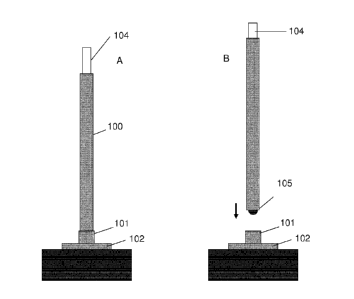
FIG. 1(A) shows the cannula (100) of the invention engaged with the
stopper/pledget (101/102) assembly which is fixed on the vessel (103); also
the
introducer (104) is shown in the figure;
FIG. 1(B) shows the cannula/introducer (100/104) assembly, according to the
invention, not yet engaged with the stopper/pledget (101/102) assembly already
fixed at the vessel (103) to be cannulated; the blade or sharp edge of the
introducer, protruding from the tip of the cannula is visible in this figure;
FIG.2 shows a detail of the stopper/pledget (101/102) assembly already fixed
at
the vessel (103) to be cannulated; in the embodiment shown in this figure the
pledget is secured to the vessel by means of a minimum of two suture lines
(106)
and the stopper is engaged to the pledget by means of a suture with an exposed
loop (107);
FIG. 3 shows the detail of the cannula (100), according to the invention, when
it is
in place into the vessel (103) and it is mechanically engaged to the
pledget/stopper (102/101) assembly by means of a mechanical joint (108) and it
is
further secured, for not protruding to far into the vessel, by means of a
molded
II
CA 02837421 2016-12-02
4
feature (109) which, stopping against the stopper, permits the cannula to
enter the
aorta only to a fixed dept;
FIG. 4 shows tourniquets (111) removably attached to the external body of the
cannula (100); the suture lines (106), which secures the pledget/stopper
(102/101)
assembly to the vessel can be introduced into said tourniquets in order to
guide
the cannula exactly to the cannulation site predisposed by the pledget/stopper
(102/101) assembly.
DETAILED DESCRIPTION OF THE INVENTION
The cannula according to the invention includes three separate components, the
cannula body (100), the pledget and stopper assembly (102/101), and the
introducer (104).
The cannula shape and its dimensions are preferably according to those already
known in the art.
The introducer (104) is a semi rigid tube placed coaxially inside the cannula
to
facilitate cannula placement and can include a hole for wire introducer
commonly
used to guide the introducer and cannula assembly to the required location.
The
introducer can be made from a semi-rigid material such as a polyolephin or
other
plastic that will assist in guiding the cannula assembly to the designated
cannulation site. Specific to this embodiment, the introducer includes a sharp
end
that protrudes past the cannula tip, either the material itself being sharp
enough to
piece the vessel at the cannulation site or a small blade 1 to 3 mm in depth
could
be included in the introducer tip to permit simple and easy vessel penetration
at
the cannulation site.
The tip of the cannula can be designed in any shape or manner desired to
reduce
the damage to the blood or improve the flow out of the cannula, in this
embodiment the minimum recommended tip depth into the vessel is 10 mm. The
tip of the cannula has a diameter smaller than the introducer in order to
avoid that
the introducer blade or sharp edge could protrude outside the cannula tip more
than 1-5 mm, more preferably 1-3 mm.
One of the essential features of the present invention is that the pledget and
stopper assembly (102/101) mates, snaps or otherwise interfaces with the
external
body of the cannula, engaged in a mechanical joint (108) based on interference
or
CA 02837421 2016-12-02
a ball-detent mechanism or male/female joint similar to that found on an ink
pen
cap. The stopper prevents the cannula and introducer assembly from moving too
deeply into the cannulation site beyond a point established by the design of
the
stopper, and acts as a safety mechanism preventing accidental puncture of the
5 opposite wall of the blood vessel being cannulated. Furthermore the
external
cannula body could present a molded feature (109) that permits the cannula to
enter the aorta to a fixed depth, no more (the maximum recommended tip depth
into the vessel is 15 mm). The molded feature (109) is a, further to the
mechanical
engagement (108), second secure stopper at the stopper.
The resulting cannulation is done in an inherently safer was compared to
similar
embodiments such as that described n US patent 6,488,63 by Gannoe, et al.
The introducer and cannula assembly piece the vessel to a fixed depth
established
by the design of the stopper. Once the cannula is in place in the stopper, the
introducer is withdrawn and thrown away. The introducer is withdrawn in a
hemostatic manner, meaning that the cannula fills with blood as the introducer
is
withdrawn, and when the introducer is almost completely out of the cannula,
the
surgeon then clamps the proximal end of the cannula, preventing blood from
coming out of the cannula.
The pledget-stopper assembly (102/101) are sutured to the vessel to be
cannulated (103) and is used to guide the cannula and introducer (100 and
104),
with the cannulation site being at the center of the pledget-stopper assembly.
The
pledget can made in such a way that once the cannula and stopper are
extracted,
the pledget (102) remains connected to the vessel (103).
The pledget-stopper assembly may include a minimum of two suture lines (106)
that are used to fix the pledget to the vessel to be cannulated; these suture
lines
can be included in the assembly or may be fixed onto the pledget by the
surgeon
manually. The stopper can be made of any material, preferably a polycarbonate
or
other moldable plastic that interfaces mechanically with the tip of the
cannula as
described above. The pledget is made from typical pledget/bandage material,
like
a sturdy wool or other bandage material that is flexible enough to take the
shape
of the curvature of the vessel (103) to which it is attached. The sutures
included on
the pledget are used to sew the assembly to the vessel. A minimum of two
sutures
CA 02837421 2013-11-26
WO 2012/168306 PCT/EP2012/060715
6
with needles included at each end of the suture line are preferable but more
than
two lines can easily be included as an improvement or other embodiment of the
invention.
As the cannula and connected stopper are removed from the cannulation site,
the
pledget is then used to close the hole left behind in the vessel (i.e.
cannulation
site) preventing blood loss. This is a very advantageous additional feature of
the
invention because the surgeon often does not have clear visible access to the
aorta and cannulation site in a MICS mini thoracotomy or mini sternotomy
approach. Being able to leave a pledget on the vessel and using the suture
lines
(106) to close the vessel, as described below, is therefore highly
advantageous
due to this lack of visualization.
Therefore the pledget and stopper are preferably each other joined
mechanically
in a manner that will permit easy detachment of the stopper once the procedure
is
complete and the cannula needs to be removed. One embodiment could be using
a suture with an exposed loop (107) joining the stopper to the pledget; the
exposed loop could be cut, breaking the suture which joins the stopper to the
pledget, so that the suture line could be extracted, freeing the stopper from
the
pledget. The stopper remains attached to the cannula and is extracted along
with
the cannula when the assembly is removed, while the pledget remains attached
to
the vessel. By crossing the sutures (106) the vessel can be easily closed when
the
sutures are crossed and tied off manually with a knot or using a more
sophisticated tie off technique, permitting simple closure of the cannulation
site.
Again, at least two suture lines would be required, however any number more
than
two could be realized in this design.
In many cases the cannulation site is not entirely visible, so a method for
aligning
the central axis of the stopper with the tip of the cannula is highly
advantageous.
Such a technique can permit the proper landing of the cannula tip to the seat
of
the pledget stopper assembly, and can be provided quite easily by the use of
the
suture strings (106) and a mechanical guide on the body of the cannula which
permits the use of the suture strings to guide the tip of the cannula directly
into the
stopper. These same suture strings (106) can then be placed into a tube
(typically
called a tourniquet (111) and mechanically attached diametrically opposed to
the
CA 02837421 2013-11-26
WO 2012/168306 PCT/EP2012/060715
7
external surface of the cannula. This allows the tourniquet assembly with the
suture lines inside to be safely secured out of the field of view, which is
important
particularly if the surgical incision and consequent surgical field is quite
small.
