Note: Descriptions are shown in the official language in which they were submitted.
CA 02837647 2013-11-28
WO 2012/166116 PCT/US2011/038593
OPTICAL COHERENCE TOMOGRAPHY PROBE
STATEMENT REGARDING FEDERALLY SPONSORED RESEARCH OR
DEVELOPMENT
[0001] This invention was made with government support under FA9550-04-1-
0045
awarded by the Air Force Office of Scientific Research ¨ DOD. The government
has certain
rights in the invention.
BACKGROUND OF THE INVENTION
[0002] Advances in Optical Coherence Tomography (OCT) technology have made
it
possible to use OCT in a wide variety of applications. One application of OCT
is in
ophthalmology for imaging eye diseases due to the high transmittance of ocular
media. OCT
technology was invented in the early 1990's to generate depth-resolved images
of tissue level
microstructures, in vivo, and without physical contact. Second generation
imaging technology,
such as frequency-domain, swept-source, and spectral-domain OCT, has improved
the signal-to-
noise ratio over first generation technology, translating to faster imaging.
As a result of this
speed increase, high resolution cross-sectional images (B-scans) can be
acquired at video-rates
and three-dimensional images can be acquired very quickly. Sunita Sayeram and
Joseph Izatt,
"High-resolution SDOCT imaging ¨ cutting-edge technology for clinical and
research
applications," Photonik (November 2008) (hereinafter referred to as the
"Photonik Article").
[0003] As noted in the Photonik Article, OCT is an imaging technique which
provides
microscopic tomographic sectioning of biological samples. By measuring singly
backscattered
light as a function of depth, OCT fills a valuable niche in imaging of tissue
ultrastructure,
providing sub-surface imaging with high spatial resolution (-5-10 gm) in three
dimensions and
high sensitivity (>110dB) in vivo with no contact needed between the probe and
the tissue.
[0004] In biological and biomedical imaging applications, OCT allows for
micrometer-scale
imaging non-invasively in transparent, translucent, and highly-scattering
biological tissues. As
illustrated in FIG. 1, the longitudinal ranging capability of OCT is based on
low-coherence
interferometry, in which light from a broadband source is split between
illuminating the sample
of interest and a reference path in a fiber optic interferometer. The
interference pattern of light
1
CA 02837647 2013-11-28
WO 2012/166116 PCT/US2011/038593
backscattered from the sample and light from the reference delay contains
information about the
location and scattering amplitude of the scatterers in the sample. This
information is recorded as
a map of the reflectivity of the sample versus depth, called an A-scan.
[0005] For two or three-dimensional OCT imaging, multiple A-scans are
acquired while the
sample beam is scanned laterally across the tissue surface, building up a map
of reflectivity
versus depth and one or two lateral dimensions. The lateral resolution of the
B-scan is given by
the confocal resolving power of the sample arm optical system.
SUMMARY OF THE INVENTION
[0006] OCT technology has had a profound effect upon ophthalmic imaging and
diagnosis.
Its capabilities are also being embraced by gastroenterology, urology,
oncology, and other
specialties. The OCT B-scan is used daily in ophthalmology clinics to evaluate
the delicate
structures within the eye for evidence of macular edema, macular holes, subtle
retinal lesions,
glaucomatous retinal nerve fiber thinning, etc. As noted in the Photonik
Article, OCT has
evolved with improved imaging speed and resolution especially of the retinal
layers in research
investigations.
[0007] Real-time OCT B-scan imaging of laser ablation has been achieved
with ultrahigh-
speed optical frequency domain imaging, but not through a miniature probe.
Large and small
OCT side-scanning probes have been developed to examine tissues within tubular
structures such
as the esophagus and coronary arteries with lateral resolution up to 10 m.
Probes as small as
0.36 mm have been developed, but they project views only from the side rather
than directly in
front of the catheter tip. OCT has been combined with the operating
microscope, but its lateral
resolution was found to be 5-times less than with the handheld OCT probe
system during
laryngoscopy. A forward-imaging OCT B-scan device has been used to image
bladders, but its
diameter is relatively large at 5.8 mm X 3 mm. The standard
microelectromechanical system
(MEMS) scanning mirror component of an OCT forward-imaging probe has been
reduced to a
diameter of 1 mm, but the mirror alone is still larger than ophthalmic probe
requirements. Others
have used a piezoelectric cantilever system with a rod lens 2.7 mm in
diameter, a lead zirconate
titanate actuator and cantilever within a 2.4 mm diameter probe, a fiber-
bundle system measuring
2
CA 02837647 2013-11-28
WO 2012/166116 PCT/US2011/038593
3.2 mm in diameter, complicated paired rotating GRIN lenses in a probe
measuring 1.65 mm in
diameter, and an electrostatic scanning probe measuring 2.2 mm in diameter. To
pass through
the 1.2 mm diameter size of the smallest endoscopic working channel, a novel
design is required.
Individual OCT A-scan components alone would permit miniaturization of the
sensing probe,
but the system would be unable to provide two-dimensional information.
Alternative designs for
permitting scanning within a miniature probe are required to break the 1.2 mm
diameter size
barrier.
[0008] Accordingly, in one construction, the invention is related to an OCT
probe
miniaturized for insertion into a working channel of an endoscope for imaging
tissue. High-
resolution OCT forward-imaging alone could be used to evaluate sub-surface
structures during
endoscopic procedures. This is likely to advance therapies within small
spaces, such as the space
behind the eye. This endoscopic-capable device has the potential for adoption
in multiple
surgical specialties.
[0009] In one embodiment, the invention provides an optical coherence
tomography probe
comprising a housing configured to support an actuator, a first conduit
connected to the housing,
a second conduit positioned within the first conduit and in communication with
the actuator, a
third conduit, and a single mode fiber. The third conduit is positioned within
the second conduit,
and the third conduit includes a first linear portion and a second curved
portion, the second
portion extending from a distal end of the second conduit. The single mode
fiber is positioned
within the third conduit, and a portion of the single mode fiber extends from
a distal end of the
third conduit. The portion of the single mode fiber is configured to move
laterally when the
actuator activates the second conduit to slide along the third conduit, and
the single mode fiber is
configured to scan light data reflected from a sample positioned in front of a
distal end of the
first conduit.
[0010] In another embodiment, the invention provides an endoscope
comprising a light
source, an imaging source, and an optical coherence tomography probe. The
probe includes a
housing configured to support an actuator, a first conduit connected to the
housing, a second
conduit positioned within the first conduit and in communication with the
actuator, a third
conduit, and a single mode fiber. The third conduit is positioned within the
second conduit, and
3
CA 02837647 2013-11-28
WO 2012/166116 PCT/US2011/038593
the third conduit includes a first linear portion and a second curved portion,
the second portion
extending from a distal end of the second conduit. The single mode fiber is
positioned within the
third conduit, and a portion of the single mode fiber extends from a distal
end of the third
conduit. The portion of the single mode fiber is configured to move laterally
when the actuator
activates the second conduit to slide along the third conduit, and the single
mode fiber is
configured to scan light data reflected from a sample positioned in front of a
distal end of the
first conduit.
[0011] In yet another embodiment, the invention provides an optical
coherence tomography
probe comprising a housing configured to support an actuator, a first conduit
connected to the
housing, a second conduit positioned within the first conduit and in
communication with the
actuator, the second conduit including a first linear portion and a second
curved portion, and a
single mode fiber positioned within the second conduit, the single mode fiber
being configured
to move laterally when the actuator activates the second conduit to slide
within the first conduit,
the single mode fiber configured to scan light data reflected from a sample
positioned in front of
a distal end of the first conduit.
[0012] In a further embodiment, the invention provides an endoscope
comprising a light
source, an imaging source, and an optical coherence tomography probe. The
probe includes a
housing configured to support an actuator, a first conduit connected to the
housing, a second
conduit positioned within the first conduit and in communication with the
actuator, the second
conduit including a first linear portion and a second curved portion, and a
single mode fiber
positioned within the second conduit, the single mode fiber being configured
to move laterally
when the actuator activates the second conduit to slide within the first
conduit, the single mode
fiber configured to scan light data reflected from a sample positioned in
front of a distal end of
the first conduit.
[0013] An additional embodiment of the invention provides an optical
coherence
tomography probe comprising a housing configured to support an actuator, a
first conduit
connected to the housing, and a single mode fiber positioned within the first
conduit, the single
mode fiber being configured to move laterally when activated by the actuator,
the single mode
4
CA 02837647 2013-11-28
WO 2012/166116 PCT/US2011/038593
fiber configured to scan light data reflected from a sample positioned in
front of a distal end of
the first conduit.
[0014] A further embodiment of the invention provides an endoscope
comprising a light
source, an imaging source, and an optical coherence tomography probe. The
probe includes a
housing configured to support an actuator, a first conduit connected to the
housing, and a single
mode fiber positioned within the first conduit, the single mode fiber being
configured to move
laterally when activated by the actuator, the single mode fiber configured to
scan light data
reflected from a sample positioned in front of a distal end of the first
conduit.
[0015] The invention also provides a method of imaging a sample. The method
includes
inserting an endoscope through a lumen toward a target in the patient, the
endoscope including
an imaging device having a single mode fiber, activating the single mode fiber
to laterally scan
for light data reflected from the target, collecting the light data reflected
from the target, and
generating a B-scan image of the collected light data, the image representing
the target
positioned about 1 mm to about 15 mm forward of a distal end of the endoscope.
[0016] Other aspects of the invention will become apparent by consideration
of the detailed
description and accompanying drawings.
BRIEF DESCRIPTION OF THE DRAWINGS
[0017] FIG. 1 is a schematic illustration of an OCT system.
[0018] FIG. 2 is a schematic illustration of an OCT system incorporating an
OCT probe
according to one embodiment of the present invention.
[0019] FIGS. 3-4 are schematic illustrations of an OCT probe according to
one embodiment
of the present invention.
[0020] FIG. 5 is a schematic illustration of an OCT probe according to one
embodiment of
the present invention.
[0021] FIGS. 6-9 are schematic illustrations of an OCT probe according to
one embodiment
of the present invention.
CA 02837647 2013-11-28
WO 2012/166116 PCT/US2011/038593
[0022] FIG. 10 is a schematic illustration of an OCT probe according to one
embodiment of
the present invention.
[0023] FIGS. 11-14 are schematic illustrations of an OCT probe according to
one
embodiment of the present invention.
[0024] FIGS. 15-16 are schematic illustrations of an OCT probe according to
one
embodiment of the present invention.
[0025] FIGS. 17-21 are schematic illustrations of an OCT probe according to
one
embodiment of the present invention.
[0026] FIGS. 22-23 are schematic illustrations of an OCT probe according to
one
embodiment of the present invention.
[0027] FIG. 24 is a schematic illustration of an OCT probe according to one
embodiment of
the present invention.
[0028] FIGS. 25-29 are schematic illustrations of an OCT probe according to
one
embodiment of the present invention positioned within the working channel of
an endoscope.
[0029] FIG. 30 is a pictorial illustration of an angle OCT image from a B-
scan forward-
imaging prototype probe.
DETAILED DESCRIPTION
[0030] Before any embodiments of the invention are explained in detail, it
is to be
understood that the invention is not limited in its application to the details
of construction and the
arrangement of components set forth in the following description or
illustrated in the following
drawings. The invention is capable of other embodiments and of being practiced
or of being
carried out in various ways. Also, it is to be understood that the phraseology
and terminology
used herein are for the purpose of description and should not be regarded as
limiting. The use of
"including," "comprising," or "having" and variations thereof herein is meant
to encompass the
items listed thereafter and equivalents thereof as well as additional items.
Unless specified or
limited otherwise, the terms "mounted," "connected," "supported," and
"coupled" and variations
6
CA 02837647 2013-11-28
WO 2012/166116 PCT/US2011/038593
thereof are used broadly and encompass both direct and indirect mountings,
connections,
supports, and couplings.
[0031] Although directional references, such as upper, lower, downward,
upward, rearward,
bottom, front, rear, etc., may be made herein in describing the drawings,
these references are
made relative to the drawings (as normally viewed) for convenience. These
directions are not
intended to be taken literally or limit the present invention in any form. In
addition, terms such
as "first," "second," and "third" are used herein for purposes of description
and are not intended
to indicate or imply relative importance or significance.
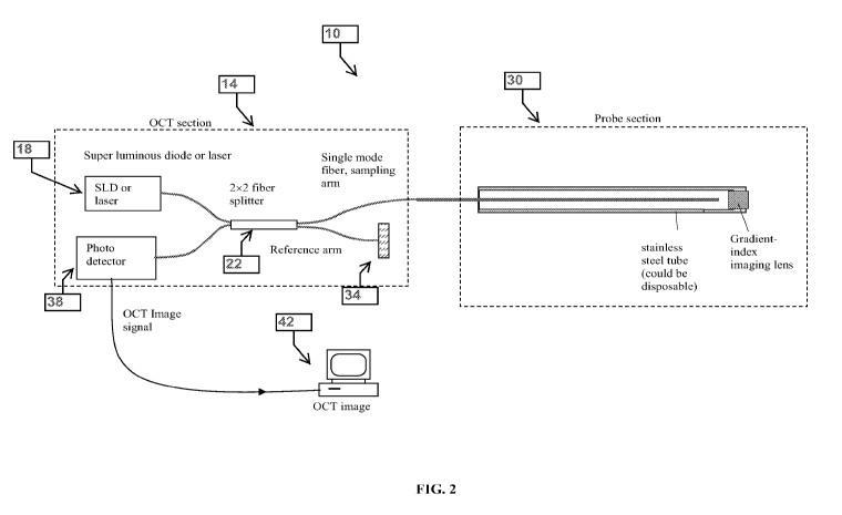
[0032] FIG. 2 schematically illustrates a combined OCT system 10 according
to one
embodiment of the present invention. The OCT system 10 includes an OCT section
14 and a
probe section 30. The OCT section 14 includes a light source 18 that outputs a
light signal,
which is then input to a beam splitter 22 where the light signal is split
between illuminating a
sample via a probe 30 and a reference device 34. The reference device 34 can
include a lens and
a reference mirror. The OCT section 14 also includes a photo detector 38 for
receiving
backscattered light from the sample that was collected by the probe 30 and
light from the
reference device 34. The photo detector 38 can convert the light signals to
digital signals to
generate an OCT image signal, which is transmitted to a computer processor 42
for generation of
an image, such as an A-scan or a B-scan. The computer processor 42 can include
software (e.g.,
stored on non-transitory computer-readable medium) for processing the data
into an A-scan
and/or a B-scan.
[0033] The probe 30 is a miniature intraoperative probe (e.g., 3 mm or
smaller such as 25
gauge) capable of forward-imaging with OCT. FIGS. 3-4 illustrate one
construction of the
probe 30. In this construction, the probe 30 can include a housing 74 having
an electromagnetic
system 78 (e.g., coil, magnet, and suitable electronic circuitry to activate
the coil). The housing
74 is connected to a first tube 82 (or conduit) that defines a first bore 86,
which is configured to
support a second tube 90. The word tube is used herein to describe various
constructions of the
probe; however a tube, as used herein, is a conduit having any cross-sectional
shape suitable to
the invention. Use of the word tube or conduit shall not limit the shape of
the probe to a circular
cross-section as other cross-sectional shapes are contemplated by the
invention.
7
CA 02837647 2013-11-28
WO 2012/166116 PCT/US2011/038593
[0034] The outer diameter of the second tube 90 is less than the inside
diameter of the first
tube 82 such that the second tube 90 can slide or resonate along a length of
the first tube 82 when
the electromagnetic system 78 is activated. The second tube 90 defines a
second bore 94
configured to receive a third tube 98. As illustrated in FIGS. 3-4, the third
tube 98 includes a
first portion 102 being substantially straight and a second portion 106 having
a somewhat S-
shaped curvature. The second portion 106 is at the distal portion of the third
tube 98. The first
tube 82, the second tube 90, and the third tube 98 can comprise stainless
steel or other suitable
materials or combinations of materials.
[0035] With continued reference to FIGS. 3-4, the third tube 98 includes a
third bore 110
configured to receive a fiber 114. A portion 118 of the fiber 114 extends from
the distal end of
the third tube 98 toward a distal end of the first tube 82. A distal end of
the fiber 114 is
positioned adjacent a GRIN imaging lens 122, which is connected to the distal
end of the first
tube 82. The portion 118 of the fiber 114 can move laterally or in the X
direction (axes
definition and used throughout the specification: the Z axis goes horizontally
across the paper,
the Y axis goes vertically top to bottom, and the X axis goes into the paper)
within the first tube
82 when the second tube 90 is activated and slides within the first tube 82.
The first tube 82 can
include an index-matching liquid.
[0036] FIG. 5 illustrates a second construction of the probe 30. In this
construction, the
probe 30 includes a housing 130 defining a bore 134, a gradient index lens rod
138 extending
from the distal end of the housing 130, and a GRIN lens 142 positioned within
a distal end of the
gradient index lens rod 138. The probe 30 also includes a single mode fiber
146 coupled to a
piezoelectric system 150 (e.g., piezo actuator and suitable electronic
circuitry to activate the
piezo actuator), which is supported within the bore 134 of the housing 130.
Activation of the
piezoelectric system 150 is controlled by a conduit 154 extending from a
proximal end of the
housing 130 and to electronic circuitry. A distal end of the single mode fiber
146 is configured
to move laterally within the bore 134 to scan light data at a proximal end of
the gradient index
lens rod 138 when the piezoelectric system 150 is activated.
[0037] FIGS. 6-9 illustrate a third construction of the probe 30. In this
construction, the
probe 30 includes a first tube 160 having a first portion 164 and a second
portion 168. The first
8
CA 02837647 2013-11-28
WO 2012/166116 PCT/US2011/038593
portion 164 is generally linear while the second portion 168 includes a
plurality of notches 172
thereby defining a plurality of rings 176 interconnected by a strip 180 that
is integral with the
first portion 164. The second portion 168 is non-linear and forms a curvature
as illustrated in
FIGS. 6-9.
[0038] The first tube 160 defines a first bore 180 configured to receive a
single mode fiber
184. In some constructions, the single mode fiber can have about a 125 m
diameter, or about an
80 m diameter, or about a 50 m diameter. Other suitable-sized diameters are
also
contemplated by this construction. The single mode fiber 184 can be connected
or secured (e.g.,
with glue or other suitable fixation method) to a distal end of the second
portion 168. A portion
170 of the single mode fiber 184 extends beyond the distal end of the second
portion 168.
[0039] With further reference to FIG. 9, the first tube 160 is at least
partially supported
within a second bore 188 of a second tube 192, which is connected or secured
to an inner wall of
a third tube 196. The third tube 196 is connected to a housing 200 having an
electromagnetic
system 204 (e.g., coil, magnet, and suitable electronic circuitry to activate
the coil) electrically
connected to suitable electronic circuitry. The housing 200 can include a
ferrule for coupling to
and supporting the proximal end of the single mode fiber 184. The outer
diameter of the first
tube 160 is less than the inside diameter of the second tube 192 such that the
first tube 160 can
slide or resonate along a length of the second tube 192 when the
electromagnetic system 204 is
activated. The first tube 160, the second tube 192, and the third tube 196 can
comprise stainless
steel or other suitable materials or combinations of materials.
[0040] With continued reference to FIG. 9, the portion 170 of the single
mode fiber 184 that
extends from the distal end of the first tube 160 toward a distal end of the
third tube 196 is
positioned adjacent a GRIN imaging lens 208, which is connected to the distal
end of the third
tube 196. The portion 170 of the single mode fiber 184 can move laterally
within the third tube
196 when the first tube 160 slides (after actuation of the electromagnetic
system 204) within the
second tube 192. When the first tube 160 slides within the second tube 192,
the first tube 160
also slides along the single mode fiber 184 to compress the plurality of rings
176, which causes
the portion 170 of the single mode fiber 184 to move laterally to scan light
data near the GRIN
imaging lens 208.
9
CA 02837647 2013-11-28
WO 2012/166116 PCT/US2011/038593
[0041] FIG. 10 illustrates a fourth construction of the probe 30. In this
construction, the
probe 30 includes a single mode fiber 220 having an actuator comprised of a
memory alloy wire
224 coupled to a portion of the fiber 220. The memory alloy wire 224 can cause
the single mode
fiber 220 to move laterally to scan light data when a current is applied to
the wire. The single
mode fiber 220 can be housed within a tube as illustrated in any one of the
constructions
described herein, but a housing is not required.
[0042] FIGS. 11-13 illustrate a fifth construction of the probe 30. In this
construction, the
probe 30 includes a first tube 230 connected to a housing 234 having a chamber
238. The
housing 234 supports a pulsed air system having an inlet 242 coupled to an air
source for
periodically injecting air into the chamber 238. The housing 234 includes a
diaphragm 246
biased in a first position by an elastic member 250 (e.g., a spring). The
diaphragm 246 and the
elastic member 250 are coupled to a second tube 254, which is positioned
within a bore 258 of
the first tube 230. The second tube 254 includes a first portion 262 and a
second portion 266.
The first portion is generally linear and is connected to the diaphragm 246
and coupled to the
elastic member 250. The second portion 266 includes a spring-like structure
that is non-linear
and forms a curvature as illustrated in the figures. A distal end of the
second portion 266 abuts
with a stopper 270 on an inner wall of the first tube 230. The second tube 254
includes a bore
274 through which a single mode fiber 278 is positioned with a portion 282 of
the single mode
fiber 278 extending beyond a distal end of the second tube 254. A proximal end
of the single
mode fiber 278 also extends through the diaphragm 246 and through an aperture
in the housing
234. The portion 282 of the single mode fiber 278 that extends from the distal
end of the second
tube 254 toward a distal end of the first tube 230 is positioned adjacent a
GRIN imaging lens
286, which is connected to the distal end of the first tube 230. When the
chamber 238 fills with
an amount of air that overcomes the biasing force of the elastic member 250,
the diaphragm 246
moves forward. When the diaphragm 246 moves forward, the second tube 254 also
moves
forward thereby causing the second portion 266 of the second tube 254 to flex
in a sinusoidal-
like pattern. This flexing of the second portion 266 causes the portion 282 of
the single mode
fiber 278 to move laterally to scan light data near the GRIN imaging lens 286.
[0043] FIG. 14 illustrates an alternative configuration of the fifth
construction of the probe
30. In this configuration, the actuator (i.e., the inlet 242, the air source,
the diaphragm 246, and
CA 02837647 2013-11-28
WO 2012/166116 PCT/US2011/038593
the elastic member 250) can be replaced with an electromagnetic system similar
to the
electromagnetic systems described above. For example, an electromagnetic
system or a motor
can be electrically coupled to the second tube 254, such that when activated,
the second tube 254
moves forward thereby causing the second portion 266 of the second tube 254 to
flex in a
sinusoidal-like pattern. This flexing of the second portion 266 causes the
portion 282 of the
single mode fiber 278 to move laterally to scan light data near the GRIN
imaging lens 286.
[0044] FIG. 15 illustrates a sixth construction of the probe 30. In this
construction, the probe
30 includes a single mode fiber 400 that goes through a bore within a magnet
404 that is
surrounded by two coils 408. Two coils 408A and 408B, which are 180 degrees
apart are
situated on each side of the magnet 404. The probe 30 includes a GRIN imaging
lens 412
connected to a distal end of the single mode fiber 400. In some alternative
constructions, the
GRIN imaging lens 412 can be connected to a distal end of a housing or tube
that supports the
single mode fiber 400. The coils 408A and 408B are connected to electronic
circuitry such that
when activated, the current through the coil 408 induces the magnet 404 to
move laterally
thereby causing the distal end of the single mode fiber 400 with GRIN imaging
lens 412 to move
laterally to scan light data at the GRIN imaging lens 412.
[0045] With reference to FIG. 16, other alternative constructions
appropriate for the
constructions illustrated in FIGS. 5 or 15 can be implemented with the single
mode fiber 400.
For example, a piezoelectric system can be connected to the single mode fiber
400 that can be
activated to rotate while adjusting the curvature of the distal portion of the
single mode fiber 400.
This rotation method can generate a scanning area of about 2mm diameter. In
other
constructions, the piezoelectric system connected to the single mode fiber 400
can be activated to
move the single mode fiber 400 forward and backward while adjusting an angle
of the distal
portion of the single mode fiber 400 with respect to the piezoelectric system.
In this particular
construction, the single mode fiber 400 can scan for light data in the X and Y
directions.
[0046] With continued reference to FIG. 16, another alternative
construction appropriate for
the construction illustrated in FIGS. 5 or 15 involves attaching two mini
magnets to the single
mode fiber 400 and by using electromagnetic coils to interact with the mini
magnets to activate
the single mode fiber 400 to move and scan for light data in the X and Y
directions. In yet
11
CA 02837647 2013-11-28
WO 2012/166116 PCT/US2011/038593
another alternative construction, a single mini magnet is connected to the
single mode fiber 400
that interacts with signals from electromagnetic coils to activate the single
mode fiber 400 to
move and scan for light data in the X and Y directions. In a further
alternative construction, a
mini magnet and a piezo sheet is connected to the single mode fiber 400. An
electromagnetic
coil interacts with the mini magnet to activate the single mode fiber 400 to
move and scan for
light data in the X direction. In addition, the electromagnetic coil interacts
with the piezo sheet
to activate the single mode fiber 400 to move and scan for light data in the Y
direction.
[0047] FIGS. 17-18 illustrate a seventh construction of the probe 30. In
this construction, the
probe 30 includes a first tube 420 that defines a first bore 424. The first
tube 420 includes a
bearing 422 connected to an inner wall and which is configured to support a
second tube 428.
The outer diameter of the second tube 428 is less than the inside diameter of
the first tube 420
such that the second tube 428 can rotate within the first tube 420 when
activated. The second
tube 428 includes a distal portion 432 having a curvature as illustrated in
the figures. The second
tube 428 defines a second bore 436 configured to receive a third tube 440. The
third tube 440
also includes a distal portion 444 having a curvature as illustrated in the
figures. A portion of the
distal portion 444 extends beyond a distal end of the second tube 428. The
first tube 420, the
second tube 428, and the third tube 440 can comprise stainless steel or other
suitable materials or
combinations of materials.
[0048] With continued reference to FIGS. 17-18, the third tube 440 defines
a third bore 448
configured to receive a single mode fiber 452. A portion 456 of the single
mode fiber 452
extends from the distal end of the third tube 440 toward a distal end of the
first tube 420. The
portion 456 is positioned through an aperture 460 of a ring 464, which is
connected to the first
tube 420. A distal end of the single mode fiber 452 is positioned adjacent a
GRIN imaging lens
468, which is connected to the distal end of the first tube 420. The portion
456 of the single
mode fiber 452 can move in a circular pattern defined by the circumference of
the aperture 460
of the ring 464 within the first tube 420. This circular movement occurs when
the second tube
428 is actuated (by any suitable actuator) to rotate around the third tube 440
and due to the
curvature of both the second tube 428 and the third tube 440. The single mode
fiber 452 scans
for light data while moving in the circular pattern.
12
CA 02837647 2013-11-28
WO 2012/166116 PCT/US2011/038593
[0049] With reference to FIGS. 19-20, an alternative configuration of the
seventh
construction is illustrated. The difference with this alternative
configuration than the
configuration illustrated in FIGS. 17-18 is that the ring 464 moves forward
and backward (i.e., in
the Z direction). The ring 464 is connected to the bearing 422, and the
bearing 422 is coupled to
an actuator. When the second tube 428 is actuated to rotate, and the bearing
422 is actuated to
move in the Z direction, the single mode fiber 452 scans for light data while
moving in a circular
pattern at different diameters. The image target is a circular band as
illustrated.
[0050] FIG. 21 illustrates another alternative configuration of the seventh
construction of the
probe 30. In this configuration, when the bearing 422 is actuated to move in
the Z direction, the
second tube 428 and the third tube 440 also move with the bearing 422. This
movement causes
the single mode fiber 452 to move in the Z direction which results in an image
target being a
circular band having a particular depth or thickness defined by how far the
single mode fiber 452
moves in the Z direction.
[0051] FIGS. 22-23 illustrate an eighth construction of the probe 30. In
this construction, the
probe 30 includes a first tube 480 that defines a first bore 484. The first
tube 480 includes a
bearing 488 connected to an inner wall and which is configured to support a
second tube 492.
The outer diameter of the second tube 492 is less than the inside diameter of
the first tube 480
such that the second tube 492 can rotate within the first tube 480 when
activated. The second
tube 492 includes a distal portion 496 having a curvature as illustrated in
the figures. The second
tube 492 defines a second bore 500 configured to receive a third tube 504. The
third tube 504
also includes a distal portion 508 having a curvature as illustrated in the
figures. A portion of the
distal portion 508 extends beyond a distal end of the second tube 492. The
first tube 480, the
second tube 492, and the third tube 504 can comprise stainless steel or other
suitable materials or
combinations of materials.
[0052] With continued reference to FIGS. 22-23, the third tube 504 defines
a third bore 512
configured to receive a single mode fiber 516. A portion 520 of the single
mode fiber 516
extends from the distal end of the third tube 504 toward a distal end of the
first tube 480. The
portion 520 is positioned through a slit 524 of a bracket 528, which is
connected to the first tube
480. A distal end of the single mode fiber 516 is positioned adjacent a GRIN
imaging lens 532,
13
CA 02837647 2013-11-28
WO 2012/166116 PCT/US2011/038593
which is connected to the distal end of the first tube 480. The portion 520 of
the single mode
fiber 516 can move in a linear pattern defined by the slit 524 of the bracket
528 within the first
tube 480. This linear movement occurs when the second tube 492 is actuated (by
any suitable
actuator) to rotate around the third tube 504. The single mode fiber 516 scans
for light data
while moving in the linear pattern.
[0053] FIG. 24 illustrates a ninth construction of the probe 30. In this
construction, the probe
30 includes a first tube 540 that defines a first bore 544, which is
configured to support a second
tube 548. The second tube 548 defines a second bore 552 configured to receive
a third tube 556
and two additional bores to receive two thin wires or strings 580, 584. As
illustrated in FIG. 24,
the third tube 556 includes a first generally linear portion 560 and a second
portion 564 having a
spring-like configuration. The second portion 564 is at the distal portion of
the third tube 556.
The first tube 540, the second tube 548, and the third tube 556 can comprise
stainless steel or
other suitable materials or combinations of materials.
[0054] The third tube 556 includes a third bore 568 configured to receive a
single mode fiber
572. A portion 576 of the single mode fiber 572 extends from the distal end of
the third tube 556
toward a distal end of the first tube 540. The distal end of the third tube
556 is connected to two
electrical conduits 580, 584, which extend through the second tube 548 and are
coupled to a
suitable actuator. FIG. 24 also illustrates several constructions of
alternative cross-sections of
the second tube 548. A distal end of the single mode fiber 572 is positioned
adjacent a GRIN
imaging lens 588, which is connected to the distal end of the first tube 540.
The portion 576 of
the single mode fiber 572 can move laterally within the first tube 540 when
the actuator
alternately pulls or activates the thin wires or strings 580, 584 causing the
second portion 564 of
the third tube 556 to bend or flex. This bending or flexing of the second
portion 564 allows the
distal portion 576 of the single mode fiber 572 to move laterally to scan
light data at the GRIN
imaging lens 588.
[0055] FIGS. 25-29 illustrate how the probe 30 is incorporated into an
endoscope. An
endoscope 600 includes a first tube 604. Within the first tube 604, the
endoscope can include a
second tube 608 and a third tube or working channel 612. The second tube 608
can support the
endoscope's image fiber bundle 616 and the imaging lens 620. The third tube
612 can support
14
CA 02837647 2013-11-28
WO 2012/166116 PCT/US2011/038593
the probe 30 (in any one of the constructions described above). The first tube
604 also includes
numerous illumination fibers that provide a light source for illuminating the
sample tissue.
[0056] The single mode fiber of each of the probes 30 described above is in
communication
with a processor for receiving the light data reflected from the sample. The
processor is
configured to generate an A-scan and/or a B-scan image from the light data.
FIG. 30 illustrates a
B-scan image from a probe 30 that was positioned within the eye. The white
arrow identifies
Schlemms canal in the eye, and the red arrow identifies the Angle.
[0057] The GRIN imaging lens of each of the probes 30 described above is
polished to a
particular length to define a focus point and focus length which matches the
OCT imaging plane.
The length of the GRIN imaging lens can be in the range of about 0.1 mm to
about 3 mm.
Although the GRIN imaging lens is illustrated in many of the constructions
described above as
being connected to the outer tube, the GRIN imaging lens can be instead
connected to the distal
end of the single mode fiber in those constructions. In addition, the imaging
lens could be a
GRIN lens, a lens ground onto a GRIN rod, an aspherical lens, a spherical
lens, or a combination
of these lenses.
[0058] The single mode fiber of each of the probes 30 described above can
have a diameter
of about 125 m. In other constructions, the single mode fiber can have a
diameter of about
50 m or about 80 m. In other constructions, the single mode fiber can have a
customized
diameter.
[0059] The probes 30 can include a single-use disposable detachable tip
which includes the
outer distal conduit and imaging lens. Similarly, the entire OCT probe could
be a disposable
single-use device.
[0060] The probe 30 can be combined with a confocal microscopy probe or an
ultrasound
probe for enhanced visualization of tissue samples.
[0061] Various features and advantages of the invention are set forth in
the following claims.