Note: Descriptions are shown in the official language in which they were submitted.
CA 02837886 2013-11-29
WO 2013/004555
PCT/EP2012/062399
1
Description
Medical Device comprising Illumination Arrangement
Field of the Invention
The present invention relates to the field of medical devices and in
particular to drug
delivery devices or lancing devices having a piercing element to penetrate the
skin of a
patient.
Background and Prior Art
User operated drug delivery devices are as such known in the art. They are
typically
applicable in circumstances, in which persons without formal medical training,
i.e.,
patients, need to administer an accurate and predefined dose of a medicament,
such as
heparin or insulin. In particular, such devices have application, where a
medicament is
administered on a regular or irregular basis over a short-term or long-term
period.
In order to accommodate these demands, such devices have to fulfil a number of
requirements. First of all, the device must be robust in construction, yet
easy to use in
terms of handling and in understanding by the user of its operation and the
delivery of
the required dose or medicament. The dose setting must be easy and
unambiguous.
Where the device is to be disposable rather than reusable, the device should
be
inexpensive to manufacture and easy to dispose.
With patients suffering diabetes for instance, a blood glucose level has to be
monitored
and according to an actual measurement of said level an appropriate dose of
insulin has
to be administered on a regular basis. Blood glucose measurements as well as
injection
of a required dose of the medicament are quite often conducted by the patients
themselves in different circumstances and situations. However, in rather dim
or even
dark environments, the process of piercing the skin may become rather
difficult. Hence,
CA 02837886 2013-11-29
WO 2013/004555
PCT/EP2012/062399
2
a required degree of brightness or visibility for a lancing and/or for an
injection process
is not always available.
Document WO 01/41837 Al discloses an injection device having a housing with a
bow
which can be pressed against and extend a skin surface where an injection is
to be
made. The bow is preferably made of a light transmitting material and can be
illuminated by a lamp.
Moreover, document DE 33 22 923 Al discloses an injection syringe with an
illumination arrangement. There, the illumination arrangement is integrated
into the
syringe and is arranged lateral to the syringe for directing a focused light
beam towards
the distal tip of the injection needle. In this way, the site of needle
puncture can be
illuminated.
Even though various illumination arrangement already exist for syringe
applications and
injection devices, control and operation of the illumination requires a
separate and
additional interaction with the user or patient. In particular, the
illumination has to be
switched on and/or off separately. Also, the operation of an illumination
arrangement is
rather uncorrelated to the main functions of the injection device.
Objects of the Invention
It is therefore an object of the present invention to provide a medical device
having an
improved illumination arrangement for illuminating a site of needle puncture
in a
comfortable and user-friendly way. The illumination arrangement should be easy
and
intuitively to control. The illumination arrangement should further improve
user comfort
and patient safety.
Summary of the Invention
The invention generally refers to a medical device for intradermal or
subcutaneous
treatment of a patient. For this purpose, the medical device comprises a
piercing
CA 02837886 2013-11-29
WO 2013/004555
PCT/EP2012/062399
3
element to penetrate a skin portion of the patient. The device further
comprises an
illumination arrangement to illuminate the piercing element and/or to
illuminate the skin
portion, hence the site of needle puncture, at least during a treatment
process. The
illumination arrangement, in particular its control, is further adapted to
visually indicate a
predefined treatment sequence of the treatment process to be executed by or
with the
help of the medical device. The illumination arrangement, in particular its
control, is
adapted to coordinate illumination with the steps in a treatment process
and/or with the
status of the medical device.
With this particular feature, the illumination arrangement is not only adapted
to
illuminate a site of needle puncture and/or to illuminate the device itself
but also to
provide treatment-related information, for example timing information, to the
user or
patient prior, during or after a respective treatment process to be conducted
and
executed by way of the medical device.
In particular, the illumination arrangement is adapted to indicate the
beginning, the end
or various distinct intermediate steps of the treatment process. The
illumination
arrangement may be adapted to visualize, that the medical device is ready for
use.
Moreover, the illumination arrangement and its control may indicate a
predefined time
interval, the piercing element has to remain in or at the site of needle
puncture. Also, the
illumination arrangement may visually indicate a predefined dwell period, the
piercing
element should remain in biological tissue after delivery of the medicament in
the tissue.
According to a preferred embodiment, the illumination arrangement is adapted
to
generate at least one light cone around the piercing element to illuminate the
skin
portion. Preferably, the illumination arrangement comprises a light source or
at least a
light-emitting aperture symmetrically arranged around the piercing element. It
is of
particular benefit, when the centre of the light cone substantially coincides
with the
piercing element. In this way, the site of puncture can be homogeneously
illuminated,
substantially irrespective of the orientation of the medical device.
CA 02837886 2013-11-29
WO 2013/004555
PCT/EP2012/062399
4
According to another aspect, it is intended, that the illumination arrangement
is adapted
to generate various or variable light cones featuring a different cone angle,
respectively.
Moreover, the illumination arrangement may comprise different or multiple
light sources,
each of which being adapted to generate a light cone with a particular cone
angle.
Depending on the cone angle and the light intensity provided by the respective
light
source, either a rather bright and focused light spot may be incident on the
skin portion
and/or a rather large area of the skin may become subject to a rather faint or
low level
of illumination.
Moreover, it is even conceivable, that the illumination arrangement produces
two
different light cones simultaneously, wherein a rather focused inner light
cone is entirely
surrounded by an outer rather diverging and less intensive light cone. In this
way, the
outer light cone may provide a rather low level and ambient illumination
wherein the
inner light cone is particularly adapted and intended to brightly illuminate
the site of skin
puncture. By sufficiently illuminating the site of puncture, penetration angle
and
penetration depth can be easily controlled by the user or patient himself,
even in a dim
environment. The outer and ambient illumination cone may thus provide an
illumination
level sufficient for a rough orientation with respect to the site of puncture.
According to a further aspect, the illumination arrangement comprises at least
one light
source. The light source is preferably electrically driven. It may comprise an
incandescent element, a gas discharge element, an electro-luminescent element
or
even laser-like optical elements providing stimulated emission radiation. In
particular,
the light source may comprise at least one light emitting diode (LED)
providing a rather
high luminous efficacy at a rather low degree of electrical power consumption.
Preferably, the illumination arrangement also comprises at least one light
reflecting
and/or light deflecting element in order to provide a required beam shape and
intensity
distribution. The illumination arrangement may comprise at least one mirror or
reflector
and/or respective refractive and/or diffractive optical elements to modify and
to shape
the emitted light in a predefined way.
CA 02837886 2013-11-29
WO 2013/004555
PCT/EP2012/062399
According to another aspect, the illumination arrangement is integrated into a
housing of
the device. Preferably, the illumination arrangement or its light emitting
aperture is
positioned near a distal end of the medical device pointing towards the skin
portion
during a treatment process. Preferably, the illumination arrangement or its
light emitting
5 aperture is arranged around the piercing element, e.g. near a distal end
of a cartridge
holder of e.g. a pen-type injector. In particular, when the piercing element
is of
disposable or replaceable type, the illumination arrangement and/or its light
emitting
aperture can be arranged near or even at an interface adapted to releasably
connect
the piercing element to the housing of the medical device.
According to a further preferred embodiment, the medical device comprises a
control
element adapted to at least partially correlate activation and/or deactivation
of the
illumination arrangement with the predefined sequence of the treatment process
to be
executed with the medical device. In particular, the control element is
adapted to
automatically activate and/or to deactivate the illumination arrangement
depending on
the user-controlled functionality of the medical device. If the medical device
is for
instance designed as a drug delivery device, such as a pen-type injector,
setting of a
dose of a medicament may be detected by the control element and may therefore
automatically switch on the illumination arrangement. Additionally, also a
dose injection
action may trigger a different type or a supplemental illumination.
Moreover, the illumination arrangement is coupled with a control element
adapted to
control and/or to initiate the treatment process. For instance, the
illumination
arrangement, in particular its light source or its control element may be
electrically
connected with e.g. a dose dial or dose button of a drug delivery device. In
this way,
setting of a dose by activating or dialling a dose dial may inherently trigger
to switch on
the illumination arrangement. In the further process of treatment, depressing
of a dose
button to initiate dose injection or dose dispensing may be monitored
accordingly and
may be used to transfer the illumination arrangement into a different mode of
operation.
Hence, light intensity, cone angle or spectral composition of the emitted
light may
change appropriately. Moreover, also the end of a dose setting procedure prior
to a
CA 02837886 2013-11-29
WO 2013/004555
PCT/EP2012/062399
6
dose dispensing action may be detected and may serve as a trigger to switch
the
illumination into a predefined illumination mode.
According to another preferred embodiment, the control element is even adapted
to
determine and/or to detect at least one distinct step of the treatment
process. For
instance, the control element may detect removal of a protective cap being
indicative of
the beginning of a treatment process. For instance, the control element may
detect
activation of an "arm" button, indicating that a dose has been set and that
the injection
process may start. Moreover, any manipulation of dose setting or dose
dispensing
elements of the medical device may be detected by the control element, which
in
response to such events may trigger a corresponding illumination, either of
the device
itself or of a skin portion to become subject to medical treatment.
Additionally, the control element may not only be adapted to switch on or to
change the
illumination. According to a further embodiment, the control element may be
also
adapted to modify and/or to deactivate the illumination provided by the
illumination
arrangement within a predefined temporal delay after reaching of or after
determination
of a distinct step of the treatment process. In particular with pen-type
injectors it may be
beneficial, when the end of an injection process is signalled by the
illumination
arrangement. Since an injection needle should stay in the skin tissue for a
predefined
dwell period after the dose of the medicament has been dispensed, the
illumination
arrangement may be used to indicate the duration of said dwell period, which
is typically
in the range of a few seconds.
Therefore and according to another preferred embodiment, the illumination
arrangement
is adapted to indicate a particular treatment sequence, e.g. a dwell period,
by at least
two different illumination colours and/or by way of different illumination
schemes.
According to one approach, the duration of a predefined treatment sequence may
be
indicated by a flashing or blinking of the illumination arrangement.
Furthermore, the
illumination sequence may intuitively indicate the end of a particular
treatment step, e.g.
by a varying illumination sequence. Hence, the frequency of a blinking may
increase or
CA 02837886 2013-11-29
WO 2013/004555
PCT/EP2012/062399
7
decrease until the light pattern emitted by the illumination arrangement
either
approaches a constant or zero intensity level. Apart from modifying the colour
or the
spectral composition of the emitted light, it is also conceivable, that the
illumination
arrangement is adapted to modify the intensity or attenuation of the light.
According to a further preferred aspect, the medical device comprises a drug
delivery
device, particularly adapted and designed for injecting a liquid medicament.
The drug
delivery device is preferably designed as a pen-type injector for conducting
self-
medication of various medicaments. In a further preferred aspect, the drug
delivery
device comprises a housing to accommodate a cartridge being at least partially
filled
with a medicament to be injected. The drug delivery device further comprises a
drive
mechanism having a piston rod to become operably engaged with a piston of the
cartridge. The piston is slidably arranged in the cartridge and provides a
proximal seal
for the cartridge.
A distal end of the cartridge, typically sealed by a pierceable or penetrable
septum is to
be coupled with the piercing element typically comprising an injection needle.
This way
and by exerting distally directed thrust to the piston of the cartridge, a
predefined dose
of the medicament can be expelled through the injection needle and can thus be
administered to biological tissue.
It may be of further benefit, when the cartridge is readily disposed and
arranged in the
housing of the medical device. The medical device, in particular the drug
delivery device
may be of disposable type. Hence, instead of replacing an empty cartridge, the
entire
device may be discarded when the content of the cartridge is used up.
Alternatively, it is
also conceivable, that the cartridge is replaceable. Hence, the housing of the
drug
delivery device can be disassembled and re-assembled in order to provide
cartridge
replacement.
In a further aspect, the medical device may comprise a lancing device adapted
to pierce
and/or to analyze biological fluid and/or biological tissue. In particular,
the lancing
CA 02837886 2013-11-29
WO 2013/004555
PCT/EP2012/062399
8
device comprises a needle to puncture a blood vessel in order to extract and
to
measure a blood glucose level or other physiological substances or parameters.
In effect and according to another preferred aspect, actuation of the control
element of
the medical device may turn on and activate the illumination arrangement at
least for a
predefined time interval. The invention therefore provides fully or semi-
automated
activation, modification and/or deactivation of an illumination arrangement of
a medical
device to enhance patient safety and to guide the user through the treatment
process to
be conducted with the medical device.
Furthermore, and according to an independent aspect, the invention also
relates to a
method of controlling an illumination arrangement of a medical device. Here,
in a first
step, at least one treatment sequence of a predefined treatment process to be
executed
by the medical device is detected. In response to said detection, the control
of the
illumination arrangement may automatically modify, activate and/or deactivate
the
illumination arrangement, wherein further parameters, like a predefined delay
or
temporal offset may be additionally implemented to correlate to at least one
particular
treatment sequence of the treatment process with a modification, activation
and/or
deactivation of the illumination arrangement.
Alternatively or additionally, the method may provide visually indicating at
least one
predefined treatment sequence of the treatment process by means of the
illumination
arrangement. In this way, the patient and/or user of the device can be
visually guided
through and informed about the treatment process in general and the single
treatment
steps to be conducted sequentially.
The term "drug" or "medicament", as used herein, means a pharmaceutical
formulation
containing at least one pharmaceutically active compound,
wherein in one embodiment the pharmaceutically active compound has a molecular
weight up to 1500 Da and/or is a peptide, a proteine, a polysaccharide, a
vaccine, a
CA 02837886 2013-11-29
WO 2013/004555
PCT/EP2012/062399
9
DNA, a RNA, an enzyme, an antibody or a fragment thereof, a hormone or an
oligonucleotide, or a mixture of the above-mentioned pharmaceutically active
compound,
wherein in a further embodiment the pharmaceutically active compound is useful
for the
treatment and/or prophylaxis of diabetes mellitus or complications associated
with
diabetes mellitus such as diabetic retinopathy, thromboembolism disorders such
as
deep vein or pulmonary thromboembolism, acute coronary syndrome (ACS), angina,
myocardial infarction, cancer, macular degeneration, inflammation, hay fever,
atherosclerosis and/or rheumatoid arthritis,
wherein in a further embodiment the pharmaceutically active compound comprises
at
least one peptide for the treatment and/or prophylaxis of diabetes mellitus or
complications associated with diabetes mellitus such as diabetic retinopathy,
wherein in a further embodiment the pharmaceutically active compound comprises
at
least one human insulin or a human insulin analogue or derivative, glucagon-
like
peptide (GLP-1) or an analogue or derivative thereof, or exendin-3 or exendin-
4 or an
analogue or derivative of exendin-3 or exendin-4.
Insulin analogues are for example Gly(A21), Arg(B31), Arg(B32) human insulin;
Lys(B3),
Glu(B29) human insulin; Lys(B28), Pro(B29) human insulin; Asp(B28) human
insulin;
human insulin, wherein proline in position B28 is replaced by Asp, Lys, Leu,
Val or Ala
and wherein in position B29 Lys may be replaced by Pro; Ala(B26) human
insulin;
Des(B28-630) human insulin; Des(B27) human insulin and Des(B30) human insulin.
Insulin derivates are for example B29-N-myristoyl-des(B30) human insulin; B29-
N-
palmitoyl-des(B30) human insulin; B29-N-myristoyl human insulin; B29-N-
palmitoyl
human insulin; B28-N-myristoyl LysB28ProB29 human insulin; B28-N-palmitoyl-
LysB28ProB29 human insulin; B30-N-myristoyl-ThrB29LysB30 human insulin; B30-N-
palmitoyl- ThrB29LysB30 human insulin; B29-N-(N-palmitoyl-Y-glutamyI)-des(B30)
human insulin; B29-N-(N-lithocholyl-Y-glutamyI)-des(B30) human insulin; B29-N-
(w-
CA 02837886 2013-11-29
WO 2013/004555
PCT/EP2012/062399
carboxyheptadecanoyI)-des(B30) human insulin and B29-N-(w-carboxyhepta-
idecanoyl)
human insulin.
Exendin-4 for example means Exendin-4(1-39), a peptide of the sequence H His-
Gly-
5 Glu-Gly-Thr-Phe-Thr-Ser-Asp-Leu-Ser-Lys-Gln-Met-Glu-Glu-Glu-Ala-Val-Arg-Leu-
Phe-
Ile-Glu-Trp-Leu-Lys-Asn-Gly-Gly-Pro-Ser-Ser-Gly-Ala-Pro-Pro-Pro-Ser-NH2.
Exendin-4 derivatives are for example selected from the following list of
compounds:
10 H-(Lys)4-des Pro36, des Pro37 Exendin-4(1-39)-NH2,
H-(Lys)5-des Pro36, des Pro37 Exendin-4(1-39)-NH2,
des Pro36 Exendin-4(1-39),
des Pro36 [Asp28] Exendin-4(1-39),
des Pro36 [IsoAsp28] Exendin-4(1-39),
des Pro36 [Met(0)14, Asp28] Exendin-4(1-39),
des Pro36 [Met(0)14, IsoAsp28] Exendin-4(1-39),
des Pro36 [Trp(02)25, Asp28] Exendin-4(1-39),
des Pro36 [Trp(02)25, IsoAsp28] Exendin-4(1-39),
des Pro36 [Met(0)14 Trp(02)25, Asp28] Exendin-4(1-39),
des Pro36 [Met(0)14 Trp(02)25, IsoAsp28] Exendin-4(1-39); or
des Pro36 [Asp28] Exendin-4(1-39),
des Pro36 [IsoAsp28] Exendin-4(1-39),
des Pro36 [Met(0)14, Asp28] Exendin-4(1-39),
des Pro36 [Met(0)14, IsoAsp28] Exendin-4(1-39),
des Pro36 [Trp(02)25, Asp28] Exendin-4(1-39),
des Pro36 [Trp(02)25, IsoAsp28] Exendin-4(1-39),
des Pro36 [Met(0)14 Trp(02)25, Asp28] Exendin-4(1-39),
des Pro36 [Met(0)14 Trp(02)25, IsoAsp28] Exendin-4(1-39),
wherein the group -Lys6-NH2 may be bound to the C-terminus of the Exendin-4
derivative;
CA 02837886 2013-11-29
WO 2013/004555
PCT/EP2012/062399
11
or an Exendin-4 derivative of the sequence
des Pro36 Exendin-4(1-39)-Lys6-NH2 (AVE0010),
H-(Lys)6-des Pro36 [Asp28] Exendin-4(1-39)-Lys6-NH2,
des Asp28 Pro36, Pro37, Pro38Exendin-4(1-39)-NH2,
H-(Lys)6-des Pro36, Pro38 [Asp28] Exendin-4(1-39)-NH2,
H-Asn-(Glu)5des Pro36, Pro37, Pro38 [Asp28] Exendin-4(1-39)-NH2,
des Pro36, Pro37, Pro38 [Asp28] Exendin-4(1-39)-(Lys)6-NH2,
H-(Lys)6-des Pro36, Pro37, Pro38 [Asp28] Exendin-4(1-39)-(Lys)6-NH2,
H-Asn-(Glu)5-des Pro36, Pro37, Pro38 [Asp28] Exendin-4(1-39)-(Lys)6-NH2,
H-(Lys)6-des Pro36 [Trp(02)25, Asp28] Exendin-4(1-39)-Lys6-NH2,
H-des Asp28 Pro36, Pro37, Pro38 [Trp(02)25] Exendin-4(1-39)-NH2,
H-(Lys)6-des Pro36, Pro37, Pro38 [Trp(02)25, Asp28] Exendin-4(1-39)-NH2,
H-Asn-(Glu)5-des Pro36, Pro37, Pro38 [Trp(02)25, Asp28] Exendin-4(1-39)-NH2,
des Pro36, Pro37, Pro38 [Trp(02)25, Asp28] Exendin-4(1-39)-(Lys)6-NH2,
H-(Lys)6-des Pro36, Pro37, Pro38 [Trp(02)25, Asp28] Exendin-4(1-39)-(Lys)6-
NH2,
H-Asn-(Glu)5-des Pro36, Pro37, Pro38 [Trp(02)25, Asp28] Exendin-4(1-39)-(Lys)6-
NH2,
H-(Lys)6-des Pro36 [Met(0)14, Asp28] Exendin-4(1-39)-Lys6-NH2,
des Met(0)14 Asp28 Pro36, Pro37, Pro38 Exendin-4(1-39)-NH2,
H-(Lys)6-desPro36, Pro37, Pro38 [Met(0)14, Asp28] Exendin-4(1-39)-NH2,
H-Asn-(Glu)5-des Pro36, Pro37, Pro38 [Met(0)14, Asp28] Exendin-4(1-39)-NH2,
des Pro36, Pro37, Pro38 [Met(0)14, Asp28] Exendin-4(1-39)-(Lys)6-NH2,
H-(Lys)6-des Pro36, Pro37, Pro38 [Met(0)14, Asp28] Exendin-4(1-39)-(Lys)6-NH2,
H-Asn-(Glu)5 des Pro36, Pro37, Pro38 [Met(0)14, Asp28] Exendin-4(1-39)-(Lys)6-
NH2,
H-Lys6-des Pro36 [Met(0)14, Trp(02)25, Asp28] Exendin-4(1-39)-Lys6-NH2,
H-des Asp28 Pro36, Pro37, Pro38 [Met(0)14, Trp(02)25] Exendin-4(1-39)-NH2,
H-(Lys)6-des Pro36, Pro37, Pro38 [Met(0)14, Asp28] Exendin-4(1-39)-NH2,
H-Asn-(Glu)5-des Pro36, Pro37, Pro38 [Met(0)14, Trp(02)25, Asp28] Exendin-4(1-
39)-
NH2,
des Pro36, Pro37, Pro38 [Met(0)14, Trp(02)25, Asp28] Exendin-4(1-39)-(Lys)6-
NH2,
H-(Lys)6-des Pro36, Pro37, Pro38 [Met(0)14, Trp(02)25, Asp28] Exendin-4(S1-39)-
(Lys)6-NH2,
CA 02837886 2013-11-29
WO 2013/004555
PCT/EP2012/062399
12
H-Asn-(Glu)5-des Pro36, Pro37, Pro38 [Met(0)14, Trp(02)25, Asp28] Exendin-4(1-
39)-
(Lys)6-NH2;
or a pharmaceutically acceptable salt or solvate of any one of the afore-
mentioned
Exendin-4 derivative.
Hormones are for example hypophysis hormones or hypothalamus hormones or
regulatory active peptides and their antagonists as listed in Rote Liste, ed.
2008,
Chapter 50, such as Gonadotropine (Follitropin, Lutropin, Choriongonadotropin,
Menotropin), Somatropine (Somatropin), Desmopressin, Terlipressin,
Gonadorelin,
Triptorelin, Leuprorelin, Buserelin, Nafarelin, Goserelin.
A polysaccharide is for example a glucosaminoglycane, a hyaluronic acid, a
heparin, a
low molecular weight heparin or an ultra low molecular weight heparin or a
derivative
thereof, or a sulphated, e.g. a poly-sulphated form of the above-mentioned
polysaccharides, and/or a pharmaceutically acceptable salt thereof. An example
of a
pharmaceutically acceptable salt of a poly-sulphated low molecular weight
heparin is
enoxaparin sodium.
Antibodies are globular plasma proteins (-150 kDa) that are also known as
immunoglobulins which share a basic structure. As they have sugar chains added
to
amino acid residues, they are glycoproteins. The basic functional unit of each
antibody
is an immunoglobulin (Ig) monomer (containing only one Ig unit); secreted
antibodies
can also be dimeric with two Ig units as with IgA, tetrameric with four Ig
units like teleost
fish IgM, or pentameric with five Ig units, like mammalian IgM.
The Ig monomer is a "Y"-shaped molecule that consists of four polypeptide
chains; two
identical heavy chains and two identical light chains connected by disulfide
bonds
between cysteine residues. Each heavy chain is about 440 amino acids long;
each light
chain is about 220 amino acids long. Heavy and light chains each contain
intrachain
disulfide bonds which stabilize their folding. Each chain is composed of
structural
domains called Ig domains. These domains contain about 70-110 amino acids and
are
CA 02837886 2013-11-29
WO 2013/004555
PCT/EP2012/062399
13
classified into different categories (for example, variable or V, and constant
or C)
according to their size and function. They have a characteristic
immunoglobulin fold in
which two [3 sheets create a "sandwich" shape, held together by interactions
between
conserved cysteines and other charged amino acids.
There are five types of mammalian Ig heavy chain denoted by a, 6, E, y, and p.
The type
of heavy chain present defines the isotype of antibody; these chains are found
in IgA,
IgD, IgE, IgG, and IgM antibodies, respectively.
Distinct heavy chains differ in size and composition; a and y contain
approximately 450
amino acids and 6 approximately 500 amino acids, while p and E have
approximately
550 amino acids. Each heavy chain has two regions, the constant region (CH)
and the
variable region (VH). In one species, the constant region is essentially
identical in all
antibodies of the same isotype, but differs in antibodies of different
isotypes. Heavy
chains y, a and 6 have a constant region composed of three tandem Ig domains,
and a
hinge region for added flexibility; heavy chains p and E have a constant
region
composed of four immunoglobulin domains. The variable region of the heavy
chain
differs in antibodies produced by different B cells, but is the same for all
antibodies
produced by a single B cell or B cell clone. The variable region of each heavy
chain is
approximately 110 amino acids long and is composed of a single Ig domain.
In mammals, there are two types of immunoglobulin light chain denoted by A and
K. A
light chain has two successive domains: one constant domain (CL) and one
variable
domain (VL). The approximate length of a light chain is 211 to 217 amino
acids. Each
antibody contains two light chains that are always identical; only one type of
light chain,
K or A, is present per antibody in mammals.
Although the general structure of all antibodies is very similar, the unique
property of a
given antibody is determined by the variable (V) regions, as detailed above.
More
specifically, variable loops, three each the light (VL) and three on the heavy
(VH) chain,
are responsible for binding to the antigen, i.e. for its antigen specificity.
These loops are
referred to as the Complementarity Determining Regions (CDRs). Because CDRs
from
CA 02837886 2013-11-29
WO 2013/004555
PCT/EP2012/062399
14
both VH and VL domains contribute to the antigen-binding site, it is the
combination of
the heavy and the light chains, and not either alone, that determines the
final antigen
specificity.
An "antibody fragment" contains at least one antigen binding fragment as
defined above,
and exhibits essentially the same function and specificity as the complete
antibody of
which the fragment is derived from. Limited proteolytic digestion with papa in
cleaves the
Ig prototype into three fragments. Two identical amino terminal fragments,
each
containing one entire L chain and about half an H chain, are the antigen
binding
fragments (Fab). The third fragment, similar in size but containing the
carboxyl terminal
half of both heavy chains with their interchain disulfide bond, is the crystal
izable
fragment (Fc). The Fc contains carbohydrates, complement-binding, and FcR-
binding
sites. Limited pepsin digestion yields a single F(ab')2 fragment containing
both Fab
pieces and the hinge region, including the H-H interchain disulfide bond.
F(ab')2 is
divalent for antigen binding. The disulfide bond of F(ab')2 may be cleaved in
order to
obtain Fab'. Moreover, the variable regions of the heavy and light chains can
be fused
together to form a single chain variable fragment (scFv).
Pharmaceutically acceptable salts are for example acid addition salts and
basic salts.
Acid addition salts are e.g. HCI or HBr salts. Basic salts are e.g. salts
having a cation
selected from alkali or alkaline, e.g. Na+, or K+, or Ca2+, or an ammonium ion
N+(R1)(R2)(R3)(R4), wherein R1 to R4 independently of each other mean:
hydrogen,
an optionally substituted Cl C6-alkyl group, an optionally substituted C2-C6-
alkenyl
group, an optionally substituted C6-C10-aryl group, or an optionally
substituted C6-C10-
heteroaryl group. Further examples of pharmaceutically acceptable salts are
described
in "Remington's Pharmaceutical Sciences" 17. ed. Alfonso R. Gennaro (Ed.),
Mark
Publishing Company, Easton, Pa., U.S.A., 1985 and in Encyclopedia of
Pharmaceutical
Technology.
Pharmaceutically acceptable solvates are for example hydrates.
CA 02837886 2013-11-29
WO 2013/004555
PCT/EP2012/062399
It will be further apparent to those skilled in the pertinent art that various
modifications
and variations can be made to the present invention without departing from the
spirit
and scope of the invention. Further, it is to be noted, that any reference
signs used in
the appended claims are not to be construed as limiting the scope of the
present
5 invention.
Brief Description of the Drawings
In the following, preferred embodiments of the invention will be described in
detail by
10 making reference to the drawings in which:
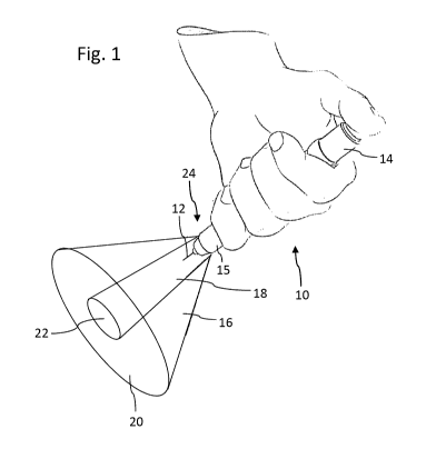
Figure 1 schematically illustrates a medical device in form of a pen-
type injector
adapted to generate two different light cones during a dose dispensing action,
15 Figure 2 schematically shows a lancing device equipped with an
illumination
arrangement,
Figure 3 shows another application scenario of a pen-type injector,
Figure 4 is illustrative of a flowchart of a first operation mode of the
medical device,
and
Figure 5 shows a flowchart of another operation mode of the
illumination
arrangement.
Detailed Description
The sketch of Figure 1 schematically illustrates a typical application
scenario when
using a pen-type injector 10 for injecting a dose of a medicament. The drug
delivery
device 10 comprises a dose button 14 at a proximal end which is to be
depressed by a
thumb of a user. The drug delivery device 10 comprises a cartridge holder 15
at a distal
portion which is further equipped with a replaceable injection needle 12.
CA 02837886 2013-11-29
WO 2013/004555
PCT/EP2012/062399
16
Typically, the injection needle 12 is provided on a needle hub to be screwed
onto a
distal interface of the cartridge holder 15. As further depicted in Figure 1,
the cartridge
holder 15 is equipped with an illumination arrangement 24 which is adapted to
generate
two different cones of light 16, 18. Whereas the rather focused inner cone of
light 18 is
intended to illuminate a site of needle puncture 22 on the skin of the
patient, the outer
cone of light 16 concentrically arranged relative to the inner cone of light
18 is adapted
to provide ambient illumination of the surrounding area 20 of the particular
skin portion
22 to become subject to medical treatment.
A rather similar device 40 is illustrated in Fig. 3 but there, instead of an
illumination
arrangement 24 concentrically enclosing the injection needle 12, a single
light source 44
is provided at a distal end face of a cartridge holder 15. As shown in Figure
3, the light
source 44 is preferably designed as LED or as a laser adapted to emit a rather
focused
light beam 46 to a target area 48 on the skin of the patient. Here, the target
area 48
marks the site of needle puncture.
Figure 2 is further illustrative of a lancing device 30 having a piercing
needle 34 adapted
to puncture or to pierce the skin tissue of the patient, e.g. for the purpose
of a blood
glucose measurement. The lancing device 30 is equipped with an illumination
arrangement 24 similar to the one already described with respect to Figure 1.
The
illumination arrangement 24 is adapted to generate two different cones of
light 16, 18,
which may be activated sequentially or simultaneously and may feature equal,
similar or
different spectral compositions and light intensities.
Furthermore, a control button 32 to be depressed by a thumb of a user's hand
is
illustrated. If, for instance, the control button 32 is depressed by the user,
the entire
device 30 will be switched on to support a medical treatment process.
Depression of
said button 32 may be detected by the control element 36, which, depending on
a
predefined schedule may activate or deactivate the cones of light 16, 18,
either for a
predefined time interval and/or until a stop signal is received by the control
element 36.
CA 02837886 2013-11-29
WO 2013/004555
PCT/EP2012/062399
17
The flowchart according to Figure 4 illustrates an example on how the
illumination
arrangement 24, 44 can be correlated with the overall handling of e.g. a drug
delivery
device 10, 40. In a first step 100, the device is activated. For instance a
semi- or fully-
automated device is switched on. Also, such activation 100 of the device 10,
30, 40 may
be automatically sensed, e.g. by removing a protective cap. In a subsequent
step 102, a
dose to be injected by means of the drug delivery device 10,40 is set, e.g. by
actuation
of a respective dose setting element 14 of the device 10. The dose setting, in
particular
the termination of a dose setting procedure may also be automatically detected
by a
control element 36, for example by detection of a button press of an "arm"
button. In
response to said detection, the illumination arrangement 24, 44 may be
switched on in
step 104.
Activation of the illumination arrangement 24, 44 may lead to a sequential or
simultaneous generation of the cones of light 16, 18. In a subsequent step 106
the dose
previously set is injected into the skin portion 22, 48 by making use of the
illumination
provided by the illumination arrangement 24, 44.
In a subsequent step 108, termination or end of the injection process is
detected. With a
semi-automated drug delivery device 10, 40, the end of an injection procedure
can be
detected by a substantial decrease of the thrust exerted by the thumb of a
user. Hence,
when the force level acting on a piston of a cartridge arranged inside a drug
delivery
device 10, 40 drops below a predefined threshold, a clear indication of the
end of an
injection process is generally given.
With respect to the illumination arrangement and its control, the end of an
injection
process may trigger a delay or a dwell period. Hence, in the following step
110, the
illumination is kept on for a predefined period of time, which is in the range
of a few
seconds, e.g. around 5 to 10 seconds. Thereafter, in step 112, the
illumination is
automatically switched off, thereby indicating to the user the end of the
dwell period.
CA 02837886 2013-11-29
WO 2013/004555
PCT/EP2012/062399
18
In this way, the illumination arrangement 24 is indicative to the user that
the predefined
dwell period has elapsed and that the injection needle 12 can now be removed
from the
skin portion 22.
Figure 5 is further illustrative of an alternative way on how to operate the
illumination
arrangement. In a first step 200, the medical device is activated. But here,
already prior
to a setting of a dose in step 204, the illumination arrangement 24, 44 is
switched on in
the first step 202. Accordingly, the illumination arrangement 24, 44 may be
particularly
adapted to illuminate a scale or a comparable display of the medical device
10, 30, 40
in order to enable and/or to facilitate the procedure of dose setting, even in
dark or dim
environments. In the proceeding step 204, the dose to be dispensed by the
device 10,
40 is set and at the end of the dose setting, prior to a dose injecting to be
conducted as
step 208, the illumination arrangement is switched into a different
illumination mode in
step 206.
The illumination mode to be activated in step 206 is particularly intended and
adapted to
accompany the injection process conducted in step 208. The illumination mode
activated in steps 202 and 206 may differ with respect to spectral
composition, spatial
light distribution, light intensity and/or with respect to a sequence of on-
off cycles.
During the injection process 208 the illumination is sustained. After
completion of the
actual injection process 208, the control of the illumination arrangement is
adapted to
provide a delay in step 210 during which the illumination is kept in an
activated state
before in a final step 212, e.g. after elapsing of a predefined dwell period,
the
illumination is finally switched off.
The described steps are only exemplary of two of a plurality of modes the
device can be
operated. In general, the device may comprise various sensors, e.g. to
automatically
detected various steps of the treatment process to be executed by the device.
Hence,
the device may be equipped with pressure and/or position sensor, in order to
track and
to monitor a dose setting and/or dispensing action conducted by the user.
Depending on
CA 02837886 2013-11-29
WO 2013/004555
PCT/EP2012/062399
19
the signals to be generated by such sensors, the illumination can be modified,
e.g
switched on and/or off appropriately without any further user interaction.
CA 02837886 2013-11-29
WO 2013/004555
PCT/EP2012/062399
List of Reference Numerals
10 drug delivery device
12 injection needle
5 14 dose button
15 cartridge holder
16 cone of light
18 cone of light
20 illuminated area
10 22 illuminated area
24 illumination arrangement
lancing device
32 control button
34 piercing needle
15 36 control element
drug delivery device
42 injection needle
44 light source
46 light beam
20 48 target area