Note: Descriptions are shown in the official language in which they were submitted.
CA 02838007 2007-05-15
WO 2006/055615 PCT/US2005/041498
1 A METHOD FOR SINGLE OXYGEN ATOM INCORPORATION INTO
2 DIGESTED PEPTIDES USING PEPTIDASES
3
4 FIELD OF THE INVENTION
The present invention relates to a method for comparative proteomics
6 using a peptidase under enzymatic conditions that incorporate a single
oxygen
7 atom into a digested peptide. The method employs a peptidase to
incorporate a
8 single 180 atom into peptide set derived from a population of proteins at
a
9 conditioned state, which is compared to a second peptide set incorporated
with a
single 160 atom derived from a population of proteins at a second conditioned
11 state. Upon combining the two peptide sets, the populations of proteins
are
12 analyzed for qualitative and quantitative differences based on the
content of 180
13 atoms and 160 atoms in digested peptides using mass spectrometry
14 instrumentation. The method is advantageous to reduce errors due to
random
incorporation of a second oxygen atom introduced during digestion and after
16 mixing the peptide sets.
17
18 BACKGROUND OF THE INVENTION
19 The completion of the genome sequencing of humans and other species
and the emergence of new technologies in mass spectrometry have together
21 fostered unprecedented opportunities for studying proteins on a large
scale. It is
22 expected that large scale quantitative measurements of protein
expressions in
23 different sets of samples, referred to as comparative proteomics, will
advance our
24 understanding of physiological processes and disease mechanisms.
Comparative
proteomic approaches have been applied to various biological samples to
identify
26 and quantify proteins that are up-. or down-regulated in response to
biological
27 conditions. To date, there are two primary strategies used in current
comparative
28 proteomics; two dimensional gel electrophoresis (2D-PAGE) based strategy
and
29 mass spectrometry based in vitro stable isotope labeling strategy.
Although 2D-PAGE based methods have been a primary choice in
31 comparative proteomics, 2D-gels are cumbersome to run, have a poor
dynamic
32 range, and are biased toward abundant and soluble proteins. In contrast,
the mass
33 spectrometry based stable isotope labeling strategy has a potential
ofoverc.oming
34 most of the weaknesses of the 2D-PAGE based methods. If the stable
isotope
1
CA 02838007 2007-05-15
WO 2006/055615 PCT/US2005/041498
1 labeling can be achieved efficiently and equivalently for each distinct
sample,
2 then two samples are compared using isotopic ratios. Among the in vitro
stable
3 isotope labeling methods, proteolytic 180 labeling is the simplest stable
isotope
4 labeling method and is expected to have the least methodological error
(technical
variations). Therefore, the proteolytic 180 labeling method has a potential to
be a
6 central method in comparative proteomics.
7 Although promising, a major drawback of the proteolytic 180 labeling
8 method has been the generation of a mixture of isotopic isoforms upon
9 proteolytic digestion resulting from the differential incorporation of
either one or
two 180 atoms (1801/ 1802) into each digested peptide species generated.
Typical
11 serine proteases used include trypsin, Lys-C or Glu-C proteases.
Unfortunately,
12 past studies have found that the ratios of the first and the second 180
atom
13 incorporation vary significantly with peptide sequences, and thus, the
ratios of
14 isor and 18-2_
peptides cannot be predicted with any certainty. The
quantifications of the peptides results in significant errors in calculating
160- and
16 1804abeled peptide ratios. In spite of more recent wide appreciation of
this
17 problem, no method has been reported to solve the problem.
18 A second significant drawback of using serine proteases that has been
19 demonstrated for 180 labeling is that digested peptide products continue
to react
with these proteases at the carboxyl termini. As a result, the serine
proteases will
21 catalyze oxygen back-exchange reaction when two digests, the first in
112160 and
22 the second in 112180, are mixed together. A previous report demonstrated
that
23 trypsin catalyzed oxygen back-exchange reaction occurs and leads to
inaccurate
24 quantification.
26
27
28 SUMMARY OF THE INVENTION
29 Unexpectedly, the present invention has found that peptidases are
able to
preferentially incorporate only a single 180 atom into each digested peptide
under
31 specific conditions. In addition, there is no evidence of significant
enzyme
32 catalyzed oxygen back-exchange reaction. Therefore, the invention has
the
33 unique property of resolving previous commercial problems in utilizing
proteases
34 in conjunction with 180-labeled peptides to accurately quantify
different protein
2
CA 02838007 2007-05-15
WO 2006/055615 PCT/US2005/041498
1 populations. The invention eliminates prior drawbacks employing 180
labeling
2 with peptidases to provide for a highly accurate quantification method
for
3 comparative proteomics.
4 The present invention is a method for incorporation of a single
oxygen
atom into a digested peptide using a peptidase. A protein or set of proteins
is
6 treated with a peptidase under specific conditions that incorporate a
single
7 oxygen atom in the carboxyl terminus of the digested peptide. The present
8 invention is further directed to the mass spectrometry comparison of
protein
9 expression in different biological conditions using a peptidase to
incorporate a
single 180 oxygen atom into peptide set derived from a population of proteins
at a
11 conditioned state which is compared to a second peptide set
incorporating 160
12 oxygen atom derived from a population of proteins at a second
conditioned state.
13 The first aspect of the invention is a method of incorporating a
single
14 oxygen atom into a digested peptide using a peptidase.
The second aspect of the invention is a method of incorporating a single
16 oxygen atom into a digested peptide using a peptidase, a protein, and
water.
17 Preferably, the oxygen atom is an 180 atom or 160 atom and the water is
160
18 water or 180 enriched water.
19 The third aspect of the invention is a method of incorporating a
single
oxygen atom under optimized conditions into a digested peptide using a
21 peptidase, a protein, and 180 enriched water. Preferably, the oxygen
atom is an
22 180 atom or 160 atom and the water is 160 water and 180 enriched water.
23 The fourth aspect of the invention is a method of incorporating a
single
24 oxygen atom into a digested peptide using a peptidase selected from a
group
consisting of exopeptidases (EC 3.4.11-19) or endopeptidases (EC 3.4.21-25 and
26 99).
27 The fifth aspect of the invention is a method of incorporating a
single
28 oxygen atom into a digested peptide using an exopeptidase selected from
a group
29 consisting of aminopeptidase (EC 3.4.11), dipeptidyl-peptidase (EC
3.4.14),
tripeptidyl-peptidase (EC 3.4.14), carboxypeptidase (EC 3.4.16-18), peptidyl-
31 dipeptidase (EC 3.4.15), dipeptidase (EC 3.4.13) or omega peptidase (EC
3.4.19).
32 The sixth aspect of the invention is a method of incorporating a
single
33 oxygen atom into a digested peptide using an endopeptidase selected from
a
34 group consisting of serine endopeptidases (EC 3.4.21), cysteine
endopeptidases
3
CA 02838007 2007-05-15
WO 2006/055615 PCT/US2005/041498
1 (EC 3.4.22), aspartic endopeptidases (EC 3.4.23), metalloendopeptidases
(EC
2 3.4.24) and threonine endopeptidases (EC 3.4.25) and unassigned
endopeptidases
3 (EC 3.4.99).
4 In the seventh aspect of the invention, a metalloendopeptidase is
peptidyl-
Lys metallopeptidase (EC 3.4.24.20, Lys-N), peptidyl-Asp metallopeptidase (EC
6 3.4.24.33, endoproteinase Asp-N), thermolysin (EC 3.4.24.27) or mycolysin
(EC
7 3.4.24.31).
8 The eighth aspect of the invention is a method for optimizing a
buffer for
9 the incorporation of a single oxygen atom into a digested peptide using a
peptidase, a protein, and 180 enriched water. Most preferably, the buffer is
11 optimized for pH.
12 The ninth aspect of the invention is a method for the comparison of
13 proteins under different biological conditions, wherein a digested
peptide of one
14 biological condition contains a single 180 atom incorporated by a
peptidase and a
digested peptide of a second biological contains a single 160 atom
incorporated
16 by the same peptidase. The digested peptidases are mixed and analyzed by
mass
17 spectrometry for the ratio of180 and 160. The ratio of180 and 160 is
used to
18 determine the increase or decrease in regulation of a specific peptide
or protein in
19 the two biological conditions.
The tenth aspect of the invention is a kit to incorporate a single oxygen
21 atom into a digested peptide containing a peptidase and enriched 180
water.
22 The eleventh aspect of the invention is a kit to incorporate a single
oxygen
23 atom into a digested peptide containing a peptidase, optimized buffer
and
24 enriched 180 water.
"Biological condition" means any physiological or cellular condition of a
26 plant, animal, microorganism, organ, cell or other biological material.
27 "Optimized buffer" means any buffer and its components that are
28 optimized for the incorporation of a single oxygen atom into a digested
peptidase
29 using a peptidase. The buffer is optimized for conditions that include,
but are not
limited to, pH and salt concentration
31 "Single oxygen atom" means at least a 90% incorporation as a single
32 oxygen atom, and more preferably, 95%, 98% or greater of the
incorporated
33 oxygen atom is incorporated as a single oxygen atom into the digested
peptide.
4
CA 02838007 2007-05-15
WO 2006/055615
PCT/US2005/041498
1 Examples of oxygen atoms include, but are not limited to 160 atoms and
180
2 atoms.
3 "Stable oxygen isotope" means any stable isotope of oxygen such as 160
4 and 180.
"180 enriched water" means water containing at least 90%180 atom, and
6 more preferably, 95%, 98% or greater, where 160 oxygen atoms comprise the
7 majority of the remainder of the oxygen atoms in water.
8 "160 water" means naturally occurring water.
9
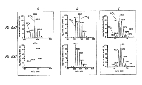
BRIEF DESCRIPTION OF THE DRAWINGS
11 FIG. 1 shows the mass spectra of three peptides (a,b,c) obtained by
12 digesting apomyoglobin with Lys-N in enriched H2180 at different pH.
13
14 Detailed description of the invention
Traditionally mass spectrometry based comparative proteomic methods
16 are based on in vitro labeling of two stable isotopes. For example, the
peptides
17 from the control sample are labeled with naturally abundant (light)
isotope(s),
18 while peptides from the experimental sample are labeled with its heavier
19 isotope(s) or vice versa. The samples are then mixed together in equal
proportion
and analyzed by mass spectrometry. Since a peptide labeled with the light
21 isotope and the same peptide labeled with the heavier isotope give
different
22 molecular weights, the light- and heavy-peptide can be distinguished by
mass
23 spectrometry. By comparing the peak areas or intensities of the light-
peptide and
24 heavy-peptide, the relative abundance of the two peptides can be
determined.
These ratios can further be used to quantify the relative abundance of each
parent
26 protein in the distinct original samples.
27 As a further illustration of the commercial application, using this
28 comparative approach a pool of isotopically labeled proteins acquired
from an
29 unstressed system is mixed with the same relative amount of an unlabeled
sample
from a second (stressed) experimental system or vise visa. The combined pool
is
31 then analyzed by mass spectrometry to rapidly determine those stressed
induced
32 proteins relative to the unstressed state. The applications of this
method would
5
CA 02838007 2007-05-15
WO 2006/055615
PCT/US2005/041498
1 be highly useful to identify and quantify changes in protein expression
in a
2 variety of diseased or physiological states in animals, plants and
microorganisms.
3 Currently, there are two ways to incorporate stable isotopes into
peptides;
4 first, by derivatization of peptides by a light- or heavy-isotope coded
reagent
(Isotope Coded Affinity Tag or ICAT) or second, by incorporation of 160 and
180
6 atom(s) into the carboxyl termini of peptides from the solvent water,
H2160 or
7 H218
respectively, upon proteolytic cleavage of proteins. The second method is
8 referred as proteolytic 180 labeling, where a peptidase is used.
9 The members of the peptidase family are any enzymes that hydrolyze
peptide bonds (EC 3.4, Enzyme Nomenclature 1992, Academic Press, San Diego,
11 California). Peptidases are present in the wide variety of biological
sources and
12 contain the amino acid sequence motif comprising His-Glu-Xaa-Xaa-His,
where
13 Xaa is any amino acid. The peptidase family can be subdivided into
14 exopeptidases (EC 3.4.11-19) and endopeptidases (EC 3.4.21-99), the
latter
referred to as proteinases, that act near the terminus of the polypeptide or
16 internally, respectively. Subclasses of exopeptidases include those
acting at a
17 free N-terminus releasing a single amino acid (aminopeptidase, EC
3.4.11), a
18 dipeptide (dipeptidyl-peptidase, EC 3.4.14), or a tripeptide
(tripeptidyl-peptidase,
19 EC 3.4.14) and those acting at a free C-terminus releasing a single
amino acid
(carboxypeptidase, EC 3.4.16-18) or a dipeptide (peptidyl-dipeptidase, EC
21 3.4.15). Other exopeptidases are specific for dipeptides (dipeptidases,
EC 3.4.13)
22 or remove terminal residues that are substituted, cyclized or linked by
isopeptide
23 bonds (omega peptidases, EC 3.4.19). Subclasses of endopeptidases (EC
3.4.21-
24 24 and EC 3.4.99) are subdivided on the basis of catalytic mechanism and
specificity is used only to identify individual enzymes within the groups.
26 Subclasses of endopeptidases include serine endopeptidases (EC 3.4.21),
cysteine
27 endopeptidases (EC 3.4.22), aspartic endopeptidases (EC 3.4.23),
28 metalloendopeptidases (EC 3.4.24) and threonine endopeptidases (EC
3.4.25).
29 Endopeptidases that could not be assigned to any of the sub-subclasses
EC
3.4.21-25 were listed in sub-subclass EC 3.4.99.
31 Unexpectedly, in the present invention members of the endopeptidase
32 subfamily circumvented the incorporation of multiple 180 atoms under
specific
33 enzymatic conditions. It is expected that conditions exist for other
6
CA 02838007 2007-05-15
WO 2006/055615 PCT/US2005/041498
1 endopeptidases and exopeptidases that facilitate the incorporation of a
single
2 oxygen atom.
3 In the example described herein, peptidyl-Lys metallopeptidase (EC
4 3.4.24.20) is shown to incorporate a single oxygen atom into the carboxyl
terminus of a digested peptide under alkaline pH conditions. Peptidyl-Lys
6 metalloendopeptidase from Grifola frondosa (Lys-N, EC 3.4.24.20), which
7 cleaves peptidyl-lysine bonds (-Xaa-Lys-) in proteins and peptides, is
referred to
8 as protease Lys-N because of its substrate specificity. The
metalloendopeptidase
9 contains one atom of zinc per molecule and is most active at pH 9.5. It
is known
to exhibit more than 50% maximal activity within the pH range of 6-10.5.
11
12 Example 1: Sample Preparation Prior to 180 Labeling of Proteolytic
Peptides
13 The invention described herein employed a peptidase and 180 enriched
14 water to preferentially label the C-terminal fragment of the digested
peptides;
however this invention is not limited to and includes water containing any
stable
16 oxygen isotope. All reagents are available and the chemistry is
generally well-
17 known to those skilled in the art. The following examples are
illustrations of
18 such technology that may be used.
19 The first step may or may not include a protein denaturation step. In
the
event that information is required about the protein or peptide conformational
21 state or structure this step would be omitted. For example, for a
protein or
22 peptide that plays a role in signal transduction and undergoes a
conformational
23 change or modification due to an altered physiological condition would
be within
24 the scope of this invention to assess changes through altered
accessibility to
proteases.
26 In cases where it is desirable to denature the protein or peptide to
examine
27 its primary structure or less structured state, the protein or peptide
is treated to
28 remove those elements required for secondary or tertiary structure. More
29 specifically, the ability of any protease to fragment a protein or
peptide is limited
by the accessibility of the protease to susceptible peptide bonds. While
31 denaturants such as acidic pH, urea, detergents, and organic co-solvents
can
32 partially denature proteins and expose many structurally shielded
peptide bonds,
33 pre-existing disulfide bonds within a protein can prevent sufficient
denaturation
34 with these agents alone. In conventional protein structural studies,
disulfides are
7
CA 02838007 2007-05-15
WO 2006/055615 PCT/US2005/041498
1 usually cleaved by reduction with 2-mercaptoethanol, dithiothreitol, and
other
2 reductants require a pH greater than pH 7 for sufficient activity. In the
present
3 experiments, reduction was achieved by using dithiothreitol and
alkylation of
4 cysteine thiol groups in proteins was established by using
iodoacetoamide. To
block thiol groups, a method used by Crestfield, et al. involved blocking the
thiol
6 (-SH) group by carbamidomethylation. The invention, however, is not
limited to
7 a specific method or agents to effectively denature part or all the
protein or
8 peptide structure. The examples described herein is presented as
illustrative,
9 where a protein or a mixture of proteins were reduced and subsequently
carbamodomethylated before digestion with a metalloendopeptidase.
11 An illustrative example of the first step included the reduction and
12 alkylation of cysteine thiol groups in a protein mixture consisting of
bovine
13 serum albumin (BSA), glycerol dehydrogenase (GDH), glyceraldehyde-3-
14 phosphate dehydrogenase (GAPDH), ACY-I, creatine phosphokinase (CPK) and
apomyoglobin. Approximately 2 nmoles of each of the protein were dissolved in
16 200 gl of 2 M Tris-HC1 buffer (pH 8.0) containing 5 M guanidine-I-IC1
and 2 mM
17 ethylenediaminetetraacetic acid (EDTA) and subsequently reduced with 1mM
18 dithiothreitol (DTT) for 60 minutes at 50 C followed by treatment with
2.5 mM
19 iodoacetamide for 30 minutes at 25 C. The proteins of the reaction
mixture were
isolated from the reagents using a PD-10 gel filtration column (Amersham
21 Biosciences AB, Uppsala, Sweden) that was equilibrated with 0.1% formic
acid.
22 The protein fractions from the PD-10 column were combined and dried in a
23 Speed-Vac concentrator and dissolved in 100 mM glycine buffer (pH 10.0)
24 containing 1M urea. The protein concentration was determined by a
modified
Bradford method. Because apomyoglobin does not contain cysteine or disulphide
26 bonds, reduction and alkylation of apomyoglobin-only samples was not
required.
27
28 Example 2: Methods of Stable Oxygen Isotope Labeling and LC/MS Analysis
29
Denatured proteins, reduced and carbamidomethylated if necessary, were
31 digested using either Lys-N or Asp-N metalloendopeptidase. The
conditions for
32 the proteolytic digestion were standardized in our laboratory for the
purpose of
33 single labeled oxygen atom incorporation. Lys-N was obtained from
Seikagaku
34 Corp. (Tokyo, Japan). The digestion of proteins by Lys-N was performed
in the
following buffer systems; 100 mM sodium phosphate at pH 6.0 or 8.0 or 100 mM
8
CA 02838007 2007-05-15
WO 2006/055615
PCT/US2005/041498
1 glycine-NaOH at pH 9.0, 9.5, or 10Ø The digestion buffers were prepared
from
2 their corresponding stock solutions by placing the required aliquot into
3 Eppendorf tubes, drying with a Speed-Vac concentrator and reconstituting
with
4 the appropriate stable oxygen isotope, preferably 112160 or enriched
112180. The
digestions of proteins were incubated at 25 C for 18 hrs using a Lys-N to
6 substrate ratio of 1:85 (w/w), unless otherwise stated. The effective
range of Lys-
7 N to substrate (protein to be digested) ratios was found to be from 1:10
to 1:85.
8 After the
incubation, the digests were diluted with 0.1% formic acid in
9 112160 to the desired concentrations for mass spectrometry analyses. The
resultant 180 labeled peptides were analyzed by liquid chromatography mass
11 spectrometry (LC-MS) that consisted of an UltiMate nano HPLC system
12 (Dionex, San Francisco, CA, USA) equipped with an isocratic pump, an
13 autosampler, a gradient pump module and a column switching module and a
14 QStar quadrupole/time-of-flight mass spectrometer (Applied Biosystem-MDS
Sciex, Foster City, CA, USA) equipped with nano-electrospray ion source
16 (Applied Biosystem-MDS Sciex, Foster City, CA, USA) and metal sprayer
(GL
17 Science, Tokyo, Japan). The protein digests (5 jtl,-1 pmol) were
injected into a
18 reverse-phase C18 trapping column (300 gm i.d. x 1 mm, Dionex,
Sunnyvale,
19 CA, USA) equilibrated with 0.1% formic acid /2% acetonitrile (v/v) and
washed
for 5 minutes with the equilibration solvent at a flow rate of 10 gL/min.
After the
21 washing, the trapping column was switched in-line with the reverse-phase
22 analytical column and the trapped peptides were chromatographed on a
column
23 (0.075 x 50 mm, New Objective Inc., Woburn., MA) packed with Jupiter C18
24 media (10 gm, 300 A, Phenomenex, Torrance, CA, USA) using a linear
gradient
of acetonitrile from 2% to 82% in water in the presence of 0.1% formic acid
over
26 a period of 80 min at a flow rate of 200 nL/min. The column effluent was
passed
27 directly into the nano-electrospray ion source. The total ion current
was obtained
28 in the mass range of m/z 300-2000 at 2,100 V and 65 V of electrospray
voltage
29 and orifice voltage, respectively, in the positive ion mode. AnalystQS
software
(version 1.1Ø6410, Applied Biosystem-MDS Sciex, CA, USA) was used for
31 instrument control, data acquisition, and data processing. In liquid
32 chromatography-tandem mass spectrometry (LC/MS/MS) analyses, the mass
33 spectrometer was operated in data-dependent MS to MS/MS switching mode
9
CA 02838007 2007-05-15
WO 2006/055615 PCT/US2005/041498
1 with the three most intense ions in each MS scan subjected to MS/MS
analysis.
2 The identities of the peptides were determined by submitting product ion
spectra
3 of the peptides to the Swiss Protein database using Mascot data base
search
4 software (Matrix Science, London, UK).
The actual 160/180 peptide ratio for each peptide was calculated from the
6 observed monoisotopic peak intensity of160- and 180-labeled peptide
present in
7 mixed samples using the following equations.
8 Equations
9 1. act160 = obs160 ¨ (0.05 x act180)
2. ac.18,,
= 0E18180 ¨ (01)8160 X Y) + (0.05 x act180)
11 3. act160 = obs160 ¨0.05 x (obs180 ¨ obs160 x Y)
12 0.95
13 4. ac=t180= 01:18180 ¨ (ObS160 X Y)
14 0.95
5. ratio of 160/180 = act160/act180
16
17 In these equations, act160 and act180 are the actual, corrected
monoisotopic peak
18 intensities (cps) arising solely from the peptides in sample 1 that were
digested in
19 100% H2160 and from the peptides in sample 2 that were digested in 95%
H2180
and 5% , H216u¨ respectively. The actual monoisotopic peak intensities are
21 derived from the observed monoisotopic peak intensities (cps) of 160-
and 180-
22 labeled peptides, obs160 and obs180, arising from either sample. Y is
the
23 theoretical fractional intensity of the M + 2 isotopic peak of the 160-
labeled
24 peptide compared to its monoisotopic peak and is calculated from the
amino acid
sequence of the peptide. The M+2 isotopic peak is naturally occurring peptide
in
26 the 160-labelled sample due to the presence of13C, 2H, 15N, 170 etc.
27 Equation 1 includes a correction factor to account for the 5%
28 incorporation of160 into peptides digested in H2180 for conversion to
the
29 observed 160 signal. To obtain the signal due only to the peptide in the
160
sample, the second term on the right side of equation 1, 0.05 x act180, is
31 subtracted from the observed signal, obs160.
32 Equation 2, for calculating the actual intensity of the 180 sample
peptide,
33 includes two correction factors. First, to obtain the signal due only to
the peptide
34 in the 180 sample, the second term on the right side, obs160 x Y is
subtracted
from the observed signal, obs180. Second, the third term in Equation 2, 0.05 x
CA 02838007 2007-05-15
WO 2006/055615 PCT/US2005/041498
1 act180, is added as the 5% correction for the 160-labelled peptides in
the 180
2 sample.
3 Equations 1 and 2 are converted further to become equations 3 and 4,
4 respectively. The ratios of 160- and 180-labeled peptide were calculated
by
dividing the actual intensity of 160 labeled peptide by the actual intensity
of180
6 labeled peptide (Equation 5).
7 Mass spectra used for the 160/ 180 peptide ratio calculations were
8 extracted from the total ion current (TIC) only if the signal intensities
of the
9 peptides were lower than 500 cps. If the signal intensities exceeded 500
cps at
the top of the TIC peak, regions of the lower slope of the TIC peaks were used
to
11 extract the mass spectra to avoid peak saturations. Approximately 1,000
cps was
12 the maximum signal intensity within the linear dynamic range of the
detector in
13 the instrument used.
14
Example 3: Optimizing Digestion Conditions for Single Oxygen Atom
16 Incorporation
17 Apomyglobin was digested by Lys-N at pH 6.0, 8.0, 9.0, 9.5 or 10.0
using
18 112180 prepared in 100 mM glycine-NaOH buffer. The resulting digests
were
19 analyzed by liquid chromatography-mass spectrometry (LC/MS). Figure 1
shows
the mass spectra of three representative apomyoglobin peptides that were
21 hydrolyzed at different pH. In Figure 1, panel a shows (M + 3H)3+ ions
of
22 peptide KALELFRNDIAA (SEQ ID NO 1), panel b shows (M + 3H)3+ ions of
23 peptide KHPGDFGADAQGAMT (SEQ ID NO 2), and panel c shows (M +
24 4H)4+ ions of peptide KVEADIAGHGQEVLIRLFTGHPETLE (SEQ ID NO 3).
The bottom most spectrum in each panel is the theoretical abundances of the
26 isotopes for each corresponding peptide containing one 180 atom. These
results
27 show that variability of the 1801- and 1802-peptide ratios is pfl
dependent. At pH
28 6.0, peptide peaks with two 180 atoms (1 8 02) were abundant in all the
three
29 peptides. As the pH is increased there is a steady decrease in the
incorporation of
the second 180 atom. In fact, the incorporation of the second 180 atom was not
31 observed at pH 9.5 and 10.0 as evidenced by the exact match of the
relative
32 intensities of the isotopes of the observed peptide mass spectrum
compared to
33 their theoretical abundances. This invention demonstrates for the first
time that
34 there exist enzymatic conditions for endopeptidases where only a single
180 atom
11
CA 02838007 2007-05-15
WO 2006/055615 PCT/US2005/041498
1 is incorporated. Enzymatic conditions were determined for incorporation
of a
2 single oxygen isotope into digested peptides using another endopeptidase.
Using
3 the same optimization method, Asp-N, peptidyl-Asp metallopeptidase (EC
4 3.4.24.33), was found to incorporate a single 180 atom in a peptide
between pH
8.0 to pH 9Ø
6 In a separate experiment, it was confirmed that there is no
detectable non-
7 enzymatic incorporation of180 atom into angiotensin II (DRVYlHPF)
incubated
8 in 100 mM glycine-NaOH buffer (pH 10.0) or 0.1% formic acid at 25 C for
24
9 hrs (data not shown), confirming that significant oxygen back-exchange
reaction
does not take place during the incubation period and LC/MS analysis.
11 It was further demonstrated that the single 180 atom incorporation
12 property of Lys-N is not affected by temperatures ranging from about 25
C to
13 about 50 C and urea concentrations ranging from about 0.5 M to about 4
M. The
14 effective range of the buffer concentration for single 180 atom
incorporation
single ranged from about 10 mM to about 500 mM glycine-NaOH buffer. The
16 activity of the enzyme was highest at about 25 C and aboutl M urea
under the
17 conditions employed as judged by the observed ion intensities and
selected for
18 further use.
19 Finally, four representative apomyoglobin peptides were hydrolyzed in
either H2160 and in H2180 in 100 mM glycine-NaOH buffer (pH 10.0) containing
21 1 M urea at 25 C. The proportional abundances of the isotopes between
160-
22 and 180-labeled peptides were identical, indicating that only one 180
atom was
23 incorporated into each peptide in the presence of urea.
24 For other examples described herein, the standard digestion protocol
of
proteins by Lys-N uses a 100 mM glycine-NaOH buffer, pH 10.0, containing 1 M
26 urea at 25 C, which is incubated for 18 hrs.
27
28 Example 4: Evaluation of Protein Mixtures by Single Oxygen Incorporation
29 Using Metalloendoproteases
The digestion was performed using the standardized digestion protocol
31 described above on a protein mixture containing six reduced and S-
32 carbamidomethylated proteins; bovine serum albumin (BSA), glutamate
33 dehydrogenase (GDH), glyceraldehydes-3-phosphate dehydrogenase (GAPDH),
34 aminoacylase-1 (ACY-1), creatine phosphokinase (CPK) and apomyoglobin.
12
CA 02838007 2007-05-15
WO 2006/055615 PCT/US2005/041498
1 This protein mixture was digested in H2160 and H2180 separately and mixed
in
2 1:1 ratio. Approximately 50 ng of this mixture was analyzed by LC/MS. A
total
3 of 50 Lys-N peptides from the six proteins were selected to calculate the
ratios of
4 160_ and 180-labeled peptides (160/180). The identities of the peptides,
based on
amino acid sequences, were determined by submitting product ion spectra of the
6 peptides to Swiss Protein database using Mascot data base search software
in a
7 separate LC/MS/MS experiment.
8 The average experimental 160/180 ratios for BSA, GDH, GAPDH, ACY-
9 1, CPK and apomyoglobin peptides were 1.08 0.22 (n=23), 1.05 0.06
(n=6),
0.92 0.17 (n=7), 1.01 0.04 (n=3), 1.12 0.18 (n=4) and 1.04 0.21 (n=7),
11 respectively. More careful analysis revealed that in all cases only a
single
12 oxygen atom was incorporated. Ratios of twelve peptides, however,
deviated
13 more than 0.25 from the predicted 1:1 ratios. Nine peptides of the 12
peptides
14 contained either Glu-Lys or Pro-Lys bond cleavage, suggesting that the
reaction
rate of Lys-N to Glu-Lys and Pro-Lys bond is slower than other Xaa-Lys bonds.
16 The average experimental 160/180 ratios and standard deviations (SD) for
BSA,
17 GAPDH, CPK and apomyoglobin become 1.01 0.08 (n=15), 0.98 0.09
(n=6),
18 1.03 0.06 (n=3) and 1.06 0.10 (n=5) when the 12 peptides are
removed,
19 demonstrating an excellent accuracy and reproducibility of the method.
The
average and the standard deviation values were calculated using different
21 peptides within a same protein.
22
23 Example 5: Dynamic Range of Metallopeptidase 180 Labeling
24 To demonstrate the utility of endopeptidase 180 labeling for
comparative
proteomics, apomyoglobin (about 3.414) was digested using either Lys-N in
26 112160 or 112180 under the standarized protocol and mixed in different
ratios.
27 Three representative peptides were analyzed by LC/MS, which was repeated
5-
28 times to obtain average experimental 160 /180 peptide ratios. The
obtained
29 average experimental 160/180 peptide ratios were plotted against their
theoretical
ratios with relative standard deviation (RSD) values to evaluate the linearity
of
31 the quantification of160 peptide ratios. The correlation coefficients
(r2) of
32 the linear regression lines for the three peptides were 0.9960 for
33 KALELFRNDIAA, 0.9977 for KHPGDFGADAQGAMT, 0.9995 for
13
CA 02838007 2007-05-15
WO 2006/055615 PCT/US2005/041498
1 KHGTVVLTALGGILK, respectively, indicating good linearity with respect to
2 the 160 ,
u peptide ratios over the range of 0.11 to 9.
3
4 Example 6: Characterization of Proteome Changes in
Cytokine/Lipopolysaccharide (LPS) Treated Versus Untreated Human Retinal
6 Pigment Epithelium (ARPE-19) Cells
7 Human retinal pigment epithelium (ARPE-19) cells were obtained from
8 the American Tissue Culture Collection (Rockville, MD). Cells were
cultured to
9 approximately 80% confluency in T-175 flasks at 37 C under 95% air and
5%
CO2 in Dulbecco's Modified Eagle Medium: Nutrient Mixture F-12 (Ham) 1:1
11 (DMEM-F12) with 10 % fetal calf serum, 2% L-glutamine and 0.5%
12 antibiotic/antimycotic. The cells, before harvesting, were either: 1)
treated in
13 growth medium for 24 h with a combination of cytokines/LPS consisting of
14 human tumor necrosis factor a (TNF-a, 3.25 ng/mL, Upstate, Lake Placid,
NY),
human interferon-y (IFN-y, 50 ng /mL, Upstate, Lake Placid, NY) and
16 Escherichia coli lipopolysaccharide (LPS, 10 jig / mL, Sigma-Aldrich, St
Louis,
17 MO) or 2) untreated for controls, keeping them in medium for 24 h. After
24 h,
18 the medium was removed from the flask and the cells were washed with
19 phosphate buffered saline (PBS) twice and DMEM-F12 once, and harvested
in
DMEM-F12 by scraping the cells from the flask. The harvested cell suspension
21 was centrifuged at 150 g for 10 min at 4 C, the supernatant removed and
the cell
22 pellet stored at -80 C until use.
23 The stored cell pellets were resuspended in 2.5 mL of 2% sodium
dodecyl
24 sulfate (SDS) in 50 mM Tris-HC1, pH 7.5 buffer and sonicated for 60
seconds.
The resulting homogenate was centrifuged at 8,000 g for 30 min at 4 C and the
26 supernatant recovered. The extracted proteins were reduced by adjusting
the
27 solution to 1 rnM dithiothreitol (DTT) and reacting for 2 h at 50 C.
28 After S-alkylation treatment was performed, protein digestion of
protein
29 samples from treated and untreated cells were carried out separately in
H2160 and
H2180 under the conditions described above. The treated and untreated digests
31 were mixed in a 1:1 ratio, separated by strong cation exchange
chromatography
32 into eight fractions, which were each analyzed by reverse phase liquid
33 chromatography-tandem mass spectrometry. Identities of the resulting
peptides
14
CA 02838007 2007-05-15
WO 2006/055615 PCT/US2005/041498
1 were determined by database searching, and the peak intensities of each
160- and
2 180-labeled peptide was obtained and corrected as described above.
3 In this study, a very large population of proteins,1046, were
sequenced
4 and quantified. Of these, 584 proteins were identified, and the relative
abundance
of 562 of these proteins was effective for complex and detailed comparative
6 analysis between proteomes in cytokine/LPS treated versus untreated ARPE-
19
7 cells. This is the most comprehensive finding of a retinal pigment
epithelium cell
8 proteome thus far and demonstrates the unique utility of the present
invention.
9 These results are detailed in Rao et al., MCP Papers in Press, July 5,
2005, DOT
10.1074/mcp.M500150-MCP200, which is incorporated by reference.
11 The description of the specific embodiments of the invention is
presented for
12 the purposed of illustration. It is not intended to be exhaustive nor to
limit the scope
13 of the invention to the specific forms described herein. Although the
invention has
14 been described with reference to several embodiments, it will be
understood by one
of ordinary skill in the art that various modifications can be made without
departing
16 from the spirit and the scope of the invention, as set forth in the
claims. All patents,
17 patent applications and publications referenced herein are hereby
incorporated by
18 reference.
19 Other embodiments are within the claims.
CA 02838007 2007-05-15
WO 2006/055615 PCT/US2005/041498
seq.list.ST25
SEQUENCE LISTING
<110> University of North Dakota
<120> A METHOD FOR SINGLE OXYGEN ATOM INCORPORATION INTO DIGESTED
PEPTIDES USING PEPTIDASES
<130> U66.12-13
<160> 3
<170> PatentIn version 3.3
<210> 1
<211> 12
<212> PRT
<213> human
<400> 1
Lys Ala Leu Glu Leu Phe Arg Asn Asp Ile Ala Ala
1 5 10
<210> 2
<211> 15
<212> PRT
<213> human
<400> 2
Lys His Pro Gly Asp Phe Gly Ala Asp Ala Gin Gly Ala Met Thr
1 5 10 15
<210> 3
<211> 26
<212> PRT
<213> human
<400> 3
_Lys Val Glu Ala Asp Ile Ala Gly His Gly Gin Glu Val Leu Ile Arg
1 5 10 15
Leu Phe Thr Gly His Pro Glu Thr Leu Glu
20 25
Page 1