Note: Descriptions are shown in the official language in which they were submitted.
CA 02838728 2013-12-06
WO 2012/170711 PCT/US2012/041387
CIRCULATING BIOMARKERS FOR CANCER
CROSS REFERENCE
[0001] This application claims the benefit of U.S. Provisional Patent
Application Nos. 61/494,196, filed June
7, 2011; 61/494,355, filed June 7, 2011; and 61/507,989, filed July 14, 2011;
all of which applications are
incorporated herein by reference in their entirety.
[0002] This application is a continuation-in-part of International Patent
Application PCT/US2012/025741,
filed February 17, 2012, which application claims the benefit of U.S.
Provisional Patent Application Nos.
61/446,313, filed February 24, 2011; 61/501,680, filed June 27, 2011;
61/471,417, filed April 4, 2011;
61/523,763, filed August 15, 2011; and 61/445,273, filed February 22, 2011;
all of which applications are
incorporated herein by reference in their entirety.
[0003] This application is also a continuation-in-part of International Patent
Application
PCT/US2011/048327, filed August 18, 2011, which application claims the benefit
of U.S. Provisional Patent
Application Nos. 61/374,951, filed August 18, 2010; 61/379,670, filed
September 2, 2010; 61/381,305, filed
September 9, 2010; 61/383,305, filed September 15, 2010; 61/391,504, filed
October 8, 2010; 61/393,823, filed
October 15, 2010; 61/411,890, filed November 9, 2010; 61/414,870, filed
November 17, 2010; 61/416,560, filed
November 23, 2010; 61/421,851, filed December 10, 2010; 61/423,557, filed
December 15, 2010; 61/428,196,
filed December 29, 2010; all of which applications are incorporated herein by
reference in their entirety.
[0004] This application is also a continuation-in-part of International Patent
Application PCT/
US2011/026750, filed March 1, 2011, which application claims is a continuation-
in-part application of U.S.
Patent Application Serial No. 12/591,226, filed November 12, 2009, which
claims the benefit of U.S.
Provisional Application Nos. 61/114,045, filed November 12, 2008; 61/114,058,
filed November 12, 2008;
61/114,065, filed November 13, 2008; 61/151,183, filed February 9, 2009;
61/278,049, filed October 2, 2009;
61/250,454, filed October 9, 2009; and 61/253,027 filed October 19, 2009; and
which application also claims
the benefit of U.S. Provisional Application Nos. 61/274,124, filed March
1,2010; 61/357,517, filed June 22,
2010; 61/364,785, filed July 15, 2010; all of which applications are
incorporated herein by reference in their
entirety.
[0005] This application is also a continuation-in-part of International Patent
Application
PCT/US2011/031479, filed April 6, 2011, which application claims the benefit
of U.S. Provisional Patent
Application Nos. 61/321,392, filed April 6,2010; 61/321,407, filed April
6,2010; 61/332,174, filed May 6,
2010; 61/348,214, filed May 25, 2010, 61/348,685, filed May 26, 2010;
61/354,125, filed June 11,2010;
61/355,387, filed June 16, 2010; 61/356,974, filed June 21, 2010; 61/357,517,
filed June 22, 2010; 61/362,674,
filed July 8,2010; 61/413,377, filed November 12, 2010; 61/322,690, filed
April 9,2010; 61/334,547, filed May
13, 2010; 61/364,785, filed July 15, 2010; 61/370,088, filed August 2, 2010;
61/379,670, filed September 2,
2010; 61/381,305, filed September 9, 2010; 61/383,305, filed September 15,
2010; 61/391,504, filed October 8,
2010; 61/393,823, filed October 15, 2010; 61/411,890, filed November 9, 2010;
and 61/416,560, filed
November 23, 2010; all of which applications are incorporated herein by
reference in their entirety.
-1-
CA 02838728 2013-12-06
WO 2012/170711 PCT/US2012/041387
BACKGROUND
[0006] Biomarkers for conditions and diseases such as cancer include
biological molecules such as proteins,
peptides, lipids, RNAs, DNA and variations and modifications thereof.
[0007] The identification of specific biomarkers, such as DNA, RNA and
proteins, can provide biosignatures
that are used for the diagnosis, prognosis, or theranosis of conditions or
diseases. Biomarkers can be detected in
bodily fluids, including circulating DNA, RNA, proteins, and vesicles.
Circulating biomarkers include proteins
such as PSA and CA125, and nucleic acids such as SEPT9 DNA and PCA3 messenger
RNA (mRNA).
Circulating biomarkers can be associated with circulating vesicles. Vesicles
are membrane encapsulated
structures that are shed from cells and have been found in a number of bodily
fluids, including blood, plasma,
serum, breast milk, ascites, bronchoalveolar lavage fluid and urine. Vesicles
can take part in the communication
between cells as transport vehicles for proteins, RNAs, DNAs, viruses, and
prions. MicroRNAs are short RNAs
that regulate the transcription and degradation of messenger RNAs. MicroRNAs
have been found in bodily
fluids and have been observed as a component within vesicles shed from tumor
cells. The analysis of circulating
biomarkers associated with diseases, including vesicles and/or microRNA, can
aid in detection of disease or
severity thereof, determining predisposition to a disease, as well as making
treatment decisions.
[0008] Vesicles present in a biological sample provide a source of biomarkers,
e.g., the markers are present
within a vesicle (vesicle payload), or are present on the surface of a
vesicle. Characteristics of vesicles (e.g.,
size, surface antigens, determination of cell-of-origin, payload) can also
provide a diagnostic, prognostic or
theranostic readout. There remains a need to identify biomarkers that can be
used to detect and treat disease.
microRNA, proteins and other biomarkers associated with vesicles as well as
the characteristics of a vesicle can
provide a diagnosis, prognosis, or theranosis.
[0009] The present invention provides methods and systems for characterizing a
phenotype by detecting
biomarkers that are indicative of disease or disease progress. The biomarkers
can be circulating biomarkers
including without limitation vesicle markers, protein, nucleic acids, mRNA, or
microRNA. The biomarkers can
be nucleic acid-protein complexes.
SUMMARY
[0010] Disclosed herein are methods and compositions for characterizing a
phenotype by analyzing circulating
biomarkers, such as a vesicle, microRNA or protein present in a biological
sample. Characterizing a phenotype
for a subject or individual may include, but is not limited to, the diagnosis
of a disease or condition, the
prognosis of a disease or condition, the determination of a disease stage or a
condition stage, a drug efficacy, a
physiological condition, organ distress or organ rejection, disease or
condition progression, therapy-related
association to a disease or condition, or a specific physiological or
biological state.
[0011] In an aspect, the present invention provides a method comprising:
determining a presence or level of
one or more biomarker in a biological sample from a subject, wherein the one
or more biomarker is selected
from the group consisting of A2ML1, BAX, ClOorf47, Clorf162, CSDA, EIFC3,
ETFB, GABARAPL2,
GUK1, GZMH, HIST1H3B, HLA-A, HSP9OAA1, NRGN, PRDX5, PTMA, RABAC1, RABAGAP1L,
RPL22,
SAP18, SEPW1, SOX1, and a combination thereof; and identifying a biosignature
comprising the presence or
level of the one or more biomarker. In an embodiment, the one or more
biomarker, e.g., 1, 2, 3, 4, 5 or 6
biomarkers, is selected from the group consisting of A2ML1, GABARAPL2, PTMA,
RABAC1, SOX1, EFTB,
and a combination thereof. The one or more biomarker can comprise PTMA.
-2-
CA 02838728 2013-12-06
WO 2012/170711 PCT/US2012/041387
[0012] The method can further comprise comparing the biosignature to a
reference biosignature, wherein the
comparison is used to characterize a cancer. In some embodiments, the
characterizing comprises identifying the
presence or risk of the cancer, or identifying the cancer as metastatic or
aggressive. In some embodiments, the
characterizing comprises determining whether the subject is responding to a
therapeutic treatment, or whether
the subject is likely to respond or not respond to a therapeutic treatment.
The treatment can be any cancer
treatment disclosed herein or known in the art, e.g., watchful waiting,
surgical pelvic lymphadenectomy, radical
prostatectomy, transurethral resection of the prostate (TURP), orchiectomy,
radiation therapy, external-beam
radiation therapy (EBRT), 1125, palladium, iridium, hormone therapy,
luteinizing hormone-releasing hormone
agonists, leuprolide, goserelin, buserelin, antiandrogens, flutamide,
bicalutamide, megestrol acetate, nilutamide,
ketoconazole, aminoglutethimide, gonadotropin-releasing hormone (GnRH),
estrogen, cryotherapy,
chemotherapy, biologic therapy, ultrasound, and proton beam radiation.
[0013] In still other embodiments, the step of comparing the biosignature to
the reference comprises
determining whether any of the one or more biomarker is altered relative to
the reference, and thereby providing
a prognostic, diagnostic or theranostic determination for the cancer.
[0014] The cancer can be any appropriate cancer disclosed herein. In an
embodiment, the cancer comprises a
prostate cancer.
[0015] In another aspect, the invention provides a method comprising,
determining a presence or level of one
or more biomarker in a biological sample from a subject, wherein the one or
more biomarker, e.g.,1, 2, 3, 4 or 5
biomarkers, is selected from the group consisting of CA-125, CA 19-9, c-
reactive protein, CD95, FAP-1 and a
combination thereof, and identifying a biosignature comprising the presence or
level of the one or more
biomarker. In an embodiment, the one or more biomarker further comprises one
or more biomarker selected
from the group consisting of EGFR, EGFRvIII, apolipoprotein Al, apolipoprotein
CIII, myoglobin, tenascin C,
MSH6, claudin-3, claudin-4, caveolin-1, coagulation factor III, CD9, CD36,
CD37, CD53, CD63, CD81,
CD136, CD147, Hsp70, Hsp90, Rab13, Desmocollin-1, EMP-2, CK7, CK20, GCDF15,
CD82, Rab-5b,
Annexin V, MFG-E8, HLA-DR, a miR200 microRNA, and a combination thereof. The
miR200 microRNA may
be miR-200c.
[0016] The method can further comprise comparing the biosignature to a
reference biosignature, wherein the
comparison is used to characterize a cancer. In some embodiments, the
characterizing comprises identifying the
presence or risk of the cancer, or identifying the cancer as metastatic or
aggressive. In some embodiments, the
characterizing comprises determining whether the subject is responding to a
therapeutic treatment, or whether
the subject is likely to respond or not respond to a therapeutic treatment. In
still other embodiments, the step of
comparing the biosignature to the reference comprises determining whether any
of the one or more biomarker is
altered relative to the reference, and thereby providing a prognostic,
diagnostic or theranostic determination for
the cancer. In an embodiment, the reference comprises a non-cancer sample and
increased levels of FAP-1 as
compared to the reference indicate a cancer or a more aggressive cancer. In a
related embodiment, the reference
comprises a non-cancer sample and decreased levels of CD95 as compared to the
reference indicate a cancer or
a more aggressive cancer. In still another related embodiment, the reference
comprises a non-cancer sample and
decreased levels of the miR200 microRNA as compared to the reference indicate
a cancer or a more aggressive
cancer. The cancer can be any appropriate cancer disclosed herein. In an
embodiment, the cancer comprises an
ovarian cancer.
-3-
CA 02838728 2013-12-06
WO 2012/170711 PCT/US2012/041387
[0017] In the methods of the invention, the biological sample may comprise a
bodily fluid. Appropriate bodily
fluids comprise without limitation peripheral blood, sera, plasma, ascites,
urine, cerebrospinal fluid (CSF),
sputum, saliva, bone marrow, synovial fluid, aqueous humor, amniotic fluid,
cerumen, breast milk,
broncheoalveolar lavage fluid, semen, prostatic fluid, cowper's fluid or pre-
ejaculatory fluid, female ejaculate,
sweat, fecal matter, hair, tears, cyst fluid, pleural and peritoneal fluid,
pericardial fluid, lymph, chyme, chyle,
bile, interstitial fluid, menses, pus, sebum, vomit, vaginal secretions,
mucosal secretion, stool water, pancreatic
juice, lavage fluids from sinus cavities, bronchopulmonary aspirates,
blastocyl cavity fluid, or umbilical cord
blood. For example, the biological sample may include urine, blood or blood
derivatives (serum, plasma and the
like).
[0018] In the methods of the invention, the biological sample may contain one
or more microvesicle. In some
embodiments, the one or more biomarker is associated with the one or more
microvesicle. The one or more
microvesicle may have a diameter between 10 nm and 2000 nm, e.g., between 20
nm and 1500 nm, between 20
nm and 1000 nm, between 20 nm and 500 nm or between 20 nm and 200 nm.
[0019] The one or more microvesicle can be isolated from the sample using
methods disclosed herein or
known in the art. In embodiments, the one or more microvesicle is subjected to
size exclusion chromatography,
density gradient centrifugation, differential centrifugation, nanomembrane
ultrafiltration, immunoabsorbent
capture, affinity purification, affinity capture, affinity selection,
immunoassay, ELISA, microfluidic separation,
flow cytometry or combinations thereof.
[0020] The one or more microvesicle may be contacted with one or more binding
agent. In some
embodiments, the one or more binding agent comprises a nucleic acid, DNA
molecule, RNA molecule,
antibody, antibody fragment, aptamer, peptoid, zDNA, peptide nucleic acid
(PNA), locked nucleic acid (LNA),
lectin, peptide, dendrimer, membrane protein labeling agent, chemical
compound, or a combination thereof. For
example, the binding agent can be an antibody or an aptamer. The one or more
binding agent can be used to
capture and/or detect the one or more microvesicle. In an embodiment, the one
or more binding agent binds to
one or more surface antigen on the one or more microvesicle. The one or more
surface antigen can comprise one
or more protein.
[0021] The one or more protein can be any useful biomarker on the vesicles of
interest, such as those disclosed
herein. In an embodiment, the one or more protein comprises one or more cell
specific or cancer specific vesicle
marker, e,g., CD9, CD63, CD81, PSMA, PCSA, B7H3, EpCam, or a protein in Tables
4 or 5. The one or more
protein may also comprise a general vesicle marker, e.g., one or more of a
tetraspanin, CD9, CD63, CD81,
CD63, CD9, CD81, CD82, CD37, CD53, Rab-5b, Annexin V, MFG-E8, or a protein in
Table 3. In
embodiments, the one or more protein comprises one or more protein in any of
Tables 3-5.
[0022] The one or more binding agent can be used to capture the one or more
microvesicle. The captured
microvesicles can be used for further assessment. For example, the payload
within the microvesicles can be
assessed. Microvesicle payload comprises one or more nucleic acid, peptide,
protein, lipid, antigen,
carbohydrate, and/or proteoglycan. The nucleic acid may comprise one or more
DNA, mRNA, microRNA,
snoRNA, snRNA, rRNA, tRNA, siRNA, hnRNA, or shRNA. In an embodiment, the one
or more biomarker
comprises payload within the one or more captured microvesicle. For example,
the one or more biomarker can
include mRNA payload. The one or more biomarker can also include microRNA
payload. The one or more
biomarker can also include protein payload, e.g., inner membrane protein or
soluble protein.
-4-
CA 02838728 2013-12-06
WO 2012/170711 PCT/US2012/041387
[0023] The methods of the invention can be performed in vitro, e.g., using an
in vitro biological sample or a
cell culture sample.
[0024] In a further embodiment, the cancer under analysis may be a lung cancer
including non-small cell lung
cancer and small cell lung cancer (including small cell carcinoma (oat cell
cancer), mixed small cell/large cell
carcinoma, and combined small cell carcinoma), colon cancer, breast cancer,
prostate cancer, liver cancer,
pancreas cancer, brain cancer, kidney cancer, ovarian cancer, stomach cancer,
skin cancer, bone cancer, gastric
cancer, breast cancer, pancreatic cancer, glioma, glioblastoma, hepatocellular
carcinoma, papillary renal
carcinoma, head and neck squamous cell carcinoma, leukemia, lymphoma, myeloma,
or a solid tumor.
[0025] In embodiments, the cancer that is characterized by the subject methods
comprises an acute
lymphoblastic leukemia; acute myeloid leukemia; adrenocortical carcinoma; AIDS-
related cancers; AIDS-
related lymphoma; anal cancer; appendix cancer; astrocytomas; atypical
teratoid/rhabdoid tumor; basal cell
carcinoma; bladder cancer; brain stem glioma; brain tumor (including brain
stem glioma, central nervous system
atypical teratoid/rhabdoid tumor, central nervous system embryonal tumors,
astrocytomas, craniopharyngioma,
ependymoblastoma, ependymoma, medulloblastoma, medulloepithelioma, pineal
parenchymal tumors of
intermediate differentiation, supratentorial primitive neuroectodermal tumors
and pineoblastoma); breast cancer;
bronchial tumors; Burkitt lymphoma; cancer of unknown primary site; carcinoid
tumor; carcinoma of unknown
primary site; central nervous system atypical teratoid/rhabdoid tumor; central
nervous system embryonal
tumors; cervical cancer; childhood cancers; chordoma; chronic lymphocytic
leukemia; chronic myelogenous
leukemia; chronic myeloproliferative disorders; colon cancer; colorectal
cancer; craniopharyngioma; cutaneous
T-cell lymphoma; endocrine pancreas islet cell tumors; endometrial cancer;
ependymoblastoma; ependymoma;
esophageal cancer; esthesioneuroblastoma; Ewing sarcoma; extracranial germ
cell tumor; extragonadal germ
cell tumor; extrahepatic bile duct cancer; gallbladder cancer; gastric
(stomach) cancer; gastrointestinal carcinoid
tumor; gastrointestinal stromal cell tumor; gastrointestinal stromal tumor
(GIST); gestational trophoblastic
tumor; glioma; hairy cell leukemia; head and neck cancer; heart cancer;
Hodgkin lymphoma; hypopharyngeal
cancer; intraocular melanoma; islet cell tumors; Kaposi sarcoma; kidney
cancer; Langerhans cell histiocytosis;
laryngeal cancer; lip cancer; liver cancer; malignant fibrous histiocytoma
bone cancer; medulloblastoma;
medulloepithelioma; melanoma; Merkel cell carcinoma; Merkel cell skin
carcinoma; mesothelioma; metastatic
squamous neck cancer with occult primary; mouth cancer; multiple endocrine
neoplasia syndromes; multiple
myeloma; multiple myeloma/plasma cell neoplasm; mycosis fungoides;
myelodysplastic syndromes;
myeloproliferative neoplasms; nasal cavity cancer; nasopharyngeal cancer;
neuroblastoma; Non-Hodgkin
lymphoma; nonmelanoma skin cancer; non-small cell lung cancer; oral cancer;
oral cavity cancer;
oropharyngeal cancer; osteosarcoma; other brain and spinal cord tumors;
ovarian cancer; ovarian epithelial
cancer; ovarian germ cell tumor; ovarian low malignant potential tumor;
pancreatic cancer; papillomatosis;
paranasal sinus cancer; parathyroid cancer; pelvic cancer; penile cancer;
pharyngeal cancer; pineal parenchymal
tumors of intermediate differentiation; pineoblastoma; pituitary tumor; plasma
cell neoplasm/multiple myeloma;
pleuropulmonary blastoma; primary central nervous system (CNS) lymphoma;
primary hepatocellular liver
cancer; prostate cancer; rectal cancer; renal cancer; renal cell (kidney)
cancer; renal cell cancer; respiratory tract
cancer; retinoblastoma; rhabdomyosarcoma; salivary gland cancer; Sezary
syndrome; small cell lung cancer;
small intestine cancer; soft tissue sarcoma; squamous cell carcinoma; squamous
neck cancer; stomach (gastric)
cancer; supratentorial primitive neuroectodermal tumors; T-cell lymphoma;
testicular cancer; throat cancer;
-5-
CA 02838728 2013-12-06
WO 2012/170711 PCT/US2012/041387
thymic carcinoma; thymoma; thyroid cancer; transitional cell cancer;
transitional cell cancer of the renal pelvis
and ureter; trophoblastic tumor; ureter cancer; urethral cancer; uterine
cancer; uterine sarcoma; vaginal cancer;
vulvar cancer; Waldenstrom macroglobulinemia; or Wilm's tumor.
[0026] In an aspect, the invention provides a reagent to carry out any of the
methods of the invention. In a
related aspect, the invention provides a kit comprising a reagent to carry out
any of the methods of the invention.
The reagent may be a binding agent, including without limitation an antibody
or aptamer to the one or more
biomarker. In some embodiments, the binding agent is labeled directly or is
configured to be indirectly labeled.
[0027] In another aspect, the invention provides an isolated vesicle
comprising one or more mRNA selected
from the group consisting of A2ML1, BAX, ClOorf47, Clorf162, CSDA, EIFC3,
ETFB, GABARAPL2,
GUK1, GZMH, HIST1H3B, HLA-A, HSP9OAA1, NRGN, PRDX5, PTMA, RABAC1, RABAGAP1L,
RPL22,
SAP18, SEPW1, SOX1, and a combination thereof. The vesicle may be isolated
from a biological sample from
a subject with a cancer, including without limitation a prostate cancer.
Alternately, the vesicle may be isolated
from a biological sample comprising a cell culture, including without
limitation a culture comprising prostate
cells.
[0028] In still another aspect, the invention provides an isolated
microvesicle population comprising CA-125,
CA 19-9, and/or c-reactive protein. In an aspect, the invention provides an
isolated microvesicle population
comprising CD95 and/or FAP-1 and one or more mir200 microRNA. In an
embodiment, the mir200 microRNA
comprises mir200c. In some embodiments, the isolated vesicle population
further comprises one or more
biomarker selected from the group consisting of CA-125, CA 19-9, c-reactive
protein, CD95, FAP-1, EGFR,
EGFRvIII, apolipoprotein Al, apolipoprotein CIII, myoglobin, tenascin C, MSH6,
claudin-3, claudin-4,
caveolin-1, coagulation factor III, CD9, CD36, CD37, CD53, CD63, CD81, CD136,
CD147, Hsp70, Hsp90,
Rab13, Desmocollin-1, EMP-2, CK7, CK20, GCDF15, CD82, Rab-5b, Annexin V, MFG-
E8, HLA-DR,
miR200 microRNAs, and a combination thereof. The vesicle may be isolated from
a biological sample from a
subject with a cancer, including without limitation an ovarian cancer.
Alternately, the vesicle may be isolated
from a biological sample comprising a cell culture, including without
limitation a culture comprising ovarian
cells.
INCORPORATION BY REFERENCE
[0029] All publications, patents and patent applications mentioned in this
specification are herein incorporated
by reference to the same extent as if each individual publication, patent or
patent application was specifically
and individually indicated to be incorporated by reference.
BRIEF DESCRIPTION OF THE DRAWINGS
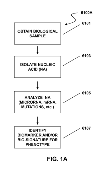
[0030] FIG. 1A depicts a method of identifying a biosignature comprising
nucleic acid to characterize a
phenotype. FIG. 1B depicts a method of identifying a biosignature of a vesicle
or vesicle population to
characterize a phenotype.
[0031] FIG. 2 illustrates methods of characterizing a phenotype by assessing
vesicle biosignatures. FIG. 2A is
a schematic of a planar substrate coated with a capture antibody, which
captures vesicles expressing that protein.
The capture antibody is for a vesicle protein that is specific or not specific
for vesicles derived from diseased
cells ("disease vesicle"). The detection antibody binds to the captured
vesicle and provides a fluorescent signal.
The detection antibody can detect an antigen that is generally associated with
vesicles, or is associated with a
-6-
CA 02838728 2013-12-06
WO 2012/170711 PCT/US2012/041387
cell-of-origin or a disease, e.g., a cancer. FIG. 2B is a schematic of a bead
coated with a capture antibody,
which captures vesicles expressing that protein. The capture antibody is for a
vesicle protein that is specific or
not specific for vesicles derived from diseased cells ("disease vesicle"). The
detection antibody binds to the
captured vesicle and provides a fluorescent signal. The detection antibody can
detect an antigen that is generally
associated with vesicles, or is associated with a cell-of-origin or a disease,
e.g., a cancer. FIG. 2C is an example
of a screening scheme that can be performed by multiplexing using the beads as
shown in FIG. 2B. FIG. 2D
presents illustrative schemes for capturing and detecting vesicles to
characterize a phenotype. FIG. 2E presents
illustrative schemes for assessing vesicle payload to characterize a
phenotype.
[0032] FIG. 3 illustrates a computer system that can be used in some exemplary
embodiments of the
invention.
[0033] FIG. 4 illustrates a method of depicting results using a bead based
method of detecting vesicles from a
subject. The number of beads captured at a given intensity is an indication of
how frequently a vesicle expresses
the detection protein at that intensity. The more intense the signal for a
given bead, the greater the expression of
the detection protein. The figure shows a normalized graph obtained by
combining normal patients into one
curve and cancer patients into another, and using bio-statistical analysis to
differentiate the curves. Data from
each individual is normalized to account for variation in the number of beads
read by the detection machine,
added together, and then normalized again to account for the different number
of samples in each population.
[0034] FIG. 5 illustrates the capture of prostate cancer cells-derived
vesicles from plasma with EpCam by
assessing TMPRSS2-ERG expression. VCaP purified vesicles were spiked into
normal plasma and then
incubated with Dynal magnetic beads coated with either the EpCam or isotype
control antibody. RNA was
isolated directly from the Dynal beads. Equal volumes of RNA from each sample
were used for RT-PCR and
subsequent Taqman assays.
[0035] FIG. 6 depicts a bar graph of miR-21 or miR-141 expression with CD9
bead capture. 1 ml of plasma
from prostate cancer patients, 250 ng/ml of LNCaP, or normal purified vesicles
were incubated with CD9 coated
Dynal beads. The RNA was isolated from the beads and the bead supernatant. One
sample (#6) was also
uncaptured for comparison. microRNA expression was measured with qRT-PCR and
the mean CT values for
each sample compared. CD9 capture improves the detection of miR-21 and miR-141
in prostate cancer samples.
[0036] FIG. 7 illustrates separation and identification of vesicles using the
MoFlo XDP.
[0037] FIG. 8 represents a schematic of detecting vesicles in a sample wherein
the presence or level of the
desired vesicles are assessed using a microsphere platform. FIG. 8A represents
a schematic of isolating vesicles
from plasma using a column based filtering method, wherein the isolated
vesicles are subsequently assessed
using a microsphere platform. FIG. 8B represents a schematic of compression of
a membrane of a vesicle due to
high-speed centrifugation, such as ultracentrifugation. FIG. 8C represents a
schematic of detecting vesicles
bound to microspheres using laser detection.
[0038] FIG. 9A illustrates the ability of a vesicle bio-signature to
discriminate between normal prostate and
PCa samples. Cancer markers included EpCam and B7H3. General vesicle markers
included CD9, CD81 and
CD63. Prostate specific markers included PCSA. PSMA can be used as well as
PCSA. The test was found to be
98% sensitive and 95% specific for PCa vs normal samples. FIG. 9B illustrates
mean fluorescence intensity
(MFI) on the Y axis for vesicle markers of FIG. 9A in normal and prostate
cancer patients.
-7-
CA 02838728 2013-12-06
WO 2012/170711 PCT/US2012/041387
[0039] FIG. 10 is a schematic for a decision tree for a vesicle prostate
cancer assay for determining whether a
sample is positive for prostate cancer.
[0040] FIG. 11 shows the results of a vesicle detection assay for prostate
cancer following the decision tree
versus detection using elevated PSA levels.
[0041] FIG. 12 illustrates levels of miR-145 in vesicles isolated from control
and PCa samples.
[0042] FIGs. 13A-13B illustrate the use of miR-107 and miR-141 to identify
false negatives from a vesicle-
based diagnostic assay for prostate cancer. FIG. 13A illustrates a scheme for
using miR analysis within vesicles
to convert false negatives into true positives, thereby improving sensitivity.
FIG. 13B illustrates a scheme for
using miR analysis within vesicles to convert false positives into true
negatives, thereby improving specificity.
Normalized levels of miR-107 (FIG. 13C) and miR-141 (FIG. 13D) are shown on
the Y axis for true positives
(TP) called by the vesicle diagnostic assay, true negatives (TN) called by the
vesicle diagnostic assay, false
positives (FP) called by the vesicle diagnostic assay, and false negatives
(FN) called by the vesicle diagnostic
assay.
[0043] FIG. 14 illustrates dot plots of raw background subtracted fluorescence
values of selected mRNAs
from microarray profiling of vesicle mRNA payload levels. In each plot, the Y
axis shows raw background
subtracted fluorescence values (Raw BGsub Florescence). The X axis shows dot
plots for four normal control
plasmas and four plasmas from prostate cancer patients. The mRNAs shown are
A2ML1 (FIG. 14A),
GABARAPL2 (FIG. 14B), PTMA (FIG. 14C), RABAC1 (FIG. 14D), SOX1 (FIG. 14E), and
ETFB
(FIG. 14F).
DETAILED DESCRIPTION OF THE INVENTION
[0044] Disclosed herein are methods and systems for characterizing a phenotype
of a biological sample, e.g., a
sample from a cell culture, an organism, or a subject. The phenotype can be
characterized by assessing one or
more biomarkers. The biomarkers can be associated with a vesicle or vesicle
population, either presented vesicle
surface antigens or vesicle payload. As used herein, vesicle payload comprises
entities encapsulated within a
vesicle. Vesicle associated biomarkers can comprise both membrane bound and
soluble biomarkers. The
biomarkers can also be circulating biomarkers, such as nucleic acids (e.g.,
microRNA) or protein/polypeptide, or
functional fragments thereof, assessed in a bodily fluid. Unless otherwise
specified, the terms "purified" or
"isolated" as used herein in reference to vesicles or biomarker components
mean partial or complete purification
or isolation of such components from a cell or organism. Furthermore, unless
otherwise specified, reference to
vesicle isolation using a binding agent includes binding a vesicle with the
binding agent whether or not such
binding results in complete isolation of the vesicle apart from other
biological entities in the starting material.
[0045] A method of characterizing a phenotype by analyzing a circulating
biomarker, e.g., a nucleic acid
biomarker, is depicted in scheme 6100A of FIG. 1A, as a non-limiting
illustrative example. In a first step 6101,
a biological sample is obtained, e.g., a bodily fluid, tissue sample or cell
culture. Nucleic acids are isolated from
the sample 6103. The nucleic acid can be DNA or RNA, e.g., microRNA.
Assessment of such nucleic acids can
provide a biosignature for a phenotype. By sampling the nucleic acids
associated with target phenotype (e.g.,
disease versus healthy, pre- and post-treatment), one or more nucleic acid
markers that are indicative of the
phenotype can be determined. Various aspects of the present invention are
directed to biosignatures determined
by assessing one or more nucleic acid molecules (e.g., microRNA) present in
the sample 6105, where the
biosignature corresponds to a predetermined phenotype 6107. FIG. 1B
illustrates a scheme 6100B of using
-8-
CA 02838728 2013-12-06
WO 2012/170711 PCT/US2012/041387
vesicles to isolate the nucleic acid molecules. In one example, a biological
sample is obtained 6102, and one or
more vesicles, e.g., vesicles from a particular cell-of-origin and/or vesicles
associated with a particular disease
state, are isolated from the sample 6104. The vesicles are analyzed 6106 by
characterizing surface antigens
associated with the vesicles and/or determining the presence or levels of
components present within the vesicles
("payload"). Unless specified otherwise, the term "antigen" as used herein
refers generally to a biomarker that
can be bound by a binding agent, whether the binding agent is an antibody,
aptamer, lectin, or other binding
agent for the biomarker and regardless of whether such biomarker illicits an
immune response in a host. Vesicle
payload may be protein, including peptides and polypeptides, and/or nucleic
acids such as DNA and RNAs.
RNA payload includes messenger RNA (mRNA) and microRNA (also referred to
herein as miRNA or miR). A
phenotype is characterized based on the biosignature of the vesicles 6108. In
another illustrative method of the
invention, schemes 6100A and 6100B are performed together to characterize a
phenotype. In such a scheme,
vesicles and nucleic acids, e.g., microRNA, are assessed, thereby
characterizing the phenotype.
[0046] In a related aspect, methods are provided herein for the discovery of
biomarkers comprising assessing
vesicle surface markers or payload markers in one sample and comparing the
markers to another sample.
Markers that distinguish between the samples can be used as biomarkers
according to the invention. Such
samples can be from a subject or group of subjects. For example, the groups
can be, e.g., known responders and
non-responders to a given treatment for a given disease or disorder.
Biomarkers discovered to distinguish the
known responders and non-responders provide a biosignature of whether a
subject is likely to respond to a
treatment such as a therapeutic agent, e.g., a drug or biologic.
Phenotypes
[0047] Disclosed herein are products and processes for characterizing a
phenotype of an individual by
analyzing a vesicle such as a membrane vesicle. A phenotype can be any
observable characteristic or trait of a
subject, such as a disease or condition, a disease stage or condition stage,
susceptibility to a disease or condition,
prognosis of a disease stage or condition, a physiological state, or response
to therapeutics. A phenotype can
result from a subject's gene expression as well as the influence of
environmental factors and the interactions
between the two, as well as from epigenetic modifications to nucleic acid
sequences.
[0048] A phenotype in a subject can be characterized by obtaining a biological
sample from a subject and
analyzing one or more vesicles from the sample. For example, characterizing a
phenotype for a subject or
individual may include detecting a disease or condition (including pre-
symptomatic early stage detecting),
determining the prognosis, diagnosis, or theranosis of a disease or condition,
or determining the stage or
progression of a disease or condition. Characterizing a phenotype can also
include identifying appropriate
treatments or treatment efficacy for specific diseases, conditions, disease
stages and condition stages,
predictions and likelihood analysis of disease progression, particularly
disease recurrence, metastatic spread or
disease relapse. A phenotype can also be a clinically distinct type or subtype
of a condition or disease, such as a
cancer or tumor. Phenotype determination can also be a determination of a
physiological condition, or an
assessment of organ distress or organ rejection, such as post-transplantation.
The products and processes
described herein allow assessment of a subject on an individual basis, which
can provide benefits of more
efficient and economical decisions in treatment.
[0049] In an aspect, the invention relates to the analysis of vesicles to
provide a biosignature to predict
whether a subject is likely to respond to a treatment for a disease or
disorder. Characterizating a phenotype
-9-
CA 02838728 2013-12-06
WO 2012/170711 PCT/US2012/041387
includes predicting the responder / non-responder status of the subject,
wherein a responder responds to a
treatment for a disease and a non-responder does not respond to the treatment.
Vesicles can be analyzed in the
subject and compared to vesicle analysis of previous subjects that were known
to respond or not to a treatment.
If the vesicle biosignature in a subject more closely aligns with that of
previous subjects that were known to
respond to the treatment, the subject can be characterized, or predicted, as a
responder to the treatment.
Similarly, if the vesicle biosignature in the subject more closely aligns with
that of previous subjects that did not
respond to the treatment, the subject can be characterized, or predicted as a
non-responder to the treatment. The
treatment can be for any appropriate disease, disorder or other condition. The
method can be used in any disease
setting where a vesicle biosignature that correlates with responder / non-
responder status is known.
[0050] The term "phenotype" as used herein can mean any trait or
characteristic that is attributed to a vesicle
biosignature that is identified utilizing methods of the invention. For
example, a phenotype can be the
identification of a subject as likely to respond to a treatment, or more
broadly, it can be a diagnostic, prognostic
or theranostic determination based on a characterized biosignature for a
sample obtained from a subject.
[0051] In some embodiments, the phenotype comprises a disease or condition
such as those listed in Table 1.
For example, the phenotype can comprise the presence of or likelihood of
developing a tumor, neoplasm, or
cancer. A cancer detected or assessed by products or processes described
herein includes, but is not limited to,
breast cancer, ovarian cancer, lung cancer, colon cancer, hyperplastic polyp,
adenoma, colorectal cancer, high
grade dysplasia, low grade dysplasia, prostatic hyperplasia, prostate cancer,
melanoma, pancreatic cancer, brain
cancer (such as a glioblastoma), hematological malignancy, hepatocellular
carcinoma, cervical cancer,
endometrial cancer, head and neck cancer, esophageal cancer, gastrointestinal
stromal tumor (GIST), renal cell
carcinoma (RCC) or gastric cancer. The colorectal cancer can be CRC Dukes B or
Dukes C-D. The
hematological malignancy can be B-Cell Chronic Lymphocytic Leukemia, B-Cell
Lymphoma-DLBCL, B-Cell
Lymphoma-DLBCL-germinal center-like, B-Cell Lymphoma-DLBCL-activated B-cell-
like, and Burkitt's
lymphoma.
[0052] The phenotype can be a premalignant condition, such as actinic
keratosis, atrophic gastritis,
leukoplakia, eiythroplasia, Lymphomatoid Granulomatosis, preleukemia,
fibrosis, cervical dysplasia, uterine
cervical dysplasia, xeroderma pigmentosum, Barrett's Esophagus, colorectal
polyp, or other abnormal tissue
growth or lesion that is likely to develop into a malignant tumor.
Transformative viral infections such as HIV
and HPV also present phenotypes that can be assessed according to the
invention.
[0053] The cancer characterized by the methods of the invention can comprise,
without limitation, a
carcinoma, a sarcoma, a lymphoma or leukemia, a germ cell tumor, a blastoma,
or other cancers. Carcinomas
include without limitation epithelial neoplasms, squamous cell neoplasms
squamous cell carcinoma, basal cell
neoplasms basal cell carcinoma, transitional cell papillomas and carcinomas,
adenomas and adenocarcinomas
(glands), adenoma, adenocarcinoma, linitis plastica insulinoma, glucagonoma,
gastrinoma, vipoma,
cholangiocarcinoma, hepatocellular carcinoma, adenoid cystic carcinoma,
carcinoid tumor of appendix,
prolactinoma, oncocytoma, hurthle cell adenoma, renal cell carcinoma, grawitz
tumor, multiple endocrine
adenomas, endometrioid adenoma, adnexal and skin appendage neoplasms,
mucoepidermoid neoplasms, cystic,
mucinous and serous neoplasms, cystadenoma, pseudomyxoma peritonei, ductal,
lobular and medullary
neoplasms, acinar cell neoplasms, complex epithelial neoplasms, warthin's
tumor, thymoma, specialized gonadal
neoplasms, sex cord stromal tumor, thecoma, granulosa cell tumor,
arrhenoblastoma, sertoli leydig cell tumor,
-10-
CA 02838728 2013-12-06
WO 2012/170711 PCT/US2012/041387
glomus tumors, paraganglioma, pheochromocytoma, glomus tumor, nevi and
melanomas, melanocytic nevus,
malignant melanoma, melanoma, nodular melanoma, dysplastic nevus, lentigo
maligna melanoma, superficial
spreading melanoma, and malignant acral lentiginous melanoma. Sarcoma includes
without limitation Askin's
tumor, botryodies, chondrosarcoma, Ewing's sarcoma, malignant hemangio
endothelioma, malignant
schwannoma, osteosarcoma, soft tissue sarcomas including: alveolar soft part
sarcoma, angiosarcoma,
cystosarcoma phyllodes, dermatofibrosarcoma, desmoid tumor, desmoplastic small
round cell tumor, epithelioid
sarcoma, extraskeletal chondrosarcoma, extraskeletal osteosarcoma,
fibrosarcoma, hemangiopericytoma,
hemangiosarcoma, kaposi's sarcoma, leiomyosarcoma, liposarcoma,
lymphangiosarcoma, lymphosarcoma,
malignant fibrous histiocytoma, neurofibrosarcoma, rhabdomyosarcoma, and
synovialsarcoma. Lymphoma and
leukemia include without limitation chronic lymphocytic leukemia/small
lymphocytic lymphoma, B-cell
prolymphocytic leukemia, lymphoplasmacytic lymphoma (such as waldenstrom
macroglobulinemia), splenic
marginal zone lymphoma, plasma cell myeloma, plasmacytoma, monoclonal
immunoglobulin deposition
diseases, heavy chain diseases, extranodal marginal zone B cell lymphoma, also
called malt lymphoma, nodal
marginal zone B cell lymphoma (nmzl), follicular lymphoma, mantle cell
lymphoma, diffuse large B cell
lymphoma, mediastinal (thymic) large B cell lymphoma, intravascular large B
cell lymphoma, primary effusion
lymphoma, burkitt lymphoma/leukemia, T cell prolymphocytic leukemia, T cell
large granular lymphocytic
leukemia, aggressive NK cell leukemia, adult T cell leukemia/lymphoma,
extranodal NK/T cell lymphoma,
nasal type, enteropathy-type T cell lymphoma, hepatosplenic T cell lymphoma,
blastic NK cell lymphoma,
mycosis fungoides / sezary syndrome, primary cutaneous CD30-positive T cell
lymphoproliferative disorders,
primary cutaneous anaplastic large cell lymphoma, lymphomatoid papulosis,
angioimmunoblastic T cell
lymphoma, peripheral T cell lymphoma, unspecified, anaplastic large cell
lymphoma, classical hodgkin
lymphomas (nodular sclerosis, mixed cellularity, lymphocyte-rich, lymphocyte
depleted or not depleted), and
nodular lymphocyte-predominant hodgkin lymphoma. Germ cell tumors include
without limitation germinoma,
dysgerminoma, seminoma, nongerminomatous germ cell tumor, embryonal carcinoma,
endodermal sinus
turmor, choriocarcinoma, teratoma, polyembryoma, and gonadoblastoma. Blastoma
includes without limitation
nephroblastoma, medulloblastoma, and retinoblastoma. Other cancers include
without limitation labial
carcinoma, larynx carcinoma, hypopharynx carcinoma, tongue carcinoma, salivary
gland carcinoma, gastric
carcinoma, adenocarcinoma, thyroid cancer (medullary and papillary thyroid
carcinoma), renal carcinoma,
kidney parenchyma carcinoma, cervix carcinoma, uterine corpus carcinoma,
endometrium carcinoma, chorion
carcinoma, testis carcinoma, urinary carcinoma, melanoma, brain tumors such as
glioblastoma, astrocytoma,
meningioma, medulloblastoma and peripheral neuroectodermal tumors, gall
bladder carcinoma, bronchial
carcinoma, multiple myeloma, basalioma, teratoma, retinoblastoma, choroidea
melanoma, seminoma,
rhabdomyosarcoma, craniopharyngeoma, osteosarcoma, chondrosarcoma, myosarcoma,
liposarcoma,
fibrosarcoma, Ewing sarcoma, and plasmocytoma.
[0054] In a further embodiment, the cancer under analysis may be a lung cancer
including non-small cell lung
cancer and small cell lung cancer (including small cell carcinoma (oat cell
cancer), mixed small cell/large cell
carcinoma, and combined small cell carcinoma), colon cancer, breast cancer,
prostate cancer, liver cancer,
pancreas cancer, brain cancer, kidney cancer, ovarian cancer, stomach cancer,
skin cancer, bone cancer, gastric
cancer, breast cancer, pancreatic cancer, glioma, glioblastoma, hepatocellular
carcinoma, papillary renal
carcinoma, head and neck squamous cell carcinoma, leukemia, lymphoma, myeloma,
or a solid tumor.
-11-
CA 02838728 2013-12-06
WO 2012/170711 PCT/US2012/041387
[0055] In embodiments, the cancer comprises an acute lymphoblastic leukemia;
acute myeloid leukemia;
adrenocortical carcinoma; AIDS-related cancers; AIDS-related lymphoma; anal
cancer; appendix cancer;
astrocytomas; atypical teratoid/rhabdoid tumor; basal cell carcinoma; bladder
cancer; brain stem glioma; brain
tumor (including brain stem glioma, central nervous system atypical
teratoid/rhabdoid tumor, central nervous
system embryonal tumors, astrocytomas, craniopharyngioma, ependymoblastoma,
ependymoma,
medulloblastoma, medulloepithelioma, pineal parenchymal tumors of intermediate
differentiation, supratentorial
primitive neuroectodermal tumors and pineoblastoma); breast cancer; bronchial
tumors; Burkitt lymphoma;
cancer of unknown primary site; carcinoid tumor; carcinoma of unknown primary
site; central nervous system
atypical teratoid/rhabdoid tumor; central nervous system embryonal tumors;
cervical cancer; childhood cancers;
chordoma; chronic lymphocytic leukemia; chronic myelogenous leukemia; chronic
myeloproliferative disorders;
colon cancer; colorectal cancer; craniopharyngioma; cutaneous T-cell lymphoma;
endocrine pancreas islet cell
tumors; endometrial cancer; ependymoblastoma; ependymoma; esophageal cancer;
esthesioneuroblastoma;
Ewing sarcoma; extracranial germ cell tumor; extragonadal germ cell tumor;
extrahepatic bile duct cancer;
gallbladder cancer; gastric (stomach) cancer; gastrointestinal carcinoid
tumor; gastrointestinal stromal cell
tumor; gastrointestinal stromal tumor (GIST); gestational trophoblastic tumor;
glioma; hairy cell leukemia; head
and neck cancer; heart cancer; Hodgkin lymphoma; hypopharyngeal cancer;
intraocular melanoma; islet cell
tumors; Kaposi sarcoma; kidney cancer; Langerhans cell histiocytosis;
laryngeal cancer; lip cancer; liver cancer;
malignant fibrous histiocytoma bone cancer; medulloblastoma;
medulloepithelioma; melanoma; Merkel cell
carcinoma; Merkel cell skin carcinoma; mesothelioma; metastatic squamous neck
cancer with occult primary;
mouth cancer; multiple endocrine neoplasia syndromes; multiple myeloma;
multiple myeloma/plasma cell
neoplasm; mycosis fungoides; myelodysplastic syndromes; myeloproliferative
neoplasms; nasal cavity cancer;
nasopharyngeal cancer; neuroblastoma; Non-Hodgkin lymphoma; nonmelanoma skin
cancer; non-small cell
lung cancer; oral cancer; oral cavity cancer; oropharyngeal cancer;
osteosarcoma; other brain and spinal cord
tumors; ovarian cancer; ovarian epithelial cancer; ovarian germ cell tumor;
ovarian low malignant potential
tumor; pancreatic cancer; papillomatosis; paranasal sinus cancer; parathyroid
cancer; pelvic cancer; penile
cancer; pharyngeal cancer; pineal parenchymal tumors of intermediate
differentiation; pineoblastoma; pituitary
tumor; plasma cell neoplasm/multiple myeloma; pleuropulmonary blastoma;
primary central nervous system
(CNS) lymphoma; primary hepatocellular liver cancer; prostate cancer; rectal
cancer; renal cancer; renal cell
(kidney) cancer; renal cell cancer; respiratory tract cancer; retinoblastoma;
rhabdomyosarcoma; salivary gland
cancer; Sezary syndrome; small cell lung cancer; small intestine cancer; soft
tissue sarcoma; squamous cell
carcinoma; squamous neck cancer; stomach (gastric) cancer; supratentorial
primitive neuroectodermal tumors;
T-cell lymphoma; testicular cancer; throat cancer; thymic carcinoma; thymoma;
thyroid cancer; transitional cell
cancer; transitional cell cancer of the renal pelvis and ureter; trophoblastic
tumor; ureter cancer; urethral cancer;
uterine cancer; uterine sarcoma; vaginal cancer; vulvar cancer; Waldenstrom
macroglobulinemia; or Wilm's
tumor. The methods of the invention can be used to characterize these and
other cancers. Thus, characterizing a
phenotype can be providing a diagnosis, prognosis or theranosis of one of the
cancers disclosed herein.
[0056] The phenotype can also be an inflammatory disease, immune disease, or
autoimmune disease. For
example, the disease may be inflammatory bowel disease (IBD), Crohn's disease
(CD), ulcerative colitis (UC),
pelvic inflammation, vasculitis, psoriasis, diabetes, autoimmune hepatitis,
Multiple Sclerosis, Myasthenia
Gravis, Type I diabetes, Rheumatoid Arthritis, Psoriasis, Systemic Lupus
Erythematosis (SLE), Hashimoto's
-12-
CA 02838728 2013-12-06
WO 2012/170711 PCT/US2012/041387
Thyroiditis, Grave's disease, Ankylosing Spondylitis Sjogrens Disease, CREST
syndrome, Scleroderma,
Rheumatic Disease, organ rejection, Primary Sclerosing Cholangitis, or sepsis.
[0057] The phenotype can also comprise a cardiovascular disease, such as
atherosclerosis, congestive heart
failure, vulnerable plaque, stroke, or ischemia. The cardiovascular disease or
condition can be high blood
pressure, stenosis, vessel occlusion or a thrombotic event.
[0058] The phenotype can also comprise a neurological disease, such as
Multiple Sclerosis (MS), Parkinson's
Disease (PD), Alzheimer's Disease (AD), schizophrenia, bipolar disorder,
depression, autism, Prion Disease,
Pick's disease, dementia, Huntington disease (HD), Down's syndrome,
cerebrovascular disease, Rasmussen's
encephalitis, viral meningitis, neurospsychiatric systemic lupus erythematosus
(NPSLE), amyotrophic lateral
sclerosis, Creutzfeldt-Jacob disease, Gerstmann-Straussler-Scheinker disease,
transmissible spongiform
encephalopathy, ischemic reperfusion damage (e.g. stroke), brain trauma,
microbial infection, or chronic fatigue
syndrome. The phenotype may also be a condition such as fibromyalgia, chronic
neuropathic pain, or peripheral
neuropathic pain.
[0059] The phenotype may also comprise an infectious disease, such as a
bacterial, viral or yeast infection. For
example, the disease or condition may be Whipple's Disease, Prion Disease,
cirrhosis, methicillin-resistant
staphylococcus aureus, HIV, hepatitis, syphilis, meningitis, malaria,
tuberculosis, or influenza. Viral proteins,
such as HIV or HCV-like particles can be assessed in a vesicle, to
characterize a viral condition.
[0060] The phenotype can also comprise a perinatal or pregnancy related
condition (e.g. preeclampsia or
preterm birth), metabolic disease or condition, such as a metabolic disease or
condition associated with iron
metabolism. For example, hepcidin can be assayed in a vesicle to characterize
an iron deficiency. The metabolic
disease or condition can also be diabetes, inflammation, or a perinatal
condition.
[0061] The methods of the invention can be used to characterize these and
other diseases and disorders that
can be assessed via biomarkers. Thus, characterizing a phenotype can be
providing a diagnosis, prognosis or
theranosis of one of the diseases and disorders disclosed herein.
Subject
[0062] One or more phenotypes of a subject can be determined by analyzing one
or more vesicles, such as
vesicles, in a biological sample obtained from the subject. A subject or
patient can include, but is not limited to,
mammals such as bovine, avian, canine, equine, feline, ovine, porcine, or
primate animals (including humans
and non-human primates). A subject can also include a mammal of importance due
to being endangered, such as
a Siberian tiger; or economic importance, such as an animal raised on a farm
for consumption by humans, or an
animal of social importance to humans, such as an animal kept as a pet or in a
zoo. Examples of such animals
include, but are not limited to, carnivores such as cats and dogs; swine
including pigs, hogs and wild boars;
ruminants or ungulates such as cattle, oxen, sheep, giraffes, deer, goats,
bison, camels or horses. Also included
are birds that are endangered or kept in zoos, as well as fowl and more
particularly domesticated fowl, i.e.
poultry, such as turkeys and chickens, ducks, geese, guinea fowl. Also
included are domesticated swine and
horses (including race horses). In addition, any animal species connected to
commercial activities are also
included such as those animals connected to agriculture and aquaculture and
other activities in which disease
monitoring, diagnosis, and therapy selection are routine practice in husbandry
for economic productivity and/or
safety of the food chain.
-13-
CA 02838728 2013-12-06
WO 2012/170711 PCT/US2012/041387
[0063] The subject can have a pre-existing disease or condition, such as
cancer. Alternatively, the subject may
not have any known pre-existing condition. The subject may also be non-
responsive to an existing or past
treatment, such as a treatment for cancer.
Samples
[0064] The biological sample obtained from the subject can be any bodily
fluid. For example, the biological
sample can be peripheral blood, sera, plasma, ascites, urine, cerebrospinal
fluid (CSF), sputum, saliva, bone
marrow, synovial fluid, aqueous humor, amniotic fluid, cerumen, breast milk,
broncheoalveolar lavage fluid,
semen (including prostatic fluid), Cowper's fluid or pre-ejaculatory fluid,
female ejaculate, sweat, fecal matter,
hair, tears, cyst fluid, pleural and peritoneal fluid, pericardial fluid,
lymph, chyme, chyle, bile, interstitial fluid,
menses, pus, sebum, vomit, vaginal secretions, mucosal secretion, stool water,
pancreatic juice, lavage fluids
from sinus cavities, bronchopulmonary aspirates or other lavage fluids. A
biological sample may also include
the blastocyl cavity, umbilical cord blood, or maternal circulation which may
be of fetal or maternal origin. The
biological sample may also be a tissue sample or biopsy from which vesicles
and other circulating biomarkers
may be obtained. For example, cells from the sample can be cultured and
vesicles isolated from the culture (see
for example, Example 1). In various embodiments, biomarkers or more
particularly biosignatures disclosed
herein can be assessed directly from such biological samples (e.g.,
identification of presence or levels of nucleic
acid or polypeptide biomarkers or functional fragments thereof) utilizing
various methods, such as extraction of
nucleic acid molecules from blood, plasma, serum or any of the foregoing
biological samples, use of protein or
antibody arrays to identify polypeptide (or functional fragment) biomarker(s),
as well as other array, sequencing,
PCR and proteomic techniques known in the art for identification and
assessment of nucleic acid and
polypeptide molecules.
[0065] Table 1 lists illustrative examples of diseases, conditions, or
biological states and a corresponding list
of biological samples from which vesicles may be analyzed.
Table 1: Examples of Biological Samples for Vesicle Analysis for
Various Diseases, Conditions, or Biological States
Illustrative Disease, Condition or Biological State Illustrative Biological
Samples
Cancers/neoplasms affecting the following tissue Blood, serum, plasma,
cerebrospinal fluid (CSF),
types/bodily systems: breast, lung, ovarian, colon, urine, sputum, ascites,
synovial fluid, semen, nipple
rectal, prostate, pancreatic, brain, bone, connective aspirates, saliva,
bronchoalveolar lavage fluid, tears,
tissue, glands, skin, lymph, nervous system, endocrine, oropharyngeal washes,
feces, peritoneal fluids, pleural
germ cell, genitourinary, hematologic/blood, bone effusion, sweat, tears,
aqueous humor, pericardial
marrow, muscle, eye, esophageal, fat tissue, thyroid, fluid, lymph, chyme,
chyle, bile, stool water, amniotic
pituitary, spinal cord, bile duct, heart, gall bladder, fluid, breast milk,
pancreatic juice, cerumen, Cowper's
bladder, testes, cervical, endometrial, renal, ovarian, fluid or pre-
ejaculatory fluid, female ejaculate,
digestive/gastrointestinal, stomach, head and neck, interstitial fluid,
menses, mucus, pus, sebum, vaginal
liver, leukemia, respiratory/thorasic, cancers of lubrication, vomit
unknown primary (CUP)
Neurodegenerative/neurological disorders: Blood, serum, plasma, CSF, urine
Parkinson's disease, Alzheimer's Disease and multiple
sclerosis, Schizophrenia, and bipolar disorder,
spasticity disorders, epilepsy
Cardiovascular Disease: atherosclerosis, Blood, serum, plasma, CSF, urine
cardiomyopathy, endocarditis, vunerable plaques,
infection
Stroke: ischemic, intracerebral hemorrhage, Blood, serum, plasma, CSF,
urine
subarachnoid hemorrhage, transient ischemic attacks
(TIA)
-14-
CA 02838728 2013-12-06
WO 2012/170711 PCT/US2012/041387
Pain disorders: peripheral neuropathic pain and Blood, serum, plasma, CSF,
urine
chronic neuropathic pain, and fibromyalgia,
Autoimmune disease: systemic and localized diseases, Blood, serum, plasma,
CSF, urine, synovial fluid
rheumatic disease, Lupus, Sjogren's syndrome
Digestive system abnormalities: Barrett's esophagus, Blood, serum, plasma,
CSF, urine
irritable bowel syndrome, ulcerative colitis, Crohn's
disease, Diverticulosis and Diverticulitis, Celiac
Disease
Endocrine disorders: diabetes mellitus, various forms Blood, serum, plasma,
CSF, urine
of Thyroiditisõ adrenal disorders, pituitary disorders
Diseases and disorders of the skin: psoriasis Blood, serum, plasma, CSF,
urine, synovial fluid, tears
Urological disorders: benign prostatic hypertrophy Blood, serum, plasma,
urine
(BPH), polycystic kidney disease, interstitial cystitis
Hepatic disease/injury: Cirrhosis, induced Blood, serum, plasma, urine
hepatotoxicity (due to exposure to natural or synthetic
chemical sources)
Kidney disease/injury: acute, sub-acute, chronic Blood, serum, plasma,
urine
conditions, Podocyte injury, focal segmental
glomerulosclerosis
Endometriosis Blood, serum, plasma, urine, vaginal
fluids
Osteoporosis Blood, serum, plasma, urine, synovial
fluid
Pancreatitis Blood, serum, plasma, urine, pancreatic
juice
Asthma Blood, serum, plasma, urine, sputum,
bronchiolar
lavage fluid
Allergies Blood, serum, plasma, urine, sputum,
bronchiolar
lavage fluid
Prion-related diseases Blood, serum, plasma, CSF, urine
Viral Infections: HIV/AIDS Blood, serum, plasma, urine
Sepsis Blood, serum, plasma, urine, tears,
nasal lavage
Organ rejection/transplantation Blood, serum, plasma, urine, various
lavage fluids
Differentiating conditions: adenoma versus Blood, serum, plasma, urine,
sputum, feces, colonic
hyperplastic polyp, irritable bowel syndrome (IBS) lavage fluid
versus normal, classifying Dukes stages A, B, C,
and/or D of colon cancer, adenoma with low-grade
hyperplasia versus high-grade hyperplasia, adenoma
versus normal, colorectal cancer versus normal, IBS
versus, ulcerative colitis (UC) versus Crohn's disease
(CD),
Pregnancy related physiological states, conditions, or Maternal serum, plasma,
amniotic fluid, cord blood
affiliated diseases: genetic risk, adverse pregnancy
outcomes
[0066] The methods of the invention can be used to characterize a phenotype
using a blood sample or blood
derivative. Blood derivatives include plasma and serum. Blood plasma is the
liquid component of whole blood,
and makes up approximately 55% of the total blood volume. It is composed
primarily of water with small
amounts of minerals, salts, ions, nutrients, and proteins in solution. In
whole blood, red blood cells, leukocytes,
and platelets are suspended within the plasma. Blood serum refers to blood
plasma without fibrinogen or other
clotting factors (i.e., whole blood minus both the cells and the clotting
factors).
[0067] The biological sample may be obtained through a third party, such as a
party not performing the
analysis of the biomarkers, whether direct assessment of a biological sample
or by profiling one or more
vesicles obtained from the biological sample. For example, the sample may be
obtained through a clinician,
-15-
CA 02838728 2013-12-06
WO 2012/170711 PCT/US2012/041387
physician, or other health care manager of a subject from which the sample is
derived. Alternatively, the
biological sample may obtained by the same party analyzing the vesicle. In
addition, biological samples be
assayed, are archived (e.g., frozen) or ortherwise stored in under
preservative conditions.
[0068] The volume of the biological sample used for biomarker analysis can be
in the range of between 0.1-
20 mL, such as less than about 20, 15, 10, 9, 8, 7, 6, 5, 4, 3, 2, 1 or 0.1
mL.
[0069] A sample of bodily fluid can be used as a sample for characterizing a
phenotype. For example,
biomarkers in the sample can be assessed to provide a diagnosis, prognosis
and/or theranosis of a disease. The
biomarkers can be circulating biomarkers, such as circulating proteins or
nucleic acids. The biomarkers can also
be associated with a vesicle or vesicle population. Methods of the invention
can be applied to assess one or more
vesicles, as well as one or more different vesicle populations that may be
present in a biological sample or in a
subject. Analysis of one or more biomarkers in a biological sample can be used
to determine whether an
additional biological sample should be obtained for analysis. For example,
analysis of one or more vesicles in a
sample of bodily fluid can aid in determining whether a tissue biopsy should
be obtained.
[0070] A sample from a patient can be collected under conditions that preserve
the circulating biomarkers and
other entities of interest contained therein for subsequent analysis. In an
embodiment, the samples are processed
using one or more of CellSave Preservative Tubes (Veridex, North Raritan, NJ),
PAXgene Blood DNA Tubes
(QIAGEN GmbH, Germany), and RNAlater (QIAGEN GmbH, Germany).
[0071] CellSave Preservative Tubes (CellSave tubes) are sterile evacuated
blood collection tubes. Each tube
contains a solution that contains Na2EDTA and a cell preservative. The EDTA
absorbs calcium ions, which can
reduce or eliminate blood clotting. The preservative preserves the morphology
and cell surface antigen
expression of epithelial and other cells. The collection and processing can be
performed as described in a
protocol provided by the manufacturer. Each tube is evacuated to withdraw
venous whole blood following
standard phlebotomy procedures as known to those of skill in the art. CellSave
tubes are disclosed in US Patent
Numbers 5,466,574; 5,512,332; 5,597,531; 5,698,271; 5,985,153; 5,993,665;
6,120,856; 6,136,182; 6,365,362;
6,551,843; 6,620,627; 6,623,982; 6,645,731; 6,660,159; 6,790,366; 6,861,259;
6,890,426; 7,011,794; 7,282,350;
7,332,288; 5,849,517 and 5,459,073, each of which is incorporated by reference
in its entirety herein.
[0072] The PAXgene Blood DNA Tube (PAXgene tube) is a plastic, evacuated tube
for the collection of
whole blood for the isolation of nucleic acids. The tubes can be used for
blood collection, transport and storage
of whole blood specimens and isolation of nucleic acids contained therein,
e.g., DNA or RNA. Blood is
collected under a standard phlebotomy protocol into an evacuated tube that
contains an additive. The collection
and processing can be performed as described in a protocol provided by the
manufacturer. PAXgene tubes are
disclosed in US Patent Nos. 5,906,744; 4,741,446; 4,991,104, each of which is
incorporated by reference in its
entirety herein.
[0073] The RNAlater RNA Stabilization Reagent (RNAlater) is used for immediate
stabilization of RNA in
tissues. RNA can be unstable in harvested samples. The aqueous RNAlater
reagent permeates tissues and other
biological samples, thereby stabilizing and protecting the RNA contained
therein. Such protection helps ensure
that downstream analyses reflect the expression profile of the RNA in the
tissue or other sample. The samples
are submerged in an appropriate volume of RNAlater reagent immediately after
harvesting. The collection and
processing can be performed as described in a protocol provided by the
manufacturer. According to the
manufacturer, the reagent preserves RNA for up to 1 day at 37 C, 7 days at 18-
25 C, or 4 weeks at 2-8 C,
-16-
CA 02838728 2013-12-06
WO 2012/170711 PCT/US2012/041387
allowing processing, transportation, storage, and shipping of samples without
liquid nitrogen or dry ice. The
samples can also be placed at ¨20 C or ¨80 C, e.g., for archival storage. The
preserved samples can be used to
analyze any type of RNA, including without limitation total RNA, mRNA, and
microRNA. RNAlater can also
be useful for collecting samples for DNA, RNA and protein analysis. RNAlater
is disclosed in US Patent Nos.
5,346,994, each of which is incorporated by reference in its entirety herein.
Vesicles
[0074] Methods of the invention can include assessing one or more vesicles,
including assessing vesicle
populations. A vesicle, as used herein, is a membrane vesicle that is shed
from cells. Vesicles or membrane
vesicles include without limitation: circulating microvesicles (cMVs),
microvesicle, exosome, nanovesicle,
dexosome, bleb, blebby, prostasome, microparticle, intralumenal vesicle,
membrane fragment, intralumenal
endosomal vesicle, endosomal-like vesicle, exocytosis vehicle, endosome
vesicle, endosomal vesicle, apoptotic
body, multivesicular body, secretory vesicle, phospholipid vesicle, liposomal
vesicle, argosome, texasome,
secresome, tolerosome, melanosome, oncosome, or exocytosed vehicle.
Furthermore, although vesicles may be
produced by different cellular processes, the methods of the invention are not
limited to or reliant on any one
mechanism, insofar as such vesicles are present in a biological sample and are
capable of being characterized by
the methods disclosed herein. Unless otherwise specified, methods that make
use of a species of vesicle can be
applied to other types of vesicles. Vesicles comprise spherical structures
with a lipid bilayer similar to cell
membranes which surrounds an inner compartment which can contain soluble
components, sometimes referred
to as the payload. In some embodiments, the methods of the invention make use
of exosomes, which are small
secreted vesicles of about 40-100 nm in diameter. For a review of membrane
vesicles, including types and
characterizations, see Thery et al., Nat Rev Immunol. 2009 Aug;9(8): 581-93.
Some properties of different types
of vesicles include those in Table 2:
Table 2: Vesicle Properties
Feature Exosomes Microvesicle Ectosomes Membrane Exosome- Apoptotic
particles like vesicles
vesicles
Size 50-100 nm 100-1,000 nm 50-200 nm 50-80 nm 20-50 nm 50-
500 nm
Density in 1.13-1.19 g/ml 1.04-1.07 1.1 g/ml 1.16-
1.28
sucrose g/ml g/ml
EM Cup shape Irregular Bilamellar Round Irregular
Heterogeneou
appearance shape, round shape
electron structures
dense
Sedimentatio 100,000 g 10,000 g 160,000- 100,000-
175,000 g 1,200 g,
200,000 g 200,000 g 10,000 g,
100,000 g
Lipid Enriched in Expose PPS Enriched in No
lipid
composition cholesterol, cholesterol and rafts
sphingomyelin diacylglycerol;
and ceramide; expose PPS
contains lipid
rafts; expose
PPS
Major protein Tetraspanins Integrins, CR1 and CD133; no TNFRI
Histones
markers (e.g., CD63, selectins and proteolytic CD63
CD9), Alix, CD40 ligand enzymes; no
TSG101 CD63
Intracellular Internal Plasma Plasma Plasma
origin compartments membrane membrane membrane
-17-
CA 02838728 2013-12-06
WO 2012/170711 PCT/US2012/041387
(endosomes)
Abbreviations: phosphatidylserine (PPS); electron microscopy (EM)
[0075] Vesicles include shed membrane bound particles, or "microparticles,"
that are derived from either the
plasma membrane or an internal membrane. Vesicles can be released into the
extracellular environment from
cells. Cells releasing vesicles include without limitation cells that
originate from, or are derived from, the
ectoderm, endoderm, or mesoderm. The cells may have undergone genetic,
environmental, and/or any other
variations or alterations. For example, the cell can be tumor cells. A vesicle
can reflect any changes in the
source cell, and thereby reflect changes in the originating cells, e.g., cells
having various genetic mutations. In
one mechanism, a vesicle is generated intracellularly when a segment of the
cell membrane spontaneously
invaginates and is ultimately exocytosed (see for example, Keller et al.,
Immunol. Lett. 107 (2): 102-8 (2006)).
Vesicles also include cell-derived structures bounded by a lipid bilayer
membrane arising from both herniated
evagination (blebbing) separation and sealing of portions of the plasma
membrane or from the export of any
intracellular membrane-bounded vesicular structure containing various membrane-
associated proteins of tumor
origin, including surface-bound molecules derived from the host circulation
that bind selectively to the tumor-
derived proteins together with molecules contained in the vesicle lumen,
including but not limited to tumor-
derived microRNAs or intracellular proteins. Blebs and blebbing are further
described in Charras et al., Nature
Reviews Molecular and Cell Biology, Vol. 9, No. 11, p. 730-736 (2008). A
vesicle shed into circulation or bodily
fluids from tumor cells may be referred to as a "circulating tumor-derived
vesicle." When such vesicle is an
exosome, it may be referred to as a circulating-tumor derived exosome (CTE).
In some instances, a vesicle can
be derived from a specific cell of origin. CTE, as with a cell-of-origin
specific vesicle, typically have one or
more unique biomarkers that permit isolation of the CTE or cell-of-origin
specific vesicle, e.g., from a bodily
fluid and sometimes in a specific manner. For example, a cell or tissue
specific markers are utilized to identify
the cell of origin. Examples of such cell or tissue specific markers are
disclosed herein and can further be
accessed in the Tissue-specific Gene Expression and Regulation (TiGER)
Database, available at
bioinfo.wilmer.jhu.edu/tiger/; Liu et al. (2008) TiGER: a database for tissue-
specific gene expression and
regulation. BMC Bioinformatics. 9:271; TissueDistributionDBs, available at
genome.dkfz-
heidelberg.de/menu/tissue_db/index.html.
[0076] A vesicle can have a diameter of greater than about 10 nm, 20 nm, or 30
nm. A vesicle can have a
diameter of greater than 40 nm, 50 nm, 100 nm, 200 nm, 500 nm, 1000 nm 1500
nm, or greater than 10,000 nm.
A vesicle can have a diameter of about 20-1500 nm, 30-1000 nm, about 30-800
nm, about 30-200 nm, or about
30-100 nm. In some embodiments, the vesicle has a diameter of less than 10,000
nm, 1500 nm, 1000 nm, 800
nm, 500 nm, 200 nm, 100 nm, 50 nm, 40 nm, 30 nm, 20 nm or less than 10 nm. As
used herein the term "about"
in reference to a numerical value means that variations of 10% above or below
the numerical value are within
the range ascribed to the specified value. Typical sizes for various types of
vesicles are shown in Table 2.
Vesicles can be assessed to measure the diameter of a single vesicle or any
number of vesicles. For example, the
range of diameters of a vesicle population or an average diameter of a vesicle
population can be determined.
Vesicle diameter can be assessed using methods known in the art, e.g., imaging
technologies such as electron
microscopy. In an embodiment, a diameter of one or more vesicles is determined
using optical particle
detection. See, e.g., U.S. Patent 7,751,053, entitled "Optical Detection and
Analysis of Particles" and issued July
6, 2010; and U.S. Patent 7,399,600, entitled "Optical Detection and Analysis
of Particles" and issued July 15,
2010.
-18-
CA 02838728 2013-12-06
WO 2012/170711 PCT/US2012/041387
[0077] In some embodiments, vesicles are directly assayed from a biological
sample without prior isolation,
purification, or concentration from the biological sample. For example, the
amount of vesicles in the sample can
by itself provide a biosignature that provides a diagnostic, prognostic or
theranostic determination.
Alternatively, the vesicle in the sample may be isolated, captured, purified,
or concentrated from a sample prior
to analysis. As noted, isolation, capture or purification as used herein
comprises partial isolation, partial capture
or partial purification apart from other components in the sample. Vesicle
isolation can be performed using
various techniques as described herein, e.g., chromatography, filtration,
centrifugation, flow cytometry, affinity
capture (e.g., to a planar surface or bead), and/or using microfluidics.
[0078] Vesicles such as exosomes can be assessed to provide a phenotypic
characterization by comparing
vesicle characteristics to a reference. In some embodiments, surface antigens
on a vesicle are assessed. The
surface antigens can provide an indication of the anatomical origin and/or
cellular of the vesicles and other
phenotypic information, e.g., tumor status. For example, wherein vesicles
found in a patient sample, e.g., a
bodily fluid such as blood, serum or plasma, are assessed for surface antigens
indicative of colorectal origin and
the presence of cancer. The surface antigens may comprise any informative
biological entity that can be detected
on the vesicle membrane surface, including without limitation surface
proteins, lipids, carbohydrates, and other
membrane components. For example, positive detection of colon derived vesicles
expressing tumor antigens can
indicate that the patient has colorectal cancer. As such, methods of the
invention can be used to characterize any
disease or condition associated with an anatomical or cellular origin, by
assessing, for example, disease-specific
and cell-specific biomarkers of one or more vesicles obtained from a subject.
[0079] In another embodiment, one or more vesicle payloads are assessed to
provide a phenotypic
characterization. The payload with a vesicle comprises any informative
biological entity that can be detected as
encapsulated within the vesicle, including without limitation proteins and
nucleic acids, e.g., genomic or cDNA,
mRNA, or functional fragments thereof, as well as microRNAs (miRs). In
addition, methods of the invention
are directed to detecting vesicle surface antigens (in addition or exclusive
to vesicle payload) to provide a
phenotypic characterization. For example, vesicles can be characterized by
using binding agents (e.g., antibodies
or aptamers) that are specific to vesicle surface antigens, and the bound
vesicles can be further assessed to
identify one or more payload components disclosed therein. As described
herein, the levels of vesicles with
surface antigens of interest or with payload of interest can be compared to a
reference to characterize a
phenotype. For example, overexpression in a sample of cancer-related surface
antigens or vesicle payload, e.g.,
a tumor associated mRNA or microRNA, as compared to a reference, can indicate
the presence of cancer in the
sample. The biomarkers assessed can be present or absent, increased or reduced
based on the selection of the
desired target sample and comparison of the target sample to the desired
reference sample. Non-limiting
examples of target samples include: disease; treated/not-treated; different
time points, such as a in a longitudinal
study; and non-limiting examples of reference sample: non-disease; normal;
different time points; and sensitive
or resistant to candidate treatment(s).
MicroRNA
[0080] Various biomarker molecules can be assessed in biological samples or
vesicles obtained from such
biological samples. MicroRNAs comprise one class biomarkers assessed via
methods of the invention.
MicroRNAs, also referred to herein as miRNAs or miRs, are short RNA strands
approximately 21-23
nucleotides in length. MiRNAs are encoded by genes that are transcribed from
DNA but are not translated into
-19-
CA 02838728 2013-12-06
WO 2012/170711 PCT/US2012/041387
protein and thus comprise non-coding RNA. The miRs are processed from primary
transcripts known as pri-
miRNA to short stem-loop structures called pre-miRNA and finally to the
resulting single strand miRNA. The
pre-miRNA typically forms a structure that folds back on itself in self-
complementary regions. These structures
are then processed by the nuclease Dicer in animals or DCL1 in plants. Mature
miRNA molecules are partially
complementary to one or more messenger RNA (mRNA) molecules and can function
to regulate translation of
proteins. Identified sequences of miRNA can be accessed at publicly available
databases, such as
www.microRNA.org, www.mirbase.org, or www.mirz.unibas.ch/cgi/miRNA.cgi.
[0081] miRNAs are generally assigned a number according to the naming
convention " mir-[number]." The
number of a miRNA is assigned according to its order of discovery relative to
previously identified miRNA
species. For example, if the last published miRNA was mir-121, the next
discovered miRNA will be named mir-
122, etc. When a miRNA is discovered that is homologous to a known miRNA from
a different organism, the
name can be given an optional organism identifier, of the form [organism
identifier]- mir-[number]. Identifiers
include hsa for Homo sapiens and mmu for Mus Musculus. For example, a human
homolog to mir-121 might be
referred to as hsa-mir-121 whereas the mouse homolog can be referred to as mmu-
mir-121.
[0082] Mature microRNA is commonly designated with the prefix "miR" whereas
the gene or precursor
miRNA is designated with the prefix "mir." For example, mir-121 is a precursor
for miR-121. When differing
miRNA genes or precursors are processed into identical mature miRNAs, the
genes/precursors can be delineated
by a numbered suffix. For example, mir-121-1 and mir-121-2 can refer to
distinct genes or precursors that are
processed into miR-121. Lettered suffixes are used to indicate closely related
mature sequences. For example,
mir-121a and mir-121b can be processed to closely related miRNAs miR-121a and
miR-121b, respectively. In
the context of the invention, any microRNA (miRNA or miR) designated herein
with the prefix mir-* or miR-*
is understood to encompass both the precursor and/or mature species, unless
otherwise explicitly stated
otherwise.
[0083] Sometimes it is observed that two mature miRNA sequences originate from
the same precursor. When
one of the sequences is more abundant that the other, a "*" suffix can be used
to designate the less common
variant. For example, miR-121 would be the predominant product whereas miR-
121* is the less common variant
found on the opposite arm of the precursor. If the predominant variant is not
identified, the miRs can be
distinguished by the suffix "5p" for the variant from the 5' arm of the
precursor and the suffix "3p" for the
variant from the 3' arm. For example, miR-121-5p originates from the 5' arm of
the precursor whereas miR-
121-3p originates from the 3' arm. Less commonly, the 5p and 3p variants are
referred to as the sense ("s") and
anti-sense ("as") forms, respectively. For example, miR-121-5p may be referred
to as miR-121-s whereas miR-
121-3p may be referred to as miR-121-as.
[0084] The above naming conventions have evolved over time and are general
guidelines rather than absolute
rules. For example, the let- and lin- families of miRNAs continue to be
referred to by these monikers. The
mir/miR convention for precursor/mature forms is also a guideline and context
should be taken into account to
determine which form is referred to. Further details of miR naming can be
found at www.mirbase.org or
Ambros et al., A uniform system for microRNA annotation, RNA 9:277-279 (2003).
[0085] Plant miRNAs follow a different naming convention as described in
Meyers et al., Plant Cell. 2008
20(12):3186-3190.
-20-
CA 02838728 2013-12-06
WO 2012/170711 PCT/US2012/041387
[0086] A number of miRNAs are involved in gene regulation, and miRNAs are part
of a growing class of non-
coding RNAs that is now recognized as a major tier of gene control. In some
cases, miRNAs can interrupt
translation by binding to regulatory sites embedded in the 3'-UTRs of their
target mRNAs, leading to the
repression of translation. Target recognition involves complementary base
pairing of the target site with the
miRNA's seed region (positions 2-8 at the miRNA's 5' end), although the exact
extent of seed complementarity
is not precisely determined and can be modified by 3' pairing. In other cases,
miRNAs function like small
interfering RNAs (siRNA) and bind to perfectly complementary mRNA sequences to
destroy the target
transcript.
[0087] Characterization of a number of miRNAs indicates that they influence a
variety of processes, including
early development, cell proliferation and cell death, apoptosis and fat
metabolism. For example, some miRNAs,
such as lin-4, let-7, mir-14, mir-23, and bantam, have been shown to play
critical roles in cell differentiation and
tissue development. Others are believed to have similarly important roles
because of their differential spatial and
temporal expression patterns.
[0088] The miRNA database available at miRBase (www.mirbase.org) comprises a
searchable database of
published miRNA sequences and annotation. Further information about miRBase
can be found in the following
articles, each of which is incorporated by reference in its entirety herein:
Griffiths-Jones et al., miRBase: tools
for microRNA genomics. NAR 2008 36(Database Issue):D154-D158; Griffiths-Jones
et al., miRBase:
microRNA sequences, targets and gene nomenclature. NAR 2006 34(Database
Issue):D140-D144; and
Griffiths-Jones, S. The microRNA Registry. NAR 2004 32(Database Issue):D109-
D111. Representative
miRNAs contained in Release 16 of miRBase, made available September 2010.
[0089] As described herein, microRNAs are known to be involved in cancer and
other diseases and can be
assessed in order to characterize a phenotype in a sample. See, e.g., Ferracin
et al., Micromarkers: miRNAs in
cancer diagnosis and prognosis, Exp Rev Mol Diag, Apr 2010, Vol. 10, No. 3,
Pages 297-308; Fabbri, miRNAs
as molecular biomarkers of cancer, Exp Rev Mol Diag, May 2010, Vol. 10, No. 4,
Pages 435-444. Techniques
to isolate and characterize vesicles and miRs are known to those of skill in
the art. In addition to the
methodology presented herein, additional methods can be found in U.S. Patent
No. 7,888,035, entitled
"METHODS FOR ASSESSING RNA PATTERNS" and issued February 15, 2011; and
International Patent
Application Nos. PCT/U52010/058461, entitled "METHODS AND SYSTEMS FOR
ISOLATING, STORING,
AND ANALYZING VESICLES" and filed November 30, 2010; and PCT/US2011/021160,
entitled
"DETECTION OF GASTROINTESTINAL DISORDERS" and filed January 13, 2011; each of
which
applications are incorporated by reference herein in their entirety.
Circulating Biomarkers
[0090] Circulating biomarkers include biomarkers that are detectable in body
fluids, such as blood, plasma,
serum. Examples of circulating cancer biomarkers include cardiac troponin T
(cTnT), prostate specific antigen
(PSA) for prostate cancer and CA125 for ovarian cancer. Circulating biomarkers
according to the invention
include any appropriate biomarker that can be detected in bodily fluid,
including without limitation protein,
nucleic acids, e.g., DNA, mRNA and microRNA, lipids, carbohydrates and
metabolites. Circulating biomarkers
can include biomarkers that are not associated with cells, such as biomarkers
that are membrane associated,
embedded in membrane fragments, part of a biological complex, or free in
solution. In one embodiment,
-21-
CA 02838728 2013-12-06
WO 2012/170711 PCT/US2012/041387
circulating biomarkers are biomarkers that are associated with one or more
vesicles present in the biological
fluid of a subject.
[0091] Circulating biomarkers have been identified for use in characterization
of various phenotypes. See, e.g.,
Ahmed N, et al., Proteomic-based identification of haptoglobin-1 precursor as
a novel circulating biomarker of
ovarian cancer. Br. J. Cancer 2004; Mathelin et al., Circulating proteinic
biomarkers and breast cancer, Gynecol
Obstet Fertil. 2006 Jul-Aug;34(7-8):638-46. Epub 2006 Jul 28; Ye et al.,
Recent technical strategies to identify
diagnostic biomarkers for ovarian cancer. Expert Rev Proteomics. 2007
Feb;4(1):121-31; Carney, Circulating
oncoproteins HER2/neu, EGFR and CAIX (MN) as novel cancer biomarkers. Expert
Rev Mol Diagn. 2007
May;7(3):309-19; Gagnon, Discovery and application of protein biomarkers for
ovarian cancer, Curr Opin
Obstet Gynecol. 2008 Feb;20(1):9-13; Pasterkamp et al., Immune regulatory
cells: circulating biomarker
factories in cardiovascular disease. Clin Sci (Lond). 2008 Aug;115(4):129-31;
Fabbri, miRNAs as molecular
biomarkers of cancer, Exp Rev Mol Diag, May 2010, Vol. 10, No. 4, Pages 435-
444; PCT Patent Publication
WO/2007/088537; U.S. Patents 7,745,150 and 7,655,479; U.S. Patent Publications
20110008808, 20100330683,
20100248290, 20100222230, 20100203566, 20100173788, 20090291932, 20090239246,
20090226937,
20090111121, 20090004687, 20080261258, 20080213907, 20060003465, 20050124071,
and 20040096915,
each of which publication is incorporated herein by reference in its entirety.
Vesicle Enrichment
[0092] A vesicle or a population of vesicles may be isolated, purified,
concentrated or otherwise enriched prior
to and/or during analysis. Unless otherwise specified, the terms "purified,"
"isolated," or similar as used herein
in reference to vesicles or biomarker components are intended to include
partial or complete purification or
isolation of such components from a cell or organism. Analysis of a vesicle
can include quantitiating the amount
one or more vesicle populations of a biological sample. For example, a
heterogeneous population of vesicles can
be quantitated, or a homogeneous population of vesicles, such as a population
of vesicles with a particular
biomarker profile, a particular biosignature, or derived from a particular
cell type can be isolated from a
heterogeneous population of vesicles and quantitated. Analysis of a vesicle
can also include detecting,
quantitatively or qualitatively, one or more particular biomarker profile or
biosignature of a vesicle, as described
herein.
[0093] A vesicle can be stored and archived, such as in a bio-fluid bank and
retrieved for analysis as
necessary. A vesicle may also be isolated from a biological sample that has
been previously harvested and
stored from a living or deceased subject. In addition, a vesicle may be
isolated from a biological sample which
has been collected as described in King et al., Breast Cancer Res 7(5): 198-
204 (2005). A vesicle can be isolated
from an archived or stored sample. Alternatively, a vesicle may be isolated
from a biological sample and
analyzed without storing or archiving of the sample. Furthermore, a third
party may obtain or store the
biological sample, or obtain or store the vesicle for analysis.
[0094] An enriched population of vesicles can be obtained from a biological
sample. For example, vesicles
may be concentrated or isolated from a biological sample using size exclusion
chromatography, density gradient
centrifugation, differential centrifugation, nanomembrane ultrafiltration,
immunoabsorbent capture, affinity
purification, microfluidic separation, or combinations thereof.
[0095] Size exclusion chromatography, such as gel permeation columns,
centrifugation or density gradient
centrifugation, and filtration methods can be used. For example, a vesicle can
be isolated by differential
-22-
CA 02838728 2013-12-06
WO 2012/170711 PCT/US2012/041387
centrifugation, anion exchange and/or gel permeation chromatography (for
example, as described in US Patent
Nos. 6,899,863 and 6,812,023), sucrose density gradients, organelle
electrophoresis (for example, as described
in U.S. Patent No. 7,198,923), magnetic activated cell sorting (MACS), or with
a nanomembrane ultrafiltration
concentrator. Various combinations of isolation or concentration methods can
be used.
[0096] Highly abundant proteins, such as albumin and immunoglobulin, may
hinder isolation of vesicles from
a biological sample. For example, a vesicle can be isolated from a biological
sample using a system that utilizes
multiple antibodies that are specific to the most abundant proteins found in a
biological sample, such as blood.
Such a system can remove up to several proteins at once, thus unveiling the
lower abundance species such as
cell-of-origin specific vesicles.
[0097] This type of system can be used for isolation of vesicles from
biological samples such as blood,
cerebrospinal fluid or urine. The isolation of vesicles from a biological
sample may also be enhanced by high
abundant protein removal methods as described in Chromy et al. J Proteome Res
2004; 3:1120-1127. In another
embodiment, the isolation of vesicles from a biological sample may also be
enhanced by removing serum
proteins using glycopeptide capture as described in Zhang et al, Mol Cell
Proteomics 2005;4:144-155. In
addition, vesicles from a biological sample such as urine may be isolated by
differential centrifugation followed
by contact with antibodies directed to cytoplasmic or anti-cytoplasmic
epitopes as described in Pisitkun et al.,
Proc Natl Acad Sci USA, 2004;101:13368-13373.
[0098] Isolation or enrichment of a vesicle from a biological sample can also
be enhanced by use of sonication
(for example, by applying ultrasound), detergents, other membrane-activating
agents, or any combination
thereof. For example, ultrasonic energy can be applied to a potential tumor
site, and without being bound by
theory, release of vesicles from a tissue can be increased, allowing an
enriched population of vesicles that can be
analyzed or assessed from a biological sample using one or more methods
disclosed herein.
[0099] Sample Handling
[00100] With methods of detecting circulating biomarkers as described here,
e.g., antibody affinity isolation,
the consistency of the results can be optimized as necessary using various
concentration or isolation procedures.
Such steps can include agitation such as shaking or vortexing, different
isolation techniques such as polymer
based isolation, e.g., with PEG, and concentration to different levels during
filtration or other steps. It will be
understood by those in the art that such treatments can be applied at various
stages of testing the vesicle
containing sample. In one embodiment, the sample itself, e.g., a bodily fluid
such as plasma or serum, is
vortexed. In some embodiments, the sample is vortexed after one or more sample
treatment step, e.g., vesicle
isolation, has occurred. Agitation can occur at some or all appropriate sample
treatment steps as desired.
Additives can be introduced at the various steps to improve the process, e.g.,
to control aggregation or
degradation of the biomarkers of interest.
[00101] The results can also be optimized as desireable by treating the sample
with various agents. Such agents
include additives to control aggregation and/or additives to adjust pH or
ionic strength. Additives that control
aggregation include blocking agents such as bovine serum albumin (BSA) and
milk, chaotropic agents such as
guanidium hydro chloride, and detergents or surfactants. Useful ionic
detergents include sodium dodecyl sulfate
(SDS, sodium lauryl sulfate (SLS)), sodium laureth sulfate (SLS, sodium lauryl
ether sulfate (SLES)),
ammonium lauryl sulfate (ALS), cetrimonium bromide, cetrimonium chloride,
cetrimonium stearate, and the
like. Useful non-ionic (zwitterionic) detergents include polyoxyethylene
glycols, polysorbate 20 (also known as
-23-
CA 02838728 2013-12-06
WO 2012/170711
PCT/US2012/041387
Tween 20), other polysorbates (e.g., 40, 60, 65, 80, etc), Triton-X (e.g.,
X100, X114), 3-[(3-
cholamidopropyl)dimethylammonio]-1-propanesulfonate (CHAPS), CHAPSO,
deoxycholic acid, sodium
deoxycholate, NP-40, glycosides, octyl-thio-glucosides, maltosides, and the
like. In some embodiments,
Pluronic F-68, a surfactant shown to reduce platelet aggregation, is used to
treat samples containing vesicles
during isolation and/or detection. F68 can be used from a 0.1% to 10%
concentration, e.g., a 1%, 2.5% or 5%
concentration. The pH and/or ionic strength of the solution can be adjusted
with various acids, bases, buffers or
salts, including without limitation sodium chloride (NaC1), phosphate-buffered
saline (PBS), tris-buffered saline
(TBS), sodium phosphate, potassium chloride, potassium phosphate, sodium
citrate and saline-sodium citrate
(SSC) buffer. In some embodiments, NaC1 is added at a concentration of 0.1% to
10%, e.g., 1%, 2.5% or 5%
final concentration. In some embodiments, Tween 20 is added to 0.005 to 2%
concentration, e.g., 0.05%, 0.25%
or 0.5 final concentration. Blocking agents for use with the invention
comprise inert proteins, e.g., milk
proteins, non-fat dry milk protein, albumin, BSA, casein, or serum such as
newborn calf serum (NBCS), goat
serum, rabbit serum or salmon serum. The proteins can be added at a 0.1% to
10% concentration, e.g., 1%, 2%,
3%, 3.5%, 4%, 5%, 6%, 7%, 8%, 9% or 10% concentration. In some embodiments,
BSA is added to 0.1% to
10% concentration, e.g., 1%, 2%, 3%, 3.5%, 4%, 5%, 6%, 7%, 8%, 9% or 10%
concentration. In an
embodiment, the sample is treated according to the methodology presented in
U.S. Patent Application
11/632946, filed July 13, 2005, which application is incorporated herein by
reference in its entirety.
Commercially available blockers may be used, such as SuperBlock,
StartingBlock, Protein-Free from Pierce (a
division of Thermo Fisher Scientific, Rockford, IL). In some embodiments,
SSC/detergent (e.g., 20X SSC with
0.5% Tween 20 or 0.1% Triton-X 100) is added to 0.1% to 10% concentration,
e.g., at 1.0% or 5.0%
concentration.
[00102] The methods of detecting vesicles and other circulating biomarkers can
be optimized as desired with
various combinations of protocols and treatments as described herein. A
detection protocol can be optimized by
various combinations of agitation, isolation methods, and additives. In some
embodiments, the patient sample is
vortexed before and after isolation steps, and the sample is treated with
blocking agents including BSA and/or
F68. Such treatments may reduce the formation of large aggregates or protein
or other biological debris and thus
provide a more consistent detection reading.
[00103] Filters
[00104] A vesicle can be isolated from a biological sample by filtering a
biological sample from a subject
through a filtration module and collecting from the filtration module a
retentate comprising the vesicle, thereby
isolating the vesicle from the biological sample. The method can comprise
filtering a biological sample from a
subject through a filtration module comprising a filter; and collecting from
the filtration module a retentate
comprising the vesicle, thereby isolating the vesicle from the biological
sample. In one embodiment, the filter
retains molecules greater than about 100 kiloDaltons.
[00105] The method can further comprise determining a biosignature of the
vesicle. The method can also
further comprise applying the retentate to a plurality of substrates, wherein
each substrate is coupled to one or
more capture agents, and each subset of the plurality of substrates comprises
a different capture agent or
combination of capture agents than another subset of the plurality of
substrates.
[00106] Also provided herein is a method of determining a biosignature of a
vesicle in a sample comprising:
filtering a biological sample from a subject with a disorder through a
filtration module, collecting from the
-24-
CA 02838728 2013-12-06
WO 2012/170711 PCT/US2012/041387
filtration module a retentate comprising one or more vesicles, and determining
a biosignature of the one or more
vesicles. In one embodiment, the filtration module comprises a filter that
retains molecules greater than about
100 or 150 kiloDaltons.
[00107] The method disclosed herein can further comprise characterizing a
phenotype in a subject by filtering a
biological sample from a subject through a filtration module, collecting from
the filtration module a retentate
comprising one or more vesicles; detecting a biosignature of the one or more
vesicles; and characterizing a
phenotype in the subject based on the biosignature, wherein characterizing is
with at least 70% sensitivity. In
some embodiments, characterizing comprises determining an amount of one or
more vesicle having the
biosignature. Furthermore, the characterizing can be from about 80% to 100%
sensitivity.
[00108] Also provided herein is a method for multiplex analysis of a plurality
of vesicles. In some
embodiments, the method comprises filtering a biological sample from a subject
through a filtration module;
collecting from the filtration module a retentate comprising the plurality of
vesicles, applying the plurality of
vesicles to a plurality of capture agents, wherein the plurality of capture
agents is coupled to a plurality of
substrates, and each subset of the plurality of substrates is differentially
labeled from another subset of the
plurality of substrates; capturing at least a subset of the plurality of
vesicles; and determining a biosignature for
at least a subset of the captured vesicles. In one embodiment, each substrate
is coupled to one or more capture
agents, and each subset of the plurality of substrates comprises a different
capture agent or combination of
capture agents as compared to another subset of the plurality of substrates.
In some embodiments, at least a
subset of the plurality of substrates is intrinsically labeled, such as
comprising one or more labels. The substrate
can be a particle or bead, or any combination thereof. In one embodiment, the
filtration module comprises a
filter that retains molecules greater than about 100 or 150 kiloDaltons.
[00109] In some embodiments, the method for multiplex analysis of a plurality
of vesicles comprises filtering a
biological sample from a subject through a filtration module, wherein the
filtration module comprises a filter
that retains molecules greater than about 100 kiloDaltons; collecting from the
filtration module a retentate
comprising the plurality of vesicles; applying the plurality of vesicles to a
plurality of capture agents, wherein
the plurality of capture agents is coupled to a microarray; capturing at least
a subset of the plurality of vesicles
on the microarray; and determining a biosignature for at least a subset of the
captured vesicles. In one
embodiment, the filtration module comprises a filter that retains molecules
greater than about 100 or 150
kiloDaltons.
[00110] The biological sample can be clarified prior to isolation by
filtration. For example, non-vesicle
components such as cellular debris can be removed. The clarification can be by
low-speed centrifugation, such
as at about 5,000x g, 4,000x g, 3,000x g, 2,000x g, 1,000x g, or less. The
supernatant, or clarified biological
sample, containing the vesicle can then be collected and filtered to isolate
the vesicle from the clarified
biological sample. In some embodiments, the biological sample is not clarified
prior to isolation of a vesicle by
filtration.
[00111] In some embodiments, isolation of a vesicle from a sample does not use
high-speed centrifugation,
such as ultracentrifugation. For example, isolation may not require the use of
centrifugal speeds, such as about
100,000x g or more. In some embodiments, isolation of a vesicle from a sample
uses speeds of less than 50,000x
g, 40,000x g, 30,000x g, 20,000x g, 15,000x g, 12,000x g, or 10,000x g.
-25-
CA 02838728 2013-12-06
WO 2012/170711 PCT/US2012/041387
[00112] The filtration module utilized to isolate the vesicle from the
biological sample can be a fiber-based
filtration cartridge. For example, the fiber can be a hollow polymeric fiber,
such as a polypropylene hollow
fiber. A biological sample can be introduced into the filtration module by
pumping the sample fluid, such as a
biological fluid as disclosed herein, into the module with a pump device, such
as a peristaltic pump. The pump
flow rate can vary, such as at about 0.25, 0.5, 1, 1.5, 2, 2.5, 3, 3.5, 4,
4.5, 5, 6, 7, 8, 9, or 10 mL/minute.
[00113] The filtration module can be a membrane filtration module. For
example, the membrane filtration
module can comprise a filter disc membrane, such as a hydrophilic
polyvinylidene difluoride (PVDF) filter disc
membrane housed in a stirred cell apparatus (e.g., comprising a magnetic
stirrer). In some embodiments, the
sample moves through the filter as a result of a pressure gradient established
on either side of the filter
membrane.
[00114] The filter can comprise a material having low hydrophobic absorptivity
and/or high hydrophilic
properties. For example, the filter can have an average pore size for vesicle
retention and permeation of most
proteins as well as a surface that is hydrophilic, thereby limiting protein
adsorption. For example, the filter can
comprise a material selected from the group consisting of polypropylene, PVDF,
polyethylene,
polyfluoroethylene, cellulose, secondary cellulose acetate, polyvinylalcohol,
and ethylenevinyl alcohol
(EVALO, Kuraray Co., Okayama, Japan). Additional materials that can be used in
a filter include, but are not
limited to, polysulfone and polyethersulfone.
[00115] The filtration module can have a filter that retains molecules greater
than about 50, 60, 70, 80, 90, 100,
110, 120, 130, 140, 150, 160, 170, 180, 190, 200, 250, 300, 400, or 500
kiloDaltons (kDa), such as a filter that
has a MWCO (molecular weight cut off) of about 50, 60, 70, 80, 90, 100, 110,
120, 130, 140, 150, 160, 170,
180, 190, 200, 250, 300, 400, or 500. In some embodiments, the filter within
the filtration module has an
average pore diameter of about 0.01 in to about 0.15 m, and in some
embodiments from about 0.05 in to
about 0.12 in. In some embodiments, the filter has an average pore diameter
of about 0.06 in, 0.07. in, 0.08
m, 0.09 m, 0.1 m, or 0.11 in.
[00116] The filtration module can be a commerically available column, such as
a column typically used for
concentrating proteins or for isoatling proteins. Examples include, but are
not limited to, columns from Millpore
(Billerica, MA), such as Amicon0 centrifugal filters, or from Pierce
(Rockford, IL), such as Pierce
Concentrator filter devices. Useful columns from Pierce include disposable
ultrafiltration centrifugal devices
with a MWCO of 9 kDa, 20 kDa and/or 150 kDa. These concentrators consist of a
high-performance
regenerated cellulose membrane welded to a conical device. The filters can be
as described in U.S. Patents
6,269,957 or 6,357,601, both of which applications are incorporated by
reference in their entirety herein.
[00117] The retentate comprising the isolated vesicle can be collected from
the filtration module. The retentate
can be collected by flushing the retentate from the filter. Selection of a
filter composition having hydrophilic
surface properties, thereby limiting protein adsorption, can be used, without
being bound by theory, for easier
collection of the retentate and minimize use of harsh or time-consuming
collection techniques.
[00118] The collected retentate can then be used subsequent analysis, such as
assessing a biosignature of one or
more vesicles in the retentate, as further described herein. The analysis can
be directly performed on the
collected retentate. Alternatively, the collected retentate can be further
concentrated or purified, prior to analysis
of one or more vesicles. For example, the retentate can be further
concentrated or vesicles further isolated from
the retentate using size exclusion chromatography, density gradient
centrifugation, differential centrifugation,
-26-
CA 02838728 2013-12-06
WO 2012/170711 PCT/US2012/041387
immunoabsorbent capture, affinity purification, microfluidic separation, or
combinations thereof, such as
described herein. In some embodiments, the retentate can undergo another step
of filtration. Alternatively, prior
to isolation of a vesicle using a filter, the vesicle is concentrated or
isolated using size exclusion
chromatography, density gradient centrifugation, differential centrifugation,
immunoabsorbent capture, affinity
purification, microfluidic separation, or combinations thereof
[00119] For example, prior to filtering a biological sample through a
filtration module with a filter that retains
molecules greater than about 50, 60, 70, 80, 90, 100, 110, 120, 130, 140, 150,
160, 170, 180, 190, 200, 250, 300,
400, or 500 kiloDaltons (kDa), such as a filter that has a MWCO (molecular
weight cut off) of about 50, 60, 70,
80, 90, 100, 110, 120, 130, 140, 150, 160, 170, 180, 190, 200, 250, 300, 400,
or 500, the biological sample may
first be filtered through a filter having a porosity or pore size of between
about 0.01 pm to about 2 m, about
0.05 in to about 1.5 in, In some embodiments, the filter has a pore size of
about 0.5, 0.6, 0.7, 0.8, 0.9, 1.0, 1.1,
1.2, 1.3, 1.4, 1.5, 1.6, 1.7, 1.8, 1.9 or 2.0 in. The filter may be a syringe
filter. Thus, in one embodiment, the
method comprises filtering the biological sample through a filter, such as a
syringe filter, wherein the syringe
filter has a porosity of greater than about 1 in, prior to filtering the
sample through a filtration module
comprising a filter that retains molecules greater than about 100 or 150
kiloDaltons. In an embodiment, the filter
is 1.2 M filter and the filtration is followed by passage of the sample
through a 7 ml or 20 ml concentrator
column with a 150 kDa cutoff.
[00120] The filtration module can be a component of a microfluidic device.
Microfluidic devices, which may
also be referred to as "lab-on-a-chip" systems, biomedical micro-electro-
mechanical systems (bioMEMs), or
multicomponent integrated systems, can be used for isolating, and analyzing,
vesicles. Such systems miniaturize
and compartmentalize processes that allow for binding of vesicles, detection
of biomarkers, and other processes,
such as further described herein
[00121] A microfluidic device can also be used for isolation of a vesicle by
comprising a filtration module. For
example, a microfluidic device can use one more channels for isolating a
vesicle from a biological sample based
on size from a biological sample. A biological sample can be introduced into
one or more microfluidic channels,
which selectively allows the passage of vesicles. The microfluidic device can
further comprise binding agents,
or more than one filtration module to select vesicles based on a property of
the vesicles, for example, size,
shape, deformability, biomarker profile, or biosignature.
[00122] Binding Agents
[00123] Binding agents (also referred to as binding reagents) include agents
that are capable of binding a target
biomarker. A binding agent can be specific for the target biomarker, meaning
the agent is capable of binding a
target biomarker. The target can be any useful biomarker disclosed herein,
such as a biomarker on the vesicle
surface. In some embodiments, the target is a single molecule, such as a
single protein, so that the binding agent
is specific to the single protein. In other embodiments, the target can be a
group of molecules, such as a family
or proteins having a similar epitope or moiety, so that the binding agent is
specific to the family or group of
proteins. The group of molecules can also be a class of molecules, such as
protein, DNA or RNA. The binding
agent can be a capture agent used to capture a vesicle by binding a component
or biomarker of a vesicle. In
some embodiments, a capture agent comprises an antibody or fragment thereof,
or an aptamer, that binds to an
antigen on a vesicle. The capture agent can be optionally coupled to a
substrate and used to isolate a vesicle, as
further described herein.
-27-
CA 02838728 2013-12-06
WO 2012/170711 PCT/US2012/041387
[00124] A binding agent is an agent that binds to a circulating biomarker,
such as a vesicle or a component of a
vesicle. The binding agent can be used as a capture agent and/or a detection
agent. A capture agent can bind and
capture a circulating biomarker, such as by binding a component or biomarker
of a vesicle. For example, the
capture agent can be a capture antibody or capture antigen that binds to an
antigen on a vesicle. A detection
agent can bind to a circulating biomarker thereby facilitating detection of
the biomarker. For example, a capture
agent comprising an antibody or aptamer that is sequestered to a substrate can
be used to capture a vesicle in a
sample, and a detection agent comprising an antibody or aptamer that carries a
label can be used to detect the
captured vesicle via detection of the detection agent's label. In some
embodiments, a vesicle is assessed using
capture and detection agents that recognize the same vesicle biomarkers. For
example, a vesicle population can
be captured using a tetraspanin such as by using an anti-CD9 antibody bound to
a substrate, and the captured
vesicles can be detected using a fluorescently labeled anti-CD9 antibody to
label the captured vesicles. In other
embodiments, a vesicle is assessed using capture and detection agents that
recognize different vesicle
biomarkers. For example, a vesicle population can be captured using a cell-
specific marker such as by using an
anti-PCSA antibody bound to a substrate, and the captured vesicles can be
detected using a fluorescently labeled
anti-CD9 antibody to label the captured vesicles. Similarly, the vesicle
population can be captured using a
general vesicle marker such as by using an anti-CD9 antibody bound to a
substate, and the captured vesicles can
be detected using a fluorescently labeled antibody to a cell-specific or
disease specific marker to label the
captured vesicles.
[00125] The biomarkers recognized by the binding agent are sometimes referred
to herein as an antigen. Unless
otherwise specified, antigen as used herein is meant to encompass any entity
that is capable of being bound by a
binding agent, regardless of the type of binding agent or the immunogenicity
of the biomarker. The antigen
further encompasses a functional fragment thereof. For example, an antigen can
encompass a protein biomarker
capable of being bound by a binding agent, including a fragment of the protein
that is capable of being bound by
a binding agent.
[00126] In one embodiment, a vesicle is captured using a capture agent that
binds to a biomarker on a vesicle.
The capture agent can be coupled to a substrate and used to isolate a vesicle,
as further described herein. In one
embodiment, a capture agent is used for affinity capture or isolation of a
vesicle present in a substance or
sample.
[00127] A binding agent can be used after a vesicle is concentrated or
isolated from a biological sample. For
example, a vesicle can first be isolated from a biological sample before a
vesicle with a specific biosignature is
isolated or detected. The vesicle with a specific biosignature can be isolated
or detected using a binding agent
for the biomarker. A vesicle with the specific biomarker can be isolated or
detected from a heterogeneous
population of vesicles. Alternatively, a binding agent may be used on a
biological sample comprising vesicles
without a prior isolation or concentration step. For example, a binding agent
is used to isolate or detect a vesicle
with a specific biosignature directly from a biological sample.
[00128] A binding agent can be a nucleic acid, protein, or other molecule that
can bind to a component of a
vesicle. The binding agent can comprise DNA, RNA, monoclonal antibodies,
polyclonal antibodies, Fabs, Fab',
single chain antibodies, synthetic antibodies, aptamers (DNA/RNA), peptoids,
zDNA, peptide nucleic acids
(PNAs), locked nucleic acids (LNAs), lectins, synthetic or naturally occurring
chemical compounds (including
but not limited to drugs, labeling reagents), dendrimers, or a combination
thereof. For example, the binding
-28-
CA 02838728 2013-12-06
WO 2012/170711 PCT/US2012/041387
agent can be a capture antibody. In embodiments of the invention, the binding
agent comprises a membrane
protein labeling agent. See, e.g., the membrane protein labeling agents
disclosed in Alroy et al., US. Patent
Publication US 2005/0158708. In an embodiment, vesicles are isolated or
captured as described herein, and one
or more membrane protein labeling agent is used to detect the vesicles.
[00129] In some instances, a single binding agent can be employed to isolate
or detect a vesicle. In other
instances, a combination of different binding agents may be employed to
isolate or detect a vesicle. For
example, at least 2, 3, 4, 5, 6, 7, 8, 9, 10, 11, 12, 13, 14, 15, 16, 17, 18,
19, 20, 25, 50, 75 or 100 different
binding agents may be used to isolate or detect a vesicle from a biological
sample. Furthermore, the one or more
different binding agents for a vesicle can form a biosignature of a vesicle,
as further described below.
[00130] Different binding agents can also be used for multiplexing. For
example, isolation or detection of more
than one population of vesicles can be performed by isolating or detecting
each vesicle population with a
different binding agent. Different binding agents can be bound to different
particles, wherein the different
particles are labeled. In another embodiment, an array comprising different
binding agents can be used for
multiplex analysis, wherein the different binding agents are differentially
labeled or can be ascertained based on
the location of the binding agent on the array. Multiplexing can be
accomplished up to the resolution capability
of the labels or detection method, such as described below. The binding agents
can be used to detect the
vesicles, such as for detecting cell-of-origin specific vesicles. A binding
agent or multiple binding agents can
themselves form a binding agent profile that provides a biosignature for a
vesicle. One or more binding agents
can be selected from Fig. 2 of International Patent Application Serial No.
PCT/US2011/031479, entitled
"Circulating Biomarkers for Disease" and filed April 6, 2011, which
application is incorporated by reference in
its entirety herein. For example, if a vesicle population is detected or
isolated using two, three, four or more
binding agents in a differential detection or isolation of a vesicle from a
heterogeneous population of vesicles,
the particular binding agent profile for the vesicle population provides a
biosignature for the particular vesicle
population. The vesicle can be detected using any number of binding agents in
a multiplex fashion. Thus, the
binding agent can also be used to form a biosignature for a vesicle. The
biosignature can be used to characterize
a phenotype.
[00131] The binding agent can be a lectin. Lectins are proteins that bind
selectively to polysaccharides and
glycoproteins and are widely distributed in plants and animals. For example,
lectins such as those derived from
Galanthus nivalis in the form of Galanthus nivalis agglutinin ("GNA"),
Narcissus pseudonarcissus in the form of
Narcissus pseudonarcissus agglutinin ("NPA") and the blue green algae Nostoc
ellipsosporum called
"cyanovirin" (Boyd et al. Antimicrob Agents Chemother 41(7): 1521 1530, 1997;
Hammar et al. Ann N Y Acad
Sci 724: 166 169, 1994; Kaku et al. Arch Biochem Biophys 279(2): 298 304,
1990) can be used to isolate a
vesicle. These lectins can bind to glycoproteins having a high mannose content
(Chervenak et al. Biochemistry
34(16): 5685 5695, 1995). High mannose glycoprotein refers to glycoproteins
having mannose-mannose
linkages in the form of a-1¨>3 or a-1¨>6 mannose-mannose linkages.
[00132] The binding agent can be an agent that binds one or more lectins.
Lectin capture can be applied to the
isolation of the biomarker cathepsin D since it is a glycosylated protein
capable of binding the lectins Galanthus
nivalis agglutinin (GNA) and concanavalin A (ConA).
[00133] Methods and devices for using lectins to capture vesicles are
described in International Patent
Applications PCT/1J52010/058461, entitled "METHODS AND SYSTEMS FOR ISOLATING,
STORING,
-29-
CA 02838728 2013-12-06
WO 2012/170711 PCT/US2012/041387
AND ANALYZING VESICLES" and filed November 30, 2010; PCT/1JS2009/066626,
entitled "AFFINITY
CAPTURE OF CIRCULATING BIOMARKERS" and filed December 3, 2009;
PCT/US2010/037467, entitled
"METHODS AND MATERIALS FOR ISOLATING EXOSOMES" and filed June 4, 2010; and
PCT/US2007/006101, entitled "EXTRACORPOREAL REMOVAL OF MICROVESICULAR
PARTICLES"
and filed March 9, 2007, each of which applications is incorporated by
reference herein in its entirety.
[00134] The binding agent can be an antibody. For example, a vesicle may be
isolated using one or more
antibodies specific for one or more antigens present on the vesicle. For
example, a vesicle can have CD63 on its
surface, and an antibody, or capture antibody, for CD63 can be used to isolate
the vesicle. Alternatively, a
vesicle derived from a tumor cell can express EpCam, the vesicle can be
isolated using an antibody for EpCam
and CD63. Other antibodies for isolating vesicles can include an antibody, or
capture antibody, to CD9, PSCA,
TNFR, CD63, B7H3, MFG-E8, EpCam, Rab, CD81, STEAP, PCSA, PSMA, or 5T4. Other
antibodies for
isolating vesicles can include an antibody, or capture antibody, to DR3,
STEAP, epha2, TMEM211, MFG-E8,
Tissue Factor (TF), unc93A, A33, CD24, NGAL, EpCam, MUC17, TROP2, or TETS.
[00135] In some embodiments, the capture agent is an antibody to CD9, CD63,
CD81, PSMA, PCSA, B7H3,
EpCam, PSCA, ICAM, STEAP, or EGFR. The capture agent can also be used to
identify a biomarker of a
vesicle. For example, a capture agent such as an antibody to CD9 would
identify CD9 as a biomarker of the
vesicle. In some embodiments, a plurality of capture agents can be used, such
as in multiplex analysis. The
plurality of captures agents can comprise binding agents to one or more of:
CD9, CD63, CD81, PSMA, PCSA,
B7H3, EpCam, PSCA, ICAM, STEAP, and EGFR. In some embodiments, the plurality
of capture agents
comprise binding agents to CD9, CD63, CD81, PSMA, PCSA, B7H3, MFG-E8, and/or
EpCam. In yet other
embodiments, the plurality of capture agents comprises binding agents to CD9,
CD63, CD81, PSMA, PCSA,
B7H3, EpCam, PSCA, ICAM, STEAP, and/or EGFR. The plurality of capture agents
comprises binding agents
to TMEM211, MFG-E8, Tissue Factor (TF), and/or CD24.
[00136] The antibodies referenced herein can be immunoglobulin molecules or
immunologically active portions
of immunoglobulin molecules, i.e., molecules that contain an antigen binding
site that specifically binds an
antigen and synthetic antibodies. The immunoglobulin molecules can be of any
class (e.g., IgG, IgE, IgM, IgD
or IgA) or subclass of immunoglobulin molecule. Antibodies include, but are
not limited to, polyclonal,
monoclonal, bispecific, synthetic, humanized and chimeric antibodies, single
chain antibodies, Fab fragments
and F(ab')2 fragments, Fv or Fv' portions, fragments produced by a Fab
expression library, anti-idiotypic (anti-
Id) antibodies, or epitope-binding fragments of any of the above. An antibody,
or generally any molecule,
"binds specifically" to an antigen (or other molecule) if the antibody binds
preferentially to the antigen, and,
e.g., has less than about 30%, 20%, 10%, 5% or 1% cross-reactivity with
another molecule.
[00137] The binding agent can also be a polypeptide or peptide. Polypeptide is
used in its broadest sense and
may include a sequence of subunit amino acids, amino acid analogs, or
peptidomimetics. The subunits may be
linked by peptide bonds. The polypeptides may be naturally occurring,
processed forms of naturally occurring
polypeptides (such as by enzymatic digestion), chemically synthesized or
recombinantly expressed. The
polypeptides for use in the methods of the present invention may be chemically
synthesized using standard
techniques. The polypeptides may comprise D-amino acids (which are resistant
to L- amino acid-specific
proteases), a combination of D- and L-amino acids, 13 amino acids, or various
other designer or non-naturally
occurring amino acids (e.g., 13-methyl amino acids, Ca- methyl amino acids,
and Na-methyl amino acids, etc.) to
-30-
CA 02838728 2013-12-06
WO 2012/170711
PCT/US2012/041387
convey special properties. Synthetic amino acids may include ornithine for
lysine, and norleucine for leucine or
isoleucine. In addition, the polypeptides can have peptidomimetic bonds, such
as ester bonds, to prepare
polypeptides with novel properties. For example, a polypeptide may be
generated that incorporates a reduced
peptide bond, i.e., R 1-CH2-NH-R2, where R1 and R2 are amino acid residues or
sequences. A reduced peptide
bond may be introduced as a dipeptide subunit. Such a polypeptide would be
resistant to protease activity, and
would possess an extended half- live in vivo. Polypeptides can also include
peptoids (N-substituted glycines), in
which the side chains are appended to nitrogen atoms along the molecule's
backbone, rather than to the a-
carbons, as in amino acids. Polypeptides and peptides are intended to be used
interchangeably throughout this
application, i.e. where the term peptide is used, it may also include
polypeptides and where the term
polypeptides is used, it may also include peptides. The term "protein" is also
intended to be used
interchangeably throughout this application with the terms "polypeptides" and
"peptides" unless otherwise
specified.
[00138] A vesicle may be isolated, captured or detected using a binding agent.
The binding agent can be an
agent that binds a vesicle "housekeeping protein," or general vesicle
biomarker. The biomarker can be CD63,
CD9, CD81, CD82, CD37, CD53, Rab-5b, Annexin V or MFG-E8. Tetraspanins, a
family of membrane
proteins with four transmembrane domains, can be used as general vesicle
markers. The tetraspanins include
CD151, CD53, CD37, CD82, CD81, CD9 and CD63. There have been over 30
tetraspanins identified in
mammals, including the TSPAN1 (TSP-1), TSPAN2 (TSP-2), TSPAN3 (TSP-3), TSPAN4
(TSP-4, NAG-2),
TSPANS (TSP-5), TSPAN6 (TSP-6), TSPAN7 (CD231, TALLA-1, A15), TSPAN8 (C0-029),
TSPAN9 (NET-
S), TSPAN10 (Oculospanin), TSPAN11 (CD151-like), TSPAN12 (NET-2), TSPAN13 (NET-
6), TSPAN14,
TSPAN15 (NET-7), TSPAN16 (TM4-B), TSPAN17, TSPAN18, TSPAN19, TSPAN20 (UP1b,
UPK1B),
TSPAN21 (UPla, UPK1A), TSPAN22 (RDS, PRPH2), TSPAN23 (ROM1), TSPAN24 (CD151),
TSPAN25
(CD53), TSPAN26 (CD37), TSPAN27 (CD82), TSPAN28 (CD81), TSPAN29 (CD9), TSPAN30
(CD63),
TSPAN31 (SAS), TSPAN32 (TSSC6), TSPAN33, and TSPAN34. Other commonly observed
vesicle markers
include those listed in Table 3. Any of these proteins can be used as vesicle
markers.
Table 3: Proteins Observed in Vesicles from Multiple Cell Types
Class Protein
Antigen Presentation MHC class I, MHC class II, Integrins, Alpha 4 beta 1,
Alpha M beta 2, Beta 2
Immunoglobulin family ICAM1/CD54, P-selection
Cell-surface peptidases Dipeptidylpeptidase IV/CD26, Aminopeptidase n/CD13
Tetraspanins CD151, CD53, CD37, CD82, CD81, CD9 and CD63
Heat-shock proteins Hsp70, Hsp84/90
Cytoskeletal proteins Actin, Actin-binding proteins, Tubulin
Membrane transport and Annexin I, Annexin II, Annexin W, Annexin V, Annexin
VI,
fusion RAB7/RAP1B/RADGDI
Signal transduction Gi2alpha/14-3-3, CBL/LCK
Abundant membrane CD63, GAPDH, CD9, CD81, ANXA2, EN01, SDCBP, MSN, MFGE8,
EZR,
proteins GK, ANXA1, LAMP2, DPP4, TSG101, HSPA1A, GDI2, CLTC,
LAMP1,
Cd86, ANPEP, TFRC, SLC3A2, RDX, RAP1B, RABSC, RABSB, MYH9,
ICAM1, FN1, RAB11B, PIGR, LGALS3, ITGB1, EHD1, CLIC1, ATP1A1,
ARF1, RAP1A, P4HB, MUC1, KRT10, HLA-A, FLOT1, CD59, Clorf58,
BASP1, TACSTD1, STOM
[00139] The binding agent can also be an agent that binds to a vesicle derived
from a specific cell type, such as
a tumor cell (e.g. binding agent for Tissue factor, EpCam, B7H3, RAGE or CD24)
or a specific cell-of-origin.
-31-
CA 02838728 2013-12-06
WO 2012/170711 PCT/US2012/041387
The binding agent used to isolate or detect a vesicle can be a binding agent
for an antigen selected from Fig. 1 of
International Patent Application Serial No. PCT/US2011/031479, entitled
"Circulating Biomarkers for Disease"
and filed April 6, 2011, which application is incorporated by reference in its
entirety herein. The binding agent
for a vesicle can also be selected from those listed in Fig. 2 of
International Patent Application Serial No.
PCT/US2011/031479. The binding agent can be for an antigen such as a
tetraspanin, MFG-E8, Annexin V, 5T4,
B7H3, caveolin, CD63, CD9, E-Cadherin, Tissue factor, MFG-E8, TMEM211, CD24,
PSCA, PCSA, PSMA,
Rab-5B, STEAP, TNFR1, CD81, EpCam, CD59, CD81, ICAM, EGFR, or CD66. A binding
agent for a platelet
can be a glycoprotein such as GpIa-IIa, GpIIb-IIIa, GpIIIb, GpIb, or GpIX. A
binding agent can be for an
antigen comprisine one or more of CD9, Erb2, Erb4, CD81, Erb3, MUC16, CD63,
DLL4, HLA-Drpe, B7H3,
IFNAR, 5T4, PCSA, MICB, PSMA, MFG-E8, Mud, PSA, Muc2, Unc93a, VEGFR2, EpCAM,
VEGF A,
TMPRSS2, RAGE, PSCA, CD40, Muc17, IL-17-RA, and CD80. For example, the binding
agent can be one or
more of CD9, CD63, CD81, B7H3, PCSA, MFG-E8, MUC2, EpCam, RAGE and Muc17. One
or more binding
agents, such as one or more binding agents for two or more of the antigens,
can be used for isolating or detecting
a vesicle. The binding agent used can be selected based on the desire of
isolating or detecting a vesicle derived
from a particular cell type or cell-of-origin specific vesicle.
[00140] A binding agent can also be linked directly or indirectly to a solid
surface or substrate. A solid surface
or substrate can be any physically separable solid to which a binding agent
can be directly or indirectly attached
including, but not limited to, surfaces provided by microarrays and wells,
particles such as beads, columns,
optical fibers, wipes, glass and modified or functionalized glass, quartz,
mica, diazotized membranes (paper or
nylon), polyformaldehyde, cellulose, cellulose acetate, paper, ceramics,
metals, metalloids, semiconductive
materials, quantum dots, coated beads or particles, other chromatographic
materials, magnetic particles; plastics
(including acrylics, polystyrene, copolymers of styrene or other materials,
polypropylene, polyethylene,
polybutylene, polyurethanes, TEFLONTm, etc.), polysaccharides, nylon or
nitrocellulose, resins, silica or silica-
based materials including silicon and modified silicon, carbon, metals,
inorganic glasses, plastics, ceramics,
conducting polymers (including polymers such as polypyrole and polyindole);
micro or nanostructured surfaces
such as nucleic acid tiling arrays, nanotube, nanowire, or nanoparticulate
decorated surfaces; or porous surfaces
or gels such as methacrylates, acrylamides, sugar polymers, cellulose,
silicates, or other fibrous or stranded
polymers. In addition, as is known the art, the substrate may be coated using
passive or chemically-derivatized
coatings with any number of materials, including polymers, such as dextrans,
acrylamides, gelatins or agarose.
Such coatings can facilitate the use of the array with a biological sample.
[00141] For example, an antibody used to isolate a vesicle can be bound to a
solid substrate such as a well, such
as commercially available plates (e.g. from Nunc, Milan Italy). Each well can
be coated with the antibody. In
some embodiments, the antibody used to isolate a vesicle is bound to a solid
substrate such as an array. The
array can have a predetermined spatial arrangement of molecule interactions,
binding islands, biomolecules,
zones, domains or spatial arrangements of binding islands or binding agents
deposited within discrete
boundaries. Further, the term array may be used herein to refer to multiple
arrays arranged on a surface, such as
would be the case where a surface bore multiple copies of an array. Such
surfaces bearing multiple arrays may
also be referred to as multiple arrays or repeating arrays.
[00142] Arrays typically contain addressable moieties that can detect the
presense of an entity, e.g., a vesicle in
the sample via a binding event. An array may be referred to as a microarray.
Arrays or microarrays include
-32-
CA 02838728 2013-12-06
WO 2012/170711 PCT/US2012/041387
without limitation DNA microarrays, such as cDNA microarrays, oligonucleotide
microarrays and SNP
microarrays, microRNA arrays, protein microarrays, antibody microarrays,
tissue microarrays, cellular
microarrays (also called transfection microarrays), chemical compound
microarrays, and carbohydrate arrays
(glycoarrays). DNA arrays typically comprise addressable nucleotide sequences
that can bind to sequences
present in a sample. MicroRNA arrays, e.g., the MMChips array from the
University of Louisville or
commercial systems from Agilent, can be used to detect microRNAs. Protein
microarrays can be used to
identify protein¨protein interactions, including without limitation
identifying substrates of protein kinases,
transcription factor protein-activation, or to identify the targets of
biologically active small molecules. Protein
arrays may comprise an array of different protein molecules, commonly
antibodies, or nucleotide sequences that
bind to proteins of interest. In a non-limiting example, a protein array can
be used to detect vesicles having
certain proteins on their surface. Antibody arrays comprise antibodies spotted
onto the protein chip that are used
as capture molecules to detect proteins or other biological materials from a
sample, e.g., from cell or tissue
lysate solutions. For example, antibody arrays can be used to detect vesicle-
associated biomarkers from bodily
fluids, e.g., serum or urine. Tissue microarrays comprise separate tissue
cores assembled in array fashion to
allow multiplex histological analysis. Cellular microarrays, also called
transfection microarrays, comprise
various capture agents, such as antibodies, proteins, or lipids, which can
interact with cells to facilitate their
capture on addressable locations. Cellular arrays can also be used to capture
vesicles due to the similarity
between a vesicle and cellular membrane. Chemical compound microarrays
comprise arrays of chemical
compounds and can be used to detect protein or other biological materials that
bind the compounds.
Carbohydrate arrays (glycoarrays) comprise arrays of carbohydrates and can
detect, e.g., protein that bind sugar
moieties. One of skill will appreciate that similar technologies or
improvements can be used according to the
methods of the invention.
[00143] A binding agent can also be bound to particles such as beads or
microspheres. For example, an
antibody specific for a component of a vesicle can be bound to a particle, and
the antibody-bound particle is
used to isolate a vesicle from a biological sample. In some embodiments, the
microspheres may be magnetic or
fluorescently labeled. In addition, a binding agent for isolating vesicles can
be a solid substrate itself. For
example, latex beads, such as aldehyde/sulfate beads (Interfacial Dynamics,
Portland, OR) can be used.
[00144] A binding agent bound to a magnetic bead can also be used to isolate a
vesicle. For example, a
biological sample such as serum from a patient can be collected for colon
cancer screening. The sample can be
incubated with anti-CCSA-3 (Colon Cancer¨Specific Antigen) coupled to magnetic
microbeads. A low-density
microcolumn can be placed in the magnetic field of a MACS Separator and the
column is then washed with a
buffer solution such as Tris-buffered saline. The magnetic immune complexes
can then be applied to the column
and unbound, non-specific material can be discarded. The CCSA-3 selected
vesicle can be recovered by
removing the column from the separator and placing it on a collection tube. A
buffer can be added to the column
and the magnetically labeled vesicle can be released by applying the plunger
supplied with the column. The
isolated vesicle can be diluted in IgG elution buffer and the complex can then
be centrifuged to separate the
microbeads from the vesicle. The pelleted isolated cell-of-origin specific
vesicle can be resuspended in buffer
such as phosphate-buffered saline and quantitated. Alternatively, due to the
strong adhesion force between the
antibody captured cell-of-origin specific vesicle and the magnetic microbeads,
a proteolytic enzyme such as
trypsin can be used for the release of captured vesicles without the need for
centrifugation. The proteolytic
-33-
CA 02838728 2013-12-06
WO 2012/170711 PCT/US2012/041387
enzyme can be incubated with the antibody captured cell-of-origin specific
vesicles for at least a time sufficient
to release the vesicles.
[00145] A binding agent, such as an antibody, for isolating vesicles is
preferably contacted with the biological
sample comprising the vesicles of interest for at least a time sufficient for
the binding agent to bind to a
component of the vesicle. For example, an antibody may be contacted with a
biological sample for various
intervals ranging from seconds days, including but not limited to, about 10
minutes, 30 minutes, 1 hour, 3 hours,
hours, 7 hours, 10 hours, 15 hours, 1 day, 3 days, 7 days or 10 days.
[00146] A binding agent, such as an antibody specific to an antigen listed in
Fig. 1 of International Patent
Application Serial No. PCT/US2011/031479, entitled "Circulating Biomarkers for
Disease" and filed April 6,
2011, which application is incorporated by reference in its entirety herein,
or a binding agent listed in Fig. 2 of
International Patent Application Serial No. PCT/US2011/031479, can be labeled
to facilitate detection.
Appropriate labels include without limitation a magnetic label, a fluorescent
moiety, an enzyme, a
chemiluminescent probe, a metal particle, a non-metal colloidal particle, a
polymeric dye particle, a pigment
molecule, a pigment particle, an electrochemically active species,
semiconductor nanocrystal or other
nanoparticles including quantum dots or gold particles, fluorophores, quantum
dots, or radioactive labels.
Protein labels include green fluorescent protein (GFP) and variants thereof
(e.g., cyan fluorescent protein and
yellow fluorescent protein); and luminescent proteins such as luciferase, as
described below. Radioactive labels
include without limitation radioisotopes (radionuclides), such as 3H, 11C,
14C, 18F, 32F, 35s, "Cu, 68Ga, , 86-
Y 99Tc,
1111n, 1231, 1241, 1251, 1311, 133xe, 177Lu, 211
2
AI or -13Bi. Fluorescent labels include without limitation a rare earth
chelate (e.g., europium chelate), rhodamine; fluorescein types including
without limitation FITC, 5-
carboxyfluorescein, 6-carboxy fluorescein; a rhodamine type including without
limitation TAMRA; dansyl;
Lissamine; cyanines; phycoerythrins; Texas Red; Cy3, Cy5, dapoxyl, NBD,
Cascade Yellow, dansyl, PyMPO,
pyrene, 7-diethylaminocoumarin-3-carboxylic acid and other coumarin
derivatives, Marina B1ueTM, Pacific
B1ueTM, Cascade B1ueTM, 2-anthracenesulfonyl, PyMPO, 3,4,9,10-perylene-
tetracarboxylic acid, 2,7-
difluorofluorescein (Oregon GreenTM 488-X), 5-carboxyfluorescein, Texas RedTm-
X, Alexa Fluor 430, 5-
carboxytetramethylrhodamine (5-TAMRA), 6-carboxytetramethylrhodamine (6-
TAMRA), BODIPY FL,
bimane, and Alexa Fluor 350, 405, 488, 500, 514, 532, 546, 555, 568, 594, 610,
633, 647, 660, 680, 700, and
750, and derivatives thereof, among many others. See, e.g., "The Handbook--A
Guide to Fluorescent Probes and
Labeling Technologies," Tenth Edition, available on the interne at probes
(dot) invitrogen (dot) com/handbook.
The fluorescent label can be one or more of FAM, dRHO, 5-FAM, 6FAM, dR6G, JOE,
HEX, VIC, TET,
dTAMRA, TAMRA, NED, dROX, PET, BHQ, Go1d540 and LIZ.
[00147] A binding agent can be directly or indirectly labeled, e.g., the label
is attached to the antibody through
biotin-streptavidin. Alternatively, an antibody is not labeled, but is later
contacted with a second antibody that is
labeled after the first antibody is bound to an antigen of interest.
[00148] For example, various enzyme-substrate labels are available or
disclosed (see for example, U.S. Pat. No.
4,275,149). The enzyme generally catalyzes a chemical alteration of a
chromogenic substrate that can be
measured using various techniques. For example, the enzyme may catalyze a
color change in a substrate, which
can be measured spectrophotometrically. Alternatively, the enzyme may alter
the fluorescence or
chemiluminescence of the substrate. Examples of enzymatic labels include
luciferases (e.g., firefly luciferase
and bacterial luciferase; U.S. Pat. No. 4,737,456), luciferin, 2,3-
dihydrophthalazinediones, malate
-34-
CA 02838728 2013-12-06
WO 2012/170711 PCT/US2012/041387
dehydrogenase, urease, peroxidase such as horseradish peroxidase (HRP),
alkaline phosphatase (AP), 0-
galactosidase, glucoamylase, lysozyme, saccharide oxidases (e.g., glucose
oxidase, galactose oxidase, and
glucose-6-phosphate dehydrogenase), heterocyclic oxidases (such as uricase and
xanthine oxidase),
lactoperoxidase, microperoxidase, and the like. Examples of enzyme-substrate
combinations include, but are not
limited to, horseradish peroxidase (HRP) with hydrogen peroxidase as a
substrate, wherein the hydrogen
peroxidase oxidizes a dye precursor (e.g., orthophenylene diamine (OPD) or
3,3',5,5'-tetramethylbenzidine
hydrochloride (TMB)); alkaline phosphatase (AP) with para-nitrophenyl
phosphate as chromogenic substrate;
and 13-D-galactosidase (f3-D-Gal) with a chromogenic substrate (e.g., p-
nitrophenyl- 13-D-galactosidase) or
fluorogenic substrate 4-methylumbelliferyl-f3-D-galactosidase.
[00149] Depending on the method of isolation or detection used, the binding
agent may be linked to a solid
surface or substrate, such as arrays, particles, wells and other substrates
described above. Methods for direct
chemical coupling of antibodies, to the cell surface are known in the art, and
may include, for example, coupling
using glutaraldehyde or maleimide activated antibodies. Methods for chemical
coupling using multiple step
procedures include biotinylation, coupling of trinitrophenol (TNP) or
digoxigenin using for example
succinimide esters of these compounds. Biotinylation can be accomplished by,
for example, the use of D-
biotinyl-N-hydroxysuccinimide. Succinimide groups react effectively with amino
groups at pH values above 7,
and preferentially between about pH 8.0 and about pH 8.5. Biotinylation can be
accomplished by, for example,
treating the cells with dithiothreitol followed by the addition of biotin
maleimide.
[00150] Flow Cytom etiy
[00151] Isolation or detection of a vesicle using a particle such as a bead or
microsphere can also be performed
using flow cytometry. Flow cytometry can be used for sorting microscopic
particles suspended in a stream of
fluid. As particles pass through they can be selectively charged and on their
exit can be deflected into separate
paths of flow. It is therefore possible to separate populations from an
original mix, such as a biological sample,
with a high degree of accuracy and speed. Flow cytometry allows simultaneous
multiparametric analysis of the
physical and/or chemical characteristics of single cells flowing through an
optical/electronic detection apparatus.
A beam of light, usually laser light, of a single frequency (color) is
directed onto a hydrodynamically focused
stream of fluid. A number of detectors are aimed at the point where the stream
passes through the light beam;
one in line with the light beam (Forward Scatter or FSC) and several
perpendicular to it (Side Scatter or SSC)
and one or more fluorescent detectors.
[00152] Each suspended particle passing through the beam scatters the light in
some way, and fluorescent
chemicals in the particle may be excited into emitting light at a lower
frequency than the light source. This
combination of scattered and fluorescent light is picked up by the detectors,
and by analyzing fluctuations in
brightness at each detector (one for each fluorescent emission peak), it is
possible to deduce various facts about
the physical and chemical structure of each individual particle. FSC
correlates with the cell size and SSC
depends on the inner complexity of the particle, such as shape of the nucleus,
the amount and type of
cytoplasmic granules or the membrane roughness. Some flow cytometers have
eliminated the need for
fluorescence and use only light scatter for measurement.
[00153] Flow cytometers can analyze several thousand particles every second in
"real time" and can actively
separate out and isolate particles having specified properties. They offer
high-throughput automated
quantification, and separation, of the set parameters for a high number of
single cells during each analysis
-35-
CA 02838728 2013-12-06
WO 2012/170711 PCT/US2012/041387
session. Flow cytomers can have multiple lasers and fluorescence detectors,
allowing multiple labels to be used
to more precisely specify a target population by their phenotype. Thus, a flow
cytometer, such as a multicolor
flow cytometer, can be used to detect one or more vesicles with multiple
fluorescent labels or colors. In some
embodiments, the flow cytometer can also sort or isolate different vesicle
populations, such as by size or by
different markers.
[00154] The flow cytometer may have one or more lasers, such as 1, 2, 3, 4, 5,
6, 7, 8, 9, 10 or more lasers. In
some embodiments, the flow cytometer can detect more than one color or
fluorescent label, such as at least 2, 3,
4, 5, 6, 7, 8, 9, 10, 11, 12, 13, 14, 15, 16, 17, 18, 19, or 20 different
colors or fluorescent labels. For example, the
flow cytometer can have at least 2, 3, 4, 5, 6, 7, 8, 9, 10, 11, 12, 13, 14,
15, 16, 17, 18, 19, or 20 fluorescence
detectors.
[00155] Examples of commercially available flow cytometers that can be used to
detect or analyze one or more
vesicles, to sort or separate different populations of vesicles, include, but
are not limited to the M0F10TM XDP
Cell Sorter (Beckman Coulter, Brea, CA), M0F10TM Legacy Cell Sorter (Beckman
Coulter, Brea, CA), BD
FACSAriaTM Cell Sorter (BD Biosciences, San Jose, CA), BDTM LSRII (BD
Biosciences, San Jose, CA), and
BD FACSCa1iburTM (BD Biosciences, San Jose, CA). Use of multicolor or multi-
fluor cytometers can be used
in multiplex analysis of vesicles, as further described below. In some
embodiments, the flow cytometer can sort,
and thereby collect or sort more than one population of vesicles based one or
more characteristics. For example,
two populations of vesicles differ in size, such that the vesicles within each
population have a similar size range
and can be differentially detected or sorted. In another embodiment, two
different populations of vesicles are
differentially labeled.
[00156] The data resulting from flow-cytometers can be plotted in 1 dimension
to produce histograms or seen
in 2 dimensions as dot plots or in 3 dimensions with newer software. The
regions on these plots can be
sequentially separated by a series of subset extractions which are termed
gates. Specific gating protocols exist
for diagnostic and clinical purposes especially in relation to hematology. The
plots are often made on
logarithmic scales. Because different fluorescent dye's emission spectra
overlap, signals at the detectors have to
be compensated electronically as well as computationally. Fluorophores for
labeling biomarkers may include
those described in Ormerod, Flow Cytomeny 2nd ed., Springer-Verlag, New York
(1999), and in Nida et al.,
Gynecologic Oncology 2005;4 889-894 which is incorporated herein by reference.
Multiplexing
[00157] Multiplex experiments comprise experiments that can simultaneously
measure multiple analytes in a
single assay. Vesicles and associated biomarkers can be assessed in a
multiplex fashion. Different binding
agents can be used for multiplexing different circulating biomarkers, e.g.,
microRNA, protein, or vesicle
populations. Different biomarkers, e.g., different vesicle populations, can be
isolated or detected using different
binding agents. Each population in a biological sample can be labeled with a
different signaling label, such as a
fluorophore, quantum dot, or radioactive label, such as described above. The
label can be directly conjugated to
a binding agent or indirectly used to detect a binding agent that binds a
vesicle. The number of populations
detected in a multiplexing assay is dependent on the resolution capability of
the labels and the summation of
signals, as more than two differentially labeled vesicle populations that bind
two or more affinity elements can
produce summed signals.
-36-
CA 02838728 2013-12-06
WO 2012/170711 PCT/US2012/041387
[00158] Multiplexing of at least 2, 3, 4, 5, 6, 7, 8,9, 10, 11, 12, 13, 14,
15, 16, 17, 18, 19, 20, 25, 50, 75 or 100
different circulating biomarkers may be performed. For example, one population
of vesicles specific to a cell-of-
origin can be assayed along with a second population of vesicles specific to a
different cell-of-origin, where
each population is labeled with a different label. Alternatively, a population
of vesicles with a particular
biomarker or biosignature can be assayed along with a second population of
vesicles with a different biomarker
or biosignature. In some cases, hundreds or thousands of vesicles are assessed
in a single assay.
[00159] In one embodiment, multiplex analysis is performed by applying a
plurality of vesicles comprising
more than one population of vesicles to a plurality of substrates, such as
beads. Each bead is coupled to one or
more capture agents. The plurality of beads is divided into subsets, where
beads with the same capture agent or
combination of capture agents form a subset of beads, such that each subset of
beads has a different capture
agent or combination of capture agents than another subset of beads. The beads
can then be used to capture
vesicles that comprise a component that binds to the capture agent. The
different subsets can be used to capture
different populations of vesicles. The captured vesicles can then be analyzed
by detecting one or more
biomarkers.
[00160] Flow cytometry can be used in combination with a particle-based or
bead based assay. Multiparametric
immunoassays or other high throughput detection assays using bead coatings
with cognate ligands and reporter
molecules with specific activities consistent with high sensitivity automation
can be used. For example, beads in
each subset can be differentially labeled from another subset. In a particle
based assay system, a binding agent
or capture agent for a vesicle, such as a capture antibody, can be immobilized
on addressable beads or
microspheres. Each binding agent for each individual binding assay (such as an
immunoassay when the binding
agent is an antibody) can be coupled to a distinct type of microsphere (i.e.,
microbead) and the binding assay
reaction takes place on the surface of the microspheres. Microspheres can be
distinguished by different labels,
for example, a microsphere with a specific capture agent would have a
different signaling label as compared to
another microsphere with a different capture agent. For example, microspheres
can be dyed with discrete
fluorescence intensities such that the fluorescence intensity of a microsphere
with a specific binding agent is
different than that of another microsphere with a different binding agent.
Biomarkers bound by different capture
agents can be differentially detected using different labels.
[00161] A microsphere can be labeled or dyed with at least 2 different labels
or dyes. In some embodiments, the
microsphere is labeled with at least 3, 4, 5, 6, 7, 8, 9, or 10 different
labels. Different microspheres in a plurality
of microspheres can have more than one label or dye, wherein various subsets
of the microspheres have various
ratios and combinations of the labels or dyes permitting detection of
different microspheres with different
binding agents. For example, the various ratios and combinations of labels and
dyes can permit different
fluorescent intensities. Alternatively, the various ratios and combinations
maybe used to generate different
detection patters to identify the binding agent. The microspheres can be
labeled or dyed externally or may have
intrinsic fluorescence or signaling labels. Beads can be loaded separately
with their appropriate binding agents
and thus, different vesicle populations can be isolated based on the different
binding agents on the differentially
labeled microspheres to which the different binding agents are coupled.
[00162] In another embodiment, multiplex analysis can be performed using a
planar substrate, wherein the
substrate comprises a plurality of capture agents. The plurality of capture
agents can capture one or more
-37-
CA 02838728 2013-12-06
WO 2012/170711 PCT/US2012/041387
populations of vesicles, and one or more biomarkers of the captured vesicles
detected. The planar substrate can
be a microarray or other substrate as further described herein.
Binding Agents
[00163] A vesicle may be isolated or detected using a binding agent for a
novel component of a vesicle, such as
an antibody for a novel antigen specific to a vesicle of interest. Novel
antigens that are specific to a vesicle of
interest may be isolated or identified using different test compounds of known
composition bound to a substrate,
such as an array or a plurality of particles, which can allow a large amount
of chemical/structural space to be
adequately sampled using only a small fraction of the space. The novel antigen
identified can also serve as a
biomarker for the vesicle. For example, a novel antigen identified for a cell-
of-origin specific vesicle can be a
useful biomarker.
[00164] The term "agent" or "reagent" as used in respect to contacting a
sample can mean any entity designed
to bind, hybridize, associate with or otherwise detect or facilitate detection
of a target molecule, including target
polypeptides, peptides, nucleic acid molecules, leptins, lipids, or any other
biological entity that can be detected
as described herein or as known in the art. Examples of such agents/reagents
are well known in the art, and
include but are not limited to universal or specific nucleic acid primers,
nucleic acid probes, antibodies,
aptamers, peptoid, peptide nucleic acid, locked nucleic acid, lectin,
dendrimer, chemical compound, or other
entities described herein or known in the art.
[00165] A binding agent can be identified by screening either a homogeneous or
heterogeneous vesicle
population against test compounds. Since the composition of each test compound
on the substrate surface is
known, this constitutes a screen for affinity elements. For example, a test
compound array comprises test
compounds at specific locations on the substrate addressable locations, and
can be used to identify one or more
binding agents for a vesicle. The test compounds can all be unrelated or
related based on minor variations of a
core sequence or structure. The different test compounds may include variants
of a given test compound (such
as polypeptide isoforms), test compounds that are structurally or
compositionally unrelated, or a combination
thereof.
[00166] A test compound can be a peptoid, polysaccharide, organic compound,
inorganic compound, polymer,
lipids, nucleic acid, polypeptide, antibody, protein, polysaccharide, or other
compound. The test compound can
be natural or synthetic. The test compound can comprise or consist of linear
or branched heteropolymeric
compounds based on any of a number of linkages or combinations of linkages
(e.g., amide, ester, ether, thiol,
radical additions, metal coordination, etc.), dendritic structures, circular
structures, cavity structures or other
structures with multiple nearby sites of attachment that serve as scaffolds
upon which specific additions are
made. Thes test compound can be spotted on a substrate or synthesized in situ,
using standard methods in the
art. In addition, the test compound can be spotted or synthesized in situ in
combinations in order to detect useful
interactions, such as cooperative binding.
[00167] The test compound can be a polypeptide with known amino acid sequence,
thus, detection of a test
compound binding with a vesicle can lead to identification of a polypeptide of
known amino sequence that can
be used as a binding agent. For example, a homogenous population of vesicles
can be applied to a spotted array
on a slide containing between a few and 1,000,000 test polypeptides having a
length of variable amino acids.
The polypeptides can be attached to the surface through the C-terminus. The
sequence of the polypeptides can
be generated randomly from 19 amino acids, excluding cysteine. The binding
reaction can include a non-
-38-
CA 02838728 2013-12-06
WO 2012/170711 PCT/US2012/041387
specific competitor, such as excess bacterial proteins labeled with another
dye such that the specificity ratio for
each polypeptide binding target can be determined. The polypeptides with the
highest specificity and binding
can be selected. The identity of the polypeptide on each spot is known, and
thus can be readily identified. Once
the novel antigens specific to the homogeneous vesicle population, such as a
cell-of-origin specific vesicle is
identified, such cell-of-origin specific vesicles may subsequently be isolated
using such antigens in methods
described hereafter.
[00168] An array can also be used for identifying an antibody as a binding
agent for a vesicle. Test antibodies
can be attached to an array and screened against a heterogeneous population of
vesicles to identify antibodies
that can be used to isolate or identify a vesicle. A homogeneous population of
vesicles such as cell-of-origin
specific vesicles can also be screened with an antibody array. Other than
identifying antibodies to isolate or
detect a homogeneous population of vesicles, one or more protein biomarkers
specific to the homogenous
population can be identified. Commercially available platforms with test
antibodies pre-selected or custom
selection of test antibodies attached to the array can be used. For example,
an antibody array from Full Moon
Biosystems can be screened using prostate cancer cell derived vesicles
identifying antibodies to Bc1-XL,
ERCC1, Keratin 15, CD81/TAPA-1, CD9, Epithelial Specific Antigen (ESA), and
Mast Cell Chymase as
binding agents, and the proteins identified can be used as biomarkers for the
vesicles. The biomarker can be
present or absent, underexpressed or overexpressed, mutated, or modified in or
on a vesicle and used in
characterizing a condition.
[00169] An antibody or synthetic antibody to be used as a binding agent can
also be identified through a peptide
array. Another method is the use of synthetic antibody generation through
antibody phage display. M13
bacteriophage libraries of antibodies (e.g. Fabs) are displayed on the
surfaces of phage particles as fusions to a
coat protein. Each phage particle displays a unique antibody and also
encapsulates a vector that contains the
encoding DNA. Highly diverse libraries can be constructed and represented as
phage pools, which can be used
in antibody selection for binding to immobilized antigens. Antigen-binding
phages are retained by the
immobilized antigen, and the nonbinding phages are removed by washing. The
retained phage pool can be
amplified by infection of an Escherichia coli host and the amplified pool can
be used for additional rounds of
selection to eventually obtain a population that is dominated by antigen-
binding clones. At this stage, individual
phase clones can be isolated and subjected to DNA sequencing to decode the
sequences of the displayed
antibodies. Through the use of phase display and other methods known in the
art, high affinity designer
antibodies for vesicles can be generated.
[00170] Bead-based assays can also be used to identify novel binding agents to
isolate or detect a vesicle. A test
antibody or peptide can be conjugated to a particle. For example, a bead can
be conjugated to an antibody or
peptide and used to detect and quantify the proteins expressed on the surface
of a population of vesicles in order
to discover and specifically select for novel antibodies that can target
vesicles from specific tissue or tumor
types. Any molecule of organic origin can be successfully conjugated to a
polystyrene bead through use of a
commercially available kit according to manufacturer's instructions. Each bead
set can be colored a certain
detectable wavelength and each can be linked to a known antibody or peptide
which can be used to specifically
measure which beads are linked to exosomal proteins matching the epitope of
previously conjugated antibodies
or peptides. The beads can be dyed with discrete fluorescence intensities such
that each bead with a different
intensity has a different binding agent as described above.
-39-
CA 02838728 2013-12-06
WO 2012/170711 PCT/US2012/041387
[00171] For example, a purified vesicle preparation can be diluted in assay
buffer to an appropriate
concentration according to empirically determined dynamic range of assay. A
sufficient volume of coupled
beads can be prepared and approximately 11.11 of the antibody-coupled beads
can be aliqouted into a well and
adjusted to a final volume of approximately 50 1. Once the antibody-
conjugated beads have been added to a
vacuum compatible plate, the beads can be washed to ensure proper binding
conditions. An appropriate volume
of vesicle preparation can then be added to each well being tested and the
mixture incubated, such as for 15-18
hours. A sufficient volume of detection antibodies using detection antibody
diluent solution can be prepared and
incubated with the mixture for 1 hour or for as long as necessary. The beads
can then be washed before the
addition of detection antibody (biotin expressing) mixture composed of
streptavidin phycoereythin. The beads
can then be washed and vacuum aspirated several times before analysis on a
suspension array system using
software provided with an instrument. The identity of antigens that can be
used to selectively extract the vesicles
can then be elucidated from the analysis.
[00172] Assays using imaging systems can be utilized to detect and quantify
proteins expressed on the surface
of a vesicle in order to discover and specifically select for and enrich
vesicles from specific tissue, cell or tumor
types. Antibodies, peptides or cells conjugated to multiple well multiplex
carbon coated plates can be used.
Simultaneous measurement of many analytes in a well can be achieved through
the use of capture antibodies
arrayed on the patterned carbon working surface. Analytes can then be detected
with antibodies labeled with
reagents in electrode wells with an enhanced electro-chemiluminescent plate.
Any molecule of organic origin
can be successfully conjugated to the carbon coated plate. Proteins expressed
on the surface of vesicles can be
identified from this assay and can be used as targets to specifically select
for and enrich vesicles from specific
tissue or tumor types.
[00173] The binding agent can also be an aptamer, which refers to nucleic
acids that can bond molecules other
than their complementary sequence. An aptamer typically contains 30-80 nucleic
acids and can have a high
affinity towards a certain target molecule (Kd's reported are between 10-11-10-
6mole/1). An aptamer for a target
can be identified using systematic evolution of ligands by exponential
enrichment (SELEX) (Tuerk & Gold,
Science 249:505-510, 1990; Ellington & Szostak, Nature 346:818-822, 1990),
such as described in U.S. Pat.
Nos. 5,270,163, 6,482, 594, 6,291, 184, 6,376, 190 and US 6,458, 539. A
library of nucleic acids can be
contacted with a target vesicle, and those nucleic acids specifically bound to
the target are partitioned from the
remainder of nucleic acids in the library which do not specifically bind the
target. The partitioned nucleic acids
are amplified to yield a ligand-enriched pool. Multiple cycles of binding,
partitioning, and amplifying (i.e.,
selection) result in identification of one or more aptamers with the desired
activity. Another method for
identifying an aptamer to isolate vesicles is described in U.S. Pat. No.
6,376,190, which describes increasing or
decreasing frequency of nucleic acids in a library by their binding to a
chemically synthesized peptide. Modified
methods, such as Laser SELEX or deSELEX as described in U.S. Patent
Publication No. 20090264508 can also
be used.
[00174] The term "specific" as used herein in regards to a binding agent can
mean that an agent has a greater
affinity for its target than other targets, typically with a much great
affinity, but does not require that the binding
agent is absolutely specific for its target.
-40-
CA 02838728 2013-12-06
WO 2012/170711 PCT/US2012/041387
Microfluidics
[00175] The methods for isolating or identifying vesicles can be used in
combination with microfluidic devices.
The methods of isolating or detecting a vesicle, such as described herien, can
be performed using a microfluidic
device. Microfluidic devices, which may also be referred to as "lab-on-a-chip"
systems, biomedical micro-
electro-mechanical systems (bioMEMs), or multicomponent integrated systems,
can be used for isolating and
analyzing a vesicle. Such systems miniaturize and compartmentalize processes
that allow for binding of
vesicles, detection of biosignatures, and other processes.
[00176] A microfluidic device can also be used for isolation of a vesicle
through size differential or affinity
selection. For example, a microfluidic device can use one more channels for
isolating a vesicle from a biological
sample based on size or by using one or more binding agents for isolating a
vesicle from a biological sample. A
biological sample can be introduced into one or more microfluidic channels,
which selectively allows the
passage of a vesicle. The selection can be based on a property of the vesicle,
such as the size, shape,
deformability, or biosignature of the vesicle.
[00177] In one embodiment, a heterogeneous population of vesicles can be
introduced into a microfluidic
device, and one or more different homogeneous populations of vesicles can be
obtained. For example, different
channels can have different size selections or binding agents to select for
different vesicle populations. Thus, a
microfluidic device can isolate a plurality of vesicles wherein at least a
subset of the plurality of vesicles
comprises a different biosignature from another subset of the plurality of
vesicles. For example, the microfluidic
device can isolate at least 2, 3, 4, 5, 6, 7, 8, 9, 10, 15, 20, 25, 30, 40,
50, 60, 70, 80, 90, or 100 different subsets
of vesicles, wherein each subset of vesicles comprises a different
biosignature.
[00178] In some embodiments, the microfluidic device can comprise one or more
channels that permit further
enrichment or selection of a vesicle. A population of vesicles that has been
enriched after passage through a first
channel can be introduced into a second channel, which allows the passage of
the desired vesicle or vesicle
population to be further enriched, such as through one or more binding agents
present in the second channel.
[00179] Array-based assays and bead-based assays can be used with microfluidic
device. For example, the
binding agent can be coupled to beads and the binding reaction between the
beads and vesicle can be performed
in a microfluidic device. Multiplexing can also be performed using a
microfluidic device. Different
compartments can comprise different binding agents for different populations
of vesicles, where each population
is of a different cell-of-origin specific vesicle population. In one
embodiment, each population has a different
biosignature. The hybridization reaction between the microsphere and vesicle
can be performed in a
microfluidic device and the reaction mixture can be delivered to a detection
device. The detection device, such
as a dual or multiple laser detection system can be part of the microfluidic
system and can use a laser to identify
each bead or microsphere by its color-coding, and another laser can detect the
hybridization signal associated
with each bead.
[00180] Any appropriate microfluidic device can be used in the methods of the
invention. Examples of
microfluidic devices that may be used, or adapted for use with vesicles,
include but are not limited to those
described in U.S. Pat. Nos. 7,591,936, 7,581,429, 7,579,136, 7,575,722,
7,568,399, 7,552,741, 7,544,506,
7,541,578, 7,518,726, 7,488,596, 7,485,214, 7,467,928, 7,452,713, 7,452,509,
7,449,096, 7,431,887, 7,422,725,
7,422,669, 7,419,822, 7,419,639, 7,413,709, 7,411,184, 7,402,229, 7,390,463,
7,381,471, 7,357,864, 7,351,592,
7,351,380, 7,338,637, 7,329,391, 7,323,140, 7,261,824, 7,258,837, 7,253,003,
7,238,324, 7,238,255, 7,233,865,
-41-
CA 02838728 2013-12-06
WO 2012/170711 PCT/US2012/041387
7,229,538, 7,201,881, 7,195,986, 7,189,581, 7,189,580, 7,189,368, 7,141,978,
7,138,062, 7,135,147, 7,125,711,
7,118,910, 7,118,661, 7,640,947, 7,666,361, 7,704,735; and International
Patent Publication WO 2010/072410;
each of which patents or applications are incorporated herein by reference in
their entirety. Another example for
use with methods disclosed herein is described in Chen et al., "Microfluidic
isolation and transcrip tome
analysis of serum vesicles," Lab on a Chip, Dec. 8, 2009 DOI:
10.1039/b916199f.
[00181] Other microfluidic devices for use with the invention include devices
comprising elastomeric layers,
valves and pumps, including without limitation those disclosed in U.S. Patent
Nos. 5,376,252, 6,408,878,
6,645,432, 6,719,868, 6,793,753, 6,899,137, 6,929,030, 7,040,338, 7,118,910,
7,144,616, 7,216,671, 7,250,128,
7,494,555, 7,501,245, 7,601,270, 7,691,333, 7,754,010, 7,837,946; U.S. Patent
Application Nos. 2003/0061687,
2005/0084421, 2005/0112882, 2005/0129581, 2005/0145496, 2005/0201901,
2005/0214173, 2005/0252773,
2006/0006067; and EP Patent Nos. 0527905 and 1065378; each of which
application is herein incorporated by
reference. In some instances, much or all of the devices are composed of
elastomeric material. Certain devices
are designed to conduct thermal cycling reactions (e.g., PCR) with devices
that include one or more elastomeric
valves to regulate solution flow through the device. The devices can comprise
arrays of reaction sites thereby
allowing a plurality of reactions to be performed. Thus, the devices can be
used to assess circulating microRNAs
in a multiplex fashion, including microRNAs isolated from vesicles. In an
embodiment, the microfluidic device
comprises (a) a first plurality of flow channels formed in an elastomeric
substrate; (b) a second plurality of flow
channels formed in the elastomeric substrate that intersect the first
plurality of flow channels to define an array
of reaction sites, each reaction site located at an intersection of one of the
first and second flow channels; (c) a
plurality of isolation valves disposed along the first and second plurality of
flow channels and spaced between
the reaction sites that can be actuated to isolate a solution within each of
the reaction sites from solutions at
other reaction sites, wherein the isolation valves comprise one or more
control channels that each overlay and
intersect one or more of the flow channels; and (d) means for simultaneously
actuating the valves for isolating
the reaction sites from each other. Various modifications to the basic
structure of the device are envisioned
within the scope of the invention. MicroRNAs can be detected in each of the
reaction sites by using PCR
methods. For example, the method can comprise the steps of the steps of: (i)
providing a microfluidic device, the
microfluidic device comprising: a first fluidic channel having a first end and
a second end in fluid
communication with each other through the channel; a plurality of flow
channels, each flow channel terminating
at a terminal wall; wherein each flow channel branches from and is in fluid
communication with the first fluidic
channel, wherein an aqueous fluid that enters one of the flow channels from
the first fluidic channel can flow
out of the flow channel only through the first fluidic channel; and, an inlet
in fluid communication with the first
fluidic channel, the inlet for introducing a sample fluid; wherein each flow
channel is associated with a valve
that when closed isolates one end of the flow channel from the first fluidic
channel, whereby an isolated reaction
site is formed between the valve and the terminal wall; a control channel;
wherein each the valve is a deflectable
membrane which is deflected into the flow channel associated with the valve
when an actuating force is applied
to the control channel, thereby closing the valve; and wherein when the
actuating force is applied to the control
channel a valve in each of the flow channels is closed, so as to produce the
isolated reaction site in each flow
channel; (ii) introducing the sample fluid into the inlet, the sample fluid
filling the flow channels; (iii) actuating
the valve to separate the sample fluid into the separate portions within the
flow channels; (iv) amplifying the
nucleic acid in the sample fluid; (v) analyzing the portions of the sample
fluid to determine whether the
-42-
CA 02838728 2013-12-06
WO 2012/170711 PCT/US2012/041387
amplifying produced the reaction. The sample fluid can contain an amplifiable
nucleic acid target, e.g., a
microRNA, and the conditions can be polymerase chain reaction (PCR)
conditions, so that the reaction results in
a PCR product being formed.
[00182] In an embodiment, the PCR used to detect microRNA is digital PCR,
which is described by Brown, et
al., U.S. Pat. No. 6,143,496, titled "Method of sampling, amplifying and
quantifying segment of nucleic acid,
polymerase chain reaction assembly having nanoliter-sized chambers and methods
of filling chambers", and by
Vogelstein, et al, U.S. Pat. No. 6,446,706, titled "Digital PCR", both of
which are hereby incorporated by
reference in their entirety. In digital PCR, a sample is partitioned so that
individual nucleic acid molecules
within the sample are localized and concentrated within many separate regions,
such as the reaction sites of the
microfluidic device described above. The partitioning of the sample allows one
to count the molecules by
estimating according to Poisson. As a result, each part will contain "0" or
"1" molecules, or a negative or
positive reaction, respectively. After PCR amplification, nucleic acids may be
quantified by counting the regions
that contain PCR end-product, positive reactions. In conventional PCR,
starting copy number is proportional to
the number of PCR amplification cycles. Digital PCR, however, is not dependent
on the number of
amplification cycles to determine the initial sample amount, eliminating the
reliance on uncertain exponential
data to quantify target nucleic acids and providing absolute quantification.
Thus, the method can provide a
sensitive approach to detecting microRNAs in a sample.
[00183] In one embodiment, a microfluidic device for isolating or detecting a
vesicle comprises a channel of
less than about 1,2, 3,4, 5,6, 7, 8,9, 10, 11, 12, 13, 14, 15, 16, 17, 18, 19,
20, 21, 22, 23, 24, 25, 26, 27, 28, 29,
30, 35, 40, 45, 50, 55, of 60 mm in width, or between about 2-60, 3-50, 3-40,
3-30, 3-20, or 4-20 mm in width.
The microchannel can have a depth of less than about 10, 11, 12, 13, 14, 15,
16, 17, 18, 19, 20, 21, 22, 23, 24,
25, 26, 27, 28, 29, 30, 31, 32, 33, 34, 35, 36, 37, 38, 39, 40, 45, 50, 55,
60, 65 or 70 in, or between about 10-
70, 10-40, 15-35, or 20-30 in. Furthermore, the microchannel can have a
length of less than about 1, 2, 3, 3.5,
4, 4.5, 5, 5.5, 6, 6.5, 7, 7.5, 8, 8.5, 9, 9.5 or 10 cm. The microfluidic
device can have grooves on its ceiling that
are less than about 40, 41, 42, 43, 44, 45, 46, 47, 48, 49, 50, 51, 52, 53,
54, 55, 56, 57, 58, 59, 6, 65, 70, 75, or
80 in wide, or between about 40-80, 40-70, 40-60 or 45-55 in wide. The
grooves can be less than about 1, 2,
3, 4, 5, 6, 7, 8, 9, 10, 11, 12, 13, 14, 15, 16, 17, 18, 19, 20, 25, 30, 35,
40, 45, or 50 in deep, such as between
about 1-50, 5-40, 5-30, 3-20 or 5-15 in.
[00184] The microfluidic device can have one or more binding agents attached
to a surface in a channel, or
present in a channel. For example, the microchannel can have one or more
capture agents, such as a capture
agent for EpCam, CD9, PCSA, CD63, CD81, PSMA, B7H3, PSCA, ICAM, STEAP, and
EGFR. In one
embodiment, a microchannel surface is treated with avidin and a capture agent,
such as an antibody, that is
biotinylated can be injected into the channel to bind the avidin. In other
embodiments, the capture agents are
present in chambers or other components of a microfluidic device. The capture
agents can also be attached to
beads that can be manipulated to move through the microfluidic channels. In
one embodiment, the capture
agents are attached to magnetic beads. The beads can be manipulated using
magnets.
[00185] A biological sample can be flowed into the microfluidic device, or a
microchannel, at rates such as at
least about 1, 2, 3, 4, 5, 6, 7, 8, 9, 10, 11, 12, 13, 14, 15, 16, 17, 18, 19,
20, 25, 30, 35, 40, 45, or 50 1 per
minute, such as between about 1-50, 5-40, 5-30, 3-20 or 5-15 1 per minute.
One or more vesicles can be
captured and directly detected in the microfluidic device. Alternatively, the
captured vesicle may be released
-43-
CA 02838728 2013-12-06
WO 2012/170711 PCT/US2012/041387
and exit the microfluidic device prior to analysis. In another embodiment, one
or more captured vesicles are
lysed in the microchannel and the lysate can be analyzed, e.g., to examine
payload within the vesicles. Lysis
buffer can be flowed through the channel and lyse the captured vesicles. For
example, the lysis buffer can be
flowed into the device or microchannel at rates such as at least about a, 2,
3, 4, 5, 6, 7, 8, 9, 10, 11, 12, 13, 14,
15, 16, 17, 18, 19, 20, 25, 26, 27, 28, 29, 30, 35, 40, 45, or 50 1 per
minute, such as between about 1-50, 5-40,
10-30, 5-30 or 10-35 1 per minute. The lysate can be collected and analyzed,
such as performing RT-PCR,
PCR, mass spectrometry, Western blotting, or other assays, to detect one or
more biomarkers of the vesicle.
[00186] The various isolation and detection systems described herein can be
used to isolate or detect circulating
biomarkers such as vesicles that are informative for diagnosis, prognosis,
disease stratification, theranosis,
prediction of responder / non-responder status, disease monitoring, treatment
monitoring and the like as related
to such diseases and disorders. Combinations of the isolation techniques are
within the scope of the invention. In
a non-limiting example, a sample can be run through a chromatography column to
isolate vesicles based on a
property such as size of electrophoretic motility, and the vesicles can then
be passed through a microfluidic
device. Binding agents can be used before, during or after these steps.
Cell-of-Origin and Disease-Specific Vesicles
[00187] The bindings agent disclosed herein can be used to isolate or detect a
vesicle, such as a cell-of-origin
vesicle or vesicle with a specific biosignature. The beinding agent can be
used to isolate or detect a
heterogeneous population of vesicles from a sample or can be used to isolate
or detect a homogeneous
population of vesicles, such as cell-of-origin specific vesicles with specific
biosignatures, from a heterogeneous
population of vesicles.
[00188] A homogeneous population of vesicles, such as cell-of-origin specific
vesicles, can be analyzed and
used to characterize a phenotype for a subject. Cell-of-origin specific
vesicles are esicles derived from specific
cell types, which can include, but are not limited to, cells of a specific
tissue, cells from a specific tumor of
interest or a diseased tissue of interest, circulating tumor cells, or cells
of maternal or fetal origin. The vesicles
may be derived from tumor cells or lung, pancreas, stomach, intestine,
bladder, kidney, ovary, testis, skin,
colorectal, breast, prostate, brain, esophagus, liver, placenta, or fetal
cells. The isolated vesicle can also be from
a particular sample type, such as urinary vesicle.
[00189] A cell-of-origin specific vesicle from a biological sample can be
isolated using one or more binding
agents that are specific to a cell-of-origin. Vesicles for analysis of a
disease or condition can be isolated using
one or more binding agent specific for biomarkers for that disease or
condition.
[00190] A vesicle can be concentrated prior to isolation or detection of a
cell-of-origin specific vesicle, such as
through centrifugation, chromatography, or filtration, as described above, to
produce a heterogeneous
population of vesicles prior to isolation of cell-of-origin specific vesicles.
Alternatively, the vesicle is not
concentrated, or the biological sample is not enriched for a vesicle, prior to
isolation of a cell-of-origin vesicle.
[00191] FIG. 1B illustrates a flowchart which depicts one method 6100B for
isolating or identifying a cell-of-
origin specific vesicle. First, a biological sample is obtained from a subject
in step 6102. The sample can be
obtained from a third party or from the same party performing the analysis.
Next, cell-of-origin specific vesicles
are isolated from the biological sample in step 6104. The isolated cell-of-
origin specific vesicles are then
analyzed in step 6106 and a biomarker or biosignature for a particular
phenotype is identified in step 6108. The
method may be used for a number of phenotypes. In some embodiments, prior to
step 6104, vesicles are
-44-
CA 02838728 2013-12-06
WO 2012/170711 PCT/US2012/041387
concentrated or isolated from a biological sample to produce a homogeneous
population of vesicles. For
example, a heterogeneous population of vesicles may be isolated using
centrifugation, chromatography,
filtration, or other methods as described above, prior to use of one or more
binding agents specific for isolating
or identifying vesicles derived from specific cell types.
[00192] A cell-of-origin specific vesicle can be isolated from a biological
sample of a subject by employing one
or more binding agents that bind with high specificity to the cell-of-origin
specific vesicle. In some instances, a
single binding agent can be employed to isolate a cell-of-origin specific
vesicle. In other instances, a
combination of binding agents may be employed to isolate a cell-of-origin
specific vesicle. For example, at least
2, 3, 4, 5, 6, 7, 8,9, 10, 11, 12, 13, 14, 15, 16, 17, 18, 19, 20, 25, 50, 75,
or 100 different binding agents may be
used to isolate a cell-of-origin vesicle. Therefore, a vesicle population
(e.g., vesicles having the same binding
agent profile) can be identified by utilizing a single or a plurality of
binding agents.
[00193] One or more binding agents can be selected based on their specificity
for a target antigen(s) that is
specific to a cell-of-origin, e.g., a cell-of-origin that is related to a
tumor, autoimmune disease, cardiovascular
disease, neurological disease, infection or other disease or disorder. The
cell-of-origin can be from a cell that is
informative for a diagnosis, prognosis, disease stratification, theranosis,
prediction of responder / non-responder
status, disease monitoring, treatment monitoring and the like as related to
such diseases and disorders. The cell-
of-origin can also be from a cell useful to discover biomarkers for use
thereto. Non-limiting examples of
antigens which may be used singularly, or in combination, to isolate a cell-of-
origin specific vesicle, disease
specific vesicle, or tumor specific vesicle, are shown in Fig. 1 of
International Patent Application Serial No.
PCT/US2011/031479, entitled "Circulating Biomarkers for Disease" and filed
April 6, 2011, which application
is incorporated by reference in its entirety herein, and are also described
herein. The antigen can comprise
membrane bound antigens which are accessible to binding agents. The antigen
can be a biomarker related to
characterizing a phenotype.
[00194] One of skill will appreciate that any applicable antigen that can be
used to isolate an informative
vesicle is contemplated by the invention. Binding agents, e.g., antibodies,
aptamers and lectins, can be chosen
that recognize surface antigens and/or fragments thereof, as outlined herein.
The binding agents can recognize
antigens specific to the desired cell type or location and/or recognize
biomarkers associated with the desired
cells. The cells can be, e.g., tumor cells, other diseased cells, cells that
serve as markers of disease such as
activated immune cells, etc. One of skill will appreciate that binding agents
for any cells of interest can be useful
for isolating vesicles associated with those cells. One of skill will further
appreciate that the binding agents
disclosed herein can be used for detecting vesicles of interest. As a non-
limiting example, a binding agent to a
vesicle biomarker can be labeled directly or indirectly in order to detect
vesicles bound by one of more of the
same or different binding agents.
[00195] A number of targets for binding agents useful for binding to vesicles
associated with cancer,
autoimmune diseases, cardiovascular diseases, neurological diseases, infection
or other disease or disorders are
presented in Table 4. A vesicle derived from a cell associated with one of the
listed disorders can be
characterized using one of the antigens in the table. The binding agent, e.g.,
an antibody or aptamer, can
recognize an epitope of the listed antigens, a fragment thereof, or binding
agents can be used against any
appropriate combination. Other antigens associated with the disease or
disorder can be recognized as well in
order to characterize the vesicle. One of skill will appreciate that any
applicable antigen that can be used to
-45-
CA 02838728 2013-12-06
WO 2012/170711 PCT/US2012/041387
assess an informative vesicle is contemplated by the invention for isolation,
capture or detection in order to
characterize a vesicle.
Table 4: Illustrative Antigens for Use in Characterizing Various Diseases and
Disorders
Disease or disorder Target
Breast cancer, e.g., glandular or stromal cells BCA-225, hsp70, MART 1, ER,
VEGFA, Class III b-
tubulin, HER2/neu (for Her2+ breast cancer), GPR30,
ErbB4 (JM) isoform, MPR8, MISIIR
Breast cancer CD9, MIS Rii, ER, CD63, MUC1, HER3,
STAT3,
VEGFA, BCA, CA125, CD24, EPCAM, ERB B4
Breast cancer BCA-225, hsp70, MART 1, ER, VEGFA, Class
III b-
tubulin, HER2/neu (e.g., for Her2+ breast cancer),
GPR30, ErbB4 (JM) isoform, MPR8, MISIIR, CD9,
EphA2, EGFR, B7H3, PSM, PCSA, CD63, STEAP,
CD81, ICAM1, A33, DR3, CD66e, MFG-E8, TROP-2,
Mammaglobin, Hepsin, NPGP/NPFF2, PSCA, 5T4,
NGAL, EpCam, neurokinin receptor-1 (NK-1 or NK-
1R), NK-2, Pai-1, CD45, CD10, HER2/ERBB2,
AGTR1, NPY1R, MUC1, ESA, CD133, GPR30,
BCA225, CD24, CA15.3 (MUC1 secreted), CA27.29
(MUC1 secreted), NMDAR1, NMDAR2, MAGEA,
CTAG1B, NY-ESO-1, SPB, SPC, NSE, PGP9.5, a
progesterone receptor (PR) or its isoform (PR(A) or
PR(B)), P2RX7, NDUFB7, NSE, GAL3, osteopontin,
CHI3L1, IC3b, mesothelin, SPA, AQP5, GPCR,
hCEA-CAM, PTP IA-2, CABYR, TMEM211,
ADAM28, UNC93A, MUC17, MUC2, IL10R-beta,
BCMA, HVEM/TNFRSF14, Trappin-2 Elafin,
5T2/IL1 R4, TNFRF14, CEACAM1, TPA1, LAMP,
WF, WH1000, PECAM, BSA, TNF
Breast cancer CD10, NPGP/NPFF2, HER2/ERBB2, AGTR1,
NPY1R, neurokinin receptor-1 (NK-1 or NK-1R), NK-
2, MUC1, ESA, CD133, GPR30, BCA225, CD24,
CA15.3 (MUC1 secreted), CA27.29 (MUC1 secreted),
NMDAR1, NMDAR2, MAGEA, CTAG1B, NY-ESO-
1
Breast cancer SPB, SPC, NSE, PGP9.5, CD9, P2RX7,
NDUFB7,
NSE, GAL3, osteopontin, CHI3L1, EGFR, B7H3,
IC3b, MUC1, mesothelin, SPA, PCSA, CD63, STEAP,
AQP5, CD81, DR3, PSM, GPCR, EphA2, hCEA-
CAM, PTP IA-2, CABYR, TMEM211, ADAM28,
UNC93A, A33, CD24, CD10, NGAL, EpCam,
MUC17, TROP-2, MUC2, IL10R-beta, BCMA,
HVEM/TNFRSF14, Trappin-2 Elafin, 5T2/IL1 R4,
TNFRF14, CEACAM1, TPA1, LAMP, WF, WH1000,
PECAM, BSA, TNFR
Breast cancer BRCA, MUC-1, MUC 16, CD24, ErbB4, ErbB2
(HER2), ErbB3, HSP70, Mammaglobin, PR, PR(B),
VEGFA
Ovarian Cancer CA125, VEGFR2, HER2, MISIIR, VEGFA, CD24,
c-
reactive protein EGFR, EGFRvIII, apolipoprotein Al,
apolipoprotein CIII, myoglobin, tenascin C, MSH6,
claudin-3, claudin-4, caveolin-1, coagulation factor III,
CD9, CD36, CD37, CD53, CD63, CD81, CD136,
CD147, Hsp70, Hsp90, Rab13, Desmocollin-1, EMP-
2, CK7, CK20, GCDF15, CD82, Rab-5b, Annexin V,
MFG-E8, HLA-DR, CD95
Lung Cancer CYFRA21-1, TPA-M, TPS, CEA, SCC-Ag, XAGE-
lb, HLA Class 1, TA-MUC1, KRAS, hENT1, kinin
-46-
CA 02838728 2013-12-06
WO 2012/170711
PCT/US2012/041387
B1 receptor, kinin B2 receptor, TSC403, HTI56, DC-
LAMP
Lung Cancer SPB, SPC, PSP9.5, NDUFB7, ga13-b2c10,
iC3b,
MUC1, GPCR, CABYR and mucl7
Colorectal Cancer CEA, MUC2, GPA33, CEACAM5, ENFB1, CCSA-3,
CCSA-4, ADAM10, CD44, NG2, ephrin Bl,
plakoglobin, galectin 4, RACK1, tetraspanin-8, FASL,
A33, CEA, EGFR, dipeptidase 1, PTEN, Na( )-
dependent glucose transporter, UDP-
glucuronosyltransferase 1A, TMEM211, CD24
Prostate Cancer PSA, TMPRSS2, FASLG, TNFSF10, PSMA, NGEP,
11-7R1, CSCR4, CysLT1R, TRPM8, Kv1.3, TRPV6,
TRPM8, PSGR, MISIIR, galectin-3, PCA3,
TMPRSS2:ERG
Brain Cancer PRMT8, BDNF, EGFR, DPPX, Elk, Densin-180,
BAI2, BAI3
Blood Cancer (hematological malignancy) CD44, CD58, CD31, CD1 la, CD49d,
GARP, BTS,
Raftlin
Melanoma DUSP1, TYRP1, SILV, MLANA, MCAM, CD63,
Alix, hsp70, meosin, p120 catenin, PGRL, syntaxin
binding protein 1 & 2, caveolin
Liver Cancer (hepatocellular carcinoma) HBxAg, HBsAg, NLT
Cervical Cancer MCT-1, MCT-2, MCT-4
Endometrial Cancer Alpha V Beta 6 integrin
Psoriasis fit-1, VPF receptors, kdr
Autoimmune Disease Tim-2
Irritable Bowel Disease (IBD or Syndrome (IBS) IL-16, IL-lbeta, IL-12, TNF-
alpha, interferon-gamma,
IL-6, Rantes, 11-12, MCP-1, 5HT
Diabetes, e.g., pancreatic cells IL-6, CRP, RBP4
Barrett's Esophagus p53, MUC1, MUC6
Fibromyalgia neopterin, gp130
Benign Prostatic Hyperplasia (BPH) KIA1, intact fibronectin
Multiple Sclerosis B7, B7-2, CD-95 (fas), Apo-1/Fas
Parkinson's Disease PARK2, ceruloplasmin, VDBP, tau, DJ-1
Rheumatic Disease Citrulinated fibrin a-chain, CD5 antigen-
like
fibrinogen fragment D, CD5 antigen-like fibrinogen
fragment B, TNF alpha
Alzheimer's Disease APP695, APP751 or APP770, BACE1, cystatin
C,
amyloid p, T-tau, complement factor H, alpha-2-
macroglobulin
Head and Neck Cancer EGFR, EphB4, Ephrin B2
Gastrointestinal Stromal Tumor (GIST) c-kit PDGFRA, NHE-3
Renal Cell Carcinoma c PDGFRA, VEGF, HIF 1 alpha
Schizophrenia ATP5B, ATP5H, ATP6V1B, DNM1
Peripheral Neuropathic Pain 0X42, ED9
Chronic Neuropathic Pain chemokine receptor (CCR2/4)
Prion Disease PrPSc, 14-3-3 zeta, S-100, AQP4
Stroke S-100, neuron specific enolase, PARK7,
NDKA,
ApoC-I, ApoC-III, SAA or AT-III fragment, Lp-
PLA2, hs-CRP
Cardiovascular Disease FATP6
Esophageal Cancer CaSR
Tuberculosis antigen 60, HSP, Lipoarabinomannan,
Sulfolipid,
antigen of acylated trehalose family, DAT, TAT,
Trehalose 6,6 - dimycolate (cord-factor) antigen
HIV gp41, gp120
Autism VIP, PACAP, CGRP, NT3
Asthma YKL-40, S-nitrosothiols, SSCA2, PAI,
amphiregulin,
periostin
-47-
CA 02838728 2013-12-06
WO 2012/170711 PCT/US2012/041387
Lupus TNFR
Cirrhosis NLT, HBsAg
Influenza hemagglutinin, neurominidase
Vulnerable Plaque Alpha v. Beta 3 integrin, MMP9
[00196] The foregoing Table 4, as well as other biomarker lists disclosed here
are illustrative, and Applicants
contemplate incorporating various biomarkers disclosed across different
disease states or conditions. For
example, method of the invention may use various biomarkers across different
diseases or conditions, where the
biomarkers are useful for providing a diagnostic, prognostic or theranostic
signature. In one embodiment,
angiogenic, inflammatory or immune-associated antigens (or biomarkers)
disclosed herein or know in the art
can be used in methods of the invention to screen a biological sample in
identification of a biosignature. Indeed,
the flexibility of Applicants' multiplex approach to assessing microvesicle
populations facilitates assessing
various markers (and in some instances overlapping markers) for different
conditions or diseases whose etiology
necessarily may share certain cellular and biological mechanisms, e.g.,
different cancers implicating biomarkers
for angiogenesis, or immune response regulation or modulation. The combination
of such overlapping
biomarkers with tissue or cell-specific biomarkers, along with microvesicle-
associated biomarkers provides a
powerful series of tools for practicing the methods and compositions of the
invention.
[00197] A cell-of-origin specific vesicle may be isolated using novel binding
agents, using methods as
described herein. Furthermore, a cell-of-origin specific vesicle can also be
isolated from a biological sample
using isolation methods based on cellular binding partners or binding agents
of such vesicles. Such cellular
binding partners can include but are not limited to peptides, proteins, RNA,
DNA, apatmers, cells or serum-
associated proteins that only bind to such vesicles when one or more specific
biomarkers are present. Isolation
or deteciton of a cell-of-origin specific vesicle can be carried out with a
single binding partner or binding agent,
or a combination of binding partners or binding agents whose singular
application or combined application
results in cell-of-origin specific isolation or detection. Non-limiting
examples of such binding agents are
provided in Fig. 2 of International Patent Application Serial No.
PCT/US2011/031479, entitled "Circulating
Biomarkers for Disease" and filed April 6, 2011, which application is
incorporated by reference in its entirety
herein. For example, a vesicle for characterizing breast cancer can be
isolated with one or more binding agents
including, but not limited to, estrogen, progesterone, trastuzumab, CCND1, MYC
PNA, IGF-1 PNA, MYC
PNA, 5C4 aptamer (Ku), AII-7 aptamer (ERB2), Galectin -3, mucin-type 0-
glycans, L-PHA, Galectin-9, or any
combination thereof.
[00198] A binding agent may also be used for isolating or detecting a cell-of-
origin specific vesicle based on: i)
the presence of antigens specific for cell-of-origin specific vesicles; ii)
the absence of markers specific for cell-
of-origin specific vesicles; or iii) expression levels of biomarkers specific
for cell-of-origin specific vesicles. A
heterogeneous population of vesicles can be applied to a surface coated with
specific binding agents designed to
rule out or identify the cell-of-origin characteristics of the vesicles.
Various binding agents, such as antibodies,
can be arrayed on a solid surface or substrate and the heterogeneous
population of vesicles is allowed to contact
the solid surface or substrate for a sufficient time to allow interactions to
take place. Specific binding or non-
binding to given antibody locations on the array surface or substrate can then
serve to identify antigen specific
characteristics of the vesicle population that are specific to a given cell-of-
origin. That is, binding events can
-48-
CA 02838728 2013-12-06
WO 2012/170711 PCT/US2012/041387
signal the presence of a vesicle having an antigen recognized by the bound
antibody. Conversely, lack of
binding events can signal the absence of vesicles having an antigen recognized
by the bound antibody.
[00199] A cell-of-origin specific vesicle can be enriched or isolated using
one or more binding agents using a
magnetic capture method, fluorescence activated cell sorting (FACS) or laser
cytometry as described above.
Magnetic capture methods can include, but are not limited to, the use of
magnetically activated cell sorter
(MACS) microbeads or magnetic columns. Examples of immunoaffinity and magnetic
particle methods that can
be used are described in U.S. Patent Nos. 4,551,435, 4,795,698, 4,925,788,
5,108,933, 5,186,827, 5,200,084 or
5,158,871. A cell-of-origin specific vesicle can also be isolated following
the general methods described in U.S.
Patent No. 7,399,632, by using combination of antigens specific to a vesicle.
[00200] Any other appropriate method for isolating or otherwise enriching the
cell-of-origin specific vesicles
with respect to a biological sample may also be used in combination with the
present invention. For example,
size exclusion chromatography such as gel permeation columns, centrifugation
or density gradient
centrifugation, and filtration methods can be used in combination with the
antigen selection methods described
herein. The cell-of-origin specific vesicles may also be isolated following
the methods described in Koga et al.,
Anticancer Research, 25:3703-3708 (2005), Taylor et al., Gynecologic Oncology,
110:13-21 (2008), Nanjee et
al., Clin Chem, 2000;46:207-223 or U.S Patent No. 7,232,653.
[00201] Vesicles can be isolated and/or detected to provide diagnosis,
prognosis, disease stratification,
theranosis, prediction of responder / non-responder status, disease
monitoring, treatment monitoring and the
like. In one embodiment, vesicles are isolated from cells having a disease or
disorder, e.g., cells derived from a
tumor or malignant growth, a site of autoimmune disease, cardiovascular
disease, neurological disease, or
infection. In some embodiments, the isolated vesicles are derived from cells
related to such diseases and
disorders, e.g., immune cells that play a role in the etiology of the disease
and whose analysis is informative for
a diagnosis, prognosis, disease stratification, theranosis, prediction of
responder / non-responder status, disease
monitoring, treatment monitoring and the like as relates to such diseases and
disorders. The vesicles are further
useful to discover novel biomarkers. By identifying biomarkers associated with
vesicles, isolated vesicles can be
assessed for characterizing a phenotype as described herein.
[00202] In some embodiments, methods of the invention are directed to
characterizing presence of a cancer or
likelihood of a cancer occurring in an individual by assessing one or more
microvesicle population present in a
biological sample from an individual. Microvesicles can be isolated using one
or more processes disclosed
herein or practiced in the art.
[00203] Such microvesicles populations can each separately or collectively
provide a disease phenotype
characterization for the individual by comparing the biomarker profile, or
biosignature, for the microvesicle
population(s) with a reference sample to provide a diagnostic, prognostic or
theranostic characterization for the
test sample.
[00204] The instant disclosure provides various biomarkers that can be
assessed in determining a biosignature
for a given test sample, and which include assessment of polypeptides and/or
nucleic acid biomarkers associated
with various cancers, as well as the state of the cancer (e.g., metastatic v.
non-metastatic).
[00205] In one example, a test sample can be assessed for a cancer by
determining the presence or level of one
or more biomarker including but not limited to CA-125, CA 19-9, and c-reactive
protein. The cancer can be a
cancer of the reproductive tract, e.g., an ovarian cancer. The one or more
biomarker can further comprise one or
-49-
CA 02838728 2013-12-06
WO 2012/170711 PCT/US2012/041387
more of CD95, FAP-1, miR-200 microRNAs, EGFR, EGFRvIII, apolipoprotein AT,
apolipoprotein CIII,
myoglobin, tenascin C, MSH6, claudin-3, claudin-4, caveolin-1, coagulation
factor III, CD9, CD36, CD37,
CD53, CD63, CD81, CD136, CD147, Hsp70, Hsp90, Rab13, Desmocollin-1, EMP-2,
CK7, CK20, GCDF15,
CD82, Rab-5b, Annexin V, MFG-E8 and HLA-DR. MiR-200 microRNAs (i.e., the miR-
200 microRNA family)
comprises miR-200a, miR-200b, miR-200c, miR-141 and miR-429. Such assessment
can include determining
the presence or levels of proteins, nucleic acids, or both for each of the
biomarkers disclosed herein.
[00206] CD95 (also called Fas, Fas antigen, Fas receptor, FasR, TNFRSF6, APT1
or APO-1) is a prototypical
death receptor that regulates tissue homeostasis mainly in the immune system
through the induction of
apoptosis. During cancer progression, CD95 is frequently downregulated and the
cells are rendered apoptosis
resistant, thereby implicating loss of CD95 as part of a mechanism for tumour
evasion. The tumorigenic activity
of CD95 is mediated by a pathway involving JNK and Jun. FAP-1 (also referred
to as Fas-associated
phosphatase 1, protein tyrosine phosphatase, non-receptor type 13 (APO-1/CD95
(Fas)-associated phosphatase),
PTPN13) is a member of the protein tyrosine phosphatase (PTP) family. FAP-1
has been reported to interact
with, and dephosphorylate, CD95, thereby implicating a role in Fas mediated
programmed cell death. MiR-200
family members can regulate CD95 and FAP-1. See Schickel et al. miR-200c
regulates induction of apoptosis
through CD95 by targeting FAP-1. Mol. Cell., 38, 908-915 (2010), which
publication is incorporated by
reference in its entirety herein.
[00207] Methods of the invention disclosed herein can utilize CD95 and/or FAP-
1 characterization or profiling
for microvesicle populations present in a biological sample to determine the
presence of or predisposition to
cancer, including without limitation any of the cancers disclosed herein.
Methods of the invention comprising
multiplexed analysis for multiple biomarkers utilize CD95 and/or FAP-1
biomarker characterization, along with
other biomarkers disclosed herein, including but not limited to miR-200
microRNAs (e.g., miR-200c). In an
embodiment, a biological test sample from an individual is assessed to
determine the presence and level of
CD95 and/or FAP-1 protein, or a presence or level of a CD95+ and/or FAP-1+
circulating microvesicle
("cMV") population, and the presence or levels are compared to a reference
(e.g., samples from non-disease or
normal, pre-treatment, or different treatment timepoints). This comparison is
used to characterize the test
sample. For example, comparison of the presence or levels of CD95 protein, FAP-
1 protein, CD95+ cMVs
and/or FAP-1+ cMVs in the test sample and reference are used to determine a
disease phenotype or predict a
response/non-response to treatment. In related embodiments, the cMV population
is further assessed to
determine a presence or level of miR-200 microRNAs, which are predetermined in
a training set of reference
samples to be indicative of disease or other prognostic, theranostic or
diagnostic readout. Increased levels of
FAP-1 in the test sample as compared to a non-cancer reference may indicate
the presence of a cancer, or the
presence of a more aggressive cancer. Decreased levels of CD95 or miR200
family members such as miR-200c
as compared to a non-cancer reference may indicate the presence of a cancer,
or the presence of a more
aggressive cancer. The cMV population to be assessed can be isolated through
immunoprecipitation, flow
cytometry, or other isolation methodology disclosed herein or known in the
art.
[00208] In a related aspect, the invention provides a method of characterizing
a cancer comprising detecting a
level of one or more biomarker, e.g., 1,2, 3,4 ,5, 6, 7, 8,9, 10, 11, 12, 13,
14, 15, 16, 17, 18, 19, 20, 21 or 22
biomarkers, selected from the group consisting of A2ML1, BAX, ClOorf47,
Clorf162, CSDA, EIFC3, ETFB,
GABARAPL2, GUK1, GZMH, HIST1H3B, HLA-A, HSP9OAA1, NRGN, PRDX5, PTMA, RABAC1,
-50-
CA 02838728 2013-12-06
WO 2012/170711 PCT/US2012/041387
RABAGAP1L, RPL22, SAP18, SEPW1, SOX1, and a combination thereof. The one or
more biomarker can
comprise PTMA (prothymosin, alpha), a member of the pro/parathymosin family
which is cleaved into
Thymosin alpha-1 and has a role in immune modulation. Thymosin alpha-1 is
approved in at least 35 countries
for the treatment of Hepatitis B and C, and it is also approved for inclusion
with vaccines to boost the immune
response in the treatment of other diseases. In an embodiment, the biomarkers
comprise mRNA. The mRNAs
can be isolated from vesicles that have been isolated as described herein. In
some embodiments, a total vesicle
population in a sample is isolated, e.g., by filtration or centrifugation. The
vesicles can also by isolated by
affinity, e.g., using a binding agent to a general vesicle biomarker, a
disease biomarker or a cell-specific
biomarker.The levels of the biomarkers can be compared to a control such as a
sample without cancer, wherein
a change between the levels of the biomarkers versus the control is used to
characterize the cancer. The cancer
can be a prostate cancer.
[00209] Furthermore, by selecting a proper reference sample for comparison,
the biosignatures identified can
provide a diagnostic readout (e.g., reference sample is normal or non-
disease), prognostic (e.g., reference sample
is for poor or good disease outcome, aggressiveness or the like), or
theranostic (e.g., reference sample is from a
cohort responsive or non-responsive to selected treatment).
[00210] The vesicle population(s) can be assessed from various biological
samples and bodily fluids such as
disclosed herein.
Biomarker Assessment
[00211] In an aspect of the invention, a phenotype of a subject is
characterized by analyzing a biological sample
and determining the presence, level, amount, or concentration of one or more
populations of circulating
biomarkers in the sample, e.g., circulating vesicles, proteins or nucleic
acids. In embodiments, characterization
includes determining whether the circulating biomarkers in the sample are
altered as compared to a reference,
which can also be referred to a standard or a control. An alteration can
include any measurable difference
between the sample and the reference, including without limitation an absolute
presence or absence, a
quantitative level, a relative level compared to a reference, e.g., the level
of all vesicles present, the level of a
housekeeping marker, and/or the level of a spiked-in marker, an elevated
level, a decreased level,
overexpression, underexpression, differential expression, a mutation or other
altered sequence, a modification
(glycosylation, phosphorylation, epigenetic change) and the like. In some
embodiments, circulating biomarkers
are purified or concentrated from a sample prior to determining their amount.
Unless otherwise specified,
"purified" or "isolated" as used herein refer to partial or complete
purification or isolation. In other
embodiments, circulating biomarkers are directly assessed from a sample,
without prior purification or
concentration. Circulating vesicles can be cell-of-origin specific vesicles or
vesicles with a specific biosignature.
A biosignature includes specific pattern of biomarkers, e.g., patterns of
biomarkers indicative of a phenotype
that is desireable to detect, such as a disease phenotype. The biosignature
can comprise one or more circulating
biomarkers. A biosignature can be used when characterizing a phenotype, such
as a diagnosis, prognosis,
theranosis, or prediction of responder / non-responder status. In some
embodiments, the biosignature is used to
determine a physiological or biological state, such as pregnancy or the stage
of pregnancy. The biosignature can
also be used to determine treatment efficacy, stage of a disease or condition,
or progression of a disease or
condition. For example, the amount of one or more vesicles can be proportional
or inversely proportional to an
-51-
CA 02838728 2013-12-06
WO 2012/170711 PCT/US2012/041387
increase in disease stage or progression. The detected amount of vesicles can
also be used to monitor
progression of a disease or condition or to monitor a subject's response to a
treatment.
[00212] The circulating biomarkers can be evaluated by comparing the level of
circulating biomarkers with a
reference level or value. The reference value can be particular to physical or
temporal endpoint. For example,
the reference value can be from the same subject from whom a sample is
assessed, or the reference value can be
from a representative population of samples (e.g., samples from normal
subjects not exhibiting a symptom of
disease). Therefore, a reference value can provide a threshold measurement
which is compared to a subject
sample's readout for a biosignature assayed in a given sample. Such reference
values may be set according to
data pooled from groups of sample corresponding to a particular cohort,
including but not limited to age (e.g.,
newborns, infants, adolescents, young, middle-aged adults, seniors and adults
of varied ages), racial/ethnic
groups, normal versus diseased subjects, smoker v. non-smoker, subject
receiving therapy versus untreated
subject, different time points of treatment for a particular individual or
group of subjects similarly diagnosed or
treated or combinations thereof. Furthermore, by determining a biosignature at
different timepoints of treatment
for a particular individual, the individual's response to the treatment or
progression of a disease or condition for
which the individual is being treated for, can be monitored.
[00213] A reference value may be based on samples assessed from the same
subject so to provide
individualized tracking. In some embodiments, frequent testing of a
biosignature in samples from a subject
provides better comparisons to the reference values previously established for
that subject. Such time course
measurements are used to allow a physician to more accurately assess the
subject's disease stage or progression
and therefore inform a better decision for treatment. In some cases, the
variance of a biosignature is reduced
when comparing a subject's own biosignature over time, thus allowing an
individualized threshold to be defined
for the subject, e.g., a threshold at which a diagnosis is made. Temporal
intrasubject variation allows each
individual to serve as their own longitudinal control for optimum analysis of
disease or physiological state. As
an illustrative example, consider that the level of vesicles derived from
prostate cells is measured in a subject's
blood over time. A spike in the level of prostate-derived vesicles in the
subject's blood can indicate
hyperproliferation of prostate cells, e.g., due to prostate cancer.
[00214] Reference values can be established for unaffected individuals (of
varying ages, ethnic backgrounds
and sexes) without a particular phenotype by determining the biosignature of
interest in an unaffected
individual. For example, a reference value for a reference population can be
used as a baseline for detection of
one or more circulating biomarker populations in a test subject. If a sample
from a subject has a level or value
that is similar to the reference, the subject can be identified to not have
the disease, or of having a low likelihood
of developing a disease.
[00215] Alternatively, reference values or levels can be established for
individuals with a particular phenotype
by determining the amount of one or more populations of vesicles in an
individual with the phenotype. In
addition, an index of values can be generated for a particular phenotype. For
example, different disease stages
can have different values, such as obtained from individuals with the
different disease stages. A subject's value
can be compared to the index and a diagnosis or prognosis of the disease can
be determined, such as the disease
stage or progression wherein the subject's levels most closely correlate with
the index. In other embodiments, an
index of values is generated for therapeutic efficacies. For example, the
level of vesicles of individuals with a
particular disease can be generated and noted what treatments were effective
for the individual. The levels can
-52-
CA 02838728 2013-12-06
WO 2012/170711 PCT/US2012/041387
be used to generate values of which is a subject's value is compared, and a
treatment or therapy can be selected
for the individual, e.g., by predicting from the levels whether the subject is
likely to be a responder or non-
responder for a treatment.
[00216] In some embodiments, a reference value is determined for individuals
unaffected with a particular
cancer, by isolating or detecting circulating biomarkers with an antigen that
specifically targets biomarkers for
the particular cancer. As a non-limiting example, individuals with varying
stages of colorectal cancer and
noncancerous polyps can be surveyed using the same techniques described for
unaffected individuals and the
levels of circulating vesicles for each group can be determined. In some
embodiments, the levels are defined as
means standard deviations from at least two separate experiments, performed
in at least duplicate or triplicate.
Comparisons between these groups can be made using statistical tests to
determine statistical significance of
distinguishing biomarkers observed. In some embodiments, statistical
significance is determined using a
parametric statistical test. The parametric statistical test can comprise,
without limitation, a fractional factorial
design, analysis of variance (ANOVA), a t-test, least squares, a Pearson
correlation, simple linear regression,
nonlinear regression, multiple linear regression, or multiple nonlinear
regression. Alternatively, the parametric
statistical test can comprise a one-way analysis of variance, two-way analysis
of variance, or repeated measures
analysis of variance. In other embodiments, statistical significance is
determined using a nonparametric
statistical test. Examples include, but are not limited to, a Wilcoxon signed-
rank test, a Mann-Whitney test, a
Kruskal-Wallis test, a Friedman test, a Spearman ranked order correlation
coefficient, a Kendall Tau analysis,
and a nonparametric regression test. In some embodiments, statistical
significance is determined at a p-value of
less than 0.05, 0.01, 0.005, 0.001, 0.0005, or 0.0001. The p-values can also
be corrected for multiple
comparisons, e.g., using a Bonferroni correction, a modification thereof, or
other technique known to those in
the art, e.g., the Hochberg correction, Holm-Bonferroni correction, µid.a.k
correction, Dunnett's correction or
Tukey's multiple comparisons. In some embodiments, an ANOVA is followed by
Tukey's correction for post-
test comparing of the biomarkers from each population.
[00217] Reference values can also be established for disease recurrence
monitoring (or exacerbation phase in
MS), for therapeutic response monitoring, or for predicting responder / non-
responder status.
[00218] In some embodiments, a reference value for vesicles is determined
using an artificial vesicle, also
referred to herein as a synthetic vesicle. Methods for manufacturing
artificial vesicles are known to those of skill
in the art, e.g., using liposomes. Artificial vesicles can be manufactured
using methods disclosed in
US20060222654 and US4448765, which are incorporated herein by reference in its
entirety. Artificial vesicles
can be constructed with known markers to facilitate capture and/or detection.
In some embodiments, artificial
vesicles are spiked into a bodily sample prior to processing. The level of
intact synthetic vesicle can be tracked
during processing, e.g., using filtration or other isolation methods disclosed
herein, to provide a control for the
amount of vesicles in the initial versus processed sample. Similarly,
artificial vesicles can be spiked into a
sample before or after any processing steps. In some embodiments, artificial
vesicles are used to calibrate
equipment used for isolation and detection of vesicles.
[00219] Artificial vesicles can be produced and used a control to test the
viability of an assay, such as a bead-
based assay. The artificial vesicle can bind to both the beads and to the
detection antibodies. Thus, the artificial
vesicle contains the amino acid sequence/conformation that each of the
antibodies binds. The artificial vesicle
can comprise a purified protein or a synthetic peptide sequence to which the
antibody binds. The artificial
-53-
CA 02838728 2013-12-06
WO 2012/170711 PCT/US2012/041387
vesicle could be a bead, e.g., a polystyrene bead, that is capable of having
biological molecules attached thereto.
If the bead has an available carboxyl group, then the protein or peptide could
be attached to the bead via an
available amine group, such as using carbodiimide coupling.
[00220] In another embodiment, the artificial vesicle can be a polystyrene
bead coated with avidin and a biotin
is placed on the protein or peptide of choice either at the time of synthesis
or via a biotin-maleimide chemistry.
The proteins/peptides to be on the bead can be mixed together in ratio
specific to the application the artificial
vesicle is being used for, and then conjugated to the bead. These artificial
vesicles can then serve as a link
between the capture beads and the detection antibodies, thereby providing a
control to show that the components
of the assay are working properly.
[00221] The value can be a quantitative or qualitative value. The value can be
a direct measurement of the level
of vesicles (example, mass per volume), or an indirect measure, such as the
amount of a specific biomarker. The
value can be a quantitative, such as a numerical value. In other embodiments,
the value is qualitiative, such as
no vesicles, low level of vesicles, medium level, high level of vesicles, or
variations thereof.
[00222] The reference value can be stored in a database and used as a
reference for the diagnosis, prognosis,
theranosis, disease stratification, disease monitoring, treatment monitoring
or prediction of non-responder /
responder status of a disease or condition based on the level or amount of
circulation biomarkers, such as total
amount of vesicles or microRNA, or the amount of a specific population of
vesicles or microRNA, such as cell-
of-origin specific vesicles or microRNA or microRNA from vesicles with a
specific biosignature. In an
illustrative example, consider a method of determining a diagnosis for a
cancer. Vesicles or other circulation
biomarkers from reference subjects with and without the cancer are assessed
and stored in the database. The
reference subjects provide biosignature indicative of the cancer or of another
state, e.g., a healthy state. A
sample from a test subject is then assayed and the microRNA biosignature is
compared against those in the
database. If the subject's biosignature correlates more closely with reference
values indicative of cancer, a
diagnosis of cancer may be made. Conversely, if the subject's biosignature
correlates more closely with
reference values indicative of a healthy state, the subject may be determined
to not have the disease. One of skill
will appreciate that this example is non-limiting and can be expanded for
assessing other phenotypes, e.g., other
diseases, prognosis, theranosis, disease stratification, disease monitoring,
treatment monitoring or prediction of
non-responder / responder status, and the like.
[00223] A biosignature for characterizing a phenotype can be determined by
detecting circulating biomarkers
such as vesicles, including biomarkers associate with vesicles such as surface
antigens or payload. The payload,
e.g., protein or species of RNA such as mRNA or microRNA, can be assessed
within a vesicle. Alternately, the
payload in a sample is analyzed to characterize the phenotype without
isolating the payload from the vesicles.
Many analytical techniques are available to assess vesicles. In some
embodiments, vesicle levels are
characterized using mass spectrometry, flow cytometry, immunocytochemical
staining, Western blotting,
electrophoresis, chromatography or x-ray crystallography in accordance with
procedures known in the art. For
example, vesicles can be characterized and quantitatively measured using flow
cytometry as described in
Clayton et al., Journal of Immunological Methods 2001; 163-174, which is
herein incorporated by reference in
its entirety. Vesicle levels may be determined using binding agents as
described above. For example, a binding
agent to vesicles can be labeled and the label detected and used to determine
the amount of vesicles in a sample.
-54-
CA 02838728 2013-12-06
WO 2012/170711 PCT/US2012/041387
The binding agent can be bound to a substrate, such as arrays or particles,
such as described above.
Alternatively, the vesicles may be labeled directly.
[00224] Electrophoretic tags or eTags can be used to determine the amount of
vesicles. eTags are small
fluorescent molecules linked to nucleic acids or antibodies and are designed
to bind one specific nucleic acid
sequence or protein, respectively. After the eTag binds its target, an enzyme
is used to cleave the bound eTag
from the target. The signal generated from the released eTag, called a
"reporter," is proportional to the amount
of target nucleic acid or protein in the sample. The eTag reporters can be
identified by capillary electrophoresis.
The unique charge-to-mass ratio of each eTag reporter¨that is, its electrical
charge divided by its molecular
weight--makes it show up as a specific peak on the capillary electrophoresis
readout Thus by targeting a
specific biomarker of a vesicle with an eTag, the amount or level of vesicles
can be determined.
[00225] The vesicle level can determined from a heterogeneous population of
vesicles, such as the total
population of vesicles in a sample. Alternatively, the vesicles level is
determined from a homogenous
population, or substantially homogenous population of vesicles, such as the
level of specific cell-of-origin
vesicles, such as vesicles from prostate cancer cells. In yet other
embodiments, the level is determined for
vesicles with a particular biomarker or combination of biomarkers, such as a
biomarker specific for prostate
cancer. Determining the level vesicles can be performed in conjunction with
determining the biomarker or
combination of biomarkers of a vesicle. Alternatively, determining the amount
of vesicle may be performed
prior to or subsequent to determining the biomarker or combination of
biomarkers of the vesicles.
[00226] Determining the amount of vesicles can be assayed in a multiplexed
manner. For example, determining
the amount of more than one population of vesicles, such as different cell-of-
origin specific vesicles with
different biomarkers or combination of biomarkers, can be performed, such as
those disclosed herein.
[00227] Performance of a diagnostic or related test is typically assessed
using statistical measures. The
performance of the characterization can be assessed by measuring sensitivity,
specificity and related measures.
For example, a level of circulation biomarkers of interest can be assayed to
characterize a phenotype, such as
detecting a disease. The sensitivity and specificity of the assay to detect
the disease is determined.
[00228] A true positive is a subject with a characteristic, e.g., a disease or
disorder, correctly identified as
having the characteristic. A false positive is a subject without the
characteristic that the test improperly identifies
as having the characteristic. A true negative is a subject without the
characteristic that the test correctly
identifies as not having the characteristic. A false negative is a person with
the characteristic that the test
improperly identifies as not having the characteristic. The ability of the
test to distinguish between these classes
provides a measure of test performance.
[00229] The specificity of a test is defined as the number of true negatives
divided by the number of actual
negatives (i.e., sum of true negatives and false positives). Specificity is a
measure of how many subjects are
correctly identified as negatives. A specificity of 100% means that the test
recognizes all actual negatives - for
example, all healthy people will be recognized as healthy. A lower specificity
indicates that more negatives will
be determined as positive.
[00230] The sensitivity of a test is defined as the number of true positives
divided by the number of actual
positives (i.e., sum of true positives and false negatives). Sensitivity is a
measure of how many subjects are
correctly identified as positives. A sensitivity of 100% means that the test
recognizes all actual positives - for
-55-
CA 02838728 2013-12-06
WO 2012/170711 PCT/US2012/041387
example, all sick people will be recognized as sick. A lower sensitivity
indicates that more positives will be
missed by being determined as negative.
[00231] The accuracy of a test is defined as the number of true positives and
true negatives divided by the sum
of all true and false positives and all true and false negatives. It provides
one number that combines sensitivity
and specificity measurements.
[00232] Sensitivity, specificity and accuracy are determined at a particular
discrimination threshold value. For
example, a common threshold for prostate cancer (PCa) detection is 4 ng/mL of
prostate specific antigen (PSA)
in serum. A level of PSA equal to or above the threshold is considered
positive for PCa and any level below is
considered negative. As the threshold is varied, the sensitivity and
specificity will also vary. For example, as the
threshold for detecting cancer is increased, the specificity will increase
because it is harder to call a subject
positive, resulting in fewer false positives. At the same time, the
sensitivity will decrease. A receiver operating
characteristic curve (ROC curve) is a graphical plot of the true positive rate
(i.e., sensitivity) versus the false
positive rate (i.e., 1 - specificity) for a binary classifier system as its
discrimination threshold is varied. The
ROC curve shows how sensitivity and specificity change as the threshold is
varied. The Area Under the Curve
(AUC) of an ROC curve provides a summary value indicative of a test's
performance over the entire range of
thresholds. The AUC is equal to the probability that a classifier will rank a
randomly chosen positive sample
higher than a randomly chosen negative sample. An AUC of 0.5 indicates that
the test has a 50% chance of
proper ranking, which is equivalent to no discriminatory power (a coin flip
also has a 50% chance of proper
ranking). An AUC of 1.0 means that the test properly ranks (classifies) all
subjects. The AUC is equivalent to
the Wilcoxon test of ranks.
[00233] A biosignature according to the invention can be used to characterize
a phenotype with at least 50, 51,
52, 53, 54, 55, 56, 57, 58, 59, 60, 61, 62, 63, 64, 65, 66, 67, 68, 69, or 70%
sensitivity, such as with at least 71,
72, 73, 74, 75, 76, 77, 78, 79, 80, 81, 82, 83, 84, 85, 86, or 87%
sensitivity. In some embodiments, the
phenotype is characterized with at least 87.1, 87.2, 87.3, 87.4, 87.5, 87.6,
87.7, 87.8, 87.9, 88.0, or 89%
sensitivity, such as at least 90% sensitivity. The phenotype can be
characterized with at least 91, 92, 93, 94, 95,
96, 97, 98, 99 or 100% sensitivity.
[00234] A biosignature according to the invention can be used to characterize
a phenotype of a subject with at
least 50, 51, 52, 53, 54, 55, 56, 57, 58, 59, 60, 61, 62, 63, 64, 65, 66, 67,
68, 69, 70, 71, 72, 73, 74, 75, 76, 77,
78, 79, 80, 81, 82, 83, 84, 85, 86, 87, 88, 89, 90, 91, 92, 93, 94, 95, 96, or
97% specificity, such as with at least
97.1, 97.2, 97.3, 97.4, 97.5, 97.6, 97.7, 97.8, 97.8, 97.9, 98.0, 98.1, 98.2,
98.3, 98.4, 98.5, 98.6, 98.7, 98.8, 98.9,
99.0, 99.1, 99.2, 99.3, 99.4, 99.5, 99.6, 99.7, 99.8, 99.9 or 100%
specificity.
[00235] A biosignature according to the invention can be used to characterize
a phenotype of a subject, e.g.,
based on a level of a circulating biomarker or other characteristic, with at
least 50% sensitivity and at least 60,
65, 70, 75, 80, 85, 90, 95, 99, or 100% specificity; at least 55% sensitivity
and at least 60, 65, 70, 75, 80, 85, 90,
95, 99, or 100% specificity; at least 60% sensitivity and at least 60, 65, 70,
75, 80, 85, 90, 95, 99, or 100%
specificity; at least 65% sensitivity and at least 60, 65, 70, 75, 80, 85, 90,
95, 99, or 100% specificity; at least
70% sensitivity and at least 60, 65, 70, 75, 80, 85, 90, 95, 99, or 100%
specificity; at least 75% sensitivity and at
least 60, 65, 70, 75, 80, 85, 90, 95, 99, or 100% specificity; at least 80%
sensitivity and at least 60, 65, 70, 75,
80, 85, 90, 95, 99, or 100% specificity; at least 85% sensitivity and at least
60, 65, 70, 75, 80, 85, 90, 95, 99, or
100% specificity; at least 86% sensitivity and at least 60, 65, 70, 75, 80,
85, 90, 95, 99, or 100% specificity; at
-56-
CA 02838728 2013-12-06
WO 2012/170711 PCT/US2012/041387
least 87% sensitivity and at least 60, 65, 70, 75, 80, 85, 90, 95, 99, or 100%
specificity; at least 88% sensitivity
and at least 60, 65, 70, 75, 80, 85, 90, 95, 99, or 100% specificity; at least
89% sensitivity and at least 60, 65, 70,
75, 80, 85, 90, 95, 99, or 100% specificity; at least 90% sensitivity and at
least 60, 65, 70, 75, 80, 85, 90, 95, 99,
or 100% specificity; at least 91% sensitivity and at least 60, 65, 70, 75, 80,
85, 90, 95, 99, or 100% specificity;
at least 92% sensitivity and at least 60, 65, 70, 75, 80, 85, 90, 95, 99, or
100% specificity; at least 93%
sensitivity and at least 60, 65, 70, 75, 80, 85, 90, 95, 99, or 100%
specificity; at least 94% sensitivity and at least
60, 65, 70, 75, 80, 85, 90, 95, 99, or 100% specificity; at least 95%
sensitivity and at least 60, 65, 70, 75, 80, 85,
90, 95, 99, or 100% specificity; at least 96% sensitivity and at least 60, 65,
70, 75, 80, 85, 90, 95, 99, or 100%
specificity; at least 97% sensitivity and at least 60, 65, 70, 75, 80, 85, 90,
95, 99, or 100% specificity; at least
98% sensitivity and at least 60, 65, 70, 75, 80, 85, 90, 95, 99, or 100%
specificity; at least 99% sensitivity and at
least 60, 65, 70, 75, 80, 85, 90, 95, 99, or 100% specificity; or
substantially 100% sensitivity and at least 60, 65,
70, 75, 80, 85, 90, 95, 99, or 100% specificity.
[00236] A biosignature according to the invention can be used to characterize
a phenotype of a subject with at
least 60, 61, 62, 63, 64, 65, 66, 67, 68, 69, 70, 71, 72, 73, 74, 75, 76, 77,
78, 79, 80, 81, 82, 83, 84, 85, 86, 87,
88, 89, 90, 91, 92, 93, 94, 95, 96, or 97% accuracy, such as with at least
97.1, 97.2, 97.3, 97.4, 97.5, 97.6, 97.7,
97.8, 97.8, 97.9, 98.0, 98.1, 98.2, 98.3, 98.4, 98.5, 98.6, 98.7, 98.8, 98.9,
99.0, 99.1, 99.2, 99.3, 99.4, 99.5, 99.6,
99.7, 99.8, 99.9 or 100% accuracy.
[00237] In some embodiments, a biosignature according to the invention is used
to characterize a phenotype of
a subject with an AUC of at least 0.60, 0.61, 0.62, 0.63, 0.64, 0.65, 0.66,
0.67, 0.68, 0.69, 0.70, 0.71, 0.72, 0.73,
0.74, 0.75, 0.76, 0.77, 0.78, 0.79, 0.80, 0.81, 0.82, 0.83, 0.84, 0.85, 0.86,
0.87, 0.88, 0.89, 0.90, 0.91, 0.92, 0.93,
0.94, 0.95, 0.96, or 0.97, such as with at least 0.971, 0.972, 0.973, 0.974,
0.975, 0.976, 0.977, 0.978, 0.978,
0.979, 0.980, 0.981, 0.982, 0.983, 0.984, 0.985, 0.986, 0.987, 0.988, 0.989,
0.99, 0.991, 0.992, 0.993, 0.994,
0.995, 0.996, 0.997, 0.998, 0.999 or 1.00.
[00238] Furthermore, the confidence level for determining the specificity,
sensitivity, accuracy or AUC, may be
determined with at least 50, 51, 52, 53, 54, 55, 56, 57, 58, 59, 60, 61, 62,
63, 64, 65, 66, 67, 68, 69, 70, 71, 72,
73, 74, 75, 76, 77, 78, 79, 80, 81, 82, 83, 84, 85, 86, 87, 88, 89, 90, 91,
92, 93, 94, 95, 96, 97, 98, or 99%
confidence.
[00239] Other related performance measures include positive and negative
likelihood ratios [positive LR =
sensitivity/(1-specificity); negative LR = (1-sensitivity)/specificity]. Such
measures can also be used to gauge
test performance according to the methods of the invention.
Classification
[00240] Biosignature according to the invention can be used to classify a
sample. Techniques for discriminate
analysis are known to those of skill in the art. For example, a sample can be
classified as, or predicted to be, a
responder or non-responder to a given treatment for a given disease or
disorder. Many statistical classification
techniques are known to those of skill in the art. In supervised learning
approaches, a group of samples from two
or more groups are analyzed with a statistical classification method.
Biomarkers can be discovered that can be
used to build a classifier that differentiates between the two or more groups.
A new sample can then be analyzed
so that the classifier can associate the new with one of the two or more
groups. Commonly used supervised
classifiers include without limitation the neural network (multi-layer
perceptron), support vector machines, k-
nearest neighbors, Gaussian mixture model, Gaussian, naive Bayes, decision
tree and radial basis function
-57-
CA 02838728 2013-12-06
WO 2012/170711 PCT/US2012/041387
(RBF) classifiers. Linear classification methods include Fisher's linear
discriminant, logistic regression, naive
Bayes classifier, perceptron, and support vector machines (SVMs). Other
classifiers for use with the invention
include quadratic classifiers, k-nearest neighbor, boosting, decision trees,
random forests, neural networks,
pattern recognition, Bayesian networks and Hidden Markov models. One of skill
will appreciate that these or
other classifiers, including improvements of any of these, are contemplated
within the scope of the invention.
[00241] Classification using supervised methods is generally performed by the
following methodology:
[00242] In order to solve a given problem of supervised learning (e.g.
learning to recognize handwriting) one
has to consider various steps:
[00243] 1. Gather a training set. These can include, for example, samples that
are from a subject with or
without a disease or disorder, subjects that are known to respond or not
respond to a treatment, subjects whose
disease progresses or does not progress, etc. The training samples are used to
"train" the classifier.
[00244] 2. Determine the input "feature" representation of the learned
function. The accuracy of the learned
function depends on how the input object is represented. Typically, the input
object is transformed into a feature
vector, which contains a number of features that are descriptive of the
object. The number of features should not
be too large, because of the curse of dimensionality; but should be large
enough to accurately predict the output.
The features might include a set of biomarkers such as those derived from
vesicles as described herein.
[00245] 3. Determine the structure of the learned function and corresponding
learning algorithm. A learning
algorithm is chosen, e.g., artificial neural networks, decision trees, Bayes
classifiers or support vector machines.
The learning algorithm is used to build the classifier.
[00246] 4. Build the classifier. The learning algorithm is run the gathered
training set. Parameters of the
learning algorithm may be adjusted by optimizing performance on a subset
(called a validation set) of the
training set, or via cross-validation. After parameter adjustment and
learning, the performance of the algorithm
may be measured on a test set of naive samples that is separate from the
training set.
[00247] Once the classifier is determined as described above, it can be used
to classify a sample, e.g., that of a
subject who is being analyzed by the methods of the invention. As an example,
a classifier can be built using
data for levels of circulating biomarkers of interest in reference subjects
with and without a disease as the
training and test sets. Circulating biomarker levels found in a sample from a
test subject are assessed and the
classifier is used to classify the subject as with or without the disease. As
another example, a classifier can be
built using data for levels of vesicle biomarkers of interest in reference
subjects that have been found to respond
or not respond to certain diseases as the training and test sets. The vesicle
biomarker levels found in a sample
from a test subject are assessed and the classifier is used to classify the
subject as with or without the disease.
[00248] Unsupervised learning approaches can also be used with the invention.
Clustering is an unsupervised
learning approach wherein a clustering algorithm correlates a series of
samples without the use the labels. The
most similar samples are sorted into "clusters." A new sample could be sorted
into a cluster and thereby
classified with other members that it most closely associates. Many clustering
algorithms well known to those of
skill in the art can be used with the invention, such as hierarchical
clustering.
Biosignatures
[00249] A biosignature can be obtained according to the invention by assessing
a vesicle population, including
surface and payload vesicle associated biomarkers, and/or circulating
biomarkers including microRNA and
protein. A biosignature derived from a subject can be used to characterize a
phenotype of the subject. A
-58-
CA 02838728 2013-12-06
WO 2012/170711 PCT/US2012/041387
biosignature can further include the level of one or more additional
biomarkers, e.g., circulating biomarkers or
biomarkers associated with a vesicle of interest. A biosignature of a vesicle
of interest can include particular
antigens or biomarkers that are present on the vesicle. The biosignature can
also include one or more antigens or
biomarkers that are carried as payload within the vesicle, including the
microRNA under examination. The
biosignature can comprise a combination of one or more antigens or biomarkers
that are present on the vesicle
with one or more biomarkers that are detected in the vesicle. The biosignature
can further comprise other
information about a vesicle aside from its biomarkers. Such information can
include vesicle size, circulating
half-life, metabolic half-life, and specific activity in vivo or in vitro. The
biosignature can comprise the
biomarkers or other characteristics used to build a classifier.
[00250] In some embodiments, the microRNA is detected directly in a biological
sample. For example, RNA in
a bodily fluid can be isolated using commercially available kits such as
mirVana kits (Applied Biosystems/
Ambion, Austin, TX), MagMAXTm RNA Isolation Kit (Applied Biosystems/ Ambion,
Austin, TX), and QIAzol
Lysis Reagent and RNeasy Midi Kit (Qiagen Inc., Valencia CA). Particular
species of microRNAs can be
determined using array or PCR techniques as described below.
[00251] In some embodiments, the microRNA payload with vesicles is assessed in
order to characterize a
phenotype. The vesicles can be purified or concentrated prior to determining
the biosignature. For example, a
cell-of-origin specific vesicle can be isolated and its biosignature
determined. Alternatively, the biosignature of
the vesicle can be directly assayed from a sample, without prior purification
or concentration. The biosignature
of the invention can be used to determine a diagnosis, prognosis, or
theranosis of a disease or condition or
similar measures described herein. A biosignature can also be used to
determine treatment efficacy, stage of a
disease or condition, or progression of a disease or condition, or responder /
non-responder status. Furthermore,
a biosignature may be used to determine a physiological state, such as
pregnancy.
[00252] A characteristic of a vesicle in and of itself can be assessed to
determine a biosignature. The
characteristic can be used to diagnose, detect or determine a disease stage or
progression, the therapeutic
implications of a disease or condition, or characterize a physiological state.
Such characteristics include without
limitation the level or amount of vesicles, vesicle size, temporal evaluation
of the variation in vesicle half-life,
circulating vesicle half-life, metabolic half-life of a vesicle, or activity
of a vesicle.
[00253] Biomarkers that can be included in a biosignature include one or more
proteins or peptides (e.g.,
providing a protein signature), nucleic acids (e.g. RNA signature as
described, or a DNA signature), lipids (e.g.
lipid signature), or combinations thereof. In some embodiments, the
biosignature can also comprise the type or
amount of drug or drug metabolite present in a vesicle, (e.g., providing a
drug signature), as such drug may be
taken by a subject from which the biological sample is obtained, resulting in
a vesicle carrying the drug or
metabolites of the drug.
[00254] A biosignature can also include an expression level, presence,
absence, mutation, variant, copy number
variation, truncation, duplication, modification, or molecular association of
one or more biomarkers. A genetic
variant, or nucleotide variant, refers to changes or alterations to a gene or
cDNA sequence at a particular locus,
including, but not limited to, nucleotide base deletions, insertions,
inversions, and substitutions in the coding
and non-coding regions. Deletions may be of a single nucleotide base, a
portion or a region of the nucleotide
sequence of the gene, or of the entire gene sequence. Insertions may be of one
or more nucleotide bases. The
genetic variant may occur in transcriptional regulatory regions, untranslated
regions of mRNA, exons, introns,
-59-
CA 02838728 2013-12-06
WO 2012/170711 PCT/US2012/041387
or exon/intron junctions. The genetic variant may or may not result in stop
codons, frame shifts, deletions of
amino acids, altered gene transcript splice forms or altered amino acid
sequence.
[00255] In an embodiment, nucleic acid biomarkers, including nucleic acid
payload within a vesicle, is assessed
for nucleotide variants. The nucleic acid biomarker may comprise one or more
RNA species, e.g., mRNA,
miRNA, snoRNA, snRNA, rRNAs, tRNAs, siRNA, hnRNA, shRNA, enhancer RNA (eRNA),
or a combination
thereof. Similarly, DNA payload can be assessed to form a DNA signature.
[00256] An RNA signature or DNA signature can also include a mutational,
epigenetic modification, or genetic
variant analysis of the RNA or DNA present in the vesicle. Epigenetic
modifications include patterns of DNA
methylation. See, e.g., Lesche R. and Eckhardt F., DNA methylation markers: a
versatile diagnostic tool for
routine clinical use. Curr Opin Mol Ther. 2007 Jun;9(3):222-30, which is
incorporated herein by reference in its
entirety. Thus, a biomarker can be the methylation status of a segment of DNA.
[00257] A biosignature can comprise one or more miRNA signatures combined with
one or more additional
signatures including, but not limited to, an mRNA signature, DNA signature,
protein signature, peptide
signature, antigen signature, or any combination thereof. For example, the
biosignature can comprise one or
more miRNA biomarkers with one or more DNA biomarkers, one or more mRNA
biomarkers, one or more
snoRNA biomarkers, one or more protein biomarkers, one or more peptide
biomarkers, one or more antigen
biomarkers, one or more antigen biomarkers, one or more lipid biomarkers, or
any combination thereof.
[00258] A biosignature can comprise a combination of one or more antigens or
binding agents (such as ability
to bind one or more binding agents), such as listed in Figs. 1 and 2,
respectively, of International Patent
Application Serial No. PCT/US2011/031479, entitled "Circulating Biomarkers for
Disease" and filed April 6,
2011, which application is incorporated by reference in its entirety herein,
or those described elsewhere herein.
The biosignature can further comprise one or more other biomarkers, such as,
but not limited to, miRNA, DNA
(e.g. single stranded DNA, complementary DNA, or noncoding DNA), or mRNA. The
biosignature of a vesicle
can comprise a combination of one or more antigens, such as shown in Fig. 1 of
International Patent Application
Serial No. PCT/US2011/031479, one or more binding agents, such as shown in
Fig. 2 of International Patent
Application Serial No. PCT/US2011/031479, and one or more biomarkers for a
condition or disease, such as
listed in Figs. 3-60 of International Patent Application Serial No.
PCT/US2011/031479. The biosignature can
comprise one or more biomarkers, for example miRNA, with one or more antigens
specific for a cancer cell (for
example, as shown in Fig. 1 of International Patent Application Serial No.
PCT/US2011/031479).
[00259] In some embodiments, a vesicle used in the subject methods has a
biosignature that is specific to the
cell-of-origin and is used to derive disease-specific or biological state
specific diagnostic, prognostic or therapy-
related biosignatures representative of the cell-of-origin. In other
embodiments, a vesicle has a biosignature that
is specific to a given disease or physiological condition that is different
from the biosignature of the cell-of-
origin for use in the diagnosis, prognosis, staging, therapy-related
determinations or physiological state
characterization. Biosignatures can also comprise a combination of cell-of-
origin specific and non-specific
vesicles.
[00260] Biosignatures can be used to evaluate diagnostic criteria such as
presence of disease, disease staging,
disease monitoring, disease stratification, or surveillance for detection,
metastasis or recurrence or progression
of disease. A biosignature can also be used clinically in making decisions
concerning treatment modalities
including therapeutic intervention. A biosignature can further be used
clinically to make treatment decisions,
-60-
CA 02838728 2013-12-06
WO 2012/170711 PCT/US2012/041387
including whether to perform surgery or what treatment standards should be
utilized along with surgery (e.g.,
either pre-surgery or post-surgery). As an illustrative example, a
biosignature of circulating biomarkers that
indicates an aggressive form of cancer may call for a more aggressive surgical
procedure and/or more
aggressive therapeutic regimen to treat the patient.
[00261] A biosignature can be used in therapy related diagnostics to provide
tests useful to diagnose a disease
or choose the correct treatment regimen, such as provide a theranosis.
Theranostics includes diagnostic testing
that provides the ability to affect therapy or treatment of a diseased state.
Theranostics testing provides a
theranosis in a similar manner that diagnostics or prognostic testing provides
a diagnosis or prognosis,
respectively. As used herein, theranostics encompasses any desired form of
therapy related testing, including
predictive medicine, personalized medicine, integrated medicine,
pharmacodiagnostics and Dx/Rx partnering.
Therapy related tests can be used to predict and assess drug response in
individual subjects, i.e., to provide
personalized medicine. Predicting a drug response can be determining whether a
subject is a likely responder or
a likely non-responder to a candidate therapeutic agent, e.g., before the
subject has been exposed or otherwise
treated with the treatment. Assessing a drug response can be monitoring a
response to a drug, e.g., monitoring
the subject's improvement or lack thereof over a time course after initiating
the treatment. Therapy related tests
are useful to select a subject for treatment who is particularly likely to
benefit from the treatment or to provide
an early and objective indication of treatment efficacy in an individual
subject. Thus, a biosignature as disclosed
herein may indicate that treatment should be altered to select a more
promising treatment, thereby avoiding the
great expense of delaying beneficial treatment and avoiding the financial and
morbidity costs of administering
an ineffective drug(s).
[00262] Therapy related diagnostics are also useful in clinical diagnosis and
management of a variety of
diseases and disorders, which include, but are not limited to cardiovascular
disease, cancer, infectious diseases,
sepsis, neurological diseases, central nervous system related diseases,
endovascular related diseases, and
autoimmune related diseases. Therapy related diagnostics also aid in the
prediction of drug toxicity, drug
resistance or drug response. Therapy related tests may be developed in any
suitable diagnostic testing format,
which include, but are not limited to, e.g., immunohistochemical tests,
clinical chemistry, immunoassay, cell-
based technologies, nucleic acid tests or body imaging methods. Therapy
related tests can further include but are
not limited to, testing that aids in the determination of therapy, testing
that monitors for therapeutic toxicity, or
response to therapy testing. Thus, a biosignature can be used to predict or
monitor a subject's response to a
treatment. A biosignature can be determined at different time points for a
subject after initiating, removing, or
altering a particular treatment.
[00263] In some embodiments, a determination or prediction as to whether a
subject is responding to a
treatment is made based on a change in the amount of one or more components of
a biosignature (i.e., the
microRNA, vesicles and/or biomarkers of interest), an amount of one or more
components of a particular
biosignature, or the biosignature detected for the components. In another
embodiment, a subject's condition is
monitored by determining a biosignature at different time points. The
progression, regression, or recurrence of a
condition is determined. Response to therapy can also be measured over a time
course. Thus, the invention
provides a method of monitoring a status of a disease or other medical
condition in a subject, comprising
isolating or detecting a biosignature from a biological sample from the
subject, detecting the overall amount of
the components of a particular biosignature, or detecting the biosignature of
one or more components (such as
-61-
CA 02838728 2013-12-06
WO 2012/170711 PCT/US2012/041387
the presence, absence, or expression level of a biomarker). The biosignatures
are used to monitor the status of
the disease or condition.
[00264] One or more novel biosignatures of a vesicle can also be identified.
For example, one or more vesicles
can be isolated from a subject that responds to a drug treatment or treatment
regimen and compared to a
reference, such as another subject that does not respond to the drug treatment
or treatment regimen. Differences
between the biosignatures can be determined and used to identify other
subjects as responders or non-responders
to a particular drug or treatment regimen.
[00265] In some embodiments, a biosignature is used to determine whether a
particular disease or condition is
resistant to a drug. If a subject is drug resistant, a physician need not
waste valuable time with such drug
treatment. To obtain early validation of a drug choice or treatment regimen, a
biosignature is determined for a
sample obtained from a subject. The biosignature is used to assess whether the
particular subject's disease has
the biomarker associated with drug resistance. Such a determination enables
doctors to devote critical time as
well as the patient's financial resources to effective treatments.
[00266] Moreover, biosignature may be used to assess whether a subject is
afflicted with disease, is at risk for
developing disease or to assess the stage or progression of the disease. For
example, a biosignature can be used
to assess whether a subject has prostate cancer, colon cancer, or other cancer
as described herein. Futhermore, a
biosignature can be used to determine a stage of a disease or condition, such
as colon cancer.
[00267] Furthermore, determining the amount of vesicles, such a heterogeneous
population of vesicles, and the
amount of one or more homogeneous population of vesicles, such as a population
of vesicles with the same
biosignature, can be used to characterize a phenotype. For example,
determination of the total amount of
vesicles in a sample (i.e. not cell-type specific) and determining the
presence of one or more different cell-of-
origin specific vesicles can be used to characterize a phenotype. Threshold
values, or reference values or
amounts can be determined based on comparisons of normal subjects and subjects
with the phenotype of
interest, as further described below, and criteria based on the threshold or
reference values determined. The
different criteria can be used to characterize a phenotype.
[00268] One criterion can be based on the amount of a heterogeneous population
of vesicles in a sample. In one
embodiment, general vesicle markers, such as CD9, CD81, and CD63 can be used
to determine the amount of
vesicles in a sample. The expression level of CD9, CD81, CD63, or a
combination thereof can be detected and if
the level is greater than a threshold level, the criterion is met. In another
embodiment, the criterion is met if if
level of CD9, CD81, CD63, or a combination thereof is lower than a threshold
value or reference value. In
another embodiment, the criterion can be based on whether the amount of
vesicles is higher than a threshold or
reference value. Another criterion can be based on the amount of vesicles with
a specific biosignature. If the
amount of vesicles with the specific biosignature is lower than a threshold or
reference value, the criterion is
met. In another embodiment, if the amount of vesicles with the specific
biosignature is higher than a threshold
or reference value, the criterion is met. A criterion can also be based on the
amount of vesicles derived from a
particular cell type. If the amount is lower than a threshold or reference
value, the criterion is met. In another
embodiment, if the amount is higher than a threshold value, the criterion is
met.
[00269] In a non-limiting example, consider that vesicles from prostate cells
are determined by detecting the
biomarker PCSA or PSCA, and that a criterion is met if the level of detected
PCSA or PSCA is greater than a
threshold level. The threshold can be the level of the same markers in a
sample from a control cell line or
-62-
CA 02838728 2013-12-06
WO 2012/170711 PCT/US2012/041387
control subject. Another criterion can be based on whether the amount of
vesicles derived from a cancer cell or
comprising one or more cancer specific biomarkers. For example, the biomarkers
B7H3, EpCam, or both, can be
determined and a criterion met if the level of detected B7H3 and/or EpCam is
greater than a threshold level or
within a pre-determined range. If the amount is lower, or higher, than a
threshold or reference value, the
criterion is met. A criterion can also be the reliability of the result, such
as meeting a quality control measure or
value. A detected amount of B7H3 and/or EpCam in a test sample that is above
the amount of these markers in a
control sample may indicate the presence of a cancer in the test sample.
[00270] As described, analysis of multiple markers can be combined to assess
whether a criterion is met. In an
illustrative example, a biosignature is used to assess whether a subject has
prostate cancer by detecting one or
more of the general vesicle markers CD9, CD63 and CD81; one or more prostate
epithelial markers including
PCSA or PSMA; and one or more cancer markers such as B7H3 and/or EpCam. Higher
levels of the markers in
a sample from a subject than in a control individual without prostate cancer
indicates the presence of the
prostate cancer in the subject. In some embodiments, the multiple markers are
assessed in a multiplex fashion.
[00271] One of skill will understand that such rules based on meeting
criterion as described can be applied to
any appropriate biomarker. For example, the criterion can be applied to
vesicle characteristics such as amount of
vesicles present, amount of vesicles with a particular biosignature present,
amount of vesicle payload
biomarkers present, amount of microRNA or other circulating biomarkers
present, and the like. The ratios of
appropriate biomarkers can be determined. As illustrative examples, the
criterion could be a ratio of an vesicle
surface protein to another vesicle surface protein, a ratio of an vesicle
surface protein to a microRNA, a ratio of
one vesicle population to another vesicle population, a ratio of one
circulating biomarker to another circulating
biomarker, etc.
[00272] A phenotype for a subject can be characterized based on meeting any
number of useful criteria. In
some embodiments, at least one criterion is used for each biomarker. In some
embodiments, at least 1, 2, 3, 4, 5,
6, 7, 8, 9, 10, 15, 20, 30, 40, 50, 60, 70, 80, 90 or at least 100 criteria
are used. For example, for the
characterizing of a cancer, a number of different criteria can be used when
the subject is diagnosed with a
cancer: 1) if the amount of microRNA in a sample from a subject is higher than
a reference value; 2) if the
amount of a microRNA within cell type specific vesicles (i.e. vesicles derived
from a specific tissue or organ) is
higher than a reference value; or 3) if the amount of microRNA within vesicles
with one or more cancer specific
biomarkers is higher than a reference value. Similar rules can apply if the
amount of microRNA is less than or
the same as the reference. The method can further include a quality control
measure, such that the results are
provided for the subject if the samples meet the quality control measure. In
some embodiments, if the criteria
are met but the quality control is questionable, the subject is reassessed.
[00273] In other embodiments, a single measure is determined for assessment of
multiple biomarkers, and the
measure is compared to a reference. For illustration, a test for prostate
cancer might comprise multiplying the
level of PSA against the level of miR-141 in a blood sample. The criterion is
met if the product of the levels is
above a threshold, indicating the presense of the cancer. As another
illustration, a number of binding agents to
general vesicle markers can carry the same label, e.g., the same fluorophore.
The level of the detected label can
be compared to a threshold.
[00274] Criterion can be applied to multiple types of biomarkers in addition
to multiple biomarkers of the same
type. For example, the levels of one or more circulating biomarkers (e.g.,
RNA, DNA, peptides), vesicles,
-63-
CA 02838728 2013-12-06
WO 2012/170711 PCT/US2012/041387
mutations, etc, can be compared to a reference. Different components of a
biosignature can have different
criteria. As a non-limiting example, a biosignature used to diagnose a cancer
can include overexpression of one
miR species as compared to a reference and underexpression of a vesicle
surface antigen as compared to another
reference.
[00275] A biosignature can be determined by comparing the amount of vesicles,
the structure of a vesicle, or
any other informative characteristic of a vesicle. Vesicle structure can be
assessed using transmission electron
microscopy, see for example, Hansen et al., Journal of Biomechanics 31,
Supplement 1: 134-134(1) (1998), or
scanning electron microscopy. Various combinations of methods and techniques
or analyzing one or more
vesicles can be used to determine a phenotype for a subject.
[00276] A biosignature can include without limitation the presence or absence,
copy number, expression level,
or activity level of a biomarker. Other useful components of a biosignature
include the presence of a mutation
(e.g., mutations which affect activity of a transcription or translation
product, such as substitution, deletion, or
insertion mutations), variant, or post-translation modification of a
biomarker. Post-translational modification of
a protein biomarker include without limitation acylation, acetylation,
phosphorylation, ubiquitination,
deacetylation, alkylation, methylation, amidation, biotinylation, gamma-
carboxylation, glutamylation,
glycosylation, glycyation, hydroxylation, covalent attachment of heme moiety,
iodination, isoprenylation,
lipoylation, prenylation, GPI anchor formation, myristoylation, farnesylation,
geranylgeranylation, covalent
attachment of nucleotides or derivatives thereof, ADP-ribosylation, flavin
attachment, oxidation, palmitoylation,
pegylation, covalent attachment of phosphatidylinositol,
phosphopantetheinylation, polysialylation,
pyroglutamate formation, racemization of proline by prolyl isomerase, tRNA-
mediation addition of amino acids
such as arginylation, sulfation, the addition of a sulfate group to a
tyrosine, or selenoylation of the biomarker.
[00277] The methods described herein can be used to identify a biosignature
that is associated with a disease,
condition or physiological state. The biosignature can also be utilized to
determine if a subject is afflicted with
cancer or is at risk for developing cancer. A subject at risk of developing
cancer can include those who may be
predisposed or who have pre-symptomatic early stage disease.
[00278] A biosignature can also be utilized to provide a diagnostic or
theranostic determination for other
diseases including but not limited to autoimmune diseases, inflammatory bowel
diseases, cardiovascular disease,
neurological disorders such as Alzheimer's disease, Parkinson's disease,
Multiple Sclerosis, sepsis or
pancreatitis or any disease, conditions or symptoms listed in Figs. 3-58 of
International Patent Application
Serial No. PCT/US2011/031479, entitled "Circulating Biomarkers for Disease"
and filed April 6, 2011, which
application is incorporated by reference in its entirety herein.
[00279] The biosignature can also be used to identify a given pregnancy state
from the peripheral blood,
umbilical cord blood, or amniotic fluid (e.g. miRNA signature specific to
Downs Syndrome) or adverse
pregnancy outcome such as pre-eclampsia, pre-term birth, premature rupture of
membranes, intrauterine growth
restriction or recurrent pregnancy loss. The biosignature can also be used to
indicate the health of the mother,
the fetus at all developmental stages, the pre-implantation embryo or a
newborn.
[00280] A biosignature can be utilized for pre-symptomatic diagnosis.
Furthermore, the biosignature can be
utilized to detect disease, determine disease stage or progression, determine
the recurrence of disease, identify
treatment protocols, determine efficacy of treatment protocols or evaluate the
physiological status of individuals
related to age and environmental exposure.
-64-
CA 02838728 2013-12-06
WO 2012/170711 PCT/US2012/041387
[00281] Monitoring a biosignature of a vesicle can also be used to identify
toxic exposures in a subject
including, but not limited to, situations of early exposure or exposure to an
unknown or unidentified toxic agent.
Without being bound by any one specific theory for mechanism of action,
vesicles can shed from damaged cells
and in the process compartmentalize specific contents of the cell including
both membrane components and
engulfed cytoplasmic contents. Cells exposed to toxic agents/chemicals may
increase vesicle shedding to expel
toxic agents or metabolites thereof, thus resulting in increased vesicle
levels. Thus, monitoring vesicle levels,
vesicle biosignature, or both, allows assessment of an individual's response
to potential toxic agent(s).
[00282] A vesicle and/or other biomarkers of the invention can be used to
identify states of drug-induced
toxicity or the organ injured, by detecting one or more specific antigen,
binding agent, biomarker, or any
combination thereof. The level of vesicles, changes in the biosignature of a
vesicle, or both, can be used to
monitor an individual for acute, chronic, or occupational exposures to any
number of toxic agents including, but
not limited to, drugs, antibiotics, industrial chemicals, toxic antibiotic
metabolites, herbs, household chemicals,
and chemicals produced by other organisms, either naturally occurring or
synthetic in nature. In addition, a
biosignature can be used to identify conditions or diseases, including cancers
of unknown origin, also known as
cancers of unknown primary (CUP).
[00283] A vesicle may be isolated from a biological sample as previously
described to arrive at a heterogeneous
population of vesicles. The heterogeneous population of vesicles can then be
contacted with substrates coated
with specific binding agents designed to rule out or identify antigen specific
characteristics of the vesicle
population that are specific to a given cell-of-origin. Further, as described
above, the biosignature of a vesicle
can correlate with the cancerous state of cells. Compounds that inhibit cancer
in a subject may cause a change,
e.g., a change in biosignature of a vesicle, which can be monitored by serial
isolation of vesicles over time and
treatment course. The level of vesicles or changes in the level of vesicles
with a specific biosignature can be
monitored.
[00284] In an aspect, characterizing a phenotype of a subject comprises a
method of determining whether the
subject is likely to respond or not respond to a therapy. The methods of the
invention also include determining
new biosignatures useful in predicting whether the subject is likely to
respond or not. One or more subjects that
respond to a therapy (responders) and one or more subjects that do not respond
to the same therapy (non-
responders) can have their vesicles interrogated. Interrogation can be
performed to identify vesicle biosignatures
that classify a subject as a responder or non-responder to the treatment of
interest. In some aspects, the presence,
quantity, and payload of a vesicle are assayed. The payload of a vesicle
includes, for example, internal proteins,
nucleic acids such as miRNA, lipids or carbohydrates.
[00285] The presence or absence of a biosignature in responders but not in the
non-responders can be used for
theranosis. A sample from responders may be analyzed for one or more of the
following: amount of vesicles,
amount of a unique subset or species of vesicles, biomarkers in such vesicles,
biosignature of such vesicles, etc.
In one instance, vesicles such as microvesicles or exosomes from responders
and non-responders are analyzed
for the presence and/or quantity of one or more miRNAs, such as miRNA 122, miR-
548c-5p, miR-362-3p, miR-
422a, miR-597, miR-429, miR-200a, and/or miR-200b. A difference in
biosignatures between responders and
non-responders can be used for theranosis. In another embodiment, vesicles are
obtained from subjects having a
disease or condition. Vesicles are also obtained from subjects free of such
disease or condition. The vesicles
from both groups of subjects are assayed for unique biosignatures that are
associated with all subjects in that
-65-
CA 02838728 2013-12-06
WO 2012/170711 PCT/US2012/041387
group but not in subjects from the other group. Such biosignatures or
biomarkers can then used as a diagnostic
for the presence or absence of the condition or disease, or to classify the
subject as belonging on one of the
groups (those with/without disease, aggressive/non-aggressive disease,
responder/non-responder, etc).
[00286] In an aspect, characterizing a phenotype of a subject comprises a
method of staging a disease. The
methods of the invention also include determining new biosignatures useful in
staging. In an illustrative
example, vesicles are assayed from patients having a stage I cancer and
patients having stage II or stage III of
the same cancer. In some embodiments, vesicles are assayed in patients with
metastatic disease. A difference in
biosignatures or biomarkers between vesicles from each group of patient is
identified (e.g., vesicles from stage
III cancer may have an increased expression of one or more genes or miRNA's),
thereby identifying a
biosignature or biomarker that distinguishes different stages of a disease.
Such biosignature can then be used to
stage patients having the disease.
[00287] In some instances, a biosignature is determined by assaying vesicles
from a subject over a period of
time, e.g., daily, semiweekly, weekly, biweekly, semimonthly, monthly,
bimonthly, semiquarterly, quarterly,
semiyearly, biyearly or yearly. For example, the biosignatures in patients on
a given therapy can be monitored
over time to detect signatures indicative of responders or non-responders for
the therapy. Similarly, patients with
differing stages of disease have their vesicles interrogated over time. The
payload or physical attributes of the
vesicles in each point in time can be compared. A temporal pattern can thus
form a biosignature that can then be
used for theranosis, diagnosis, prognosis, disease stratification, treatment
monitoring, disease monitoring or
making a prediction of responder / non-responder status. As an illustrative
example only, an increasing amount
of a biomarker (e.g., miR 122) in vesicles over a time course is associated
with metastatic cancer, as opposed to
a stagnant amounts of the biomarker in vesicles over the time course that are
associated with non-metastatic
cancer. A time course may last over at least 1 week, 2 weeks, 3 weeks, 4
weeks, 1 month, 6 weeks, 8 weeks, 2
months, 10 weeks, 12 weeks, 3 months, 4 months, 5 months, 6 months, 7 months,
8 months, 9 months, 10
months, 11 months, 12 months, one year, 18 months, 2 years, or at least 3
years.
[00288] The level of vesicles, level of vesicles with a specific biosignature,
or a biosignature of a vesicle can
also be used to assess the efficacy of a therapy for a condition. For example,
the level of vesicles, level of
vesicles with a specific biosignature, or a biosignature of a vesicle can be
used to assess the efficacy of a cancer
treatment, e.g., chemotherapy, radiation therapy, surgery, or any other
therapeutic approach useful for inhibiting
cancer in a subject. In addition, a biosignature can be used in a screening
assay to identify candidate or test
compounds or agents (e.g., proteins, peptides, peptidomimetics, peptoids,
small molecules or other drugs) that
have a modulatory effect on the biosignature of a vesicle. Compounds
identified via such screening assays may
be useful, for example, for modulating, e.g., inhibiting, ameliorating,
treating, or preventing conditions or
diseases.
[00289] For example, a biosignature for a vesicle can be obtained from a
patient who is undergoing successful
treatment for a particular cancer. Cells from a cancer patient not being
treated with the same drug can be
cultured and vesicles from the cultures obtained for determining
biosignatures. The cells can be treated with test
compounds and the biosignature of the vesicles from the cultures can be
compared to the biosignature of the
vesicles obtained from the patient undergoing successful treatment. The test
compounds that results in
biosignatures that are similar to those of the patient undergoing successful
treatment can be selected for further
studies.
-66-
CA 02838728 2013-12-06
WO 2012/170711 PCT/US2012/041387
[00290] The biosignature of a vesicle can also be used to monitor the
influence of an agent (e.g., drug
compounds) on the biosignature in clinical trials. Monitoring the level of
vesicles, changes in the biosignature of
a vesicle, or both, can also be used in a method of assessing the efficacy of
a test compound, such as a test
compound for inhibiting cancer cells.
[00291] In addition to diagnosing or confirming the presence of or risk for
developing a disease, condition or a
syndrome, the methods and compositions disclosed herein also provide a system
for optimizing the treatment of
a subject having such a disease, condition or syndrome. The level of vesicles,
the biosignature of a vesicle, or
both, can also be used to determine the effectiveness of a particular
therapeutic intervention (pharmaceutical or
non-pharmaceutical) and to alter the intervention to 1) reduce the risk of
developing adverse outcomes, 2)
enhance the effectiveness of the intervention or 3) identify resistant states.
Thus, in addition to diagnosing or
confirming the presence of or risk for developing a disease, condition or a
syndrome, the methods and
compositions disclosed herein also provide a system for optimizing the
treatment of a subject having such a
disease, condition or syndrome. For example, a therapy-related approach to
treating a disease, condition or
syndrome by integrating diagnostics and therapeutics to improve the real-time
treatment of a subject can be
determined by identifying the biosignature of a vesicle.
[00292] Tests that identify the level of vesicles, the biosignature of a
vesicle, or both, can be used to identify
which patients are most suited to a particular therapy, and provide feedback
on how well a drug is working, so
as to optimize treatment regimens. For example, in pregnancy-induced
hypertension and associated conditions,
therapy-related diagnostics can flexibly monitor changes in important
parameters (e.g., cytokine and/or growth
factor levels) over time, to optimize treatment.
[00293] Within the clinical trial setting of investigational agents as defined
by the FDA, MDA, EMA, USDA,
and EMEA, therapy-related diagnostics as determined by a biosignature
disclosed herein, can provide key
information to optimize trial design, monitor efficacy, and enhance drug
safety. For instance, for trial design,
therapy-related diagnostics can be used for patient stratification,
determination of patient eligibility
(inclusion/exclusion), creation of homogeneous treatment groups, and selection
of patient samples that are
optimized to a matched case control cohort. Such therapy-related diagnostic
can therefore provide the means for
patient efficacy enrichment, thereby minimizing the number of individuals
needed for trial recruitment. For
example, for efficacy, therapy-related diagnostics are useful for monitoring
therapy and assessing efficacy
criteria. Alternatively, for safety, therapy-related diagnostics can be used
to prevent adverse drug reactions or
avoid medication error and monitor compliance with the therapeutic regimen.
[00294] In some embodiments, the invention provides a method of identifying
responder and non-responders to
a treatment undergoing clinical trials, comprising detecting biosignatures
comprising circulating biomarkers in
subjects enrolled in the clinical trial, and identifying biosignatures that
distinguish between responders and non-
responders. In a further embodiment, the biosignatures are measured in a drug
naive subject and used to predict
whether the subject will be a responder or non-responder. The prediction can
be based upon whether the
biosignatures of the drug naive subject correlate more closely with the
clinical trial subjects identified as
responders, thereby predicting that the drug naive subject will be a
responder. Conversely, if the biosignatures of
the drug naive subject correlate more closely with the clinical trial subjects
identified as non-responders, the
methods of the invention can predict that the drug naive subject will be a non-
responder. The prediction can
therefore be used to stratify potential responders and non-responders to the
treatment. In some embodiments, the
-67-
CA 02838728 2013-12-06
WO 2012/170711 PCT/US2012/041387
prediction is used to guide a course of treatment, e.g., by helping treating
physicians decide whether to
administer the drug. In some embodiments, the prediction is used to guide
selection of patients for enrollment in
further clinical trials. In a non-limiting example, biosignatures that predict
responder / non-responder status in
Phase II trials can be used to select patients for a Phase III trial, thereby
increasing the likelihood of response in
the Phase III patient population. One of skill will appreciate that the method
can be adapted to identify
biosignatures to stratify subjects on criteria other than responder / non-
responder status. In one embodiment, the
criterion is treatment safety. Therefore the method is followed as above to
identify subjects who are likely or not
to have adverse events to the treatment. In a non-limiting example,
biosignatures that predict safety profile in
Phase II trials can be used to select patients for a Phase III trial, thereby
increasing the treatment safety profile in
the Phase III patient population.
[00295] Therefore, the level of vesicles, the biosignature of a vesicle, or
both, can be used to monitor drug
efficacy, determine response or resistance to a given drug, or both, thereby
enhancing drug safety. For example,
in colon cancer, vesicles are typically shed from colon cancer cells and can
be isolated from the peripheral blood
and used to isolate one or more biomarkers e.g., KRAS mRNA which can then be
sequenced to detect KRAS
mutations. In the case of mRNA biomarkers, the mRNA can be reverse transcribed
into cDNA and sequenced
(e.g., by Sanger sequencing, pyrosequencing, NextGen sequencing, RT-PCR
assays) to determine if there are
mutations present that confer resistance to a drug (e.g., cetuximab or
panitumimab). In another example,
vesicles that are specifically shed from lung cancer cells are isolated from a
biological sample and used to
isolate a lung cancer biomarker, e.g., EGFR mRNA. The EGFR mRNA is processed
to cDNA and sequenced to
determine if there are EGFR mutations present that show resistance or response
to specific drugs or treatments
for lung cancer.
[00296] One or more biosignatures can be grouped so that information obtained
about the set of biosignatures
in a particular group provides a reasonable basis for making a clinically
relevant decision, such as but not
limited to a diagnosis, prognosis, or management of treatment, such as
treatment selection.
[00297] As with most diagnostic markers, it is often desirable to use the
fewest number of markers sufficient to
make a correct medical judgment. This prevents a delay in treatment pending
further analysis as well
inappropriate use of time and resources.
[00298] Also disclosed herein are methods of conducting retrospective analysis
on samples (e.g., serum and
tissue biobanks) for the purpose of correlating qualitative and quantitative
properties, such as biosignatures of
vesicles, with clinical outcomes in terms of disease state, disease stage,
progression, prognosis; therapeutic
efficacy or selection; or physiological conditions. Furthermore, methods and
compositions disclosed herein are
utilized for conducting prospective analysis on a sample (e.g., serum and/or
tissue collected from individuals in
a clinical trial) for the purpose of correlating qualitative and quantitative
biosignatures of vesicleswith clinical
outcomes in terms of disease state, disease stage, progression, prognosis;
therapeutic efficacy or selection; or
physiological conditions can also be performed. As used herein, a biosignature
for a vesicle can be used to
identify a cell-of-origin specific vesicle. Furthermore, a biosignature can be
determined based on a surface
marker profile of a vesicle or contents of a vesicle.
[00299] The biosignatures used to characterize a phenotype according to the
invention can comprise multiple
components (e.g., microRNA, vesicles or other biomarkers) or characteristics
(e.g., vesicle size or morphology).
The biosignatures can comprise at least 2, 3, 4, 5, 6, 7, 8, 9, 10, 11, 12,
13, 14, 15, 16, 17, 18, 19, 20, 25, 30, 40,
-68-
CA 02838728 2013-12-06
WO 2012/170711 PCT/US2012/041387
50, 75, or 100 components or characteristics. A biosignature with more than
one component or characteristic,
such as at least 2, 3, 4, 5, 6, 7, 8,9, 10, 11, 12, 13, 14, 15, 16, 17, 18,
19, 20, 25, 30, 40, 50, 75, or 100
components, may provide higher sensitivity and/or specificity in
characterizing a phenotype. In some
embodiments, assessing a plurality of components or characteristics provides
increased sensitivity and/or
specificity as compared to assessing fewer components or characteristics. On
the other hand, it is often desirable
to use the fewest number of components or characteristics sufficient to make a
correct medical judgment. Fewer
markers can avoid statistical overfitting of a classifier and can prevent a
delay in treatment pending further
analysis as well inappropriate use of time and resources. Thus, the methods of
the invention comprise
determining an optimal number of components or characteristics.
[00300] A biosignature according to the invention can be used to characterize
a phenotype with a sensitivity,
specificity, accuracy, or similar performance metric as described above. The
biosignatures can also be used to
build a classifier to classify a sample as belonging to a group, such as
belonging to a group having a disease or
not, a group having an aggressive disease or not, or a group of responders or
non-responders. In one
embodiment, a classifier is used to determine whether a subject has an
aggressive or non-aggressive cancer. In
the illustrative case of prostate cancer, this can help a physician to
determine whether to watch the cancer, i.e.,
prescribe "watchful waiting," or perform a prostatectomy. In another
embodiment, a classifier is used to
determine whether a breast cancer patient is likely to respond or not to
tamoxifen, thereby helping the physician
to determine whether or not to treat the patient with tamoxifen or another
drug.
Biomarkers
[00301] A biosignature used to characterize a phenotype can comprise one or
more biomarkers. The biomarker
can be a circulating marker, a membrane associated marker, or a component
present within a vesicle or on a
vesicle's surface. These biomarkers include without limitation a nucleic acid
(e.g. RNA (mRNA, miRNA, etc.)
or DNA), protein, peptide, polypeptide, antigen, lipid, carbohydrate, or
proteoglycan.
[00302] The biosignature can include the presence or absence, expression
level, mutational state, genetic
variant state, or any modification (such as epigenetic modification, or post-
translation modification) of a
biomarker (e.g. any one or more biomarker listed in Figs. 1, 3-60 of
International Patent Application Serial No.
PCT/US2011/031479, entitled "Circulating Biomarkers for Disease" and filed
April 6, 2011, which application
is incorporated by reference in its entirety herein). The expression level of
a biomarker can be compared to a
control or reference, to determine the overexpression or underexpression (or
upregulation or downregulation) of
a biomarker in a sample. In some embodiments, the control or reference level
comprises the amount of a same
biomarker, such as a miRNA, in a control sample from a subject that does not
have or exhibit the condition or
disease. In another embodiment, the control of reference levels comprises that
of a housekeeping marker whose
level is minimally affected, if at all, in different biological settings such
as diseased versus non-diseased states.
In yet another embodiment, the control or reference level comprises that of
the level of the same marker in the
same subject but in a sample taken at a different time point. Other types of
controls are described herein.
[00303] Nucleic acid biomarkers include various RNA or DNA species. For
example, the biomarker can be
mRNA, microRNA (miRNA), small nucleolar RNAs (snoRNA), small nuclear RNAs
(snRNA), ribosomal
RNAs (rRNA), heterogeneous nuclear RNA (hnRNA), ribosomal RNAS (rRNA), siRNA,
transfer RNAs
(tRNA), or shRNA. The DNA can be double-stranded DNA, single stranded DNA,
complementary DNA, or
noncoding DNA. miRNAs are short ribonucleic acid (RNA) molecules which average
about 22 nucleotides
-69-
CA 02838728 2013-12-06
WO 2012/170711 PCT/US2012/041387
long. miRNAs act as post-transcriptional regulators that bind to complementary
sequences in the three prime
untranslated regions (3' UTRs) of target messenger RNA transcripts (mRNAs),
which can result in gene
silencing. One miRNA may act upon 1000s of mRNAs. miRNAs play multiple roles
in negative regulation, e.g.,
transcript degradation and sequestering, translational suppression, and may
also have a role in positive
regulation, e.g., transcriptional and translational activation. By affecting
gene regulation, miRNAs can influence
many biologic processes. Different sets of expressed miRNAs are found in
different cell types and tissues.
[00304] Biomarkers for use with the invention further include peptides,
polypeptides, or proteins, which terms
are used interchangeably throughout unless otherwise noted. In some
embodiments, the protein biomarker
comprises its modification state, truncations, mutations, expression level
(such as overexpression or
underexpression as compared to a reference level), and/or post-translational
modifications, such as described
above. In a non-limiting example, a biosignature for a disease can include a
protein having a certain post-
translational modification that is more prevalent in a sample associated with
the disease than without.
[00305] A biosignature may include a number of the same type of biomarkers
(e.g., two or more different
microRNA or mRNA species) or one or more of different types of biomarkers
(e.g. mRNAs, miRNAs, proteins,
peptides, ligands, and antigens).
[00306] One or more biosignatures can comprise at least one biomarker selected
from those listed in Figs. 1, 3-
60 of International Patent Application Serial No. PCT/US2011/031479, entitled
"Circulating Biomarkers for
Disease" and filed April 6, 2011, which application is incorporated by
reference in its entirety herein. A specific
cell-of-origin biosignature may include one or more biomarkers. Figs. 3-58 of
International Patent Application
Serial No. PCT/US2011/031479 depict tables which lists a number of disease or
condition specific biomarkers
that can be derived and analyzed from a vesicle. The biomarker can also be
CD24, midkine, hepcidin,
TMPRSS2-ERG, PCA-3, PSA, EGFR, EGFRvIII, BRAF variant, MET, cKit, PDGFR, Wnt,
beta-catenin, K-
ras, H-ras, N-ras, Raf, N-myc, c-myc, IGFR, PI3K, Akt, BRCA1, BRCA2, PTEN,
VEGFR-2, VEGFR-1, Tie-2,
TEM-1, CD276, HER-2, HER-3, or HER-4. The biomarker can also be annexin V,
CD63, Rab-5b, or caveolin,
or a miRNA, such as let-7a; miR-15b; miR-16; miR-19b; miR-21; miR-26a; miR-
27a; miR-92; miR-93; miR-
320 or miR-20. The biomarker can also be of any gene or fragment thereof as
disclosed in PCT Publication No.
W02009/100029, such as those listed in Tables 3-15 therein.
[00307] In another embodiment, a vesicle comprises a cell fragment or cellular
debris derived from a rare cell,
such as described in PCT Publication No. W02006054991. One or more biomarkers,
such as CD 146, CD 105,
CD31, CD 133, CD 106, or a combination thereof, can be assessed for the
vesicle. In one embodiment, a capture
agent for the one or more biomarkers is used to isolate or detect a vesicle.
In some embodiments, one or more of
the biomarkers CD45, cytokeratin (CK) 8, CK18, CK19, CK20, CEA, EGFR, GUC,
EpCAM, VEGF, TS, Muc-
1, or a combination thereof is assessed for a vesicle. In one embodiment, a
tumor-derived vesicle is CD45-, CK+
and comprises a nucleic acid, wherein the membrane vesicle has an absence of,
or low expression or detection
of CD45, has detectable expression of a cytokeratin (such as CK8, CK18, CK19,
or CK20), and detectable
expression of a nucleic acid.
[00308] Any number of useful biomarkers that can be assessed as part of a
vesicle biosignature are disclosed
throughout the application, including without limitation CD9, EphA2, EGFR,
B7H3, PSM, PCSA, CD63,
STEAP, CD81, ICAM1, A33, DR3, CD66e, MFG-E8, TROP-2, Mammaglobin, Hepsin,
NPGP/NPFF2, PSCA,
5T4, NGAL, EpCam, neurokinin receptor-1 (NK-1 or NK-1R), NK-2, Pai-1, CD45,
CD10, HER2/ERBB2,
-70-
CA 02838728 2013-12-06
WO 2012/170711 PCT/US2012/041387
AGTR1, NPY1R, MUC1, ESA, CD133, GPR30, BCA225, CD24, CA15.3 (MUC1 secreted),
CA27.29 (MUC1
secreted), NMDAR1, NMDAR2, MAGEA, CTAG1B, NY-ESO-1, SPB, SPC, NSE, PGP9.5,
P2RX7,
NDUFB7, NSE, GAL3, osteopontin, CHI3L1, IC3b, mesothelin, SPA, AQP5, GPCR,
hCEA-CAM, PTP IA-2,
CABYR, TMEM211, ADAM28, UNC93A, MUC17, MUC2, IL10R-beta, BCMA, HVEM/TNFRSF14,
Trappin-2 Elafin, 5T2/IL1 R4, TNFRF14, CEACAM1, TPA1, LAMP, WF, WH1000, PECAM,
BSA, TNFR, or
a combination thereof.
[00309] Other biomarkers useful for assessment in methods and compositions
disclosed herein include those
associated with conditions or physiological states as disclosed in U.S. Patent
No. 6329179 and 7,625,573; U.S.
Patent Publication Nos. 2002/106684, 2004/005596, 2005/0159378, 2005/0064470,
2006/116321,
2007/0161004, 2007/0077553, 2007/104738, 2007/0298118, 2007/0172900,
2008/0268429, 2010/0062450,
2007/0298118, 2009/0220944 and 2010/0196426; U.S. Patent Application Nos.
12/524,432, 12/524,398,
12/524,462; Canadian Patent CA 2453198; and International PCT Patent
Publication Nos. W01994022018,
W02001036601, W02003063690, W02003044166, W02003076603, W02005121369,
W02005118806,
WO/2005/078124, W02007126386, W02007088537, W02007103572, W02009019215,
W02009021322,
W02009036236, W02009100029, W02009015357, W02009155505, WO 2010/065968 and WO
2010/070276; each of which patent or application is incorporated herein by
reference in their entirety. The
biomarkers disclosed in these patents and applications, including vesicle
biomarkers and microRNAs, can be
assessed as part of a signature for characterizing a phenotype, such as
providing a diagnosis, prognosis or
theranosis of a cancer or other disease. Furthermore, the methods and
techniques disclosed therein can be used
to assess biomarkers, including vesicle biomarkers and microRNAs.
[00310] Another group of useful biomarkers for assessment in methods and
compositions disclosed herein
include those associated with cancer diagnostics, prognostics and theranostics
as disclosed in US Patents
6,692,916, 6,960,439, 6,964,850, 7,074,586; U.S. Patent Application Nos.
11/159,376, 11/804,175, 12/594,128,
12/514,686, 12/514,775, 12/594,675, 12/594,911, 12/594,679, 12/741,787,
12/312,390; and International PCT
Patent Application Nos. PCT/1J52009/049935, PCT/US2009/063138,
PCT/US2010/000037; each of which
patent or application is incorporated herein by reference in their entirety.
Usefule biomarkers further include
those described in U.S. Patent Application Nos., 10/703,143 and US 10/701,391
for inflammatory disease;
11/529,010 for rheumatoid arthritis; 11/454,553 and 11/827,892 for multiple
sclerosis; 11/897,160 for transplant
rejection; 12/524,677 for lupus; PCT/U52009/048684 for osteoarthritis;
10/742,458 for infectious disease and
sepsis; 12/520,675 for sepsis; each of which patent or application is
incorporated herein by reference in their
entirety. The biomarkers disclosed in these patents and applications,
including mRNAs, can be assessed as part
of a signature for characterizing a phenotype, such as providing a diagnosis,
prognosis or theranosis of a cancer
or other disease. Furthermore, the methods and techniques disclosed therein
can be used to assess biomarkers,
including vesicle biomarkers and microRNAs.
[00311] Still other biomarkers useful for assessment in methods and
compositions disclosed herein include
those associated with conditions or physiological states as disclosed in
Wieczorek et al., Isolation and
characterization of an RNA-proteolipid complex associated with the malignant
state in humans, Proc Natl Acad
Sci U S A. 1985 May;82(10):3455-9; Wieczorek et al., Diagnostic and prognostic
value of RNA-proteolipid in
sera of patients with malignant disorders following therapy: first clinical
evaluation of a novel tumor marker,
Cancer Res. 1987 Dec 1;47(23):6407-12; Escola et al. Selective enrichment of
tetraspan proteins on the internal
-71-
CA 02838728 2013-12-06
WO 2012/170711 PCT/US2012/041387
vesicles of multivesicular endosomes and on exosomes secreted by human B-
lymphocytes. J. Biol. Chem. (1998)
273:20121-27; Pileri et al. Binding of hepatitis C virus to CD81 Science,
(1998) 282:938-41); Kopreski et al.
Detection of Tumor Messenger RNA in the Serum of Patients with Malignant
Melanoma, Clin. Cancer Res.
(1999) 5:1961-1965; Can et al. Circulating Membrane Vesicles in Leukemic
Blood, Cancer Research, (1985)
45:5944-51; Weichert et al. Cytoplasmic CD24 expression in colorectal cancer
independently correlates with
shortened patient survival. Clinical Cancer Research, 2005, 11:6574-81); Iorio
et al. MicroR1VA gene expression
deregulation in human breast cancer. Cancer Res (2005) 65:7065-70; Taylor et
al. Tumour-derived exosomes
and their role in cancer-associated T-cell signaling defects British J Cancer
(2005) 92:305-11; Valadi et al.
Exosome-mediated transfer of mRNAs and microR1VAs is a novel mechanism of
genetic exchange between cells
Nature Cell Biol (2007) 9:654-59; Taylor et al. Pregnancy-associated exosomes
and their modulation of T cell
signaling J Immunol (2006) 176:1534-42; Koga et al. Purification,
characterization and biological significance
of tumor-derived exosomes Anticancer Res (2005) 25:3703-08; Seligson et al.
Epithelial cell adhesion molecule
(ESA) expression: pathobiology and its role as an independent predictor of
survival in renal cell carcinoma
Clin Cancer Res (2004) 10:2659-69; Clayton et al. (Antigen-presenting cell
exosomes are protected from
complement-mediated lysis by expression of CD55 and CD59. Eur J Immunol (2003)
33:522-31); Simak et al.
Cell Membrane Microparticles in Blood and Blood Products: Potentially
Pathogenic Agents and Diagnostic
Markers Trans Med Reviews (2006) 20:1-26; Choi et al. Proteomic analysis of
microvesicles derived from
human colorectal cancer cells J Proteome Res (2007) 6:4646-4655; Iero et al.
Tumour-released exosomes and
their implications in cancer immunity Cell Death Diff (2008) 15:80-88; Baj-
Krzyworzeka et al. Tumour-derived
microvesicles carry several surface determinants and mRNA of tumour cells and
transfer some of these
determinants to monocytes Cencer Immunol Immunother (2006) 55:808-18; Admyre
et al. B cell-derived
exosomes can present allergen peptides and activate allergen-specific T cells
to proliferate and produce TH2-
like cytokines J Allergy Clin Immunol (2007) 120:1418-1424; Aoki et al.
Identification and characterization of
microvesicles secreted by 3T3-L1 adipocytes: redox- and hormone dependent
induction of milk fat globule-
epidermal growth factor 8-associated microvesicles Endocrinol (2007) 148:3850-
3862; Baj-Krzyworzeka et al.
Tumour-derived microvesicles carry several surface determinants and mR1VA of
tumour cells and transfer some
of these determinants to monocytes Cencer Immunol Immunother (2006) 55:808-18;
Skog et al. Glioblastoma
microvesicles transport RNA and proteins that promote tumour growth and
provide diagnostic biomarkers
Nature Cell Biol (2008) 10:1470-76; El-Hefnawy et al. Characterization of
amplifiable, circulating R1VA in
plasma and its potential as a tool for cancer diagnostics Clin Chem (2004)
50:564-573; Pisitkun et al., Proc
Natl Acad Sci USA, 2004; 101:13368-13373; Mitchell et al., Can urinary
exosomes act as treatment response
markers in Prostate Cancer?, Journal of Translational Medicine 2009, 7:4;
Clayton et al., Human Tumor-
Derived Exosomes Selectively Impair Lymphocyte Responses to Interleukin-2,
Cancer Res 2007; 67: (15).
August 1, 2007; Rabesandratana et al. Decay-accelerating factor (CD55) and
membrane inhibitor of reactive
lysis (CD59) are released within exosomes during In vitro maturation of
reticulocytes. Blood 91:2573-2580
(1998); Lamparski et al. Production and characterization of clinical grade
exosomes derived from dendritic
cells. J Immunol Methods 270:211-226 (2002); Keller et al. CD24 is a marker of
exosomes secreted into urine
and amniotic fluid. Kidney Int'l 72:1095-1102 (2007); Runz et al. Malignant
ascites-derived exosomes of
ovarian carcinoma patients contain CD24 and EpCAM. Gyn Oncol 107:563-571
(2007); Redman et al.
Circulating microparticles in normal pregnancy and preeclampsia placenta.
29:73-77 (2008); Gutwein et al.
-72-
CA 02838728 2013-12-06
WO 2012/170711 PCT/US2012/041387
Cleavage of L 1 in exosomes and apoptotic membrane vesicles released from
ovarian carcinoma cells. Clin
Cancer Res 11:2492-2501 (2005); Kristiansen et al., CD24 is an independent
prognostic marker of survival in
nonsmall cell lung cancer patients, Brit J Cancer 88:231- 236 (2003); Lim and
Oh, The Role of CD24 in Various
Human Epithelial Neoplasias, Pathol Res Pract 201:479-86 (2005); Matutes et
al., The Immunophenotype of
Splenic Lymphoma with Villous Lymphocytes and its Relevance to the
Differential Diagnosis With Other B-
Cell Disorders, Blood 83:1558-1562 (1994); Pirruccello and Lang, Differential
Expression of CD24-Related
Epitopes in Mycosis Fungoides/Sezary Syndrome: A Potential Marker for
Circulating Sezary Cells, Blood
76:2343-2347 (1990). The biomarkers disclosed in these publications, including
vesicle biomarkers and
microRNAs, can be assessed as part of a signature for characterizing a
phenotype, such as providing a diagnosis,
prognosis or theranosis of a cancer or other disease. Furthermore, the methods
and techniques disclosed therein
can be used to assess biomarkers, including vesicle biomarkers and microRNAs.
[00312] Still other biomarkers useful for assessment in methods and
compositions disclosed herein include
those associated with conditions or physiological states as disclosed in
Rajendran et al., Proc Natl Acad Sci US
A 2006;103:11172-11177, Taylor et al., Gynecol Oncol 2008;110:13-21, Zhou et
al., Kidney Int 2008;74:613-
621, Blitzing et al., Immunology 2008, Prado et al. J Immunol 2008;181:1519-
1525, Vella et al. (2008) Vet
Immunol Immunopathol 124(3-4): 385-93, Gould et al. (2003). Proc Natl Acad Sci
US A 100(19): 10592-7,
Fang et al. (2007). PLoS Biol 5(6): e158, Chen, B. J and R. A. Lamb (2008).
Virology 372(2): 221-32,
Bhatnagar, S. andj S. Schorey (2007). J Biol Chem 282(35): 25779-89, Bhatnagar
et al. (2007) Blood 110(9):
3234-44, Yuyama, et al. (2008). J Neurochem 105(1): 217-24, Gomes et al.
(2007). Neurosci Lett 428(1): 43-6,
Nagahama et al. (2003). Autoimmunity 36(3): 125-31, Taylor, D. D., S. Akyol,
et al. (2006). J Immunol 176(3):
1534-42, Peche, et al. (2006). Am J Transplant 6(7): 1541-50, Iero, M., M.
Valenti, et al. (2008). Cell Death
and Differentiation 15: 80-88, Gesierich, S., I. Berezoversuskiy, et al.
(2006), Cancer Res 66(14): 7083-94,
Clayton, A., A. Turkes, et al. (2004). Faseb J 18(9): 977-9, Skriner., K.
Adolph, et al. (2006). Arthritis Rheum
54(12): 3809-14, Brouwer, R., G. J Pruijn, et al. (2001). Arthritis Res 3(2):
102-6, Kim, S. H, N Bianco, et al.
(2006). Mol Ther 13(2): 289-300, Evans, C. H, S. C. Ghivizzani, et al. (2000).
Clin Orthop Relat Res (379
Suppl): S300-7, Zhang, H G., C. Liu, et al. (2006). J Immunol 176(12): 7385-
93, Van Niel, G., J. Mallegol, et
al. (2004). Gut 52: 1690-1697, Fiasse, R. and 0. Dewit (2007). Expert Opinion
on Therapeutic Patents 17(12):
1423-1441(19). The biomarkers disclosed in these publications, including
vesicle biomarkers and microRNAs,
can be assessed as part of a signature for characterizing a phenotype, such as
providing a diagnosis, prognosis or
theranosis of a cancer or other disease. Furthermore, the methods and
techniques disclosed therein can be used
to assess biomarkers, including vesicle biomarkers and microRNAs.
[00313] In another aspect, the invention provides a method of assessing a
cancer comprising detecting a level of
one or more circulating biomarkers in a sample from a subject selected from
the group consisting of CD9,
HSP70, Ga13, MIS, EGFR, ER, ICB3, CD63, B7H4, MUC1, DLL4, CD81, ERB3, VEGF,
BCA225, BRCA,
CA125, CD174, CD24, ERB2, NGAL, GPR30, CYFRA21, CD31, cMET, MUC2 or ERB4. CD9,
HSP70, Ga13,
MIS, EGFR, ER, ICB3, CD63, B7H4, MUC1, DLL4, CD81, ERB3, VEGF, BCA225, BRCA,
BCA200,
CA125, CD174, CD24, ERB2, NGAL, GPR30, CYFRA21, CD31, cMET, MUC2 or ERB4. In
another
embodiment, the one or more circulating biomarkers are selected from the group
consisting of CD9, EphA2,
EGFR, B7H3, PSMA, PCSA, CD63, STEAP, STEAP, CD81, B7H3, STEAP1, ICAM1 (CD54),
PSMA, A33,
DR3, CD66e, MFG-8e, EphA2, Hepsin, TMEM211, EphA2, TROP-2, EGFR, Mammoglobin,
Hepsin,
-73-
CA 02838728 2013-12-06
WO 2012/170711 PCT/US2012/041387
NPGP/NPFF2, PSCA, 5T4, NGAL, NK-2, EpCam, NGAL, NK-1R, PSMA, 5T4, PAI-1, and
CD45. In still
another embodiment, the one or more circulating biomarkers are selected from
the group consisting of CD9,
MIS Rii, ER, CD63, MUC1, HER3, STAT3, VEGFA, BCA, CA125, CD24, EPCAM, and ERB
B4. Any
number of useful biomarkers can be assessed from these groups, e.g., 1, 2, 3,
4, 5, 6, 7, 8, 9, 10 or more. In some
embodiments, the one or more biomarkers are one or more of Ga13, BCA200, OPN
and NCAM, e.g., Ga13 and
BCA200, OPN and NCAM, or all four. Assessing the cancer may comprise
diagnosing, prognosing or
theranosing the cancer. The cancer can be a breast cancer. The markers can be
associated with a vesicle or
vesicle population. For example, the one or more circulating biomarker can be
a vesicle surface antigen or
vesicle payload. Vesicle surface antigens can further be used as capture
antigens, detector antigens, or both.
[00314] The invention further provides a method of predicted response to a
therapeutic agent comprising
detecting a level of one or more circulating biomarkers in a sample from a
subject selected from the group
consisting of CD9, HSP70, Ga13, MIS, EGFR, ER, ICB3, CD63, B7H4, MUC1, DLL4,
CD81, ERB3, VEGF,
BCA225, BRCA, CA125, CD174, CD24, ERB2, NGAL, GPR30, CYFRA21, CD31, cMET, MUC2
or ERB4.
In another embodiment, the one or more circulating biomarkers are selected
from the group consisting of CD9,
EphA2, EGFR, B7H3, PSMA, PCSA, CD63, STEAP, STEAP, CD81, B7H3, STEAP1, ICAM1
(CD54),
PSMA, A33, DR3, CD66e, MFG-8e, EphA2, Hepsin, TMEM211, EphA2, TROP-2, EGFR,
Mammoglobin,
Hepsin, NPGP/NPFF2, PSCA, 5T4, NGAL, NK-2, EpCam, NGAL, NK-1R, PSMA, 5T4, PAI-
1, and CD45. In
still another embodiment, the one or more circulating biomarkers are selected
from the group consisting of CD9,
MIS Rii, ER, CD63, MUC1, HER3, STAT3, VEGFA, BCA, CA125, CD24, EPCAM, and ERB
B4. Any
number of useful biomarkers can be assessed from these groups, e.g., 1, 2, 3,
4, 5, 6, 7, 8, 9, 10 or more. In some
embodiments, the one or more biomarkers are one or more of Ga13, BCA200, OPN
and NCAM, e.g., Ga13 and
BCA200, OPN and NCAM, or all four. The therapeutic agent can be a therapeutic
agent for treating cancer. The
cancer can be a breast cancer. The markers can be associated with a vesicle or
vesicle population. For example,
the one or more circulating biomarker can be a vesicle surface antigen or
vesicle payload. Vesicle surface
antigens can further be used as capture antigens, detector antigens, or both.
[00315] The one or more biomarkers can be detected using an antibody array,
microbeads, or other method
disclosed herein or known in the art. For example, a capture antibody or
aptamer to the one or more biomarkers
can be bound to the array or bead. The captured vesicles can then be detected
using a detectable agent. In some
embodiments, captured vesicles are detected using an agent, e.g., an antibody
or aptamer, that recognizes
general vesicle biomarkers that detect the overall population of vesicles,
such as a tetraspanin or MFG-E8.
These can include tetraspanins such as CD9, CD63 and/or CD81. In other
embodiments, the captured vesicles
are detected using markers specific for vesicle origin, e.g., a type of tissue
or organ. In some embodiments, the
captured vesicles are detected using CD31, a marker for cells or vesicles of
endothelial origin. As desired, the
biomarkers used for capture can also be used for detection, and vice versa.
[00316] In an aspect, the invention provides a method of assessing a cancer
comprising detecting a level of one
or more circulating biomarker in a sample from a subject selected from the
group consisting of 5T4
(trophoblast), ADAM10, AGER/RAGE, APC, APP (13-amyloid), ASPH (A-10), B7H3
(CD276), BACE1, BAI3,
BRCA1, BDNF, BIRC2, C1GALT1, CA125 (MUC16), Calmodulin 1, CCL2 (MCP-1), CD9,
CD10, CD127
(IL7R), CD174, CD24, CD44, CD63, CD81, CEA, CRMP-2, CXCR3, CXCR4, CXCR6, CYFRA
21, derlin 1,
DLL4, DPP6, E-CAD, EpCaM, EphA2 (H-77), ER(1) ESR1 a, ER(2) ESR213, Erb B4,
Erbb2, erb3 (Erb-B3),
-74-
CA 02838728 2013-12-06
WO 2012/170711 PCT/US2012/041387
PA2G4, FRT (FLT1), Ga13, GPR30 (G-coupled ER1), HAP1, HER3, HSP-27, HSP70,
IC3b, IL8, insig,
junction plakoglobin, Keratin 15, KRAS, Mammaglobin, MARTI, MCT2, MFGE8, MMP9,
MRP8, Mud,
MUC17, MUC2, NCAM, NG2 (CSPG4), Ngal, NHE-3, NTSE (CD73), ODC1, OPG, OPN, p53,
PARK7,
PCSA, PGP9.5 (PARKS), PR(B), PSA, PSMA, RAGE, STXBP4, Survivin, TFF3
(secreted), TIMP1, TIMP2,
TMEM211, TRAF4 (scaffolding), TRAIL-R2 (death Receptor 5), TrkB, Tsg 101,
UNC93a, VEGF A, VEGFR2,
YB-1, VEGFR1, GCDPF-15 (PIP), BigH3 (TGFbl-induced protein), 5HT2B (serotonin
receptor 2B), BRCA2,
BACE 1, CDH1-cadherin. The detected biomarker can comprise protein, RNA or
DNA. The one or more
marker can be associated with a vesicle, e.g., as a vesicle surface antigen or
as vesicle payload (e.g., soluble
protein, mRNA or DNA). Any number of useful biomarkers can be assessed from
the group, e.g., 1, 2, 3, 4, 5, 6,
7, 8, 9, 10 or more. The cancer can be a breast cancer. The markers can be
associated with a vesicle or vesicle
population. For example, the one or more circulating biomarker can be a
vesicle surface antigen or vesicle
payload. Vesicle surface antigens can further be used as capture antigens,
detector antigens, or both.
[00317] The invention also provides a method of assessing a cancer, comprising
detecting in a sample from a
subject a level of one or more circulating biomarker for immunomodulation, one
or more circulating biomarker
for metastasis, and one or more circulating biomarker for angiogenesis; and
comparing the level to a reference,
thereby assessing the cancer. The one or more circulating biomarker for
immunomodulation can be one or more
of CD45, FasL, CTLA4, CD80 and CD83. The one or more circulating biomarker for
metastatis can be one or
more of Mucl, CD147, TIMP1, TIMP2, MMP7, and MMP9. The one or more circulating
biomarker for
angiogenesis can be one or more of HIF2a, Tie2, Angl, DLL4 and VEGFR2. Any
number of useful biomarkers
can be assessed from the groups, e.g., 1, 2, 3, 4, 5, 6, 7, 8, 9, 10 or more.
The cancer can be a breast cancer. The
markers can be associated with a vesicle or vesicle population. For example,
the one or more circulating
biomarker can be a vesicle surface antigen or vesicle payload. Vesicle surface
antigens can further be used as
capture antigens, detector antigens, or both.
[00318] In some embodiments, the one or more biomarkers comprise DLL4 or cMET.
Delta-like 4 (DLL4) is a
Notch-ligand and is up-regulated during angiogenesis. cMET (also referred to
as c-Met, MET, or MNNG HOS
Transforming gene) is a proto-oncogene that encodes a membrane receptor
tyrosine kinase whose ligand is
hepatocyte growth factor (HGF). The MET protein is sometimes referred to as
the hepatocyte growth factor
receptor (HGFR). MET is normally expressed on epithelial cells, and improper
activation can trigger tumor
growth, angiogenesis and metastasis. DLL4 and cMET can be used as biomarkers
to detect a vesicle population.
[00319] Biomarkers that can be derived and analyzed from a vesicle include
miRNA (miR), miRNA*nonsense
(miR*), and other RNAs (including, but not limited to, mRNA, preRNA, priRNA,
hnRNA, snRNA, siRNA,
shRNA). A miRNA biomarker can include not only its miRNA and microRNA*
nonsense, but its precursor
molecules: pri-microRNAs (pri-miRs) and pre-microRNAs (pre-miRs). The sequence
of a miRNA can be
obtained from publicly available databases such as http://www.mirbase.org/,
http://www.microrna.org/, or any
others available. Unless noted, the terms miR, miRNA and microRNA are used
interchangeably throughout
unless noted. In some embodiments, the methods of the invention comprise
isolating vesicles, and assessing the
miRNA payload within the isolated vesicles. The biomarker can also be a
nucleic acid molecule (e.g. DNA),
protein, or peptide. The presence or absence, expression level, mutations (for
example genetic mutations, such
as deletions, translocations, duplications, nucleotide or amino acid
substitutions, and the like) can be determined
for the biomarker. Any epigenetic modulation or copy number variation of a
biomarker can also be analyzed.
-75-
CA 02838728 2013-12-06
WO 2012/170711 PCT/US2012/041387
[00320] The one or more biomarkers analyzed can be indicative of a particular
tissue or cell of origin, disease,
or physiological state. Furthermore, the presence, absence or expression level
of one or more of the biomarkers
described herein can be correlated to a phenotype of a subject, including a
disease, condition, prognosis or drug
efficacy. The specific biomarker and biosignature set forth below constitute
non-inclusive examples for each of
the diseases, condition comparisons, conditions, and/or physiological states.
Furthermore, the one or more
biomarker assessed for a phenotype can be a cell-of-origin specific vesicle.
[00321] The one or more miRNAs used to characterize a phenotype may be
selected from those disclosed in
PCT Publication No. W02009/036236. For example, one or more miRNAs listed in
Tables I-VI (Figures 6-11)
therein can be used to characterize colon adenocarcinoma, colorectal cancer,
prostate cancer, lung cancer, breast
cancer, b- cell lymphoma, pancreatic cancer, diffuse large BCL cancer, CLL,
bladder cancer, renal cancer,
hypoxia-tumor, uterine leiomyomas, ovarian cancer, hepatitis C virus-
associated hepatocellular carcinoma,
ALL, Alzheimer's disease, myelofibrosis, myelofibrosis, polycythemia vera,
thrombocythemia, HIV, or HIV-I
latency, as further described herein.
[00322] The one or more miRNAs can be detected in a vesicle. The one or more
miRNAs can be miR-223,
miR-484, miR-191, miR-146a, miR-016, miR-026a, miR-222, miR-024, miR-126, and
miR-32. One or more
miRNAs can also be detected in PBMC. The one or more miRNAs can be miR-223,
miR-150, miR-146b, miR-
016, miR-484, miR-146a, miR-191, miR-026a, miR-019b, or miR-020a. The one or
more miRNAs can be used
to characterize a particular disease or condition. For example, for the
disease bladder cancer, one or more
miRNAs can be detected, such as miR-223, miR-26b, miR-221, miR-103-1, miR-185,
miR-23b, miR-203, miR-
1'7-5p, miR-23a, miR-205 or any combination thereof. The one or more miRNAs
may be upregulated or
overexpressed.
[00323] In some embodiments, the one or more miRNAs is used to characterize
hypoxia-tumor. The one or
more miRNA may be miR-23, miR-24, miR-26, miR-27, miR-103, miR-107, miR-181,
miR-210, or miR-213,
and may be upregulated. One or more miRNAs can also be used to characterize
uterine leiomyomas. For
example, the one or more miRNAs used to characterize a uterine leiomyoma may
be a let-7 family member,
miR-21, miR-23b, miR-29b, or miR-197. The miRNA can be upregulated.
[00324] Myelofibrosis can also be characterized by one or more miRNAs, such as
miR-190, which can be
upregulated; miR-31, miR-150 and miR-95, which can be downregulated, or any
combination thereof.
Furthermore, myelofibrosis, polycythemia vera or thrombocythemia can also be
characterized by detecting one
or more miRNAs, such as, but not limited to, miR-34a, miR-342, miR-326, miR-
105, miR-149, miR- 147, or
any combination thereof. The one or more miRNAs may be downregulated.
[00325] Other examples of phenotypes that can be characterized by assessing a
vesicle for one or more
biomarkers are futher described herein.
[00326] The one or more biomarkers can be detected using a probe. A probe can
comprise an oligonucleotide,
such as DNA or RNA, an aptamer, monoclonal antibody, polyclonal antibody,
Fabs, Fab', single chain antibody,
synthetic antibody, peptoid, zDNA, peptide nucleic acid (PNA), locked nucleic
acid (LNA), lectin, synthetic or
naturally occurring chemical compound (including but not limited to a drug or
labeling reagent), dendrimer, or a
combination thereof. The probe can be directly detected, for example by being
directly labeled, or be indirectly
detected, such as through a labeling reagent. The probe can selectively
recognize a biomarker. For example, a
probe that is an oligonucleotide can selectively hybridize to a miRNA
biomarker.
-76-
CA 02838728 2013-12-06
WO 2012/170711 PCT/US2012/041387
[00327] In aspects, the invention provides for the diagnosis, theranosis,
prognosis, disease stratification, disease
staging, treatment monitoring or predicting responder / non-responder status
of a disease or disorder in a subject.
The invention comprises assessing vesicles from a subject, including assessing
biomarkers present on the
vesicles and/or assessing payload within the vesicles, such as protein,
nucleic acid or other biological molecules.
Any appropriate biomarker that can be assessed using a vesicle and that
relates to a disease or disorder can be
used the carry out the methods of the invention. Furthermore, any appropriate
technique to assess a vesicle as
described herein can be used. Exemplary biomarkers for specific diseases that
can be assessed according to the
methods of the invention include the biomarkers described in International
Patent Application Serial No.
PCT/US2011/031479, entitled "Circulating Biomarkers for Disease" and filed
April 6, 2011, which application
is incorporated by reference in its entirety herein.
[00328] Any of the types of biomarkers or specific biomarkers described herein
can be assessed as part of a
biosignature. Exemplary biomarkers include without limitation those in Table
5. The markers in the table can be
used for capture and/or detection of vesicles for characterizing phenotypes as
disclosed herein. In some cases,
multiple capture and/or detectors are used to enhance the characterization.
The markers can be detected as
protein or as mRNA, which can be circulating freely or in complex. The markers
can be detected as vesicle
surface antigens or and vesicle payload. The "Illustrative Class" indicates
indications for which the markers are
known markers. Those of skill will appreciate that the markers can also be
used in alternate settings in certain
instances. For example, a marker which can be used to characterize one type
disease may also be used to
characterize another disease as appropriate.
Table 5: Illustrative Vesicle Associated Biomarkers
Illustrative Class Biomarkers
Drug associated ABCC1, ABCG2, ACE2, ADA, ADH1C, ADH4, AGT, AR, AREG, ASNS,
targets and BCL2, BCRP, BDCA1, beta III tubulin, BIRC5, B-RAF, BRCA1,
BRCA2, CA2,
prognostic markers caveolin, CD20, CD25, CD33, CD52, CDA, CDKN2A, CDKN1A,
CDKN1B,
CDK2, CDW52, CES2, CK 14, CK 17, CK 5/6, c-KIT, c-Met, c-Myc, COX-2,
Cyclin D1, DCK, DHFR, DNMT1, DNMT3A, DNMT3B, E-Cadherin, ECGF1,
EGFR, EML4-ALK fusion, EPHA2, Epiregulin, ER, ERBR2, ERCC1, ERCC3,
EREG, ESR1, FLT1, folate receptor, FOLR1, FOLR2, FSHB, FSHPRH1, FSHR,
FYN, GART, GNAll, GNAQ, GNRH1, GNRHR1, GSTP1, HCK, HDAC1,
hENT-1, Her2/Neu, HGF, HIF1A, HIG1, HSP90, HSP9OAA1, HSPCA, IGF-1R,
IGFRBP, IGFRBP3, IGFRBP4, IGFRBP5, IL13RA1, IL2RA, KDR, Ki67, KIT,
K-RAS, LCK, LTB, Lymphotoxin Beta Receptor, LYN, MET, MGMT, MLH1,
MMR, MRP1, MS4A1, MSH2, MSH5, Myc, NFKB1, NFKB2, NFKBIA, NRAS,
ODC1, OGFR, p16, p21, p27, p53, p95, PARP-1, PDGFC, PDGFR, PDGFRA,
PDGFRB, PGP, PGR, PI3K, POLA, POLA1, PPARG, PPARGC1, PR, PTEN,
PTGS2, PTPN12, RAF1, RARA, ROS1, RRM1, RRM2, RRM2B, RXRB, RXRG,
5IK2, SPARC, SRC, SSTR1, SSTR2, SSTR3, SSTR4, SSTR5, Survivin, TK1,
TLE3, TNF, TOP1, TOP2A, TOP2B, TS, TUBB3, TXN, TXNRD1, TYMS,
VDR, VEGF, VEGFA, VEGFC, VHL, YES1, ZAP70
Cancer treatment AR, AREG (Amphiregulin), BRAF, BRCA1, cKIT, cMET, EGFR,
EGFR
associated markers w/T790M, EML4-ALK, ER, ERBB3, ERBB4, ERCC1, EREG, GNAll,
GNAQ,
hENT-1, Her2, Her2 Exon 20 insert, IGF1R, Ki67, KRAS, MGMT, MGMT
methylation, MSH2, MSI, NRAS, PGP (MDR1), PIK3CA, PR, PTEN, ROS1,
ROS1 translocation, RRM1, SPARC, TLE3, TOP01, TOPO2A, TS, TUBB3,
VEGFR2
Cancer treatment AR, AREG, BRAF, BRCA1, cKIT , cMET, EGFR, EGFR w/T790M,
EML4-
associated markers ALK , ER, ERBB3, ERBB4, ERCC1, EREG, GNAll, GNAQ, Her2,
Her2 Exon
20 insert, IGFR1, Ki67, KRAS, MGMT-Me, MSH2, MSI, NRAS, PGP (MDR-1),
PIK3CA, PR, PTEN, ROS1 translocation, RRM1, SPARC, TLE3, TOP01,
TOPO2A, TS, TUBB3, VEGFR2
-77-
CA 02838728 2013-12-06
WO 2012/170711
PCT/US2012/041387
Colon cancer AREG, BRAF, EGFR, EML4-ALK, ERCC1, EREG, KRAS, MSI, NRAS,
treatment associated PIK3CA, PTEN, TS, VEGFR2
markers
Colon cancer AREG, BRAF, EGFR, EML4-ALK, ERCC1, EREG, KRAS, MSI, NRAS,
treatment associated PIK3CA, PTEN, TS, VEGFR2
markers
Melanoma treatment BRAF, cKIT, ERBB3, ERBB4, ERCC1, GNAll, GNAQ, MGMT, MGMT
associated markers methylation, NRAS, PIK3CA, TUBB3, VEGFR2
Melanoma treatment BRAF, cKIT, ERBB3, ERBB4, ERCC1, GNAll, GNAQ, MGMT-Me,
NRAS,
associated markers PIK3CA, TUBB3, VEGFR2
Ovarian cancer BRCA1, cMET, EML4-ALK, ER, ERBB3, ERCC1, hENT-1, HER2,
IGF1R,
treatment associated PGP(MDR1), PIK3CA, PR, PTEN, RRM1, TLE3, TOP01, TOPO2A,
TS
markers
Ovarian cancer BRCA1, cMET, EML4-ALK (translocation), ER, ERBB3, ERCC1,
HER2,
treatment associated PIK3CA, PR, PTEN, RRM1, TLE3, TS
markers
Breast cancer BRAF, BRCA1, EGFR, EGFR T790M, EML4-ALK, ER, ERBB3, ERCC1,
treatment associated HER2, Ki67, PGP (MDR1), PIK3CA, PR, PTEN, ROS1, ROS1
translocation,
markers RRM1, TLE3, TOP01, TOPO2A, TS
Breast cancer BRAF, BRCA1, EGFR w/T790M, EML4-ALK, ER, ERBB3, ERCC1, HER2,
treatment associated Ki67, KRAS, PIK3CA, PR, PTEN, ROS1 translocation, RRM1,
TLE3, TOP01,
markers TOPO2A, TS
NSCLC cancer BRAF, BRCA1, cMET, EGFR, EGFR w/T790M, EML4-ALK, ERCC1, Her2
treatment associated Exon 20 insert, KRAS, MSH2, PIK3CA, PTEN, ROS1 (trans),
RRM1, TLE3, TS,
markers VEGFR2
NSCLC cancer BRAF, cMET, EGFR, EGFR w/T790M, EML4-ALK, ERCC1, Her2 Exon 20
treatment associated insert, KRAS, MSH2, PIK3CA, PTEN, ROS1 translocation,
RRM1, TLE3 , TS
markers
Cancer/Angio Erb 2, Erb 3, Erb 4, UNC93a, B7H3, MUC1, MUC2, MUC16, MUC17,
5T4,
RAGE, VEGF A, VEGFR2, FLT1, DLL4, Epcam
Tissue (Breast) BIG H3, GCDFP-15, PR(B), GPR 30, CYFRA 21, BRCA 1, BRCA 2,
ESR 1,
ESR2
Tissue (Prostate) PSMA, PCSA, PSCA, PSA, TMPRSS2
Inflammation/Immu MFG-E8, IFNAR, CD40, CD80, MICB, HLA-DRb, IL-17-Ra
ne
Common vesicle HSPA8, CD63, Actb, GAPDH, CD9, CD81, ANXA2, HSP9OAA1, EN01,
markers YWHAZ, PDCD6IP, CFL1, SDCBP, PKN2, MSN, MFGE8, EZR, YWHAG,
PGK1, EEF1A1, PPIA, GLC1F, GK, ANXA6, ANXA1, ALDOA, ACTG1, TPI1,
LAMP2, HSP90AB1, DPP4, YWHAB, TSG101, PFN1, LDHB, HSPA1B,
HSPA1A, GSTP1, GNAI2, GDI2, CLTC, ANXA5, YWHAQ, TUBA1A, THBS1,
PRDX1, LDHA, LAMP1, CLU, CD86
Common vesicle CD63, GAPDH, CD9, CD81, ANXA2, EN01, SDCBP, MSN, MFGE8, EZR,
membrane markers GK, ANXA1, LAMP2, DPP4, TSG101, HSPA1A, GDI2, CLTC, LAMP1,
CD86,
ANPEP, TFRC, SLC3A2, RDX, RAP1B, RAB5C, RAB5B, MYH9, ICAM1,
FN1, RAB11B, PIGR, LGALS3, ITGB1, EHD1, CLIC1, ATP1A1, ARF1,
RAP1A, P4HB, MUC1, KRT10, HLA-A, FLOT1, CD59, Clorf58, BASP1,
TACSTD1, STOM
Common vesicle MHC class I, MHC class II, Integrins, Alpha 4 beta 1, Alpha
M beta 2, Beta 2,
markers ICAM1/CD54, P-selection, Dipeptidylpeptidase IV/CD26,
Aminopeptidase
n/CD13, CD151, CD53, CD37, CD82, CD81, CD9, CD63, Hsp70, Hsp84/90
Actin, Actin-binding proteins, Tubulin, Annexin I, Annexin II, Annexin IV,
Annexin V, Annexin VI, RAB7/RAP1B/RADGDI, Gi2alpha/14-3-3, CBL/LCK,
CD63, GAPDH, CD9, CD81, ANXA2, EN01, SDCBP, MSN, MFGE8, EZR,
GK, ANXA1, LAMP2, DPP4, TSG101, HSPA1A, GDI2, CLTC, LAMP1, Cd86,
ANPEP, TFRC, SLC3A2, RDX, RAP1B, RAB5C, RAB5B, MYH9, ICAM1,
FN1, RAB11B, PIGR, LGALS3, ITGB1, EHD1, CLIC1, ATP1A1, ARF1,
RAP1A, P4HB, MUC1, KRT10, HLA-A, FLOT1, CD59, Clorf58, BASP1,
TACSTD1, STOM
Vesicle markers A33, a33 n15, AFP, ALA, ALIX, ALP, AnnexinV, APC, ASCA,
ASPH (246-
-78-
CA 02838728 2013-12-06
WO 2012/170711
PCT/US2012/041387
260), ASPH (666-680), ASPH (A-10), ASPH (DO1P), ASPH (D03), ASPH (G-
20), ASPH (H-300), AURKA, AURKB, B7H3, B7H4, BCA-225, BCNP, BDNF,
BRCA, CA125 (MUC16), CA-19-9, C-Bir, CD1.1, CD10, CD174 (Lewis y),
CD24, CD44, CD46, CD59 (MEM-43), CD63, CD66e CEA, CD73, CD81, CD9,
CDA, CDAC1 1a2, CEA, C-Erb2, C-erbB2, CRMP-2, CRP, CXCL12,
CYFRA21-1, DLL4, DR3, EGFR, Epcam, EphA2, EphA2 (H-77), ER, ErbB4,
EZH2, FASL, FRT, FRT c.f23, GDF15, GPCR, GPR30, Gro-alpha, HAP, HBD 1,
HBD2, HER 3 (ErbB3), HSP, HSP70, hVEGFR2, iC3b, IL 6 Unc, IL-1B, IL6
Unc, IL6R, IL8, IL-8, INSIG-2, KLK2, L1CAM, LAMN, LDH, MACC-1,
MAPK4, MART-1, MCP-1, M-CSF, MFG-E8, MIC1, MIF, MIS RII, MMG,
MMP26, MMP7, MMP9, MS4A1, MUC1, MUC1 seql, MUC1 seql1A, MUC17,
MUC2, Ncam, NGAL, NPGP/NPFF2, OPG, OPN, p53, p53, PA2G4, PBP,
PCSA, PDGFRB, PGP9.5, PIM1, PR (B), PRL, PSA, PSMA, PSME3, PTEN, R5-
CD9 Tube 1, Reg IV, RUNX2, SCRN1, seprase, SERPINB3, SPARC, SPB,
SPDEF, SRVN, STAT 3, STEAP1, TF (FL-295), TFF3, TGM2, TIMP-1, TIMP1,
TIMP2, TMEM211, TMPRSS2, TNF-alpha, Trail-R2, Trail-R4, TrKB, TROP2,
Tsg 101, TWEAK, UNC93A, VEGF A, YPSMA-1
Vesicle markers NSE, TRIM29, CD63, CD151, ASPH, LAMP2, TSPAN1, SNAIL, CD45,
CKS1,
NSE, FSHR, OPN, FTH1, PGP9, ANNEXIN 1, SPD, CD81, EPCAM, PTH1R,
CEA, CYTO 7, CCL2, SPA, KRAS, TWIST1, AURKB, MMP9, P27, MMP1,
HLA, HIF, CEACAM, CENPH, BTUB, INTG b4, EGFR, NACC1, CYTO 18,
NAP2, CYTO 19, ANNEXIN V, TGM2, ERB2, BRCA1, B7H3, SFTPC, PNT,
NCAM, MS4A1, P53, INGA3, MUC2, SPA, OPN, CD63, CD9, MUC1, UNCR3,
PAN ADH, HCG, TIMP, PSMA, GPCR, RACK1, PSCA, VEGF, BMP2, CD81,
CRP, PRO GRP, B7H3, MUC1, M2PK, CD9, PCSA, PSMA
Vesicle markers TFF3, MS4A1, EphA2, GAL3, EGFR, N-gal, PCSA, CD63, MUC1,
TGM2,
CD81, DR3, MACC-1, TrKB, CD24, TIMP-1, A33, CD66 CEA, PRL, MMP9,
MMP7, TMEM211, SCRN1, TROP2, TWEAK, CDACC1, UNC93A, APC, C-
Erb, CD10, BDNF, FRT, GPR30, P53, SPR, OPN, MUC2, GRO-1, tsg 101,
GDF15
Vesicle markers CD9, Erb2, Erb4, CD81, Erb3, MUC16, CD63, DLL4, HLA-Drpe,
B7H3,
IFNAR, 5T4, PCSA, MICB, PSMA, MFG-E8, Mucl, PSA, Muc2, Unc93a,
VEGFR2, EpCAM, VEGF A, TMPRSS2, RAGE, PSCA, CD40, Muc17, IL-17-
RA, CD80
Benign Prostate BCMA, CEACAM-1, HVEM, IL-1 R4, IL-10 Rb, Trappin-2, p53,
hsa-miR-329,
Hyperplasia (BPH) hsa-miR-30a, hsa-miR-335, hsa-miR-152, hsa-miR-151-5p,
hsa-miR-200a, hsa-
miR-145, hsa-miR-29a, hsa-miR-106b, hsa-miR-595, hsa-miR-142-5p, hsa-miR-
99a, hsa-miR-20b, hsa-miR-373, hsa-miR-502-5p, hsa-miR-29b, hsa-miR-142-3p,
hsa-miR-663, hsa-miR-423-5p, hsa-miR-15a, hsa-miR-888, hsa-miR-361-3p, hsa-
miR-365, hsa-miR-10b, hsa-miR-199a-3p, hs a-miR-181 a, hs a-miR-19 a, hsa-miR-
125b, hsa-miR-760, hsa-miR-7a, hsa-miR-671-5p, hsa-miR-7c, hsa-miR-1979,
hsa-miR-103
Metastatic Prostate hsa-miR-100, hsa-miR-1236, hsa-miR-1296, hsa-miR-141,
hsa-miR-146b-5p, hs a-
Cancer miR-17*, hsa-miR-181a, hsa-miR-200b, hsa-miR-20a*, hsa-miR-
23a*, hsa-miR-
331-3p, hsa-miR-375, hsa-miR-452, hsa-miR-572, hsa-miR-574-3p, hsa-miR-577,
hsa-miR-582-3p, hsa-miR-937, miR-10a, miR-134, miR-141, miR-200b, miR-30a,
miR-32, miR-375, miR-495, miR-564, miR-570, miR-574-3p, miR-885-3p
Metastatic Prostate hsa-miR-200b, hsa-miR-375, hsa-miR-141, hs a-miR-331 -
3p, hsa-miR-181 a, hsa-
Cancer miR-574-3p
Metastatic Prostate FOX01A, 50X9, CLNS1A, PTGDS, XP01, LETMD1, RAD23B,
ABCC3, APC,
Cancer CHES1, EDNRA, FRZB, HSPG2, TMPRSS2_ETV1 fusion
Prostate Cancer hsa-let-7b, hsa-miR-107, hsa-miR-1205, hsa-miR-1270, hsa-
miR-130b, hsa-miR-
141, hsa-miR-143, hsa-miR-148b*, hsa-miR-150, hsa-miR-154*, hsa-miR-181a*,
hsa-miR-181a-2*, hsa-miR-18a*, hsa-miR-19b-1*, hsa-miR-204, hsa-miR-2110,
hsa-miR-215, hsa-miR-217, hsa-miR-219-2-3p, hsa-miR-23b*, hsa-miR-299-5p,
hsa-miR-301a, hsa-miR-301 a, hsa-miR-326, hsa-miR-331-3p, hsa-miR-365*, hsa-
miR-373*, hsa-miR-424, hsa-miR-424*, hsa-miR-432, hsa-miR-450a, hsa-miR-
451, hsa-miR-484, hsa-miR-497, hsa-miR-517*, hsa-miR-517a, hsa-miR-518f,
hsa-miR-574-3p, hsa-miR-595, hsa-miR-617, hsa-miR-625*, hsa-miR-628-5p,
hsa-miR-629, hsa-miR-634, hsa-miR-769-5p, hsa-miR-93, hsa-miR-96
-79-
CA 02838728 2013-12-06
WO 2012/170711
PCT/US2012/041387
Prostate Cancer CD9, PSMA, PCSA, CD63, CD81, B7H3, IL 6, OPG-13, IL6R,
PA2G4, EZH2,
RUNX2, SERPINB3, EpCam
Prostate Cancer A33, a33 n15, AFP, ALA, ALIX, ALP, AnnexinV, APC, ASCA,
ASPH (246-
260), ASPH (666-680), ASPH (A-10), ASPH (DO1P), ASPH (D03), ASPH (G-
20), ASPH (H-300), AURKA, AURKB, B7H3, B7H4, BCA-225, BCNP, BDNF,
BRCA, CA125 (MUC16), CA-19-9, C-Bir, CD1.1, CD10, CD174 (Lewis y),
CD24, CD44, CD46, CD59 (MEM-43), CD63, CD66e CEA, CD73, CD81, CD9,
CDA, CDAC1 1a2, CEA, C-Erb2, C-erbB2, CRMP-2, CRP, CXCL12,
CYFRA21-1, DLL4, DR3, EGFR, Epcam, EphA2, EphA2 (H-77), ER, ErbB4,
EZH2, FASL, FRT, FRT c.f23, GDF15, GPCR, GPR30, Gro-alpha, HAP, HBD 1,
HBD2, HER 3 (ErbB3), HSP, HSP70, hVEGFR2, iC3b, IL 6 Unc, IL-1B, IL6
Unc, IL6R, IL8, IL-8, INSIG-2, KLK2, L1CAM, LAMN, LDH, MACC-1,
MAPK4, MART-1, MCP-1, M-CSF, MFG-E8, MIC1, MIF, MIS RII, MMG,
MMP26, MMP7, MMP9, MS4A1, MUC1, MUC1 seql, MUC1 seql1A, MUC17,
MUC2, Ncam, NGAL, NPGP/NPFF2, OPG, OPN, p53, p53, PA2G4, PBP,
PCSA, PDGFRB, PGP9.5, PIM1, PR (B), PRL, PSA, PSMA, PSME3, PTEN, R5-
CD9 Tube 1, Reg IV, RUNX2, SCRN1, seprase, SERPINB3, SPARC, SPB,
SPDEF, SRVN, STAT 3, STEAP1, TF (FL-295), TFF3, TGM2, TIMP-1, TIMP1,
TIMP2, TMEM211, TMPRSS2, TNF-alpha, Trail-R2, Trail-R4, TrKB, TROP2,
Tsg 101, TWEAK, UNC93A, VEGF A, YPSMA-1
Prostate Cancer 5T4, ACTG1, ADAM10, ADAM15, ALDOA, ANXA2, ANXA6, AP0A1,
Vesicle Markers ATP1A1, BASP1, Clorf58, C20orf114, C8B, CAPZA1, CAV1,
CD151, CD2AP,
CD59, CD9, CD9, CFL1, CFP, CHMP4B, CLTC, COTL1, CTNND1, CTSB,
CTSZ, CYCS, DPP4, EEF1A1, EHD1, EN01, Fl1R, F2, F5, FAM125A,
FNBP1L, FOLH1, GAPDH, GLB1, GPX3, HIST1H1C, HIST1H2AB,
HSP90AB1, HSPA1B, HSPA8, IGSF8, ITGB1, ITIH3, SUP, LDHA, LDHB,
LUM, LYZ, MFGE8, MGAM, MMP9, MYH2, MYL6B, NME1, NME2,
PABPC1, PABPC4, PACSIN2, PCBP2, PDCD6IP, PRDX2, PSA, PSMA,
PSMA1, PSMA2, PSMA4, PSMA6, PSMA7, PSMB1, PSMB2, PSMB3, PSMB4,
PSMB5, PSMB6, PSMB8, PTGFRN, RPS27A, SDCBP, SERINC5, SH3GL1,
SLC3A2, SMPDL3B, SNX9, TACSTD1, TCN2, THBS1, TPI1, TSG101, TUBB,
VDAC2, VPS37B, YWHAG, YWHAQ, YWHAZ
Prostate Cancer FLNA, DCRN, HER 3 (ErbB3), VCAN, CD9, GAL3, CDADC1, GM-CSF,
Vesicle Markers EGFR, RANK, CSA, PSMA, ChickenIgY, B7H3, PCSA, CD63, CD3,
MUC1,
TGM2, CD81, S100-A4, MFG-E8, Integrin, NK-2R(C-21), PSA, CD24, TIMP-1,
IL6 Unc, PBP, PIM1, CA-19-9, Trail-R4, MMP9, PRL, EphA2, TWEAK, NY-
ESO-1, Mammaglobin, UNC93A, A33, AURKB, CD41, XAGE-1, SPDEF,
AMACR, seprase/FAP, NGAL, CXCL12, FRT, CD66e CEA, 5IM2 (C-15), C-
Bir, STEAP, PSIP1/LEDGF, MUC17, hVEGFR2, ERG, MUC2, ADAM10,
ASPH (A-10), CA125, Gro-alpha, Tsg 101, 55X2, Trail-R4
Prostate Cancer NT5E (CD73), A33, ABL2, ADAM10, AFP, ALA, ALIX, ALPL,
AMACR, Apo
Vesicle Markers J/CLU, ASCA, ASPH (A-10), ASPH (DO1P), AURKB, B7H3, B7H4,
BCNP,
BDNF, CA125 (MUC16), CA-19-9, C-Bir (Flagellin), CD10, CD151, CD24,
CD3, CD41, CD44, CD46, CD59(MEM-43), CD63, CD66e CEA, CD81, CD9,
CDA, CDADC1, C-erbB2, CRMP-2, CRP, CSA, CXCL12, CXCR3, CYFRA21-
1, DCRN, DDX-1, DLL4, EGFR, EpCAM, EphA2, ERG, EZH2, FASL, FLNA,
FRT, GAL3, GATA2, GM-CSF, Gro-alpha, HAP, HER3 (ErbB3), HSP70,
HSPB1, hVEGFR2, iC3b, IL-1B, IL6 R, IL6 Unc, IL7 R alpha/CD127, IL8,
INSIG-2, Integrin, KLK2, Label, LAMN, Mammaglobin, M-CSF, MFG-E8, MIF,
MIS RII, MMP7, MMP9, MS4A1, MUC1, MUC17, MUC2, Ncam, NDUFB7,
NGAL, NK-2R(C-21), NY-ESO-1, p53, PBP, PCSA, PDGFRB, PIM1, PRL,
PSA, PSIP1/LEDGF, PSMA, RAGE, RANK, Reg IV, RUNX2, S100-A4,
seprase/FAP, SERPINB3, 5IM2 (C-15), SPARC, SPC, SPDEF, SPP1, 55X2,
55X4, STEAP, STEAP4, TFF3, TGM2, TIMP-1, TMEM211, Trail-R2, Trail-R4,
TrKB (poly), Trop2, Tsg 101, TWEAK, UNC93A, VCAN, VEGF A, wnt-5a(C-
16), XAGE, XAGE-1
Prostate Cancer hsa-miR-1974, hsa-miR-27b, hsa-miR-103, hsa-miR-146a, hsa-
miR-22, hsa-miR-
Treatment 382, hsa-miR-23a, hsa-miR-376c, hsa-miR-335, hsa-miR-142-5p,
hsa-miR-221,
hsa-miR-142-3p, hsa-miR-151 -3p, hsa-miR-21, hsa-miR-16
Prostate Cancer let-7d, miR-148a, miR-195, miR-25, miR-26b, miR-329, miR-
376c, miR-574-3p,
-80-
CA 02838728 2013-12-06
WO 2012/170711
PCT/US2012/041387
miR-888, miR-9, miR1204, miR-16-2*, miR-497, miR-588, miR-614, miR-765,
miR92b*, miR-938, let-7f-2*, miR-300, miR-523, miR-525-5p, miR-1182, miR-
1244, miR-520d-3p, miR-379, let-7b, miR-125a-3p, miR-1296, miR-134, miR-
149, miR-150, miR-187, miR-32, miR-324-3p, miR-324-5p, miR-342-3p, miR-
378, miR-378*, miR-384, miR-451, miR-455-3p, miR-485-3p, miR-487a, miR-
490-3p, miR-502-5p, miR-548a-5p, miR-550, miR-562, miR-593, miR-593*,
miR-595, miR-602, miR-603, miR-654-5p, miR-877*, miR-886-5p, miR-125a-5p,
miR-140-3p, miR-192, miR-196a, miR-2110, miR-212, miR-222, miR-224*,
miR-30b*, miR-499-3p, miR-505*
Prostate Cancer hsa-miR-451, hsa-miR-223, hsa-miR-593*, hsa-miR-1974, hsa-
miR-486-5p, hsa-
miR-19b, hsa-miR-320b, hsa-miR-92a, hsa-miR-21, hsa-miR-675*, hsa-miR-16,
hsa-miR-876-5p, hsa-miR-144, hsa-miR-126, hsa-miR-137, hsa-miR-1913, hsa-
miR-29b-1*, hsa-miR-15a, hsa-miR-93, hsa-miR-1266
Prostate Cancer miR-148a, miR-329, miR-9, miR-378*, miR-25, miR-614, miR-
518c*, miR-378,
miR-765, 1et-7f-2*, miR-574-3p, miR-497, miR-32, miR-379, miR-520g, miR-
542-5p, miR-342-3p, miR-1206, miR-663, miR-222
Prostate Cancer hsa-miR-877*, hsa-miR-593, hsa-miR-595, hsa-miR-300, hsa-
miR-324-5p, hsa-
miR-548a-5p, hsa-miR-329, hsa-miR-550, hsa-miR-886-5p, hsa-miR-603, hsa-
miR-490-3p, hsa-miR-938, hsa-miR-149, hsa-miR-150, hsa-miR-1296, hsa-miR-
384, hsa-miR-487a, hsa-miRPlus-C1089, hsa-miR-485-3p, hsa-miR-525-5p
Prostate Cancer miR-588, miR-1258, miR-16-2*, miR-938, miR-526b, miR-92b*,
let-7d, miR-
378*, miR-124, miR-376c, miR-26b, miR-1204, miR-574-3p, miR-195, miR-499-
3p, miR-2110, miR-888
Prostate Cancer miR-183-96-182 cluster (miRs-183, 96 and 182), metal ion
transporter such as
hZIP1, SLC39A1, SLC39A2, SLC39A3, SLC39A4, SLC39A5, SLC39A6,
SLC39A7, SLC39A8, SLC39A9, SLC39A10, SLC39A11, SLC39Al2,
SLC39A13, SLC39A14
Prostate Cancer RAD23B, FBP1, TNFRSF1A, CCNG2, NOTCH3, ETV1, BID, SIM2,
LETMD1,
ANXA1, miR-519d, and miR-647
Prostate Cancer RAD23B, FBP1, TNFRSF1A, NOTCH3, ETV1, BID, SIM2, ANXA1 and
BCL2
Prostate Cancer ANPEP, ABL1, PSCA, EFNA1, HSPB1, INMT and TRIP13
Prostate Cancer E2F3, c-met, pRB, EZH2, e-cad, CAXII, CAIX, HIF-la, Jagged,
PIM-1, hepsin,
RECK, Clusterin, MMP9, MTSP-1, MMP24, MMP15, IGFBP-2, IGFBP-3, E2F4,
caveolin, EF-1A, KalRhein 2, KalRhein 3, PSGR
Prostate Cancer A2ML1, BAX, ClOorf47, Clorf162, CSDA, EIFC3, ETFB,
GABARAPL2,
GUK1, GZMH, HIST1H3B, HLA-A, HSP9OAA1, NRGN, PRDX5, PTMA,
RABAC1, RABAGAP1L, RPL22, SAP18, SEPW1, SOX1
Colorectal cancer CD9, EGFR, NGAL, CD81, STEAP, CD24, A33, CD66E, EPHA2,
Ferritin,
GPR30, GPR110, MMP9, OPN, p53, TMEM211, TROP2, TGM2, TIMP, EGFR,
DR3, UNC93A, MUC17, EpCAM, MUC1, MUC2, TSG101, CD63, B7H3
Colorectal cancer DR3, STEAP, epha2, TMEM211, unc93A, A33, CD24, NGAL,
EpCam, MUC17,
TROP2, TETS
Colorectal cancer A33, AFP, ALIX, ALX4, ANCA, APC, ASCA, AURKA, AURKB,
B7H3,
BANK1, BCNP, BDNF, CA-19-9, CCSA-2, CCSA-3&4, CD10, CD24, CD44,
CD63, CD66 CEA, CD66e CEA, CD81, CD9, CDA, C-Erb2, CRMP-2, CRP,
CRTN, CXCL12, CYFRA21-1, DcR3, DLL4, DR3, EGFR, Epcam, EphA2,
FASL, FRT, GAL3, GDF15, GPCR (GPR110), GPR30, GRO-1, HBD 1, HBD2,
HNP1-3, IL-1B, IL8, IMP3, L1CAM, LAMN, MACC-1, MGC20553, MCP-1, M-
CSF, MIC1, MIF, MMP7, MMP9, MS4A1, MUC1, MUC17, MUC2, Ncam,
NGAL, NNMT, OPN, p53, PCSA, PDGFRB, PRL, PSMA, PSME3, Reg IV,
SCRN1, Sept-9, SPARC, SPON2, SPR, SRVN, TFF3, TGM2, TIMP-1,
TMEM211, TNF-alpha, TPA, TPS, Trail-R2, Trail-R4, TrKB, TROP2, Tsg 101,
TWEAK, UNC93A, VEGFA
Colorectal cancer miR 92, miR 21, miR 9, miR 491
Colorectal cancer miR-12'7-3p, miR-92a, miR-486-3p, miR-378
Colorectal cancer TMEM211, MUC1, CD24 and/or GPR110 (GPCR 110)
Colorectal cancer hsa-miR-376c, hsa-miR-215, hsa-miR-652, hsa-miR-582-5p,
hsa-miR-324-5p,
hsa-miR-1296, hsa-miR-28-5p, hsa-miR-190, hsa-miR-590-5p, hsa-miR-202, hsa-
miR-195
-81-
CA 02838728 2013-12-06
WO 2012/170711
PCT/US2012/041387
Colorectal cancer A26C1A, A26C1B, A2M, ACAA2, ACE, ACOT7, ACP1, ACTA1,
ACTA2,
vesicle markers ACTB, ACTBL2, ACTBL3, ACTC1, ACTG1, ACTG2, ACTN1, ACTN2,
ACTN4, ACTR3, ADAM10, ADSL, AGR2, AGR3, AGRN, AHCY, AHNAK,
AKR1B10, ALB, ALDH16A1, ALDH1A1, ALDOA, ANXA1, ANXA11,
ANXA2, ANXA2P2, ANXA4, ANXA5, ANXA6, AP2A1, AP2A2, AP0A1,
ARF1, ARF3, ARF4, ARF5, ARF6, ARHGDIA, ARPC3, ARPC5L, ARRDC1,
ARVCF, ASCC3L1, ASNS, ATP1A1, ATP1A2, ATP1A3, ATP1B1, ATP4A,
ATP5A1, ATP5B, ATP5I, ATP5L, ATP50, ATP6AP2, B2M, BAIAP2,
BAIAP2L1, BRI3BP, BSG, BUB3, Clorf58, C5orf32, CAD, CALM1, CALM2,
CALM3, CANDI, CANX, CAPZA1, CBR1, CBR3, CCT2, CCT3, CCT4, CCT5,
CCT6A, CCT7, CCT8, CD44, CD46, CD55, CD59, CD63, CD81, CD82, CD9,
CDC42, CDH1, CDH17, CEACAM5, CFL1, CFL2, CHMP1A, CHMP2A,
CHMP4B, CKB, CLDN3, CLDN4, CLDN7, CLIC1, CLIC4, CLSTN1, CLTC,
CLTCL1, CLU, COL12A1, COPB1, COPB2, CORO1C, COX4I1, COX5B,
CRYZ, CSPG4, CSRP1, CST3, CTNNA1, CTNNB1, CTNND1, CTTN, CYFIP1,
DCD, DERA, DIP2A, DIP2B, DIP2C, DMBT1, DPEP1, DPP4, DYNC1H1,
EDIL3, EEF1A1, EEF1A2, EEF1AL3, EEF1G, EEF2, EFNB1, EGFR, EHD1,
EHD4, EIF3EIP, EIF3I, EIF4A1, EIF4A2, EN01, EN02, EN03, EPHA2,
EPHA5, EPHB1, EPHB2, EPHB3, EPHB4, EPPK1, ESD, EZR, Fl1R, F5, F7,
FAM125A, FAM125B, FAM129B, FASLG, FASN, FAT, FCGBP, FER1L3,
FKBP1A, FLNA, FLNB, FLOT1, FLOT2, G6PD, GAPDH, GARS, GCN1L1,
GDI2, GK, GMDS, GNA13, GNAI2, GNAI3, GNAS, GNB1, GNB2, GNB2L1,
GNB3, GNB4, GNG12, GOLGA7, GPA33, GPI, GPRC5A, GSN, GSTP1,
H2AFJ, HADHA, hCG_1757335, HEPH, HIST1H2AB, HIST1H2AE,
HIST1H2AJ, HIST1H2AK, HIST1H4A, HIST1H4B, HIST1H4C, HIST1H4D,
HIST1H4E, HIST1H4F, HIST1H4H, HIST1H4I, HIST1H4J, HIST1H4K,
HIST1H4L, HIST2H2AC, HIST2H4A, HIST2H4B, HIST3H2A, HIST4H4, HLA-
A, HLA-A29.1, HLA-B, HLA-C, HLA-E, HLA-H, HNRNPA2B1, HNRNPH2,
HPCAL1, HRAS, HSD17B4, HSP9OAA1, HSP9OAA2, HSP9OAA4P,
HSP90AB1, HSP90AB2P, HSP90AB3P, HSP90B1, HSPA1A, HSPA1B,
HSPAlL, HSPA2, HSPA4, HSPA5, HSPA6, HSPA7, HSPA8, HSPA9, HSPD1,
HSPE1, HSPG2, HYOU1, IDH1, IFITM1, IFITM2, IFITM3, IGH@, IGHG1,
IGHG2, IGHG3, IGHG4, IGHM, IGHV4-31, IGK@, IGKC, IGKV1-5, IGKV2-
24, IGKV3-20, IGSF3, IGSF8, IQGAP1, IQGAP2, ITGA2, ITGA3, ITGA6,
ITGAV, ITGB1, ITGB4, SUP, KIAA0174, KIAA1199, KPNB1, KRAS, KRT1,
KRT10, KRT13, KRT14, KRT15, KRT16, KRT17, KRT18, KRT19, KRT2,
KRT20, KRT24, KRT25, KRT27, KRT28, KRT3, KRT4, KRT5, KRT6A,
KRT6B, KRT6C, KRT7, KRT75, KRT76, KRT77, KRT79, KRT8, KRT9,
LAMAS, LAMP1, LDHA, LDHB, LFNG, LGALS3, LGALS3BP, LGALS4,
LIMA1, LIN7A, LIN7C, L0C100128936, L0C100130553, L0C100133382,
LOC100133739, L0C284889, LOC388524, LOC388720, L0C442497,
L00653269, LRP4, LRPPRC, LRSAM1, LSR, LYZ, MAN1A1, MAP4K4,
MARCKS, MARCKSL1, METRNL, MFGE8, MICA, MIF, MINK1, MITD1,
MMP7, MOBKL1A, MSN, MTCH2, MUC13, MYADM, MYH10, MYH11,
MYH14, MYH9, MYL6, MYL6B, MY01C, MY01D, NARS, NCALD, NCSTN,
NEDD4, NEDD4L, NME1, NME2, NOTCH1, NQ01, NRAS, P4HB, PCBP1,
PCNA, PCSK9, PDCD6, PDCD6IP, PDIA3, PDXK, PEBP1, PFN1, PGK1, PHB,
PHB2, PKM2, PLEC1, PLEKHB2, PLSCR3, PLXNA1, PLXNB2, PPIA, PPIB,
PPP2R1A, PRDX1, PRDX2, PRDX3, PRDX5, PRDX6, PRKAR2A, PRKDC,
PRSS23, PSMA2, PSMC6, PSMD11, PSMD3, PSME3, PTGFRN, PTPRF,
PYGB, QPCT, QS0X1, RAB10, RAB11A, RAB11B, RAB13, RAB14, RAB15,
RAB1A, RAB1B, RAB2A, RAB33B, RAB35, RAB43, RAB4B, RAB5A,
RAB5B, RAB5C, RAB6A, RAB6B, RAB7A, RAB8A, RAB8B, RAC1, RAC3,
RALA, RALB, RAN, RANP1, RAP1A, RAP1B, RAP2A, RAP2B, RAP2C,
RDX, REG4, RHOA, RHOC, RHOG, ROCK2, RP11-631M21.2, RPL10A,
RPL12, RPL6, RPL8, RPLPO, RPLPO-like, RPLP1, RPLP2, RPN1, RPS13,
RPS14, RPS15A, RPS16, RPS18, RPS20, RPS21, RPS27A, RPS3, RPS4X,
RPS4Y1, RPS4Y2, RPS7, RPS8, RPSA, RPSAP15, RRAS, RRAS2, RUVBL1,
RUVBL2, S100A10, S100A11, S100A14, S100A16, S100A6, SlOOP, SDC1,
SDC4, SDCBP, SDCBP2, SERINC1, SERINC5, SERPINA1, SERPINF1,
-82-
CA 02838728 2013-12-06
WO 2012/170711
PCT/US2012/041387
SETD4, SFN, SLC12A2, SLC12A7, SLC16A1, SLC1A5, SLC25A4, SLC25A5,
SLC25A6, SLC29A1, SLC2A1, SLC3A2, SLC44A1, SLC7A5, SLC9A3R1,
SMPDL3B, SNAP23, SND1, SOD1, SORT1, SPTAN1, SPTBN1, SSBP1, SSR4,
TACSTD1, TAGLN2, TBCA, TCEB1, TCP1, TF, TFRC, THBS1, TJP2, TKT,
TMED2, TNFSF10, TNIK, TNKS1BP1, TNP03, TOLLIP, TOMM22, TPI1,
TPM1, TRAP1, TSG101, TSPAN1, TSPAN14, TSPAN15, TSPAN6, TSPAN8,
TSTA3, TTYH3, TUBA1A, TUBA1B, TUBA1C, TUBA3C, TUBA3D,
TUBA3E, TUBA4A, TUBA4B, TUBA8, TUBB, TUBB2A, TUBB2B, TUBB2C,
TUBB3, TUBB4, TUBB4Q, TUBB6, TUFM, TXN, UBA1, UBA52, UBB, UBC,
UBE2N, UBE2V2, UGDH, UQCRC2, VAMP1, VAMP3, VAMP8, VCP, VILl,
VPS25, VPS28, VPS35, VPS36, VPS37B, VPS37C, WDR1, YWHAB, YWHAE,
YWHAG, YWHAH, YWHAQ, YWHAZ
Colorectal Cancer hsa-miR-16, hsa-miR-25, hsa-miR-125b, hsa-miR-451, hsa-
miR-200c, hsa-miR-
140-3p, hsa-miR-658, hsa-miR-370, hsa-miR-1296, hsa-miR-636, hsa-miR-502-
5p
Prostate Cancer NY-ESO-1, SSX-2, SSX-4, XAGE-lb, AMACR, p90 autoantigen,
LEDGF
Breast cancer miR-21, miR-155, miR-206, miR-122a, miR-210, miR-21, miR-155,
miR-206,
miR-122a, miR-210, let-7, miR-10b, miR-125a, miR-125b, miR-145, miR-143,
miR-145, miR- lb
Breast cancer GASS
Breast cancer ER, PR, HER2, MUC1, EGFR, KRAS, B-Raf, CYP2D6, hsp70, MART-1,
TRP,
HER2, hsp70, MART-1, TRP, HER2, ER, PR, Class III b-tubulin, VEGFA,
ETV6-NTRK3, BCA-225, hsp70, MART 1, ER, VEGFA, Class III b-tubulin,
HER2/neu (e.g., for Her2+ breast cancer), GPR30, ErbB4 (JM) isoform, MPR8,
MISIIR, CD9, EphA2, EGFR, B7H3, PSM, PCSA, CD63, STEAP, CD81,
ICAM1, A33, DR3, CD66e, MFG-E8, TROP-2, Mammaglobin, Hepsin,
NPGP/NPFF2, PSCA, 5T4, NGAL, EpCam, neurokinin receptor-1 (NK-1 or NK-
1R), NK-2, Pai-1, CD45, CD10, HER2/ERBB2, AGTR1, NPY1R, MUC1, ESA,
CD133, GPR30, BCA225, CD24, CA15.3 (MUC1 secreted), CA27.29 (MUC1
secreted), NMDAR1, NMDAR2, MAGEA, CTAG1B, NY-ES0-1, SPB, SPC,
NSE, PGP9.5, progesterone receptor (PR) or its isoform (PR(A) or PR(B)),
P2RX7, NDUFB7, NSE, GAL3, osteopontin, CHI3L1, IC3b, mesothelin, SPA,
AQP5, GPCR, hCEA-CAM, PTP IA-2, CABYR, TMEM211, ADAM28,
UNC93A, MUC17, MUC2, IL10R-beta, BCMA, HVEM/TNFRSF14, Trappin-2,
Elafin, 5T2/IL1 R4, TNFRF14, CEACAM1, TPA1, LAMP, WF, WH1000,
PECAM, BSA, TNFR
Breast cancer CD9, MIS Rii, ER, CD63, MUC1, HER3, STAT3, VEGFA, BCA, CA125,
CD24,
EPCAM, ERB B4
Breast cancer CD10, NPGP/NPFF2, HER2/ERBB2, AGTR1, NPY1R, neurokinin
receptor-1
(NK-1 or NK-1R), NK-2, MUC1, ESA, CD133, GPR30, BCA225, CD24, CA15.3
(MUC1 secreted), CA27.29 (MUC1 secreted), NMDAR1, NMDAR2, MAGEA,
CTAG1B, NY-ES0-1
Breast cancer SPB, SPC, NSE, PGP9.5, CD9, P2RX7, NDUFB7, NSE, GAL3,
osteopontin,
CHI3L1, EGFR, B7H3, IC3b, MUC1, mesothelin, SPA, PCSA, CD63, STEAP,
AQP5, CD81, DR3, PSM, GPCR, EphA2, hCEA-CAM, PTP IA-2, CABYR,
TMEM211, ADAM28, UNC93A, A33, CD24, CD10, NGAL, EpCam, MUC17,
TROP-2, MUC2, IL10R-beta, BCMA, HVEM/TNFRSF14, Trappin-2 Elafin,
5T2/IL1 R4, TNFRF14, CEACAM1, TPA1, LAMP, WF, WH1000, PECAM,
BSA, TNFR
Breast cancer BRCA, MUC-1, MUC 16, CD24, ErbB4, ErbB2 (HER2), ErbB3, HSP70,
Mammaglobin, PR, PR(B), VEGFA
Breast cancer CD9, HSP70, Ga13, MIS, EGFR, ER, ICB3, CD63, B7H4, MUC1,
DLL4, CD81,
ERB3, VEGF, BCA225, BRCA, CA125, CD174, CD24, ERB2, NGAL, GPR30,
CYFRA21, CD31, cMET, MUC2, ERBB4
Breast cancer CD9, EphA2, EGFR, B7H3, PSMA, PCSA, CD63, STEAP, CD81,
STEAP1,
ICAM1 (CD54), PSMA, A33, DR3, CD66e, MFG-8e, TMEM211, TROP-2,
EGFR, Mammoglobin, Hepsin, NPGP/NPFF2, PSCA, 5T4, NGAL, NK-2,
EpCam, NK-1R, PSMA, 5T4, PAT-1, CD45
Breast cancer PGP9.5, CD9, HSP70, ga13-b2c10, EGFR, iC3b, PSMA, PCSA, CD63,
MUC1,
DLL4, CD81, B7-H3, HER 3 (ErbB3), MART-1, PSA, VEGF A, TIMP-1, GPCR
-83-
CA 02838728 2013-12-06
WO 2012/170711
PCT/US2012/041387
GPR110, EphA2, MMP9, mmp7, TMEM211, UNC93a, BRCA, CA125
(MUC16), Mammaglobin, CD174 (Lewis y), CD66e CEA, CD24 c.sn3, C-erbB2,
CD10, NGAL, epcam, CEA (carcinoembryonic Antigen), GPR30, CYFRA21-1,
OPN, MUC17, hVEGFR2, MUC2, NCAM, ASPH, ErbB4, SPB, SPC, CD9,
MS4A1, EphA2, MIS RII, HER2 (ErbB2), ER, PR (B), MRP8, CD63, B7H4,
TGM2, CD81, DR3, STAT 3, MACC-1, TrKB, IL 6 Unc, OPG - 13, IL6R, EZH2,
SCRN1, TWEAK, SERPINB3, CDAC1, BCA-225, DR3, A33, NPGP/NPFF2,
TIMP1, BDNF, FRT, Fenitin heavy chain, seprase, p53, LDH, HSP, ost, p53,
CXCL12, HAP, CRP, Gro-alpha, Tsg 101, GDF15
Breast cancer CD9, HSP70, Ga13, MIS (RII), EGFR, ER, ICB3, CD63, B7H4,
MUC1, CD81,
ERB3, MARTI, STAT3, VEGF, BCA225, BRCA, CA125, CD174, CD24, ERB2,
NGAL, GPR30, CYFRA21, CD31, cMET, MUC2, ERB4, TMEM211
Breast Cancer 5T4 (trophoblast), ADAM10, AGER/RAGE, APC, APP (13-amyloid),
ASPH (A-
10), B7H3 (CD276), BACE1, BAI3, BRCA1, BDNF, BIRC2, C1GALT1, CA125
(MUC16), Calmodulin 1, CCL2 (MCP-1), CD9, CD10, CD127 (IL7R), CD174,
CD24, CD44, CD63, CD81, CEA, CRMP-2, CXCR3, CXCR4, CXCR6, CYFRA
21, derlin 1, DLL4, DPP6, E-CAD, EpCaM, EphA2 (H-77), ER(1) ESR1 a, ER(2)
ESR2 p, Erb B4, Erbb2, erb3 (Erb-B3), PA2G4, FRT (FLT1), Ga13, GPR30 (G-
coupled ER1), HAP1, HER3, HSP-27, HSP70, IC3b, IL8, insig, junction
plakoglobin, Keratin 15, KRAS, Mammaglobin, MART 1, MCT2, MFGE8,
MMP9, MRP8, Mud, MUC17, MUC2, NCAM, NG2 (CSPG4), Ngal, NHE-3,
NTSE (CD73), ODC1, OPG, OPN, p53, PARK7, PCSA, PGP9.5 (PARKS),
PR(B), PSA, PSMA, RAGE, STXBP4, Survivin, TFF3 (secreted), TIMP1,
TIMP2, TMEM211, TRAF4 (scaffolding), TRAIL-R2 (death Receptor 5), TrkB,
Tsg 101, UNC93a, VEGF A, VEGFR2, YB-1, VEGFR1, GCDPF-15 (PIP),
BigH3 (TGFbl-induced protein), 5HT2B (serotonin receptor 2B), BRCA2, BACE
1, CDH1-cadherin
Breast Cancer AK5.2, ATP6V1B1, CRABP1
Breast Cancer DST.3, GATA3, KRT81
Breast Cancer AK5.2, ATP6V1B1, CRABP1, DST.3, ELFS, GATA3, KRT81, LALBA,
OXTR,
RASL10A, SERHL, TFAP2A.1, TFAP2A.3, TFAP2C, VTCN1
Breast Cancer TRAP; Renal Cell Carcinoma; Filamin; 14.3.3, Pan; Prohibitin;
c-fos; Ang-2;
GSTmu; Ang-1; FHIT; Rad51; Inhibin alpha; Cadherin-P; 14.3.3 gamma;
pl8INK4c; P504S; XRCC2; Caspase 5; CREB-Binding Protein; Estrogen
Receptor; IL17; Claudin 2; Keratin 8; GAPDH; CD1; Keratin, LMW; Gamma
Glutamylcysteine Synthetase(GCS)/Glutamate-cysteine Ligase; a-B-Crystallin;
Pax-5; MMP-19; APC; IL-3; Keratin 8 (phospho-specific 5er73); TGF-beta 2;
ITK; Oct-2/; DJ-1; B7-H2; Plasma Cell Marker; Rad18; Estriol; Chkl; Prolactin
Receptor; Laminin Receptor; Histone Hl; CD45RO; GnRH Receptor;
IP10/CRG2; Actin, Muscle Specific; S100; Dystrophin; Tubulin-a; CD3zeta;
CDC37; GABA a Receptor 1; MMP-7 (Matrilysin); Heregulin; Caspase 3;
CD56/NCAM-1; Gastrin 1; SREBP-1 (Sterol Regulatory Element Binding
Protein-1); MLH1; PGP9.5; Factor VIII Related Antigen; ADP-ribosylation Factor
(ARF-6); MHC II (HLA-DR) Ia; Survivin; CD23; G-CSF; CD2; Calretinin;
Neuron Specific Enolase; CD165; Calponin; CD95 / Fas; Urocortin; Heat Shock
Protein 27/hsp27; Topo II beta; Insulin Receptor; Keratin 5/8; sm; Actin,
skeletal
muscle; CA19-9; GluRl; GRIP1; CD79a mb-1; TdT; HRP; CD94; CCK-8;
Thymidine Phosphorylase; CD57; Alkaline Phosphatase (AP); CD59 / MACIF /
MIRL / Protectin; GLUT-1; alpha-l-antitrypsin; Presenillin; Mucin 3 (MUC3);
pS2; 14-3-3 beta; MMP-13 (Collagenase-3); Fli-1; mGluRS; Mast Cell Chymase;
Laminin Bl/b1; Neurofilament (160kDa); CNPase; Amylin Peptide; Gail; CD6;
alpha-l-antichymotrypsin; E2F-2; MyoD1
Ductal carcinoma in Laminin Bl/b1; E2F-2; TdT; Apolipoprotein D; Granulocyte;
Alkaline
situ (DCIS) Phosphatase (AP); Heat Shock Protein 27/hsp27; CD95 / Fas; p52;
Estriol;
GLUT-1; Fibronectin; CD6; CCK-8; sm; Factor VIII Related Antigen; CD57;
Plasminogen; CD71 / Transferrin Receptor; Keratin 5/8; Thymidine
Phosphorylase; CD45/T200/LCA; Epithelial Specific Antigen; Macrophage;
CD10; MyoDl; Gail; bcl-XL; hPL; Caspase 3; Actin, skeletal muscle;
IP10/CRG2; GnRH Receptor; p35nck5a; ADP-ribosylation Factor (ARF-6); Cdk4
; alpha-l-antitrypsin; IL17; Neuron Specific Enolase; CD56/NCAM-1; Prolactin
-84-
CA 02838728 2013-12-06
WO 2012/170711
PCT/US2012/041387
Receptor; Cdk7; CD79a mb-1; Collagen W; CD94; Myeloid Specific Marker;
Keratin 10; Pax-5; IgM (m-Heavy Chain); CD45RO; CA19-9; Mucin 2;
Glucagon; Mast Cell Chymase; MLH1; CD1; CNPase; Parkin; MHC II (HLA-
DR) Ia; B7-H2; Chkl; Lambda Light Chain; MHC II (HLA-DP and DR);
Myogenin; MMP-7 (Matrilysin); Topo II beta; CD53; Keratin 19; Rad18; Ret
Oncoprotein; MHC II (HLA-DP); E3-binding protein (ARM1); Progesterone
Receptor; Keratin 8; IgG; IgA; Tubulin; Insulin Receptor Substrate-1; Keratin
15;
DR3; IL-3; Keratin 10/13; Cyclin D3; MHC I (HLA25 and HLA-Aw32);
Calmodulin; Neurofilament (160kDa)
Ductal carcinoma in Macrophage; Fibronectin; Granulocyte; Keratin 19; Cyclin
D3; CD45/T200/LCA;
situ (DCIS) v. other EGFR; Thrombospondin; CD81/TAPA-1; Ruv C; Plasminogen;
Collagen IV;
Breast cancer Laminin Bl/bl; CD10; TdT; Filamin; bcl-XL; 14.3.3 gamma;
14.3.3, Pan; p170;
Apolipoprotein D; CD71 / Transferrin Receptor; FHIT
Lung cancer Pgrmcl (progesterone receptor membrane component 1)/sigma-2
receptor,
STEAP, EZH2
Lung cancer Prohibitin, CD23, Amylin Peptide, HRP, Rad51, Pax-5, Oct-3/,
GLUT-1, PSCA,
Thrombospondin, FHIT, a-B-Crystallin, LewisA, Vacular Endothelial Growth
Factor(VEGF), Hepatocyte Factor Homologue-4, Flt-4, G1uR6/7, Prostate
Apoptosis Response Protein-4, GluR1, Fli-1, Urocortin, S100A4, 14-3-3 beta,
P504S, HDAC1, PGP9.5, DJ-1, COX2, MMP-19, Actin, skeletal muscle, Claudin
3, Cadherin-P, Collagen IX, p27Kipl, Cathepsin D, CD30 (Reed-Sternberg Cell
Marker) , Ubiquitin, FSH-b, TrxR2, CCK-8, Cyclin C, CD138, TGF-beta 2,
Adrenocorticotrophic Hormone, PPAR-gamma, Bc1-6, GLUT-3, IGF-I,
mRANKL, Fas-ligand, Filamin, Calretinin, 0 ct-1, Parathyroid Hormone, Claudin
5, Claudin 4, Raf-1 (Phospho-specific), CDC14A Phosphatase, Mitochondria,
APC, Gastrin 1, Ku (p80), Gail, XPA, Maltose Binding Protein, Melanoma
(gp100), Phosphotyrosine, Amyloid A, CXCR4 / Fusin, Hepatic Nuclear Factor-
3B, Caspase 1, HPV 16-E7, Axonal Growth Cones, Lck, Ornithine
Decarboxylase, Gamma Glutamylcysteine Synthetase(GCS)/Glutamate-cysteine
Ligase, ERCC1, Calmodulin, Caspase 7 (Mch 3), CD137 (4-1BB), Nitric Oxide
Synthase, brain (bNOS), E2F-2, IL-10R, L-Plastin, CD18, Vimentin,
CD50/ICAM-3, Superoxide Dismutase, Adenovirus Type 5 ElA, PHAS-I,
Progesterone Receptor (phospho-specific) - Serine 294, MHC II (HLA-DQ), XPG,
ER Ca+2 ATPase2, Laminin-s, E3-binding protein (ARM1), CD45RO, CD1,
Cdk2 , MMP-10 (Stromilysin-2), sm, Surfactant Protein B (Pro), Apolipoprotein
D, CD46, Keratin 8 (phospho-specific 5er73), PCNA, PLAP, CD20, Syk, LH,
Keratin 19, ADP-ribosylation Factor (ARF-6), Int-2 Oncoprotein, Luciferase,
AIF
(Apoptosis Inducing Factor), Grb2, bcl-X, CD16, Paxillin, MHC II (HLA-DP and
DR), B-Cell, p21WAF1, MHC II (HLA-DR), Tyrosinase, E2F-1, Pdsl, Calponin,
Notch, CD26/DPP W, 5V40 Large T Antigen, Ku (p70/p80), Perforin, XPF, SIM
Ag (SIMA-4D3), Cdkl/p34cdc2, Neuron Specific Enolase, b-2-Microglobulin,
DNA Polymerase Beta, Thyroid Hormone Receptor, Human, Alkaline
Phosphatase (AP), Plasma Cell Marker, Heat Shock Protein 70/hsp70, TRP75 /
gp75, SRF (Serum Response Factor), Laminin Bl/bl, Mast Cell Chymase,
Caldesmon, CEA / CD66e, CD24, Retinoid X Receptor (hRXR),
CD45/T200/LCA, Rabies Virus, Cytochrome c, DR3, bcl-XL, Fascin, CD71 /
Transferrin Receptor
Ovarian Cancer CA-125, CA 19-9, c-reactive protein, CD95(also called Fas,
Fas antigen, Fas
receptor, FasR, TNFRSF6, APT1 or APO-1), FAP-1, miR-200 microRNAs,
EGFR, EGFRvIII, apolipoprotein AT, apolipoprotein CIII, myoglobin, tenascin C,
MSH6, claudin-3, claudin-4, caveolin-1, coagulation factor III, CD9, CD36,
CD37, CD53, CD63, CD81, CD136, CD147, Hsp70, Hsp90, Rab13, Desmocollin-
1, EMP-2, CK7, CK20, GCDF15, CD82, Rab-5b, Annexin V, MFG-E8 and HLA-
DR. MiR-200 microRNAs (miR-200a, miR-200b, miR-200c), miR-141, miR-429,
JNK, Jun
Integrins ITGA1 (CD49a, VLA1), ITGA2 (CD49b, VLA2), ITGA3 (CD49c, VLA3),
ITGA4 (CD49d, VLA4), ITGA5 (CD49e, VLA5), ITGA6 (CD49f, VLA6),
ITGA7 (FLJ25220), ITGA8, ITGA9 (RLC), ITGA10, ITGAll (HsT18964),
ITGAD (CD11D, FLJ39841), ITGAE (CD103, HUMINAE), ITGAL (CD1 1 a,
LFA1A), ITGAM (CD1 lb, MAC-1), ITGAV (CD51, VNRA, MSK8), ITGAW,
-85-
CA 02838728 2013-12-06
WO 2012/170711
PCT/US2012/041387
ITGAX (CD11c), ITGB1 (CD29, FNRB, MSK12, MDF20), ITGB2 (CD18, LFA-
1, MAC-1, MFI7), ITGB3 (CD61, GP3A, GPIIIa), ITGB4 (CD104), ITGB5
(FLJ26658), ITGB6, ITGB7, ITGB8
Glycoprotein GpIa-IIa, GpIIb-IIIa, GpIIIb, GpIb, GpIX
Transcription factors STAT3, EZH2, p53, MACC1, SPDEF, RUNX2, YB-1
Kinases AURKA, AURKB
Disease Markers 6Ckine, Adiponectin, Adrenocorticotropic Hormone, Agouti-
Related Protein,
Aldose Reductase, Alpha-1 -Antichymotrypsin, Alpha-1 -Antitrypsin, Alpha-1 -
Microglobulin, Alpha-2-Macroglobulin, Alpha-Fetoprotein, Amphiregulin,
Angiogenin, Angiopoietin-2, Angiotensin-Converting Enzyme, Angiotensinogen,
Annexin Al, Apolipoprotein A-I, Apolipoprotein A-II, Apolipoprotein A-TV,
Apolipoprotein B, Apolipoprotein C-I, Apolipoprotein C-III, Apolipoprotein D,
Apolipoprotein E, Apolipoprotein H, Apolipoprotein(a), AXL Receptor Tyrosine
Kinase, B cell-activating Factor, B Lymphocyte Chemoattractant, Bc1-2-like
protein 2, Beta-2-Microglobulin, Betacellulin, Bone Morphogenetic Protein 6,
Brain-Derived Neurotrophic Factor, Calbindin, Calcitonin, Cancer Antigen 125,
Cancer Antigen 15-3, Cancer Antigen 19-9, Cancer Antigen 72-4,
Carcinoembryonic Antigen, Cathepsin D, CD 40 antigen, CD40 Ligand, CD5
Antigen-like, Cellular Fibronectin, Chemokine CC-4, Chromogranin-A, Ciliary
Neurotrophic Factor, Clusterin, Collagen IV, Complement C3, Complement
Factor H, Connective Tissue Growth Factor, Cortisol, C-Peptide, C-Reactive
Protein, Creatine Kinase-MB, Cystatin-C, Endoglin, Endostatin, Endothelin-1,
EN-RAGE, Eotaxin-1, Eotaxin-2, Eotaxin-3, Epidermal Growth Factor,
Epiregulin, Epithelial cell adhesion molecule, Epithelial-Derived Neutrophil-
Activating Protein 78, Erythropoietin, E-Selectin, Ezrin, Factor VII, Fas
Ligand,
FASLG Receptor, Fatty Acid-Binding Protein (adipocyte), Fatty Acid-Binding
Protein (heart), Fatty Acid-Binding Protein (liver), Ferritin, Fetuin-A,
Fibrinogen,
Fibroblast Growth Factor 4, Fibroblast Growth Factor basic, Fibulin-1C,
Follicle-
Stimulating Hormone, Galectin-3, Gelsolin, Glucagon, Glucagon-like Peptide 1,
Glucose-6-phosphate Isomerase, Glutamate-Cysteine Ligase Regulatory subunit,
Glutathione S-Transferase alpha, Glutathione S-Transferase Mu 1, Granulocyte
Colony-Stimulating Factor, Granulocyte-Macrophage Colony-Stimulating Factor,
Growth Hormone, Growth-Regulated alpha protein, Haptoglobin, HE4, Heat
Shock Protein 60, Heparin-Binding EGF-Like Growth Factor, Hepatocyte Growth
Factor, Hepatocyte Growth Factor Receptor, Hepsin, Human Chorionic
Gonadotropin beta, Human Epidermal Growth Factor Receptor 2,
Immunoglobulin A, Immunoglobulin E, Immunoglobulin M, Insulin, Insulin-like
Growth Factor I, Insulin-like Growth Factor-Binding Protein 1, Insulin-like
Growth Factor-Binding Protein 2, Insulin-like Growth Factor-Binding Protein 3,
Insulin-like Growth Factor Binding Protein 4, Insulin-like Growth Factor
Binding
Protein 5, Insulin-like Growth Factor Binding Protein 6, Intercellular
Adhesion
Molecule 1, Interferon gamma, Interferon gamma Induced Protein 10, Interferon-
inducible T-cell alpha chemoattractant, Interleukin-1 alpha, Interleukin-1
beta,
Interleukin-1 Receptor antagonist, Interleukin-2, Interleukin-2 Receptor
alpha,
Interleukin-3, Interleukin-4, Interleukin-5, Interleukin-6, Interleukin-6
Receptor,
Interleukin-6 Receptor subunit beta, Interleukin-7, Interleukin-8, Interleukin-
10,
Interleukin-11, Interleukin-12 Subunit p40, Interleukin-12 Subunit p70,
Interleukin-13, Interleukin-15, Interleukin-16, Interleukin-25, KalRhein 5,
Kallikrein-7, Kidney Injury Molecule-1, Lactoylglutathione lyase, Latency-
Associated Peptide of Transforming Growth Factor beta 1, Lectin-Like Oxidized
LDL Receptor 1, Leptin, Luteinizing Hormone, Lymphotactin, Macrophage
Colony-Stimulating Factor 1, Macrophage Inflammatory Protein-1 alpha,
Macrophage Inflammatory Protein-1 beta, Macrophage Inflammatory Protein-3
alpha, Macrophage inflammatory protein 3 beta, Macrophage Migration Inhibitory
Factor, Macrophage-Derived Chemokine, Macrophage-Stimulating Protein,
Malondialdehyde-Modified Low-Density Lipoprotein, Maspin, Matrix
Metalloproteinase-1, Matrix Metalloproteinase-2, Matrix Metalloproteinase-3,
Matrix Metalloproteinase-7, Matrix Metalloproteinase-9, Matrix
Metalloproteinase-9, Matrix Metalloproteinase-10, Mesothelin, MHC class I
chain-related protein A, Monocyte Chemotactic Protein 1, Monocyte Chemotactic
-86-
CA 02838728 2013-12-06
WO 2012/170711
PCT/US2012/041387
Protein 2, Monocyte Chemotactic Protein 3, Monocyte Chemotactic Protein 4,
Monokine Induced by Gamma Interferon, Myeloid Progenitor Inhibitory Factor 1,
Myeloperoxidase, Myoglobin, Nerve Growth Factor beta, Neuronal Cell Adhesion
Molecule, Neuron-Specific Enolase, Neuropilin-1, Neutrophil Gelatinase-
Associated Lipocalin, NT-proBNP, Nucleoside diphosphate kinase B,
Osteopontin, Osteoprotegerin, Pancreatic Polypeptide, Pepsinogen I, Peptide
YY,
Peroxiredoxin-4, Phosphoserine Aminotransferase, Placenta Growth Factor,
Plasminogen Activator Inhibitor 1, Platelet-Derived Growth Factor BB,
Pregnancy-Associated Plasma Protein A, Progesterone, Proinsulin (inc. Total or
Intact), Prolactin, Prostasin, Prostate-Specific Antigen (inc. Free PSA),
Prostatic
Acid Phosphatase, Protein S100-A4, Protein S100-A6, Pulmonary and Activation-
Regulated Chemokine, Receptor for advanced glycosylation end products,
Receptor tyrosine-protein kinase erbB-3, Resistin, S100 calcium-binding
protein
B, Secretin, Serotransferrin, Serum Amyloid P-Component, Serum Glutamic
Oxaloacetic Transaminase, Sex Hormone-Binding Globulin, Sortilin, Squamous
Cell Carcinoma Antigen-1, Stem Cell Factor, Stromal cell-derived Factor-1,
Superoxide Dismutase 1 (soluble), T Lymphocyte-Secreted Protein 1-309, Tamm-
Horsfall Urinary Glycoprotein, T-Cell-Specific Protein RANTES, Tenascin-C,
Testosterone, Tetranectin, Thrombomodulin, Thrombopoietin, Thrombospondin-1,
Thyroglobulin, Thyroid-Stimulating Hormone, Thyroxine-Binding Globulin,
Tissue Factor, Tissue Inhibitor of Metalloproteinases 1, Tissue type
Plasminogen
activator, TNF-Related Apoptosis-Inducing Ligand Receptor 3, Transforming
Growth Factor alpha, Transforming Growth Factor beta-3, Transthyretin, Trefoil
Factor 3, Tumor Necrosis Factor alpha, Tumor Necrosis Factor beta, Tumor
Necrosis Factor Receptor I, Tumor necrosis Factor Receptor 2, Tyrosine kinase
with Ig and EGF homology domains 2, Urokinase-type Plasminogen Activator,
Urokinase-type plasminogen activator Receptor, Vascular Cell Adhesion
Molecule-1, Vascular Endothelial Growth Factor, Vascular endothelial growth
Factor B, Vascular Endothelial Growth Factor C, Vascular endothelial growth
Factor D, Vascular Endothelial Growth Factor Receptor 1, Vascular Endothelial
Growth Factor Receptor 2, Vascular endothelial growth Factor Receptor 3,
Vitamin K-Dependent Protein S, Vitronectin, von Willebrand Factor, YKL-40
Disease Markers Adiponectin, Adrenocorticotropic Hormone, Agouti-Related
Protein, Alpha-1-
Antichymotrypsin, Alpha-l-Antitrypsin, Alpha-l-Microglobulin, Alpha-2-
Macroglobulin, Alpha-Fetoprotein, Amphiregulin, Angiopoietin-2, Angiotensin-
Converting Enzyme, Angiotensinogen, Apolipoprotein A-I, Apolipoprotein A-II,
Apolipoprotein A-IV, Apolipoprotein B, Apolipoprotein C-I, Apolipoprotein C-
M, Apolipoprotein D, Apolipoprotein E, Apolipoprotein H, Apolipoprotein(a),
AXL Receptor Tyrosine Kinase, B Lymphocyte Chemoattractant, Beta-2-
Microglobulin, Betacellulin, Bone Morphogenetic Protein 6, Brain-Derived
Neurotrophic Factor, Calbindin, Calcitonin, Cancer Antigen 125, Cancer Antigen
19-9, Carcinoembryonic Antigen, CD 40 antigen, CD40 Ligand, CD5 Antigen-
like, Chemokine CC-4, Chromogranin-A, Ciliary Neurotrophic Factor, Clusterin,
Complement C3, Complement Factor H, Connective Tissue Growth Factor,
Cortisol, C-Peptide, C-Reactive Protein, Creatine Kinase-MB, Cystatin-C,
Endothelin-1, EN-RAGE, Eotaxin-1, Eotaxin-3, Epidermal Growth Factor,
Epiregulin, Epithelial-Derived Neutrophil-Activating Protein 78,
Erythropoietin,
E-Selectin, Factor VII, Fas Ligand, FASLG Receptor, Fatty Acid-Binding Protein
(heart), Ferritin, Fetuin-A, Fibrinogen, Fibroblast Growth Factor 4,
Fibroblast
Growth Factor basic, Follicle-Stimulating Hormone, Glucagon, Glucagon-like
Peptide 1, Glutathione S-Transferase alpha, Granulocyte Colony-Stimulating
Factor, Granulocyte-Macrophage Colony-Stimulating Factor, Growth Hormone,
Growth-Regulated alpha protein, Haptoglobin, Heat Shock Protein 60, Heparin-
Binding EGF-Like Growth Factor, Hepatocyte Growth Factor, Immunoglobulin
A, Immunoglobulin E, Immunoglobulin M, Insulin, Insulin-like Growth Factor I,
Insulin-like Growth Factor-Binding Protein 2, Intercellular Adhesion Molecule
1,
Interferon gamma, Interferon gamma Induced Protein 10, Interleukin-1 alpha,
Interleukin-1 beta, Interleukin-1 Receptor antagonist, Interleukin-2,
Interleukin-3,
Interleukin-4, Interleukin-5, Interleukin-6, Interleukin-6 Receptor,
Interleukin-7,
Interleukin-8, Interleukin-10, Interleukin-11, Interleukin-12 Subunit p40,
-87-
CA 02838728 2013-12-06
WO 2012/170711
PCT/US2012/041387
Interleukin-12 Subunit p'70, Interleukin-13, Interleukin-15, Interleukin-16,
Interleukin-25, Kidney Injury Molecule-1, Lectin-Like Oxidized LDL Receptor 1,
Leptin, Luteinizing Hormone, Lymphotactin, Macrophage Colony-Stimulating
Factor 1, Macrophage Inflammatory Protein-1 alpha, Macrophage Inflammatory
Protein-1 beta, Macrophage Inflammatory Protein-3 alpha, Macrophage Migration
Inhibitory Factor, Macrophage-Derived Chemokine, Malondialdehyde-Modified
Low-Density Lipoprotein, Matrix Metalloproteinase-1, Matrix Metalloproteinase-
2, Matrix Metalloproteinase-3, Matrix Metalloproteinase-7, Matrix
Metalloproteinase-9, Matrix Metalloproteinase-9, Matrix Metalloproteinase-10,
Monocyte Chemotactic Protein 1, Monocyte Chemotactic Protein 2, Monocyte
Chemotactic Protein 3, Monocyte Chemotactic Protein 4, Monokine Induced by
Gamma Interferon, Myeloid Progenitor Inhibitory Factor 1, Myeloperoxidase,
Myoglobin, Nerve Growth Factor beta, Neuronal Cell Adhesion Molecule,
Neutrophil Gelatinase-Associated Lipocalin, NT-proBNP, Osteopontin, Pancreatic
Polypeptide, Peptide YY, Placenta Growth Factor, Plasminogen Activator
Inhibitor 1, Platelet-Derived Growth Factor BB, Pregnancy-Associated Plasma
Protein A, Progesterone, Proinsulin (inc. Intact or Total), Prolactin,
Prostate-
Specific Antigen (inc. Free PSA), Prostatic Acid Phosphatase, Pulmonary and
Activation-Regulated Chemokine, Receptor for advanced glycosylation end
products, Resistin, S100 calcium-binding protein B, Secretin, Serotransferrin,
Serum Amyloid P-Component, Serum Glutamic Oxaloacetic Transaminase, Sex
Hormone-Binding Globulin, Sortilin, Stem Cell Factor, Superoxide Dismutase 1
(soluble), T Lymphocyte-Secreted Protein 1-309, Tamm-Horsfall Urinary
Glycoprotein, T-Cell-Specific Protein RANTES, Tenascin-C, Testosterone,
Thrombomodulin, Thrombopoietin, Thrombospondin-1, Thyroid-Stimulating
Hormone, Thyroxine-Binding Globulin, Tissue Factor, Tissue Inhibitor of
Metalloproteinases 1, TNF-Related Apoptosis-Inducing Ligand Receptor 3,
Transforming Growth Factor alpha, Transforming Growth Factor beta-3,
Transthyretin, Trefoil Factor 3, Tumor Necrosis Factor alpha, Tumor Necrosis
Factor beta, Tumor necrosis Factor Receptor 2, Vascular Cell Adhesion Molecule-
1, Vascular Endothelial Growth Factor, Vitamin K-Dependent Protein S,
Vitronectin, von Willebrand Factor
Oncology 6Ckine, Aldose Reductase, Alpha-Fetoprotein, Amphiregulin,
Angiogenin,
Annexin Al, B cell-activating Factor, B Lymphocyte Chemoattractant, Bc1-2-like
protein 2, Betacellulin, Cancer Antigen 125, Cancer Antigen 15-3, Cancer
Antigen
19-9, Cancer Antigen 72-4, Carcinoembryonic Antigen, Cathepsin D, Cellular
Fibronectin, Collagen IV, Endoglin, Endostatin, Eotaxin-2, Epidermal Growth
Factor, Epiregulin, Epithelial cell adhesion molecule, Ezrin, Fatty Acid-
Binding
Protein (adipocyte), Fatty Acid-Binding Protein (liver), Fibroblast Growth
Factor
basic, Fibulin-1C, Galectin-3, Gelsolin, Glucose-6-phosphate Isomerase,
Glutamate-Cysteine Ligase Regulatory subunit, Glutathione S-Transferase Mu 1,
HE4, Heparin-Binding EGF-Like Growth Factor, Hepatocyte Growth Factor,
Hepatocyte Growth Factor Receptor, Hepsin, Human Chorionic Gonadotropin
beta, Human Epidermal Growth Factor Receptor 2, Insulin-like Growth Factor-
Binding Protein 1, Insulin-like Growth Factor-Binding Protein 2, Insulin-like
Growth Factor-Binding Protein 3, Insulin-like Growth Factor Binding Protein 4,
Insulin-like Growth Factor Binding Protein 5, Insulin-like Growth Factor
Binding
Protein 6, Interferon gamma Induced Protein 10, Interferon-inducible T-cell
alpha
chemoattractant, Interleukin-2 Receptor alpha, Interleukin-6, Interleukin-6
Receptor subunit beta, KalRhein 5, Kallikrein-7, Lactoylglutathione lyase,
Latency-Associated Peptide of Transforming Growth Factor beta 1, Leptin,
Macrophage inflammatory protein 3 beta, Macrophage Migration Inhibitory
Factor, Macrophage-Stimulating Protein, Maspin, Matrix Metalloproteinase-2,
Mesothelin, MHC class I chain-related protein A, Monocyte Chemotactic Protein
1, Monokine Induced by Gamma Interferon, Neuron-Specific Enolase, Neuropilin-
1, Neutrophil Gelatinase-Associated Lipocalin, Nucleoside diphosphate kinase
B,
Osteopontin, Osteoprotegerin, Pepsinogen I, Peroxiredoxin-4, Phosphoserine
Aminotransferase, Placenta Growth Factor, Platelet-Derived Growth Factor BB,
Prostasin, Protein S100-A4, Protein S100-A6, Receptor tyrosine-protein kinase
erbB-3, Squamous Cell Carcinoma Antigen-1, Stromal cell-derived Factor-1,
-88-
CA 02838728 2013-12-06
WO 2012/170711
PCT/US2012/041387
Tenascin-C, Tetranectin, Thyroglobulin, Tissue type Plasminogen activator,
Transforming Growth Factor alpha, Tumor Necrosis Factor Receptor I, Tyrosine
kinase with Ig and EGF homology domains 2, Urokinase-type Plasminogen
Activator, Urokinase-type plasminogen activator Receptor, Vascular Endothelial
Growth Factor, Vascular endothelial growth Factor B, Vascular Endothelial
Growth Factor C, Vascular endothelial growth Factor D, Vascular Endothelial
Growth Factor Receptor 1, Vascular Endothelial Growth Factor Receptor 2,
Vascular endothelial growth Factor Receptor 3, YKL-40
Disease Adiponectin, Alpha-l-Antitrypsin, Alpha-2-Macroglobulin, Alpha-
Fetoprotein,
Apolipoprotein A-I, Apolipoprotein C-III, Apolipoprotein H, Apolipoprotein(a),
Beta-2-Microglobulin, Brain-Derived Neurotrophic Factor, Calcitonin, Cancer
Antigen 125, Cancer Antigen 19-9, Carcinoembryonic Antigen, CD 40 antigen,
CD40 Ligand, Complement C3, C-Reactive Protein, Creatine Kinase-MB,
Endothelin-1, EN-RAGE, Eotaxin-1, Epidermal Growth Factor, Epithelial-
Derived Neutrophil-Activating Protein 78, Erythropoietin, Factor VII, Fatty
Acid-
Binding Protein (heart), Ferritin, Fibrinogen, Fibroblast Growth Factor basic,
Granulocyte Colony-Stimulating Factor, Granulocyte-Macrophage Colony-
Stimulating Factor, Growth Hormone, Haptoglobin, Immunoglobulin A,
Immunoglobulin E, Immunoglobulin M, Insulin, Insulin-like Growth Factor I,
Intercellular Adhesion Molecule 1, Interferon gamma, Interleukin-1 alpha,
Interleukin-1 beta, Interleukin-1 Receptor antagonist, Interleukin-2,
Interleukin-3,
Interleukin-4, Interleukin-5, Interleukin-6, Interleukin-7, Interleukin-8,
Interleukin-10, Interleukin-12 Subunit p40, Interleukin-12 Subunit p70,
Interleukin-13, Interleukin-15, Interleukin-16, Leptin, Lymphotactin,
Macrophage
Inflammatory Protein-1 alpha, Macrophage Inflammatory Protein-1 beta,
Macrophage-Derived Chemokine, Matrix Metalloproteinase-2, Matrix
Metalloproteinase-3, Matrix Metalloproteinase-9, Monocyte Chemotactic Protein
1, Myeloperoxidase, Myoglobin, Plasminogen Activator Inhibitor 1, Pregnancy-
Associated Plasma Protein A, Prostate-Specific Antigen (inc. Free PSA),
Prostatic
Acid Phosphatase, Serum Amyloid P-Component, Serum Glutamic Oxaloacetic
Transaminase, Sex Hormone-Binding Globulin, Stem Cell Factor, T-Cell-Specific
Protein RANTES, Thrombopoietin, Thyroid-Stimulating Hormone, Thyroxine-
Binding Globulin, Tissue Factor, Tissue Inhibitor of Metalloproteinases 1,
Tumor
Necrosis Factor alpha, Tumor Necrosis Factor beta, Tumor Necrosis Factor
Receptor 2, Vascular Cell Adhesion Molecule-1, Vascular Endothelial Growth
Factor, von Willebrand Factor
Neurological Alpha-l-Antitrypsin, Apolipoprotein A-I, Apolipoprotein A-II,
Apolipoprotein B,
Apolipoprotein C-I, Apolipoprotein H, Beta-2-Microglobulin, Betacellulin,
Brain-
Derived Neurotrophic Factor, Calbindin, Cancer Antigen 125, Carcinoembryonic
Antigen, CD5 Antigen-like, Complement C3, Connective Tissue Growth Factor,
Cortisol, Endothelin-1, Epidermal Growth Factor Receptor, Ferritin, Fetuin-A,
Follicle-Stimulating Hormone, Haptoglobin, Immunoglobulin A, Immunoglobulin
M, Intercellular Adhesion Molecule 1, Interleukin-6 Receptor, Interleukin-7,
Interleukin-10, Interleukin-11, Interleukin-17, Kidney Injury Molecule-1,
Luteinizing Hormone, Macrophage-Derived Chemokine, Macrophage Migration
Inhibitory Factor, Macrophage Inflammatory Protein-1 alpha, Matrix
Metalloproteinase-2, Monocyte Chemotactic Protein 2, Peptide YY, Prolactin,
Prostatic Acid Phosphatase, Serotransferrin, Serum Amyloid P-Component,
Sortilin, Testosterone, Thrombopoietin, Thyroid-Stimulating Hormone, Tissue
Inhibitor of Metalloproteinases 1, TNF-Related Apoptosis-Inducing Ligand
Receptor 3, Tumor necrosis Factor Receptor 2, Vascular Endothelial Growth
Factor, Vitronectin
Cardiovascular Adiponectin, Apolipoprotein A-I, Apolipoprotein B,
Apolipoprotein
Apolipoprotein D, Apolipoprotein E, Apolipoprotein H, Apolipoprotein(a),
Clusterin, C-Reactive Protein, Cystatin-C, EN-RAGE, E-Selectin, Fatty Acid-
Binding Protein (heart), Ferritin, Fibrinogen, Haptoglobin, Immunoglobulin M,
Intercellular Adhesion Molecule 1, Interleukin-6, Interleukin-8, Lectin-Like
Oxidized LDL Receptor 1, Leptin, Macrophage Inflammatory Protein-1 alpha,
Macrophage Inflammatory Protein-1 beta, Malondialdehyde-Modified Low-
Density Lipoprotein, Matrix Metalloproteinase-1, Matrix Metalloproteinase-10,
-89-
CA 02838728 2013-12-06
WO 2012/170711
PCT/US2012/041387
Matrix Metalloproteinase-2, Matrix Metalloproteinase-3, Matrix
Metalloproteinase-7, Matrix Metalloproteinase-9, Monocyte Chemotactic Protein
1, Myeloperoxidase, Myoglobin, NT-proBNP, Osteopontin, Plasminogen
Activator Inhibitor 1, P-Selectin, Receptor for advanced glycosylation end
products, Serum Amyloid P-Component, Sex Hormone-Binding Globulin, T-Cell-
Specific Protein RANTES, Thrombomodulin, Thyroxine-Binding Globulin,
Tissue Inhibitor of Metalloproteinases 1, Tumor Necrosis Factor alpha, Tumor
necrosis Factor Receptor 2, Vascular Cell Adhesion Molecule-1, von Willebrand
Factor
Inflammatory Alpha- 1-Antitrypsin, Alpha-2-Macroglobulin, Beta-2-
Microglobulin, Brain-
Derived Neurotrophic Factor, Complement C3, C-Reactive Protein, Eotaxin-1,
Factor VII, Ferritin, Fibrinogen, Granulocyte-Macrophage Colony-Stimulating
Factor, Haptoglobin, Intercellular Adhesion Molecule 1, Interferon gamma,
Interleukin-1 alpha, Interleukin-1 beta, Interleukin-1 Receptor antagonist,
Interleukin-2, Interleukin-3, Interleukin-4, Interleukin-5, Interleukin-6,
Interleukin-7, Interleukin-8, Interleukin-10, Interleukin-12 Subunit p40,
Interleukin- 12 Subunit p'70, Interleukin- 15, Interleukin- 1 7, Interleukin-
23,
Macrophage Inflammatory Protein-1 alpha, Macrophage Inflammatory Protein-1
beta, Matrix Metalloproteinase-2, Matrix Metalloproteinase-3, Matrix
Metalloproteinase-9, Monocyte Chemotactic Protein 1, Stem Cell Factor, T-Cell-
Specific Protein RANTES, Tissue Inhibitor of Metalloproteinases 1, Tumor
Necrosis Factor alpha, Tumor Necrosis Factor beta, Tumor necrosis Factor
Receptor 2, Vascular Cell Adhesion Molecule-1, Vascular Endothelial Growth
Factor, Vitamin D-Binding Protein, von Willebrand Factor
Metabolic Adiponectin, Adrenocorticotropic Hormone, Angiotensin-
Converting Enzyme,
Angiotensinogen, Complement C3 alpha des arg, Cortisol, Follicle-Stimulating
Hormone, Galanin, Glucagon, Glucagon-like Peptide 1, Insulin, Insulin-like
Growth Factor I, Leptin, Luteinizing Hormone, Pancreatic Polypeptide, Peptide
YY, Progesterone, Prolactin, Resistin, Secretin, Testosterone
Kidney Alpha-l-Microglobulin, Beta-2-Microglobulin, Calbindin,
Clusterin, Connective
Tissue Growth Factor, Creatinine, Cystatin-C, Glutathione S-Transferase alpha,
Kidney Injury Molecule-1, Microalbumin, Neutrophil Gelatinase-Associated
Lipocalin, Osteopontin, Tamm-Horsfall Urinary Glycoprotein, Tissue Inhibitor
of
Metalloproteinases 1, Trefoil Factor 3, Vascular Endothelial Growth Factor
Cytokines Granulocyte-Macrophage Colony-Stimulating Factor, Interferon
gamma,
Interleukin-2, Interleukin-3, Interleukin-4, Interleukin-5, Interleukin-6,
Interleukin-7, Interleukin-8, Interleukin-10, Macrophage Inflammatory Protein-
1
alpha, Macrophage Inflammatory Protein-1 beta, Matrix Metalloproteinase-2,
Monocyte Chemotactic Protein 1, Tumor Necrosis Factor alpha, Tumor Necrosis
Factor beta, Brain-Derived Neurotrophic Factor, Eotaxin-1, Intercellular
Adhesion
Molecule 1, Interleukin-1 alpha, Interleukin-1 beta, Interleukin-1 Receptor
antagonist, Interleukin-12 Subunit p40, Interleukin-12 Subunit p70,
Interleukin-
15, Interleukin-17, Interleukin-23, Matrix Metalloproteinase-3, Stem Cell
Factor,
Vascular Endothelial Growth Factor
Protein 14.3.3 gamma, 14.3.3 (Pan), 14-3-3 beta, 6-Histidine, a-B-
Crystallin, Acinus,
Actin beta, Actin (Muscle Specific), Actin (Pan), Actin (skeletal muscle),
Activin
Receptor Type II, Adenovirus, Adenovirus Fiber, Adenovirus Type 2 ElA,
Adenovirus Type 5 ElA, ADP-ribosylation Factor (ARF-6), Adrenocorticotrophic
Hormone, AIF (Apoptosis Inducing Factor), Alkaline Phosphatase (AP), Alpha
Fetoprotein (AFP), Alpha Lactalbumin, alpha- 1-antichymotrypsin, alpha-1-
antitrypsin, Amphiregulin, Amylin Peptide, Amyloid A, Amyloid A4 Protein
Precursor, Amyloid Beta (APP), Androgen Receptor, Ang-1, Ang-2, APC,
APC11, APC2, Apolipoprotein D, A-Raf, ARC, Askl / MAPKKK5, ATM,
Axonal Growth Cones, b Galactosidase, b-2-Microglobulin, B7-H2, BAG-1, Bak,
Bax, B-Cell, B-cell Linker Protein (BLNK), Bell / CIPER / CLAP / mE10, bcl-
2a, Bc1-6, bcl-X, bcl-XL, Bim (BOD), Biotin, Bonzo / STRL33 / TYMSTR,
Bovine Serum Albumin, BRCA2 (aa 1323-1346), BrdU, Bromodeoxyuridine
(BrdU), CA125, CA19-9, c-Abl, Cadherin (Pan), Cadherin-E, Cadherin-P,
Calcitonin, Calcium Pump ATPase, Caldesmon, Calmodulin, Calponin, Calretinin,
Casein, Caspase 1, Caspase 2, Caspase 3, Caspase 5, Caspase 6 (Mch 2), Caspase
-90-
CA 02838728 2013-12-06
WO 2012/170711
PCT/US2012/041387
7 (Mch 3), Caspase 8 (FLICE), Caspase 9, Catenin alpha, Catenin beta, Catenin
gamma, Cathepsin D, CCK-8, CD1, CD10, CD100/Leukocyte Semaphorin,
CD105, CD106 / VCAM, CD115/c-fms/CSF-1R/M-CSFR, CD137 (4-1BB),
CD138, CD14, CD15, CD155/PVR (Polio Virus Receptor), CD16, CD165, CD18,
CD1a, CD1b, CD2, CD20, CD21, CD23, CD231, CD24, CD25/IL-2 Receptor a,
CD26/DPP IV, CD29, CD30 (Reed-Sternberg Cell Marker), CD32/Fcg Receptor
II, CD35/CR1, CD36GPIIIb/GPIV, CD3zeta, CD4, CD40, CD42b, CD43,
CD45/T200/LCA, CD45RB, CD45RO, CD46, CD5, CD50/ICAM-3, CD53,
CD54/ICAM-1, CD56/NCAM-1, CD57, CD59 / MACIF / MIRL / Protectin, CD6,
CD61 / Platelet Glycoprotein IIIA, CD63, CD68, CD71 / Transferrin Receptor,
CD79a mb-1, CD79b, CD8, CD81/TAPA-1, CD84, CD9, CD94, CD95 / Fas,
CD98, CDC14A Phosphatase, CDC25C, CDC34, CDC37, CDC47, CDC6, cdhl,
Cdkl/p34cdc2, Cdk2, Cdk3, Cdk4, Cdk5, Cdk7, Cdk8, CDw17, CDw60, CDw75,
CDw78, CEA / CD66e, c-erbB-2/HER-2/neu Ab-1 (21N), c-erbB-4/HER-4, c-fos,
Chkl, Chorionic Gonadotropin beta (hCG-beta), Chromogranin A, CIDE-A,
CIDE-B, CITED1, c-jun, Clathrin, claudin 11, Claudin 2, Claudin 3, Claudin 4,
Claudin 5, CLAUDIN 7, Claudin-1, CNPase, Collagen II, Collagen IV, Collagen
IX, Collagen VII, Connexin 43, COX2, CREB, CREB-Binding Protein,
Cryptococcus neoformans, c-Src, Cullin-1 (CUL-1), Cullin-2 (CUL-2), Cullin-3
(CUL-3), CXCR4 / Fusin, Cyclin Bl, Cyclin C, Cyclin D1, Cyclin D3, Cyclin E,
Cyclin E2, Cystic Fibrosis Transmembrane Regulator, Cytochrome c, D4-GDI,
Daxx, DcR1, DcR2 / TRAIL-R4 / TRUNDD, Desmin, DFF40 (DNA
Fragmentation Factor 40) / CAD, DFF45 / ICAD, DJ-1, DNA Ligase I, DNA
Polymerase Beta, DNA Polymerase Gamma, DNA Primase (p49), DNA Primase
(p58), DNA-PKcs, DP-2, DR3, DR5, Dysferlin, Dystrophin, E2F-1, E2F-2, E2F-3,
E2F-4, E2F-5, E3-binding protein (ARM1), EGFR, EMA/CA15-3/MUC-1,
Endostatin, Epithelial Membrane Antigen (EMA / CA15-3 / MUC-1), Epithelial
Specific Antigen, ER beta, ER Ca+2 ATPase2, ERCC1, Erkl, ERK2, Estradiol,
Estriol, Estrogen Receptor, Exol, Ezrin/p81/80K/Cytovillin, F.VIIINWF, Factor
VIII Related Antigen, FADD (FAS-Associated death domain-containing protein),
Fascin, Fas-ligand, Fenitin, FGF-1, FGF-2, FHIT, Fibrillin-1, Fibronectin,
Filaggrin, Filamin, FITC, Fli-1, FLIP, Flk-1 / KDR / VEGFR2, Flt-1 / VEGFR1,
Flt-4, Fra2, FSH, FSH-b, Fyn, Ga0, Gab-1, GABA a Receptor 1, GAD65, Gail,
Gamma Glutamyl Trans ferase (gGT), Gamma Glutamylcysteine
Synthetase(GCS)/Glutamate-cysteine Ligase, GAPDH, Gastrin 1, GCDFP-15, G-
CSF, GFAP, Glicentin, Glucagon, Glucose-Regulated Protein 94, GluR 2/3,
GluR1, G1uR4, G1uR6/7, GLUT-1, GLUT-3, Glycogen Synthase Kinase 3b
(GSK3b), Glycophorin A, GM-CSF, GnRH Receptor, Golgi Complex,
Granulocyte, Granzyme B, Grb2, Green Fluorescent Protein (GFP), GRIP 1,
Growth Hormone (hGH), GSK-3, GST, GSTmu, H.Pylori, HDAC1, HDJ-
2/DNAJ, Heat Shock Factor 1, Heat Shock Factor 2, Heat Shock Protein 27/hsp27,
Heat Shock Protein 60/hsp60, Heat Shock Protein 70/hsp70, Heat Shock Protein
75/hsp75, Heat Shock Protein 90a/hsp86, Heat Shock Protein 90b/hsp84,
Helicobacter pylon, Heparan Sulfate Proteoglycan, Hepatic Nuclear Factor-3B,
Hepatocyte, Hepatocyte Factor Homologue-4, Hepatocyte Growth Factor,
Heregulin, HIF-la, Histone H1, hPL, HPV 16, HPV 16-E7, HRP, Human Sodium
Iodide Symporter (hNIS), I-FLICE / CASPER, IFN gamma, IgA, IGF-1R, IGF-I,
IgG, IgM (m-Heavy Chain), I-Kappa-B Kinase b (IKKb), IL-1 alpha, IL-1 beta,
IL-10, IL-10R, IL17, IL-2, IL-3, IL-30, IL-4, IL-5, IL-6, IL-8, Inhibin alpha,
Insulin, Insulin Receptor, Insulin Receptor Substrate-1, Int-2 Oncoprotein,
Integrin beta5, Interferon-a(II), Interferon-g, Involucrin, IP10/CRG2, IPO-38
Proliferation Marker, IRAK, ITK, INK Activating kinase (JKK1), Kappa Light
Chain, Keratin 10, Keratin 10/13, Keratin 14, Keratin 15, Keratin 16, Keratin
18,
Keratin 19, Keratin 20, Keratin 5/6/18, Keratin 5/8, Keratin 8, Keratin 8
(phospho-
specific 5er73), Keratin 8/18, Keratin (LMW), Keratin (Multi), Keratin (Pan),
Ki67, Ku (p70/p80), Ku (p80), Li Cell Adhesion Molecule, Lambda Light Chain,
Laminin Bl/bl, Laminin B2/gl, Laminin Receptor, Laminin-s, Lck, Lck (p561ck),
Leukotriene (C4, D4, E4), LewisA, LewisB, LH, L-Plastin, LRP / MVP,
Luciferase, Macrophage, MADD, MAGE-1, Maltose Binding Protein, MAP1B,
MAP2a,b, MART-1/Melan-A, Mast Cell Chymase, Mc1-1, MCM2, MCM5,
-91-
CA 02838728 2013-12-06
WO 2012/170711
PCT/US2012/041387
MDM2, Medroxyprogesterone Acetate (MPA), Mekl, Mek2, Mek6, Mekk-1,
Melanoma (gp100), mGluR1, mGluR5, MGMT, MHC I (HLA25 and HLA-
Aw32), MHC I (HLA-A), MHC I (HLA-A,B,C), MHC I (HLA-B), MHC II
(HLA-DP and DR), MHC II (HLA-DP), MHC II (HLA-DQ), MHC II (HLA-DR),
MHC II (HLA-DR) Ia, Microphthalmia, Milk Fat Globule Membrane Protein,
Mitochondria, MLH1, MMP-1 (Collagenase-I), MMP-10 (Stromilysin-2), MMP-
11 (Stromelysin-3), MMP-13 (Collagenase-3), MMP-14 / MT1-MMP, MMP-15 /
MT2-MMP, MMP-16 / MT3-MMP, MMP-19, MMP-2 (72kDa Collagenase IV),
MMP-23, MMP-7 (Matrilysin), MMP-9 (92kDa Collagenase IV), Moesin,
mRANKL, Muc-1, Mucin 2, Mucin 3 (MUC3), Mucin 5AC, MyD88, Myelin /
Oligodendrocyte, Myeloid Specific Marker, Myeloperoxidase, MyoD1,
Myogenin, Myoglobin, Myosin Smooth Muscle Heavy Chain, Nck, Negative
Control for Mouse IgGl, Negative Control for Mouse IgG2a, Negative Control for
Mouse IgG3, Negative Control for Mouse IgM, Negative Control for Rabbit IgG,
Neurofilament, Neurofilament (160kDa), Neurofilament (200kDa), Neurofilament
(68kDa), Neuron Specific Enolase, Neutrophil Elastase, NF kappa B / p50, NF
kappa B / p65 (Rd l A), NGF-Receptor (p75NGFR), brain Nitric Oxide Synthase
(bNOS), endothelial Nitric Oxide Synthase (eNOS), nm23, NOS-i, NOS-u, Notch,
Nucleophosmin (NPM), NuMA, 0 ct-1, Oct-2/, Oct-3/, Ornithine Decarboxylase,
Osteopontin, p130, p130cas, p14ARF, pl5INK4b, pl6INK4a, p170, p170 / MDR-
1, pl8INK4c, p19ARF, pl9Skpl, p21WAF1, p27Kipl, p300 / CBP, p35nck5a,
P504S, p53, p57Kip2 Ab-7, p63 (p53 Family Member), p'73, p73a, p73a/b,
p95VAV, Parathyroid Hormone, Parathyroid Hormone Receptor Type 1, Parkin,
PARP, PARP (Poly ADP-Ribose Polymerase), Pax-5, Paxillin, PCNA,
PCTAIRE2, PDGF, PDGFR alpha, PDGFR beta, Pdsl, Perforin, PGP9.5, PHAS-
I, PHAS-II, Phospho-Ser/Thr/Tyr, Phosphotyrosine, PLAP, Plasma Cell Marker,
Plasminogen, PLC gamma 1, PMP-22, Pneumocystis jiroveci, PPAR-gamma, PR3
(Proteinase 3), Presenillin, Progesterone, Progesterone Receptor, Progesterone
Receptor (phospho-specific) - Serine 190, Progesterone Receptor (phospho-
specific) - Serine 294, Prohibitin, Prolactin, Prolactin Receptor, Prostate
Apoptosis
Response Protein-4, Prostate Specific Acid Phosphatase, Prostate Specific
Antigen, pS2, PSCA, Rabies Virus, RAD1, Rad51, Rafl, Raf-1 (Phospho-
specific), RAIDD, Ras, Radl 8, Renal Cell Carcinoma, Ret Oncoprotein,
Retinoblastoma, Retinoblastoma (Rb) (Phospho-specific Serine608), Retinoic
Acid Receptor (b), Retinoid X Receptor (hRXR), Retinol Binding Protein,
Rhodopsin (Opsin), ROC, RPA/p32, RPA/p70, Ruv A, Ruv B, Ruv C, S100,
S100A4, S100A6, SHP-1, SIM Ag (SIMA-4D3), SIRP al, sm, SODD (Silencer of
Death Domain), Somatostatin Receptor-I, SRC1 (Steroid Receptor Coactivator-1)
Ab-1, SREBP-1 (Sterol Regulatory Element Binding Protein-1), SRF (Serum
Response Factor), Stat-1, Stat3, Stat5, Stat5a, Stat5b, Stat6, Streptavidin,
Superoxide Dismutase, Surfactant Protein A, Surfactant Protein B, Surfactant
Protein B (Pro), Survivin, 5V40 Large T Antigen, Syk, Synaptophysin,
Synuclein,
Synuclein beta, Synuclein pan, TACE (TNF-alpha converting enzyme) /
ADAM17, TAG-72, tau, TdT, Tenascin, Testosterone, TGF beta 3, TGF-beta 2,
Thomsen-Friedenreich Antigen, Thrombospondin, Thymidine Phosphorylase,
Thymidylate Synthase, Thymine Glycols, Thyroglobulin, Thyroid Hormone
Receptor beta, Thyroid Hormone Receptor, Thyroid Stimulating Hormone (TSH),
TID-1, TIMP-1, TIMP-2, TNF alpha, TNFa, TNR-R2, Topo II beta,
Topoisomerase IIa, Toxoplasma Gondii, TR2, TRADD, Transforming Growth
Factor a, Transglutaminase II, TRAP, Tropomyosin, TRP75 / gp75, TrxR2, TTF-
1, Tubulin, Tubulin-a, Tubulin-b, Tyrosinase, Ubiquitin, UCP3, uPA, Urocortin,
Vacular Endothelial Growth Factor(VEGF), Vimentin, Vinculin, Vitamin D
Receptor (VDR), von Hippel-Lindau Protein, Wnt-1, Xanthine Oxidase, XPA,
XPF, XPG, XRCC1, XRCC2, ZAP-70, Zip kinase
Known Cancer ABL1, ABL2, ACSL3, AF15Q14, AF1Q, AF3p21, AF5q31, AKAP9, AKT1,
Genes AKT2, ALDH2, ALK, AL017, APC, ARHGEF12, ARHH, ARID1A, ARID2,
ARNT, ASPSCR1, ASXL1, ATF1, ATIC, ATM, ATRX, BAP1, BCL10,
BCL11A, BCL11B, BCL2, BCL3, BCL5, BCL6, BCL7A, BCL9, BCOR, BCR,
BHD, BIRC3, BLM, BMPR1A, BRAF, BRCA1, BRCA2, BRD3, BRD4, BRIP1,
BTG1, BUB1B, Cl20149, Cl5orf21, Cl5orf55, Cl6or175, CANT1, CARD11,
-92-
CA 02838728 2013-12-06
WO 2012/170711
PCT/US2012/041387
CARS, CBFA2T1, CBFA2T3, CBFB, CBL, CBLB, CBLC, CCNB1IP1, CCND1,
CCND2, CCND3, CCNE1, CD273, CD274, CD74, CD79A, CD79B, CDH1,
CDH11, CDK12, CDK4, CDK6, CDKN2A , CDKN2a(p14), CDKN2C, CDX2,
CEBPA, CEP1, CHCHD7, CHEK2, CHIC2, CHN1, CIC, CIITA, CLTC,
CLTCL1, CMKOR1, COL1A1, COPEB, COX6C, CREB1, CREB3L1,
CREB3L2, CREBBP, CRLF2, CRTC3, CTNNB1, CYLD, D105170, DAXX,
DDB2, DDIT3, DDX10, DDX5, DDX6, DEK, DICER1, DNMT3A, DUX4,
EBF1, EGFR, EIF4A2, ELF4, ELK4, ELKS, ELL, ELN, EML4, EP300, EPS15,
ERBB2, ERCC2, ERCC3, ERCC4, ERCC5, ERG, ETV1, ETV4, ETV5, ETV6,
EVI1, EWSR1, EXT1, EXT2, EZH2, FACL6, FAM22A, FAM22B, FAM46C,
FANCA, FANCC, FANCD2, FANCE, FANCF, FANCG, FBX011, FBXW7,
FCGR2B, FEY, FGFR1, FGFR1OP, FGFR2, FGFR3, FH, FHIT, FIP1L1, FLI1,
FLJ27352, FLT3, FNBP1, FOXL2, FOX01A, FOX03A, FOXP1, FSTL3,
FUBP1, FUS, FVT1, GAS7, GATA1, GATA2, GATA3, GMPS, GNAll, GNAQ,
GNAS, GOLGA5, GOPC, GPC3, GPHN, GRAF, HCMOGT-1, HEAB,
HERPUD1, HEY1, HIP1, HIST1H4I, HLF, HLXB9, HMGA1, HMGA2,
HNRNPA2B1, HOOK3, HOXA11, HOXA13, HOXA9, HOXC11, HOXC13,
HOXD11, HOXD13, HRAS, HRPT2, HSPCA, HSPCB, IDH1, IDH2, IGH@,
IGK@, IGL@, IKZFl, IL2, IL21R, IL6ST, IL7R, IRF4, IRTA1, ITK, JAK1,
JAK2, JAK3, JAZFl, JUN, KDM5A, KDM5C, KDM6A, KDR, KIAA1549, KIT,
KLK2, KRAS, KTN1, LAF4, LASP1, LCK, LCP1, LCX, LHFP, LIFR, LM01,
LM02, LPP, LYL1, MADH4, MAF, MAFB, MALT1, MAML2, MAP2K4,
MDM2, MDM4, MDS1, MDS2, MECT1, MED12, MEN1, MET, MITF, MKL1,
MLF1, MLH1, MLL, MLL2, MLL3, MLLT1, MLLT10, MLLT2, MLLT3,
MLLT4, MLLT6, MLLT7, MN1, MPL, MSF, MSH2, MSH6, M5I2, MSN,
MTCP1, MUC1, MUTYH, MYB, MYC, MYCL1, MYCN, MYD88, MYH11,
MYH9, MYST4, NACA, NBS1, NCOA1, NCOA2, NCOA4, NDRG1, NF1, NF2,
NFE2L2, NFIB, NFKB2, NIN, NKX2-1, NONO, NOTCH1, NOTCH2, NPM1,
NR4A3, NRAS, NSD1, NTRK1, NTRK3, NUMA1, NUP214, NUP98, OLIG2,
OMD, P2RY8, PAFAH1B2, PALB2, PAX3, PAX5, PAX7, PAX8, PBRM1,
PBX1, PCM1, PCSK7, PDE4DIP, PDGFB, PDGFRA, PDGFRB, PERI,
PHOX2B, PICALM, PIK3CA, PIK3R1, PIM1, PLAG1, PML, PMS1, PMS2,
PMX1, PNUTL1, POU2AF1, POU5F1, PPARG, PPP2R1A, PRCC, PRDM1,
PRDM16, PRF1, PRKAR1A, PR01073, PSIP2, PTCH, PTEN, PTPN11,
RAB5EP, RAD51L1, RAF1, RALGDS, RANBP17, RAP1GDS1, RARA, RB1,
RBM15, RECQL4, REL, RET, ROS1, RPL22, RPN1, RUNDC2A, RUNX1,
RUNXBP2, SBDS, SDH5, SDHB, SDHC, SDHD, SEPT6, SET, SETD2, SF3B1,
SFPQ, SFRS3, SH3GL1, SIL, 5LC45A3, SMARCA4, SMARCB1, SMO,
SOCS1, 50X2, SRGAP3, SRSF2, SS18, 5518L1, SSH3BP1, SSX1, 55X2,
55X4, STK11, STL, SUFU, SUZ12, SYK, TAF15, TALI, TAL2, TCEA1, TCF1,
TCF12, TCF3, TCF7L2, TCL1A, TCL6, TET2, TFE3, TFEB, TFG, TFPT, TFRC,
THRAP3, TIF1, TLX1, TLX3, TMPRSS2, TNFAIP3, TNFRSF14, TNFRSF17,
TNFRSF6, TOP1, TP53, TPM3, TPM4, TPR, TRA@, TRB@, TRD@, TRIM27,
TRIM33, TRIP11, TSC1, TSC2, TSHR, TTL, U2AF1, USP6, VHL, VTI1A,
WAS, WHSC1, WHSC1L1, WIF1, WRN, WT1, WTX, XPA, XPC, XP01,
YWHAE, ZNF145, ZNF198, ZNF278, ZNF331, ZNF384, ZNF521, ZNF9,
ZRSR2
Known Cancer AR, androgen receptor; ARPC1A, actin-related protein complex
2/3 subunit A;
Genes AURKA, Aurora kinase A; BAG4, BC1-2 associated anthogene 4;
BC1212, BC1-2
like 2; BIRC2, Baculovirus TAP repeat containing protein 2; CACNA1E, calcium
channel voltage dependent alpha-1E subunit; CCNE1, cyclin El; CDK4, cyclin
dependent kinase 4; CHD1L, chromodomain helicase DNA binding domain 1-
like; CKS1B, CDC28 protein kinase 1B; COPS3, COP9 subunit 3; DCUN1D1,
DCN1 domain containing protein 1; DYRK2, dual specificity tyrosine
phosphorylation regulated kinase 2; EEF1A2, eukaryotic elongation
transcription
factor 1 alpha 2; EGFR, epidermal growth factor receptor; FADD, Fas-associated
via death domain; FGFR1, fibroblast growth factor receptor 1, GATA6, GATA
binding protein 6; GPC5, glypican 5; GRB7, growth factor receptor bound
protein
7; MAP3K5, mitogen activated protein kinase kinase kinase 5; MED29, mediator
complex subunit 5; MITF, microphthalmia associated transcription factor; MTDH,
-93-
CA 02838728 2013-12-06
WO 2012/170711 PCT/US2012/041387
metadherin; NCOA3, nuclear receptor coactivator 3; NKX2-1, NK2 homeobox 1;
PAK1, p21/CDC42/RAC1-activated kinase 1; PAX9, paired box gene 9; PIK3CA,
phosphatidylinosito1-3 kinase catalytic a; PLA2G10, phopholipase A2, group X;
PPM1D, protein phosphatase magnesium-dependent 1D; PTK6, protein tyrosine
kinase 6; PRKCI, protein kinase C iota; RPS6KB1, ribosomal protein s6 kinase
70kDa; SKP2, s-phase kinase associated protein; SMURF1, sMAD specific E3
ubiquitin protein ligase 1; SHH, sonic hedgehog homologue; STARD3, sTAR-
related lipid transfer domain containing protein 3; YWHAQ, tyrosine 3-
monooxygenase/tryptophan 5-monooxygenase activation protein, zeta isoform;
ZNF217, zinc finger protein 217
Mitotic Related Aurora kinase A (AURKA); Aurora kinase B (AURKB);
Baculoviral TAP repeat-
Cancer Genes containing 5, survivin (BIRC5); Budding uninhibited by
benzimidazoles 1
homolog (BUB1); Budding uninhibited by benzimidazoles 1 homolog beta,
BUBR1 (BUB1B); Budding uninhibited by benzimidazoles 3 homolog (BUB3);
CDC28 protein kinase regulatory subunit 1B (CKS1B); CDC28 protein kinase
regulatory subunit 2 (CKS2); Cell division cycle 2 (CDC2)/CDK1 Cell division
cycle 20 homolog (CDC20); Cell division cycle-associated 8, borealin (CDCA8);
Centromere protein F, mitosin (CENPF); Centrosomal protein 110 kDa (CEP110);
Checkpoint with forkhead and ring finger domains (CHFR); Cyclin B1 (CCNB1);
Cyclin B2 (CCNB2); Cytoskeleton-associated protein 5 (CKAP5/ch-TOG);
Microtubule-associated protein RP/ EB family member 1. End-binding protein 1,
EB1 (MAPRE1); Epithelial cell transforming sequence 2 oncogene (ECT2); Extra
spindle poles like 1, separase (ESPL1); Forkhead box M1 (FOXM1); H2A histone
family, member X (H2AFX); Kinesin family member 4A (KIF4A); Kinetochore-
associated 1 (KNTC1/ROD); Kinetochore-associated 2; highly expressed in
cancer 1 (KNTC2/HEC1); Large tumor suppressor, homolog 1 (LATS1); Large
tumor suppressor, homolog 2 (LATS2); Mitotic arrest deficient-like 1; MAD1
(MAD1L1); Mitotic arrest deficient-like 2; MAD2 (MAD2L1); Mpsl protein
kinase (TTK); Never in mitosis gene a-related kinase 2 (NEK2); Ninein, GSK3b
interacting protein (NIN); Non-SMC condensin I complex, subunit D2
(NCAPD2/CNAP1); Non-SMC condensin I complex, subunit H
(NACPH/CAPH); Nuclear mitotic apparatus protein 1 (NUMA1); Nucleophosmin
(nucleolar phosphoprotein B23, numatrin); (NPM1); Nucleoporin (NUP98);
Pericentriolar material 1 (PCM1); Pituitary tumor-transforming 1, securin
(PTTG1); Polo-like kinase 1 (PLK1); Polo-like kinase 4 (PLK4/SAK); Protein
(peptidylprolyl cis/trans isomerase) NIMA-interacting 1 (PIN1); Protein
regulator
of cytokinesis 1 (PRC1); RAD21 homolog (RAD21); Ras association
(Ra1GDS/AF-6); domain family 1 (RASSF1); Stromal antigen 1 (STAG1);
Synuclein-c, breast cancer-specific protein 1 (SNCG, BCSG1); Targeting protein
for Xklp2 (TPX2); Transforming, acidic coiled-coil containing protein 3
(TACC3); Ubiquitin-conjugating enzyme E2C (UBE2C); Ubiquitin-conjugating
enzyme E21 (UBE2I/UBC9); ZW10 interactor, (ZWINT); ZW10, kinetochore-
associated homolog (ZW10); Zwilch, kinetochore-associated homolog (ZWILCH)
[00329] Additional biomarkers that can be used in the methods of the invention
include those disclosed in
International Patent Application PCT/US2012/025741, filed February 17, 2012;
International Patent Application
PCT/US2011/048327, filed August 18, 2011; International Patent Application
PCT/ US2011/026750, filed
March 1, 2011; and International Patent Application PCT/US2011/031479, filed
April 6, 2011; each of which is
incorporated by reference herein in its entirety.
[00330] Gene Fusions
[00331] The one or more biomarkers assessed of vesicle, can be a gene fusion.
A fusion gene is a hybrid gene
created by the juxtaposition of two previously separate genes. This can occur
by chromosomal translocation or
inversion, deletion or via trans-splicing. The resulting fusion gene can cause
abnormal temporal and spatial
expression of genes, such as leading to abnormal expression ofcell growth
factors, angiogenesis factors, tumor
-94-
CA 02838728 2013-12-06
WO 2012/170711 PCT/US2012/041387
promoters or other factors contributing to the neoplastic transformation of
the cell and the creation of a tumor.
Such fusion genes can be oncogenic due to the juxtaposition of: 1) a strong
promoter region of one gene next to
the coding region of a cell growth factor, tumor promoter or other gene
promoting oncogenesis leading to
elevated gene expression, or 2) due to the fusion of coding regions of two
different genes, giving rise to a
chimeric gene and thus a chimeric protein with abnormal activity.
[00332] An example of a fusion gene is BCR-ABL, a characteristic molecular
aberration in ¨90% of chronic
myelogenous leukemia (CML) and in a subset of acute leukemias (Kurzrock et
al., Annals of Internal Medicine
2003; 138(10):819-830). The BCR-ABL results from a translocation between
chromosomes 9 and 22. The
translocation brings together the 5' region of the BCR gene and the 3' region
of ABL1, generating a chimeric
BCR-ABL1 gene, which encodes a protein with constitutively active tyrosine
kinase activity (Mittleman et al.,
Nature Reviews Cancer 2007; 7(4):233-245). The aberrant tyrosine kinase
activity leads to de-regulated cell
signaling, cell growth and cell survival, apoptosis resistance and growth
factor independence, all of which
contribute to the pathophysiology of leukemia (Kurzrock et al., Annals of
Internal Medicine 2003; 138(10):819-
830).
[00333] Another fusion gene is IGH-MYC, a defining feature of ¨80% of
Burkitt's lymphoma (Ferry et al.
Oncologist 2006; 11(4):375-83). The causal event for this is a translocation
between chromosomes 8 and 14,
bringing the c-Myc oncogene adjacent to the strong promoter of the
immunoglobin heavy chain gene, causing c-
myc overexpression (Mittleman et al., Nature Reviews Cancer 2007; 7(4):233-
245). The c-myc rearrangement is
a pivotal event in lymphomagenesis as it results in a perpetually
proliferative state. It has wide ranging effects
on progression through the cell cycle, cellular differentiation, apoptosis,
and cell adhesion (Ferry et al.
Oncologist 2006; 11(4):375-83).
[00334] A number of recurrent fusion genes have been catalogued in the
Mittleman database
(cgap.nci.nih.gov/Chromosomes/Mitelman) and can be assessed in a vesicle, and
used to characterize a
phenotype. The gene fusion can be used to characterize a hematological
malignancy or epithelial tumor. For
example, TMPRSS2-ERG, TMPRSS2-ETV and 5LC45A3-ELK4 fusions can be detected and
used to
characterize prostate cancer; and ETV6-NTRK3 and ODZ4-NRG1 for breast cancer.
[00335] Furthermore, assessing the presence or absence, or expression level of
a fusion gene can be used to
diagnosis a phenotype such as a cancer as well as a monitoring a therapeutic
response to selecting a treatment.
For example, the presence of the BCR-ABL fusion gene is a characteristic not
only for the diagnosis of CML,
but is also the target of the Novartis drug Imatinib mesylate (Gleevec), a
receptor tyrosine kinase inhibitor, for
the treatment of CML. Imatinib treatment has led to molecular responses
(disappearance of BCR-ABL+ blood
cells) and improved progression-free survival in BCR-ABL+ CML patients
(Kantarjian et al., Clinical Cancer
Research 2007; 13(4):1089-1097).
[00336] Assessing a vesicle for the presence, absence, or expression level of
a gene fusion can be of by
assessing a heterogeneous population of vesicles for the presence, absence, or
expression level of a gene fusion.
Alternatively, the vesicle that is assessed can be derived from a specific
cell type, such as cell-of-origin specific
vesicle, as described above. Illustrative examples of use of fusions that can
be assessed to characterize a
phenotype include those described in International Patent Application Serial
No. PCT/US2011/031479, entitled
"Circulating Biomarkers for Disease" and filed April 6, 2011, which
application is incorporated by reference in
its entirety herein.
-95-
CA 02838728 2013-12-06
WO 2012/170711
PCT/US2012/041387
[00337] Gene-Associated MiRNA Biomarkers
[00338] Illustrative examples of use of miRNA biomarkers known to interact
with certain transcripts and that
can be assessed to characterize a phenotype include those described in
International Patent Application Serial
No. PCT/US2011/031479, entitled "Circulating Biomarkers for Disease" and filed
April 6, 2011, which
application is incorporated by reference in its entirety herein.
[00339] Nucleic acid - Protein Complex Biomarkers
[00340] MicroRNAs in human plasma have been found associated with circulating
microvesicles, Argonaute
proteins, and HDL and LDL complexes. See, e.g., Arroyo et al., Argonaute2
complexes carry a population of
circulating microRNAs independent of vesicles in human plasma. Proc Natl Acad
Sci USA. 2011. 108:5003-08.
Epub 2011 Mar 7; Collino et al., Microvesicles derived from adult human bone
marrow and tissue specific
mesenchymal stem cells shuttle selected pattern of miRNAs. PLOS One. 2010
5(7):e11803. The Argonaute
family of proteins plays a role in RNA interference (RNAi) gene silencing.
Argonaute proteins bind short RNAs
such as microRNAs (miRNAs) or short interfering RNAs (siRNAs), and repress the
translation of their
complementary mRNAs. They are also involved in transcriptional gene silencing
(TGS), in which short RNAs
known as antigene RNAs or agRNAs direct the transcriptional repression of
complementary promoter regions.
Argonaute family members include Argonaute 1 ("eukaryotic translation
initiation factor 2C, 1", EIF2C1,
AG01), Argonaute 2 ("eukaryotic translation initiation factor 2C, 2", EIF2C2,
AG02), Argonaute 3
("eukaryotic translation initiation factor 2C, 3", EIF2C3, AG03), and
Argonaute 4 ("eukaryotic translation
initiation factor 2C, 4", EIF2C4, AG04). Several Argonaute isotypes have been
identified. Argonaute 2 is an
effector protein within the RNA-Induced Silencing Complex (RISC) where it
plays a role in the silencing of
target messenger RNAs in the microRNA silencing pathway.
[00341] The protein GW182 associates with microvesicles and also has the
capacity to bind all human
Argonaute proteins (e.g., Agol, Ago2, Ago3, Ago4) and their associated miRNAs.
See, e.g., Gibbings et al.,
Multivesicular bodies associate with components of miRNA effector complexes
and modulate miRNA activity,
Nat Cell Biol 2009 11:1143-1149. Epub 2009 Aug 16; Lazzaretti et al., The C-
terminal domains of human
TNRC6A, TNRC6B, and TNRC6C silence bound transcripts independently of
Argonaute proteins. RNA. 2009
15:1059-66. Epub 2009 Apr 21. GW182, which is encoded by the TNRC6A gene
(trinucleotide repeat
containing 6A), functions in post-transcriptional gene silencing through the
RNA interference (RNAi) and
microRNA pathways. TNRC6B and TNRC6C are also members of the trinucleotide
repeat containing 6 family
and play similar roles in gene silencing. GW182 associates with mRNAs and
Argonaute proteins in cytoplasmic
bodies known as GW-bodies or P-bodies. GW182 is involved in miRNA-dependent
repression of translation
and for siRNA-dependent endonucleolytic cleavage of complementary mRNAs by
argonaute family proteins.
[00342] In an aspect, the invention provides a method of characterizing a
phenotype comprising analyzing
nucleic acid - protein complex biomarkers. As used herein, a nucleic acid -
protein complex comprises at least
one nucleic acid and at least one protein, and can also include other
components such as lipids. A nucleic acid -
protein complex can be associated with a vesicle. In an embodiment, RNA -
protein complexes are isolated and
the levels of the associated RNAs are assessed, wherein the levels are used
for characterizing the phenotype,
e.g., providing a diagnosis, prognosis, theranosis, or other phenotype as
described herein. The RNA can be
microRNA. MicroRNAs have been found associated with vesicles and proteins. In
some cases, this association
may serve to protect miRNAs from degradation via RNAses or other factors.
Content of various populations of
-96-
CA 02838728 2013-12-06
WO 2012/170711 PCT/US2012/041387
microRNA can be assessed in a sample, including without limitation vesicle
associated miRs, Ago-associated
miRs, cell-of-origin vesicle associated miRs, circulating Ago-bound miRs,
circulating HDL-bound miRs, and
the total miR content.
[00343] The protein biomarker used to isolate the complexes can be one or more
Argonaute protein, or other
protein that associates with Argonaute family members. These include without
limitation the Argonaute proteins
Ago 1, Ago2, Ago3, Ago4, and various isoforms thereof. The protein biomarker
can be GW182 (TNRC6A),
TNRC6B and/or TNRC6C. The protein biomarker can be a protein associated with a
P-body or a GW-body,
such as SW182, an argonaute, decapping enzyme or RNA helicase. See, e.g.,
Kulkarni et al. On track with P-
bodies. Biochem Soc Trans 2010, 38:242-251. The protein biomarker can also be
one or more of HNRNPA2B1
(Heterogeneous nuclear ribonucleoprotein a2/b1), HNRPAB (Heterogeneous nuclear
ribonucleoprotein A/B),
ILF2 (Interleukin enhancer binding factor 2, 45 kda), NCL (Nucleolin), NPM1
(Nucleophosmin (nucleolar
phosphoprotein b23, numatrin)), RPL10A (Ribosomal protein 110a), RPL5
(Ribosomal protein 15), RPLP1
(Ribosomal protein, large, pl), RPS12 (Ribosomal protein s12), RPS19
(Ribosomal protein s19), SNRPG
(Small nuclear ribonucleoprotein polypeptide g), TROVE2 (Trove domain family,
member 2). See Wang et al.,
Export of microRNAs and microRNA-protective protein by mammalian cells.
Nucleic Acids Res. 38:7248-59.
Epub 2010 Jul 7. The protein biomarker can also be an apolipoprotein, which
are proteins that bind to lipids (oil-
soluble substances such as fat and cholesterol) to form lipoproteins, which
transport the lipids through the
lymphatic and circulatory systems. See Vickers et al., MicroRNAs are
transported in plasma and delivered to
recipient cells by high-density lipoproteins, Nat Cell Biol 201113:423-33,
Epub 2011 Mar 20. The
apolipoprotein can be apolipoprotein A (including apo A-I, apo A-II, apo A-IV,
and apo A-V), apolipoprotein B
(including apo B48 and apo B100), apolipoprotein C (including apo C-I, apo C-
II, apo C-III, and apo C-IV),
apolipoprotein D (ApoD), apolipoprotein E (ApoE), apolipoprotein H (ApoH), or
a combination thereof. The
apolipoprotein can be apolipoprotein L, including APOL1, APOL2, APOL3, APOL4,
APOL5, APOL6,
APOLD1, or a combination thereof. Apolipoprotein L (Apo L) belongs to the high
density lipoprotein family
that plays a central role in cholesterol transport. The protein biomarker can
be a component of a lipoprotein,
such as a component of a chylomicron, very low density lipoprotein (VLDL),
intermediate density lipoprotein
(IDL), low density lipoprotein (LDL) and/or high density lipoprotein (HDL). In
an embodiment, the protein
biomarker is a component of a LDL or HDL. The component can be ApoE. The
component can be ApoAl. The
protein biomarker can be a general vesicle marker, such as a tetraspanin or
other protein listed in Table 3,
including without limitation CD9, CD63 and/or CD81. The protein biomarker can
be a cancer marker such as
EpCam, B7H3 and/or CD24. The protein biomarker can be a tissue specific
biomarker, such as the prostate
biomarkers PSCA, PCSA and/or PSMA. Combinations of these or other useful
protein biomarkers can be used
to isolate specific populations of complexes of interest.
[00344] The nucleic acid ¨ protein complexes can be isolated by using a
binding agent to one or more
component of the complexes. Various techniques for isolating proteins are
known to those of skill in the art
and/or presented herein, including without limitation affinity isolation,
immunocapture, immunoprecipitation,
and flow cytometry. The binding agent can be any appropriate binding agent,
including those described herein
such as the one or more binding agent comprises a nucleic acid, DNA molecule,
RNA molecule, antibody,
antibody fragment, aptamer, peptoid, zDNA, peptide nucleic acid (PNA), locked
nucleic acid (LNA), lectin,
peptide, dendrimer, membrane protein labeling agent, chemical compound, or a
combination thereof. In an
-97-
CA 02838728 2013-12-06
WO 2012/170711 PCT/US2012/041387
embodiment, the binding agent comprises an antibody, antibody conjugate,
antibody fragment, and/or aptamer.
For additional methods of assessing protein ¨ nucleic acid complexes that can
be used with the subject
invention, see also Wang et al., Export of microRNAs and microRNA-protective
protein by mammalian cells.
Nucleic Acids Res. 38:7248-59. Epub 2010 Jul 7; Keene et al., RIP-Chip: the
isolation and identification of
mRNAs, microRNAs and protein components of ribonucleoprotein complexes from
cell extracts. Nat Protoc
2006 1:302-07; Hafner, Transcriptome-wide identification of RNA-binding
protein and microRNA target sites
by PAR-CLIP. Cell 2010 141:129-41.
[00345] The present invention further provides a method of identifying miRNAs
that are found in complex with
proteins. In one embodiment, a population of protein¨nucleic acid complexes is
isolated as described above.
The miRNA content of the population is assessed. This method can be used on
various samples of interest (e.g.,
diseased, non-diseased, responder, non-responder) and the miRNA content in the
samples can be compared to
identify miRNAs that differentiate between the samples. Methods of detecting
miRNA are provided herein
(arrays, per, etc). The identified miRNAs can be used to characterize a
phenotype according to the methods
herein. For example, the samples used for discovery can be cancer and non-
cancer plasma samples. Protein-
complexed miRNAs can be identified that distinguish between the cancer and non-
cancer samples, and the
distinguishing miRNAs can be assessed in order to detect a cancer in a plasma
sample.
[00346] The present invention also provides a method of distinguishing
microRNA payload within vesicles by
removing non-payload miRs from a vesicle-containing sample, then assessing the
miR content within the
vesicles. miRs can be removed from the sample using RNAses or other entities
that degrade miRNA. In some
embodiments, the sample is treated with an agent to remove microRNAs from
protein complexes prior to the
RNAse treatment. The agent can be an enzyme that degrades protein, e.g., a
proteinase such as Proteinase K or
Trypsin, or any other appropriate enzyme. The method can be used to
characterize a phenotype according to the
methods herein by assessing the microRNA fraction contained with vesicles
apart from free miRNA or miRNA
in circulating protein complexes.
Biomarker Detection
[00347] A biosignature can be detected qualitatively or quantitatively by
detecting a presence, level or
concentration of a circulating biomarker, e.g., a microRNA, protein, vesicle
or other biomarker, as disclosed
herein. These biosignature components can be detected using a number of
techniques known to those of skill in
the art. For example, a biomarker can be detected by microarray analysis,
polymerase chain reaction (PCR)
(including PCR-based methods such as real time polymerase chain reaction (RT-
PCR), quantitative real time
polymerase chain reaction (Q-PCR/qPCR) and the like), hybridization with
allele-specific probes, enzymatic
mutation detection, ligation chain reaction (LCR), oligonucleotide ligation
assay (OLA), flow-cytometric
heteroduplex analysis, chemical cleavage of mismatches, mass spectrometry,
nucleic acid sequencing, single
strand conformation polymorphism (SSCP), denaturing gradient gel
electrophoresis (DGGE), temperature
gradient gel electrophoresis (TGGE), restriction fragment polymorphisms,
serial analysis of gene expression
(SAGE), or combinations thereof. A biomarker, such as a nucleic acid, can be
amplified prior to detection. A
biomarker can also be detected by immunoassay, immunoblot,
immunoprecipitation, enzyme-linked
immunosorbent assay (ELISA; EIA), radioimmunoassay (RIA), flow cytometry, or
electron microscopy (EM).
[00348] Biosignatures can be detected using capture agents and detection
agents, as described herein. A capture
agent can comprise an antibody, aptamer or other entity which recognizes a
biomarker and can be used for
-98-
CA 02838728 2013-12-06
WO 2012/170711 PCT/US2012/041387
capturing the biomarker. Biomarkers that can be captured include circulating
biomarkers, e.g., a protein, nucleic
acid, lipid or biological complex in solution in a bodily fluid. Similarly,
the capture agent can be used for
capturing a vesicle. A detection agent can comprise an antibody or other
entity which recognizes a biomarker
and can be used for detecting the biomarker vesicle, or which recognizes a
vesicle and is useful for detecting a
vesicle. In some embodiments, the detection agent is labeled and the label is
detected, thereby detecting the
biomarker or vesicle. The detection agent can be a binding agent, e.g., an
antibody or aptamer. In other
embodiments, the detection agent comprises a small molecule such as a membrane
protein labeling agent. See,
e.g., the membrane protein labeling agents disclosed in Alroy et al., US.
Patent Publication US 2005/0158708.
In an embodiment, vesicles are isolated or captured as described herein, and
one or more membrane protein
labeling agent is used to detect the vesicles. In many cases, the antigen or
other vesicle-moiety that is recognized
by the capture and detection agents are interchangeable. As a non-limiting
example, consider a vesicle having a
cell-of-origin specific antigen on its surface and a cancer-specific antigen
on its surface. In one instance, the
vesicle can be captured using an antibody to the cell-of-origin specific
antigen, e.g., by tethering the capture
antibody to a substrate, and then the vesicle is detected using an antibody to
the cancer-specific antigen, e.g., by
labeling the detection antibody with a fluorescent dye and detecting the
fluorescent radiation emitted by the dye.
In another instance, the vesicle can be captured using an antibody to the
cancer specific antigen, e.g., by
tethering the capture antibody to a substrate, and then the vesicle is
detected using an antibody to the cell-of-
origin specific antigen, e.g., by labeling the detection antibody with a
fluorescent dye and detecting the
fluorescent radiation emitted by the dye.
[00349] In some embodiments, a same biomarker is recognized by both a capture
agent and a detection agent.
This scheme can be used depending on the setting. In one embodiment, the
biomarker is sufficient to detect a
vesicle of interest, e.g., to capture cell-of-origin specific vesicles. In
other embodiments, the biomarker is
multifunctional, e.g., having both cell-of-origin specific and cancer specific
properties. The biomarker can be
used in concert with other biomarkers for capture and detection as well.
[00350] One method of detecting a biomarker comprises purifying or isolating a
heterogeneous population of
vesicles from a biological sample, as described above, and performing a
sandwich assay. A vesicle in the
population can be captured with a capture agent. The capture agent can be a
capture antibody, such as a primary
antibody. The capture antibody can be bound to a substrate, for example an
array, well, or particle. The captured
or bound vesicle can be detected with a detection agent, such as a detection
antibody. For example, the detection
antibody can be for an antigen of the vesicle. The detection antibody can be
directly labeled and detected.
Alternatively, the detection agent can be indirectly labeled and detected,
such as through an enzyme linked
secondary antibody that can react with the detection agent. A detection
reagent or detection substrate can be
added and the reaction detected, such as described in PCT Publication No.
W02009092386. In an illustrative
example wherein the capture agent binds Rab-5b and the detection agent binds
or detects CD63 or caveolin-1,
the capture agent can be an anti-Rab 5b antibody and the detection agent can
be an anti-CD63 or anti-caveolin-1
antibody. In some embodiments, the capture agent binds CD9, PSCA, TNFR, CD63,
B7H3, MFG-E8, EpCam,
Rab, CD81, STEAP, PCSA, PSMA, or 5T4. For example, the capture agent can be an
antibody to CD9, PSCA,
TNFR, CD63, B7H3, MFG-E8, EpCam, Rab, CD81, STEAP, PCSA, PSMA, or 5T4. The
capture agent can also
be an antibody to MFG-E8, Annexin V, Tissue Factor, DR3, STEAP, epha2,
TMEM211, unc93A, A33, CD24,
NGAL, EpCam, MUC17, TROP2, or TETS. The detection agent can be an agent that
binds or detects CD63,
-99-
CA 02838728 2013-12-06
WO 2012/170711 PCT/US2012/041387
CD9, CD81, B7H3, or EpCam, such as a detection antibody or aptamer to CD63,
CD9, CD81, B7H3, or
EpCam. Various combinations of capture and/or detection agents can be used in
concert. In an embodiment, the
capture agents comprise PCSA, PSMA, B7H3 and optionally EpCam, and the
detection agents comprise one or
more general vesicle biomarker, e.g., a tetraspanin such as CD9, CD63 and
CD81. In another embodiment, the
capture agents comprise TMEM211 and CD24, and the detection agents comprise
one or more tetraspanin such
as CD9, CD63 and CD81. In another embodiment, the capture agents comprise CD66
and EpCam, and the
detection agents comprise one or more tetraspanin such as CD9, CD63 and CD81.
The capture agent and/or
detection agent can be to an antigen comprising one or more of CD9, Erb2,
Erb4, CD81, Erb3, MUC16, CD63,
DLL4, HLA-Drpe, B7H3, IFNAR, 5T4, PCSA, MICB, PSMA, MFG-E8, Mud, PSA, Muc2,
Unc93a,
VEGFR2, EpCAM, VEGF A, TMPRSS2, RAGE*, PSCA, CD40, Muc17, IL-17-RA, and CD80.
For example,
capture agent and/or detection agent can be to one or more of CD9, CD63, CD81,
B7H3, PCSA, MFG-E8,
MUC2, EpCam, RAGE and Muc17. Increasing numbers of such tetraspanins and/or
other general vesicle
markers can improve the detection signal in some cases. Proteins or other
circulating biomarkers can also be
detected using sandwich approaches. The captured vesicles can be collected and
used to analyze the payload
contained therein, e.g., mRNA, microRNAs, DNA and soluble protein.
[00351] In some embodiments, the capture agent binds or targets EpCam, B7H3,
RAGE or CD24, and the one
or more biomarkers detected on the vesicle are CD9 and/or CD63. In one
embodiment, the capture agent binds
or targets EpCam, and the one or more biomarkers detected on the vesicle are
CD9, EpCam and/or CD81. The
single capture agent can be selected from CD9, PSCA, TNFR, CD63, B7H3, MFG-E8,
EpCam, Rab, CD81,
STEAP, PCSA, PSMA, or 5T4. The single capture agent can also be an antibody to
DR3, STEAP, epha2,
TMEM211, unc93A, A33, CD24, NGAL, EpCam, MUC17, TROP2, MFG-E8, TF, Annexin V
or TETS. In
some embodiments, the single capture agent is selected from PCSA, PSMA, B7H3,
CD81, CD9 and CD63.
[00352] In other embodiments, the capture agent targets PCSA, and the one or
more biomarkers detected on the
captured vesicle are B7H3 and/or PSMA. In other embodiments, the capture agent
targets PSMA, and the one or
more biomarkers detected on the captured vesicle are B7H3 and/or PCSA. In
other embodiments, the capture
agent targets B7H3, and the one or more biomarkers detected on the captured
vesicle are PSMA and/or PCSA.
In yet other embodiments, the capture agent targets CD63 and the one or more
biomarkers detected on the
vesicle are CD81, CD83, CD9 and/or CD63. The different capture agent and
biomarker combinations disclosed
herein can be used to characterize a phenotype, such as detecting, diagnosing
or prognosing a disease, e.g., a
cancer. In some embodiments, vesicles are analyzed to characterize prostate
cancer using a capture agent
targeting EpCam and detection of CD9 and CD63; a capture agent targeting PCSA
and detection of B7H3 and
PSMA; or a capture agent of CD63 and detection of CD81. In other embodiments,
vesicles are used to
characterize colon cancer using capture agent targeting CD63 and detection of
CD63, or a capture agent
targeting CD9 coupled with detection of CD63. One of skill will appreciate
that targets of capture agents and
detection agents can be used interchangeably. In an illustrative example,
consider a capture agent targeting
PCSA and detection agents targeting B7H3 and PSMA. Because all of these
markers are useful for detecting
PCa derived vesicles, B7H3 or PSMA could be targeted by the capture agent and
PCSA could be recognized by
a detection agent. For example, in some embodiments, the detection agent
targets PCSA, and one or more
biomarkers used to capture the vesicle comprise B7H3 and/or PSMA. In other
embodiments, the detection agent
targets PSMA, and the one or more biomarkers used to capture the vesicle
comprise B7H3 and/or PCSA. In
-100-
CA 02838728 2013-12-06
WO 2012/170711 PCT/US2012/041387
other embodiments, the detection agent targets B7H3, and the one or more
biomarkers used to capture the
vesicle comprise PSMA and/or PCSA. In some embodiments, the invention provides
a method of detecting
prostate cancer cells in bodily fluid using capture agents and/or detection
agents to PSMA, B7H3 and/or PCSA.
The bodily fluid can comprise blood, including serum or plasma. The bodily
fluid can comprise ejaculate or
sperm. In further embodiments, the methods of detecting prostate cancer
further use capture agents and/or
detection agents to CD81, CD83, CD9 and/or CD63. The method further provides a
method of characterizing a
GI disorder, comprising capturing vesicles with one or more of DR3, STEAP,
epha2, TMEM211, unc93A, A33,
CD24, NGAL, EpCam, MUC17, TROP2, and TETS, and detecting the captured vesicles
with one or more
general vesicle antigen, such as CD81, CD63 and/or CD9. Additional agents can
improve the test performance,
e.g., improving test accuracy or AUC, either by providing additional
biological discriminatory power and/or by
reducing experimental noise.
[00353] Techniques of detecting biomarkers for use with the invention include
the use of a planar substrate
such as an array (e.g., biochip or microarray), with molecules immobilized to
the substrate as capture agents that
facilitate the detection of a particular biosignature. The array can be
provided as part of a kit for assaying one or
more biomarkers or vesicles. A molecule that identifies the biomarkers
described above and shown in Figs. 1 or
3-60 of International Patent Application Serial No. PCT/US2011/031479,
entitled "Circulating Biomarkers for
Disease" and filed April 6, 2011, which application is incorporated by
reference in its entirety herein, can be
included in an array for detection and diagnosis of diseases including
presymptomatic diseases. In some
embodiments, an array comprises a custom array comprising biomolecules
selected to specifically identify
biomarkers of interest. Customized arrays can be modified to detect biomarkers
that increase statistical
performance, e.g., additional biomolecules that identifies a biosignature
which lead to improved cross-validated
error rates in multivariate prediction models (e.g., logistic regression,
discriminant analysis, or regression tree
models). In some embodiments, customized array(s) are constructed to study the
biology of a disease, condition
or syndrome and profile biosignatures in defined physiological states. Markers
for inclusion on the customized
array be chosen based upon statistical criteria, e.g., having a desired level
of statistical significance in
differentiating between phenotypes or physiological states. In some
embodiments, standard significance of p-
value = 0.05 is chosen to exclude or include biomolecules on the microarray.
The p-values can be corrected for
multiple comparisons. As an illustrative example, nucleic acids extracted from
samples from a subject with or
without a disease can be hybridized to a high density microarray that binds to
thousands of gene sequences.
Nucleic acids whose levels are significantly different between the samples
with or without the disease can be
selected as biomarkers to distinguish samples as having the disease or not. A
customized array can be
constructed to detect the selected biomarkers. In some embodiments, customized
arrays comprise low density
microarrays, which refer to arrays with lower number of addressable binding
agents, e.g., tens or hundreds
instead of thousands. Low density arrays can be formed on a substrate. In some
embodiments, customizable low
density arrays use PCR amplification in plate wells, e.g., TaqMan0 Gene
Expression Assays (Applied
Biosystems by Life Technologies Corporation, Carlsbad, CA).
[00354] A planar array generally contains addressable locations (e.g., pads,
addresses, or micro-locations) of
biomolecules in an array format. The size of the array will depend on the
composition and end use of the array.
Arrays can be made containing from 2 different molecules to many thousands.
Generally, the array comprises
from two to as many as 100,000 or more molecules, depending on the end use of
the array and the method of
-101-
CA 02838728 2013-12-06
WO 2012/170711 PCT/US2012/041387
manufacture. A microarray for use with the invention comprises at least one
biomolecule that identifies or
captures a biomarker present in a biosignature of interest, e.g., a microRNA
or other biomolecule or vesicle that
makes up the biosignature. In some arrays, multiple substrates are used,
either of different or identical
compositions. Accordingly, planar arrays may comprise a plurality of smaller
substrates.
[00355] The present invention can make use of many types of arrays for
detecting a biomarker, e.g., a
biomarker associated with a biosignature of interest. Useful arrays or
microarrays include without limitation
DNA microarrays, such as cDNA microarrays, oligonucleotide microarrays and SNP
microarrays, microRNA
arrays, protein microarrays, antibody microarrays, tissue microarrays,
cellular microarrays (also called
transfection microarrays), chemical compound microarrays, and carbohydrate
arrays (glycoarrays). These arrays
are described in more detail above. In some embodiments, microarrays comprise
biochips that provide high-
density immobilized arrays of recognition molecules (e.g., antibodies), where
biomarker binding is monitored
indirectly (e.g., via fluorescence). FIG. 2A shows an illustrative
configuration in which capture antibodies
against a vesicle antigen of interest are tethered to a surface. The captured
vesicles are then detected using
detector antibodies against the same or different vesicle antigens of
interest. The capture antibodies can be
substituted with tethered aptamers as available and desirable. Fluorescent
detectors are shown. Other detectors
can be used similarly, e.g., enzymatic reaction, detectable nanoparticles,
radiolabels, and the like. In other
embodiments, an array comprises a format that involves the capture of proteins
by biochemical or
intermolecular interaction, coupled with detection by mass spectrometry (MS).
The vesicles can be eluted from
the surface and the payload therein, e.g., microRNA, can be analyzed.
[00356] An array or microarray that can be used to detect one or more
biomarkers of a biosignature can be
made according to the methods described in U.S. Pat. Nos. 6,329,209;
6,365,418; 6,406,921; 6,475,808; and
6,475,809, and U.S. Patent Application Ser. No. 10/884,269, each of which is
herein incorporated by reference
in its entirety. Custom arrays to detect specific selections of sets of
biomarkers described herein can be made
using the methods described in these patents. Commercially available
microarrays can also be used to carry out
the methods of the invention, including without limitation those from
Affymetrix (Santa Clara, CA), Illumina
(San Diego, CA), Agilent (Santa Clara, CA), Exiqon (Denmark), or Invitrogen
(Carlsbad, CA). Custom and/or
commercial arrays include arrays for detection proteins, nucleic acids, and
other biological molecules and
entities (e.g., cells, vesicles, virii) as described herein.
[00357] In some embodiments, molecules to be immobilized on an array comprise
proteins or peptides. One or
more types of proteins may be immobilized on a surface. In certain
embodiments, the proteins are immobilized
using methods and materials that minimize the denaturing of the proteins, that
minimize alterations in the
activity of the proteins, or that minimize interactions between the protein
and the surface on which they are
immobilized.
[00358] Array surfaces useful may be of any desired shape, form, or size. Non-
limiting examples of surfaces
include chips, continuous surfaces, curved surfaces, flexible surfaces, films,
plates, sheets, or tubes. Surfaces
can have areas ranging from approximately a square micron to approximately 500
cm2. The area, length, and
width of surfaces may be varied according to the requirements of the assay to
be performed. Considerations may
include, for example, ease of handling, limitations of the material(s) of
which the surface is formed,
requirements of detection systems, requirements of deposition systems (e.g.,
arrayers), or the like.
-102-
CA 02838728 2013-12-06
WO 2012/170711 PCT/US2012/041387
[00359] In certain embodiments, it is desirable to employ a physical means for
separating groups or arrays of
binding islands or immobilized biomolecules: such physical separation
facilitates exposure of different groups
or arrays to different solutions of interest. Therefore, in certain
embodiments, arrays are situated within
microwell plates having any number of wells. In such embodiments, the bottoms
of the wells may serve as
surfaces for the formation of arrays, or arrays may be formed on other
surfaces and then placed into wells. In
certain embodiments, such as where a surface without wells is used, binding
islands may be formed or
molecules may be immobilized on a surface and a gasket having holes spatially
arranged so that they correspond
to the islands or biomolecules may be placed on the surface. Such a gasket is
preferably liquid tight. A gasket
may be placed on a surface at any time during the process of making the array
and may be removed if separation
of groups or arrays is no longer necessary.
[00360] In some embodiments, the immobilized molecules can bind to one or more
biomarkers or vesicles
present in a biological sample contacting the immobilized molecules. In some
embodiments, the immobilized
molecules modify or are modified by molecules present in the one or more
vesicles contacting the immobilized
molecules. Contacting the sample typically comprises overlaying the sample
upon the array.
[00361] Modifications or binding of molecules in solution or immobilized on an
array can be detected using
detection techniques known in the art. Examples of such techniques include
immunological techniques such as
competitive binding assays and sandwich assays; fluorescence detection using
instruments such as confocal
scanners, confocal microscopes, or CCD-based systems and techniques such as
fluorescence, fluorescence
polarization (FP), fluorescence resonant energy transfer (FRET), total
internal reflection fluorescence (TIRF),
fluorescence correlation spectroscopy (FCS); colorimetric/spectrometric
techniques; surface plasmon resonance,
by which changes in mass of materials adsorbed at surfaces are measured;
techniques using radioisotopes,
including conventional radioisotope binding and scintillation proximity assays
(SPA); mass spectroscopy, such
as matrix-assisted laser desorption/ionization mass spectroscopy (MALDI) and
MALDI-time of flight (TOF)
mass spectroscopy; ellipsometry, which is an optical method of measuring
thickness of protein films; quartz
crystal microbalance (QCM), a very sensitive method for measuring mass of
materials adsorbing to surfaces;
scanning probe microscopies, such as atomic force microscopy (AFM), scanning
force microscopy (SFM) or
scanning electron microscopy (SEM); and techniques such as electrochemical,
impedance, acoustic, microwave,
and IR/Raman detection. See, e.g., Mere L, et al., 'Miniaturized FRET assays
and microfluidics: key components
for ultra-high-throughput screening," Drug Discovery Today 4(8):363-369
(1999), and references cited therein;
Lakowicz J R, Principles of Fluorescence Spectroscopy, 2nd Edition, Plenum
Press (1999), or JaM KK:
Integrative Omics, Pharmacoproteomics, and Human Body Fluids. In:
Thongboonkerd V, ed., ed. Proteomics of
Human Body Fluids: Principles, Methods and Applications. Volume 1: Totowa,
NJ.: Humana Press, 2007, each
of which is herein incorporated by reference in its entirety.
[00362] Microarray technology can be combined with mass spectroscopy (MS)
analysis and other tools.
Electrospray interface to a mass spectrometer can be integrated with a
capillary in a microfluidics device. For
example, one commercially available system contains eTag reporters that are
fluorescent labels with unique and
well-defined electrophoretic mobilities; each label is coupled to biological
or chemical probes via cleavable
linkages. The distinct mobility address of each eTag reporter allows mixtures
of these tags to be rapidly
deconvoluted and quantitated by capillary electrophoresis. This system allows
concurrent gene expression,
protein expression, and protein function analyses from the same sample JaM KK:
Integrative Omics,
-103-
CA 02838728 2013-12-06
WO 2012/170711 PCT/US2012/041387
Pharmacoproteomics, and Human Body Fluids. In: Thongboonkerd V, ed., ed.
Proteomics of Human Body
Fluids: Principles, Methods and Applications. Volume 1: Totowa, NJ.: Humana
Press, 2007, which is herein
incorporated by reference in its entirety.
[00363] A biochip can include components for a microfluidic or nanofluidic
assay. A microfluidic device can
be used for isolating or analyzing biomarkers, such as determining a
biosignature. Microfluidic systems allow
for the miniaturization and compartmentalization of one or more processes for
isolating, capturing or detecting a
vesicle, detecting a microRNA, detecting a circulating biomarker, detecting a
biosignature, and other processes.
The microfluidic devices can use one or more detection reagents in at least
one aspect of the system, and such a
detection reagent can be used to detect one or more biomarkers. In one
embodiment, the device detects a
biomarker on an isolated or bound vesicle. Various probes, antibodies,
proteins, or other binding agents can be
used to detect a biomarker within the microfluidic system. The detection
agents may be immobilized in different
compartments of the microfluidic device or be entered into a hybridization or
detection reaction through various
channels of the device.
[00364] A vesicle in a microfluidic device can be lysed and its contents
detected within the microfluidic device,
such as proteins or nucleic acids, e.g., DNA or RNA such as miRNA or mRNA. The
nucleic acid may be
amplified prior to detection, or directly detected, within the microfluidic
device. Thus microfluidic system can
also be used for multiplexing detection of various biomarkers. In an
embodiment, vesicles are captured within
the microfluidic device, the captured vesicles are lysed, and a biosignature
of microRNA from the vesicle
payload is determined. The biosignature can further comprise the capture agent
used to capture the vesicle.
[00365] Novel nanofabrication techniques are opening up the possibilities for
biosensing applications that rely
on fabrication of high-density, precision arrays, e.g., nucleotide-based chips
and protein arrays otherwise know
as heterogeneous nanoarrays. Nanofluidics allows a further reduction in the
quantity of fluid analyte in a
microchip to nanoliter levels, and the chips used here are referred to as
nanochips. (See, e.g., Unger Met al.,
Biotechniques 1999; 27(5):1008-14, Kartalov EP et al., Biotechniques 2006;
40(1):85-90, each of which are
herein incorporated by reference in their entireties.) Commercially available
nanochips currently provide simple
one step assays such as total cholesterol, total protein or glucose assays
that can be run by combining sample
and reagents, mixing and monitoring of the reaction. Gel-free analytical
approaches based on liquid
chromatography (LC) and nanoLC separations (Cutillas et al. Proteomics,
2005;5:101-112 and Cutillas et al.,
Mol Cell Proteomics 2005;4:1038-1051, each of which is herein incorporated by
reference in its entirety) can be
used in combination with the nanochips.
[00366] An array suitable for identifying a disease, condition, syndrome or
physiological status can be included
in a kit. A kit can include, as non-limiting examples, one or more reagents
useful for preparing molecules for
immobilization onto binding islands or areas of an array, reagents useful for
detecting binding of a vesicle to
immobilized molecules, and instructions for use.
[00367] Further provided herein is a rapid detection device that facilitates
the detection of a particular
biosignature in a biological sample. The device can integrate biological
sample preparation with polymerase
chain reaction (PCR) on a chip. The device can facilitate the detection of a
particular biosignature of a vesicle in
a biological sample, and an example is provided as described in Pipper et al.,
Angewandte Chemie, 47(21), p.
3900-3904 (2008), which is herein incorporated by reference in its entirety. A
biosignature can be incorporated
-104-
CA 02838728 2013-12-06
WO 2012/170711 PCT/US2012/041387
using micro-/nano-electrochemical system (MEMS/NEMS) sensors and oral fluid
for diagnostic applications as
described in Li et al., Adv Dent Res 18(1): 3-5 (2005), which is herein
incorporated by reference in its entirety.
[00368] As an alternative to planar arrays, assays using particles, such as
bead based assays as described herein,
can be used in combination with flow cytometry. Multiparametric assays or
other high throughput detection
assays using bead coatings with cognate ligands and reporter molecules with
specific activities consistent with
high sensitivity automation can be used. In a bead based assay system, a
binding agent for a biomarker or
vesicle, such as a capture agent (e.g. capture antibody), can be immobilized
on an addressable microsphere.
Each binding agent for each individual binding assay can be coupled to a
distinct type of microsphere (i.e.,
microbead) and the assay reaction takes place on the surface of the
microsphere, such as depicted in FIG. 2B. A
binding agent for a vesicle can be a capture antibody coupled to a bead. Dyed
microspheres with discrete
fluorescence intensities are loaded separately with their appropriate binding
agent or capture probes. The
different bead sets carrying different binding agents can be pooled as
necessary to generate custom bead arrays.
Bead arrays are then incubated with the sample in a single reaction vessel to
perform the assay. Examples of
microfluidic devices that may be used, or adapted for use with the invention,
include but are not limited to those
described herein.
[00369] Product formation of the biomarker with an immobilized capture
molecule or binding agent can be
detected with a fluorescence based reporter system (see for example, FIG. 2A-
B). The biomarker can either be
labeled directly by a fluorophore or detected by a second fluorescently
labeled capture biomolecule. The signal
intensities derived from captured biomarkers can be measured in a flow
cytometer. The flow cytometer can first
identify each microsphere by its individual color code. For example, distinct
beads can be dyed with discrete
fluorescence intensities such that each bead with a different intensity has a
different binding agent. The beads
can be labeled or dyed with at least 2 different labels or dyes. In some
embodiments, the beads are labeled with
at least 3, 4, 5, 6, 7, 8, 9, or 10 different labels. The beads with more than
one label or dye can also have various
ratios and combinations of the labels or dyes. The beads can be labeled or
dyed externally or may have intrinsic
fluorescence or signaling labels.
[00370] The amount of captured biomarkers on each individual bead can be
measured by the second color
fluorescence specific for the bound target. This allows multiplexed
quantitation of multiple targets from a single
sample within the same experiment. Sensitivity, reliability and accuracy are
compared or can be improved to
standard microtiter ELISA procedures. An advantage of a bead-based system is
the individual coupling of the
capture biomolecule or binding agent for a vesicle to distinct microspheres
provides multiplexing capabilities.
For example, as depicted in FIG. 2C, a combination of 5 different biomarkers
to be detected (detected by
antibodies to antigens such as CD63, CD9, CD81, B7H3, and EpCam) and 20
biomarkers for which to capture a
vesicle, (using capture antibodies, such as antibodies to CD9, PSCA, TNFR,
CD63, B7H3, MFG-E8, EpCam,
Rab, CD81, STEAP, PCSA, PSMA, 5T4, and/or CD24) can result in approximately
100 combinations to be
detected. As shown in FIG. 2C as "EpCam 2x," "CD63 2X," multiple antibodies to
a single target can be used
to probe detection against various epitopes. In another example, multiplex
analysis comprises capturing a
vesicle using a binding agent to CD24 and detecting the captured vesicle using
a binding agent for CD9, CD63,
and/or CD81. The captured vesicles can be detected using a detection agent
such as an antibody. The detection
agents can be labeled directly or indirectly, as described herein.
-105-
CA 02838728 2013-12-06
WO 2012/170711 PCT/US2012/041387
[00371] Multiplexing of at least 2, 3, 4, 5, 6, 7, 8,9, 10, 11, 12, 13, 14,
15, 16, 17, 18, 19, 20, 25, 50, 75 or 100
different biomarkers may be performed. For example, an assay of a
heterogeneous population of vesicles can be
performed with a plurality of particles that are differentially labeled. There
can be at least 2, 3, 4, 5, 6, 7, 8, 9,
10, 11, 12, 13, 14, 15, 16, 17, 18, 19, 20, 25, 50, 75 or 100 differentially
labeled particles. The particles may be
externally labeled, such as with a tag, or they may be intrinsically labeled.
Each differentially labeled particle
can be coupled to a capture agent, such as a binding agent, for a vesicle,
resulting in capture of a vesicle. The
multiple capture agents can be selected to characterize a phenotype of
interest, including capture agents against
general vesicle biomarkers, cell-of-origin specific biomarkers, and disease
biomarkers. One or more biomarkers
of the captured vesicle can then be detected by a plurality of binding agents.
The binding agent can be directly
labeled to facilitate detection. Alternatively, the binding agent is labeled
by a secondary agent. For example, the
binding agent may be an antibody for a biomarker on the vesicle. The binding
agent is linked to biotin. A
secondary agent comprises streptavidin linked to a reporter and can be added
to detect the biomarker. In some
embodiments, the captured vesicle is assayed for at least 2, 3, 4, 5, 6, 7, 8,
9, 10, 11, 12, 13, 14, 15, 16, 17, 18,
19, 20, 25, 50, 75 or 100 different biomarkers. For example, multiple
detectors, i.e., detection of multiple
biomarkers of a captured vesicle or population of vesicles, can increase the
signal obtained, permitted increased
sensitivity, specificity, or both, and the use of smaller amounts of samples.
For example, detection with more
than one general vesicle marker can improve the signal as compared to using a
lesser number of detection
markers, such as a single marker. To illustrate, detection of vesicles with
labeled binding agents to two or three
of CD9, CD63 and CD81 can improve the signal compared to detection with any
one of the tetraspanins
individually.
[00372] An immunoassay based method or sandwich assay can also be used to
detect a biomarker of a vesicle.
An example includes ELISA. A binding agent or capture agent can be bound to a
well. For example an antibody
to an antigen of a vesicle can be attached to a well. A biomarker on the
captured vesicle can be detected based
on the methods described herein. FIG. 2A shows an illustrative schematic for a
sandwich-type of immunoassay.
The capture antibody can be against a vesicle antigen of interest, e.g., a
general vesicle biomarker, a cell-of-
origin marker, or a disease marker. In the figure, the captured vesicles are
detected using fluorescently labeled
antibodies against vesicle antigens of interest. Multiple capture antibodies
can be used, e.g., in distinguishable
addresses on an array or different wells of an immunoassay plate. The
detection antibodies can be against the
same antigen as the capture antibody, or can be directed against other
markers. The capture antibodies can be
substituted with alternate binding agents, such as tethered aptamers or
lectins, and/or the detector antibodies can
be similarly substituted, e.g., with detectable (e.g., labeled) aptamers,
lectins or other binding proteins or
entities. In an embodiment, one or more capture agents to a general vesicle
biomarker, a cell-of-origin marker,
and/or a disease marker are used along with detection agents against general
vesicle biomarker, such as
tetraspanin molecules including without limitation one or more of CD9, CD63
and CD81.
[00373] FIG. 2D presents an illustrative schematic for analyzing vesicles
according to the methods of the
invention. Capture agents are used to capture vesicles, detectors are used to
detect the captured vesicles, and the
level or presence of the captured and detected antibodies is used to
characterize a phenotype. Capture agents,
detectors and characterizing phenotypes can be any of those described herein.
For example, capture agents
include antibodies or aptamers tethered to a substrate that recognize a
vesicle antigen of interest, detectors
include labeled antibodies or aptamers to a vesicle antigen of interest, and
characterizing a phenotype includes a
-106-
CA 02838728 2013-12-06
WO 2012/170711 PCT/US2012/041387
diagnosis, prognosis, or theranosis of a disease. In the scheme shown in FIG.
2D i), a population of vesicles is
captured with one or more capture agents against general vesicle biomarkers
(6300). The captured vesicles are
then labeled with detectors against cell-of-origin biomarkers (6301) and/or
disease specific biomarkers (6302).
If only cell-of-origin detectors are used (6301), the biosignature used to
characterize the phenotype (6303) can
include the general vesicle markers (6300) and the cell-of-origin biomarkers
(6301). If only disease detectors are
used (6302), the biosignature used to characterize the phenotype (6303) can
include the general vesicle markers
(6300) and the disease biomarkers (6302). Alternately, detectors are used to
detect both cell-of-origin
biomarkers (6301) and disease specific biomarkers (6302). In this case, the
biosignature used to characterize the
phenotype (6303) can include the general vesicle markers (6300), the cell-of-
origin biomarkers (6301) and the
disease biomarkers (6302). The biomarkers combinations are selected to
characterize the phenotype of interest
and can be selected from the biomarkers and phenotypes described herein.
[00374] In the scheme shown in FIG. 2D ii), a population of vesicles is
captured with one or more capture
agents against cell-of-origin biomarkers (6310) and/or disease biomarkers
(6311). The captured vesicles are then
detected using detectors against general vesicle biomarkers (6312). If only
cell-of-origin capture agents are used
(6310), the biosignature used to characterize the phenotype (6313) can include
the cell-of-origin biomarkers
(6310) and the general vesicle markers (6312). If only disease biomarker
capture agents are used (6311), the
biosignature used to characterize the phenotype (6313) can include the disease
biomarkers (6311) and the
general vesicle biomarkers (6312). Alternately, capture agents to one or more
cell-of-origin biomarkers (6310)
and one or more disease specific biomarkers (6311) are used to capture
vesicles. In this case, the biosignature
used to characterize the phenotype (6313) can include the cell-of-origin
biomarkers (6310), the disease
biomarkers (6311), and the general vesicle markers (6313). The biomarkers
combinations are selected to
characterize the phenotype of interest and can be selected from the biomarkers
and phenotypes described herein.
[00375] Biomarkers comprising vesicle payload can be analyzed to characterize
a phenotype. Payload
comprises the biological entities contained within a vesicle membrane. These
entities include without limitation
nucleic acids, e.g., mRNA, microRNA, or DNA fragments; protein, e.g., soluble
and membrane associated
proteins; carbohydrates; lipids; metabolites; and various small molecules,
e.g., hormones. The payload can be
part of the cellular milieu that is encapsulated as a vesicle is formed in the
cellular environment. In some
embodiments of the invention, the payload is analyzed in addition to detecting
vesicle surface antigens. Specific
populations of vesicles can be captured as described above then the payload in
the captured vesicles can be used
to characterize a phenotype. For example, vesicles captured on a substrate can
be further isolated to assess the
payload therein. Alternately, the vesicles in a sample are detected and sorted
without capture. The vesicles so
detected can be further isolated to assess the payload therein. In an
embodiment, vesicle populations are sorted
by flow cytometry and the payload in the sorted vesicles is analyzed. In the
scheme shown in FIG. 2E iii), a
population of vesicles is captured and/or detected (6320) using one or more of
cell-of-origin biomarkers (6320),
disease biomarkers (6321), and general vesicle markers (6322). The payload of
the isolated vesicles is assessed
(6323). A biosignature detected within the payload can be used to characterize
a phenotype (6324). In a non-
limiting example, a vesicle population can be analyzed in a plasma sample from
a patient using antibodies
against one or more vesicle antigens of interest. The antibodies can be
capture antibodies which are tethered to a
substrate to isolate a desired vesicle population. Alternately, the antibodies
can be directly labeled and the
labeled vesicles isolated by sorting with flow cytometry. The presence or
level of microRNA or mRNA
-107-
CA 02838728 2013-12-06
WO 2012/170711 PCT/US2012/041387
extracted from the isolated vesicle population can be used to detect a
biosignature. The biosignature is then used
to diagnose, prognose or theranose the patient.
[00376] In other embodiments, vesicle payload is analyzed in a vesicle
population without first capturing or
detected subpopulations of vesicles. For example, vesicles can be generally
isolated from a sample using
centrifugation, filtration, chromatography, or other techniques as described
herein. The payload of the isolated
vesicles can be analyzed thereafter to detect a biosignature and characterize
a phenotype. In the scheme shown
in FIG. 2E iv), a population of vesicles is isolated (6330) and the payload of
the isolated vesicles is assessed
(6331). A biosignature detected within the payload can be used to characterize
a phenotype (6332). In a non-
limiting example, a vesicle population is isolated from a plasma sample from a
patient using size exclusion and
membrane filtration. The presence or level of microRNA or mRNA extracted from
the vesicle population is
used to detect a biosignature. The biosignature is then used to diagnose,
prognose or theranose the patient.
[00377] The methods of characterizing a phenotype can employ a combination of
techniques to assess a vesicle
population in a sample of interest. In an embodiment, the sample is split into
various aliquots and each is
analyzed separately. For example, protein content of one or more aliquot is
determined and microRNA content
of one or more other aliquot is determined. The protein content and microRNA
content can be combined to
characterize a phenotype. In another embodiment, vesicles of interest are
isolated and the payload therein is
assessed. For example, a population of vesicles with a given surface marker
can be isolated by affinity isolation
such as flow cytometry immunoprecipitation, or other immunocapture technique
using a binding agent to the
surface marker of interest. The isolated vesicles can then be assessed for
biomarkers such as surface content or
payload. The biomarker profile of vesicles having the given surface marker can
be used to characterize a
phenotype. As a non-limiting example, a PCSA+ capture agent can be used to
isolate a prostate specific vesicle
population. Levels of surface antigens such as PCSA itself, PSMA, B7H3, or
EpCam can be assessed from the
PCSA+ vesicles. Levels of payload in the PCSA+ can also be assessed, e.g.,
microRNA or mRNA content. A
biosignature can be constructed from a combination of the markers in the PCSA+
vesicle population.
[00378] A peptide or protein biomarker can be analyzed by mass spectrometry or
flow cytometry. Proteomic
analysis of a vesicle may be carried out by immunocytochemical staining,
Western blotting, electrophoresis,
SDS-PAGE, chromatography, x-ray crystallography or other protein analysis
techniques in accordance with
procedures well known in the art. In other embodiments, the protein
biosignature of a vesicle may be analyzed
using 2 D differential gel electrophoresis as described in, Chromy et al. J
Proteome Res, 2004;3:1120-1127,
which is herein incorporated by reference in its entirety, or with liquid
chromatography mass spectrometry as
described in Zhang et al. Mol Cell Proteomics, 2005;4:144-155, which is herein
incorporated by reference in its
entirety. A vesicle may be subjected to activity-based protein profiling
described for example, in Berger et al.,
Am J Pharmacogenomics, 2004;4:371-381, which is in incorporated by reference
in its entirety. In other
embodiments, a vesicle may be profiled using nanospray liquid chromatography-
tandem mass spectrometry as
described in Pisitkun et al., Proc Natl Acad Sci USA, 2004; 101:13368-13373,
which is herein incorporated by
reference in its entirety. In another embodiment, the vesicle may be profiled
using tandem mass spectrometry
(MS) such as liquid chromatography/MS/MS (LC-MS/MS) using for example a LTQ
and LTQ-FT ion trap mass
spectrometer. Protein identification can be determined and relative
quantitation can be assessed by comparing
spectral counts as described in Smalley et al., J Proteome Res, 2008;7:2088-
2096, which is herein incorporated
by reference in its entirety.
-108-
CA 02838728 2013-12-06
WO 2012/170711 PCT/US2012/041387
[00379] The expression of circulating protein biomarkers or protein payload
within a vesicle can also be
identified. The latter analysis can optionally follow the isolation of
specific vesicles using capture agents to
capture populations of interest. In an embodiment, immunocytochemical staining
is used to analyze protein
expression. The sample can be resuspended in buffer, centrifuged at 100 x g
for example, for 3 minutes using a
cytocentrifuge on adhesive slides in preparation for immunocytochemical
staining. The cytospins can be air-
dried overnight and stored at -80 C until staining. Slides can then be fixed
and blocked with serum-free
blocking reagent. The slides can then be incubated with a specific antibody to
detect the expression of a protein
of interest. In some embodiments, the vesicles are not purified, isolated or
concentrated prior to protein
expression analysis.
[00380] Biosignatures comprising vesicle payload can be characterized by
analysis of a metabolite marker or
metabolite within the vesicle. Various metabolite-oriented approaches have
been described such as metabolite
target analyses, metabolite profiling, or metabolic fingerprinting, see for
example, Denkert et al., Molecular
Cancer 2008; 7: 4598-4617, Ellis et al., Analyst 2006; 8: 875-885, Kuhn et
al., Clinical Cancer Research 2007;
24: 7401-7406, Fiehn 0., Comp Funct Genomics 2001;2:155-168, Fancy et al.,
Rapid Commun Mass Spectrom
20(15): 2271-80 (2006), Lindon et al., Pharm Res, 23(6): 1075-88 (2006),
Holmes et al., Anal Chem. 2007 Apr
1;79(7):2629-40. Epub 2007 Feb 27. Erratum in: Anal Chem. 2008 Aug
1;80(15):6142-3, Stanley et al., Anal
Biochem. 2005 Aug 15;343(2):195-202., Lehtimaki et al., J Biol Chem. 2003 Nov
14;278(46):45915-23, each of
which is herein incorporated by reference in its entirety.
[00381] Peptides can be analyzed by systems described in JaM KK: Integrative
Omics, Pharmacoproteomics,
and Human Body Fluids. In: Thongboonkerd V, ed., ed. Proteomics of Human Body
Fluids: Principles, Methods
and Applications. Volume 1: Totowa, NJ.: Humana Press, 2007, which is herein
incorporated by reference in
its entirety. This system can generate sensitive molecular fingerprints of
proteins present in a body fluid as well
as in vesicles. Commercial applications which include the use of
chromatography/mass spectroscopy and
reference libraries of all stable metabolites in the human body, for example
Paradigm Genetic's Human
Metabolome Project, may be used to determine a metabolite biosignature. Other
methods for analyzing a
metabolic profile can include methods and devices described in U.S. Patent No.
6,683,455 (Metabometrix), U.S.
Patent Application Publication Nos. 20070003965 and 20070004044 (Biocrates
Life Science), each of which is
herein incorporated by reference in its entirety. Other proteomic profiling
techniques are described in Kennedy,
Toxicol Lett 120:379-384 (2001), Berven et al., Curr Pharm Biotechnol 7(3):
147-58 (2006), Conrads et al.,
Expert Rev Proteomics 2(5): 693-703, Decramer et al., World J Urol 25(5): 457-
65 (2007), Decramer et al.,
Mol Cell Proteomics 7(10): 1850-62 (2008), Decramer et al., Contrib Nephrol,
160: 127-41 (2008), Diamandis,
J Proteome Res 5(9): 2079-82 (2006), Immler et al., Proteomics 6(10): 2947-58
(2006), Khan et al., J Proteome
Res 5(10): 2824-38 (2006), Kumar et al., Biomarkers 11(5): 385-405 (2006),
Noble et al., Breast Cancer Res
Treat 104(2): 191-6 (2007), Omenn, Dis Markers 20(3): 131-4 (2004), Powell et
al., Expert Rev Proteomics
3(1): 63-74 (2006), Rai et al., Arch Pathol Lab Med, 126(12): 1518-26 (2002),
Ramstrom et al., Proteomics,
3(2): 184-90 (2003), Tammen et al., Breast Cancer Res Treat, 79(1): 83-93
(2003), Theodorescu et al., Lancet
Oncol, 7(3): 230-40 (2006), or Zurbig et al., Electrophoresis, 27(11): 2111-25
(2006).
[00382] For analysis of mRNAs, miRNAs or other small RNAs, the total RNA can
be isolated using any known
methods for isolating nucleic acids such as methods described in U.S. Patent
Application Publication No.
2008132694, which is herein incorporated by reference in its entirety. These
include, but are not limited to, kits
-109-
CA 02838728 2013-12-06
WO 2012/170711 PCT/US2012/041387
for performing membrane based RNA purification, which are commercially
available. Generally, kits are
available for the small-scale (30 mg or less) preparation of RNA from cells
and tissues, for the medium scale
(250 mg tissue) preparation of RNA from cells and tissues, and for the large
scale (1 g maximum) preparation of
RNA from cells and tissues. Other commercially available kits for effective
isolation of small RNA-containing
total RNA are available. Such methods can be used to isolate nucleic acids
from vesicles.
[00383] Alternatively, RNA can be isolated using the method described in U.S.
Patent No. 7,267,950, which is
herein incorporated by reference in its entirety. U.S. Patent No. 7,267,950
describes a method of extracting
RNA from biological systems (cells, cell fragments, organelles, tissues,
organs, or organisms) in which a
solution containing RNA is contacted with a substrate to which RNA can bind
and RNA is withdrawn from the
substrate by applying negative pressure. Alternatively, RNA may be isolated
using the method described in U.S.
Patent Application No. 20050059024, which is herein incorporated by reference
in its entirety, which describes
the isolation of small RNA molecules. Other methods are described in U.S.
Patent Application No.
20050208510, 20050277121, 20070238118, each of which is incorporated by
reference in its entirety.
[00384] In one embodiment, mRNA expression analysis can be carried out on
mRNAs from a vesicle isolated
from a sample. In some embodiments, the vesicle is a cell-of-origin specific
vesicle. An expression pattern
generated from a vesicle can be indicative of a given disease state, disease
stage, therapy related signature, or
physiological condition.
[00385] In one embodiment, once the total RNA has been isolated, cDNA can be
synthesized and either qRT-
PCR assays (e.g. Applied Biosystem's Taqman0 assays) for specific mRNA targets
can be performed according
to manufacturer's protocol, or an expression microarray can be performed to
look at highly multiplexed sets of
expression markers in one experiment. Methods for establishing gene expression
profiles include determining
the amount of RNA that is produced by a gene that can code for a protein or
peptide. This can be accomplished
by quantitative reverse transcriptase PCR (qRT-PCR), competitive RT-PCR, real
time RT-PCR, differential
display RT-PCR, Northern Blot analysis or other related tests. While it is
possible to conduct these techniques
using individual PCR reactions, it is also possible to amplify complementary
DNA (cDNA) or complementary
RNA (cRNA) produced from mRNA and analyze it via microarray.
[00386] The level of a miRNA product in a sample can be measured using any
appropriate technique that is
suitable for detecting mRNA expression levels in a biological sample,
including but not limited to Northern blot
analysis, RT-PCR, qRT-PCR, in situ hybridization or microarray analysis. For
example, using gene specific
primers and target cDNA, qRT-PCR enables sensitive and quantitative miRNA
measurements of either a small
number of target miRNAs (via singleplex and multiplex analysis) or the
platform can be adopted to conduct
high throughput measurements using 96-well or 384-well plate formats. See for
example, Ross JS et al,
Oncologist. 2008 May; 13(5):477-93, which is herein incorporated by reference
in its entirety. A number of
different array configurations and methods for microarray production are known
to those of skill in the art and
are described in U.S. patents such as: U.S. Pat. Nos. 5,445,934; 5,532,128;
5,556,752; 5,242,974; 5,384,261;
5,405,783; 5,412,087; 5,424,186; 5,429,807; 5,436,327; 5,472,672; 5,527,681;
5,529,756; 5,545,531; 5,554,501;
5,561,071; 5,571,639; 5,593,839; 5,599,695; 5,624,711; 5,658,734; or
5,700,637; each of which is herein
incorporated by reference in its entirety. Other methods of profiling miRNAs
are described in Taylor et al.,
Gynecol Oncol. 2008 Jul; 110(1): 13-21, Gilad et al, PLoS ONE. 2008 Sep 5
;3(9):e3148, Lee et al., Annu Rev
PathoL 2008 Sep 25 and Mitchell et al, Proc Natl Acad Sci USA. 2008 Jul 29;
105(30): 10513-8, Shen R et al,
-110-
CA 02838728 2013-12-06
WO 2012/170711 PCT/US2012/041387
BMC Genoinics. 2004 Dec 14;5(1):94, Mina L et al, Breast Cancer Res Treat.
2007 Jun; 103(2): 197-208, Zhang
L et al, Proc Natl Acad Sci USA. 2008 May 13;105(19):7004-9, Ross JS et al,
Oncologist. 2008
May; 13(5):477-93, Schetter AJ et al, JAMA. 2008 Jan 30;299(4):425-36, Staudt
Lm, N Engl J Med
2003;348:1777-85, Mulligan G et al, Blood. 2007 Apr 15;109(8):3177-88. Epub
2006 Dec 21, McLendon R et
al, Nature. 2008 Oct 23;455(7216):1061-8, and U.S. Patent Nos. 5,538,848,
5,723,591, 5,876,930, 6,030,787,
6,258,569, and 5,804,375, each of which is herein incorporated by reference.
In some embodiments, arrays of
microRNA panels are use to simultaneously query the expression of multiple
miRs. The Exiqon mIRCURY
LNA microRNA PCR system panel (Exiqon, Inc., Woburn, MA) or the TaqMan0
MicroRNA Assays and
Arrays systems from Applied Biosystems (Foster City, CA) can be used for such
purposes.
[00387] Microarray technology allows for the measurement of the steady-state
mRNA or miRNA levels of
thousands of transcripts or miRNAs simultaneously thereby presenting a
powerful tool for identifying effects
such as the onset, arrest, or modulation of uncontrolled cell proliferation.
Two microarray technologies, such as
cDNA arrays and oligonucleotide arrays can be used. The product of these
analyses are typically measurements
of the intensity of the signal received from a labeled probe used to detect a
cDNA sequence from the sample that
hybridizes to a nucleic acid sequence at a known location on the microarray.
Typically, the intensity of the
signal is proportional to the quantity of cDNA, and thus mRNA or miRNA,
expressed in the sample cells. A
large number of such techniques are available and useful. Methods for
determining gene expression can be
found in U.S. Pat. No. 6,271,002 to Linsley, et al.; U.S. Pat. No. 6,218,122
to Friend, et al.; U.S. Pat. No.
6,218,114 to Peck et al.; or U.S. Pat. No. 6,004,755 to Wang, et al., each of
which is herein incorporated by
reference in its entirety.
[00388] Analysis of an expression level can be conducted by comparing such
intensities. This can be performed
by generating a ratio matrix of the expression intensities of genes in a test
sample versus those in a control
sample. The control sample may be used as a reference, and different
references to account for age, ethnicity
and sex may be used. Different references can be used for different conditions
or diseases, as well as different
stages of diseases or conditions, as well as for determining therapeutic
efficacy.
[00389] For instance, the gene expression intensities of mRNA or miRNAs
derived from a diseased tissue,
including those isolated from vesicles, can be compared with the expression
intensities of the same entities in
normal tissue of the same type (e.g., diseased breast tissue sample versus
normal breast tissue sample). A ratio
of these expression intensities indicates the fold-change in gene expression
between the test and control
samples. Alternatively, if vesicles are not normally present in from normal
tissues (e.g. breast) then absolute
quantitation methods, as is known in the art, can be used to define the number
of miRNA molecules present
without the requirement of miRNA or mRNA isolated from vesicles derived from
normal tissue.
[00390] Gene expression profiles can also be displayed in a number of ways. A
common method is to arrange
raw fluorescence intensities or ratio matrix into a graphical dendogram where
columns indicate test samples and
rows indicate genes. The data is arranged so genes that have similar
expression profiles are proximal to each
other. The expression ratio for each gene is visualized as a color. For
example, a ratio less than one (indicating
down-regulation) may appear in the blue portion of the spectrum while a ratio
greater than one (indicating up-
regulation) may appear as a color in the red portion of the spectrum.
Commercially available computer software
programs are available to display such data.
-111-
CA 02838728 2013-12-06
WO 2012/170711 PCT/US2012/041387
[00391] mRNAs or miRNAs that are considered differentially expressed can be
either over expressed or under
expressed in patients with a disease relative to disease free individuals.
Over and under expression are relative
terms meaning that a detectable difference (beyond the contribution of noise
in the system used to measure it) is
found in the amount of expression of the mRNAs or miRNAs relative to some
baseline. In this case, the baseline
is the measured mRNA/miRNA expression of a non-diseased individual. The
mRNA/miRNA of interest in the
diseased cells can then be either over or under expressed relative to the
baseline level using the same
measurement method. Diseased, in this context, refers to an alteration of the
state of a body that interrupts or
disturbs, or has the potential to disturb, proper performance of bodily
functions as occurs with the uncontrolled
proliferation of cells. Someone is diagnosed with a disease when some aspect
of that person's genotype or
phenotype is consistent with the presence of the disease. However, the act of
conducting a diagnosis or
prognosis includes the determination of disease/status issues such as
determining the likelihood of relapse or
metastasis and therapy monitoring. In therapy monitoring, clinical judgments
are made regarding the effect of a
given course of therapy by comparing the expression of genes over time to
determine whether the
mRNA/miRNA expression profiles have changed or are changing to patterns more
consistent with normal
tissue.
[00392] Levels of over and under expression are distinguished based on fold
changes of the intensity
measurements of hybridized microarray probes. A 2X difference is preferred for
making such distinctions or a
p-value less than 0.05. That is, before an mRNA/miRNA is the to be
differentially expressed in
diseased/relapsing versus normal/non-relapsing cells, the diseased cell is
found to yield at least 2 times more, or
2 times less intensity than the normal cells. The greater the fold difference,
the more preferred is use of the gene
as a diagnostic or prognostic tool. mRNA/miRNAs selected for the expression
profiles of the instant invention
have expression levels that result in the generation of a signal that is
distinguishable from those of the normal or
non-modulated genes by an amount that exceeds background using clinical
laboratory instrumentation.
[00393] Statistical values can be used to confidently distinguish modulated
from non-modulated
mRNA/miRNA and noise. Statistical tests find the mRNA/miRNA most significantly
different between diverse
groups of samples. The Student's t-test is an example of a robust statistical
test that can be used to find
significant differences between two groups. The lower the p-value, the more
compelling the evidence that the
gene shows a difference between the different groups. Nevertheless, since
microarrays measure more than one
mRNA/miRNA at a time, tens of thousands of statistical tests may be performed
at one time. Because of this,
one is unlikely to see small p-values just by chance and adjustments for this
using a Sidak correction as well as a
randomization/permutation experiment can be made. A p-value less than 0.05 by
the t-test is evidence that the
gene is significantly different. More compelling evidence is a p-value less
then 0.05 after the Sidak correction is
factored in. For a large number of samples in each group, a p-value less than
0.05 after the
randomization/permutation test is the most compelling evidence of a
significant difference.
[00394] In one embodiment, a method of generating a posterior probability
score to enable diagnostic,
prognostic, therapy-related, or physiological state specific biosignature
scores can be arrived at by obtaining
circulating biomarker expression data from a statistically significant number
of patients; applying linear
discrimination analysis to the data to obtain selected biomarkers; and
applying weighted expression levels to the
selected biomarkers with discriminate function factor to obtain a prediction
model that can be applied as a
-112-
CA 02838728 2013-12-06
WO 2012/170711 PCT/US2012/041387
posterior probability score. Other analytical tools can also be used to answer
the same question such as, logistic
regression and neural network approaches.
[00395] For instance, the following can be used for linear discriminant
analysis:
where,
I(psid) = The log base 2 intensity of the probe set enclosed in parenthesis.
d(cp) = The
discriminant function for the disease positive class d(CN) = The discriminant
function for the disease negative
class
P(p) = The posterior p-value for the disease positive class
P(cN) = The posterior p-value for the disease negative class
[00396] Numerous other well-known methods of pattern recognition are
available. The following references
provide some examples: Weighted Voting: Golub et al. (1999); Support Vector
Machines: Su et al. (2001); and
Ramaswamy et al. (2001); K-nearest Neighbors: Ramaswamy (2001); and
Correlation Coefficients: van 't Veer
et al. (2002), all of which are herein incorporated by reference in their
entireties.
[00397] A biosignature portfolio, further described below, can be established
such that the combination of
biomarkers in the portfolio exhibit improved sensitivity and specificity
relative to individual biomarkers or
randomly selected combinations of biomarkers. In one embodiment, the
sensitivity of the biosignature portfolio
can be reflected in the fold differences, for example, exhibited by a
transcript's expression in the diseased state
relative to the normal state. Specificity can be reflected in statistical
measurements of the correlation of the
signaling of transcript expression with the condition of interest. For
example, standard deviation can be a used
as such a measurement. In considering a group of biomarkers for inclusion in a
biosignature portfolio, a small
standard deviation in expression measurements correlates with greater
specificity. Other measurements of
variation such as correlation coefficients can also be used in this capacity.
[00398] Another parameter that can be used to select mRNA/miRNA that generate
a signal that is greater than
that of the non-modulated mRNA/miRNA or noise is the use of a measurement of
absolute signal difference.
The signal generated by the modulated mRNA/miRNA expression is at least 20%
different than those of the
normal or non-modulated gene (on an absolute basis). It is even more preferred
that such mRNA/miRNA
produce expression patterns that are at least 30% different than those of
normal or non-modulated
mRNA/miRNA.
[00399] MiRNA can also be detected and measured by amplification from a
biological sample and measured
using methods described in U.S. Patent No. 7,250,496, U.S. Application
Publication Nos. 20070292878,
20070042380 or 20050222399 and references cited therein, each of which is
herein incorporated by reference in
its entirety. The microRNA can be assessed as in U.S. Patent No. 7,888,035,
entitled "METHODS FOR
ASSESSING RNA PATTERNS," issued February 15, 2011, which application is
incorporated by reference
herein in its entirety.
[00400] The levels of microRNA can be normalized using various techniques
known to those of skill in the art.
For example, relative quantification of miRNA expression can be performed
using the 2-AAcT method (Applied
Biosystems User Bulletin N 2). The levels of microRNA can also be normalized
to housekeeping nucleic acids,
such as housekeeping mRNAs, microRNA or snoRNA. Further methods for
normalizing miRNA levels that can
be used with the invention are described further in Vasilescu, MicroRNA
fingerprints identify miR-150 as a
plasma prognostic marker in patients with sepsis. PLoS One. 2009 Oct
12;4(10):e7405; and Peltier and Latham,
-113-
CA 02838728 2013-12-06
WO 2012/170711 PCT/US2012/041387
Normalization of microRNA expression levels in quantitative RT-PCR assays:
identification of suitable
reference RNA targets in normal and cancerous human solid tissues. RNA. 2008
May;14(5):844-52. Epub 2008
Mar 28; each of which reference is herein incorporated by reference in its
entirety.
[00401] Peptide nucleic acids (PNAs) which are a new class of synthetic
nucleic acid analogs in which the
phosphate¨sugar polynucleotide backbone is replaced by a flexible pseudo-
peptide polymer may be utilized in
analysis of a biosignature. PNAs are capable of hybridizing with high affinity
and specificity to complementary
RNA and DNA sequences and are highly resistant to degradation by nucleases and
proteinases. Peptide nucleic
acids (PNAs) are an attractive new class of probes with applications in
cytogenetics for the rapid in situ
identification of human chromosomes and the detection of copy number variation
(CNV). Multicolor peptide
nucleic acid-fluorescence in situ hybridization (PNA-FISH) protocols have been
described for the identification
of several human CNV-related disorders and infectious diseases. PNAs can also
be utilized as molecular
diagnostic tools to non-invasively measure oncogene mRNAs with tumor targeted
radionuclide-PNA-peptide
chimeras. Methods of using PNAs are described further in Pellestor F et al,
Curr Pharm Des.
2008;14(24):2439-44, Tian X et al, Ann N Y Acad Sci. 2005 Nov;1059:106-44,
Paulasova P and Pellestor F,
Annales de Genetique, 47 (2004) 349-358, Stender H Expert Rev Mol Diagn. 2003
Sep;3(5):649-55. Review,
Vigneault et al., Nature Methods, 5(9), 777 ¨ 779 (2008), each reference is
herein incorporated by reference in
its entirety. These methods can be used to screen the genetic materials
isolated from a vesicle. When applying
these techniques to a cell-of-origin specific vesicle, they can be used to
identify a given molecular signal that
directly pertains to the cell of origin.
[00402] Mutational analysis may be carried out for mRNAs and DNA, including
those that are identified from a
vesicle. For mutational analysis of a target or biomarker that is of RNA
origin, the RNA (mRNA, miRNA or
other) can be reverse transcribed into cDNA and subsequently sequenced or
assayed, such as for known SNPs
(by Taqman SNP assays, for example) or single nucleotide mutations, as well as
using sequencing to look for
insertions or deletions to determine mutations present in the cell-of-origin.
Multiplexed ligation dependent probe
amplification (MLPA) could alternatively be used for the purpose of
identifying CNV in small and specific
areas of interest. For example, once the total RNA has been obtained from
isolated colon cancer-specific
vesicles, cDNA can be synthesized and primers specific for exons 2 and 3 of
the KRAS gene can be used to
amplify these two exons containing codons 12, 13 and 61 of the KRAS gene. The
same primers used for PCR
amplification can be used for Big Dye Terminator sequence analysis on the ABI
3730 to identify mutations in
exons 2 and 3 of KRAS. Mutations in these codons are known to confer
resistance to drugs such as Cetuximab
and Panitumimab. Methods of conducting mutational analysis are described in
Maheswaran Set al, July 2, 2008
(10.1056/NEJMoa0800668) and Orita, Met al, PNAS 1989, (86): 2766-70, each of
which is herein incorporated
by reference in its entirety.
[00403] Other methods of conducting mutational analysis include miRNA
sequencing. Applications for
identifying and profiling miRNAs can be done by cloning techniques and the use
of capillary DNA sequencing
or "next-generation" sequencing technologies. The new sequencing technologies
currently available allow the
identification of low-abundance miRNAs or those exhibiting modest expression
differences between samples,
which may not be detected by hybridization-based methods. Such new sequencing
technologies include the
massively parallel signature sequencing (MPSS) methodology described in Nakano
et al. 2006, Nucleic Acids
Res. 2006;34:D731¨D735. doi: 10.1093/nar/gkj077, the Roche/454 platform
described in Margulies et al. 2005,
-114-
CA 02838728 2013-12-06
WO 2012/170711 PCT/US2012/041387
Nature. 2005;437:376-380 or the Illumina sequencing platform described in
Berezikov et al. Nat. Genet.
2006b;38:1375-1377, each of which is incorporated by reference in its
entirety.
[00404] Additional methods to determine a biosignature includes assaying a
biomarker by allele-specific PCR,
which includes specific primers to amplify and discriminate between two
alleles of a gene simultaneously,
single-strand conformation polymorphism (SSCP), which involves the
electrophoretic separation of single-
stranded nucleic acids based on subtle differences in sequence, and DNA and
RNA aptamers. DNA and RNA
aptamers are short oligonucleotide sequences that can be selected from random
pools based on their ability to
bind a particular molecule with high affinity. Methods of using aptamers are
described in Ulrich H et al, Comb
Chem High Throughput Screen. 2006 Sep;9(8):619-32, Ferreira CS et al, Anal
Bioanal Chem. 2008
Feb;390(4):1039-50, Ferreira CS et al, Tumour Biol. 2006;27(6):289-301, each
of which is herein incorporated
by reference in its entirety.
[00405] Biomarkers can also be detected using fluorescence in situ
hybridization (FISH). Methods of using
FISH to detect and localize specific DNA sequences, localize specific mRNAs
within tissue samples or identify
chromosomal abnormalities are described in Shaffer DR et al, Clin Cancer Res.
2007 Apr 1;13(7):2023-9,
Cappuzo F et al, Journal of Thoracic Oncology, Volume 2, Number 5, May 2007,
Moroni Met al, Lancet Oncol.
2005 May; 6(5):279-86, each of which is herein incorporated by reference in
its entirety.
[00406] An illustrative schematic for analyzing a population of vesicles for
their payload is presented in FIG.
2E. In an embodiment, the methods of the invention include characterizing a
phenotype by capturing vesicles
(6330) and determining a level of microRNA species contained therein (6331),
thereby characterizing the
phenotype (6332).
[00407] A biosignature comprising a circulating biomarker or vesicle can
comprise a binding agent thereto. The
binding agent can be a DNA, RNA, aptamer, monoclonal antibody, polyclonal
antibody, Fabs, Fab', single chain
antibody, synthetic antibody, aptamer (DNA/RNA), peptoid, zDNA, peptide
nucleic acid (PNA), locked nucleic
acid (LNA), lectin, synthetic or naturally occurring chemical compounds
(including but not limited to drugs and
labeling reagents).
[00408] A binding agent can used to isolate or detect a vesicle by binding to
a component of the vesicle, as
described above. The binding agent can be used to detect a vesicle, such as
for detecting a cell-of-origin specific
vesicle. A binding agent or multiple binding agents can themselves form a
binding agent profile that provides a
biosignature for a vesicle. For example, if a vesicle population is detected
or isolated using two, three, four or
more binding agents in a differential detection or isolation of a vesicle from
a heterogeneous population of
vesicles, the particular binding agent profile for the vesicle population
provides a biosignature for the particular
vesicle population.
[00409] As an illustrative example, a vesicle for characterizing a cancer can
be detected with one or more
binding agents including, but not limited to, PSA, PSMA, PCSA, PSCA, B7H3,
EpCam, TMPRSS2, mAB 5D4,
XPSM-A9, XPSM-A10, Galectin-3, E-selectin, Galectin-1, or E4 (IgG2a kappa), or
any combination thereof.
[00410] The binding agent can also be for a general vesicle biomarker, such as
a "housekeeping protein" or
antigen. The biomarker can be CD9, CD63, or CD81. For example, the binding
agent can be an antibody for
CD9, CD63, or CD81. The binding agent can also be for other proteins, such as
for tissue specific or cancer
specific vesicles. The binding agent can be for PCSA, PSMA, EpCam, B7H3, or
STEAP. The binding agent can
be for DR3, STEAP, epha2, TMEM211, MFG-E8, Annexin V, TF, unc93A, A33, CD24,
NGAL, EpCam,
-115-
CA 02838728 2013-12-06
WO 2012/170711 PCT/US2012/041387
MUC17, TROP2, or TETS. For example, the binding agent can be an antibody or
aptamer for PCSA, PSMA,
EpCam, B7H3, DR3, STEAP, epha2, TMEM211, MFG-E8, Annexin V, TF, unc93A, A33,
CD24, NGAL,
EpCam, MUC17, TROP2, or TETS.
[00411] Various proteins are not typically distributed evenly or uniformly on
a vesicle shell. Vesicle-specific
proteins are typically more common, while cancer-specific proteins are less
common. In some embodiments,
capture of a vesicle is accomplished using a more common, less cancer-specific
protein, such as one or more
housekeeping proteins or antigen or general vesicle antigen (e.g., a
tetraspanin), and one or more cancer-specific
biomarkers and/or one or more cell-of-origin specific biomarkers is used in
the detection phase. In another
embodiment, one or more cancer-specific biomarkers and/or one or more cell-of-
origin specific biomarkers are
used for capture, and one or more housekeeping proteins or antigen or general
vesicle antigen (e.g., a
tetraspanin) is used for detection. In embodiments, the same biomarker is used
for both capture and detection.
Different binding agents for the same biomarker can be used, such as
antibodies or aptamers that bind different
epitopes of an antigen.
[00412] Additional cellular binding partners or binding agents may be
identified by any conventional methods
known in the art, or as described herein, and may additionally be used as a
diagnostic, prognostic or therapy-
related marker. For example, vesicles can be detected using one or more
binding agent listed in Tables 3, 4 or 5
herein. For example, the binding agent can also be for a general vesicle
biomarker, such as a "housekeeping
protein" or antigen. The general vesicle biomarker can be CD9, CD63, or CD81,
or other biomarker in Table 3.
The binding agent can also be for other proteins, such as for cell of origin
specific or cancer specific vesicles. As
a non-limiting example, in the case of prostate cancer, the binding agent can
be for PCSA, PSMA, EpCam,
B7H3, RAGE or STEAP. For example, the binding agent can be an antibody or
aptamer for PCSA, PSMA,
EpCam, B7H3, RAGE or STEAP.
[00413] Various proteins may not be distributed evenly or uniformly on a
vesicle surface. For example, vesicle-
specific proteins are typically more common, while cancer-specific proteins
are less common. In some
embodiments, capture of a vesicle is accomplished using a more common, less
cancer-specific protein, such as a
housekeeping protein or antigen, and cancer-specific proteins is used in the
detection phase. Depending on the
sensitivity of the detection system, the opposite method can also be used
wherein a large vesicle population is
captured using a binding agent to a general vesicle marker and then cell-
specific vesicles are detected with
detection agents specific to a sub-population of interest.
[00414] Furthermore, additional cellular binding partners or binding agents
may be identified by any
conventional methods known in the art, or as described herein, and may
additionally be used as a diagnostic,
prognostic or therapy-related marker.
Biosignatures for Cancer
[00415] As described herein, biosignatures comprising circulating biomarkers
can be used to characterize a
cancer. This Section presents a non-exclusive list of biomarkers that can be
used as part of a biosignature, e.g.,
for prostate, GI, or ovarian cancer. In some embodiments, the circulating
biomarkers are associated with a
vesicle or with a population of vesicles. For example, circulating biomarkers
associated with vesicles can be
used to capture and/or to detect a vesicle or a vesicle population.
[00416] It will be appreciated that the biomarkers presented herein may be
useful in biosignatures for other
diseases, e.g., other proliferative disorders and cancers of other cellular or
tissue origins. For example,
-116-
CA 02838728 2013-12-06
WO 2012/170711 PCT/US2012/041387
transformation in various cell types can be due to common events, e.g.,
mutation in p53 or other tumor
suppressor. A biosignature comprising cell-of-origin biomarkers and cancer
biomarkers can be used to further
assess the nature of the cancer. Biomarkers for metastatic cancer may be used
with cell-of-origin biomarkers to
assess a metastatic cancer. Such biomarkers for use with the invention include
those in Dawood, Novel
biomarkers of metastatic cancer, Exp Rev Mol Diag July 2010, Vol. 10, No. 5,
Pages 581-590, which
publication is incorporated herein by reference in its entirety.
[00417] The biosignatures of the invention may comprise markers that are
upregulated, dowrn-egulated, or have
no change, depending on the reference. Solely for illustration, if the
reference is a normal sample, the
biosignature may indicate that the subject is normal if the subject's
biosignature is not changed compared to the
reference. Alternately, the biosignature may comprise a mutated nucleic acid
or amino acid sequence so that the
levels of the components in the biosignature are the same between a normal
reference and a diseased sample. In
another case, the reference can be a cancer sample, such that the subject's
biosignature indicates cancer if the
subject's biosignature is substantially similar to the reference. The
biosignature of the subject can comprise
components that are both upregulated and dowrn-egulated compared to the
reference. Solely for illustration, if
the reference is a normal sample, a cancer biosignature can comprise both
upregulated oncogenes and
dowrn-egulated tumor suppressors. Vesicle markers can also be differentially
expressed in various settings. For
example, tetraspanins may be overexpressed in cancer vesicles compared to non-
cancer vesicles, whereas MFG-
E8 can be overexpressed in non-cancer vesicles as compared to cancer vesicles.
Theranosis
[00418] As disclosed herein, methods are disclosed for characterizing a
phenotype for a subject by assessing
one or more biomarkers, including vesicle biomarkers and/or circulating
biomarkers. The biomarkers can be
assessed using methods for multiplexed analysis of vesicle biomarkers
disclosed herein. Characterizing a
phenotype can include providing a theranosis for a subject, such as
determining if a subject is predicted to
respond to a treatment or is predicted to be non-responsive to a treatment. A
subject that responds to a treatment
can be termed a responder whereas a subject that does not respond can be
termed a non-responder. A subject
suffering from a condition can be considered to be a responder for a treatment
based on, but not limited to, an
improvement of one or more symptoms of the condition; a decrease in one or
more side effects of an existing
treatment; an increased improvement, or rate of improvement, in one or more
symptoms as compared to a
previous or other treatment; or prolonged survival as compared to without
treatment or a previous or other
treatment. For example, a subject suffering from a condition can be considered
to be a responder to a treatment
based on the beneficial or desired clinical results including, but are not
limited to, alleviation or amelioration of
one or more symptoms, diminishment of extent of disease, stabilized (i.e., not
worsening) state of disease,
preventing spread of disease, delay or slowing of disease progression,
amelioration or palliation of the disease
state, and remission (whether partial or total), whether detectable or
undetectable. Treatment also includes
prolonging survival as compared to expected survival if not receiving
treatment or if receiving a different
treatment.
[00419] The systems and methods disclosed herein can be used to select a
candidate treatment for a subject in
need thereof. Selection of a therapy can be based on one or more
characteristics of a vesicle, such as the
biosignature of a vesicle, the amount of vesicles, or both. Vesicle typing or
profiling, such as the identification
of the biosignature of a vesicle, the amount of vesicles, or both, can be used
to identify one or more candidate
-117-
CA 02838728 2013-12-06
WO 2012/170711 PCT/US2012/041387
therapeutic agents for an individual suffering from a condition. For example,
vesicle profiling can be used to
determine if a subject is a non-responder or responder to a particular
therapeutic, such as a cancer therapeutic if
the subject is suffering from a cancer.
[00420] Vesicle profiling can be used to provide a diagnosis or prognosis for
a subject, and a therapy can be
selected based on the diagnosis or prognosis. Alternatively, therapy selection
can be directly based on a
subject's vesicle profile. Furthermore, a subject's vesicle profile can be
used to follow the evolution of a
disease, to evaluate the efficacy of a medication, adapt an existing treatment
for a subject suffering from a
disease or condition, or select a new treatment for a subject suffering from a
disease or condition.
[00421] A subject's response to a treatment can be assessed using biomarkers,
including vesicles, microRNA,
and other circulating biomarkers. In one embodiment, a subject is determined,
classified, or identified as a non-
responder or responder based on the subject's vesicle profile assessed prior
to any treatment. During
pretreatment, a subject can be classifed as a non-responder or responder,
thereby reducing unnecessary
treatment options, and avoidance of possible side effects from ineffective
therapeutics. Furthermore, the subject
can be identified as a responder to a particular treatment, and thus vesicle
profiling can be used to prolong
survival of a subject, improve the subject's symptoms or condition, or both,
by providing personalized treatment
options. Thus, a subject suffering from a condition can have a biosignature
generated from vesicles and other
circulating biomarkers using one or more systems and methods disclosed herein,
and the profile can then be
used to determine whether a subject is a likely non-responder or responder to
a particular treatment for the
condition. Based on use of the biosignature to predict whether the subject is
a non-responder or responder to the
initially contemplated treatment, a particular treatment contemplated for
treating the subject's condition can be
selected for the subject, or another potentially more optimal treatment can be
selected.
[00422] In one embodiment, a subject suffering from a condition is currently
being treated with a therapeutic. A
sample can be obtained from the subject before treatment and at one or more
timepoints during treatment. A
biosignature including vesicles or other biomarkers from the samples can be
assessed and used to determine the
subject's response to the drug, such as based on a change in the biosignature
over time. If the subject is not
responding to the treatment, e.g., the biosignature does not indicate that the
patient is responding, the subject can
be classified as being non-responsive to the treatment, or a non-responder.
Similarly, one or more biomarkers
associated with a worsening condition may be detected such that the
biosignature is indicative of patient's
failure to respond favorably to the treatment. In another example, one or more
biomarkers associated with the
condition remain the same despite treatment, indicating that the condition is
not improving. Thus, based on the
biosignature, a treatment regimen for the subject can be changed or adapted,
including selection of a different
therapeutic.
[00423] Alternatively, the subject can be determined to be responding to the
treatment, and the subject can be
classified as being responsive to the treatment, or a responder. For example,
one or more biomarkers associated
with an improvement in the condition or disorder may be detected. In another
example, one or more biomarkers
associated with the condition changes, thus indicating an improvement. Thus,
the existing treatment can be
continued. In another embodiment, even when there is an indiciation of
improvement, the existing treatment
may be adapted or changed if the biosignature indicates that another line of
treatment may be more effective.
The existing treatment may be combined with another therapeutic, the dosage of
the current therapeutic may be
increased, or a different candidate treatment or therapeutic may be selected.
Criteria for selecting the different
-118-
CA 02838728 2013-12-06
WO 2012/170711 PCT/US2012/041387
candidate treatment can depend on the setting. In one embodiment, the
candidate treatment may have been
known to be effective for subjects with success on the existing treatment. In
another embodiment, the candidate
treatment may have been known to be effective for other subjects with a
similar biosignature.
[00424] In some embodiments, the subject is undergoing a second, third or more
line of treatment, such as
cancer treatment. A biosignature according to the invention can be determined
for the subject prior to a second,
third or more line of treatment, to determine whether a subject would be a
responder or non-resonder to the
second, third or more line of treatment. In another embodiment, a biosignature
is determined for the subject
during the second, third or more line of treatment, to determine if the
subject is responding to the second, third
or more line of treatment.
[00425] The methods and systems described herein for assessing one or more
vesicles can be used to determine
if a subject suffering from a condition is responsive to a treatment, and thus
can be used to select a treatment
that improves one or more symptoms of the condition; decreases one or more
side effects of an existing
treatment; increases the improvement, or rate of improvement, in one or more
symptoms as compared to a
previous or other treatment; or prolongs survival as compared to without
treatment or a previous or other
treatment. Thus, the methods described herein can be used to prolong survival
of a subject by providing
personalized treatment options, and/or may reduce unnecessary treatment
options and unnecessary side effects
for a subject.
[00426] The prolonged survival can be an increased progression-free survival
(PFS), which denotes the chances
of staying free of disease progression for an individual or a group of
individuals suffering from a disease, e.g., a
cancer, after initiating a course of treatment. It can refer to the percentage
of individuals in the group whose
disease is likely to remain stable (e.g., not show signs of progression) after
a specified duration of time.
Progression-free survival rates are an indication of the effectiveness of a
particular treatment. In other
embodiments, the prolonged survival is disease-free survival (DFS), which
denotes the chances of staying free
of disease after initiating a particular treatment for an individual or a
group of individuals suffering from a
cancer. It can refer to the percentage of individuals in the group who are
likely to be free of disease after a
specified duration of time. Disease-free survival rates are an indication of
the effectiveness of a particular
treatment. Two treatment strategies can be compared on the basis of the
disease-free survival that is achieved in
similar groups of patients. Disease-free survival is often used with the term
overall survival when cancer
survival is described.
[00427] The candidate treatment selected by vesicle profiling as described
herein can be compared to a non-
vesicle profiling selected treatment by comparing the progression free
survival (PFS) using therapy selected by
vesicle profiling (period B) with PFS for the most recent therapy on which the
subject has just progressed
(period A). In one setting, a PFSB/PFSA ratio? 1.3 is used to indicate that
the vesicle profiling selected therapy
provides benefit for subject (see for example, Robert Temple, Clinical
measurement in drug evaluation. Edited
by Wu Ningano and G.T. Thicker John Wiley and Sons Ltd. 1995; Von Hoff D.D.
Clin Can Res. 4: 1079, 1999:
Dhani et al. Clin Cancer Res. 15: 118-123, 2009).
[00428] Other methods of comparing the treatment selected by vesicle profiling
can be compared to a non-
vesicle profiling selected treatment by determine response rate (RECIST) and
percent of subjects without
progression or death at 4 months. The term "about" as used in the context of a
numerical value for PFS means a
variation of +/- ten percent (10%) relative to the numerical value. The PFS
from a treatment selected by vesicle
-119-
CA 02838728 2013-12-06
WO 2012/170711 PCT/US2012/041387
profiling can be extended by at least 10%, 15%, 20%, 30%, 40%, 50%, 60%, 70%,
80%, or at least 90% as
compared to a non- vesicle profiling selected treatment. In some embodiments,
the PFS from a treatment
selected by vesicle profiling can be extended by at least 100%, 150%, 200%,
300%, 400%, 500%, 600%, 700%,
800%, 900%, or at least about 1000% as compared to a non-vesicle profiling
selected treatment. In yet other
embodiments, the PFS ratio (PFS on vesicle profiling selected therapy or new
treatment / PFS on prior therapy
or treatment) is at least about 1.3. In yet other embodiments, the PFS ratio
is at least about 1.1, 1.2, 1.3, 1.4, 1.5,
1.6, 1.7, 1.8, 1.9, or 2Ø In yet other embodiments, the PFS ratio is at
least about 3, 4, 5, 6, 7, 8, 9 or 10.
[00429] Similarly, the DFS can be compared in subjects whose treatment is
selected with or without
determining a biosignature according to the invention. The DFS from a
treatment selected by vesicle profiling
can be extended by at least 10%, 15%, 20%, 30%, 40%, 50%, 60%, 70%, 80%, or at
least 90% as compared to a
non- vesicle profiling selected treatment. In some embodiments, the DFS from a
treatment selected by vesicle
profiling can be extended by at least 100%, 150%, 200%, 300%, 400%, 500%,
600%, 700%, 800%, 900%, or at
least about 1000% as compared to a non-vesicle profiling selected treatment.
In yet other embodiments, the DFS
ratio (DFS on vesicle profiling selected therapy or new treatment / DFS on
prior therapy or treatment) is at least
about 1.3. In yet other embodiments, the DFS ratio is at least about 1.1, 1.2,
1.3, 1.4, 1.5, 1.6, 1.7, 1.8, 1.9, or
2Ø In yet other embodiments, the DFS ratio is at least about 3, 4, 5, 6, 7,
8, 9 or 10.
[00430] In some embodiments, the candidate treatment selected by microvescile
profiling does not increase the
PFS ratio or the DFS ratio in the subject; nevertheless vesicle profiling
provides subject benefit. For example, in
some embodiments no known treatment is available for the subject. In such
cases, vesicle profiling provides a
method to identify a candidate treatment where none is currently identified.
The vesicle profiling may extend
PFS, DFS or lifespan by at least 1 week, 2 weeks, 3 weeks, 4 weeks, 1 month, 5
weeks, 6 weeks, 7 weeks, 8
weeks, 2 months, 9 weeks, 10 weeks, 11 weeks, 12 weeks, 3 months, 4 months, 5
months, 6 months, 7 months,
8 months, 9 months, 10 months, 11 months, 12 months, 13 months, 14 months, 15
months, 16 months, 17
months, 18 months, 19 months, 20 months, 21 months, 22 months, 23 months, 24
months or 2 years. The vesicle
profiling may extend PFS, DFS or lifespan by at least 2 1/2 years, 3 years, 4
years, 5 years, or more. In some
embodiments, the methods of the invention improve outcome so that subject is
in remission.
[00431] The effectiveness of a treatment can be monitored by other measures. A
complete response (CR)
comprises a complete disappearance of the disease: no disease is evident on
examination, scans or other tests. A
partial response (PR) refers to some disease remaining in the body, but there
has been a decrease in size or
number of the lesions by 30% or more. Stable disease (SD) refers to a disease
that has remained relatively
unchanged in size and number of lesions. Generally, less than a 50% decrease
or a slight increase in size would
be described as stable disease. Progressive disease (PD) means that the
disease has increased in size or number
on treatment. In some embodiments, vesicle profiling according to the
invention results in a complete response
or partial response. In some embodiments, the methods of the invention result
in stable disease. In some
embodiments, the invention is able to achieve stable disease where non-vesicle
profiling results in progressive
disease.
[00432] The theranosis based on a biosignature of the invention can be for a
phenotype including without
limitation those listed herein. Characterizing a phenotype includes
determining a theranosis for a subject, such
as predicting whether a subject is likely to respond to a treatment
("responder") or be non-responsive to a
treatment ("non-responder"). As used herein, identifying a subject as a
"responder" to a treatment or as a "non-
-120-
CA 02838728 2013-12-06
WO 2012/170711 PCT/US2012/041387
responder" to the treatment comprises identifying the subject as either likely
to respond to the treatment or likely
to not respond to the treatment, respectively, and does not require
determining a definitive prediction of the
subject's response. One or more vesicles, or populations of vesicles, obtained
from subject are used to determine
if a subject is a non-responder or responder to a particular therapeutic, by
assessing biomarkers disclosed herein,
e.g., those listed in Table 7. Detection of a high or low expression level of
a biomarker, or a mutation of a
biomarker, can be used to select a candidate treatment, such as a
pharmaceutical intervention, for a subject with
a condtion. Table 7 contains illustrative conditions and pharmaceutical
interventions for those conditions. The
table lists biomarkers that affect the efficacy of the intervention. The
biomarkers can be assessed using the
methods of the invention, e.g., as circulating biomarkers or in association
with a vesicle.
Table 7: Examples of Biomarkers and Pharmaceutical Intervention for a
Condition
Condition Pharmaceutial intervention Biomarker
Peripheral Arterial Atorvastatin, Simvastatin,
Rosuvastatin, C-reactive protein(CRP), serum
Disease Pravastatin, Fluvastatin, Lovastatin Amylyoid A (SAA),
interleukin-6,
intracellular adhesion molecule
(ICAM), vascular adhesion
molecule (VCAM), CD4OL,
fibrinogen, fibrin D-dimer,
fibrinopeptide A, von Willibrand
factor, tissue plasminogen activator
antigen (t-PA), factor VII,
prothrombin fragment 1, oxidized
low density lipoprotein (oxLDL),
lipoprotein A
Non-Small Cell Erlotinib, Carboplatin, Paclitaxel, Gefitinib EGFR,
excision repair cross-
Lung Cancer complementation group 1
(ERCC1), p53, Ras, p2'7, class III
beta tubulin, breast cancer gene 1
(BRCA1), breast cancer gene 1
(BRCA2), ribonucleotide reductase
messenger 1 (RRM1)
Colorectal Cancer Panitumumab, Cetuximab K-
ras
Breast Cancer Trastuzumab, Anthracyclines, Taxane, HER2, toposiomerase
II alpha,
Methotrexate, fluorouracil estrogen receptor,
progesterone
receptor
Alzheimer's Disease Donepezil, Galantamine, Memantine, beta-amyloid
protein, amyloid
Rivastigmine, Tacrine precursor protein (APP),
APP670/671, APP693, APP692,
APP715, APP716, APP717,
APP723, presenilin 1, presenilin 2,
cerebrospinal fluid amyloid beta
protein 42 (CSF-Abeta42),
cerebrospinal fluid amyloid beta
protein 40 (CSF-Abeta40), F2
isoprostane, 4-hydroxynonenal, F4
neuroprostane, acrolein
Arrhythmia Disopyramide, Flecainide, Lidocaine, Mexiletine, SERCA, AAP,
Connexin 40,
Moricizine, Procainamide, Propafenone, Connexin 43, ATP-sensitive
Quinidine, Tocainide, Acebutolol, Atenolol, potassium channel, Kv1.5
channel,
Betaxolol, Bisoprolol, Carvedilol, Esmolol, acetylcholine-activated
posassium
Metoprolol, Nadolol, Propranolol, Sotalol, channel
Timolol, Amiodarone, Azimilide, Bepridil,
Dofetilide, Ibutilide, Tedisamil, Diltiazem,
Verapamil, Azimilide, Dronedarone,
Amiodarone, PM101, ATI-2042, Tedisamil,
Nifekalant, Ambasilide, Ersentilide, Trecetilide,
-121-
CA 02838728 2013-12-06
WO 2012/170711 PCT/US2012/041387
Almokalant, D-sotalol, BRL-32872, HMR1556,
L768673, Vernakalant, AZD70009, AVE0118,
S9947, NIP-141/142, XEN-D0101/2, Ranolazine,
Pilsicainide, JTV519, Rotigaptide, GAP-134
Rheumatoid arthritis Methotrexate, infliximab, adalimumab, 677CC/1298AA
MTHFR,
etanercept, sulfasalazine 677CT/1298AC MTHFR, 677CT
MTHFR, G80AA RFC-1, 3435TT
MDR1 (ABCB1), 3435TT ABCB1,
AMPD1/ATIC/ITPA, IL1 -RN3,
HLA-DRB103, CRP, HLA-D4,
HLA DRB-1, anti-citrulline epitope
containing peptides, anti-A1/RA33,
Erythrocyte sedimentation rate
(ESR), C-reactive protein (CRP),
SAA (serum amyloid-associated
protein), rheumatoid factor, IL-1,
TNF, IL-6, IL-8, IL-1Ra,
Hyaluronic acid, Aggrecan, Glc-
Gal-PYD, osteoprotegerin,
RNAKL, carilage oligomeric
matrix protein (COMP),
calprotectin
Arterial Fibrillation warfarin, aspirin,
anticoagulants, heparin, F1.2, TAT, FPA, beta-
ximelagatran tImpboglobulin, platelet
factor 4,
soluble P-selectin, IL-6, CRP
HIV Infection Zidovudine, Didanosine, Zalcitabine, Stavudine, HIV p24
antigen, TNF-alpha,
Lamivudine, Saquinavir, Ritonavir, Indinavir, TNFR-II, CD3, CD14, CD25,
Nevirane, Nelfinavir, Delavirdine, Stavudine, CD27, Fas, FasL, beta2
Efavirenz, Etravirine, Enfuvirtide, Darunavir, microglobulin, neopterin,
HIV
Abacavir, Amprenavir, Lonavir/Ritonavirc, RNA, HLA-B *5701
Tenofovir, Tipranavir
Cardiovascular lisinopril, candesartan, enalapril ACE inhibitor,
angiotensin
Disease
[00433] Cancer
[00434] Vesicle biosignatures can be used in the theranosis of a cancer, such
as identifying whether a subject
suffering from cancer is a likely responder or non-responder to a particular
cancer treatment. The subject
methods can be used to theranose cancers including those listed herein, e.g.,
in the "Phenotype" section above.
These include without limitation lung cancer, non-small cell lung cancerm
small cell lung cancer (including
small cell carcinoma (oat cell cancer), mixed small cell/large cell carcinoma,
and combined small cell
carcinoma), colon cancer, breast cancer, prostate cancer, liver cancer,
pancreatic cancer, brain cancer, kidney
cancer, ovarian cancer, stomach cancer, melanoma, bone cancer, gastric cancer,
breast cancer, glioma,
gliobastoma, hepatocellular carcinoma, papillary renal carcinoma, head and
neck squamous cell carcinoma,
leukemia, lymphoma, myeloma, or other solid tumors.
[00435] A biosignature of circulating biomarkers, including markers associated
with vesicle, in a sample from a
subject suffering from a cancer can be used select a candidate treatment for
the subject. The biosignature can be
determined according to the methods of the invention presented herein. In some
embodiments, the candidate
treatment comprises a standard of care for the cancer. The biosignature can be
used to determine if a subject is a
non-responder or responder to a particular treatment or standard of care. The
treatment can be a cancer treatment
such as radiation, surgery, chemotherapy or a combination thereof The cancer
treatment can be a therapeutic
such as anti-cancer agents and chemotherapeutic regimens. Cancer treatments
for use with the methods of the
invention include without limitation those listed in Table 8:
-122-
CA 02838728 2013-12-06
WO 2012/170711 PCT/US2012/041387
Table 8: Cancer Treatments
Treatment or Agent
Cancer therapies Radiation, Surgery, Chemotherapy, Biologic therapy, Neo-
adjuvant therapy, Adjuvant
therapy, Palliative therapy, Watchful waiting
Anti-cancer agents 13-cis-Retinoic Acid, 2-CdA, 2-Chlorodeoxyadenosine, 5-
Azacitidine, 5-Fluorouracil,
(chemotherapies and 5-FU, 6-Mercaptopurine, 6-MP, 6-TG, 6-Thioguanine,
Abraxane, Accutane0,
biologics) Actinomycin-D, AdriamycinO, Adruci10, Afinitor0, AgrylinO, Ala-
Cort0,
Aldesleukin, Alemtuzumab, ALIMTA, Alitretinoin, Alkaban-AQO, AlkeranO, All-
transretinoic Acid, Alpha Interferon, Altretamine, Amethopterin, Amifostine,
Aminoglutethimide, Anagrelide, Anandron0, Anastrozole, Arabinosylcytosine, Ara-
C,
AranespO, Aredia0, Arimidex0, AromasinO, Arranon0, Arsenic Trioxide,
Asparaginase, ATRA, AvastinO, Azacitidine, BCG, BCNU, Bendamustine,
Bevacizumab, Bexarotene, BEXXARO, Bicalutamide, BiCNU, Blenoxane0,
Bleomycin, Bortezomib, Busulfan, Busulfex0, C225, Calcium Leucovorin,
Campath0,
Camptosar0, Camptothecin-11, Capecitabine, CaracTM, Carboplatin, Carmustine,
Carmustine Wafer, Casodex0, CC-5013, CCI-779, CCNU, CDDP, CeeNU,
Cerubidine0, Cetuximab, Chlorambucil, Cisplatin, Citrovorum Factor,
Cladribine,
Cortisone, Cosmegen0, CPT-11, Cyclophosphamide, Cytadren0, Cytarabine,
Cytarabine Liposomal, Cytosar-U , CytoxanO, Dacarbazine, Dacogen,
Dactinomycin,
Darbepoetin Alfa, Dasatinib, Daunomycin Daunorubicin, Daunorubicin
Hydrochloride,
Daunorubicin Liposomal, DaunoXome0, Decadron, Decitabine, Delta-Cortef0,
Deltasone0, Denileukin, Diftitox, DepoCytTM, Dexamethasone, Dexamethasone
Acetate
Dexamethasone Sodium Phosphate, Dexasone, Dexrazoxane, DHAD, DIC, Diodex
Docetaxel, Doxi10, Doxorubicin, Doxorubicin Liposomal, DroxiaTM, DTIC, DTIC-
Dome , Duralone0, Efudex0, EligardTM, EllenceTM, EloxatinTM, Elspar0, EmcytO,
Epirubicin, Epoetin Alfa, Erbitux, Erlotinib, Erwinia L-asparaginase,
Estramustine,
Ethyol Etopophos0, Etoposide, Etoposide Phosphate, EulexinO, Everolimus,
Evista0,
Exemestane, Fareston0, Faslodex0, Femara0, Filgrastim, Floxuridine, Fludara0,
Fludarabine, Fluoroplex0, Fluorouracil, Fluorouracil (cream), Fluoxymesterone,
Flutamide, Folinic Acid, FUDRO, Fulvestrant, G-CSF, Gefitinib, Gemcitabine,
Gemtuzumab ozogamicin, Gemzar, GleevecTM, Gliadel0 Wafer, GM-CSF, Goserelin,
Granulocyte - Colony Stimulating Factor, Granulocyte Macrophage Colony
Stimulating
Factor, HalotestinO, HerceptinO, Hexadrol, Hexalen0, Hexamethylmelamine, HMM,
HycamtinO, Hydrea0, Hydrocort Acetate , Hydrocortisone, Hydrocortisone Sodium
Phosphate, Hydrocortisone Sodium Succinate, Hydrocortone Phosphate,
Hydroxyurea,
Ibritumomab, Ibritumomab, Tiuxetan, IdamycinO, Idarubicin, Ifex0, IFN-alpha,
Ifosfamide, IL-11, IL-2, Imatinib mesylate, Imidazole Carboxamide, Interferon
alfa,
Interferon Alfa-2b (PEG Conjugate), Interleukin-2, Interleukin-11, Intron
(interferon alfa-2b), Iressa0, Irinotecan, Isotretinoin, Ixabepilone,
IxempraTM, Kidrolase
(t), Lanacort0, Lapatinib, L-asparaginase, LCR, Lenalidomide, Letrozole,
Leucovorin,
Leukeran, LeukineTM, Leuprolide, Leurocristine, LeustatinTM, Liposomal Ara-C
Liquid
Pred0, Lomustine, L-PAM, L-Sarcolysin, Lupron0, Lupron Depot , Matulane0,
Maxidex, Mechlorethamine, Mechlorethamine Hydrochloride, Medralone0, Medro10,
Megace0, Megestrol, Megestrol Acetate, Melphalan, Mercaptopurine, Mesna,
MesnexTM, Methotrexate, Methotrexate Sodium, Methylprednisolone, Meticorten0,
Mitomycin, Mitomycin-C, Mitoxantrone, M-PrednisolO, MTC, MTX, Mustargen0,
Mustine, MutamycinO, MyleranO, MylocelTM, MylotargO, Navelbine0, Nelarabine,
Neosar0, NeulastaTM, Neumega0, Neupogen0, Nexavar0, Nilandron0, Nilutamide,
NipentO, Nitrogen Mustard, Novaldex0, Novantrone0, Octreotide, Octreotide
acetate,
Oncospar0, OncovinO, Ontak0, OnxalTM, Oprevelkin, Orapred0, Orasone0,
Oxaliplatin, Paclitaxel, Paclitaxel Protein-bound, Pamidronate, Panitumumab,
PanretinO, ParaplatinO, Pediapred0, PEG Interferon, Pegaspargase,
Pegfilgrastim,
PEG-INTRONTm, PEG-L-asparaginase, PEMETREXED, Pentostatin, Phenylalanine
Mustard, Platino10, Platinol-AQO, Prednisolone, Prednisone, Prelone0,
Procarbazine,
PROCRITO, ProleukinO, Prolifeprospan 20 with Carmustine Implant, PurinetholO,
Raloxifene, RevlimidO, Rheumatrex0, RituxanO, Rituximab, Roferon-A
(Interferon
Alfa-2a), Rubex0, Rubidomycin hydrochloride, SandostatinO, Sandostatin LARO,
Sargramostim, Solu-Cortef0, Solu-Medro10, Sorafenib, SPRYCELTM, STI-571,
Streptozocin, SU11248, Sunitinib, SutentO, Tamoxifen, Tarceva0, TargretinO,
Taxo10,
Taxotere0, Temodar0, Temozolomide, Temsirolimus, Teniposide, TESPA,
-123-
CA 02838728 2013-12-06
WO 2012/170711 PCT/US2012/041387
Thalidomide, Thalomid , TheraCys , Thioguanine, Thioguanine Tabloid ,
Thiophosphoamide, Thioplex , Thiotepa, TICE , Toposar , Topotecan, Toremifene,
Torisel , Tositumomab, Trastuzumab, Treanda , Tretinoin, TrexallTm, Trisenox ,
TSPA, TYKERB , VCR, VectibixTM, Velban , Velcade , VePesid , Vesanoid ,
ViadurTM, Vidaza , Vinblastine, Vinblastine Sulfate, Vincasar Pfs ,
Vincristine,
Vinorelbine, Vinorelbine tartrate, VLB, VM-26, Vorinostat, VP-16, Vumon ,
Xeloda ,
Zanosar , ZevalinTM, Zinecard , Zoladex , Zoledronic acid, Zolinza, Zometa
Combination CHOP (cyclophosphamide, doxorubicin, vincristine, and
prednisone); CVP
Therapies (cyclophosphamide, vincristine, and prednisone); RCVP
(Rituximab+CVP); RCHOP
(Rituximab+CHOP); RICE (Rituximab+ifosamide, carboplatin, etoposide); RDHAP,
(Rituximab+dexamethasone, cytarabine, cisplatin); RESHAP (Rituximab+etoposide,
methylprednisolone, cytarabine, cisplatin); combination treatment with
vincristine,
prednisone, and anthracycline, with or without asparaginase; combination
treatment with
daunorubicin, vincristine, prednisone, and asparaginase; combination treatment
with
teniposide and Ara-C (cytarabine); combination treatment with methotrexate and
leucovorin; combination treatment with bleomycin, doxorubicin, etoposide,
mechlorethamine, prednisone, vinblastine, and vincristine; FOLFOX4 regimen
(oxaliplatin, leucovorin, and fluorouracil [5-FU]); FOLFIRI regimen
(Irinotecan
Hydrochloride, Fluorouracil, and Leucovorin Calcium); Levamisole regimen (5-FU
and
levamisole); NCCTG regimen (5-FU and low-dose leucovorin); NSABP regimen (5-FU
and high-dose leucovorin); XAD (Xelox (Capecitabine + Oxaliplatin) +
Bevacizumab +
Dasatinib); FOLFOX/Bevacizumab/Hydroxychloroquine; German AIO regimen (folic
acid, 5-FU, and irinotecan); Douillard regimen (folic acid, 5-FU, and
irinotecan);
CAPDX regimen (Capecitabine, oxaliplatin); FOLFOX6 regimen (oxaliplatin,
leucovorin, and 5-FU); FOLFIRI regimen (folic acid, 5-FU, and irinotecan);
FUFOX
regimen (oxaliplatin, leucovorin, and 5-FU); FUOX regimen (oxaliplatin and 5-
FU);
IFL regimen (irinotecan, 5-FU, and leucovorin); XELOX regimen (capecitabine
oxaliplatin); KHAD-L (ketoconazole, hydrocortisone, dutasteride and
lapatinib);
Biologics anti-CD52 antibodies (e.g., Alemtuzumab), anti-CD20 antibodies
(e.g., Rituximab),
anti-CD40 antibodies (e.g., SGN40)
Classes of Anthracyclines and related substances, Anti-androgens, Anti-
estrogens, Antigrowth
Treatments hormones (e.g., Somatostatin analogs), Combination therapy
(e.g., vincristine, bcnu,
melphalan, cyclophosphamide, prednisone (VBMCP)), DNA methyltransferase
inhibitors, Endocrine therapy - Enzyme inhibitor, Endocrine therapy - other
hormone
antagonists and related agents, Folic acid analogs (e.g., methotrexate), Folic
acid
analogs (e.g., pemetrexed), Gonadotropin releasing hormone analogs,
Gonadotropin-
releasing hormones, Monoclonal antibodies (EGFR-Targeted - e.g., panitumumab,
cetuximab), Monoclonal antibodies (Her2-Targeted - e.g., trastuzumab),
Monoclonal
antibodies (Multi-Targeted - e.g., alemtuzumab), Other alkylating agents,
Antineoplastic
agents (e.g., asparaginase, ATRA, bexarotene, celecoxib, gemcitabine,
hydroxyurea,
irinotecan, topotecan, pentostatin), Cytotoxic antibiotics, Platinum
compounds,
Podophyllotoxin derivatives (e.g., etoposide), Progestogens, Protein kinase
inhibitors
(EGFR-Targeted), Protein kinase inhibitors (Her2 targeted therapy - e.g.,
lapatinib),
Pyrimidine analogs (e.g., cytarabine), Pyrimidine analogs (e.g.,
fluoropyrimidines),
Salicylic acid and derivatives (e.g., aspirin), Src-family protein tyrosine
kinase inhibitors
(e.g., dasatinib), Taxanes (e.g., nab-paclitaxel), Vinca Alkaloids and
analogs, Vitamin D
and analogs, Monoclonal antibodies (Multi-Targeted - e.g., bevacizumab),
Protein
kinase inhibitors (e.g., imatinib, sorafenib, sunitinib)
Prostate Cancer Watchful waiting (i.e., monitor without treatment); Surgery
(e.g., Pelvic
Treatments lymphadenectomy, Radical prostatectomy, Transurethral resection
of the prostate
(TURP); Orchiectomy); Radiation therapy (e.g., external-beam radiation therapy
(EBRT), Proton beam radiation; implantation of radioisotopes (i.e., iodine
1125,
palladium, and iridium)); Hormone therapy (e.g., Luteinizing hormone-releasing
hormone agonists such as leuprolide, goserelin, buserelin or ozarelix;
Antiandrogens
such as flutamide, 2-hydroxyflutamide, bicalutamide, megestrol acetate,
nilutamide,
ketoconazole, aminoglutethimide; calcitriol, gonadotropin-releasing hormone
(GnRH),
estrogens (DES, chlorotrianisene, ethinyl estradiol, conjugated estrogens USP,
and DES-
diphosphate), triptorelin, finasteride, cyproterone acetate, ASP3550);
Cryosurgery/cryotherapy; Chemotherapy and Biologic therapy (dutasteride,
zoledronate,
azacitidine, docetaxel, prednisolone, celecoxib, atorvastatin, AMT2003, soy
protein,
-124-
CA 02838728 2013-12-06
WO 2012/170711 PCT/US2012/041387
LHRH agonist, PD-103, pomegranate extract, soy extract, taxotere, 1-125,
zoledronic
acid, dasatinib, vitamin C, vitamin D, vitamin D3, vitamin E, gemcitabine,
cisplatin,
lenalidomide, prednisone, degarelix, OGX-011, OGX-427, MDV3100, tasquinimod,
cabazitaxel, TOOKADO, lanreotide, PROSTVAC, GM-CSF, lenalidomide, samarium
Sm-153 lexidronam, N-Methyl-D-Aspartate (NMDA)-Receptor Antagonist, sorafenib,
sorafenib tosylate, mitoxantrone, ABI-008, hydrocortisone, panobinostat, soy-
tomato
extract, KHAD-L, TOK-001, cixutumumab, temsirolimus, ixabepilone, TAK-700,
TAK-448, TRC105, cyclophosphamide, lenalidomide, MLN8237, GDC-0449,
AlpharadinO, ARN-509, PX-866, ISIS EIF4E Rx, AEZS-108, 131I-F16SIP Monoclonal
Antibody, anti-0X40 antibody, Muscadine Plus, ODM-201, BBI608, ZD4054,
erlotinib,
rIL-2, epirubicin, estramustine phosphate, HuJ591-GS monoclonal (177Lu-J591),
abraxane, IVIG, fermented wheat germ nutriment (FWGE), 153Sm-EDTMP,
estramustine, mitoxantrone, vinblastine, carboplatin, paclitaxel, pazopanib,
cytarabine,
testosterone replacement, Zoledronic Acid, Strontium Chloride Sr 89,
paricalcitol,
satraplatin, RAD001 (everolimus), valproic acid, tea extract, Hamsa-1,
hydroxychloroquine, sipuleucel-T, selenomethionine, selenium, lycopene,
sunitinib,
vandetanib, IMC-Al2 antibody, monoclonal antibody IMC-3G3, ixabepilone,
diindolylmethane, metformin, efavirenz, dasatinib, nilutamide, abiraterone,
cabozantinib
(XL184), isoflavines, cinacalcet hydrochloride, 5B939, LY2523355, KX2-391,
olaparib,
genestein, digoxin, R04929097, ipilimumab, bafetinib, cediranib maleate,
MK2206,
phenelzine sulfate, triptorelin pamoate, saracatinib, STA-9090, tesetaxel,
pasireotide,
afatinib, GTx 758, lonafarnib, satraplatin, radiolabeled antibody 7E11,
FP253/fludarabine, Coxsackie A21 (CVA21) virus, ARRY-380, ARRY-382, anti-
PSMA designer T cells, pemetrexed disodium, bortezomib, MDX-1106, white button
mushroom extract, SU011248, MLN9708, BMTP-11, ABT-888, CX-4945, 45C-205,
temozolomide, MGAH22, vinorelbine ditartrate, Sodium Selenite, vorinostat, Ad-
REIC/Dkk-3, ASG-5ME, IMF-001, PROHIBITIN-TP01, DSTP3086S, ridaforolimus,
MK-2206, MK-0752, polyunsaturated fatty acids, 1-125, statins,
cholecalciferol, omega-
3 fatty acids, raloxifene, etoposide, POMELLATm extract, Lucrin depot); Cancer
vaccines (e.g., DNA vaccines, peptide vaccines, dendritic cell vaccines,
PEP223,
PSA/TRICOM, PROSTVAC-V/TRICOM, PROSTVAC-F/TRICOM, PSA vaccine,
TroVax0, GI-6207, PSMA and TARP Peptide Vaccine); Ultrasound; Proton beam
radiation
Colorectal Cancer Primary Surgical Therapy (e.g., local excision; resection
and anastomosis of primary
Treatments lesion and removal of surrounding lymph nodes); Adjuvant
Therapy (e.g., fluorouracil
(5-FU), capecitabine, leucovorin, oxaliplatin, erlotinib, irinotecan, aspirin,
mitomycin C,
suntinib, cetuximab, bevacizumab, pegfilgrastim, panitumumab, ramucirumab,
curcumin, celecoxib, FOLFOX4 regimen, FOLFOX6 regimen, FOLFIRI regimen,
FUFOX regimen, FUOX regimen, IFL regimen, XELOX regimen, 5-FU and levamisole
regimens, German AIO regimen, CAPDX regimen, Douillard regimen, XAD, RAD001
(everolimus), ARQ 197, BMS-908662, JI-101, hydroxychloroquine (HCQ), Yttrium
Microspheres, EZN-2208, CS-7017, IMC-1121B, IMC-18F1, docetaxel, lonafarnib,
Maytansinoid DM4-Conjugated Humanized Monoclonal Antibody huC242, paclitaxel,
ARRY-380, ARRY-382, IM0-2055, MDX1105-01, CX-4945, Pazopanib, Ixabepilone,
OSI-906, NPC-1C Chimeric Monoclonal Antibody, brivanib, Poly-ADP Ribose (PARP)
Inhibitor, R04929097, Anti-cancer vaccine, CEA vaccine, cyclophosphamide,
yttrium
Y 90 DOTA anti-CEA monoclonal antibody M5A, MEHD7945A, ABT-806, ABT-888,
MEDI-565, LY2801653, AZD6244, PRI-724, BKM120, tivozanib, floxuridine,
dexamethosone, NKTR-102, perifosine, regorafenib, EP0906, Celebrex, PHY906,
KRN330, imatinib mesylate, azacitidine, entinostat, PX-866, ABX-EGF, BAY 43-
9006,
ESO-1 Lymphocytes and Aldesleukin, LBH589, olaparib, fostamatinib, PD 0332991,
STA-9090, cholecalciferol, GI-4000, IL-12, AMG 706, temsirolimus, dulanermin,
bortezomib, ursodiol, ridaforolimus, veliparib, NK012, Dalotuzumab, MK-2206,
MK-
0752, lenalidomide, REOLYSINO, AUY922, PRI-724, BKM120, avastin, dasatinib);
Adjuvant Radiation Therapy (particularly for rectal cancer)
[00436] As shown in Table 8, cancer treatments include various surgical and
therapeutic treatments. Anti-
cancer agents include drugs such as small molecules and biologicals. The
methods of the invention can be used
to identify a biosignature comprising circulating biomarkers that can then be
used for theranostic purposes such
-125-
CA 02838728 2013-12-06
WO 2012/170711 PCT/US2012/041387
as monitoring a treatment efficacy, classifying a subject as a responder or
non-responder to a treatment, or
selecting a candidate therapeutic agent. The invention can be used to provide
a theranosis for any cancer
treatments, including without limitation thernosis involving the cancer
treatments in Tables 8-10. Cancer
therapies that can be identified as candidate treatments by the methods of the
invention include without
limitation the chemotherapeutic agents listed in Tables 8-10 and any
appropriate combinations thereof. In one
embodiment, the treatments are specific for a specific type of cancer, such as
the treatments listed for prostate
cancer, colorectal cancer, breast cancer and lung cancer in Table 8. In other
embodiments, the treatments are
specific for a tumor regardless of its origin but that displays a certain
biosignature, such as a biosignature
comprising a marker listed in Tables 9-10.
[00437] The invention provides methods of monitoring a cancer treatment
comprising identifying a series of
biosignatures in a subject over a time course, such as before and after a
treatment, or over time after the
treatment. The biosignatures are compared to a reference to determine the
efficacy of the treatment. In an
embodiment, the treatment is selected from Tables 8-10, such as radiation,
surgery, chemotherapy, biologic
therapy, neo-adjuvant therapy, adjuvant therapy, or watchful waiting. The
reference can be from another
individual or group of individuals or from the same subject. For example, a
subject with a biosignature
indicative of a cancer pre-treatment may have a biosignature indicative of a
healthy state after a successful
treatment. Conversely, the subject may have a biosignature indicative of
cancer after an unsuccessful treatment.
The biosignatures can be compared over time to determine whether the subject's
biosignatures indicate an
improvement, worsening of the condition, or no change. Additional treatments
may be called for if the cancer is
worsening or there is no change over time. For example, hormone therapy may be
used in addition to surgery or
radiation therapy to treat more aggressive prostate cancers. One or more of
the following miRs can be used in a
biosignature for monitoring an efficacy of prostate cancer treatment: hsa-miR-
1974, hsa-miR-27b, hsa-miR-103,
hsa-miR-146a, hsa-miR-22, hsa-miR-382, hsa-miR-23a, hsa-miR-376c, hsa-miR-335,
hsa-miR-142-5p, hsa-
miR-221, hsa-miR-142-3p, hsa-miR-151-3p, hsa-miR-21, hsa-miR-16. One or more
miRs listed in the following
publication can be used in a biosignature for monitoring treatment of a cancer
of the GI tract: Albulescu et al.,
Tissular and soluble miRNAs for diagnostic and therapy improvement in
digestive tract cancers, Exp Rev Mol
Diag, 11:1, 101-120.
[00438] In some embodiments, the invention provides a method of identifying a
biosignature in a sample from a
subject in order to select a candidate therapeutic. For example, the
biosignature may indicate that a drug-
associated target is mutated or differentially expressed, thereby indicating
that the subject is likely to respond or
not respond to certain treatments. The candidate treatments can be chosen from
the anti-cancer agents or classes
of therapeutic agents identified in Tables 8-10. In some embodiments, the
candidate treatments identified
according to the subject methods are chosen from at least the groups of
treatments consisting of 5-fluorouracil,
abarelix, alemtuzumab, aminoglutethimide, anastrozole, asparaginase, aspirin,
ATRA, azacitidine, bevacizumab,
bexarotene, bicalutamide, calcitriol, capecitabine, carboplatin, celecoxib,
cetuximab, chemotherapy,
cholecalciferol, cisplatin, cytarabine, dasatinib, daunorubicin, decitabine,
doxorubicin, epirubicin, erlotinib,
etoposide, exemestane, flutamide, fulvestrant, gefitinib, gemcitabine,
gonadorelin, goserelin, hydroxyurea,
imatinib, irinotecan, lapatinib, letrozole, leuprolide, liposomal-doxorubicin,
medroxyprogesterone, megestrol,
megestrol acetate, methotrexate, mitomycin, nab-paclitaxel, octreotide,
oxaliplatin, paclitaxel, panitumumab,
-126-
CA 02838728 2013-12-06
WO 2012/170711 PCT/US2012/041387
pegaspargase, pemetrexed, pentostatin, sorafenib, sunitinib, tamoxifen,
taxanes, temozolomide, toremifene,
trastuzumab, VBMCP, and vincristine.
[00439] Similar to selecting a candidate treatment, the invention also
provides a method of determining whether
to treat a cancer at all. For example, prostate cancer can be a non-aggressive
disease that is unlikely to
substantially harm the subject. Radiation therapy with androgen ablation
(hormone reduction) is the standard
method of treating locally advanced prostate cancer. Morbidities of hormone
therapy include impotence, hot
flashes, and loss of libido. In addition, a treatment such as prostatectomy
can have morbidities such as
impotence or incontinence. Therefore, the invention provides biosignatures
that indicate aggressiveness or a
progression (e.g., stage or grade) of the cancer. A non-aggressive cancer or
localized cancer might not require
immediate treatment but rather be watched, e.g., "watchful waiting" of a
prostate cancer. Whereas an aggressive
or advanced stage lesion would require a concomitantly more aggressive
treatment regimen.
[00440] Examples of biomarkers that can be detected, and treatment agents that
can be selected or possibly
avoided are listed in Table 9. For example, a biosignature is identified for a
subject with a prostate cancer,
wherein the biosignature comprises levels of androgen receptor (AR).
Overexpression or overproduction of AR,
such as high levels of mRNA levels or protein levels in a vesicle, provides an
identification of candidate
treatments for the subject. Such treatments include agents for treating the
subject such as Bicalutamide,
Flutamide, Leuprolide, or Goserelin. The subject is accordingly identified as
a responder to Bicalutamide,
Flutamide, Leuprolide, or Goserelin. In another illustrative example, BCRP
mRNA, protein, or both is detected
at high levels in a vesicle from a subject suffering from NSCLC. The subject
may then be classified as a non-
responder to the agents Cisplatin and Carboplatin, or the agents are
considered to be less effective than other
agents for treating NSCLC in the subject and not selected for use in treating
the subject. Any of the following
biomarkers can be assessed in a vesicle obtained from a subject, and the
biomarker can be in the form including
but not limited to one or more of a nucleic acid, polypeptide, peptide or
peptide mimetic. In yet another
illustrative example, a mutation in one or more of KRAS, BRAF, PIK3CA, and/or
c¨kit can be used to select a
candidate treatment. For example, a mutation in KRAS or BRAF in a patient may
indicate that cetuximab and/or
panitumumab are likely to be less effective in treating the patient.
Table 9: Examples of Biomarkers, Lineage and Agents
Biomarker Lineage Possibly Less Effective
Possible Agents to
Agents Consider
AR (high expression) Prostate
Bicalutamide, Flutamide,
Leuprolide, Goserelin
AR (high expression) default
Bicaluamide, Flutamide,
Leuprolide, Goserelin
BCRP (high Non-small cell lung cancer Cisplatin, Carboplatin
expression) (NSCLC)
BCRP (low Non-small cell lung cancer Cisplatin,
Carboplatin
expression) (NSCLC)
BCRP (high default Cisplatin, Carboplatin
expression)
BCRP (low default Cisplatin,
Carboplatin
expression)
BRAF V600E Colorectal Cetuximab, Panitumumab
(mutation positive)
BRAF V600E Colorectal Cetuximab,
Panitumumab
(mutation negative)
BRAF V600E All other Cetuximab, Panitumumab
-127-
CA 02838728 2013-12-06
WO 2012/170711 PCT/US2012/041387
(mutation positive)
BRAF V600E All other Cetuximab,
Panitumumab
(mutation negative)
BRAF V600E default Cetuximab, Panitumumab
(mutation positive)
BRAF V600E default Cetuximab,
Panitumumab
(mutation negative)
CD52 (high Leukemia Alemtuzumab
expression)
CD52 (low Leukemia Alemtuzumab
expression)
CD52 (high default (Hematologic Alemtuzumab
expression) malignancies only)
CD52 (low default (Hematologic Alemtuzumab
expression) malignancies only)
c-kit Uveal Melanoma
c-kit (high expression) Gastrointestinal Stromal Imatinib
Tumors [GIST]; cKIT will
not be performed on Uveal
Melanoma as imatinib is
not useful in the setting of
WT cKIT positive uveal
melanoma (see Hofmann
et al. 2009)
c-kit (high expression) Extrahepatic Bile Duct Imatinib
Tumors; cKIT will not be
performed on Uveal
Melanoma as imatinib is
not useful in the setting of
WT cKIT positive uveal
melanoma (see Hofmann
et al. 2009)
c-kit (high expression) Acute myeloid leukemia Imatinib
(AML)
c-kit (high expression) default; cKIT will not be Imatinib
performed on Uveal
Melanoma as imatinib is
not useful in the setting of
WT cKIT positive uveal
melanoma (see Hofmann
et al. 2009)
EGFR (high copy Head and neck squamous Erlotinib, Gefitinib
number) cell carcinoma (HNSCC)
EGFR Head and neck squamous Erlotinib, Gefitinib
cell carcinoma (HNSCC)
EGFR (high copy Non-small cell lung cancer Erlotinib, Gefitinib
number) (NSCLC)
EGFR (low copy Non-small cell lung cancer Erlotinib, Gefitinib
number) (NSCLC)
EGFR (high copy default Cetuxumab,
Panitumumab,
number) Erlotinib, Gefitinib
EGFR (low copy default Cetuxumab, Panitumumab,
number) Erlotinib, Gefitinib
ER (high expression) Breast Ixabepilone
Tamoxifen-based treatment,
aromatase inhibitors
(anastrazole, letrozole)
ER (low expression) Breast
Ixabepilone
ER (high expression) Ovarian Tamoxifen-
based treatment,
aromatase inhibitors
-128-
CA 02838728 2013-12-06
WO 2012/170711 PCT/US2012/041387
(anastrazole, letrozole)
ER (high expression) default Tamoxifen-
based treatment,
aromatase inhibitors
(anastrazole, letrozole)
ERCC1 (high Non-small cell lung cancer Carboplatin, Cisplatin
expression) (NSCLC)
ERCC1 (low Non-small cell lung cancer Carboplatin,
Cisplatin
expression) (NSCLC)
ERCC1 (high Small Cell Lung Cancer Carboplatin, Cisplatin
expression) (SCLC)
ERCC1 (low Small Cell Lung Cancer Carboplatin,
Cisplatin
expression) (SCLC)
ERCC1 (high Gastric Oxaliplatin
expression)
ERCC1 (low Gastric Oxaliplatin
expression)
ERCC1 (high default Carboplatin, Cisplatin,
expression) Oxaliplatin
ERCC1 (low default Carboplatin,
Cisplatin,
expression) Oxaliplatin
HER-2 (high Breast Lapatinib,
Trastuzumab
expression)
HER-2 (high default Lapatinib,
Trastuzumab
expression)
KRAS (mutation Colorectal cancer Cetuximab, Panitumumab
positive)
KRAS (mutation Colorectal cancer Cetuximab,
Panitumumab
negative)
KRAS (mutation Non-small cell lung cancer Erlotinib, Gefitinib
positive) (NSCLC)
KRAS (mutation Non-small cell lung cancer Erlotinib, Gefitinib
negative) (NSCLC)
KRAS (mutation Bronchioloalveolar Erlotinib
positive) carcinoma (BAC) or
adenocarcinoma (BAC
subtype)
KRAS (mutation Bronchioloalveolar Erlotinib
negative) carcinoma (BAC) or
adenocarcinoma (BAC
subtype)
KRAS (mutation Multiple myeloma VBMCP/Cyclophosphamid
positive) e
KRAS (mutation Multiple myeloma
VBMCP/Cyclophosphamid
negative) e
KRAS (mutation default Cetuximab, Panitumumab
positive)
KRAS (mutation default Cetuximab,
panitumumab
negative)
KRAS (mutation default Cetuximab, Erlotinib,
positive) Panitumumab, Gefitinib
KRAS (mutation default Cetuximab,
Erlotinib,
negative) Panitumumab,
Gefitinib
MGMT (high Pituitary tumors, Temozolomide
expression) oligodendroglioma
MGMT (low Pituitary tumors, Temozolomide
expression) oligodendroglioma
MGMT (high Neuroendocrine tumors Temozolomide
expression)
MGMT (low Neuroendocrine tumors Temozolomide
-129-
CA 02838728 2013-12-06
WO 2012/170711 PCT/US2012/041387
expression)
MGMT (high default Temozolomide
expression)
MGMT (low default Temozolomide
expression)
MRP1 (high Breast Cyclophosphamide
expression)
MRP1 (low Breast Cyclophosphamide
expression)
MRP1 (high Small Cell Lung Cancer Etoposide
expression) (SCLC)
MRP1 (low Small Cell Lung Cancer Etoposide
expression) (SCLC)
MRP1 (high Nodal Diffuse Large B- CyclophosphamideNincrist
expression) Cell Lymphoma inc
MRP1 (low Nodal Diffuse Large B-
CyclophosphamideNincrist
expression) Cell Lymphoma inc
MRP1 (high default Cyclophosphamide,
expression) Etoposide, Vincristine
MRP1 (low default Cyclophosphamide,
expression) Etoposide,
Vincristine
PDGFRA (high Malignant Solitary Fibrous Imatinib
expression) Tumor of the Pleura
(MSFT)
PDGFRA (high Gastrointestinal stromal Imatinib
expression) tumor (GIST)
PDGFRA (high Default Imatinib
expression)
p-glycoprotein (high Acute myeloid leukemia Etoposide
expression) (AML)
p-glycoprotein (low Acute myeloid
leukemia Etoposide
expression) (AML)
p-glycoprotein (high Diffuse Large B-cell Doxorubicin
expression) Lymphoma (DLBCL)
p-glycoprotein (low Diffuse Large B-cell
Doxorubicin
expression) Lymphoma (DLBCL)
p-glycoprotein (high Lung Etoposide
expression)
p-glycoprotein (low Lung Etoposide
expression)
p-glycoprotein (high Breast Doxorubicin
expression)
p-glycoprotein (low Breast
Doxorubicin
expression)
p-glycoprotein (high Ovarian Paclitaxel
expression)
p-glycoprotein (low Ovarian
Paclitaxel
expression)
p-glycoprotein (high Head and neck squamous Vincristine
expression) cell carcinoma (HNSCC)
p-glycoprotein (low Head and neck
squamous Vincristine
expression) cell carcinoma (HNSCC)
p-glycoprotein (high default Vincristine, Etoposide,
expression) Doxorubicin, Paclitaxel
p-glycoprotein (low default
Vincristine, Etoposide,
expression) Doxorubicin,
Paclitaxel
PR (high expression) Breast Chemoendocrine
therapy Tamoxifen, Anastrazole,
Letrozole
PR (low expression) default Chemoendocrine
therapy Tamoxifen, Anastrazole,
-130-
CA 02838728 2013-12-06
WO 2012/170711 PCT/US2012/041387
Letrozole
PTEN (high Breast Trastuzumab
expression)
PTEN (low Breast Trastuzumab
expression)
PTEN (high Non-small cell Lung Gefitinib
expression) Cancer (NSCLC)
PTEN (low Non-small cell Lung Gefitinib
expression) Cancer (NSCLC)
PTEN (high Colorectal Cetuximab,
Panitumumab
expression)
PTEN (low Colorectal Cetuximab, Panitumumab
expression)
PTEN (high Glioblastoma Erlotinib, Gefitinib
expression)
PTEN (low Glioblastoma Erlotinib, Gefitinib
expression)
PTEN (high default Cetuximab,
Panitumumab,
expression) Erlotinib, Gefitinib
and
Trastuzumab
PTEN (low default Cetuximab, Panitumumab,
expression) Erlotinib, Gefitinib and
Trastuzumab
RRM1 (high Non-small cell lung cancer Gemcitabine
experssion) (NSCLC)
RRM1 (low Non-small cell lung cancer Gemcitabine
expression) (NSCLC)
RRM1 (high Pancreas Gemcitabine
experssion)
RRM1 (low Pancreas Gemcitabine
expression)
RRM1 (high default Gemcitabine
experssion)
RRM1 (low default Gemcitabine
expression)
SPARC (high Breast nab-paclitaxel
expression)
SPARC (high default nab-paclitaxel
expression)
TS (high expression) Colorectal fluoropyrimidines
TS (low expression) Colorectal
fluoropyrimidines
TS (high expression) Pancreas fluoropyrimidines
TS (low expression) Pancreas
fluoropyrimidines
TS (high expression) Head and Neck Cancer fluoropyrimidines
TS (low expression) Head and Neck Cancer
fluoropyrimidines
TS (high expression) Gastric fluoropyrimidines
TS (low expression) Gastric
fluoropyrimidines
TS (high expression) Non-small cell lung cancer fluoropyrimidines
(NSCLC)
TS (low expression) Non-small cell lung
cancer fluoropyrimidines
(NSCLC)
TS (high expression) Liver fluoropyrimidines
TS (low expression) Liver
fluoropyrimidines
TS (high expression) default fluoropyrimidines
TS (low expression) default
fluoropyrimidines
TOP01 (high Colorectal Irinotecan
expression)
TOP01 (low Colorectal Irinotecan
expression)
-13 1-
CA 02838728 2013-12-06
WO 2012/170711 PCT/US2012/041387
TOP01 (high Ovarian Irinotecan
expression)
TOP01 (low Ovarian Irinotecan
expression)
TOP01 (high default Irinotecan
expression)
TOP01 (low default Irinotecan
expression)
TopoHa (high Breast Doxorubicin,
liposomal-
epxression) Doxorubicin,
Epirubicin
TopoHa (low Breast Doxorubicin, liposomal-
expression) Doxorubicin, Epirubicin
TopoHa (high default Doxorubicin,
liposomal-
epxression) Doxorubicin,
Epirubicin
TopoHa (low default Doxorubicin, liposomal-
expression) Doxorubicin, Epirubicin
[00441] Other examples of biomarkers that can be detected and the treatment
agents that can be selected or
possibly avoided based on the biomarker signatures are listed in Table 10. For
example, for a subject suffering
from cancer, detecting overexpression of ADA in vesicles from a subject is
used to classify the subject as a
responder to pentostatin, or pentostatin identified as an agent to use for
treating the subject. In another example,
for a subject suffering from cancer, detecting overexpression of BCRP in
vesicles from the subject is used to
classify the subject as a non-responder to cisplatin, carboplatin, irinotecan,
and topotecan, meaning that
cisplatin, carboplatin, irinotecan, and topotecan are identified as agents
that are suboptimal for treating the
subject.
Table 10: Examples of Biomarkers, Agents and Resistance
Gene Name Expression Status Candidate Agent(s)
Possible Resistance
ADA Overexpressed pentostatin
ADA Underexpressed cytarabine
AR Overexpressed abarelix, bicalutamide,
flutamide, gonadorelin,
goserelin, leuprolide
ASNS Underexpressed asparaginase, pegaspargase
BCRP (ABCG2) Overexpressed cisplatin,
carboplatin,
irinotecan, topotecan
BRCA1 Underexpressed mitomycin
BRCA2 Underexpressed mitomycin
CD52 Overexpressed alemtuzumab
CDA Overexpressed cytarabine
c-erbB2 High levels of Trastuzumab, c-erbB2
phosphorylation in kinase inhibitor, lapatinib
epithelial cells
CES2 Overexpressed irinotecan
c-kit Overexpressed sorafenib, sunitinib,
imatinib
COX-2 Overexpressed celecoxib
DCK Overexpressed gemcitabine cytarabine
DHFR Underexpressed methotrexate, pemetrexed
DHFR Overexpressed methotrexate
DNMT1 Overexpressed azacitidine, decitabine
DNMT3A Overexpressed azacitidine, decitabine
DNMT3B Overexpressed azacitidine, decitabine
EGFR Overexpressed erlotinib, gefitinib,
cetuximab, panitumumab
-132-
CA 02838728 2013-12-06
WO 2012/170711 PCT/US2012/041387
EML4-ALK Overexpressed (present) crizotinib
EPHA2 Overexpressed dasatinib
ER Overexpressed anastrazole, exemestane,
fulvestrant, letrozole,
megestrol, tamoxifen,
medroxyprogesterone,
toremifene,
aminoglutethimide
ERCC1 Overexpressed carboplatin,
cisplatin
GART Underexpressed pemetrexed
GRN (PCDGF, PGRN) Overexpressed anti-oestrogen
therapy,
tamoxifen, faslodex,
letrozole, herceptin in
Her-2 overexpressing
cells, doxorubicin
HER-2 (ERBB2) Overexpressed trastuzumab, lapatinib
HIF-la Overexpressed sorafenib, sunitinib,
bevacizumab
IKB-a Overexpressed bortezomib
MGMT Underexpressed temozolomide
MGMT Overexpressed temozolomide
MRP 1 (ABCC 1) Overexpressed etoposide,
paclitaxel,
docetaxel, vinblastine,
vinorelbine, topotecan,
teniposide
P-gp (ABCB1) Overexpressed doxorubicin,
etoposide,
epirubicin, paclitaxel,
docetaxel, vinblastine,
vinorelbine, topotecan,
teniposide, liposomal
doxorubicin
PDGFR-a Overexpressed sorafenib, sunitinib,
imatinib
PDGFR-I3 Overexpressed sorafenib, sunitinib,
imatinib
PR Overexpressed exemestane, fulvestrant,
gonadorelin, goserelin,
medroxyprogesterone,
megestrol, tamoxifen,
toremifene
RARA Overexpressed ATRA
RRM 1 Underexpressed gemcitabine, hydroxyurea
RRM2 Underexpressed gemcitabine, hydroxyurea
RRM2B Underexpressed gemcitabine, hydroxyurea
RXR-a Overexpressed bexarotene
RXR-I3 Overexpressed bexarotene
SPARC Overexpressed nab-paclitaxel
SRC Overexpressed dasatinib
SSTR2 Overexpressed octreotide
SSTR5 Overexpressed octreotide
TOPO I Overexpressed irinotecan, topotecan
TOPO Ha Overexpressed doxorubicin, epirubicin,
liposomal- doxorubicin
TOPO 1113 Overexpressed doxorubicin, epirubicin,
liposomal- doxorubicin
TS Underexpressed capecitabine, 5-
fluorouracil, pemetrexed
TS Overexpressed capecitabine, 5-
fluorouracil
-133-
CA 02838728 2013-12-06
WO 2012/170711
PCT/US2012/041387
VDR Overexpressed calcitriol, cholecalciferol
VEGFR1 (FM) Overexpressed sorafenib, sunitinib,
bevacizumab
VEGFR2 Overexpressed sorafenib, sunitinib,
bevacizumab
VHL Underexpressed sorafenib, sunitinib
[00442] Further drug associations and rules that are used in embodiments of
the invention are found in U.S.
Patent Application 12/658,770, filed February 12, 2010; International PCT
Patent Application
PCT/US2010/000407, filed February 11, 2010; International PCT Patent
Application PCT/US2010/54366, filed
October 27, 2010; and U.S. Provisional Patent Application 61/427,788, filed
December 28, 2010; au of which
applications are incorporated by reference herein in their entirety. See,
e.g., "Table 4: Rules Summary for
Treatment Selection" of PCT/US2010/54366.
[00443] Any drug-associated target can be part of a biosignature for providing
a theranosis. A "druggable
target" comprising a target that can be modulated with a therapeutic agent
such as a small molecule or biologic,
is a candidate for inclusion in the biosignature of the invention. Drug-
associated targets also include biomarkers
that can confer resistance to a treatment, such as shown in Tables 9 and 10.
The biosignature can be based on
either the gene, e.g., DNA sequence, and/or gene product, e.g., mRNA or
protein, or the drug-associated target.
Such nucleic acid and/or polypeptide can be profiled as applicable as to
presence or absence, level or amount,
activity, mutation, sequence, haplotype, rearrangement, copy number, or other
measurable characteristic. The
gene or gene product can be associated with a vesicle population, e.g., as a
vesicle surface marker or as vesicle
payload. In an embodiment, the invention provides a method of theranosing a
cancer, comprising identifying a
biosignature that comprises a presence or level of one or more drug-associated
target, and selecting a candidate
therapeutic based on the biosignature. The drug-associated target can be a
circulating biomarker, a vesicle, or a
vesicle associated biomarker. Because drug-associated targets can be
independent of the tissue or cell-of-origin,
biosignatures comprising drug-associated targets can be used to provide a
theranosis for any proliferative
disease, such as cancers from various anatomical origins, including cancers of
unknown origin such as CUPS.
[00444] The drug-associated targets assessed using the methods of the
invention comprise without limitation
ABCC1, ABCG2, ACE2, ADA, ADH1C, ADH4, AGT, AR, AREG, ASNS, BCL2, BCRP, BDCA1,
beta III
tubulin, BIRC5, B-RAF, BRCA1, BRCA2, CA2, caveolin, CD20, CD25, CD33, CD52,
CDA, CDKN2A,
CDKN1A, CDKN1B, CDK2, CDW52, CES2, CK 14, CK 17, CK 5/6, c-KIT, c-Met, c-Myc,
COX-2, Cyclin
D1, DCK, DHFR, DNMT1, DNMT3A, DNMT3B, E-Cadherin, ECGF1, EGFR, EML4-ALK
fusion, EPHA2,
Epiregulin, ER, ERBR2, ERCC1, ERCC3, EREG, ESR1, FLT1, folate receptor, FOLR1,
FOLR2, FSHB,
FSHPRH1, FSHR, FYN, GART, GNAll, GNAQ, GNRH1, GNRHR1, GSTP1, HCK, HDAC1, hENT-
1,
Her2/Neu, HGF, HIF1A, HIG1, HSP90, HSP9OAA1, HSPCA, IGF-1R, IGFRBP, IGFRBP3,
IGFRBP4,
IGFRBP5, IL13RA1, IL2RA, KDR, Ki67, KIT, K-RAS, LCK, LTB, Lymphotoxin Beta
Receptor, LYN, MET,
MGMT, MLH1, MMR, MRP1, MS4A1, MSH2, MSH5, Myc, NFKB1, NFKB2, NFKBIA, NRAS,
ODC1,
OGFR, p16, p21, p2'7, p53, p95, PARP-1, PDGFC, PDGFR, PDGFRA, PDGFRB, PGP,
PGR, PI3K, POLA,
POLA1, PPARG, PPARGC1, PR, PTEN, PTGS2, PTPN12, RAF1, RARA, ROS1, RRM1, RRM2,
RRM2B,
RXRB, RXRG, 5IK2, SPARC, SRC, SSTR1, SSTR2, SSTR3, SSTR4, SSTR5, Survivin,
TK1, TLE3, TNF,
TOP1, TOP2A, TOP2B, TS, TUBB3, TXN, TXNRD1, TYMS, VDR, VEGF, VEGFA, VEGFC,
VHL, YES1,
ZAP70, or any combination thereof. A biosignature including one or combination
of these markers can be used
-134-
CA 02838728 2013-12-06
WO 2012/170711 PCT/US2012/041387
to characterize a phenotype according to the invention, such as providing a
theranosis. These markers are known
to play a role in the efficacy of various chemotherapeutic agents against
proliferative diseases. Accordingly, the
markers can be assessed to select a candidate treatment for the cancer
independent of the origin or type of
cancer. In an embodiment, the invention provides a method of selecting a
candidate therapeutic for a cancer,
comprising identifying a biosignature comprising a level or presence of one or
more drug associated target, and
selecting the candidate therapeutic based on its predicted efficacy for a
patient with the biosignature. The one or
more drug-associated target can be one of the targets listed above, or in
Tables 9-10. In some embodiments, at
least 2, 3, 4, 5, 6, 7, 8, 9, 10, 12, 15, 20, 25, 30, 35, 40, 45, or at least
50 of the one or more drug-associated
targets are assessed. The one or more drug-associated target can be associated
with a vesicle, e.g., as a vesicle
surface marker or as vesicle payload as either nucleic acid (e.g., DNA, mRNA)
or protein. In some
embodiments, the presence or level of a microRNA known to interact with the
one or more drug-associated
target is assessed, wherein a high level of microRNA known to suppress the one
or more drug-associated target
can indicate a lower expression of the one or more drug-associated target and
thus a lower likelihood of
response to a treatment against the drug-associated target. The one or more
drug-associated target can be
circulating biomarkers. The one or more drug-associated target can be assessed
in a tissue sample. The predicted
efficacy can be determined by comparing the presence or level of the one or
more drug-associated target to a
reference value, wherein a higher level that the reference indicates that the
subject is a likely responder. The
predicted efficacy can be determined using a classifier algorithm, wherein the
classifier was trained by
comparing the biosignature of the one or more drug-associated target in
subjects that are known to be responders
or non-responders to the candidate treatment. Molecular associations of the
one or more drug-associated target
with appropriate candidate targets are displayed in Tables 9-10 herein and
U.S. Patent Application 12/658,770,
filed February 12, 2010; International PCT Patent Application
PCT/US2010/000407, filed February 11, 2010;
International PCT Patent Application PCT/US2010/54366, filed October 27, 2010;
International Patent
Application Serial No. PCT/US2011/031479, entitled "Circulating Biomarkers for
Disease" and filed April 6,
2011; and U.S. Provisional Patent Application 61/427,788, filed December 28,
2010; all of which applications
are incorporated by reference herein in their entirety.
[00445] Table 11 of International Patent Application Serial No.
PCT/US2011/031479, provides a listing of gene
and corresponding protein symbols and names of many of the theranostic targets
that are analyzed according to
the methods of the invention. As understood by those of skill in the art,
genes and proteins have developed a
number of alternative names in the scientific literature. Thus, the listing in
Table 11 of PCT/US2011/031479
comprises an illustrative but not exhaustive compilation. A further listing of
gene aliases and descriptions can be
found using a variety of online databases, including GeneCards0
(www.genecards.org), HUGO Gene
Nomenclature (www.genenames.org), Entrez Gene
(www.ncbi.nlm.nih.gov/entrez/query.fcgi?db=gene),
UniProtKB/Swiss-Prot (www.uniprot.org), UniProtKB/TrEMBL (www.uniprot.org),
OMIM
(www.ncbi.nlm.nih.gov/entrez/query.fcgi?db=0MIM), GeneLoc
(genecards.weizmann.ac.il/geneloc/), and
Ensembl (www.ensembl.org). Generally, gene symbols and names below correspond
to those approved by
HUGO, and protein names are those recommended by UniProtKB/Swiss-Prot. Common
alternatives are
provided as well. Where a protein name indicates a precursor, the mature
protein is also implied. Throughout the
application, gene and protein symbols may be used interchangeably and the
meaning can be derived from
context as necessary.
-135-
CA 02838728 2013-12-06
WO 2012/170711 PCT/US2012/041387
[00446] As an illustration, a treatment can be selected for a subject
suffering from Non-Small Cell Lung
Cancer. One or more biomarkers, such as, but not limited to, EGFR, excision
repair cross-complementation
group 1 (ERCC1), p53, Ras, p2'7, class III beta tubulin, breast cancer gene 1
(BRCA1), breast cancer gene 1
(BRCA2), and ribonucleotide reductase messenger 1 (RRM1), can be assessed from
a vesicle from the subject.
Based on one or more characteristics of the one or more biomarkers, the
subject can be determined to be a
responder or non-responder for a treatment, such as, but not limited to,
Erlotinib, Carboplatin, Paclitaxel,
Gefitinib, or a combination thereof.
[00447] In another embodiment, a treatment can be selected for a subject
suffering from Colorectal Cancer, and
a biomarker, such as, but not limited to, K-ras, can be assessed from a
vesicle from the subject. Based on one or
more characteristics of the one or more biomarkers, the subject can be
determined to be a responder or non-
responder for a treatment, such as, but not limited to, Panitumumab,
Cetuximab, or a combination thereof.
[00448] In another embodiment, a treatment can be selected for a subject
suffering from Breast Cancer. One or
more biomarkers, such as, but not limited to, HER2, toposiomerase II a,
estrogen receptor, and progesterone
receptor, can be assessed from a vesicle from the subject. Based on one or
more characteristics of the one or
more biomarkers, the subject can be determined to be a responder or non-
responder for a treatment, such as, but
not limited to, trastuzumab, anthracyclines, taxane, methotrexate,
fluorouracil, or a combination thereof.
[00449] As described, the biosignature used to theranose a cancer can comprise
analysis of one or more
biomarker, which can be a protein or nucleic acid, including a mRNA or a
microRNA. The biomarker can be
detected in a bodily fluid and/or can be detected associated with a vesicle,
e.g., as a vesicle antigen or as vesicle
payload. In an illustrative example, the biosignature is used to identify a
patient as a responder or non-responder
to a tyrosine kinase inhibitor. The biomarkers can be one or more of those
described in WO/2010/121238,
entitled "METHODS AND KITS TO PREDICT THERAPEUTIC OUTCOME OF TYROSINE KINASE
INHIBITORS" and filed April 19, 2010; or WO/2009/105223, entitled "SYSTEMS AND
METHODS OF
CANCER STAGING AND TREATMENT" and filed February 19, 2009; both of which
applications are
incorporated herein by reference in their entirety.
[00450] In an aspect, the present invention provides a method of determining
whether a subject is likely to
respond or not to a tyrosine kinase inhibitor, the method comprising
identifying one or more biomarker in a
vesicle population in a sample from the subject, wherein differential
expression of the one or more biomarker in
the sample as compared to a reference indicates that the subject is a
responder or non-responder to the tyrosine
kinase inhibitor. In an embodiment, the one or more biomarker comprises miR-
497, wherein reduced expression
of miR-497 indicates that the subject is a responder (i.e., sensitive to the
tyrosine kinase inhibitor). In another
embodiment, the one or more biomarker comprises onr or more of miR-21, miR-
23a, miR-23b, and miR-29b,
wherein upregulation of the microRNA indicates that the subject is a likely
non-responder (i.e., resistant to the
tyrosine kinase inhibitor). In some embodiments, the one or more biomarker
comprises onr or more of hsa-miR-
029a, hsa-let-7d, hsa-miR-100, hsa-miR-1260, hsa-miR-025, hsa-let-7i, hsa-miR-
146a, hsa-miR-594-Pre, hsa-
miR-024, FGFR1, MET, RAB25, EGFR, KIT and VEGFR2. In another embodiment, the
one or more
biomarker comprises FGF1, HOXC10 or LHFP, wherein higher expression of the
biomarker indicates that the
subject is a non-responder (i.e., resistant to the tyrosine kinase inhibitor).
The method can be used to determine
the sensitivity of a cancer to the tyrosine kinase inhibitor, e.g., a non-
small cell lung cancer cell, kidney cancer
or GIST. The tyrosine kinase inhibitor can be erlotinib, vandetanib, sunitinib
and/or sorafenib, or other
-136-
CA 02838728 2013-12-06
WO 2012/170711 PCT/US2012/041387
inhibitors that operate by a similar mechanism of action. A tyrosine kinase
inhibitor includes any agent that
inhibits the action of one or more tyrosine kinases in a specific or non-
specific fashion. Tyrosine kinase
inhibitors include small molecules, antibodies, peptides, or any appropriate
entity that directly, indirectly,
allosterically, or in any other way inhibits tyrosine residue phosphorylation.
Specific examples of tyrosine
kinase inhibitors include N-(trifluoromethylpheny1)-5 -methylisoxazol-4-
carboxamide, 3 - [(2,4-dimethylpyrrol-
- yl)methylidenyl)indolin-2-one, 17-(allylamino)- 17-demethoxygeldanamycin, 4-
(3-chloro-4-
fluorophenylamino)-7-methoxy-643-(4-morpholinyl)propoxyl]q- uinazoline, N-(3-
ethynylpheny1)-6,7-bis(2-
methoxyethoxy)-4-quinazolinamine, BIBX1382, 2,3,9,10,11,12- hexahydro- 10-
(hydroxymethyl)- 10-hydroxy-
9-methy1-9, 12-epox- y- 1H-diindolo[ 1,2,3-fg:3,2', 1 '-kl]pyrrolo[3,4-
i][1,6]benzodiazocin-l-one, SH268,
genistein, 5TI571, CEP2563, 4-(3- chlorophenylamino)-5,6-dimethy1-7H-
pyrrolo[2,3-d]pyrimidinemethane
sulfonate, 4-(3-bromo- 4-hydroxyphenyl)amino-6,7-dimethoxyquinazoline, 4-(4'-
hydroxyphenyl)amino-6,7-
dimethoxyquinazoline, 5U6668, STI571A, N-4-chloropheny1-4-(4-pyridylmethyl)-1-
phthalazinamine, N-[2-
(diethylamino)ethy1]-5-[(Z)-(5-fluoro- 1,2- dihydro-2-oxo-3H-indol- 3-
ylidine)methy1]-2,4-dimethy1-1H-pyrrole-
3-carboxamide (commonly known as sunitinib), A- [A- [ [4-chloro-3
(trifluoromethyl)phenyl] carbamoylamino]
phenoxy] -N-methyl-pyridine-2- carboxamide (commonly known as sorafenib),
EMD121974, and N-(3-
ethynylpheny1)-6, 7- bis(2-methoxyethoxy)quinazolin-4-amine (commonly known as
erlotinib). In some
embodiments, the tyrosine kinase inhibitor has inhibitory activity upon the
epidermal growth factor receptor
(EGFR), VEGFR, PDGFR beta, and/or FLT3.
[00451] Thus, a treatment can be selected for the subject suffering from a
cancer, based on a biosignature
identified by the methods of the invention. Accordingly, the biosignature can
comprise a presence or level of a
circulating biomarker, including a microRNA, a vesicle, or any useful vesicle
associated biomarker.
[00452] Biomarkers that can be used for theranosis of other diseases using the
methods of the invention,
including cardiovascular disease, neurological diseases and disorders, immune
diseases and disorders and
infectious disease, are described in International Patent Application Serial
No. PCT/US2011/031479, entitled
"Circulating Biomarkers for Disease" and filed April 6, 2011, which
application is incorporated by reference in
its entirety herein.
Vesicle Compositions
[00453] Also provided herein is an isolated vesicle with a particular
biosignature. The isolated vesicle can
comprise one or more biomarkers or biosignatures specific for specific cell
type, or for characterizing a
phenotype, such as described above. An isolated vesicle can also comprise one
or more biomarkers, wherein the
expression level of the one or more biomarkers is higher, lower, or the same
for an isolated vesicle as compared
to an isolated vesicle derived from a normal cell (ie. a cell derived from a
subject without a phenotype of
interest). For example, an isolated vesicle can comprise one or more
biomarkers selected from the group
consisting of: B7H3, PSCA, MFG-E8, Rab, STEAP, PSMA, PCSA, 5T4, miR-9, miR-
629, miR-141, miR-671-
3p, miR-491, miR-182, miR-125a-3p, miR-324-5p, miR-148b, and miR-222, wherein
the expression level of the
one or more biomarkers is higher for an isolated vesicle as compared those
derived from a normal cell. The
isolated vesicle can comprise at least 2, 3, 4, 5, 6, 7, 8, 9, 10, 11, 13, 14,
15, 16, 17, 18, or 19 of the biomarkers
selected from the group. The isolated vesicle can further comprising one or
more biomarkers selected from the
group consisting of: EpCam, B7H3, PSMA, PSCA, PCSA, CD63, CD59, CD81, or CD9.
-137-
CA 02838728 2013-12-06
WO 2012/170711 PCT/US2012/041387
[00454] A composition comprising an isolated vesicle is also provided herein.
The composition can comprise
one or more isolated vesicles. For example, the composition can comprise a
plurality of vesicles, or one or more
populations of vesicles. The composition can be substantially enriched for
vesicles. For example, the
composition can be substantially absent of cellular debris, cells, or non-
exosomal proteins, peptides, or nucleic
acids (such as biological molecules not contained within the vesicles). The
cellular debris, cells, or non-
exosomal proteins, peptides, or nucleic acids, can be present in a biological
sample along with vesicles. A
composition can be substantially absent of cellular debris, cells, or non-
exosomal proteins, peptides, or nucleic
acids (such as biological molecules not contained within the vesicles), can be
obtained by any method disclosed
herein, such as through the use of one or more binding agents or capture
agents for one or more vesicles. The
vesicles can comprise at least 30, 40, 50, 60, 70, 80, 90, 95 or 99% of the
total composition, by weight or by
mass. The vesicles of the composition can be a heterogeneous or homogeneous
population of vesicles. For
example, a homogeneous population of vesicles comprises vesicles that are
homogeneous as to one or more
properties or characteristics. For example, the one or more characteristics
can be selected from a group
consisting of: one or more of the same biomarkers, a substantially similar or
identical biosignature, derived from
the same cell type, vesicles of a particular size, and a combination thereof.
[00455] Thus, in some embodiments, the composition comprises a substantially
enriched population of
vesicles. The composition can be enriched for a population of vesicles that
are at least 30, 40, 50, 60, 70, 80, 90,
95 or 99% homogeneous as to one or more properties or characteristics. For
example, the one or more
characteristics can be selected from a group consisting of: one or more of the
same biomarkers, a substantially
similar or identical biosignature, derived from the same cell type, vesicles
of a particular size, and a combination
thereof. For example, the population of vesicles can be homogeneous by all
having a particular biosignature,
having the same biomarker, having the same biomarker combination, or derived
from the same cell type. In
some embodients, the composition comprises a substantially homogeneous
population of vesicles, such as a
population with a specific biosignature, derived from a specific cell, or
both.
[00456] The population of vesicles can comprise one or more of the same
biomarkers. The biomarker can be
any component such as any nucleic acid (e.g. RNA or DNA), protein, peptide,
polypeptide, antigen, lipid,
carbohydrate, or proteoglycan. For example, each vesicle in a population can
comprise the same or identical one
or more biomarkers. In some embodiments, each vesicle comprises the same 1, 2,
3, 4, 5, 6, 3, 4, 5, 6, 7, 8, 9,
10, 11, 12, 13, 14, 15, 16, 17, 18, 19, 20, 25, 50,75 or 100 biomarkers.
[00457] The vesicle population comprising the same or identical biomarker can
include each vesicle in the
population having the same presence or absence, expression level, mutational
state, or modification of the
biomarker. For example, an enriched population of vesicle can comprise
vesicles wherein each vesicle has the
same biomarker present, the same biomarker absent, the same expression level
of a biomarker, the same
modification of a biomarker, or the same mutation of a biomarker. The same
expression level of a biomarker
can refer to a quantitative or qualitive measurement, such as the vesicles in
the population underexpress,
overexpress, or have the same expression level of a biomarker as compared to a
reference level.
[00458] Alternatively, the same expression level of a biomarker can be a
numerical value representing the
expression of a biomarker that is similar for each vesicle in a population.
For example the copy number of a
miRNA, the amount of protein, or the level of mRNA of each vesicle, can be
quantitatively similar for each
-138-
CA 02838728 2013-12-06
WO 2012/170711 PCT/US2012/041387
vesicle in a population, such that the numerical amount of each vesicle is 1,
2, 3, 4, 5, 6, 7, 8, 9, 10, 15, or 20%
from the amount in each other vesicle in the population, as such variations
are appropriate.
[00459] In some embodiments, the composition comprises a substantially
enriched population of vesicles,
wherein the vesicles in the enriched population has a substantially similar or
identical biosignature. The
biosignature can comprise one or more characteristic of the vesicle, such as
the level or amount of vesicles,
temporal evaluation of the variation in vesicle half-life, circulating vesicle
half-life, metabolic half-life of a
vesicle, or the activity of a vesicle. The biosignature can also comprise the
presence or absence, expression
level, mutational state, or modification of a biomarker, such as those
described herein.
[00460] The biosignature of each vesicle in the population can be at least 30,
40, 50, 60, 70, 80, 90, 95, or 99%
identical. In some embodiments, the biosignature of each vesicle is 100%
identical. The biosignature of each
vesicle in the enriched population can have the same 1, 2, 3, 4, 5, 6, 3, 4,
5, 6, 7, 8, 9, 10, 11, 12, 13, 14, 15, 16,
17, 18, 19, 20, 25, 50, 75 or 100 characteristics. For example, a biosignature
of a vesicle in an enriched
population can be the presence of a first biomarker, the presence of a second
biomarker, and the
underexpression of a third biomarker. Another vesicle in the same population
can be 100% identical, having the
same first and second biomarkers present and underexpression of the third
biomarker. Alternatively, a vesicle in
the same population can have the same first and second biomarkers, but not
have underexpression of the third
biomarker.
[00461] In some embodiments, the composition comprises a substantially
enriched population of vesicles,
wherein the vesicles are derived from the same cell type. For example, the
vesicles can all be derived from cells
of a specific tissue, cells from a specific tumor of interest or a diseased
tissue of interest, circulating tumor cells,
or cells of maternal or fetal origin. The vesicles can all be derived from
tumor cells. The vesicles can all be
derived from the same tissue or cells, including without limitation lung,
pancreas, stomach, intestine, bladder,
kidney, ovary, testis, skin, colorectal, breast, prostate, brain, esophagus,
liver, placenta, or fetal cells.
[00462] The composition comprising a substantially enriched population of
vesicles can also comprise vesicles
are of a particular size. For example, the vesicles can all a diameter of
greater than about 10, 20, or 30nm. They
can all have a diameter of about 10-1000nm, e.g., about 30-800nm, about 30-
200nm, or about 30-100nm. In
some embodiments, the vesicles can all have a diameter of less than 10,000nm,
1000nm, 800nm, 500nm,
200nm, 100nm or 50 nm.
[00463] The population of vesicles homogeneous for one or more characteristics
can comprises at least about
30, 40, 50, 60, 70, 80, 90, 95, or 99% of the total vesicle population of the
composition. In some embodiments, a
composition comprising a substantially enriched population of vesicles
comprises at least 2, 3, 4, 5, 10, 20, 25,
50, 100, 250, 500, or 1000 times the concentration of vesicle as compared to a
concentration of the vesicle in a
biological sample from which the composition was derived. In yet other
embodiments, the composition can
further comprise a second enriched population of vesicles, wherein the
poplulation of vesicles is at least 30%
homogeneous as to one or more characteristics, as described herein.
[00464] Multiplex analysis can be used to obtain a composition substantially
enriched for more than one
population of vesicles, such as at least 2, 3, 4, 5, 6, 7, 8, 9, 10 vesicle,
populations. Each substantially enriched
vesicle population can comprise at least 5, 10, 15, 20, 25, 30, 35, 40, 45,
46, 47, 48, or 49% of the composition,
by weight or by mass. In some embodiments, the substantially enriched vesicle
population comprises at least
about 30, 40, 50, 60, 70, 80, 90, 95, or 99% of the composition, by weight or
by mass.
-139-
CA 02838728 2013-12-06
WO 2012/170711 PCT/US2012/041387
[00465] A substantially enriched population of vesicles can be obtained by
using one or more methods,
processes, or systems as disclosed herein. For example, isolation of a
population of vesicles from a sample can
be performed by using one or more binding agents for one or more biomarkers of
a vesicle, such as using two or
more binding agents that target two or more biomarkers of a vesicle. One or
more capture agents can be used to
obtain a substantially enriched population of vesicles. One or more detection
agents can be used to identify a
substantially enriched population of vesicles.
[00466] In one embodiment, a population of vesicles with a particular
biosignature is obtained by using one or
more binding agents for the biomarkers of the biosignature. The vesicles can
be isolated resulting in a
composition comprising a substantially enriched population of vesicles with
the particular biosignature. In
another embodiment, a population of vesicles with a particular biosignature of
interest can be obtained by using
one or more binding agents for biomarkers that are not a component of the
biosignature of interest. Thus, the
binding agents can be used to remove the vesicles that do not have the
biosignature of interest and the resulting
composition is substantially enriched for the population of vesicles with the
particular biosignature of interest.
The resulting composition can be substantially absent of the vesicles
comprising a biomarker for the binding
agent.
[00467] International Patent Application Serial No. PCT/US2011/031479,
entitled "Circulating Biomarkers for
Disease" and filed April 6, 2011, which application is incorporated by
reference in its entirety herein.
Detection System and Kits
[00468] Also provided is a detection system configured to determine one or
more biosignatures for a vesicle.
The detection system can be used to detect a heterogeneous population of
vesicles or one or more homogeneous
population of vesicles. The detection system can be configured to detect a
plurality of vesicles, wherein at least
a subset of the plurality of vesicles comprises a different biosignature from
another subset of the plurality of
vesicles. The detection system detect at least 2, 3, 4, 5, 6, 7, 8, 9, 10, 15,
20, 25, 30, 40, 50, 60, 70, 80, 90, or
100 different subsets of vesicles, wherein each subset of vesicles comprises a
different biosignature. For
example, a detection system, such as using one or more methods, processes, and
compositions disclosed herein,
can be used to detect at least 2, 3, 4, 5, 6, 7, 8, 9, 10, 15, 20, 25, 30, 40,
50, 60, 70, 80, 90, or 100 different
populations of vesicles.
[00469] The detection system can be configured to assess at least 2, 3, 4, 5,
6, 7, 8, 9, 10, 15, 20, 25, 30, 40, 50,
60, 70, 80, 90, 100, 1000, 2500, 5000, 7500, 10,000, 100,000, 150,000,
200,000, 250,000, 300,000, 350,000,
400,000, 450,000, 500,000, 750,000, or 1,000,000 different biomarkers for one
or more vesicles. In some
embodiments, the one or more biomarkers are selected from any of Tables 3-5,
or as disclosed herein. The
detection system can be configured to assess a specific population of
vesicles, such as vesicles from a specific
cell-of-origin, or to assess a plurality of specific populations of vesicles,
wherein each population of vesicles has
a specific biosignature.
[00470] The detection system can be a low density detection system or a high
density detection system. For
example, a low density detection system can detect up to 1, 2, 3, 4, 5, 6, 7,
8, 9, or 10 different vesicle
populations, whereas a high density detection system can detect at least about
15, 20, 25, 50, or 100 different
vesicle populations In another embodiment, a low density detection system can
detect up to about 100, 200, 300,
400, or 500 different biomarkers, whereas a high density detection system can
detect at least about 750, 1000,
2000, 3000, 4000, 5000, 6000, 7000, 8000, 9,000, 10,000, 15,000, 20,000,
25,000, 50,000, or 100,000 different
-140-
CA 02838728 2013-12-06
WO 2012/170711 PCT/US2012/041387
biomarkers. In yet another embodiment, a low density detection system can
detect up to about 100, 200, 300,
400, or 500 different biosignatures or biomarker combinations, whereas a high
density detection system can
detect at least about 750, 1000, 2000, 3000, 4000, 5000, 6000, 7000, 8000,
9,000, 10,000, 15,000, 20,000,
25,000, 50,000, or 100,000 biosignatures or biomarker combinations.
[00471] The detection system can comprise a probe that selectively hybridizes
to a vesicle. The detection
system can comprise a plurality of probes to detect a vesicle. In some
embodiments, a plurality of probes is used
to detect the amount of vesicles in a heterogeneous population of vesicles. In
yet other embodiments, a plurality
of probes is used to detect a homogeneous population of vesicles. A plurality
of probes can be used to isolate or
detect at least two different subsets of vesicles, wherein each subset of
vesicles comprises a different
biosignature.
[00472] A detection system, such as using one or more methods, processes, and
compositions disclosed herein,
can comprise a plurality of probes configured to detect, or isolate, such as
using one or more methods,
processes, and compositions disclosed herein at least 2, 3, 4, 5, 6, 7, 8, 9,
10, 15, 20, 25, 30, 40, 50, 60, 70, 80,
90, or 100 different subsets of vesicles, wherein each subset of vesicles
comprises a different biosignature.
[00473] For example, a detection system can comprise a plurality of probes
configured to detect at least 2, 3, 4,
5, 6, 7, 8,9, 10, 15, 20, 25, 30, 40, 50, 60, 70, 80, 90, or 100 different
populations of vesicles. The detection
system can comprise a plurality of probes configured to selectively hybridize
to at least 2, 3, 4, 5, 6, 7, 8, 9, 10,
15, 20, 25, 30, 40, 50, 60, 70, 80, 90, 100, 1000, 2500, 5000, 7500, 10,000,
100,000, 150,000, 200,000, 250,000,
300,000, 350,000, 400,000, 450,000, 500,000, 750,000, or 1,000,000 different
biomarkers for one or more
vesicles. In some embodiments, the one or more biomarkers are selected from
any of Tables 3-5, or as disclosed
herein. The plurality of probes can be configured to assess a specific
population of vesicles, such as vesicles
from a specific cell-of-origin, or to assess a plurality of specific
populations of vesicles, wherein each
population of vesicles has a specific biosignature.
[00474] The detection system can be a low density detection system or a high
density detection system
comprising probes to detect vesicles. For example, a low density detection
system can comprise probes to detect
up to 1, 2, 3, 4, 5, 6, 7, 8, 9, or 10 different vesicle populations, whereas
a high density detection system can
comprise probes to detect at least about 15, 20, 25, 50, or 100 different
vesicle populations. In another
embodiment, a low density detection system can comprise probes to detect up to
about 100, 200, 300, 400, or
500 different biomarkers, whereas a high density detection system can comprise
probes to detect at least about
750, 1000, 2000, 3000, 4000, 5000, 6000, 7000, 8000, 9,000, 10,000, 15,000,
20,000, 25,000, 50,000, or
100,000 different biomarkers. In yet another embodiment, a low density
detection system can comprise probes
to detect up to about 100, 200, 300, 400, or 500 different biosignatures or
biomarker combinations, whereas a
high density detection system can comprise probes to detect at least about
750, 1000, 2000, 3000, 4000, 5000,
6000, 7000, 8000, 9,000, 10,000, 15,000, 20,000, 25,000, 50,000, or 100,000
biosignatures or biomarker
combinations.
[00475] The probes can be specific for detecting a specific vesicle
population, for example a vesicle with a
particular biosignature, and as described above. A plurality of probes for
detecting prostate specific vesicles is
also provided. A plurality of probes can comprise probes for detecting one or
more of the biomarkers in Tables
3-5. The plurality of probes can also comprise one or more probes for
detecting one or more of the biomarkers
in Tables 3-5.
-141-
CA 02838728 2013-12-06
WO 2012/170711 PCT/US2012/041387
[00476] A plurality of probes for detecting one or more miRNAs of a vesicle
can comprise probes for detecting
one or more of the following miRNAs: miR-9, miR-629, miR-141, miR-671-3p, miR-
491, miR-182, miR-125a-
3p, miR-324-5p, miR-148b, and miR-222. In another embodiment, the plurality of
probes comprises one or
more probes for detecting EpCam, CD9, PCSA, CD63, CD81, PSMA, B7H3, PSCA,
ICAM, STEAP, and
EGFR. In some embodiments, the plurality of probes comprises one or more
probes for detecting EpCam, CD9,
PCSA, CD63, CD81, PSMA, and B7H3. In other embodiments, the plurality of
probes comprises one or more
probes for detecting EpCam, CD9, PCSA, CD63, CD81, PSMA, B7H3, PSCA, ICAM,
STEAP, and EGFR. In
yet another embodiment, a subset of the plurality of probes are capture agents
for one or more of EpCam, CD9,
PCSA, CD63, CD81, PSMA, B7H3, PSCA, ICAM, STEAP, and EGFR, and another subset
are probes for
detecting one or more of CD9, CD63, and CD81. A plurality of probes can also
comprises one or more probes
for detecting r miR-92a-2*, miR-147, miR-574-5p, or a combination thereof. A
plurality of probes can also
comprise one or more probes for detecting miR-548c-5p, miR-362-3p, miR-422a,
miR-597, miR-429, miR-
200a, miR-200b or a combination thereof. A plurality of probes can also
comprise one or more probes for
detecting EpCam, CK, and CD45. In some embodiments, the one or more probes may
be capture agents. In
another embodiment, the probes may be detection agents. In yet another
embodiment, the plurality of probes
comprises capture and detection agents.
[00477] The probes, such as capture agents, may be attached to a solid
substrate, such as an array or bead.
Alternatively, the probes, such as detection agents, are not attached. The
detection system may be an array based
system, a sequencing system, a PCR-based system, or a bead-based system, such
as described above. The
detection system can also be a microfluidic device as described above.
[00478] The detection system may be part of a kit. Alternatively, the kit may
comprise the one or more probe
sets or plurality of probes, as described herein. The kit may comprise probes
for detecting a vesicle or a plurality
of vesicles, such as vesicles in a heterogeneous population. The kit may
comprise probes for detecting a
homogeneous population of vesicles. For example, the kit may comprise probes
for detecting a population of
specific cell-of-origin vesicles, or vesicles with the same specific
biosignature
Computer Systems
[00479] A vesicle can be assayed for molecular features, for example, by
determining an amount, presence or
absence of one or more biomarkers. The data generated can be used to produce a
biosignature, which can be
stored and analyzed by a computer system, such as shown in FIG. 3. The
assaying or correlating of the
biosignature with one or more phenotypes can also be performed by computer
systems, such as by using
computer executable logic.
[00480] A computer system, such as shown in FIG. 3, can be used to transmit
data and results following
analysis. Accordingly, FIG. 3 is a block diagram showing a representative
example logic device through which
results from a vesicle can be analyzed and the analysis reported or generated.
FIG. 3 shows a computer system
(or digital device) 800 to receive and store data generated from a vesicle,
analyze of the data to generate one or
more biosignatures, and produce a report of the one or more biosignatures or
phenotype characterization. The
computer system can also perform comparisons and analyses of biosignatures
generated, and transmit the
results. Alternatively, the computer system can receive raw data of vesicle
analysis, such as through
transmission of the data over a network, and perform the analysis.
-142-
CA 02838728 2013-12-06
WO 2012/170711 PCT/US2012/041387
[00481] The computer system 800 may be understood as a logical apparatus that
can read instructions from
media 811 and/or network port 805, which can optionally be connected to server
809 having fixed media 812.
The system shown in FIG. 3 includes CPU 801, disk drives 803, optional input
devices such as keyboard 815
and/or mouse 816 and optional monitor 807. Data communication can be achieved
through the indicated
communication medium to a server 809 at a local or a remote location. The
communication medium can include
any means of transmitting and/or receiving data. For example, the
communication medium can be a network
connection, a wireless connection or an interne connection. Such a connection
can provide for communication
over the World Wide Web. It is envisioned that data relating to the present
invention can be transmitted over
such networks or connections for reception and/or review by a party 822. The
receiving party 822 can be but is
not limited to an individual, a health care provider or a health care manager.
Thus, the information and data on a
test result can be produced anywhere in the world and transmitted to a
different location. For example, when an
assay is conducted in a differing building, city, state, country, continent or
offshore, the information and data on
a test result may be generated and cast in a transmittable form as described
above. The test result in a
transmittable form thus can be imported into the U.S. to receiving party 822.
Accordingly, the present invention
also encompasses a method for producing a transmittable form of information on
the diagnosis of one or more
samples from an individual. The method comprises the steps of (1) determining
a diagnosis, prognosis,
theranosis or the like from the samples according to methods of the invention;
and (2) embodying the result of
the determining step into a transmittable form. The transmittable form is the
product of the production method.
In one embodiment, a computer-readable medium includes a medium suitable for
transmission of a result of an
analysis of a biological sample, such as biosignatures. The medium can include
a result regarding a vesicle, such
as a biosignature of a subject, wherein such a result is derived using the
methods described herein.
EXAMPLES
Example 1: Purification of Vesicles From Prostate Cancer Cell Lines
[00482] Prostate cancer cell lines are cultured for 3-4 days in culture media
containing 20% FBS (fetal bovine
serum) and 1% P/S/G. The cells are then pre-spun for 10 minutes at 400x g at 4
C. The supernatant is kept and
centrifuged for 20 minutes at 2000 x g at 4. The supernatant containing
vesicles can be concentrated using a
Millipore Centricon Plus-70 (Cat # UFC710008 Fisher).
[00483] The Centricon is pre washed with 30mls of PBS at 1000 x g for 3
minutes at room temperature. Next,
15- 70 mls of the pre-spun cell culture supernatant is poured into the
Concentrate Cup and is centrifuged in a
Swing Bucket Adapter (Fisher Cat # 75-008-144) for 30 minutes at 1000 x g at
room temperature.
[00484] The flow through in the Collection Cup is poured off. The volume in
the Concentrate Cup is brought
back up to 60mls with any additional supernatant. The Concentrate Cup is
centrifuged for 30 minutes at 1000 x
g at room temperature to concentrate the cell supernatant.
[00485] The Concentrate Cup is washed by adding 70mls of PBS and centrifuged
for 30-60 minutes at 1000 x g
until approximately 2 mls remains. The vesicles are removed from the filter by
inverting the concentrate into the
small sample cup and centrifuge for 1 minute at 4 C. The volume is brought up
to 25 mls with PBS. The
vesicles are now concentrated and are added to a 30% Sucrose Cushion.
[00486] To make a cushion, 4 mls of Tris/30%Sucrose/D20 solution (30g protease-
free sucrose, 2.4g Tris base,
50m1 D20, adjust pH to 7.4 with lON NCL drops, adjust volume to 100mls with
D20, sterilize by passing thru
-143-
CA 02838728 2013-12-06
WO 2012/170711 PCT/US2012/041387
a 0.22-um filter) is loaded to the bottom of a 30m1 V bottom thin walled
Ultracentrifuge tube. The diluted 25
mls of concentrated vesicles is gently added above the sucrose cushion without
disturbing the interface and is
centrifuged for 75 minutes at 100,000 x g at 4 C. The ¨25mls above the sucrose
cushion is carefully removed
with a 10m1 pipet and the ¨3.5mls of vesicles is collected with a fine tip
transfer pipet (SAMCO 233) and
transferred to a fresh ultracentrifuge tube, where 30 mls PBS is added. The
tube is centrifuged for 70 minutes at
100,000 x g at 4 C. The supernatant is poured off carefully. The pellet is
resuspended in 200u1 PBS and can be
stored at 4 C or used for assays. A BCA assay (1:2) can be used to determine
protein content and Western
blotting or electron micrography can be used to determine vesicle
purification.
Example 2: Purification of Vesicles from VCaP and 22Rv1
[00487] Vesicles from Vertebral-Cancer of the Prostate (VCaP) and 22Rv1, a
human prostate carcinoma cell
line, derived from a human prostatic carcinoma xenograft (CWR22R) were
collected by ultracentrifugation by
first diluting plasma with an equal volume of PBS (1 m1). The diluted fluid
was transferred to a 15 ml falcon
tube and centrifuged 30 minutes at 2000 x g 4 C. The supernatant (-2 mls) was
transferred to an ultracentrifuge
tube 5.0 ml PA thinwall tube (Sorvall # 03127) and centrifuged at 12,000 x g,
4 C for 45 minutes.
[00488] The supernatant (-2 mls) was transferred to a new 5.0 ml
ultracentrifuge tubes and filled to maximum
volume with addition of 2.5 mls PBS and centrifuged for 90 minutes at 110,000
x g, 4 C. The supernatant was
poured off without disturbing the pellet and the pellet resuspended with 1 ml
PBS. The tube was filled to
maximum volume with addition of 4.5 ml of PBS and centrifuged at 110,000 x g,
4 C for 70 minutes.
[00489] The supernatant was poured off without disturbing the pellet and an
additional 1 ml of PBS was added
to wash the pellet. The volume was increased to maximum volume with the
addition of 4.5 mls of PBS and
centrifuged at 110,000 x g for 70 minutes at 4 C. The supernatant was removed
with P-1000 pipette until ¨ 100
IA of PBS was in the bottom of the tube. The ¨ 90 IA remaining was removed
with P-200 pipette and the pellet
collected with the ¨10 IA of PBS remaining by gently pipetting using a P-20
pipette into the microcentrifuge
tube. The residual pellet was washed from the bottom of a dry tube with an
additional 5 IA of fresh PBS and
collected into microcentrifuge tube and suspended in phosphate buffered saline
(PBS) to a concentration of 500
lig/mi.
Example 3: Plasma Collection and Vesicle Purification
[00490] Blood is collected via standard veinpuncture in a 7m1K2-EDTA tube. The
sample is spun at 400g for
minutes in a 4 C centrifuge to separate plasma from blood cells (SORVALL
Legend RT+ centrifuge). The
supernatant (plasma) is transferred by careful pipetting to 15m1 Falcon
centrifuge tubes. The plasma is spun at
2,000g for 20 minutes and the supernatant is collected.
[00491] For storage, approximately lml of the plasma (supernatant) is
aliquoted to a cryovials, placed in dry ice
to freeze them and stored in -80 C. Before vesicle purification, if samples
were stored at -80 C, samples are
thawed in a cold water bath for 5 minutes. The samples are mixed end over end
by hand to dissipate insoluble
material.
[00492] In a first prespin, the plasma is diluted with an equal volume of PBS
(example, approximately 2 ml of
plasma is diluted with 2 ml of PBS). The diluted fluid is transferred to a 15
ml Falcon tube and centrifuged for
30 minutes at 2000 x g at 4 C.
[00493] For a second prespin, the supernatant (approximately 4 mls) is
carefully transferred to a 50 ml Falcon
tube and centrifuged at 12,000 x g at 4 C for 45 minutes in a Sorval.
-144-
CA 02838728 2013-12-06
WO 2012/170711 PCT/US2012/041387
[00494] In the isolation step, the supernatant (approximately 2 mls) is
carefully transferred to a 5.0 ml
ultracentrifuge PA thinwall tube (Sorvall # 03127) using a P1000 pipette and
filled to maximum volume with an
additional 0.5 mls of PBS. The tube is centrifuged for 90 minutes at 110,000 x
g at 4 C.
[00495] In the first wash, the supernatant is poured off without disturbing
the pellet. The pellet is resuspended
or washed with 1 ml PBS and the tube is filled to maximum volume with an
additional 4.5 ml of PBS. The tube
is centrifuged at 110,000 x g at 4 C for 70 minutes. A second wash is
performed by repeating the same steps.
[00496] The vesicles are collected by removing the supernatant with P-1000
pipette until approximately 100 Ill
of PBS is in the bottom of the tube. Approximately 90 1111 of the PBS is
removed and discarded with P-200
pipette. The pellet and remaining PBS is collected by gentle pipetting using a
P-20 pipette. The residual pellet is
washed from the bottom of the dry tube with an additional 5 Ill of fresh PBS
and collected into a
microcentrifuge tube.
Example 4: Analysis of Vesicles Using Antibody-Coupled Microspheres and
Directly Conjugated
Antibodies
[00497] This example demonstrates the use of particles coupled to an antibody,
where the antibody captures
the vesicles. See, e.g., FIG. 2A. An antibody, the detector antibody, is
directly coupled to a label, and is used to
detect a biomarker on the captured vesicle.
[00498] First, an antibody-coupled microsphere set is selected (Luminex,
Austin, TX). The microsphere set can
comprise various antibodies, and thus allows multiplexing. The microspheres
are resuspended by vortex and
sonication for approximately 20 seconds. A Working Microsphere Mixture is
prepared by diluting the coupled
microsphere stocks to a final concentration of 100 microspheres of each set/4
in Startblock (Pierce (37538)).
50 [LL of Working Microsphere Mixture is used for each well. Either PBS-1% BSA
or PBS-BN (PBS, 1% BSA,
0.05% Azide, pH 7.4) may be used as Assay Buffer.
[00499] A 1.2 [Lin Millipore filter plate is pre-wet with 100 of
PBS-1% BSA (Sigma (P3688-10PAK +
0.05% NaAzide (S8032))) and aspirated by vacuum manifold. An aliquot of 50 pA
of the Working Microsphere
Mixture is dispensed into the appropriate wells of the filter plate (Millipore
Multiscreen HTS (MSBVN1250)).
A 50 Ill aliquot of standard or sample is dispensed into to the appropriate
wells. The filter plate is covered and
incubated for 60 minutes at room temperature on a plate shaker. The plate is
covered with a sealer, placed on the
orbital shaker and set to 900 for 15-30 seconds to re-suspend the beads.
Following that the speed is set to 550
for the duration of the incubation.
[00500] The supernatant is aspirated by vacuum manifold (less than 5 inches Hg
in all aspiration steps). Each
well is washed twice with 100 Ill of PBS-1% BSA (Sigma (P3688-10PAK + 0.05%
NaAzide (S8032))) and is
aspirated by vacuum manifold. The microspheres are resuspended in 50 [LL of
PBS-1% BSA (Sigma (P3688-
10PAK + 0.05% NaAzide (S8032))). The PE conjugated detection antibody is
diluted to 4 [tg/mL (or
appropriate concentration) in PBS-1% BSA (Sigma (P3688-10PAK + 0.05% NaAzide
(S8032))). (Note: 50 [LL
of diluted detection antibody is required for each reaction.) A 50 Ill aliquot
of the diluted detection antibody is
added to each well. The filter plate is covered and incubated for 60 minutes
at room temperature on a plate
shaker. The filter plate is covered with a sealer, placed on the orbital
shaker and set to 900 for 15-30 seconds to
re-suspend the beads. Following that the speed is set to 550 for the duration
of the incubation. The supernatant is
aspirated by vacuum manifold. The wells are washed twice with 100 Ill of PBS-
1% BSA (Sigma (P3688-
10PAK + 0.05% NaAzide (S8032))) and aspirated by vacuum manifold. The
microspheres are resuspended in
-145-
CA 02838728 2013-12-06
WO 2012/170711 PCT/US2012/041387
100 IA of PBS-1% BSA (Sigma (P3688-10PAK + 0.05% NaAzide (S8032))). The
microspheres are analyzed on
a Luminex analyzer according to the system manual.
Example 5: Analysis of Vesicles Using Antibody-Coupled Microspheres and
Biotinylated Antibody
[00501] This example demonstrates the use of particles coupled to an antibody,
where the antibody captures the
vesicles. An antibody, the detector antibody, is biotinylated. A label coupled
to streptavidin is used to detect the
biomarker.
[00502] First, the appropriate antibody-coupled microsphere set is selected
(Luminex, Austin, TX). The
microspheres are resuspended by vortex and sonication for approximately 20
seconds. A Working Microsphere
Mixture is prepared by diluting the coupled microsphere stocks to a final
concentration of 50 microspheres of
each set/1AL in Startblock (Pierce (37538)). (Note: 50 IA of Working
Microsphere Mixture is required for each
well.) Beads in Start Block should be blocked for 30 minutes and no more than
1 hour.
[00503] A 1.2 [tin Millipore filter plate is pre-wet with 100 IA /well of PBS-
1% BSA + Azide (PBS-
BN)((Sigma (P3688-10PAK + 0.05% NaAzide (S8032))) and is aspirated by vacuum
manifold. A 50 IA aliquot
of the Working Microsphere Mixture is dispensed into the appropriate wells of
the filter plate (Millipore
Multiscreen HTS (MSBVN1250)). A 50 IA aliquot of standard or sample is
dispensed to the appropriate wells.
The filter plate is covered with a seal and is incubated for 60 minutes at
room temperature on a plate shaker. The
covered filter plate is placed on the orbital shaker and set to 900 for 15-30
seconds to re-suspend the beads.
Following that, the speed is set to 550 for the duration of the incubation.
[00504] The supernatant is aspirated by a vacuum manifold (less than 5 inches
Hg in all aspiration steps).
Aspiration can be done with the Pall vacuum manifold. The valve is place in
the full off position when the plate
is placed on the manifold. To aspirate slowly, the valve is opened to draw the
fluid from the wells, which takes
approximately 3 seconds for the 100 IA of sample and beads to be fully
aspirated from the well. Once the sample
drains, the purge button on the manifold is pressed to release residual vacuum
pressure from the plate.
[00505] Each well is washed twice with 100 IA of PBS-1% BSA + Azide (PBS-
BN)(Sigma (P3688-10PAK +
0.05% NaAzide (S8032))) and is aspirates by vacuum manifold. The microspheres
are resuspended in 50 IA of
PBS-1% BSA+ Azide (PBS-BN)( (Sigma (P3688-10PAK + 0.05% NaAzide (S8032)))
[00506] The biotinylated detection antibody is diluted to 4 [tg/mL in PBS-1%
BSA + Azide (PBS-BN)( (Sigma
(P3688-10PAK + 0.05% NaAzide (S8032))). (Note: 50 IA of diluted detection
antibody is required for each
reaction.) A 50 IA aliquot of the diluted detection antibody is added to each
well.
[00507] The filter plate is covered with a sealer and is incubated for 60
minutes at room temperature on a plate
shaker. The plate is placed on the orbital shaker and set to 900 for 15-30
seconds to re-suspend the beads.
Following that, the speed is set to 550 for the duration of the incubation.
[00508] The supernatant is aspirated by vacuum manifold. Aspiration can be
done with the Pall vacuum
manifold. The valve is place in the full off position when the plate is placed
on the manifold. To aspirate slowly,
the valve is opened to draw the fluid from the wells, which takes
approximately 3 seconds for the 100 ul of
sample and beads to be fully aspirated from the well. Once all of the sample
is drained, the purge button on the
manifold is pressed to release residual vacuum pressure from the plate.
[00509] Each well is washed twice with 100 IA of PBS-1% BSA + Azide (PBS-BN)(
(Sigma (P3688-10PAK +
0.05% NaAzide (S8032))) and is aspirated by vacuum manifold. The microspheres
are resuspended in 50 IA of
PBS-1% BSA (Sigma (P3688-10PAK + 0.05% NaAzide (S8032))).
-146-
CA 02838728 2013-12-06
WO 2012/170711 PCT/US2012/041387
[00510] The streptavidin-R-phycoerythrin reporter (Molecular Probes 1 mg/ml)
is diluted to 4 [tg/mL in PBS-
1% BSA+ Azide (PBS-BN). 50 IA of diluted streptavidin-R-phycoerythrin was used
for each reaction. A 50 IA
aliquot of the diluted streptavidin-R-phycoerythrin is added to each well.
[00511] The filter plate is covered with a sealer and is incubated for 60
minutes at room temperature on a plate
shaker. The plate is placed on the orbital shaker and set to 900 for 15-30
seconds to re-suspend the beads.
Following that, the speed is set to 550 for the duration of the incubation.
[00512] The supernatant is aspirated by vacuum manifold. Aspiration can be
done with the Pall vacuum
manifold. The valve is place in the full off position when the plate is placed
on the manifold. To aspirate slowly,
the valve is opened to draw the fluid from the wells, which takes
approximately 3 seconds for the 100 ul of
sample and beads to be fully aspirated from the well. Once all of the sample
is drained, the purge button on the
manifold is pressed to release residual vacuum pressure from the plate.
[00513] Each well is washed twice with 100 IA of PBS-1% BSA + Azide (PBS-BN)(
(Sigma (P3688-10PAK +
0.05% NaAzide (S8032))) and is aspirated by vacuum manifold. The microspheres
are resuspended in 100 IA of
PBS-1% BSA+ Azide (PBS-BN)( (Sigma (P3688-10PAK + 0.05% NaAzide (S8032))) and
analyzed on the
Luminex analyzer according to the system manual.
Example 6: Vesicle Concentration from Plasma
[00514] Supplies and Equipment: Pall life sciences Acrodisc, 25mm syringe
filter w/1.2 um, Versapor
membrane (sterile) Part number: 4190; Pierce concentrators 7 m1/150 K MWCO
(molecular weight cut off), Part
number: 89922; BD syringe filter, 10 ml, Part number: 305482; Sorvall Legend
RT Plus Series Benchtop
Centrifuge w 15 ml swinging bucket rotor; PBS, pH 7.4, Sigma cat#P3813-10PAK
prepared in Sterile
Molecular grade water; Co-polymer 1.7m1microfuge tubes, USA Scientific,
cat#1415-2500. Water used for
reagents is Sterile Filtered Molecular grade water (Sigma, cat#W4502).
Handling of patient plasma is done in a
biosafety hood.
Procedure:
1. Filter procedure for plasma samples
1.1. Remove plasma samples from -80 C (-65 C to -85 C) freezer
1.2. Thaw samples in room temperature water (10-15 minutes).
1.3. Prepare syringe and filter by removing the number necessary from their
casing.
1.4. Pull plunger to draw 4mL of sterile molecular grade water into the
syringe.
Attach a 1.21.tm filter to the syringe tip and pass contents through the
filter onto
the 7 m1/150 K MWCO Pierce column.
1.5. Cap the columns and place in the swing bucket centrifuge at spin at
1000xg in
Sorvall Legend RT plus centrifuge for 4 minutes at 20 C (16 C-24 C).
1.6. While spinning, disassemble the filter from syringe. Then remove
plunger
from syringe.
1.7. Discard flow through from the tube and gently tap column on paper
towels to
remove any residual water.
1.8. Measure and record starting volumes for all plasma samples. Samples
with a
volume less than 9001.11 may not be processed.
1.9. Place open syringe and filter on open Pierce column. Fill open end of
syringe
with 5.2mL of 1X PBS and pipette mix plasma into PBS three to four times.
1.10. Replace the plunger of the syringe and slowly depress the plunger
until the
contents of the syringe have passed through the filter onto the Pierce column.
Contents should pass through the filter drop wise.
2. Microvesicle concentration centrifugation protocol
-147-
CA 02838728 2013-12-06
WO 2012/170711 PCT/US2012/041387
2.1. Spin 7 m1/150 K MWCO Pierce columns at 2000xg at 20 C
(16 C-24 C) for
60 minutes or until volume is reduced to 250-3001.IL. If needed, spin for
additional 15 minutes increments to reach required volume.
2.2. At the conclusion of the spin, pipette mix on the column
15x (avoid creating
bubbles) and withdraw volume (3001.IL or less) and transfer to a new 1.7mL
co-polymer tube.
2.3. The final volume of the plasma concentrate is dependent
on the initial volume
of plasma. Plasma is concentrated to 300u1 if the original plasma volume is
lml. If the original volume of plasma is less than lml, then the volume of
concentrate should be consistent with that ratio. For example, if the original
volume is 900u1, then the volume of concentrate is 270u1. The equation to
follow is: x=(y/1000)*300, where xis the final volume of concentrate and y is
the initial volume of plasma.
2.4. Record the sample volume and add 1X PBS to the sample to
make the final
sample volume.
2.5. Store concentrated microvesicle sample at 4 C (2 C to 8
C).
Calculations:
1. Final volume of concentrated plasma sample
x=(y/1000)*300, where x is the final volume of concentrate and y is the
initial
volume of plasma.
Example 7: Capture of Vesicles Using Magnetic Beads
[00515] Vesicles isolated as described in Example 2 are used. Approximately 40
IA of the vesicles are
incubated with approximately 5 lig (-50 Ill) of EpCam antibody coated Dynal
beads (Invitrogen, Carlsbad, CA)
and 50 IA of Starting Block. The vesicles and beads are incubated with shaking
for 2 hours at 45 C in a shaking
incubator. The tube containing the Dynal beads is placed on the magnetic
separator for 1 minute and the
supernatant removed. The beads are washed twice and the supernatant removed
each time. Wash beads twice,
discarding the supernatant each time.
Example 8: Detection of mRNA Transcripts in Vesicles
[00516] RNA from the bead-bound vesicles of Example 7 was isolated using the
Qiagen miRneasyTM kit, (Cat.
No. 217061), according to the manufacturer's instructions.
[00517] The vesicles are homogenized in QIAz01TM Lysis Reagent (Qiagen Cat.
No. 79306). After addition of
chloroform, the homogenate is separated into aqueous and organic phases by
centrifugation. RNA partitions to
the upper, aqueous phase, while DNA partitions to the interphase and proteins
to the lower, organic phase or the
interphase. The upper, aqueous phase is extracted, and ethanol is added to
provide appropriate binding
conditions for all RNA molecules from 18 nucleotides (nt) upwards. The sample
is then applied to the
RNeasyTM Mini spin column, where the total RNA binds to the membrane and
phenol and other contaminants
are efficiently washed away. High quality RNA is then eluted in RNase-free
water.
[00518] RNA from the VCAP bead captured vesicles was measured with the Taqman
TMPRSS:ERG fusion
transcript assay (Kirsten D. Mertz et al. Neoplasia. 2007 March; 9(3): 200-
206.). RNA from the 22Rv 1 bead
captured vesicles was measured with the Taqman SPINK1 transcript assay (Scott
A. Tomlins et al. Cancer Cell
2008 June 13(6):519-528). The GAPDH transcript (control transcript) was also
measured for both sets of
vesicle RNA.
[00519] Higher CT values indicate lower transcript expression. One change in
cycle threshold (CT) is
equivalent to a 2 fold change, 3 CT difference to a 4 fold change, and so
forth, which can be calculated with the
following: 2ACT1-CT2.
This experiment shows a difference in CT of the expression of the fusion
transcript
TMPRSS:ERG and the equivalent captured with the IgG2 negative control bead
(FIG. 5). The same comparison
-148-
CA 02838728 2013-12-06
WO 2012/170711 PCT/US2012/041387
of the SPINK1 transcript in 22RV1 vesicles showed a CT difference of 6.14 for
a fold change of 70.5. Results
with GAPDH were similar (not shown).
Example 9: Obtaining Serum Samples from Subjects
[00520] Blood is collected from subjects (both healthy subjects and subjects
with cancer) in EDTA tubes,
citrate tubes or in a 10 ml Vacutainer SST plus Blood Collection Tube
(BD367985 or BD366643, BD
Biosciences). Blood is processed for plasma isolation within 2 h of
collection.
[00521] Samples are allowed to sit at room temperature for a minimum of 30 min
and a max of 2 h. Separation
of the clot is accomplished by centrifugation at 1,000-1,300 x g at 4 C for 15-
20 min. The serum is removed
and dispensed in aliquots of 500 IA into 500 to 750 tl cryotubes. Specimens
are stored at -80 C.
[00522] At a given sitting, the amount of blood drawn can range from ¨20 to
¨90 ml. Blood from several
EDTA tubes is pooled and transferred to RNase/DNase-free 50-ml conical tubes
(Greiner), and centrifuged at
1,200 x g at room temperature in a Hettich Rotanta 460R benchtop centrifuge
for 10 min. Plasma is transferred
to a fresh tube, leaving behind a fixed height of 0.5 cm plasma supernatant
above the pellet to avoid disturbing
the pellet. Plasma is aliquoted, with inversion to mix between each aliquot,
and stored at -80 C.
Example 10: RNA Isolation From Human Plasma and Serum Samples
[00523] Four hundred IA of human plasma or serum is thawed on ice and lysed
with an equal volume of 2X
Denaturing Solution (Ambion). RNA is isolated using the mirVana PARIS kit
following the manufacturer's
protocol for liquid samples (Ambion), modified such that samples are extracted
twice with an equal volume of
acid-phenol chloroform (as supplied by the Ambion kit). RNA is eluted with 105
IA of Ambion elution solution
according to the manufacturer's protocol. The average volume of eluate
recovered from each column is about 80
[00524] A scaled-up version of the mirVana PARIS (Ambion) protocol is also
used: 10 ml of plasma is thawed
on ice, two 5-ml aliquots are transferred to 50-ml tubes, diluted with an
equal volume of mirVana PARIS 2X
Denaturing Solution, mixed thoroughly by vortexing for 30 s and incubated on
ice for 5 min. An equal volume
(10 ml) of acid/phenol/chloroform (Ambion) is then added to each aliquot. The
resulting solutions are vortexed
for 1 min and spun for 5 min at 8,000 rpm, 20 C in a JA17 rotor. The
acid/phenol/chloroform extraction is
repeated three times. The resulting aqueous volume is mixed thoroughly with
1.25 volumes of 100% molecular-
grade ethanol and passed through a mirVana PARIS column in sequential 700111
aliquots. The column is
washed following the manufacturer's protocol, and RNA is eluted in 105 IA of
elution buffer (95 C). A total of
1.5 IA of the eluate is quantified by Nanodrop.
Example 11: Measurement of miRNA Levels in RNA from Plasma and Serum using
011T-PCR
[00525] A fixed volume of 1.67 IA of RNA solution from about ¨80 tl -eluate
from RNA isolation of a given
sample is used as input into the reverse transcription (RT) reaction. For
samples in which RNA is isolated from
a 400- IA plasma or serum sample, for example, 1.67 IA of RNA solution
represents the RNA corresponding to
(1.67/80) X 400 = 8.3 IA plasma or serum. For generation of standard curves of
chemically synthesized RNA
oligonucleotides corresponding to known miRNAs, varying dilutions of each
oligonucleotide are made in water
such that the final input into the RT reaction has a volume of 1.67 Ill. Input
RNA is reverse transcribed using the
TaqMan miRNA Reverse Transcription Kit and miRNA-specific stem-loop primers
(Applied BioSystems) in a
small-scale RT reaction comprised of 1.387 IA of H20, 0.5 IA of 10X Reverse-
Transcription Buffer, 0.063 IA of
-149-
CA 02838728 2013-12-06
WO 2012/170711
PCT/US2012/041387
RNase-Inhibitor (20 units / 0.05 IA of 100 mM dNTPs with dTTP, 0.33 IA of
Multiscribe Reverse-
Transcriptase, and 1.67 IA of input RNA; components other than the input RNA
can be prepared as a larger
volume master mix, using a Tetrad2 Peltier Thermal Cycler (BioRad) at 16 C for
30 min, 42 C for 30 min and
85 C for 5 min. Real-time PCR is carried out on an Applied BioSystems 7900HT
thermocycler at 95 C for 10
min, followed by 40 cycles of 95 C for 15 s and 60 C for 1 min. Data is
analyzed with SDS Relative
Quantification Software version 2.2.2 (Applied BioSystems.), with the
automatic Ct setting for assigning
baseline and threshold for Ct determination.
[00526] The protocol can also be modified to include a preamplification step,
such as for detecting miRNA. A
1.25-1.11 aliquot of undiluted RT product is combined with 3.75 IA of
Preamplification PCR reagents [comprised,
per reaction, of 2.5 IA of TaqMan PreAmp Master Mix (2X) and 1.25 IA of 0.2X
TaqMan miRNA Assay
(diluted in TE)] to generate a 5.01.11 preamplification PCR, which is carried
out on a Tetrad2 Peltier Thermal
Cycler (BioRad) by heating to 95 C for 10 min, followed by 14 cycles of 95 C
for 15 s and 60 C for 4 min. The
preamplification PCR product is diluted (by adding 20 IA of H20 to the 5-1.11
preamplification reaction product),
following which 2.25 IA of the diluted material is introduced into the real-
time PCR and carried forward as
described.
Example 12: Extracting microRNA from Vesicles
[00527] MicroRNA is extracted from vesicles isolated from patient samples as
described herein. See, e.g.,
Example 6. Methods for isolation and concentration of vesicles are presented
herein. The methods in this
Example can also be used to isolate microRNA from patient samples without
first isolating vesicles.
[00528] Protocol Using Trizol
[00529] This protocol uses the QIAzol Lysis Reagent and RNeasy Midi Kit from
Qiagen Inc., Valencia CA to
extract microRNA from concentrated vesicles. The steps of the method comprise:
1. Add 2 IA of RNase A to 50 IA of vesicle concentrate, incubate at 37 C for
20 min.
2. Add 700 IA of QIAzol Lysis Reagent, vortex 1 minute. Spike samples with 25
fmo1/1.1L of C. elegans
microRNA (1 L) after the addition of QIAzol, making a 75 fmo1/1.1L spike in
for each total sample (3 aliquots
combined).
3. Incubate at 55 C for 5 min.
4. Add 140 IA chloroform and shake vigorously for 15 sec.
5. Cool on ice for 2-3 min.
6. Centrifuge @ 12,000 x g at 4 C for 15 min.
7. Transfer aqueous phase (300 L) to a new tube and add 1.5 volumes of 100%
Et0H (i.e., 450 L).
8. Pipet up to 4 ml of sample into an RNeasy Midi spin column in a 15 ml
collection tube (combining lysis from
3 50 IA of concentrate)
9. Spin at 2700 x g for 5 min at room temperature.
10. Discard flowthrough from the spin.
11. Add 1 ml of Buffer RWT to column and centrifuge at 2700 x g for 5 min at
room temperature. Do not use
Buffer RW1 supplied in the Midi kit. Buffer RW1 can wash away miRNA. Buffer
RWT is supplied in the Mini
kit from Qiagen Inc.
12. Discard flowthrough.
13. Add 1 ml of Buffer RPE onto the column and centrifuge at 2700 x g for 2
min at room temperature.
14. Repeat steps 12 and 13.
16. Place column into a new 15 ml collection tube and add 150 IA Elution
Buffer. Incubate at room temperature
for 3 min.
17. Centrifuge at 2700 x g for 3 min at room temperature.
18. Vortex the sample and transfer to 1.7 mL tube. Store the extracted sample
at -80 C.
[00530] Modified Trizol Protocol
-150-
CA 02838728 2013-12-06
WO 2012/170711 PCT/US2012/041387
1. Add Epicentre RNase A to final concentration of 229 1.1g/m1 (Epicentre , an
Illumina0 company, Madison,
WI). (For example, to 150 ul of concentrate, add 450 Ill PBS and 28.8 Ill
Epicentre Rnase A [51g/1l].) Vortex
briefly. Incubate for 20 min at 37 C. Aliquot "babies" in increments of 100
Ill using reverse pipetting.
2. Set temperature on centrifuge to 4 C.
3. Add 750 IA of Trizol LS to each 100 IA sample and immediately vortex.
5. Incubate on benchtop at room temperature (RT) for 5 mins.
6. Vortex all samples for 30 min. at 1400 rpm at RT in the MixMate. While
vortexing, add BCP phase
separation agent to the plate.
7. Briefly centrifuge tubes. Transfer the sample to the collection microtube
rack.
8. Add 150 tl BCP to the samples in the plate. Cap the plate and shake
vigorously for 15 sec.
9. Incubate at RT for 3 min.
10. Centrifuge at 6,000 x g at 4 C for 15 min. Reset centrifuge temperature to
24 C (RT).
11. Add 500 Ill 100% Et0H to the appropriate wells of a new S-block. Transfer
200 IA aqueous phase to new 5-
block, mix the aqueous/Et0H by pipetting 10X.
12. Briefly centrifuge.
13. Place an RNeasy 96 (Qiagen, Inc., Valencia, CA) plate on top of a new S-
block. Pipette the aqueous/Et0H
sample mixture into the wells of the RNeasy 96 plate. Seal the RNeasy 96 plate
with AirPore tape.
14. Spin at 6000 rpm (-5600 x g) for 4 min at RT. Avoid temps below 24 C.
15. Empty the S-block by discarding the flowthrough and remove the AirPore
tape.
14. Add 700 IA of Buffer RWT to the plate, seal with AirPore tape, and
centrifuge at 6,000 rpm for 4 min at RT.
Empty the S-block and remove the AirPore tape.
15. Add 500 IA of Buffer RPE to the plate, seal with AirPore tape, and
centrifuge at 6,000 rpm for 4 min at RT.
Empty the S-block and remove the AirPore tape.
16. Add another 500 IA of Buffer RPE to the plate, seal with AirPore tape, and
centrifuge at 6,000 rpm for 10
min at RT. Empty the S-block and remove the AirPore tape.
17. Place the Rneasy 96 plate on top of a clean elution microtube rack. Pipet
30 IA of RNase-free water onto the
columns of the Rneasy 96 plate. Seal with AirPore tape.
18. Allow water to sit on column for 5 min.
19. Centrifuge column for 4 min at 6,000 rpm to elute RNA. Cap the microtubes
with elution microtube caps.
Pool babies together.
20. Store @ -80 C.
[00531] Protocol using MagMax
[00532] This protocol uses the MagMAXTm RNA Isolation Kit from Applied
Biosystems/Ambion, Austin, TX
to extract microRNA from concentrated vesicles. The steps of the method
comprise:
1. Add 700 ml of QIAzol Lysis Reagent and vortex 1 minute.
2. Incubate on benchtop at room temperature for 5 min.
3. Add 140 IA chloroform and shake vigorously for 15 sec.
4. Incubate on benchtop for 2-3 min.
5. Centrifuge at 12,000 x g at 4 C for 15 min.
6. Transfer aqueous phase to a deep well plate and add 1.25 volumes of 100%
Isopropanol.
7. Shake MagMAXTm binding beads well. Pipet 10 IA of RNA binding beads into
each well.
8. Gather two elution plates and two additional deep well plates.
9. Label one elution plate "Elution" and the other "Tip Comb."
10. Label one deep well as "1st Wash 2" and the other as "2nd Wash 2."
11. Fill both Wash 2 deep well plates with 150 IA of Wash 2, being sure to add
ethanol to wash beforehand. Fill
in the same number of wells as there are samples.
12. Select the appropriate collection program on the MagMax Particle
Processor.
13. Press start and load each appropriate plate.
14. Transfer samples to microcentrifuge tubes.
15. Vortex and store at -80 C. Residual beads will be seen in sample.
Example 13: MicroRNA Arrays
[00533] MicroRNA levels in a sample can be analyzed using an array format,
including both high density and
low density arrays. Array analysis can be used to discover differentially
expressed in a desired setting, e.g., by
analyzing the expression of a plurality of miRs in two samples and performing
a statistical analysis to determine
which ones are differentially expressed between the samples and can therefore
be used in a biosignature. The
-151-
CA 02838728 2013-12-06
WO 2012/170711 PCT/US2012/041387
arrays can also be used to identify a presence or level of one or more
microRNAs in a single sample in order to
characterize a phenotype by identifying a biosignature in the sample. This
Example describes commercially
available systems that are used to carry out the methods of the invention.
[00534] TaqMan Low Density Array
[00535] TaqMan Low Density Array (TLDA) miRNA cards are used to compare
expression of miRNA in
various sample groups as desired. The miRNA are collected and analyzed using
the TaqMan MicroRNA
Assays and Arrays systems from Applied Biosystems, Foster City, CA. Applied
Biosystems TaqMan Human
MicroRNA Arrays are used according to the MegaplexTM Pools Quick Reference
Card protocol supplied by the
manufacturer.
[00536] Exiqon mIRCURY LNA microRNA
[00537] The Exiqon miRCURY LNATM Universal RT microRNA PCR Human Panels I and
II (Exiqon, Inc,
Woburn, MA) are used to compare expression of miRNA in various sample groups
as desired. The Exiqon 384
well panels include 750 miRs. Samples are normalized to control primers
towards synthetic RNA spike-in from
Universal cDNA synthesis kit (UniSp6 CP). Results were normalized to inter-
plate calibrator probes.
[00538] With either system, quality control standards are implemented.
Normalized values for each probe
across three data sets for each indication are averaged. Probes with an
average CV% higher than 20% are not
used for analysis. Results are subjected to a paired t-test to find
differentially expressed miRs between two
sample groups. P-values are corrected with a Benjamini and Hochberg false-
discovery rate test. Results are
analyzed using using GeneSpring software (Agilent Technologies, Inc., Santa
Clara, CA).
Example 14: MicroRNA Profiles in Vesicles
[00539] Vesicles were collected by ultracentrifugation from 22Rv1, LNCaP, Vcap
and normal plasma (pooled
from 16 donors) as described in Examples 1-3. RNA was extracted using the
Exiqon miR isolation kit (Cat.
Nos. 300110, 300111). Equals amounts of vesicles (301.1g) were used as
determined by BCA assay.
[00540] Equal volumes (5 1.11) were put into a reverse-transcription reaction
for microRNA. The reverse-
transcriptase reactions were diluted in 81 Ill of nuclease-free water and then
9 Ill of this solution was added to
each individual miR assay. MiR-629 was found to only be expressed in PCa
(prostate cancer) vesicles and was
virtually undetectable in normal plasma vesicles. MiR-9 was found to be highly
overexpressed (-704 fold
increase over normal as measured by copy number) in all PCa cell lines, and
has very low expression in normal
plasma vesicles.
Example 15: MicroRNA Profiles of Magnetic EpCam-Captured Vesicles
[00541] The bead-bound vesicles of Example 7 were placed in QIAz01TM Lysis
Reagent (Qiagen Cat. #79306).
An aliquot of 125 fmol of c. elegans miR-39 was added. The RNA was isolated
using the Qiagen miRneasyTM
kit, (Cat. # 217061), according to the manufacturer's instructions, and eluted
in 30 ul RNAse free water.
[00542] 10 Ill of the purified RNA was placed into a pre-amplification
reaction for miR-9, miR-141 and miR-
629 using a Veriti 96-well thermocycler. A 1:5 dilution of the pre-
amplification solution was used to set up a
qRT-PCR reaction for miR9 (ABI 4373285), miR-141 (ABI 4373137) and miR-629
(ABI 4380969) as well as
c. elegans miR-39 (ABI 4373455). The results were normalized to the c. elegans
results for each sample.
-152-
CA 02838728 2013-12-06
WO 2012/170711 PCT/US2012/041387
Example 16: MicroRNA Profiles of CD9-Captured Vesicles
[00543] CD9 coated Dynal beads (Invitrogen, Carlsbad, CA) were used instead of
EpCam coated beads as in
Example 15. Vesicles from prostate cancer patients, LNCaP, or normal purified
vesicles were incubated with
the CD9 coated beads and the RNA isolated as described in Example 15. The
expression of miR-21 and miR-
141 was detected by qRT-PCR and the results depicted in FIG. 6.
Example 17: Isolation of Vesicles Using a Filtration Module
[00544] Six mL of PBS is added to 1 mL of plasma. The sample is then put
through a 1.2 micron (m) Pall
syringe filter directly into a 100 kDa MWCO (Millipore, Billerica, MA), 7 ml
column with a 150 kDa MWCO
(Pierce , Rockford, IL), 15 ml column with a 100 kDa MWCO (Millipore,
Billerica, MA), or 20 ml column
with a 150 kDa MWCO (Pierce , Rockford, IL).
[00545] The tube is centrifuged for between 60 to 90 minutes until the volume
is about 250 1. The retentate is
collected and PBC added to bring the sample up to 300 1. Fifty IA of the
sample is then used for further vesicle
analysis, such as further described in the examples below.
Example 18: Multiplex Analysis of Vesicles Isolated with Filters
[00546] The vesicle samples obtained using methods as described in Example 17
are used in multiplexing
assays as described herein. See, e.g., Examples 23-24 below. The capture
antibodies are CD9, CD63, CD81,
PSMA, PCSA, B7H3, and EpCam. The detection antibodies are for biomarkers CD9,
CD81, and CD63 or B7H3
and EpCam.
Example 19: Flow Cytometry Analysis of Vesicles
[00547] Purified plasma vesicles are assayed using the MoFlo XDP (Beckman
Coulter, Fort Collins, CO, USA)
and the median fluorescent intensity analyzed using the Summit 4.3 Software
(Beckman Coulter). Vesicles are
labeled directly with antibodies, or beads or microspheres (e.g., magnetic,
polystyrene, including BD FACS 7-
color setup, catalog no. 335775) can be incorporated. Vesicles can be detected
with binding agents against the
following vesicle antigens: CD9 (Mouse anti-human CD9, MAB1880, R&D Systems,
Minneapolis, MN, USA),
PSM (Mouse anti-human PSM, sc-73651, Santa Cruz, Santa Cruz, CA, USA), PCSA
(Mouse anti-human
Prostate Cell Surface Antigen, MAB4089, Millipore, MA, USA), CD63 (Mouse anti-
human CD63, 556019, BD
Biosciences, San Jose, CA, USA), CD81 (Mouse anti-human CD81, 555675, BD
Biosciences, San Jose, CA,
USA) B7-H3 (Goat anti-human B7-H3, AF1027, R&D Systems, Minneapolis, MN, USA),
EpCAM (Mouse
anti-human EpCAM, MAB9601, R&D Systems, Minneapolis, MN, USA). Vesicles can be
detected with
fluorescently labeled antibodies against the desired vesicle antigens. For
example, FITC, phycoerythrin (PE) and
Cy7 are commonly used to label the antibodies.
[00548] To capture the antibodies with multiplex microspheres, the
microspheres can be obtained from
Luminex (Austin, TX, USA) and conjugated to the desired antibodies using
micros using Sulfo-NHS and EDC
obtained from Pierce Thermo (Cat. No. 24510 and 22981, respectively, Rockford,
Ill, USA).
[00549] Purified vesicles (lOug/m1) are incubated with 5,000 microspheres for
one hour at room temperature
with shaking. The samples are washed in FACS buffer (0.5% FBS/PBS) for 10
minutes at 1700 rpms. The
detection antibodies are incubated at the manufacturer's recommended
concentrations for one hour at room
temperature with shaking. Following another wash with FACS buffer for 10
minutes at 1700 rpms, the samples
are resuspended in 100u1 FACS buffer and run on the FACS machine.
-153-
CA 02838728 2013-12-06
WO 2012/170711
PCT/US2012/041387
[00550] Further when using microspheres to detect vesicles, the labeled
vesicles can be sorted according to
their detection antibody content into different tubes. For example, using FITC
or PE labeled microspheres, a
first tube contains the population of microspheres with no detectors, the
second tube contains the population
with PE detectors, the third tube contains the population with FITC detectors,
and the fourth tube contains the
population with both PE and FITC detectors. The sorted vesicle populations can
be further analyzed, e.g., by
examining payload such as mRNA, microRNA or protein content.
[00551] FIG. 7 shows separation and identification of vesicles using the MoFlo
XDP. In this set of
experiments, there were about 3000 trigger events with just buffer (i.e.
particulates about the size of a large
vesicle). There were about 46,000 trigger events with unstained vesicles
(43,000 vesicles of sufficient size to
scatter the laser). There were 500,000 trigger events with stained vesicles.
Vesicles were detected using
detection agents for tetraspanins CD9, CD63, and CD81 all labeled with FITC.
The smaller vesicles can be
detected when they are stained with detection agents.
[00552] Physical isolation by sorting of specific populations of vesicles
facilitates additional studies such as
microRNA analysis on the partially or wholly purified vesicle populations.
Example 20: Antibody Detection of Vesicles
[00553] Vesicles in a patient sample are assessed using antibody-coated beads
to detect the vesicles in the
sample using techniques as described herein. The following general protocol is
used:
a. Blood is drawn from a patient at a point of care (e.g., clinic, doctor's
office, hospital).
b. The plasma fraction of the blood is used for further analysis.
c. To remove large particles and isolate a vesicle containing fraction, the
plasma sample is
filtered, e.g., with a 0.8 or 1.2 micron ( m) syringe filter, and then passed
through a size
exclusion column, e.g., with a 150 kDa molecular weight cut off. A general
schematic is
shown in FIG. 8A. Filtration may be preferable to ultracentrifugation, as
illustrated in FIG.
8B. Without being bound by theory, high-speed centrifugation may remove
protein targets
weakly anchored in the membrane as opposed to the tetraspanins which are more
solidly
anchored in the membrane, and may reduce the cell specific targets in the
vesicle, which
would then not be detected in subsequent analysis of the biosignature of the
vesicle.
d. The vesicle fraction is incubated with beads conjugated with a "capture"
antibody to a marker
of interest. The captured vesicles are then tagged with labeled "detection"
antibodies, e.g.,
phycoerythrin or FITC conjugated antibodies. The beads can be labeled as well.
e. Captured and tagged vesicles in the sample are detected. Fluorescently
labeled beads and
detection antibodies can be detected as shown in FIG. 8C. Use of the labeled
beads and
labeled detection antibodies allows assessment of beads with vesicles bound
thereto by the
capture antibody.
f. Data is analyzed. A threshold can be set for the median fluorescent
intensity (MFI) of a
particular capture antibody. A reading for that capture antibody above the
threshold can
indicate a certain phenotype. As an illustrative example, an MFI above the
threshold for a
capture antibody directed to a cancer marker can indicate the presense of
cancer in the patient
sample.
-154-
CA 02838728 2013-12-06
WO 2012/170711 PCT/US2012/041387
[00554] In FIG. 8, the beads 816 flow through a capillary 811. Use of dual
lasers 812 at different wavelengths
allows separate detection at detector 813 of both the capture antibody 818
from the fluorescent signal derived
from the bead, as well as the median fluorescent intensity (MFI) resulting
from the labeled detection antibodies
819. Use of labeled beads conjugated to different capture antibodies of
interest, each bead labeled with a
different fluor, allows for mulitiplex analysis of different vesicle 817
populations in a single assay as shown.
Laser 1 815 allows detection of bead type (i.e., the capture antibody) and
Laser 2 814 allows measurement of
detector antibodies, which can include general vesicle markers such as
tetraspanins including CD9, CD63 and
CD81. Use of different populations of beads and lasers allows simultaneous
multiplex analysis of many
different populations of vesicles in a single assay.
Example 21: Detection of Prostate Cancer
[00555] High quality training set samples were obtained from commercial
suppliers. The samples comprised
plasma from 42 normal prostate, 42 PCa and 15 BPH patients. The PCa samples
included 4 stage III and the
remainder state II. The samples were blinded until all laboratory work was
completed.
[00556] The vesicles from the samples were obtained by filtration to eliminate
particles greater than 1.5
microns, followed by column concentration and purification using hollow fiber
membrane tubes. The samples
were analyzed using a multiplexed bead-based assay system as described above.
[00557] Antibodies to the following proteins were analyzed:
a. General Vesicle (MV) markers: CD9, CD81, and CD63
b. Prostate MV markers: PCSA
c. Cancer-Associated MV markers: EpCam and B7H3
[00558] Samples were required to pass a quality test as follows: if
multiplexed median fluorescence intensity
(MFI) PSCA + MFI B7H3 + MFI EpCam < 200 then sample fails due to lack of
signal above background. In the
training set, six samples (three normals and three prostate cancers) did not
achieve an adequate quality score and
were excluded. An upper limit on the MFI was also established as follows: if
MFI of EpCam is > 6300 then test
is over the upper limit score and samples are deemed not cancer (i.e.,
"negative" for purposes of the test).
[00559] The samples were classified according to the result of MFI scores for
the six antibodies to the training
set proteins, wherein the following conditions must be met for the sample to
be classified as PCa positive:
a. Average MFI of General MV markers > 1500
b. PCSA MFI > 300
c. B7H3 MFI > 550
d. EpCam MFI between 550 and 6300
[00560] Using the 84 normal and PCa training data samples, the test was found
to be 98% sensitive and 95%
specific for PCa vs normal samples. See FIG. 9A. The increased MFI of the PCa
samples compared to normals
is shown in FIG. 9B. Compared to PSA and PCA3 testing, the PCa Test presented
in this Example can result in
saving ¨220 men without PCa in every 1000 normal men screened from having an
unnecessary biopsy.
Example 22: Microsphere Vesicle Prostate Cancer Assay Protocol
[00561] In this example, the vesicle PCa test is a microsphere based
immunoassay for the detection of a set of
protein biomarkers present on the vesicles from plasma of patients with
prostate cancer. The test employs
specific antibodies to the following protein biomarkers: CD9, CD59, CD63,
CD81, PSMA, PCSA, B7H3 and
EpCAM. After capture of the vesicles by antibody coated microspheres,
phycoerythrin-labeled antibodies are
-155-
CA 02838728 2013-12-06
WO 2012/170711 PCT/US2012/041387
used for the detection of vesicle specific biomarkers. Depending on the level
of binding of these antibodies to
the vesicles from a patient's plasma a determination of the presence or
absence of prostate cancer is made.
[00562] Vesicles are isolated as described above.
[00563] Microspheres
[00564] Specific antibodies are conjugated to microspheres (Luminex) after
which the microspheres are
combined to make a Microsphere Master Mix consisting of L100-C105-01; L100-
C115-01; L100-C119-01;
L100-C120-01; L100-C122-01; L100-C124-01; L100-C135-01; and L100-C175-01.
xMAPO Classification
Calibration Microspheres L100-CAL1 (Luminex) are used as instrument
calibration reagents for the Luminex
LX200 instrument. xMAPO Reporter Calibration Microspheres L100-CAL2 (Luminex)
are used as instrument
reporter calibration reagents for the Luminex LX200 instrument. xMAPO
Classification Control Microspheres
L100-CON1 (Luminex) are used as instrument control reagents for the Luminex
LX200 instrument. xMAP
Reporter Control Microspheres L100-CON2 (Luminex) and are used as reporter
control reagents for the
Luminex LX200 instrument.
[00565] Capture Antibodies
[00566] The following antibodies are used to coat Luminex microspheres for use
in capturing certain
populations of vesicles by binding to their respective protein targets on the
vesicles in this Example: a. Mouse
anti-human CD9 monoclonal antibody is an IgG2b used to coat microsphere L100-
C105 to make
*EPCLMACD9-C105; b. Mouse anti-human PSMA monoclonal antibody is an IgG1 used
to coat microsphere
L100-C115 to make EPCLMAPSMA-C115; c. Mouse anti-human PCSA monoclonal
antibody is an IgG1 used
to coat microsphere L100-C119 to make EPCLMAPCSA-C119; d. Mouse anti-human
CD63monoclonal
antibody is an IgG1 used to coat microsphere L100-C120 to make EPCLMACD63-
C120; e. Mouse anti-human
CD81 monoclonal antibody is an IgG1 used to coat microsphere L100-C124 to make
EPCLMACD81-C124; f.
Goat anti-human B7-H3 polyclonal antibody is an IgG purified antibody used to
coat microsphere L100-C125 to
make EPCLGAB7-H3-C125; and g. Mouse anti-human EpCAM monoclonal antibody is an
IgG2b purified
antibody used to coat microsphere L100-C175 to make EPCLMAEpCAM-C175.
[00567] Detection Antibodies
[00568] The following phycoerythrin (PE) labeled antibodies are used as
detection probes in this assay: a.
EPCLMACD81PE: Mouse anti-human CD81 PE labeled antibody is an IgG1 antibody
used to detect CD81 on
captured vesicles; b. EPCLMACD9PE: Mouse anti-human CD9 PE labeled antibody is
an IgG1 antibody used
to detect CD9 on captured vesicles; c. EPCLMACD63PE: Mouse anti-human CD63 PE
labeled antibody is an
IgG1 antibody used to detect CD63 on captured vesicles; d. EPCLMAEpCAMPE:
Mouse anti-human EpCAM
PE labeled antibody is an IgG1 antibody used to detect EpCAM on captured
vesicles; e. EPCLMAPSMAPE:
Mouse anti-human PSMA PE labeled antibody is an IgG1 antibody used to detect
PSMA on captured vesicles;
f. EPCLMACD59PE: Mouse anti-human CD59 PE labeled antibody is an IgG1 antibody
used to detect CD59
on captured vesicles; and g. EPCLMAB7-H3PE: Mouse anti-human B7-H3 PE labeled
antibody is an IgG1
antibody used to detect B7-H3 on captured vesicles.
[00569] Reagent Preparation
[00570] Antibody Purification: The following antibodies in Table 12 are
received from vendors and purified
and adjusted to the desired working concentrations according to the following
protocol.
-156-
CA 02838728 2013-12-06
WO 2012/170711 PCT/US2012/041387
Table 12: Antibodies for PCa Assay
Antibody Use
EPCLMACD9 Coating of microspheres for vesicle capture
EPCLMACD63 Coating of microspheres for vesicle capture
EPCLMACD81 Coating of microspheres for vesicle capture
EPCLMAPSMA Coating of microspheres for vesicle capture
EPCLGAB7-H3 Coating of microspheres for vesicle capture
EPCLMAEpCAM Coating of microspheres for vesicle capture
EPCLMAPCSA Coating of microspheres for vesicle capture
EPCLMACD81PE PE coated antibody for vesicle biomarker detection
EPCLMACD9PE PE coated antibody for vesicle biomarker detection
EPCLMACD63PE PE coated antibody for vesicle biomarker detection
EPCLMAEpCAMPE PE coated antibody for vesicle biomarker detection
EPCLMAPSMAPE PE coated antibody for vesicle biomarker detection
EPCLMACD59PE PE coated antibody for vesicle biomarker detection
EPCLMAB7-H3PE PE coated antibody for vesicle biomarker detection
[00571] Antibody Purification Protocol: Antibodies are purified using Protein
G resin from Pierce (Protein G
spin kit, prod # 89979). Micro-chromatography columns made from filtered P-200
tips are used for purification.
[00572] One hundred 1 of Protein G resin is loaded with 100 1 buffer from the
Pierce kit to each micro
column. After waiting a few minutes to allow the resin to settle down, air
pressure is applied with a P-200
Pipettman to drain buffer when needed, ensuring the column is not let to dry.
The column is equilibrated with
0.6m1 of Binding Buffer (pH 7.4, 100mM Phosphate Buffer, 150mM NaCl; (Pierce,
Prod # 89979). An antibody
is applied to the column (<1mg of antibody is loaded on the column). The
column is washed with 1.5m1 of
Binding Buffer. Five tubes (1.5 ml micro centrifuge tubes) are prepared and 10
1 of neutralization solution
(Pierce, Prod # 89979) is applied to each tube. The antibody is eluted with
the elution buffer from the kit to each
of the five tubes, 100u1 for each tube (for a total of 500 1). The relative
absorbance of each fraction is measured
at 280nm using Nanodrop (Thermo scientific, Nanodrop 1000 spectrophotometer).
The fractions with highest
OD reading are selected for downstream usage. The samples are dialyzed against
0.25 liters PBS buffer using
Pierce Slide-A-Lyzer Dialysis Cassette (Pierce, prod 66333, 3KDa cut off). The
buffer is exchanged every 2
hours for minimum three exchanges at 4 C with continuous stirring. The
dialyzed samples are then transferred
to 1.5m1microcentifuge tubes, and can be labeled and stored at 4 C (short
term) or -20 C (long term).
[00573] Microsphere Working Mix Assembly: A microsphere working mix MWM101
includes the first four
rows of antibody, microsphere and coated microsphere of Table 13.
Table 13: Antibody-Microsphere Combinations
Antibody Microsphere Coated Microsphere
EPCLMACD9 L100-C105 EPCLMACD9-C105
EPCLMACD63 L100-C120 EPCLMACD63-C120
EPCLMACD81 L100-C124 EPCLMACD81 -C124
EPCLMAPSMA L100-C115 EPCLMAPSMA-C115
EPCLGAB7-H3 L100-C125 EPCLGAB7-H3-C125
bEPCLMAEpCAM L100-C175 EPCLMAEpCAM-C175
EPCLMAPCSA L100-C119 EPCLMAPCSA-C119
[00574] Microspheres are coated with their respective antibodies as listed
above according to the following
protocol.
-157-
CA 02838728 2013-12-06
WO 2012/170711 PCT/US2012/041387
[00575] Protocol for Two-Step Carbodiimide Coupling of Protein to Carboxylated
Microspheres: The
microspheres should be protected from prolonged exposure to light throughout
this procedure. The stock
uncoupled microspheres are resuspended according to the instructions described
in the Product Information
Sheet provided with the microspheres (xMAP technologies, MicroPlex TM
Microspheres). Five x 106 of the
stock microspheres are transferred to a USA Scientific 1.5m1microcentrifuge
tube. The stock microspheres are
pelleted by microcentrifugation at? 8000 x g for 1-2 minutes at room
temperature. The supernatant is removed
and the pelleted microspheres are resuspended in 100 pA of dH20 by vortex and
sonication for approximately 20
seconds. The microspheres are pelleted by microcentrifugation at? 8000 x g for
1-2 minutes at room
temperature. The supernatant is removed and the washed microspheres are
resuspended in 80 pA of 100 mM
Monobasic Sodium Phosphate, pH 6.2 by vortex and sonication (Branson 1510,
Branson UL Trasonics Corp.)
for approximately 20 seconds. Ten pA of 50 mg/ml Sulfo-NHS (Thermo Scientific,
Cat#24500) (diluted in
dH20) is added to the microspheres and is mixed gently by vortex. Ten pA of 50
mg/ml EDC (Thermo Scientific,
Cat# 25952-53-8) (diluted in dH20) is added to the microspheres and gently
mixed by vortexing. The
microspheres are incubated for 20 minutes at room temperature with gentle
mixing by vortex at 10 minute
intervals. The activated microspheres are pelleted by microcentrifugation at?
8000 x g for 1-2 minutes at room
temperature. The supernatant is removed and the microspheres are resuspended
in 250 pA of 50 mM MES, pH
5.0 (MES, Sigma, Cat# M2933) by vortex and sonication for approximately 20
seconds. (Only PBS-1% BSA+
Azide (PBS-BN)( (Sigma (P3688-10PAK + 0.05% NaAzide (S8032))) should be used
as assay buffer as well as
wash buffer.). The microspheres are then pelleted by microcentrifugation at?
8000 x g for 1-2 minutes at room
temperature.
[00576] The supernatant is removed and the microspheres are resuspended in 250
pA of 50 mM MES, pH 5.0
(MES, Sigma, Cat# M2933) by vortex and sonication for approximately 20
seconds. (Only PBS-1% BSA+
Azide (PBS-BN) ((Sigma (P3688-10PAK + 0.05% NaAzide (S8032))) should be used
as assay buffer as well as
wash buffer.). The microspheres are then pelleted by microcentrifugation at?
8000 x g for 1-2 minutes at room
temperature, thus completing two washes with 50 mM MES, pH 5Ø
[00577] The supernatant is removed and the activated and washed microspheres
are resuspended in 100 IA of 50
mM MES, pH 5.0 by vortex and sonication for approximately 20 seconds. Protien
in the amount of 125, 25, 5 or
1 [tg is added to the resuspended microspheres. (Note: Titration in the 1 to
125 [tg range can be performed to
determine the optimal amount of protein per specific coupling reaction.). The
total volume is brought up to 500
pA with 50 mM MES, pH 5Ø The coupling reaction is mixed by vortex and is
incubated for 2 hours with mixing
(by rotating on Labquake rotator, Barnstead) at room temperature. The coupled
microspheres are pelleted by
microcentrifugation at? 8000 x g for 1-2 minutes at room temperature. The
supernatant is removed and the
pelleted microspheres are resuspended in 5004 of PBS-TBN by vortex and
sonication for approximately 20
seconds. (Concentrations can be optimized for specific reagents, assay
conditions, level of multiplexing, etc. in
use.).
[00578] The microspheres are incubated for 30 minutes with mixing (by rotating
on Labquake rotator,
Barnstead) at room temperature. The coupled microspheres are pelleted by
microcentrifugation at? 8000 x g for
1-2 minutes at room temperature. The supernatant is removed and the
microspheres are resuspended in 1 ml of
PBS-TBN by vortex and sonication for approximately 20 seconds. (Each time
there is the addition of samples,
detector antibody or SA-PE the plate is covered with a sealer and light
blocker (such as aluminum foil), placed
-158-
CA 02838728 2013-12-06
WO 2012/170711 PCT/US2012/041387
on the orbital shaker and set to 900 for 15-30 seconds to re-suspend the
beads. Following that the speed should
be set to 550 for the duration of the incubation.).
[00579] The microspheres are pelleted by microcentrifugation at? 8000 x g for
1-2 minutes. The supernatant is
removed and the microspheres are resuspended in 1 ml of PBS-TBN by vortex and
sonication for approximately
20 seconds. The microspheres are pelleted by microcentrifugation at? 8000 x g
for 1-2 minutes (resulting in a
total of two washes with 1 ml PBS-TBN).
[00580] Protocol for microsphere assay: The preparation for multiple
phycoeiythrin detector antibodies is used
as described in Example 4. One hundred pA is analyzed on the Luminex analyzer
(Luminex 200, xMAP
technologies) according to the system manual (High PMT setting).
[00581] Decision Tree: A decision tree as in FIG. 10 is used to assess the
results from the microsphere assay to
determine if a subject has cancer. Threshold limits on the MFI is established
and samples classified according to
the result of MFI scores for the antibodies, to determine whether a sample has
sufficient signal to perform
analysis (e.g., is a valid sample for analysis or an invalid sample for
further analysis, in which case a second
patient sample may be obtained) and whether the sample is PCa positive. FIG.
10 shows a decision tree using
the MFI obtained with PCSA, PSMA, B7-H3, CD9, CD81 and CD63. A sample is
classified as indeterminate if
the MFI is within the standard deviation of the predetermined threshold (TH).
In this case, a second patient
sample can be obtained. For validation, the sample must have sufficient signal
when capturing vesicles with the
individual tetraspanins and labeling with all tetraspanins. A sample that
passes validation is called positive if
either of the prostate-specific markers (PSMA or PCSA) is considered positive,
and the cancer marker (B7-H3)
is also considered positive.
[00582] Results: See Example 23.
Example 23: Microsphere Vesicle PCa Assay Performance
[00583] In this example, the vesicle PCa test is a microsphere based
immunoassay for the detection of a set of
protein biomarkers present on the vesicles from plasma of patients with
prostate cancer. The test is performed
similarly to that of Example 22 with modifications indicated below.
[00584] The test uses a multiplexed immunoassay designed to detect circulating
microvesicles. The test uses
PCSA, PSMA and B7H3 to capture the microvesicles present in patient samples
such as plasma and uses CD9,
CD81, and CD63 to detect the captured microvesicles. The output of this assay
is the median fluorescent
intensity (MFI) that results from the antibody capture and fluorescently
labeled antibody detection of
microvesicles that contain both the individual capture protein and the
detector proteins on the microvesicle. A
sample is "POSITIVE" by this test if the MFI levels of PSMA or PCSA, and B7H3
protein-containing
microvesicles are above the empirically determined threshold. A method for
determining the threshold is
presented in Example 33 of International Patent Application Serial No.
PCT/US2011/031479, entitled
"Circulating Biomarkers for Disease" and filed April 6, 2011, which
application is incorporated by reference in
its entirety herein. A sample is determined to be "NEGATIVE" if any one of
these two microvesicle capture
categories exhibit an MFI level that is below the empirically determined
threshold. Alternatively, a result of
"INDETERMINATE" will be reported if the sample MFI fails to clearly produce a
positive or negative result
due to MFI values not meeting certain thresholds or the replicate data showed
too much statistical variation. A
"NON-EVALUABLE" interpretation for this test indicates that this patient
sample contained inadequate
-159-
CA 02838728 2013-12-06
WO 2012/170711 PCT/US2012/041387
microvesicle quality for analysis. See Example 33 of International Patent
Application Serial No.
PCT/US2011/031479 for a method to determine the empirically derived threshold
values.
[00585] The test employs specific antibodies to the following protein
biomarkers: CD9, CD59, CD63, CD81,
PSMA, PCSA, and B7H3 as in Example 22. Decision rules are set to determine if
a sample is called positive,
negative or indeterminate, as outlined in Table 14. See also Example 22. For a
sample to be called positive the
replicates must exceed all four of the MFI cutoffs determined for the
tetraspanin markers (CD9, CD63, CD81),
prostate markers (PSMA or PCSA), and B7H3. Samples are called indeterminate if
both of the three replicates
from PSMA and PCSA or any of the three replicates from B7H3 antibodies span
the cutoff MFI value. Samples
are called negative if there is at least one of the tetraspanin markers (CD9,
CD63, and CD81), prostate markers
(PSMA or PCSA), B7H3 that fall below the MFI cutoffs.
Table 14: MFI Parameter for Each Capture Antibody
Tetraspanin Markers Prostate Markers B7H3 Result
(CD9, CD63, CD81) (PSMA, PCSA) Determination
Average of all All replicates from All replicates from
If all 3 are true,
replicates from the either of the two B7H3 have a MFI
then the sample is
three tetraspanins have prostate markers have >300 called Positive
a MFI >500 a MFI >350 for PCSA
and >90 for PSMA
Both replicate sets Any replicates If either are
true,
from either prostate from B7H3 have then the sample is
marker have values values both above called
both above and below and below a MFI indeterminate
a MFI =350 for PCSA =300
and =90 for PSMA
All replicates from the All replicates from All replicates from
If any of the 3 are
three tetraspanins have either of the two B7H3 have a MFI
true, then the
a MFI <500 prostate markers have <300 sample
is called
a MFI <350 for PCSA Negative, given the
and <90 for PSMA sample doesn't
qualify as
indeterminate
[00586] The vesicle PCa test was compared to elevated PSA on a cohort of 296
patients with or without PCa as
confirmed by biopsy. An ROC curve of the results is shown in FIG. 11. As
shown, the area under the curve
(AUC) for the vesicle PCa test was 0.94 whereas the AUC for elevated PSA on
the same samples was only 0.68.
The PCa samples were likely found due to a high PSA value. Thus this
population is skewed in favor of PSA,
accounting for the higher AUC than is observed in a true clinical setting.
[00587] The vesicle PCa test was further performed on a cohort of 933 patient
plasma samples. Results are
summarized in Table 15:
Table 15: Performance of vesicle PCa test on 933 patient cohort
True Positive 409
True Negative 307
False Positive 50
False Negative 72
Non-evaluable 63
-160-
CA 02838728 2013-12-06
WO 2012/170711 PCT/US2012/041387
Indeterminate 32
Total 933
Sensitivity 85%
Specificity 86%
Accuracy 85%
Non-evaluable Rate 8%
Indeterminate Rate 5%
[00588] As shown in Table 15, the vesicle PCa test achieved an 85% sensitivity
level at a 86% specificity level,
for an accuracy of 85%. In contrast, PSA at a sensitivity of 85% had a
specificity of about 55%, and PSA at a
specificity of 86% had a sensitivity of about 5%. FIG. 11. About 12% of the
933 samples were non-evaluable or
indeterminate. Samples from the patients could be recollected and re-
evaluated. The vesicle PCa test had an
AUC of 0.92 for the 933 samples.
Example 24: Vesicle Protein Array to Detect Prostate Cancer
[00589] In this example, the vesicle PCa test is performed using a protein
array, more specifically an antibody
array, for the detection of a set of protein biomarkers present on the
vesicles from plasma of patients with
prostate cancer. The array comprises capture antibodies specific to the
following protein biomarkers: CD9,
CD59, CD63, CD81. Vesicles are isolated as described above, e.g., in Example
6. After filtration and isolation
of the vesicles from plasma of men at risk for PCa, such as those over the age
of 50, the plasma samples are
incubated with an array harboring the various capture antibodies. Depending on
the level of binding of
fluorescently labeled detection antibodies to PSMA, PCSA, B7H3 and EpCAM that
bind to the vesicles from a
patient's plasma that hybridize to the array, a determination of the presence
or absence of prostate cancer is
made.
[00590] In a second array format, the vesicles are isolated from plasma and
hybridized to an array containing
CD9, CD59, CD63, CD81, PSMA, PCSA, B7H3 and EpCam. The captured vesicles are
tagged with non-
specific vesicle antibodies labeled with Cy3 and/or Cy5. The fluorescence is
detected. Depending on the pattern
of binding, a determination of the presence or absence of prostate cancer is
made.
Example 25: Distinguishing BPH and PCa using miRs
[00591] RNA from the plasma derived vesicles of nine normal male individuals
and nine individuals with stage
3 prostate cancers were analyzed on the Exiqon mIRCURY LNA microRNA PCR system
panel. The Exiqon
384 well panels measure 750 miRs. Samples were normalized to control primers
towards synthetic RNA spike-
in from Universal cDNA synthesis kit (UniSp6 CP). Normalized values for each
probe across three data sets for
each indication (BPH or PCa) were averaged. Probes with an average CV% higher
than 20% were not used for
analysis.
[00592] Analysis of the results revealed several microRNAs that were 2 fold or
more over-expressed in BPH
samples compared to Stage 3 prostate cancer samples. These miRs include: hsa-
miR-329, hsa-miR-30a, hsa-
miR-335, hsa-miR-152, hsa-miR-151-5p, hsa-miR-200a and hsa-miR-145, as shown
in Table 16:
Table 16: miRs overexpressed in BPH vs PCa
Overexpressed in BPH v PCa Fold Change
hsa-miR-329 12.32
hsa-miR-30a 6.16
hsa-miR-335 6.00
-161-
CA 02838728 2013-12-06
WO 2012/170711 PCT/US2012/041387
hsa-miR-152 4.73
hsa-miR-151-5p 3.16
hsa-miR-200a 3.16
hsa-miR-145 2.35
Example 26: miR-145 in controls and PCa samples
[00593] FIG. 12 illustrates a comparison of miR-145 in control and prostate
cancer samples. RNA was
collected as in Example 12. The controls include Caucasians >75 years old and
African Americans >65 years
old with PSA < 4 ng/ml and a benign digital rectal exam (DRE). As seen in the
figure, miR-145 was under
expressed in PCa samples. miR-145 is useful for identifying those with
early/indolent PCa vs those with benign
prostate changes (e.g., BPH).
Example 27: miRs to enhance vesicle diagnostic assay performance
[00594] As described herein, vesicles are concentrated in plasma patient
samples and assessed to provide a
diagnostic, prognostic or theranostic readout. Vesicle analysis of patient
samples includes the detection of
vesicle surface biomarkers, e.g., surface antigens, and/or vesicle payload,
e.g., mRNAs and microRNAs, as
described herein. The payload within the vesicles can be assessed to enhance
assay performance. For example,
FIG. 13A illustrates a scheme for using miR analysis within vesicles to
convert false negatives into true
positives, thereby improving sensitivity. In this scheme, samples called
negative by the vesicle surface antigen
analysis are further confirmed as true negatives or true positives by
assessing payload with the vesicles.
Similarly, FIG. 13B illustrates a scheme for using miR analysis within
vesicles to convert false positives into
true negatives, thereby improving specificity. In this scheme, samples called
positive by the vesicle surface
antigen analysis are further confirmed as true negatives or true positives by
assessing payload with the vesicles.
[00595] A diagnostic test for prostate cancer includes isolating vesicles from
a blood sample from a patient to
detect vesicles indicative of the presence or absence of prostate cancer. See,
e.g., Examples 20-23. The blood
can be serum or plasma. The vesicles are isolated by capture with "capture
antibodies" that recognize specific
vesicle surface antigens. The surface antigens for the prostate cancer
diagnostic assay include the tetraspanins
CD9, CD63 and CD81, which are generally present on vesicles in the blood and
therefore act as general vesicle
biomarkers, the prostate specific biomarkers PSMA and PCSA, and the cancer
specific biomarker B7H3. The
capture antibodies are tethered to fluorescently labeled beads, wherein the
beads are differentially labeled for
each capture antibody. Captured vesicles are further highlighted using
fluorescently labeled "detection
antibodies" to the tetraspanins CD9, CD63 and CD81. Fluorescence from the
beads and the detection antibodies
is used to determine an amount of vesicles in the plasma sample expressing the
surface antigens for the prostate
cancer diagnostic assay. The fluorescence levels in a sample are compared to a
reference level that can
distinguish samples having prostate cancer. In this Example, microRNA analysis
is used to enhance the
performance of the vesicle-based prostate cancer diagnostic assay.
[00596] FIG. 13C shows the results of detection of miR-107 in samples assessed
by the vesicle-based prostate
cancer diagnostic assay. FIG. 13D shows the results of detection of miR-141 in
samples assessed by the vesicle-
based prostate cancer diagnostic assay. In the figure, normalized levels of
the indicated miRs are shown on the
Y axis for true positives (TP) called by the vesicle diagnostic assay, true
negatives (TN) called by the vesicle
diagnostic assay, false positives (FP) called by the vesicle diagnostic assay,
and false negatives (FN) called by
the vesicle diagnostic assay. As shown in FIG. 13C, the use of miR-107
enhances the sensitivity of the vesicle
-162-
CA 02838728 2013-12-06
WO 2012/170711 PCT/US2012/041387
assay by distinguishing false negatives from true negative (p=0.0008).
Similarly, FIG. 13D also shows that the
use of miR-141 enhances the sensitivity of the vesicle assay by distinguishing
false negatives from true negative
(p=0.0001). Results of adding miR-141 are shown in Table 17. miR-574-3p
performs similarly.
Table 17: Addition of miR-141 to vesicle-based test for PCa
Without miR-141 With miR-141
Sensitivity 85% 98%
Specificity 86% 86%
[00597] In this Example, vesicles are detected via surface antigens that are
indicative of prostate cancer, and the
performance of the signature is further bolstered by examining miRs within the
vesicles, i.e., sensitivity is
increased without negatively affecting specificity. This general methodology
can be extended for any setting in
which vesicles are profiled for surface antigens or other informative
characteristic, then one or more additional
biomarker is used to enhance characterization. Here, the one or more
additional biomarkers are miRs. They
could also comprise mRNA, soluble protein, lipids, carbohydrates and any other
vesicle-associated biological
entities that are useful for characterizing the phenotype of interest.
Example 28: Vesicle Isolation and Detection Methods
[00598] A number of technologies known to those of skill in the art can be
used for isolation and detection of
vesicles to carry out the methods of the invention in addition to those
described above. The following is an
illustrative description of several such methods.
[00599] Glass Microbeads. Available as VeraCode / BeadXpress from Illumina,
Inc. San Diego, CA, USA.
The steps are as follows:
1. Prepare the beads by direct conjugation of antibodies to available
carboxyl groups.
2. Block non specific binding sites on the surface of the beads.
3. Add the beads to the vesicle concentrate sample.
4. Wash the samples so that unbound vesicles are removed.
5. Apply fluorescently labeled antibodies as detection antibodies which
will bind specifically to the
vesicles.
6. Wash the plate, so that the unbound detection antibodies are removed.
7. Measure the fluorescence of the plate wells to determine the presence
the vesicles.
[00600] Enzyme Linked Immunosorbent Assay (ELISA). Methods of performing ELISA
are well known to
those of skill in the art. The steps are generally as follows:
1. Prepare a surface to which a known quantity of capture antibody is
bound.
2. Block non specific binding sites on the surface.
3. Apply the vesicle sample to the plate.
4. Wash the plate, so that unbound vesicles are removed.
5. Apply enzyme linked primary antibodies as detection antibodies which
also bind specifically to the
vesicles.
6. Wash the plate, so that the unbound antibody-enzyme conjugates are
removed.
7. Apply a chemical which is converted by the enzyme into a color,
fluorescent or electrochemical signal.
8. Measure the absorbency, fluorescence or electrochemical signal (e.g.,
current) of the plate wells to
determine the presence and quantity of vesicles.
[00601] Electrochemiluminescence detection arrays. Available from Meso Scale
Discovery, Gaithersburg,
MD, USA:
-163-
CA 02838728 2013-12-06
WO 2012/170711
PCT/US2012/041387
1. Prepare plate coating buffer by combining 5 iriL buffer of choice (e.g.
PBS, TBS, HEPES) and 75 uL
of I% Triton X-100 (0.015% final).
2. Dilute capture antibody to be coated.
3. Prepare 5 tL of diluted a capture ntibody per well using plate coating
buffer (with Triton).
4. Apply 5 !AL of diluted capture antibody directly to the center of the
working electrode surface being
careful not to breach the dielectric. The droplet should spread over time to
the edge of the dielectric
barrier but not cross it.
5. Allow plates to sit uncovered and undisturbed overnight.
[00602] The vesicle containing sample and a solution containing the labeled
detection antibody are added to the
plate wells. The detection antibody is an anti-target antibody labeled with an
electrochemiluminescent
compound, MSD SULFO-TAG label. Vesicles present in the sample bind the capture
antibody immobilized on
the electrode and the labeled detection antibody binds the target on the
vesicle, completing the sandwich. MSD
read buffer is added to provide the necessary environment for
electrochemiluminescence detection. The plate is
inserted into a reader wherein a voltage is applied to the plate electrodes,
which causes the label bound to the
electrode surface to emit light. The reader detects the intensity of the
emitted light to provide a quantitative
measure of the amount of vesicles in the sample.
[00603] Nanoparticles. Multiple sets of gold nanoparticles are prepared with a
separate antibody bound to
each. The concentrated microvesicles are incubated with a single bead type for
4 hours at 37 C on a glass slide.
If sufficient quantities of the target are present, there is a colorimetric
shift from red to purple. The assay is
performed separately for each target. Gold nanoparticles are available from
Nanosphere, Inc. of Northbrook,
Illinois, USA.
[00604] Nanosight. A diameter of one or more vesicles can be determined using
optical particle detection. See
U.S. Patent 7,751,053, entitled "Optical Detection and Analysis of Particles"
and issued July 6, 2010; and U.S.
Patent 7,399,600, entitled "Optical Detection and Analysis of Particles" and
issued July 15, 2010. The particles
can also be labeled and counted so that an amount of distinct vesicles or
vesicle populations can be assessed in a
sample.
Example 29: Microarray Profiling of mRNA from Circulating Microvesicles (cMVs)
[00605] Large scale screening on high density arrays or mRNA levels within
cMVs can be hindered by sample
quantity and quality. A protocol was developed to allow robust analysis of cMV
payload mRNAs that
distinguish prostate cancer from normals.
[00606] cMVs were isolated from 1 ml of plasma from four prostate cancer and
four non-cancer control
samples using filtration and concentration as described in Example 6. RNA was
extracted from 100 ul of
plasma concentrate, which was then subdivided into 25 Ill aliquots for lysis
with Trizol LS (Invitrogen, by life
technologies, Carlsbad, CA) after treatment with RNASE A. The aqueous phase
from each of the four aliquots
was precipitated with 70% ethanol, combined on a single Qiagen mini RNA
extraction column (Qiagen, Inc.,
Valencia, CA), and eluted in a 30 ul volume. The eluted RNA can be difficult
to reliably quantify by standard
means. Thus, a 10 ul volume was used for the subsequent labeling reactions.
Samples were cy-3 labeled with
"Low Input Quick Amp Labeling" kit from Agilent for one-color gene expression
analysis according to the
manufacturer's instructions (Agilent Technologies, Santa Clara, CA), with the
following modifications: 1) The
spike-in mix for Cy3 labeling was altered so that the third dilution was 1:5
and 1 ul was added to each sample;
2) 10 ul of sample was reduced in volume to 2.5 ul using a vacufuge in
duplicate for each sample; 3) Every
sample was processed in duplicate throughout the protocol until the
purification step of the amplified samples.
-164-
CA 02838728 2013-12-06
WO 2012/170711 PCT/US2012/041387
At the beginning of the purification protocol, the duplicate samples were
combined and subsequently passed
through the column; 4) The samples were not quantified after purification but
rather the full volume of the
purified sample was hybridized to the array. Labeled samples were then
hybridized to Agilent Whole Genome
44K microarrays according to manufacturer's instructions (Agilent
Technologies). Data was extracted with
Feature Extractor software (Agilent Technologies) and analyzed with GeneSpring
GX (Agilent Technologies).
Genes with expression in at least 50% of the samples were included in the
final analysis. 2155 probes were
detected that met these criteria. Of these 2155, 24 were found to have
significantly different expression (p value
<0.05) between the prostate cancer group and the control group. See Table 18
and FIG. 14. Table 18 shows 24
genes that were significantly differently expressed between the mRNA payload
from cMVs in the four prostate
cancer patient samples and four healthy control samples. FIG. 14 shows dot
plots of raw background subtracted
fluorescence values of selected genes from the microarray.
Table 18: Differentially expressed mRNAs in cMVs from PCa and healthy samples
GeneSymbol p-value Change in normal FCAbsolute
A2ML1 0.001 down 1.88
GABARAPL2 0.002 up 1.36
PTMA 0.002 up 1.76
ETFB 0.003 up 1.16
RPL22 0.008 down 1.36
GUK1 0.009 up 1.28
PRDX5 0.011 up 1.48
HIST1H3B 0.014 up 1.29
RABAC1 0.022 up 1.33
PTMA 0.024 up 1.65
Clorf162 0.026 down 1.35
HLA-A 0.031 up 1.23
SEPW1 0.033 up 1.31
SOX1 0.034 down 1.38
EIF3C 0.034 down 1.30
GZMH 0.037 up 1.81
CSDA 0.040 up 1.79
SAP18 0.040 down 1.36
BAX 0.043 up 1.20
RABGAP1L 0.045 up 2.19
ClOorf47 0.047 down 1.58
HSP9OAA1 0.047 up 1.46
PTMA 0.048 up 1.52
NRGN 0.049 up 2.57
[00607] Abbreviations in Table 18: "GeneSymbol" references nomenclature
available for each gene feature on
the array. Details for each gene are available from Agilent
(www.chem.agilent.com) or the HUGO database
(www.genenames.org). "FCAbsolute" shows absolute fold-change in mRNA levels
detected between groups.
Example 30: Circulating Microvesicle Assay for Ovarian Cancer
[00608] In this Example, the vesicle ovarian cancer test is a microsphere
based immunoassay for the detection
of a set of protein biomarkers present on the vesicles from plasma of patients
with ovarian cancer. The test
employs antibodies or other ligand or binding agent (e.g., aptamer, peptides,
peptid-nucleic acid) with binding
specificity to the following protein biomarkers: CD95, CD9, CD59, CD63, CD81,
and EpCAM. After capture of
the vesicles by antibody (or other binding agent) coated microspheres to CD95
and EpCAM, phycoerythrin-
-165-
CA 02838728 2013-12-06
WO 2012/170711 PCT/US2012/041387
labeled antibodies are used for the detection of general vesicle biomarkers
(here CD9, CD59, CD63, and/or
CD81). Depending on the level of binding of these antibodies to the vesicles
from a patient's plasma a
determination of the presence or absence of ovarian cancer is made.
[00609] Vesicles are isolated as described above, e.g., in Examples 22 and 23.
The profiling for such protein
biomarkers can itself represent a diagnostic, prognostic or theranostic
readout, by comparing the profile in a test
sample to that of a reference sample. The reference sample can be a level of
microvesicles in a normal sample
without cancer, wherein an elevated level of vesicles comprising CD95, CD9,
CD59, CD63, CD81, and EpCAM
indicates the presence of ovarian cancer.
[00610] In addition, the biomarkers are used to profile, identify or isolate a
particular test sample that can be
further interrogated for additional biomarkers that may be present in or
associated with the microvesicle
population. For example, the input sample of microvesicles is subjected to an
affinity or immunoprecipitation
step utilizing a binding agent specific to a biomarker (here, substrate-bound
antibody binding CD95 and/or
EpCam), and the isolated biomarker-positive (BM+) subpopulation is further
processed utilizing methods
disclosed herein or known in the art to characterize and determine the
presence of additional biomarkers (e.g.,
proteins, peptides, RNA, DNA) present in the subpopulation of microvesicles.
[00611] The test can further comprises assessing levels of microRNA within the
captured vesicles, using
methodology presented herein, e.g., in Examples 14-16. The microRNA comprises
members of the miR200
family, including miR-200c. Decreased levels of the miR200 microRNA as
compared to a non-cancer reference
indicate the presence of ovarian cancer. Lower levels of miR200 further
indicate a more aggressive cancer.
[00612] Although preferred embodiments of the present invention have been
shown and described herein, it
will be obvious to those skilled in the art that such embodiments are provided
by way of example only.
Numerous variations, changes, and substitutions will now occur to those
skilled in the art without departing
from the invention. It should be understood that various alternatives to the
embodiments of the invention
described herein may be employed in practicing the invention. It is intended
that the following claims define the
scope of the invention and that methods and structures within the scope of
these claims and their equivalents be
covered thereby.
-166-