Note: Descriptions are shown in the official language in which they were submitted.
CA 02838818 2013-12-09
WO 2012/170918
PCT/US2012/041701
1
Description
METHODS OF TREATMENT FOR RETINAL DISEASES
CROSS-REFERENCE TO RELATED APPLICATION
The present application claims the benefit of U.S.
Provisional Application Ser. No. 61/495,182, filed June 9, 2011,
which is incorporated herein by reference in its entirety.
STATEMENT REGARDING FEDERALLY-SPONSORED RESEARCH AND
DEVELOPMENT
This invention was made with U.S. government support under
grant numbers RO-1 EY-018586, R0-1 EY-015289, P30 EY-14801 awarded
by the National Eye Institute, National Institutes of Health
(NEI/NIH) and grant number W81XWH-09-1-0674 awarded by the United
States Department of Defense. The U.S. government may have certain
rights in the invention.
BACKGROUND OF THE INVENTION
Field of the Invention
This invention relates generally to the field of retinal
degenerative disorders. More particularly, it concerns methods of
treating retinal degenerative disorders using neurotrophic factors
and compositions and kits comprising neurotrophic factors.
Description of the Related Art
Mesencephalic astrocyte-derived neurotrophic factor
(MANF) and conserved dopamine neurotrophic factor (CDNF) are two
known members of a novel evolutionarily conserved protein family
with neurotrophic capabilities (Petrova et al., 2003; Lindholm et
al., 2007). The first member of the family, MANF, was identified
from the conditional medium of a rat type-1 astrocyte cell line,
namely, the ventral mesencephalic cell line 1 (VMCL1), to be a
factor that promotes the survival of cultured embryonic
dopaminergic neurons (Petrova et al., 2003). MANF also
significantly reduces infarction in the ischemic cortex in a rat
CA 02838818 2013-12-09
WO 2012/170918
PCT/US2012/041701
2
model of stroke (Airavaara et al., 2009) and promotes the survival
of cultured heart muscle cells (Tadimalla et al., 2008). CDNF, on
the other hand, was first identified in silico and then
biochemically characterized (Lindholm et al., 2007). It was
expressed in murine and human tissues, including the brain. A
single injection of CDNF rescues amphetamine-induced loss of
dopaminergic neurons in the substantia nigra (Lindholm et al.,
2007). Structural analysis showed that both MANF and CDNF have an
N-terminal saposin-like lipid-binding domain and a C-terminal
domain that may be responsible for the endoplasmic reticulum (ER)
stress response, and neither protein resembles any known growth
factor (Parkash et al., 2009). The receptors and signaling
pathways of CDNF and MANF are unknown. While these two proteins
have been considered to be potential treatments for Parkinson's
disease, the inventor herein has considered them to be potential
treatments for other neurodegenerative disorders, including
retinal degenerative disorders, such as inherited retinal
disorders, age-related macular degeneration, and glaucoma.
Summary of the Invention
In light of their neurotrophic capabilities and treatment
potential in neurodegenerative disorders, the present invention
discloses the neurotrophic factors, MANF and CDNF, being used to
rescue photoreceptors and retinal ganglion cells in retinal
degenerative disorders in patients, including inherited retinal
disorders, age-related macular degeneration, and glaucoma.
Specifically, the present invention provides a method of
treating a retinal disorder comprising administering an effective
amount of a neurotrophic factor to a subject having the retinal
disorder. The subject in need of treatment may be an animal, which
may include a mammal (e.g., a human). The retinal disorders
amenable to treatment or suppression by the methods of the
invention comprise neurodegenerative disorders, such as age-
related macular degeneration, glaucoma, inherited retinal
disorders, sporadic retinal disorders, other degenerative retinal
disorders, or retinal injuries.
CA 02838818 2013-12-09
WO 2012/170918
PCT/US2012/041701
3
The neurotrophic factors that may be administered in the
embodiments of the present invention include MANF and CDNF,
individually or in combination. The neurotrophic factor may be a
recombinant or isolated factor, and in particularly useful
embodiments, the neurotrophic factor is a human neurotrophic
factor.
In particular embodiments, the neurotrophic factor is
administered in a pharmaceutically acceptable vehicle. In some
embodiments, the neurotrophic factor is injected into an eye of a
subject in need thereof, which may also be administered using a
sustained-releasing vehicle.
The present invention also provides a method for promoting
neuroprotection in a neuronal cell comprising contacting the
neuronal cell with a neurotrophic factor, which may include the
neurotrophic factors MANF and CDNF, either individually or in
combination. The contacting of neuronal cells can take place in
vitro or in vivo.
The cell types amenable to treatment by the methods of the
invention comprise ganglion cells or photoreceptor cells.
The present invention also provides a pharmaceutical
composition comprising a neurotrophic factor, which may include
MANF and CDNF, individually or in combination. The present
invention is also directed to kits of parts comprising
neurotrophic factors, reagents, and instructions for use thereof.
Furthermore, the present invention may utilize neurotrophic
factors having the sequences of SEQ ID NOS: 1, 2, 3, or 4.
The methods, compositions and kits herein described can be
used in connection with pharmaceutical, medical, and veterinary
applications, as well as fundamental scientific research and
methodologies, as would be identifiable by a skilled person upon
reading of the present disclosure. These and other objects,
features and advantages of the present invention will become
clearer when the drawings as well as the detailed description are
taken into consideration.
CA 02838818 2013-12-09
WO 2012/170918
PCT/US2012/041701
4
These and other objects, features and advantages of the
present invention will become clearer when the drawings as well as
the detailed description are taken into consideration.
Brief Description of the Drawings
For a fuller understanding of the nature of the present
invention, reference should be had to the following detailed
description taken in connection with the accompanying figures in
which:
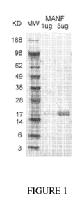
Figure 1 shows a photograph of gel electrophoresis of
purified recombinant human MANF protein, at 1 g and 5 g amounts,
compared to a standard molecular weight (MW) ladder showing the
size of purified MANF protein as approximately 20 kilodaltons
(KD).
Figure 2 shows a photograph of gel electrophoresis of
purified recombinant human CDNF protein, at a 5 g amount, compared
to a standard molecular weight (MW) ladder showing the size of
purified CDNF protein as approximately 18 kilodaltons (KD).
Figures 3A-3C show photographs of sections of the outer
nuclear layers of the retina of control- and MANF-treated S334ter3
rats under light microscopy, as well as quantitative analysis of
the thickness of the outer nuclear layer in each. Figure 3A shows
the control, PBS (Phosphate-Buffered Saline) treated retina.
Figure 3B is representative of a MANF-treated retina. Scale bar,
25 m. Figure 3C is a graphical representation of the quantitative
analysis of the thickness of the outer nuclear layer of the retina
in PBS treated and MANF treated retinas.
Figures 4A-4C show light microscopy photographs of sections
of the outer nuclear layers of the retina in control- and CDNF-
treated 5334ter3 rats, as well as quantitative analysis of the
thickness of the outer nuclear layer in each. Figure 4A is
representative of control PBS (Phosphate-Buffered Saline) treated
retinas. Figure 4B is representative of CDNF-treated retinas.
Scale bar, 25 m. Figure 4C is a graphical representation of the
quantitative analysis of the thickness of the outer nuclear layer
CA 02838818 2013-12-09
WO 2012/170918
PCT/US2012/041701
of the retina in PBS-treated and CDNF-treated retinas.
Figures 5A-5C show fluorescent microscopy photographs of cone
outer segment (COS) of whole mounted retinas stained with Alexa
Fluor 488 conjugated PNA (peanut agglutinin) in control- and MANF-
5 treated 5334ter3 rats (Figures 5A and 5B), as well as quantitative
analysis of each (Figure 5C). Figure 5A is representative of a
control, PBS-treated retina. Figure 5B is representative of a
MANF-treated retina. Scale bar, 50 m. Figure 5C is a graphical
representation of the quantitative analysis of the number of
labeled cells of the retina in PBS-treated and MANF-treated
retinas.
Figures 6A-6C show fluorescent microscopy photographs of COS
of whole mounted retinas stained with Alexa Fluor 488 conjugated
PNA in control- and CDNF-treated 5334ter3 rats (Figures 6A and
6B), as well as quantitative analysis of each (Figure 6C). Figure
6A is representative of a control PBS-treated retina. Figure 6B is
representative of a CDNF-treated retina. Figure 6C is a graphical
representation of the quantitative analysis of the number of
labeled cells of the retina in PBS-treated and CDNF-treated
retinas.
Figures 7A-7C show representative micrographs of Fluoro-Gold
retralabeled ganglion cells of whole mounted retinas of control
rats (Figure 7A), rats after optical nerve crush in addition to
PBS treatment as a control (Figure 7B), and rats after optic nerve
crush in addition to MANF-treatment (Figure 7C). Figure 7A is
representative of control retinal ganglion cells experiencing no
optic nerve crush. Figure 7B is representative of retinal ganglion
cells two weeks after optic nerve crush and PBS treatment. Figure
7C is representative of retinal ganglion cells two weeks after
optic nerve crush and MANF treatment.
Figure 8 shows a photograph of a Western blot probed for MANF
and 13-Actin (loading control). MANF expression levels in extracts
from retinas of wild-type Sprague Dawley rats at PD 1, PD 5, PD 8,
PD 10, PD 12, PD 16, PD 25, PD 30, PD 40 and PD 60 are shown.
Figure 9 shows a fluorescent microscopy photograph of a
cryosection of rat retina probed with anti-MANF antibodies. Scale
CA 02838818 2013-12-09
WO 2012/170918
PCT/US2012/041701
6
bar, 50 m. Layers labeled on the section include the retinal
pigment epithelium (RPE), the outer photoreceptor segment (OS),
the inner photoreceptor segment (IS), the outer nuclear layer
(ONL), the inner nuclear layer (INL), the inner plexiform layer
(IPL), and the ganglion cell layer (GCL). The immunoactivity of
MANF is shown in the RPE cells, Muller cell fibers and cell
bodies, as well as in the GCL.
Figure 10 shows the nucleic acid sequence of SEQ ID NO: 1.
Figure 11 shows the nucleic acid sequence of SEQ ID NO: 2.
Figure 12 shows the amino acid sequence of SEQ ID NO: 3.
Figure 13 shows the amino acid sequence of SEQ ID NO: 4.
Like reference numerals refer to like parts throughout the
several views of the drawings.
Detailed Description of the Preferred Embodiment
The present invention is directed to methods of treatment,
compositions and kits for treating retinal disorders.
Several aspects of the invention are described below, with
reference to examples for illustrative purposes only. It should be
understood that numerous specific details, relationships, and
methods are set forth to provide a full understanding of the
invention. One having ordinary skill in the relevant art, however,
will readily recognize that the invention can be practiced without
one or more of the specific details or practiced with other
methods, protocols, reagents, cell lines and animals. The present
invention is not limited by the illustrated ordering of acts or
events, as some acts may occur in different orders and/or
concurrently with other acts or events. Many of the techniques and
procedures described, or referenced herein, are well understood
and commonly employed using conventional methodology by those
skilled in the art.
Unless otherwise defined, all terms of art, notations and
other scientific terms or terminology used herein are intended to
have the meanings commonly understood by those of skill in the art
to which this invention pertains.
In some cases, terms with
commonly understood meanings are defined herein for clarity and/or
CA 02838818 2013-12-09
WO 2012/170918
PCT/US2012/041701
7
for ready reference, and the inclusion of such definitions herein
should not necessarily be construed to represent a substantial
difference over what is generally understood in the art. It will
be further understood that terms, such as those defined in
commonly used dictionaries, should be interpreted as having a
meaning that is consistent with their meaning in the context of
the relevant art and/or as otherwise defined herein.
The terminology used herein is for the purpose of describing
particular embodiments only and is not intended to be limiting of
the invention. As used herein, the indefinite articles "a", "an"
and "the" should be understood to include plural reference unless
the context clearly indicates otherwise.
The phrase "and/or," as used herein, should be understood to
mean "either or both" of the elements so conjoined, i.e., elements
that are conjunctively present in some cases and disjunctively
present in other cases.
As used herein, "or" should be understood to have the same
meaning as "and/or" as defined above. For example, when separating
a listing of items, "and/or" or "or" shall be interpreted as being
inclusive, i.e., the inclusion of at least one, but also including
more than one, of a number of items, and, optionally, additional
unlisted items. Only terms clearly indicated to the contrary,
such as "only one of" or "exactly one of," or, when used in the
claims, "consisting of," will refer to the inclusion of exactly
one element of a number or list of elements. In general, the term
"or" as used herein shall only be interpreted as indicating
exclusive alternatives (i.e., "one or the other but not both")
when preceded by terms of exclusivity, such as "either," "one of,"
"only one of," or "exactly one of."
As used herein, the terms "including", "includes", "having",
"has", "with", or variants thereof, are intended to be inclusive
similar to the term "comprising."
All genes and gene products (including RNA and proteins), and
their respective names, disclosed herein are intended to
correspond to homologs from any species for which the compositions
and methods disclosed herein are applicable. When a gene or gene
CA 02838818 2013-12-09
WO 2012/170918
PCT/US2012/041701
8
product from a particular species is disclosed, it is understood
that this disclosure is intended to be exemplary only and is not
to be interpreted as a limitation unless the context in which it
appears clearly indicates otherwise. For example, the genes and
gene products disclosed herein, which in some embodiments relate
to mammalian (including human) nucleic acid and/or amino acid
sequences, are intended to encompass homologous and/or orthologous
and/or paralogous genes and gene products from other animals
including, but not limited to, other mammals, fish, reptiles,
amphibians, birds, and other vertebrates.
In the context of the present invention, the terms
"polypeptide" and "protein" are equivalent and mutually
interchangeable. They refer to any amino acid chain, and include
any post-translational modifications thereto (for example
phosphorylation or glycosylation).
As used herein, the term "subject" refers to any animal
(e.g., mammals, birds, reptiles, amphibians, fish), including, but
not limited to, humans, non-human primates, rodents, and the like,
which is to be the recipient of a particular treatment. Typically,
the terms "subject" and "patient" may be used interchangeably
herein in reference to a subject. Furthermore, transgenic animals
(e.g., transgenic rats and mice) are useful in the methods of the
present invention.
As used herein, the term "compound" refers to a neurotrophic
factor, unless clearly indicated otherwise. The neurotrophic
factor can be represented, described, and/or applied for the
purposes of the present invention in recombinant DNA, RNA or
protein form. The neurotrophic factor can also be in an isolated
form, as isolated and purified from an animal, which could also be
a subject. In some embodiments, the neurotrophic factor may be a
polypeptide, polynucleotide, or fragment thereof. The term
"biologic" may also be used interchangeably with "compound" herein
to refer to a neurotrophic factor of the present invention.
As used herein, the term "fragment" refers to a portion of a
compound. For example, when referring to a protein, a fragment is
a plurality of consecutive amino acids comprising less than the
CA 02838818 2013-12-09
WO 2012/170918
PCT/US2012/041701
9
entire length of the polypeptide. For instance, a fragment of a
compound can share up to 99%, 95%, 90%, 85%, 80%, 75%, 70%, 65%,
or 60% of its sequence with the parent compound.
As used herein, the term "administering" refers to providing
a therapeutically effective amount of a chemical or biological
compound or pharmaceutical composition to a subject, using
intravitreal, intraocular, ocular, subretinal, intrathecal,
intravenous, subcutaneous, transcutaneous, intracutaneous,
intracranial, topical and the like administration. The chemical or
biological compound of the present invention can be administered
alone, but may be administered with other compounds, excipients,
fillers, binders, carriers or other vehicles selected based upon
the chosen route of administration and standard pharmaceutical
practice. Administration may be by way of carriers or vehicles,
such as injectable solutions, including sterile aqueous or non-
aqueous solutions, or saline solutions; creams; lotions; capsules;
tablets; granules; pellets; powders; suspensions, emulsions, or
microemulsions; patches; micelles; liposomes; vesicles; implants,
including microimplants; eye drops; other proteins and peptides;
synthetic polymers; microspheres; nanoparticles; and the like.
The chemical or biological compound or pharmaceutical
composition of the present invention may also be included, or
packaged, with other non-toxic compounds, such as pharmaceutically
acceptable carriers, excipients, binders and fillers including,
but not limited to, glucose, lactose, gum acacia, gelatin,
mannitol, xanthan gum, locust bean gum, galactose,
oligosaccharides and/or polysaccharides, starch paste, magnesium
trisilicate, talc, corn starch, starch fragments, keratin,
colloidal silica, potato starch, urea, dextrans, dextrins, and the
like. Specifically, the pharmaceutically acceptable carriers,
excipients, binders, and fillers contemplated for use in the
practice of the present invention are those which render the
compounds of the invention amenable to intravitreal delivery,
intraocular delivery, ocular delivery, subretinal delivery,
intrathecal delivery, intravenous delivery, subcutaneous delivery,
transcutaneous delivery, intracutaneous delivery, intracranial
CA 02838818 2013-12-09
WO 2012/170918
PCT/US2012/041701
delivery, topical delivery and the like. Moreover, the packaging
material may be biologically inert or lack bioactivity, such as
plastic polymers, silicone, etc. and may be processed internally
by the subject without affecting the effectiveness of the
5 neurotrophic factor packaged and/or delivered therewith.
It is also contemplated that the compounds of the present
invention can be administered by way of an implantation vehicle,
such as Encapsulated Cell Technology (ECT) or other similar or
future-derived micro-implantation technologies. ECT is described
10 in Tao, W. et al., 2006, Tao, W. and Wen, R., 2007 and Sieving et
al., 2006, which are incorporated herein by reference. In some
embodiments, the ECT vehicle may release the compounds of the
present invention at the rate of about 250 ng to about 800 ng per
1x10⁶ cells per day. The implanted vehicle may also be any other
similar sustained-release vehicle, or the like, that is later
developed.
The term "effective amount," as applied to the compound(s),
biologics and pharmaceutical compositions described herein, means
the quantity necessary to render the desired therapeutic result.
For example, an effective amount is a level effective to treat,
cure, or alleviate the symptoms of a disorder for which the
therapeutic compound, biologic or composition is being
administered. Amounts effective for the particular therapeutic
goal sought will depend upon a variety of factors including the
disorder being treated and its severity and/or stage of
development/progression; the bioavailability, and activity of the
specific compound, biologic or pharmaceutical composition used;
the route or method of administration and introduction site on the
subject; the rate of clearance of the specific compound or
biologic and other pharmacokinetic properties; the duration of
treatment; inoculation regimen; drugs used in combination or
coincident with the specific compound, biologic or composition;
the age, body weight, sex, diet, physiology and general health of
the subject being treated; and like factors well known to one of
skill in the relevant scientific art. Some variation in dosage
will necessarily occur depending upon the condition of the subject
CA 02838818 2013-12-09
WO 2012/170918
PCT/US2012/041701
11
being treated, and the physician or other individual administering
treatment will, in any event, determine the appropriate dose for
an individual patient.
As used herein, "disorder" refers to a disorder, disease or
condition, or other departure from healthy or normal biological
activity, and the terms can be used interchangeably. The terms
would refer to any condition that impairs normal function. The
condition may be caused by sporadic or heritable genetic
abnormalities. The condition may also be caused by non-genetic
abnormalities. The condition may also be caused by injuries to a
subject from environmental factors, such as, but not limited to,
cutting, crushing, burning, piercing, stretching, shearing,
injecting, or otherwise modifying a subject's cell(s), tissue(s),
organ(s), system(s), or the like.
As used herein, "treatment" or "treating" refers to arresting
or inhibiting, or attempting to arrest or inhibit, the development
or progression of a disorder and/or causing, or attempting to
cause, the reduction, suppression, regression, or remission of a
disorder and/or a symptom thereof. As would be understood by those
skilled in the art, various clinical and scientific methodologies
and assays may be used to assess the development or progression of
a disorder, and similarly, various clinical and scientific
methodologies and assays may be used to assess the reduction,
regression, or remission of a disorder or its symptoms.
Additionally, treatment can be applied to a subject or to a cell
culture.
In accordance with at least one embodiment of the present
invention, a method for treating a retinal disorder in a subject
in need thereof comprises administering an effective amount of a
compound as described herein to the subject. In one embodiment,
this compound is a neurotrophic factor.
The term "neurotrophic factor" refers to deoxyribonucleic
acids (DNA), and ribonucleic acids (RNA) and proteins derived
therefrom, in addition to fragments thereof, that are responsible
for the growth and survival of nerve cells during development and
for the maintenance of adult nerve cells. In some embodiments of
CA 02838818 2013-12-09
WO 2012/170918
PCT/US2012/041701
12
the present invention, the neurotrophic factor is Mesencephalic
astrocyte-derived neurotrophic factor (MANF). In additional
embodiments, the neurotrophic factor is conserved dopamine
neurotrophic factor (CDNF). Also, the neurotrophic factor
administered may be a combination of MANF and CDNF. As would be
understood by those skilled in the art, MANF and CDNF may have
homologs, orthologs and/or paralogs that would additionally be
contemplated for use in the present invention.
In one embodiment of the present invention, the neurotrophic
factor is a recombinant polypeptide. The recombinant polypeptide
may be the recombinant MANF or CDNF protein. It is also
contemplated in the present invention that recombinant MANF and/or
CDNF polypeptide fragments can be used in the methods and kits
described herein. It is further contemplated that recombinant MANF
and/or CDNF full length DNA, cDNA or mRNA (or fragments thereof)
may be utilized in the methods and kits described herein. In some
embodiments, the DNA, cDNA or mRNA (or fragments thereof) may be
comprised in a plasmid, vector, or the like. For example,
polynucleotides, and fragments thereof, may be utilized by way of
gene therapy techniques or encapsulated cell technology (ECT).
Additionally, the present invention may utilize neurotrophic
factors having the sequences of SEQ ID NOS: 1, 2, 3, or 4 (or
fragments, recombinants, chimerics, or combinations thereof).
In the method of the present invention, the neurotrophic
factor is administered with a pharmaceutically acceptable carrier
or vehicle. For instance, the pharmaceutically acceptable carrier
or vehicle can be a saline solution or any other vehicle
contemplated herein.
In particular, in one embodiment, the neurotrophic factor is
administered by way of injection. In some embodiments, the
injection site is an eye of the subject and can be intraocular,
intravitreal, subretinal and the like administration. In other
embodiments, the neurotrophic factor is administered in an area
adjacent to the eye, and may be through injection or other methods
of delivery as described herein.
The neurotrophic factor may also be administered by way of
CA 02838818 2013-12-09
WO 2012/170918
PCT/US2012/041701
13
implantation of a vehicle into an eye of the subject to be
treated. The vehicle may be a microimplantation device, such as
Encapsulated Cell Technology (ECT) or other similar or future-
derived micro-implantation technologies.
The retinal disorder treated by the method of the present
invention may be the result of an injury to a tissue or a cell of
the central nervous system. The retinal disorder treated can also
be a neurodegenerative disorder (e.g., retinitis pigmentosa). The
tissue or cell that is injured or afflicted with a
neurodegenerative disorder can be a ganglion cell, such as a
retinal ganglion cell, or a photoreceptor cell. In some
embodiments, the retinal disorder treated involves ganglion cell
degeneration. Such ganglion cell degeneration may be induced by
glaucoma.
The neurodegenerative disorders contemplated for the
treatment as described herein can be genetic or sporadic (i.e.,
happening as an isolated, non-heritable event) in nature. As would
be understood by those of skill in the art, neurodegenerative
disorders also embrace conditions other than retinal
neurodegenerative disease, and the methods, compositions and kits
of the present invention are contemplated to be applicable to
other such disorders. Such disorders include Alzheimer's disease,
Huntington's disease, Parkinson's disease, amyotrophic lateral
sclerosis, glaucoma, age-related hearing loss, progressive
supranuclear palsy, mild cognitive impairment, dementia,
spinocerebellar ataxias, and the like.
In at least one embodiment, the neurotrophic factor is
administered at the site of injury or affliction with the
neurodegenerative disorder or in an area adjacent to the site of
injury or affliction. The neurotrophic factor may also be
administered by way of a vehicle that releases the factor in a
controlled (i.e., time and/or dose dependant) manner, for example,
as in ECT, discussed previously herein.
In accordance with another embodiment of the present
invention, a method for promoting neuroprotection in a neuronal
cell comprises contacting the neuronal cell with a neurotrophic
CA 02838818 2013-12-09
WO 2012/170918
PCT/US2012/041701
14
factor, such as MANF, CDNF or combinations thereof. As used
herein, the term "neuroprotection" refers to preventing, halting,
inhibiting, or slowing nerve damage, neuron deterioration, and/or
death of neurons. Neuroprotection may be elicited following damage
or deterioration caused by aging, genetic factors, environmental
changes, physical stress or injury, endogenous or exogenous
biological or chemical factors (e.g., neurotropins, vitamins,
alcohol, pharmaceutical agents, ischemia and the like), stroke, or
the like.
As used herein, the term "contacting" refers to actions
directed to creation of a spatial relationship between the cell(s)
and the neurotrophic factor(s) (or vehicle containing the
neurotrophic factor(s)), provided for a predetermined and
specified time and under conditions appropriate to render a
desired biological response in the contacted cell(s), such as
neuroprotection. The spatial relationship between the cell(s) and
the neurotrophic factor(s) can include direct contact, whereby the
factor elicits a response on the contacted cell's surface directly
or enters the cell for further action, or indirect contact,
whereby the factor elicits a response on the cell through
extracellular signaling (e.g., following activation or
modification of another substance which interacts with the
contacted cell). As applied herein, a biological response includes
a neuroprotective response or any other response by the cell(s)
that causes an arrest, inhibition, reduction, or regression of a
disorder of the cell(s).
In particular embodiments of the invention, contacting
neuronal cells by a neurotrophic factor takes place in vitro. "In
vitro" can include in cell or tissue cultures, or test tube
cultures. In other embodiments, the contacting of neuronal cells
takes place in vivo. "In vivo" can include animal models (e.g.,
transgenic animals such as mice or rats) or living subjects as
defined herein, including humans. In yet other embodiments, the
contacting of neuronal cells takes place ex vivo. "Ex vivo" can
include intact tissues, organs or systems, or portions thereof,
derived from a subject that have been isolated or extracted from
CA 02838818 2013-12-09
WO 2012/170918
PCT/US2012/041701
their source. As used herein, the term "isolated" means that the
item described is segregated or separated (physically or
chemically). Something that is isolated may still be within a
subject or exist outside a subject. As used herein, the term
5 "extracted" means that the item described is removed from the
subject and exists outside the subject.
In some embodiments of the invention, the neuronal cell types
amenable to treatment by the methods of the invention comprise
ganglion cells or photoreceptor cells. In one particular
10 embodiment, the neuronal cell type amenable to treatment by the
methods of the invention comprises retinal ganglion cells.
The neurotrophic factors of the present invention may be
recombinant or isolated neurotrophic factors and may also be
either a recombinant or isolated human neurotrophic factor. As
15 used herein with regard to genes, or fragments thereof, or gene
products, or fragments thereof, the term "isolated" is defined as
being removed from cells of an animal and/or purified for use in
the methods described. As used herein, the term "gene" refers to a
polynucleotide derived from a chromosome that codes for RNA and
proteins. A gene, as used herein, may or may not include all
introns, exons, promoter regions, non-coding regions, and the
like, that are associated with the specific gene.
The present invention is also directed to a pharmaceutical
composition or medicament comprising a neurotrophic factor, such
as MANF, CDNF or combinations thereof. The pharmaceutical
composition can also include other pharmaceutically acceptable
compounds, excipients, additives, fillers, binders, adjuvants, or
carriers or vehicles selected based upon the chosen route of
administration and standard pharmaceutical practice. As such, the
neurotrophic factor(s) may be used in the manufacture or
preparation of medicaments and pharmaceutical compositions. Also,
the medicaments and pharmaceutical compositions comprising the
neurotrophic factors described may be used for the treatment of
disorders as described herein.
The present invention is also directed to a kit of parts
comprising neurotrophic factor(s) and other reagents needed to
CA 02838818 2013-12-09
WO 2012/170918
PCT/US2012/041701
16
perform the method(s) of the present invention. The kit of parts
can also include instructions for use. The neurotrophic factor(s)
and reagents can be included in one or more compositions, and each
neurotrophic factor and reagent can be in a composition in
combination with a suitable vehicle, or can be present
independently. The kit of parts may include MANF, CDNF, or
combinations thereof, as purified proteins (recombinant or
isolated from an animal) or as purified polynucleotides
(recombinant or isolated from an animal).
In other embodiments, the kit of parts includes labeled
biomarkers specific to particular neural cell types, such as
photoreceptor biomarkers or retinal ganglion cell biomarkers,
reference standards, and additional components that would be
identifiable by those skilled in the art upon reading the present
disclosure.
Without further elaboration, it is believed that one skilled
in the art can, using the preceding description, utilize the
present invention to its fullest extent. The following examples
are offered by way of illustration, not by way of limitation.
While specific examples have been provided, the above description
is illustrative and not restrictive. Anyone or more of the
features of the previously described embodiments can be combined
in any manner with one or more features of any other embodiments
in the present invention. Furthermore, many variations of the
invention will become apparent to those skilled in the art upon
review of the specification.
All publications and patent documents cited in this
application are incorporated by reference in pertinent part for
all purposes to the same extent as if each individual publication
or patent document were so individually denoted. By citation of
various references in this document, Applicant does not admit any
particular reference is "prior art" to their invention.
Examples
The methods and compositions herein described and the related
kits are further illustrated in the following examples, which are
provided by way of illustration and are not intended to be
CA 02838818 2013-12-09
WO 2012/170918
PCT/US2012/041701
17
limiting. It will be appreciated that variations in proportions
and alternatives in elements of the components shown will be
apparent to those skilled in the art and are within the scope of
embodiments of the present invention. Theoretical aspects are
presented with the understanding that Applicant does not seek to
be bound by the theory presented.
The following material and methods were used for all the
methods and compositions exemplified herein.
Cloning of recombinant human MANF and CDNF proteins: The open
reading frames (ORF) of MANF (SEQ ID NO: 1) and CDNF (SEQ ID NO:
2) were each cloned by polymerase chain reaction (PCR) from human
brain cDNAs, and resulting cloned sequences were confirmed. Each
ORF was subcloned into the expression vector pQE30 (Qiagen,
Valencia, CA), containing a 6xHis-tag coding sequence to the N-
terminus in frame. Next, the expression vectors containing each of
MANF and CDNF sequences were expressed in E. coil (XL-blue,
Stratagene, La Jolla, CA), and the corresponding expressed
proteins were purified by immobilized-metal affinity
chromatography on Ni-NTA Agarose columns (Qiagen) under native
conditions. The eluted protein was buffer-exchanged to phosphate-
buffered saline (PBS) and stored at -80 C in small aliquots until
use. The appropriate human MANF protein sequence is represented by
SEQ ID NO: 3, and the appropriate human CDNF protein sequence is
represented by SEQ ID NO: 4.
Visualization of purified MANF and CDNF proteins: 1 g and/or
5 g of purified protein were electrophoresed on a 4-12% NuPAEG
gel and visualized with Coomassie blue to confirm purification and
proper molecular weights. Molecular weight markers (MW) were
electrophoresed in a lane next to the 1 g and/or 5 g samples.
Transgenic animals: Transgenic rats carrying the murine
rhodopsin mutation 5334ter, known as 5334ter-3 rats, were
generated and utilized as previously described (Liu et al.,
(1999)).
Photoreceptor protection assay: Single intravitreal
injections of MANF and PBS (control) were given to 5334ter3 rats.
CA 02838818 2013-12-09
WO 2012/170918
PCT/US2012/041701
18
Specifically, 6 g MANF was injected into one eye of a rat at
postnatal day (PD) 9, and 3 L PBS was contemporaneously injected
into the remaining eye of the same rat as a control. Injections
were performed through a 33-gauge needle connected to a 101L
microsyringe (Hamilton, Reno, NV). Animals were sacrificed at PD21
and each eye harvested, plastic embedded, and sectioned as
previously described (Liu et al., (1999)). The resulting semi-thin
retinal sections were stained with toluidine blue and examined by
light microscopy. Similar experiments were performed using 6 g
CDNF.
Cone photoreceptor outer segment (COS) protection assay:
Single intravitreal injections of MANF and PBS (control) were
given to 5334ter3 rats. 6 g MANF was injected into one eye of a
rat at postnatal day (PD) 20, and 3 L PBS was contemporaneously
injected into the remaining eye of the same rat as a control.
Injections were performed through a 33-gauge needle connected to a
101L microsyringe (Hamilton, Reno, NV). Animals were sacrificed 10
days after treatment at PD30 and each eye harvested. Whole-mounted
retinas were stained with Alexa Fluor 488 conjugated PNA (peanut
glutinin), which specifically binds to the outer segments of cone
photoreceptors, and examined by fluorescence microscopy. Similar
experiments were performed using 6 g CDNF.
Optic nerve crush assay: Retinal ganglion cells of wild-type
Sprague Dawley rats were labeled by retralabeling with Fluoro-
Gold. One week after labeling, the optic nerves were crushed and
immediately followed with intravitreal injection of 6 g MANF. Two
weeks after the nerve crush and treatment, the rats were
sacrificed and retinas harvested. Whole-mounted retinas were
examined by fluorescence microscopy.
MANF protein expression analysis of retina: Equal amounts of
protein extracts from retinas of wild-type Sprague Dawley rats at
PD 1, PD 5, PD 8, PD 10, PD 12, PD 16, PD 25, PD 30, PD 40 and PD
60 were run on polyacrylamide gels, transferred to membranes and
probed with antibodies for MANF and 13-Actin. Structural protein,
CA 02838818 2013-12-09
WO 2012/170918
PCT/US2012/041701
19
13-Actin, expression was analyzed to ensure consistent loading of
protein extracts at each time-point analyzed.
Example 1: Purification of recombinant human mesencephalic
astrocyte-derived neurotrophic factor (MANF)
To test candidate neurotrophic factors for neuroprotective
properties, recombinant human MANF and CDNF proteins were
generated and purified for further experimentation. Recombinant
human MANF was expressed in E. coil, purified, and visualized as
described above in materials and methods. The results illustrated
in FIGURE 1 show that 1 g and 5 g of purified MANF are
visualized as a single band of 20kDa. Lane 1 depicts the molecular
weight markers (MW); Lane 2 depicts 1 g purified MANF; and Lane 3
depicts 5 g purified MANF. "KD" refers to "kilodaltons."
Example 2: Purification of recombinant human dopamine neurotrophic
factor (CDNF)
CDNF was also utilized as a candidate neurotrophic factor
with neuroprotective properties. Recombinant human CDNF was
expressed in E. coil, purified, and visualized as described above
in materials and methods. The results illustrated in FIGURE 2 show
that 5 g of purified CDNF is visualized as a single band of
18kDa. Lane 1 depicts the molecular weight markers (MW); Lane 2
depicts 5 g purified CDNF. "KD" refers to "kilodaltons."
Example 3: Protection of photoreceptors by MANF in the retina of a
retinal degeneration rodent model
In search of neurotrophic factors that could rescue
photoreceptors in retinal degenerative disorders, including
inherited retinal disorders (e.g., retinitis pigmentosa), age-
related macular degeneration, and glaucoma, recombinant human MANF
protein was tested for photoreceptor protective properties in a
retinal degeneration rodent model. To this end, S334ter3
transgenic rats were utilized because of their characterized
CA 02838818 2013-12-09
WO 2012/170918
PCT/US2012/041701
progressive retinal photoreceptor degeneration (Liu et al.,
(1999)).
The results illustrated in FIGURE 3A show that, in PBS
(control) treated 5334ter3 rats, the outer nuclear layer of the
5 retina had only one row of nuclei (see arrowhead in FIGURE 3A) at
PD 21. The results illustrated in FIGURE 3B show that a single
intravitreal injection of MANF at PD 9 in the remaining eye of the
same animal protected the photoreceptors from degeneration at the
PD 21 time-point, as the outer nuclear layer contained three to
10 four rows of nuclei (see between two arrowheads in FIGURE 3B).
Quantitative analysis of the thickness of the outer nuclear
layer of the superior retina, as shown in FIGURE 3C, indicates
that the outer nuclear layer of MANF treated retinas
(17.47 3.96 M, n=5) was significantly thicker than the outer
15 nuclear layer of control PBS treated retinas
(7.07 1.12 M, n=5)(mean SD). P<0.001 (student t-test).
Example 4: Protection of photoreceptors by CDNF in the retina of a
retinal degeneration rodent model
In search of other neurotrophic factors that could rescue
photoreceptors in retinal degenerative disorders, recombinant
human CDNF protein was tested for photoreceptor protective
properties in the same retinal degeneration rodent model used for
the MANF studies previously described herein.
The results illustrated in FIGURE 4A show that, in PBS
(control) treated 5334ter3 rats, the outer nuclear layer of the
retina had only one row of nuclei (see arrowhead in FIGURE 4A) at
PD 21. The results illustrated in FIGURE 4B show that a single
intravitreal injection of CDNF at PD 9 in the remaining eye of the
same animal protected the photoreceptors from degeneration at the
PD 21 time-point, as the outer nuclear layer contained three to
four rows of nuclei (see between two arrowheads in FIGURE 4B).
Quantitative analysis of the thickness of the outer nuclear
layer of the superior retina, as shown in FIGURE 4C, indicates
that the outer nuclear layer of CDNF-treated retinas
CA 02838818 2013-12-09
WO 2012/170918
PCT/US2012/041701
21
(16.61 2.87 M, n=6) was significantly thicker than the outer
nuclear layer in control PBS-treated
retinas
(7.4 3.43 M, n=6)(meaniSD). P<0.001 (student t-test).
Example 5: Protection of the outer segment of cone photoreceptors
by MANF in the retina of a retinal degeneration rodent model
As another test of photoreceptor protective abilities of
neurotrophic factors, the cone outer segment was analyzed in the
same retinal degeneration rodent model previously described herein
following recombinant human MANF protein exposure.
The results illustrated in FIGURE 5B show that a single
intravitreal injection of MANF at PD 20 into the eye of the
retinal degeneration rodent model protected the cone outer segment
from degeneration, as measured at the PD 30 time-point, versus the
remaining eye of the same animal being injected with PBS (control)
(FIGURE 5A).
The results shown in FIGURE 5C are a quantitative analysis of
the number of Alexa Fluor 488 conjugated PNA-positively stained
cells, indicative of the presence of cone photoreceptors. The
graph indicates that MANF-treated retinas contained a
significantly larger number of cone photoreceptors (569.5 46.5 M)
versus the control PBS-treated retinas (398.7 25.4 M) (C, mean SD).
P<0.001 (student t-test).
Example 6: Protection of the outer segment of cone photoreceptors
by CDNF in the retina of a retinal degeneration rodent model
The same experimental procedure in Example 5 was utilized to
further test the photoreceptor protective abilities of recombinant
human CDNF protein.
The results illustrated in FIGURE 6B show that a single
intravitreal injection of CDNF at PD 20 into the eye of the
retinal degeneration rodent model protected the cone outer segment
from degeneration, as measured at the PD 30 time-point, versus the
CA 02838818 2013-12-09
WO 2012/170918
PCT/US2012/041701
22
remaining eye of the same animal being injected with PBS (control)
(FIGURE 6A).
The results shown in FIGURE 6C are a quantitative analysis of
the number of Alexa Fluor 488 conjugated PNA-positively stained
cells, indicative of the presence of cone photoreceptors. The
graph indicates that CDNF-treated retinas contained a
significantly larger number of cone photoreceptors (561.5 81.3 M)
versus the control PBS-treated retinas (412.75 40.9 M)
(C,
mean SD). P=0.012 (student t-test).
Example 7: Protection of retinal ganglion cells by CDNF after
optic nerve crush in rats
As an additional test for the neuroprotective capabilities of
the neurotrophic factor MANF, retinal ganglion cells were analyzed
following optic nerve crush and MANF exposure in rats.
The results illustrated in FIGURE 7A-C show the ability of
MANF to protect retinal ganglion cells following an optic nerve
crush. As shown in FIGURE 7A, retralabeled ganglion cells are
distributed throughout the representative micrograph in control
mice. As shown in FIGURE 7B, retralabeled ganglion cells are
mostly degenerated two weeks after optic nerve crush in PBS-
treated retinas (control). On the contrary, MANF treatment, as
shown in FIGURE 7C, is able to rescue many of the retinal ganglion
cells two weeks following optic nerve crush and treatment.
Example 8: MANF protein expression in the retina is higher during
development of photoreceptors in rats
Protein extracts from retinas of wild-type Sprague Dawley
rats at PD 1, PD 5, PD 8, PD 10, PD 12, PD 16, PD 25, PD 30, PD 40
and PD 60 were analyzed by Western blot analysis for MANF
expression levels. As shown in FIGURE 8, high levels of MANF
expression were detected during postnatal development (from PD 1
to PD 16). As the retinas mature past PD 16, the expression
decreases (see the continued expression level decrease from PD 25
to PD 60 in FIGURE 8).
Example 9: MANF immunoactivity on the retina
CA 02838818 2013-12-09
WO 2012/170918
PCT/US2012/041701
23
As shown in FIGURE 9, cryosections probed with anti-MANF
antibodies demonstrated that immunoactivity of MANF was located in
the retinal pigment epithelium (RPE) cells, Muller cell fibers and
cell bodies, as well as in the retinal ganglion cell layer.
Together, the results presented in the Examples reveal that
MANF and CDNF have neuroprotective properties for photoreceptors,
and at least MANF additionally has neuroprotective properties in
retinal ganglion cells. Additionally, at least MANF has a high
expression level during development of photoreceptors, and its
level decreases as photoreceptors mature. These results suggest
MANF and CDNF are therapeutic agents for retinal degenerative
disorders.
It is to be appreciated that the Detailed Description
section, and not the Abstract section, is intended to be used to
interpret the claims. The Abstract section may set forth one or
more but not all exemplary embodiments of the present invention as
contemplated by the inventor(s), and thus, are not intended to
limit the present invention and the appended claims in any way.
The foregoing description of the specific embodiments should
fully reveal the general nature of the invention so that others
can, by applying knowledge within the skill of the art, readily
modify and/or adapt for various applications such specific
embodiments, without undue experimentation, without departing from
the general concept of the present invention. Since many
modifications, variations and changes in detail can be made to the
described preferred embodiment of the invention, it is intended
that all matters in the foregoing description and shown in the
accompanying drawings be interpreted as illustrative and not in a
limiting sense.
Thus, the scope of the invention should be
determined by the appended claims and their legal equivalents.
Moreover, the breadth and scope of the present invention should
not be limited by any of the above-described exemplary
embodiments, but should similarly be defined only in accordance
with the following claims and their equivalents.
REFERENCES
CA 02838818 2013-12-09
WO 2012/170918
PCT/US2012/041701
24
Airavaara, M., Shen, H., Kuo, C., Peranen, J., Saarma, M.,
Hoffer, B. and Wang, Y. (2009). Mesencephalic astrocyte-derived
neurotrophic factor reduces ischemic brain injury and promotes
behavioral recovery in rats. Journal of Comparative Neurology
515:116-124.
Lindholm, P., Voutilainen, M.H., Lauren, J., Peranen, J.,
Leppanen, V., Andressoo, J., Lindahl, M., Janhunen, S., Kalkkinen,
N., Timmusk, T., Tuominen, R.K. and Saarma, M. (2007). Novel
neurotrophic factor CDNF protects and rescues midbrain dopamine
neurons in vivo. Nature 448:73-78.
Liu, C., Li, Y., Peng, M., Laties, A.M. and Wen, R. (1999).
Activation of caspase-3 in the retina of transgenic rats with the
rhodopsin mutation S334ter during photoreceptor degeneration.
Journal of Neuroscience 19:4778-4785.
Parkash, V., Lindholm, P., Peranen, J., Kalkkinen, N., Oksanen,
E., Saarma, M., Leppanen, V. and Goldman, A. (2009). The structure
of the conserved neurotrophic factors MANF and CDNF explains why
they are bifunctional. Protein Engineering, Design & Selection
22:233-241.
Parkash, V. (2009). Neurotrophic factors and their receptors.
Dissertation. University of Helsinki. Helsinki University Press.
Petrova, P.S., Raibekas, A., Pevsner, J., Vigo, N., Anafi, M.,
Moore, M.K., Peaire, A.E., Shridhar, V., Smith, D.I., Kelly, J.,
Durocher, Y. and Commissiong, J.W. (2003). A new mesencephalic,
astrocyte-derived neurotrophic factor with selectivity for
dopaminergic neurons. Journal of Molecular Neuroscience 20:173-
187.
Sieving, P.A., Caruso, R.C., Tao, W. Coleman, H.R., Thompson,
D.J.S., Fullmer, K.R., and Bush, R.A. (2006). Ciliary neurotrophic
factor (CNTF) for human retinal degeneration: Phase I trial of
CNTF delivered by encapsulated cell intraocular implants.
Proceedings of the National Academy of Science 103: 3896-3901.
Tadimalla, A., Belmont, P.J., Thuerauf, D.J., Glassy, M.S.,
Martindale, J.J., Gude, N., Sussman, M.A. and Glembotski, C.C.
(2008). Mesencephalic astrocyte-derived neurotrophic factor is an
CA 02838818 2013-12-09
WO 2012/170918
PCT/US2012/041701
ischemia-inducible secreted endoplasmic reticulum stress response
protein in the heart. Circulation Research 103:1249-1258.
Tao, W., Wen, R., Aguirre, G.D., Laties, A.M. (2006). Cell-
Based delivery systems: development of encapsulated cell
5 technology for ophthalmic applications. In G.J. Jaffe, P. Ashton
(Eds.), Intraocular drug delivery: principles and clinical
applications (Ch. 8). Taylor & Francis.
Tao, W. and Wen, R. (2007). Application of Encapsulated Cell
Technology for Retinal Degenerative Diseases. In J. Tombran-Tink &
10 C. J. Barnstable (Eds.), Ophthalmology Research: Retinal
Degenerations: Biology, Diagnostics, and Therapeutics (401-413).
New Jersey: Humana Press, Inc.
Since many modifications, variations and changes in detail
can be made to the described preferred embodiment of the
15 invention, it is intended that all matters in the foregoing
description and shown in the accompanying drawings be interpreted
as illustrative and not in a limiting sense. Thus, the scope of
the invention should be determined by the appended claims and
their legal equivalents.
20 Now that the invention has been described,