Note: Descriptions are shown in the official language in which they were submitted.
CA 02839102 2013-12-10
WO 2013/003729
PCT/US2012/044946
COMPOSITIONS, METHODS AND KITS FOR TREATING LEUKEMIA
CROSS-REFERENCE TO RELATED APPLICATIONS
[0001] This
application claims the benefit of U.S. Provisional Application Serial No.
61/502,677, filed June 29, 2011, U.S. Provisional Application Serial No.
61/535,149, filed
September 15, 2011, and U.S. Provisional Application Serial No. 61/635,458,
filed
April 19, 2012, all of which are hereby incorporated by reference in their
entireties, for all
purposes, herein.
FIELD OF THE INVENTION
[0002] The
invention relates generally to the fields of molecular genetics, molecular
biology, and oncology.
BACKGROUND
[0003] Leukemia
is a highly prevalent disease that signifies uncontrolled production of
white blood cells. Currently, there is no cure for leukemia. Current therapies
of leukemia
include chemotherapy, radiation therapy, stem cell therapy, and biological
therapy. All of
these therapies suffer from many side effects. Use of anti-leukemic drugs only
prolong the
life of the patient by targeting the bulk cancer cells, but not the cancer
stem cells.
SUMMARY
[0004]
Described herein are compositions, methods and kits for treating cancers such
as
leukemia. Targeting cancer stem cells (CSC) is of paramount importance to
successfully
combat the relapse of cancer. It is shown herein that Al2-PGJ3, a novel and
naturally
produced cyclopentenone prostaglandin, CyPG, from the dietary fish-oil omega-3
polyunsaturated fatty acid (n-3 PUFA), eicosapentaenoic acid (EPA; 20:5),
alleviates the
development of leukemia in two well-studied murine models of leukemia.
Intraperitoneal
administration of Al2-PGJ3 to mice infected with Friend erythroleukemia virus
(FV) or those
expressing chronic myelogenous leukemia (CML) oncoprotein BCR-ABL in the
hematopoietic stem cell (HSC) pool completely restored normal hematological
parameters,
splenic histology, and enhanced the survival of such mice. More importantly,
Al2-PGJ3
selectively targeted leukemia stem cells (LSC) for apoptosis in the spleen and
bone marrow.
This treatment completely eradicated LSCs in vivo as demonstrated by the
inability of donor
cells from treated mice to cause leukemia in secondary transplants. This is
the first example
of a compound that eradicates leukemia stem cells and effectively "cures" CML
in a mouse
1
CA 02839102 2013-12-10
WO 2013/003729
PCT/US2012/044946
model and prolongs the life of the leukemic mice indefinitely. Given the
potency of n-
3 PUFA-derived CyPG and the well-known refractoriness of LSC to currently used
clinical
agents, Al2-PGJ3 represents a new chemotherapeutic for leukemia that targets
LSCs.
[0005]
Accordingly, described herein is a composition including a therapeutically
effective amount of a first anti-cancer drug, the first anti-cancer drug being
isolated or
synthesized 412-PGJ3 or a derivative thereof, for inhibiting LSC growth in a
subject having
LSCs (e.g., a subject suffering from leukemia) and a pharmaceutically
acceptable carrier. The
composition can further include a second anti-cancer drug (e.g., imatinib
(Gleevec Novartis,
East Hanover, NJ)).
[0006] Also
described herein is a composition including a therapeutically effective
amount of a first anti-cancer drug that is an isolated or synthesized
prostaglandin D receptor
(DP) agonist for inhibiting LSC growth in a subject having LSCs and a
pharmaceutically
acceptable carrier. The composition can further include a second anti-cancer
drug (e.g.,
imatinib). The DP agonist can be, for example, one or more of :Al2-PGJ3,
ZK118182, and
PGD2ME.
[0007] Further
described herein is a method of treating leukemia in a subject. The method
includes administering to the subject having leukemia a composition including
a
therapeutically effective amount of a first anti-cancer drug that is one or
more of: isolated or
synthesized 412-PGJ3 or a derivative thereof, a derivative of prostaglandin
D3, and an isolated
or synthesized DP agonist, for inducing death of LSCs in the subject. The LSCs
can be, for
example, chronic myeloid leukemia stem cells or acute myeloid leukemia cells.
In some
embodiments, the subject is resistant to an anti-cancer drug (e.g., imatinib).
In the method,
the composition can further include a therapeutically effective amount of a
second anti-cancer
drug (e.g., imatinib, standard chemotherapy agents such as cytarabine or
doxorubicin, etc.).
[0008] Yet
further described herein is a method of treating leukemia in a subject (e.g.
human). The method includes administering to the subject having leukemia a
composition
including a therapeutically effective amount of a DP agonist for inducing
death of LSCs in
the subject. The LSCs can be, for example, chronic myeloid leukemia stem
cells. In some
embodiments, the subject is resistant to imatinib. In the method, the
composition can further
include a therapeutically effective amount of an anti-cancer drug (e.g.,
imatinib, standard
chemotherapy agents such as cytarabine or doxorubicin, etc.).
[0009]
Additionally described herein is a kit for treating leukemia in a subject
(e.g.,
human). The kit includes a composition including a therapeutically effective
amount of a
2
CA 02839102 2013-12-10
WO 2013/003729
PCT/US2012/044946
first anti-cancer drug that is one of: an isolated or synthesized DP agonist,
an isolated or
synthesized 412-PGJ3, and a derivative of 412-PGJ3, for inducing death of LSCs
in the subject;
instructions for use, and packaging. The kit can further include a second anti-
cancer drug
(e.g., imatinib, standard chemotherapy agents such as cytarabine or
doxorubicin, etc.).
[0010] Unless
otherwise defined, all technical terms used herein have the same meaning
as commonly understood by one of ordinary skill in the art to which this
invention belongs.
[0011] As used
herein, "protein" and "polypeptide" are used synonymously to mean any
peptide-linked chain of amino acids, regardless of length or post-
translational modification,
e.g., glycosylation or phosphorylation.
[0012] By the
term "gene" is meant a nucleic acid molecule that codes for a particular
protein, or in certain cases, a functional or structural RNA molecule.
[0013] As used
herein, a "nucleic acid" or a "nucleic acid molecule" means a chain of
two or more nucleotides such as RNA (ribonucleic acid) and DNA
(deoxyribonucleic acid).
[0014] The
terms "patient," "subject" and "individual" are used interchangeably herein,
and mean a mammalian (e.g., human, rodent, non-human primates, canine, bovine,
ovine,
equine, feline, etc.) subject to be treated and/or to obtain a biological
sample from.
[0015] As used
herein, "bind," "binds," or "interacts with" means that one molecule
recognizes and adheres to a particular second molecule in a sample or
organism, but does not
substantially recognize or adhere to other structurally unrelated molecules in
the sample.
Generally, a first molecule that "specifically binds" a second molecule has a
binding affinity
greater than about 10-8 to 10-12 moles/liter for that second molecule and
involves precise
"hand-in-a-glove" docking interactions that can be covalent and noncovalent
(hydrogen
bonding, hydrophobic, ionic, and van der waals).
[0016] The term
"labeled," with regard to a probe or antibody, is intended to encompass
direct labeling of the probe or antibody by coupling (i.e., physically
linking) a detectable
substance to the probe or antibody.
[0017] When
referring to a nucleic acid molecule or polypeptide, the term "native" refers
to a naturally-occurring (e.g., a wild type or WT) nucleic acid or
polypeptide.
[0018] As used
herein, the term "regulating", "regulation", "modulating" or "modulation"
refers to the ability of an agent to either inhibit or enhance or maintain
activity and/or
function of a molecule (e.g., a receptor). For example, an inhibitor of a DP
would down-
regulate, decrease, reduce, suppress, or inactivate at least partially the
activity and/or function
of the DP. Up-regulation refers to a relative increase in function and/or
activity.
3
CA 02839102 2013-12-10
WO 2013/003729
PCT/US2012/044946
[0019] By the
term "A'2-PGJ3" is meant 412-prostaglandin .13, an omega-3 fatty acid-
derived metabolite.
[0020] By the
phrase "DP agonist" is meant any agent (e.g., drug, compound, hormone,
etc.) that forms a complex with or binds to a DP site on a cell, thereby
triggering an active
response from the cell. DP agonists can be naturally occurring or synthetic,
or a combination
thereof.
[0021] By the
phrase "leukemia stem cells" is meant leukemia initiating cells that are
functionally defined to possess the property to generate more leukemia stem
cells (self
renewal) and non-stem cell leukemia cells. Additionally, these cells are
characterized by the
expression of certain cell surface markers, which include but are not limited
to CD34,
CD123, and CD117.
[0022] The
phrases "isolated" or biologically pure" refer to material, which is
substantially or essentially free from components which normally accompany it
as found in
its native state.
[0023] The term
"antibody" is meant to include polyclonal antibodies, monoclonal
antibodies (mAbs), chimeric antibodies, humanized antibodies, anti-idiotypic
(anti-Id)
antibodies to antibodies that can be labeled in soluble or bound form, as well
as fragments,
regions or derivatives thereof, provided by any known technique, such as, but
not limited to,
enzymatic cleavage, peptide synthesis or recombinant techniques.
[0024] As used
herein, the terms "diagnostic," "diagnose" and "diagnosed" mean
identifying the presence or nature of a pathologic condition (e.g., leukemia).
[0025] The term
"sample" is used herein in its broadest sense. A sample including
polynucleotides, polypeptides, peptides, antibodies and the like may include a
bodily fluid, a
soluble fraction of a cell preparation or media in which cells were grown,
genomic DNA,
RNA or cDNA, a cell, a tissue, skin, hair and the like. Examples of samples
include saliva,
serum, blood, urine and plasma.
[0026] As used
herein, the term "treatment" is defined as the application or
administration of a therapeutic agent to a patient, or application or
administration of the
therapeutic agent to an isolated tissue or cell line from a patient, who has a
disease, a
symptom of disease or a predisposition toward a disease, with the purpose to
cure, heal,
alleviate, relieve, alter, remedy, ameliorate, improve or affect the disease,
the symptoms of
disease, or the predisposition toward disease. Treatment can include, for
example,
4
CA 02839102 2013-12-10
WO 2013/003729
PCT/US2012/044946
ameliorating, preventing or eliminating splenomegaly, reducing the number of
LSCs in a
subject, eliminating LSCs in a subject, etc.
[0027] As used
herein, the term "safe and effective amount" refers to the quantity of a
component, which is sufficient to yield a desired therapeutic response without
undue adverse
side effects (such as toxicity, irritation, or allergic response) commensurate
with a reasonable
benefit/risk ratio when used in the manner of this invention. By
"therapeutically effective
amount" is meant an amount of a composition of the present invention effective
to yield the
desired therapeutic response. For example, an amount effective to delay the
growth of or to
cause a cancer (e.g., CML) to shrink or prevent metastasis. The specific safe
and effective
amount or therapeutically effective amount will vary with such factors as the
particular
condition being treated, the physical condition of the patient, the type of
mammal or animal
being treated, the duration of the treatment, the nature of concurrent therapy
(if any), and the
specific formulations employed and the structure of the compounds or its
derivatives.
[0028] Although
compositions, kits, and methods similar or equivalent to those described
herein can be used in the practice or testing of the present invention,
suitable compositions,
kits, and methods are described below. All publications, patent applications,
and patents
mentioned herein are incorporated by reference in their entirety. In the case
of conflict, the
present specification, including definitions, will control. The particular
embodiments
discussed below are illustrative only and not intended to be limiting.
BRIEF DESCRIPTION OF THE DRAWINGS
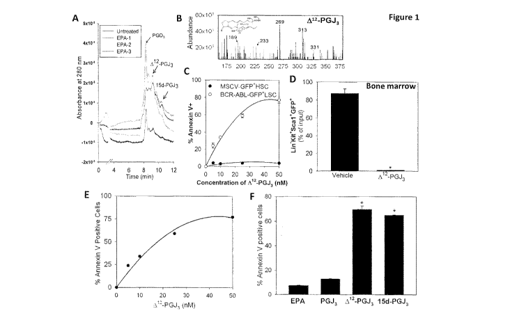
[0029] FIG. 1
shows endogenous production and pro-apoptotic properties of Al2-PGJ3.
(A) Endogenous formation of PGD3, Al2-PGJ3, 15d-PGJ3 in RAW264.7 macrophages,
LC-
UV trace; N = 3 for EPA treated. (B) Representative LC-MS of Al2-PGJ3
containing eluates
with characteristic fragmentation pattern is shown. (C) Dose-response
demonstrating the
effect of Al2-PGJ3 on BCR-ABL LSC compared with normal HSCs (MSCV-GFP FISC).
Cells were treated ex vivo with Al2-PGJ3 for 36 h. Apoptosis was measured by
annexin V
staining. (D) Kit+Sca-1 Lin-BCR-ABL-GFP cells sorted from the bone marrow and
cultured
ex vivo in media containing Al2-PGJ3 (25 nM) or vehicle control for 36 h
followed by flow
cytometric analysis of GFP cells. N= 3; Mean s.e.m. shown. * p<0.005.
Expressed as
percent of input GFP cells. (E) Dose response of LSCs isolated from FV mice
with indicated
concentrations of Al2-PGJ3 at the end of 36 h of incubation. Apoptosis of LSCs
was examined
by Annexin V staining followed by flow cytometry. (F) FV-LSCs were cultured ex
vivo with
CA 02839102 2013-12-10
WO 2013/003729
PCT/US2012/044946
25 nM of each compound for 36 h. N = 3; Mean s.e.m. shown * P<0.0001
(compared to
PGJ3).
[0030] FIG. 2
shows intraperitoneal administration of Al2-PGJ3 eradicates FV-leukemia
in mice. (A) Spleen weight of FV-infected mice treated with various doses of
Al2-PGJ3
(mg/kg body weight). N = 10 per treatment group. Al2-PGJ3 treatment at
indicated dosage
was started at 1 week post infection for a period of 7 days. * P<0.05. Inset:
representative
spleens from each treatment group. UI: Uninfected mice. (B) Analysis of LSCs
(M34+Kit+Sca1+) in the spleens of FV-infected mice treated with Al2-PGJ3 or
vehicle (Veh)
control. (C) CFU-FV colony formation in Al2-PGJ3 and vehicle control treated
mice,
*P<0.001. (D) H&E staining of spleen sections from uninfected (left), FV-
infected-vehicle
treated (middle), and FV-infected- Al2-PGJ3 -treated mice (right) on day 14
post infection.
Small box indicated on each section on the left is magnified on the right
side. Scale bars, 500
pm.
[0031] FIG. 3
shows the effect of Al2-PGJ3 treatment on leukemia induced by
transplanting FV-induced LSCs expanded in-vitro into FV-resistant Stk-/- mice.
(A)
Photograph of spleens from Stk-/- mice seven weeks after transplant with FV-
LSCs followed
by treatment with vehicle, 0.05 mg/kg, or 0.025 mg/kg Al2-PGJ3 for 1 week. (B)
Spleen
weights are shown for the conditions in panel A. N= 5 per group, *P<0.05
compared to
infected vehicle group. (C) WBC counts in LSC-transplanted Stk-/- mice treated
with
indicated amounts of Al2-PGJ3 or vehicle control. N = 5 per group, *P<0.05
compared to
infected vehicle group. (D) M34+Kit+Sca1 cells in Stk-/- mice transplanted
with LSCs.
Spleen cells that were isolated and gated on Kit+, expression of M34 and Scat
is shown. N =
per group.
[0032] FIG. 4
shows that intraperitoneal administration of Al2-PGJ3 eradicates LSCs and
prolongs survival in a murine CML model. (A) Analysis of the effect of Al2-
PGJ3 treatment
on the development of splenomegaly in mice transplanted with BCR-ABL-GFP+LSCs.
Representative photographs of spleens from control and BCR-ABL transplanted
mice treated
with Al2-PGJ3 (0.025 mg/kg) or vehicle control with corresponding spleen
weights. N = 10
per treatment group, *P<0.05. (B) Analysis of WBC counts of BCR-ABL+LSC or
MSCV-
HSC transplanted mice treated with Al2-PGJ3 or vehicle control. *P<0.0001. (C)
Flow
cytometric analysis of Sca-l+Kit+GFP+ cells in the spleen of mice transplanted
with BCR-
ABL+ LSC or MSCV+HSC treated with Al2-PGJ3 or vehicle control. N= 5 per group;
*
p<0.001 (D) Analysis of LSCs (Kit+Sca-l+Lin-GFP+) in the bone marrow of BCR-
ABL+
6
CA 02839102 2013-12-10
WO 2013/003729
PCT/US2012/044946
LSC transplanted and Al2-PGJ3-treated mice after 5 weeks of last dose of Al2-
PGJ3 (0.025
mg/kg). As a control, BCR-ABL+ LSC transplanted mice treated with vehicle for
1 week was
used for comparison. (E) Survival curves of mice transplanted with BCR-ABL
LSCs or
MSCV-GFP FISCs upon treatment with Al2-PGJ3 (0.025 mg/kg) or vehicle. N= 8 per
treatment group. (F). HSC were isolated from the bone marrow of C57BL/6 mice
and plated
in methylcellulose (lx 106 cells/ml/well; Epo, SCF, IL-3, and BMP4) with PBS
or Al2-PGJ3
(25 nM) and cultured for a week. Hematopoietic colonies (colony forming cells
in culture,
CFC) were scored. Data shown is representative of triplicate experiments.
[0033] FIG. 5 demonstrates that secondary transplantation of spleen cells
from Al2-PGJ3-
treated recipients show absence of leukemia. Panels A-C represent secondary
transplantation
of CD45.1+ BCR-ABL mice treated with Al2-PGJ3 or vehicle control transplanted
into
CD45.2 recipient mice. Panels D-E represent FV-LSCs from Al2-PGJ3 or vehicle
control
treated mice were transplanted into secondary BALB/c-Stk-/- recipients. (A).
Spleen
morphology (upper left), spleen weight (lower left) and WBC counts of
secondary
transplant mice receiving donor cells from vehicle treated or Al2-PGJ3 treated
donor cells
(right). (B) Flow cytometry analysis of spleen cells from secondary
transplants. Cells were
gated on GFP and the expression of Kit and Scat are shown. (C). Analysis of
donor CD45.1
expression in spleen cells. (D) Spleen morphology (upper left), spleen weight
(lower left),
and WBC counts of secondary transplant mice receiving donor cells from vehicle
treated or
A'2-PGJ3 treated donor cells (right). (E) Flow cytometry analysis of spleen
cells from
secondary transplants. Cells are gated on M34+ and the expression of Kit and
Scat is shown.
[0034] FIG. 6 shows spontaneous conversion of PGD3 to PGJ3, Al2-PGJ3, and
15d-PGJ3
in-vitro.
[0035] FIG. 7 shows a dose-dependent pro-apoptotic effect of CyPGs on LSCs.
[0036] FIG. 8 is a graph showing that imatinib-resistant BCR-ABL(GFP)+
cells are
targeted by 412-PGJ3. The LSCs were isolated from mice treated with imatinib
(75 mg/kg) for
one week following which the treatment was stopped. The mice were followed for
the
development of leukemia. Mice that developed leukemia were euthanized and
spleens were
used as the source of LSCs.
[0037] FIG. 9 is a graph showing apoptosis of BCR-ABL LSCs by synthetic
agonists of
the DPs.
[0038] FIG. 10 is a graph showing that A12-PGJ3 and related agonists do not
affect normal
human hematopoiesis as measured by the ability of bone marrow cells to form
differentiated
7
CA 02839102 2013-12-10
WO 2013/003729
PCT/US2012/044946
colonies when cultured in vitro. Human unfractionated bone marrow cells (5x10⁵
per well)
were plated in methylcellulose complete media containing IL-3, GM-CSF, G-CSF,
SCF and
Epo supplemented with the indicated concentrations of drugs. Total colonies
were counted
after 12 days.
[0039] FIG. 11
is a graph showing that Al2-PGJ3 does not affect the ability of normal
bone marrow cells to differentiate in to cells of the erythroid lineage (Burst
Forming Units ¨
erythroid, BFU-E).
[0040] FIG. 12
is a pair of graphs showing that DP mediate the Al2-PGJ3-dependent
apoptosis of blast crisis CML cells from a patient (#011711).
[0041] FIG. 13
shows results from an experiment in which DP mediate the Al2-PGJ3-
dependent apoptosis of AML cells from a patient (#100810). Furthermore, Al2-
PGJ3 also
specifically targeted Leukemia stem cells (CD34+CD38-CD123+ cells) for
apoptosis.
[0042] FIG. 14
is a graph showing results from a comparison of Al2-PGJ3 with Imatinib
(Gleevec Novartis, East Hanover, NJ) in the BCR-ABL LSC transplant CML model
in
mice.
[0043] FIG. 15
is a Table listing the effect of Al2-PGJ3 on LSCs from AML and blast-
crisis CML patients.
[0044] FIG. 16
is a pair of graphs showing apoptosis of human primary AML cells by DP
agonists (endogenous and exogenous) and DP antagonists.
8
CA 02839102 2013-12-10
WO 2013/003729
PCT/US2012/044946
DETAILED DESCRIPTION
[0045] AML is
one of the most common types of leukemia in adults. Unfortunately, the
five year relative survival rates for AML are the lowest when compared to
other forms of
leukemia. AML is a stem cell disease where LSCs occupy the apex of the disease
hierarchy.
LSCs can self renew and generate non-stem cell progeny that make up the bulk
of the
leukemia cells. Although chemotherapy agents can effectively target bulk
leukemia cells,
LSCs have active mechanisms to avoid killing by these drugs. As a consequence,
failure to
eliminate LSCs results in relapse of the disease. Because of this property,
specific targeting
of LSCs is essential for successful treatment. Although the need for new anti-
LSC based
therapies is well recognized, the identification of mechanism-based drugs to
target LSCs has
been lacking. Clearly new approaches are needed. Described herein are
compositions,
methods and kits for treating cancer (e.g., leukemia). A metabolite derived
from to-3 fatty
acids, Al2-PGJ3, was discovered which effectively eradicates LSCs in two mouse
models of
chronic leukemia. In the experiments described herein, these findings were
extended to show
that Al2-PGJ3effectively targets AML LSCs by inducing apoptosis in murine
models of AML
and in human AML leukemia samples. In contrast, Al2-PGJ3 has no effect on
normal
hematopoietic stem cells or the differentiation of hematopoietic progenitors.
Al2-PGJ3 acts by
inducing the expression of p53 in LSCs and leukemia cells. High-level
expression of p53 in
LSCs is incompatible with self renewal and leads to apoptosis. These data
suggest that 412-
PGJ3 is a chemotherapeutic agent for treating AML. This is the first example
of a compound
that eradicates leukemia stem cells and effectively "cures" CML in a mouse
model and
prolongs the life of the leukemic mice indefinitely.
Biological Methods
[0046] Methods
involving conventional molecular biology techniques are described
herein. Such techniques are generally known in the art and are described in
detail in
methodology treatises such as Molecular Cloning: A Laboratory Manual, 3rd ed.,
vol. 1-3,
ed. Sambrook et al., Cold Spring Harbor Laboratory Press, Cold Spring Harbor,
N.Y., 2001;
and Current Protocols in Molecular Biology, ed. Ausubel et al., Greene
Publishing and
Wiley-Interscience, New York, 1992 (with periodic updates).
Compositions for Treating Leukemia In A Subject
[0047]
Described herein are compositions for treating leukemia in a subject (e.g., a
human subject). Examples of leukemias that can be treated using the
compositions include
Acute Myelogenous Leukemia (AML), CML, Acute Lymphocytic Leukemia (ALL) and
9
CA 02839102 2013-12-10
WO 2013/003729
PCT/US2012/044946
Chronic Lymphocytic Leukemia (CLL). In one embodiment, a composition includes
a
therapeutically effective amount of 412-prostaglandin J3, or a derivative
thereof (a first anti-
cancer drug), for inhibiting LSC growth in a subject having LSCs, and a
pharmaceutically
acceptable carrier. Inhibiting LSC growth includes inducing death (killing of)
of the cancer
cells, and/or inducing differentiation of the cancer cells (promoting a more
differentiated
phenotype, e.g., causing differentiation of LSCs into terminally
differentiated cells). Any
suitable form of 412-prostaglandin J3 or derivative thereof can be used (e.g.,
synthesized,
isolated). 412-prostaglandin J3 derivatives that may find particular use in
the compositions
and methods described herein are those that induce apoptosis or
differentiation of LSCs (e.g.,
16,16-dimethy1-412-PGJ3). In such
embodiments, when administered to a subject, the
composition induces apoptosis of LSCs. The composition can further include one
or more
additional anti-cancer drugs (e.g., a second anti-cancer drug). Examples of
additional anti-
cancer drugs include imatinib, nilotinib, dasafanib, new generation BCR-ABL
inhibitors, and
standard chemotherapy drugs such as cytarabine or doxorubicin or similar
classes of drugs. In
one embodiment, a combination therapy including imatinib or a new generation
BCR-ABL
inhibitor and 412-PGJ3 may be particularly therapeutic.
[0048] In
another embodiment, a composition includes a therapeutically effective amount
of a DP agonist (a first anti-cancer drug) for inhibiting LSC growth in a
subject having LSCs
and a pharmaceutically acceptable carrier. Examples of DP agonists include a
small
molecule, a protein, a peptide, a polynucleotide, an oligonucleotide, an
organic compound, an
inorganic compound, synthetic compounds or compounds isolated from unicellular
or
multicellular organisms. Specific examples of DP agonists include PGD2ME
(Prostaglandin
D2 methyl ester (9a,15S-dihydroxy-11-oxo-prosta-5Z,13E-dien-l-oic acid, methyl
ester) and
ZK118182 (114-15R-
chloro-2Z-13R-cyclohexy1-3S-hydroxy-1R-propenyll -3S-
hydroxycyclopenty11-2R-butenyll oxyl -acetic acid, isopropyl ester). An
agonist of a DP is any
agent that activates the DP. Any agent that activates DP can be used in
compositions and
methods described herein for inducing death of LSCs and treating leukemia. A
composition
including a DP agonist can further include one or more additional anti-cancer
drugs (e.g, a
second anti-cancer drug). As noted above, examples of additional anti-cancer
drugs include
imatinib, nilotinib, dasatanib, new generation BCR-ABL inhibitors, standard
chemotherapy
drugs such as cytarabine or doxorubicin, etc.
[0049] In the
compositions described herein, 412-prostaglandin J3 can be obtained
commercially or synthesized according to the methods described, for example,
in the
CA 02839102 2013-12-10
WO 2013/003729
PCT/US2012/044946
Examples section below. Similarly, 412-prostaglandin J3 derivatives can be
synthesized as
described by Kimball et al. (Kimball FA, Bundy GL, Robert A, and Weeks JR
(1979),
Synthesis and biological properties of 9-deoxo-16,16-9-methylene-PGE2.
Prostaglandins 17:
657-66).
Effective Doses
[0050] The
compositions described above are preferably administered to a mammal (e.g.,
rodent, human, non-human primates, canine, bovine, ovine, equine, feline,
etc.) in an
effective amount, that is, an amount capable of producing a desirable result
in a treated
subject (e.g., inhibiting growth of LSCs and/or inducing death of LSCs in the
subject).
Toxicity and therapeutic efficacy of the compositions utilized in methods of
the invention can
be determined by standard pharmaceutical procedures. As is well known in the
medical and
veterinary arts, dosage for any one animal depends on many factors, including
the subject's
size, body surface area, body weight, age, the particular composition to be
administered, time
and route of administration, general health, the clinical symptoms of the
cancer and other
drugs being administered concurrently. A composition as described herein is
typically
administered at a dosage that induces death of LSCs (e.g., induces apoptosis
of LSCs), as
assayed by identifying a reduction in hematological parameters (Complete blood
count
(CBC)), or cancer cell growth or proliferation. In the experiments described
herein, the
amount of Al2-PGJ3 used to eradicate LSCs was calculated to be 0.6
micrograms/day/gram
mouse for 7 days. Generally, the dose is in mg/Kg subject/day = ug/g
subject/day. In a
typical embodiment, a dose in the range of about 0.025 to about 0.05 mg/Kg/day
is
administered. Such a dose is typically administered once a day for a few
weeks.
Methods of Treating Cancer
[0051]
Described herein are methods of treating cancer (e.g., leukemia) and/or
disorders
or symptoms thereof. The methods include administering a therapeutically
effective amount
of a pharmaceutical composition including a pharmaceutically acceptable
carrier and an
amount of 412-PGJ3, a derivative thereof, or a DP agonist (a first anti-cancer
drug) sufficient
to treat the disease or disorder or symptom thereof to a subject (e.g., a
mammal such as a
human). In the method, an amount of 412-PGJ3, a derivative thereof, or a DP
agonist
sufficient to induce death of LSCs in the subject is typically administered.
In a typical
embodiment, the LSCs are CML stem cells. In some embodiments, the composition
can be
administered to a subject who is resistant to imatinib or other anti-cancer
drug. In the
methods, the composition can further include a therapeutically effective
amount of one or
11
CA 02839102 2013-12-10
WO 2013/003729
PCT/US2012/044946
more additional anti-cancer drugs (e.g., a second anti-cancer drug such as
imatinib) or
standard chemotherapy.
[0052] The
therapeutic methods of the invention (which include prophylactic treatment)
in general include administration of a therapeutically effective amount of the
compositions
described herein to a subject in need thereof, including a mammal,
particularly a human.
Such treatment will be suitably administered to subjects, particularly humans,
suffering from,
having, susceptible to, or at risk for a disease, disorder, or symptom
thereof. Determination
of those subjects "at risk" can be made by any objective or subjective
determination by a
diagnostic test or opinion of a subject or health care provider (e.g., genetic
test, enzyme or
protein marker, marker (as defined herein), family history, and the like).
[0053] The
administration of a composition including 412-PGJ3, a derivative thereof, or a
DP agonist for the treatment of cancer (e.g., leukemia) may be by any suitable
means that
results in a concentration of the therapeutic that, (e.g., when combined with
other
components), is effective in ameliorating, reducing, or stabilizing a cancer.
The 412-PGJ3, a
derivative thereof, or a DP agonist may be contained in any appropriate amount
in any
suitable carrier substance, and is generally present in an amount of 1-95% by
weight of the
total weight of the composition. The composition may be provided in a dosage
form that is
suitable for local or systemic administration (e.g., parenteral,
subcutaneously, intravenously,
intramuscularly, or intraperitoneally). The pharmaceutical compositions may be
formulated
according to conventional pharmaceutical practice (see, e.g., Remington: The
Science and
Practice of Pharmacy (20th ed.), ed. A. R. Gennaro, Lippincott Williams &
Wilkins, 2000
and Encyclopedia of Pharmaceutical Technology, eds. J. Swarbrick and J. C.
Boylan, 1988-
1999, Marcel Dekker, New York).
[0054]
Compositions as described herein may be administered parenterally by
injection,
infusion or implantation (subcutaneous, intravenous, intramuscular,
intraperitoneal, or the
like) in dosage forms, formulations, or via suitable delivery devices or
implants containing
conventional, non-toxic pharmaceutically acceptable carriers and adjuvants.
The formulation
and preparation of such compositions are well known to those skilled in the
art of
pharmaceutical formulation. Formulations can be found in Remington: The
Science and
Practice of Pharmacy, supra.
[0055]
Compositions for parenteral use may be provided in unit dosage forms (e.g., in
single-dose ampoules), or in vials containing several doses and in which a
suitable
preservative may be added (see below). The composition may be in the form of a
solution, a
suspension, an emulsion, an infusion device, or a delivery device for
implantation, or it may
12
CA 02839102 2013-12-10
WO 2013/003729
PCT/US2012/044946
be presented as a dry powder to be reconstituted with water or another
suitable vehicle before
use. Apart from the active agent that reduces or ameliorates a cancer, the
composition may
include suitable parenterally acceptable carriers and/or excipients. The
active therapeutic
agent(s) may be incorporated into microspheres, microcapsules, nanoparticles,
liposomes, or
the like for controlled release. Furthermore, the composition may include
suspending,
solubilizing, stabilizing, pH-adjusting agents, tonicity adjusting agents,
and/or dispersing
agents.
[0056] As
indicated above, the pharmaceutical compositions described herein may be in a
form suitable for sterile injection. To prepare such a composition, the
suitable active
therapeutic(s) are dissolved or suspended in a parenterally acceptable liquid
vehicle. Among
acceptable vehicles and solvents that may be employed are water, water
adjusted to a suitable
pH by addition of an appropriate amount of hydrochloric acid, sodium hydroxide
or a suitable
buffer, 1,3-butanediol, Ringer's solution, and isotonic sodium chloride
solution and dextrose
solution. The aqueous formulation may also contain one or more preservatives
(e.g., methyl,
ethyl or n-propyl p-hydroxybenzoate). In cases where one of the compounds is
only
sparingly or slightly soluble in water, a dissolution enhancing or
solubilizing agent can be
added, or the solvent may include 10-60% w/w of propylene glycol or the like.
[0057]
Materials for use in the preparation of microspheres and/or microcapsules are,
e.g., biodegradable/bioerodible polymers such as polygalactin, poly-(isobutyl
cyanoacrylate),
poly(2-hydroxyethyl-L-glutam- nine) and, poly(lactic acid). Biocompatible
carriers that may
be used when formulating a controlled release parenteral formulation are
carbohydrates (e.g.,
dextrans), proteins (e.g., albumin), lipoproteins, or antibodies. Materials
for use in implants
can be non-biodegradable (e.g., polydimethyl siloxane) or biodegradable (e.g.,
poly(caprolactone), poly(lactic acid), poly(glycolic acid) or poly(ortho
esters) or
combinations thereof).
[0058]
Formulations for oral use include tablets containing the active ingredient(s)
(e.g.,
. 12_
zx PGJ3 or a derivative thereof, a DP agonist) in a mixture with non-toxic
pharmaceutically
acceptable excipients. Such formulations are known to the skilled artisan.
Excipients may
be, for example, inert diluents or fillers (e.g., sucrose, sorbitol, sugar,
mannitol,
microcrystalline cellulose, starches including potato starch, calcium
carbonate, sodium
chloride, lactose, calcium phosphate, calcium sulfate, or sodium phosphate);
granulating and
disintegrating agents (e.g., cellulose derivatives including microcrystalline
cellulose, starches
including potato starch, croscarmellose sodium, alginates, or alginic acid);
binding agents
(e.g., sucrose, glucose, sorbitol, acacia, alginic acid, sodium alginate,
gelatin, starch,
13
CA 02839102 2013-12-10
WO 2013/003729
PCT/US2012/044946
pregelatinized starch, microcrystalline cellulose, magnesium aluminum
silicate,
carboxymethylcellulose sodium, methylcellulose, hydroxypropyl methylcellulose,
ethylcellulose, polyvinylpyrrolidone, or polyethylene glycol); and lubricating
agents,
glidants, and antiadhesives (e.g., magnesium stearate, zinc stearate, stearic
acid, silicas,
hydrogenated vegetable oils, or talc). Other pharmaceutically acceptable
excipients can be
colorants, flavoring agents, plasticizers, humectants, buffering agents, and
the like.
[0059] The
tablets may be uncoated or they may be coated by known techniques,
optionally to delay disintegration and absorption in the gastrointestinal
tract and thereby
providing a sustained action over a longer period. The coating may be adapted
to release the
active drug in a predetermined pattern (e.g., in order to achieve a controlled
release
formulation) or it may be adapted not to release the active drug until after
passage of the
stomach (enteric coating). The coating may be a sugar coating, a film coating
(e.g., based on
hydroxypropyl methylcellulose, methylcellulose, methyl hydroxyethylcellulose,
hydroxypropylcellulose, carboxymethylcellulose, acrylate copolymers,
polyethylene glycols
and/or polyvinylpyrrolidone), or an enteric coating (e.g., based on
methacrylic acid
copolymer, cellulose acetate phthalate, hydroxypropyl methylcellulose
phthalate,
hydroxypropyl methylcellulose acetate succinate, polyvinyl acetate phthalate,
shellac, and/or
ethylcellulose). Furthermore, a time delay material, such as, e.g., glyceryl
monostearate or
glyceryl distearate may be employed.
[0060]
Optionally, a composition as described herein may be administered in
combination with any other anti-cancer therapy (e.g., imatinib); such methods
are known to
the skilled artisan and described in Remington: The Science and Practice of
Pharmacy, supra.
In one example, an effective amount of 412-PGJ3, a derivative thereof, or a DP
agonist is
administered in combination with radiation therapy. Combinations are expected
to be
advantageously synergistic. Therapeutic combinations that inhibit cancer
(e.g., leukemia) cell
growth and/or induce apoptosis of LSCs are identified as useful in the methods
described
herein.
[0061] In one
embodiment, the invention provides a method of monitoring treatment
progress. The method includes the step of determining a level of changes in
hematological
parameters and LSC analysis with cell surface proteins as diagnostic markers
(which can
include, for example, but are not limited to CD34, CD38, CD90, and CD117) or
diagnostic
measurement (e.g., screen, assay) in a subject suffering from or susceptible
to a disorder or
symptoms thereof associated with cancer (e.g., leukemia) in which the subject
has been
administered a therapeutic amount of a composition as described herein. The
level of marker
14
CA 02839102 2013-12-10
WO 2013/003729
PCT/US2012/044946
determined in the method can be compared to known levels of marker in either
healthy
normal controls or in other afflicted patients to establish the subject's
disease status. In
preferred embodiments, a second level of marker in the subject is determined
at a time point
later than the determination of the first level, and the two levels are
compared to monitor the
course of disease or the efficacy of the therapy. In certain preferred
embodiments, a pre-
treatment level of marker in the subject is determined prior to beginning
treatment according
to the methods described herein; this pre-treatment level of marker can then
be compared to
the level of marker in the subject after the treatment commences, to determine
the efficacy of
the treatment.
Kits for Treating Leukemia In a Subject
[0062]
Described herein are kits for treating leukemia in a subject. A typical kit
includes
a composition including a therapeutically effective amount of a DP agonist or
of 412-
prostaglandin J3 or a derivative thereof (a first anti-cancer drug) for
inducing death of LSCs
in a subject, packaging, and instructions for use. In a kit, the composition
may further
include a pharmaceutically acceptable carrier in unit dosage form. If desired,
the kit also
contains an effective amount of an additional anti-cancer drug (e.g., a second
anti-cancer drug
such as imatinib). In some embodiments, the kit includes a sterile container
which contains a
therapeutic or prophylactic composition; such containers can be boxes,
ampules, bottles,
vials, tubes, bags, pouches, blister-packs, or other suitable container forms
known in the art.
Such containers can be made of plastic, glass, laminated paper, metal foil, or
other materials
suitable for holding medicaments.
EXAMPLES
[0063] The
present invention is further illustrated by the following specific examples.
The examples are provided for illustration only and should not be construed as
limiting the
scope of the invention in any way.
Example 1 ¨ 412-prostaglandin .13, an omega-3 fatty acid-derived metabolite,
selectively
ablates LSCs in mice
[0064] The
endogenous formation of A12-PGJ3 from EPA was investigated and the ability
of this novel n-3 PUFA metabolite to target LSCs was examined in two well-
studied models
of leukemia, Friend Virus (FV)-induced erythroleukemia (Ben-David Y &
Bernstein A, Cell.
1991;66:831-834) and a well-established model for inducing CML in mice, which
utilizes
BCR-ABL-IRESGFP retrovirus (Schemionek M et al., Blood. 2010;115:3185-3195;
Pear WS
et al., Blood. 1998;92:3780-3792; Hu Y et al., Proc Natl Acad Sci USA.
2006;103:16870-
CA 02839102 2013-12-10
WO 2013/003729
PCT/US2012/044946
16875; Zhao C et al., Nature. 2009;458:776-779), wherein transplantation of
transduced
HSCs into mice results in pathology similar to the chronic phase of CML. FV-
induces
leukemia by activating the bone morphogenetic protein-4 (BMP4)-dependent
stress
erythropoiesis pathway, which leads to a rapid amplification of target cells
and acute disease
(Subramanian A et al., J Virol. 2008;82:382-393). The results described herein
demonstrate
that Al2-PGJ3 administration (at doses as low as 0.6 gig mouse/day) to FV-
infected and
BCR-ABL transduced HSC (hereafter referred to as BCR-ABL LSC) transplanted
mice
completely ablates leukemia, restores the hematological parameters, and
eradicates LSC via
the activation of ATM/p53 pathway of apoptosis in these cells.
METHODS
[0065] Cell
culture. Murine erythroleukemia (MEL) cells were cultured in DMEM with
% FBS. In order to examine the production 3-series PGs, BALB/c-derived
RAW264.7
macrophage-like cells (ATCC) were cultured in DMEM containing 5 % FBS, 250 nM
sodium selenite, and 50 p M EPA (as BSA conjugate) for 72 h followed by
stimulation with
E.coli endotoxin lipopolysaccharide (LPS; Serotype 0111B4; 50 ng/ml) for 30
mm. The cells
were cultured in fresh DMEM for an additional 24-144 h. Cells cultured with
cell culture-
grade fatty acid free BSA (Sigma Aldrich) served as a control. Culture media
was withdrawn
at various times and analyzed for 3-series PGs as described below. Total RNA
was isolated
from cells or tissues using Trizol reagent as per the instructions of the
supplier (Invitrogen,
Carlsbad, CA) and cDNA was prepared using a High Capacity cDNA Reverse
Transcriptase
kit (Applied Biosystems, Foster City, CA). Semiquantitative RT-PCR for p53 and
[3-actin
was performed with primers as described in Supplementary Methods. Nuclear and
cytoplasmic protein extracts of LSCs were prepared using standard methods
previously
described (Vunta H et al., J Biol Chem. 2007;282:17964-17973).
[0066]
Preparation, isolation, and spectroscopic characterization of PGD3
metabolites.
PGD3 (Cayman Chemicals) was incubated with 0.1 M sodium phosphate buffer, pH
7.4,
containing 0.9 % NaC1 at a final concentration of 100 p g/ml with shaking at
37 C for
varying periods (24 h-144 h). The reaction products and those from the cell
culture media
supernatants were purified by HPLC and analyzed by UV and MS as described
below in
Supplementary Information and Methods.
[0067]
Apoptosis. Apoptosis of LSCs was performed using annexin V as described below
in Supplementary Information and Methods.
16
CA 02839102 2013-12-10
WO 2013/003729
PCT/US2012/044946
[0068] FV-
induced erythroleukemia and production of FV leukemia stem cells (FV-
LSCs): BALB/c mice were infected with FV as previously described (Subramanian
A et al., J
Virol. 2008;82:382-393; Harandi OF et al., J Clin Invest. 2010;120:4507-4519).
On day 14
after infection, spleens were isolated and a single cell suspension of spleen
cells was
generated. The cells were filtered through a 70 p m sterile filter and flow-
through cells were
resuspended in RBC lysis buffer followed by centrifugation. Leukemia stem
cells were
isolated by FACS. Spleen cells were labeled with anti-Kit, Scat (BioLegend,
San Diego, CA)
and M34 antibodies. M34 is a monoclonal antibody that recognizes the envelope
protein of
SFFV (Chesebro B et al., Virology. 1981;112:131-144) and was used as
previously described
(Subramanian A et al., J Virol. 2008;82:382-393). As indicated M34+Kit+Sca1+
cells were
cultured in Methocult media (Stem Cell Technologies Vancouver BC) M3334
supplemented
with 200 ng/ml Sonic Hedgehog (Shh), 15 ng/ml bone morphogenetic protein-4
(BMP4)
(both from R&D Systems Minneapolis, MN), and 50 ng/ml stem cell factor (SCF;
Peprotech). For CFU-FV assays, cells were plated in methylcellulose media
containing fetal
calf serum, but lacking added growth factors as previously described (Mager DL
et al., Proc
Natl Acad Sci USA. 1981;78:1703-1707).
[0069]
Transplant of FV-LSCs into BALB/c-Stk --mice: FV-LSCs were generated as
described above. 2.5x10⁵ FV-LSCs were transplanted into BALB/c-Stk-/- mice by
retro-
orbital injection. Six weeks after transplant the mice were treated with CyPGs
or vehicle
control as indicated in the text.
[0070]
Induction of CML using MIGR-BCR-ABL retrovirus: MIGR-BCR-ABL and
control MSCV-GFP retroviruses were obtained. Viral stocks were generated in
HEK293 cells
as previously described (Finkelstein L et al., Oncogene. 2002;21:3562-3570).
C57BL/6 mice
were treated with 5-fluorouracil (5-FU; 150 mg/Kg, Sigma, St. Louis, MO) to
enrich for
cycling HSCs. On day four after treatment bone marrow cells were harvested and
infected
with MIGR-BCR-ABL or MSCV-GFP control virus overnight in IMDM media containing
5% FCS and supplemented with 2.5 ng/ml IL-3 and 15 ng/ml SCF (R&D Systems
Minneapolis, MN). 0.5 x 10⁶ transduced cells were transplanted by retro-
orbital injection into
C57BL/6 recipient mice that were preconditioned with 950 Rads of irradiation.
In order to
increase the number of CML and control mice, 17 days after transplant GFP
spleen cells
were isolated by FACS and 1x10⁵ GFP cells were transplanted into irradiated
(950 Rads)
secondary C57BL/6 recipients. Two weeks after transplant, mice were treated as
indicated
with CyPGs and vehicle control. For ex-vivo experiments, Kit+Scal+Lin-GFP
cells were
isolated from the bone marrow or spleen of transplanted mice by FACS. The
sorted cells
17
CA 02839102 2013-12-10
WO 2013/003729
PCT/US2012/044946
were cultured in Methocult media M3334 (Stem Cell Technologies Vancouver BC)
M3334
supplemented with Shh, SCF, and BMP4 and treated with the indicated CyPGs and
vehicle
controls for indicated time periods. To demonstrate the effect of 412-PGJ3 on
normal
hematopoietic progenitors, HSCs isolated from the bone marrow of C57BL/6 mice
were
cultured in methylcellulose media (1 x10⁶ cells/ml/well) containing Epo
(3U/m1), SCF, IL-3,
and BMP4 in the presence or absence of 412-PGJ3 (25 nM). The hematopoietic
colonies
(colony forming cells in culture, CFC) were scored.
[0071]
Secondary transplants to test for residual LSCs after treatment with A12-PGJ3:
For
the CML model, B6.SJLPtprca Pep3b/BoyJ (CD45.1 ) mice were treated with 5-FU
and
bone marrow cells enriched in cycling HSCs were isolated followed by infection
with MIGR-
BCR-ABL virus or control MSCV-GFP virus as described above. The cells were
transplanted
into C57BL/6 (CD45.2) recipient mice as mentioned earlier. The mice were
treated with Al2-
PGJ3 or vehicle control as indicated. Two weeks after treatment, spleen cells
were isolated
and transplanted into irradiated secondary C57BL/6 (CD45.2) recipients as
described above.
Two weeks after secondary transplant, mice were analyzed for WBC counts,
splenomegaly
and the presence of GFP or CD45.1 donor cells in the bone marrow and spleen
by flow
cytometry. Secondary transplants were also done with FV infected mice treated
with 412-
PGJ3 or vehicle control. BALB/c mice were infected with Friend virus as
described above.
The mice were treated with 412-PGJ3 or vehicle control as indicated. Two weeks
after
treatment, spleen cells isolated from FV-infected mice and transplanted into
BALB/c-Stk-/-
recipient mice (1x10⁵ cells per mouse). Five weeks post transplant, the mice
with secondary
transplants were tested for WBC counts, splenomegaly and for the presence of
M34 Kit+Scal+ FV-LSCs by flow cytometry.
[0072]
Treatment of Mice with PGs: Mice with FV-induced erythroleukemia or MIGR-
BCR-ABL induced CML were treated on the indicated days with CyPGs. Mice were
treated
with a daily intraperitoneal injection of 412-PGJ3 (0.01-0.1 mg/kg), 15d-PGJ2
(0.1 mg/kg), or
9,10-dihydro-15d-PGJ2 (0.1 mg/kg) for 7 days. All three compounds were
formulated with
hydroxypropy113-cyclodextrin (30 % w/v; Sigma; vehicle control). All
experiments utilizing
mice were approved by the IACUC of the Pennsylvania State University.
[0073]
Inhibition of ATM kinase in LSC. LSCs isolated from FV-infected mice or BCR-
ABL LSCs transplanted mice were treated with indicated concentrations of
either ATM-
specific inhibitor (MTPO, 2-Morpholin-4- y1-6-thianthren- 1- yl-pyran-4-one ;
KU55933; 50
nM; Calbiochem) or ATM/ATR-specific inhibitor (CGK-733; 1 p M; Calbiochem)
followed
by treatment with CyPGs.
18
CA 02839102 2013-12-10
WO 2013/003729
PCT/US2012/044946
[0074]
Statistical analysis. The results are expressed as means s.e.m. and the
differences between groups were analyzed using Student's t test using GraphPad
Prism. The
criterion for statistical significance was P<0.05.
RESULTS
[0075]
Endogenous metabolites of EPA: To relate the potent antileukemic effects of
EPA-derived CyPGs to their endogenous production, the cellular biosynthesis of
PGD3, Al2-
PGJ3 and 15d-PGJ3 was examined in murine macrophage-like cells (RAW264.7)
cultured
with EPA (50 p M). RAW264.7 cells, which express H-PGDS26, were stimulated
with
bacterial endotoxin lipopolysaccharide (LPS; 50 ng/ml) to induce expression of
COX-2.
Treated cells produced detectable amounts of PGD3 and its metabolites at 48 h
post-LPS
treatment. LC-MS analysis of culture media supernatants confirmed the
increased production
of PGD3, Al2-PGJ3, and 15d-PGJ3 (Fig. 1A; Fig. 6) only in cells treated with
EPA. Based on
the LC-retention times and mass fragmentation patterns, the cellular
metabolites were
identified as PGD3 (m/z 349; Fig. 6A), Al2-PGJ3 (m/z 331.45; Fig. 1B) and 15d-
PGJ3 (m/z
313.45; Fig. SlA). These metabolites were not seen in cells cultured without
exogenous EPA
(Fig. 1A). It was estimated that treatment of macrophages with 50 p M EPA
produced ¨0.15
p M of Al2-PGJ3/106 cells in 48 h. Non-enzymatic dehydration of PGD3 in
phosphate buffered
saline produced PGJ3, Al2-PGJ3, and 15d-PGJ3 in-vitro. Incubation of PGD3 (100
p g/ml;
Cayman Chemicals) in a serum free environment for 24-48 h at 37 C led to the
formation of
PGJ3 . 13_
PGJ3) (PGJ3 is also called D13-PGJ3 due to the unsaturation at carbon
13; this is a isomer that is formed), and 15d-PGJ3 that were well resolved on
a reverse phase
LC (C18) column with retention times 9.63, 9.97, and 11.02 mm, respectively
(Fig. 6B, C).
Prolonged incubation of PGD3 up to 144 h at 37 C also produced these
metabolites, with
A'2-PGJ3
predominating over the others (Fig. 6C). Presence of serum (10 %) did not
affect
the conversion of PGD3 (Fig. 6E). UV-spectroscopic analysis of the purified
Al2-PGJ3
confirmed the presence of a conjugated diene-like structure with a 2,õIax of
242 nm; while
PGJ3 and 15d-PGJ3 showed a distinct peak at ¨300 nm, which is characteristic
of the
cyclopentenone structure. Together, these data confirm the endogenous
production of PGD3
metabolites and the enhanced stability of Al2-PGJ3 in an aqueous environment.
[0076] A12-PGJ3
induces apoptosis of LSCs: Here, the pro-apoptotic properties of PGD3
metabolites were examined in the two well-studied murine models of leukemia.
Mice were
transplanted with HSCs transduced with a BCR-ABL expressing retrovirus
(hereafter referred
to as BCR-ABL mice). Incubation of Kit+Scal+Lin-GFP LSCs isolated from the
spleen of
19
CA 02839102 2013-12-10
WO 2013/003729
PCT/US2012/044946
BCR-ABL mice with low doses of 412-PGJ3 significantly increased apoptosis of
these cells
with an IC50 of ¨12 nM, but did not affect the normal HSCs, that are
represented by
Kit+Scal+Lin-GFP cells isolated from mice transplanted with HSCs transduced
with a
MSCV-GFP control virus (hereafter referred to as MSCV-control mice) (Fig 1C).
Similar
effects were also observed when BCR-ABL LSCs (Kit+Scal+Lin-GFP ) isolated from
the
bone marrow were treated ex vivo with Al2-PGJ3 (Fig. 1D). An identical effect
was also
observed with FV-LSCs (Fig. 1E). Incubation of FV-LSCs with EPA had no effect,
while
PGJ3 displayed only a 2-fold increase in apoptosis, 412-PGJ3 and 15d-PGJ3
treatment at 25
nM led to a significant increase (¨ 75 %) in apoptosis (Fig. 1F). The effects
of ARA-derived
PGJ2, 412-PGJ2 and 15d-PGJ2 on FV-LSCs and LSCs derived from BCR-ABL mice were
also examined. Responses similar to 412-PGJ3 with 412-PGJ2 and 15d-PGJ2 were
observed,
while PGJ2 was largely ineffective (Fig. 7A). In contrast, there was no
apoptosis of FV-LSC
treated with 9,10-dihydro-15d-PGJ2, a 15d-PGJ2 derivative that lacks an
unsaturation at
carbon-9 (Fig. 7B). Ex vivo treatment of Scal+GFP Kit+ BCR-ABL LSC sorted from
the
spleen of transplanted mice with 25-1000 nM of 412-PGJ3 or 15d-PGJ2
significantly increased
their apoptosis; while 9,10-dihydro-15d-PGJ2 was ineffective even at high
concentrations up
to 1 p M (Fig. 7C). While all the data described herein clearly demonstrated
the proapoptotic
ability of 412-PGJ3, it was next examined if 412-PGJ3 modulated NF-KB or
PPARy, which has
been shown to be the mechanism by which 15d-PGJ2 induces apoptosis (Rossi et
al., Nature.
2000;403:103-108; Forman BM et al., Cell. 1995;83:803-812). 412-PGJ3 did not
affect the
NF-KB pathway as seen by gel shift analysis at concentrations in high nM range
in LPS-
treated RAW264.7 cells. Furthermore, analysis of the NF-KB activation in
sorted BCR-
ABL LSCs treated with 412-PGJ3 by EMSA and Western blotting of nuclear
extracts also
demonstrated lack of activation of NF-KB. Also, 412-PGJ3 was unable to
activate PPARy in
reporter assays at nanomolar concentrations that caused apoptosis of LSC.
Along the same
lines, treatment of FV-LSCs with rosiglitazone, a synthetic agonist of PPARy
(Nolte RT et
al., Nature. 1998;395:137-143) did not affect proliferation of LSC indicating
that the
apoptotic pathway did not involve PPARy (Fig. 7B, inset). Taken together, the
data indicates
that an alkylidenecyclopentenone structure in CyPGs is absolutely essential to
effectively
induce apoptosis of LSCs from two murine models of leukemia by a mechanism
that does not
involve PPARy or NF-KB.
[0077] 412-PGJ3
eradicates leukemia and alleviates splenomegaly in the FV-infected
mice. Given the potent proapoptotic potential of 412-PGJ3 on LSC in vitro, the
ability of 412-
CA 02839102 2013-12-10
WO 2013/003729
PCT/US2012/044946
PGJ3 to ablate LSCs in FV-infected leukemic mice was tested. Seven days post
infection with
FV, the mice were treated with 412-PGJ3 at 0.01and 0.05 mg/kg/day for an
additional week
and the mice were euthanized on day 14-post infection. Compared to the vehicle
treated mice,
FV-infected mice treated with 412-PGJ3 at 0.05 (Fig. 2A) and 0.1 mg/kg showed
no signs of
splenomegaly. Although 0.01 mg/kg treatment did not completely ablate
splenomegaly, there
was a significant reduction (-50 %) (Fig. 2A). A similar trend was also seen
with 15d-PGJ2;
while 9,10-dihydro-15d-PGJ2 did not have any effect on the amelioration of
splenomegaly.
Flow cytometric analysis clearly demonstrated that 412-PGJ3 (0.05 mg/kg)
completely
eradicated the Seal Kit M34 Ter1191 cells in the spleen (Fig. 2B), which
represents the LSC
population. Identical results were obtained with 15d-PGJ2; while 9,10-dihydro-
15d-PGJ2-
treatment was ineffective. In agreement with the absence of splenomegaly and
complete
ablation of LSCs, total leukocyte and reticulocyte counts were decreased to
normal levels in
= 12
A -PGJ3 as well as in 15d-PGJ2-treated mice. Previous work has shown that
transformed
leukemia cells form colony forming units-Friend virus (CFU-FV) that exhibit
factor-
independent growth, which can be measured by plating infected spleen cells in
methylcellulose media without growth factors (Mager DL et al., Proc Natl Acad
Sci USA.
1981;78:1703-1707). CFU-FV in the Al2-PGJ3-treated mice was completely reduced
to
background levels, similar to those in the uninfected mice (Fig. 2C).
Histological
examination of the vehicle-treated FV-infected spleen showed complete
effacement of
splenic architecture as a result of infiltration of leukemic blasts, with
erythroid progenitor
expansion replacing the sinusoids (Fig. 2D). Consistent with the results of
decreased
splenomegaly, treatment of FV-infected mice with Al2-PGJ3 led to the better
demarcation of
peri-arteriolar lymphoid tissue (Fig. 2D). The erythroid progenitor cells were
substantially
lower and a few giant cells were seen accompanied by an increase in the number
of apoptotic
bodies with increased individual tumor cell necrosis in the CyPG treated group
when
compared to the vehicle-treated FV-infected group (Fig. 2D). CyPG treatment of
FV-
infected mice restored the splenic architecture, with well-defined red and
white pulp regions,
as in the uninfected mice.
[0078] A12-PGJ3
inhibits the expansion of LSCs, but not the viral replication. To rule out
the possibility that 412-PGJ3 blocks FV-induced leukemia by inhibiting viral
replication, a
second model of FV-induced leukemia was used. Here, the FV-LSCs were
transplanted into
Stk-/- mice. Short-form Stk (Sf-Stk), a naturally occurring truncated form of
Stk/Ron receptor
tyrosine kinase, is encoded by the FV-susceptibility locus 2 (Fv2) (Persons DA
et al., Nat
21
CA 02839102 2013-12-10
WO 2013/003729
PCT/US2012/044946
Genet. 1999;23:159-165). Fv2 resistant mice express low levels of Sf-Stk,
which fails to
support the proliferation of infected cells. Thus, transplantation of FV-LSC
into Stk-/- mice
results in leukemia caused by the expansion of donor cells and not by the
spread of viral
infection. LSCs generated from wild type mice were transplanted into syngeneic
Stk-/- mice.
Treatment with Al2-PGJ3 (at 0.025 mg/kg and 0.05 mg/kg) led to significantly
decreased
splenomegaly with a concomitant decrease in leukocyte counts (Fig. 3A-C). Flow
cytometric
analysis of LSCs in the spleens of transplanted Stk-/- mice indicated complete
ablation of
M34 Sca1Kit+ cells upon treatment with Al2-PGJ3 (Fig. 3D); while the LSCs from
Stk-/-
mice treated with 9,10-dihydro-15d-PGJ2 or the vehicle did not have any effect
on their
viability nor alleviated splenomegaly. Treatment of FV-induced leukemia with
Al2-PGJ3 or
15d-PGJ2 significantly decreased the hematocrit, WBC counts, and reticulocyte
counts that
are all hallmarks of leukemia; while 9,10-dihydro-15d-PGJ2 had no effect on
any parameter
tested above (Fig. 3C).
[0079] Al2-PGJ3
alleviates leukemia caused by transplantation of BCR-ABL+LSCs. The
in vitro studies of Fig. 1 showed that Al2-PGJ3 treatment caused apoptosis of
BCR-
ABL+LSCs, but not the normal HSCs (MSCV-GFP+ HSCs). Next, the anti-leukemic
activity
of Al2-PGJ3 in BCR-ABL mice, which is a model for the chronic phase of CML
(Pear WS et
al., Blood. 1998;92:3780-3792), was examined. As shown in Fig. 4, treatment of
mice
transplanted with BCR-ABL+LSC with 0.05 mg/kg of Al2-PGJ3 for 1 week
completely
alleviated splenomegaly with spleen weights close to those transplanted with
the MSCV-
GFP+ HSCs (Fig. 4A). Furthermore, Al2-PGJ3 treatment also significantly
decreased the
leukocyte counts in the peripheral blood (Fig. 4B), decreased Kit+Scar GFP-
ISCs in the
spleen (Fig. 4C), as well as eradicated Kit + Scar Lin- GFP+ LSCs in the bone
marrow of the
BCR-ABL+LSC transplanted mice (Fig. 4D). More importantly, treatment of BCR-
ABL+LSC
transplanted mice with Al2-PGJ3 rescued all of the mice; while those treated
with vehicle died
two weeks after transplantation of LSCs (Fig. 4E). In contrast, treatment of
mice transplanted
with MSCV-GFP+ HSC with Al2-PGJ3 had no effect on WBC counts or other
hematological
parameters or survival, suggesting that Al2-PGJ3 does not affect steady state
hematopoiesis
(Fig 4E). To further demonstrate that Al2-PGJ3 does not affect normal
hematopoietic
differentiation, it was next tested whether Al2-PGJ3 treatment had an adverse
effect on
hematopoietic progenitors by testing its effect on colony forming ability in
CFC assays.
Bone marrow from 5-FU treated mice was plated in methylcellulose media
containing
22
CA 02839102 2013-12-10
WO 2013/003729
PCT/US2012/044946
multiple cytokines in the absence or presence of 25 nM of Al2-PGJ3. There was
no difference
in the number of CFC in Al2-PGJ3 treated compared to control (PBS)-treated
cells (Fig. 4F).
[0080] In order
to confirm that A12-PGJ3 had eradicated LSCs, secondary transplants
using splenocytes from Al2-PGJ3 or vehicle treated BCR-ABL mice were
performed. The
original donor MIGR-BCR-ABL transduced bone marrow cells were marked with
CD45.1 ;
while the primary and secondary recipients were CD45.2 . Secondary transplants
that
received donor cells from vehicle-treated mice rapidly developed splenomegaly
and high
WBC counts indicative of leukemia. In contrast, second recipients of donor
cells from Al2-
PGJ3-treated mice failed to develop splenomegaly or high WBC counts (Fig. 5A).
Further
analysis of spleen for LSCs showed that recipients of donor cells from Al2-
PGJ3-treated mice
lacked Kit+Scal+GFP cells. In addition, analysis of CD45.1 expression also
showed that
CD45.1 donor cells were not present in the spleen (Fig.5C). Secondary
recipients of donor
cells from vehicle-treated mice exhibited large numbers of donor-derived
Kit+Scal+GFP and
CD45.1 donor cells in their spleens (Fig 5B and C). Similar secondary
transplant
experiments were performed with donor spleen cells isolated from FV-LSC
transplanted
BALB/c-Stk-/- mice treated with Al2-PGJ3 or vehicle. Similar to the BCR-ABL
secondary
transplants, mice receiving donor cells from Al2-PGJ3 treated mice failed to
develop
splenomegaly or high WBC counts and lacked LSCs in their spleens (Fig. 5D and
E). Taken
together, these data clearly demonstrate the ability of Al2-PGJ3 to eradicate
LSCs in two
diverse murine models of myeloid leukemia.
[0081] EPA-
metabolites selectively activate p53 in LSC: Ex-vivo treatment of sorted
LSCs from FV-infected mice with 10 or 25 nM of Al2-PGJ3 for 12 h led to
significant
upregulation of p53 expression at the transcript level. Similarly, treatment
of LSCs with 15d-
PGJ2 also showed a similar trend; while 9,10-dihydro-15d-PGJ2 was ineffective.
Interestingly, PGJ2 treatment upregulated the expression of p53 to only a
minor degree, but
not to the extent observed with other CyPGs. However, preincubation of PGJ2
with media (at
37 C) for 42 h prior to addition of LSCs led to increased expression of p53,
which suggests a
time-dependent isomerization event that possibly converts PGJ2 (413-PGJ2) to
Al2-PGJ2 or
15d-PGJ2 and PGJ3 (413-PGJ3) to Al2-PGJ3 or 15d-PGJ3 that makes the latter
metabolites
more potent than the precursor (Fig. 1E). Treatment of BCR-ABL LSCs ex vivo
with Al2-
PGJ3 or 15d-PGJ2 (25 nM) also led to a similar increase in p53 mRNA; while
9,10-dihydro-
15d-PGJ2-treatment was ineffective. Time course analysis showed that p53
transcript levels
rapidly increased following treatment of FV-LSCs ex vivo with Al2-PGJ3 such
that by 12 h
maximal p53 expression was observed. Analysis of p53 expression in total
spleen of
23
CA 02839102 2013-12-10
WO 2013/003729
PCT/US2012/044946
uninfected and vehicle-treated FV infected mice clearly showed no increase;
however Al2-
PGJ3-treated mice (treated for 1 week) showed a significant expression of p53
in the spleen.
Similarly, an increase in the nuclear levels of p53 protein was observed in
sorted LSC treated
with 412-PGJ3 for 12 h, but not in the untreated or vehicle-treated cells. To
confirm the role of
p53 as a critical mediator of Al2-PGJ3-dependent LSC apoptosis, the pro-
apoptotic role of
CyPGs was examined in murine erythroleukemia (MEL) cells that lack functional
p53. MEL
cells are derived from CFU-FV that has been expanded into a cell line.
Treatment of MEL
cells with 412-PGJ3 failed to initiate apoptosis. MEL cells exhibited
sensitivity to treatment
with anti-leukemic drugs such as daunorubicin, mitoxantrone, and cytarabine,
but not to
nutlin, a small molecule inhibitor of MDM2-p53 interaction that causes
reactivation of p53.
These data confirm the role of Al2-PGJ3-dependent activation of the p53
pathway in apoptosis
of LSCs.
[0082] The
activation of p53 activity is known to be regulated by an ATM-dependent
signaling pathway. It was next examined if ATM played a critical role in the
pro-apoptotic
activity of 412-PGJ3. In addition to increased phosphorylated-p53 protein, an
increase in the
levels of pChk2 was observed only in the total spleen extracts from the Al2-
PGJ3-treated mice
transplanted with FV-LSCs. TUNEL staining of splenic sections from FV-infected
mice
showed increased apoptosis only in the Al2-PGJ3-treated group. In agreement
with the
TUNEL staining results, activation of Bax expression was observed, which is a
downstream
mediator of apoptosis in the spleens of Al2-PGJ3-treated FV-infected mice, but
not the FV-
infected vehicle-treated mice. Taken together, the above experiments suggested
the
involvement of ATM-p53 signaling axis in promoting Al2-PGJ3-dependent
apoptosis. To
further confirm the involvement of ATM, two well characterized inhibitors of
ATM were
utilized. Preincubation of sorted FV-LSC ex vivo with a high-affinity
inhibitor of ATM
(MTPO) as well as a dual inhibitor of ATM and the related ATR kinase (CGK-733)
followed
by treatment with 412-PGJ3 blocked the CyPG-dependent expression of p53, which
indicated
that ATM served as a critical mediator of apoptosis by 412-PGJ3. Similar to
what was
observed with FV, treatment of BCR-ABL LSCs (Kit+Scal+Lin-GFP ) with 25 nM of
412-
PGJ3 led to a significant increase in apoptosis as seen by increased annexin V
staining and
western blot analysis of caspase 3 and caspase 8 activation. 412-PGJ3
treatment led to an
increase in p53 transcription; while there was a concomitant decrease in GFP
cells. Similar
to what was observed in FV-LSCs, pretreatment of these cells with MTPO blocked
the effect
of 412-PGJ3 on apoptosis and induction of p53 expression.
24
CA 02839102 2013-12-10
WO 2013/003729
PCT/US2012/044946
DISCUSSION
[0083] In the
present study, the metabolism of EPA-derived PGD3 to cyclopentenone
PGJ3, 412-PGJ3, and 15d-PGJ3 in macrophages was demonstrated. Of these stable
metabolites
that were detected in the macrophage culture media, only 412-PGJ3, and 15d-
PGJ3 targeted
LSCs for apoptosis in FV-induced leukemia and BCR-ABL retrovirus-based murine
model
of CML. In contrast, EPA and PGJ3 were ineffective. These data suggest a
structure-function
relationship in the form of an alkylidenecyclopentenone structure with an
unsaturation at
carbon-12 as a requirement for the apoptotic activity of CyPGs.
[0084] Based on
LC-MS/MS studies, a sufficient quantity of 412-PGJ3 (-0.15 p M/ 106
macrophages) was produced by macrophages that were well within the
concentration
required to cause apoptosis of LSCs (IC50 = 7 nM). Despite its pro-apoptotic
effects on
LSCs, 412-PGJ3 had no adverse effects on HSCs or downstream progenitors. These
studies
indicate that LSCs exhibit increased sensitivity to Al2-PGJ3 and other related
CyPGs in a
stereoselective manner. The induction of apoptosis in LSCs by these endogenous
metabolites
requires the ATM/p53 signaling axis, which causes complete ablation of
leukemia in-vivo, as
seen in two different mouse models of leukemia. These data show that treatment
eliminates
LSCs to such an extent that no LSC activity is observed on secondary
transplant. These
studies support the role of ATM as an important mediator of electrophilic
"stress" response
pathway in LSCs.
[0085] In
summary, the ability of macrophages to produce endogenous A12-PGJ3 that
displays potent proapoptotic activity towards LSCs was demonstrated in two
murine models
of leukemia by activating the ATM-p53 pathway of apoptosis. Intraperitoneal
administration
of Al2-PGJ3 eradicated LSCs in a BCR-ABL retroviral model of CML with no
relapse noted
five weeks post administration of last dose of Al2-PGJ3. In contrast, vehicle-
treated mice
transplanted with LSC failed to survive past day 16 post-transplantation (Fig.
4F). Current
anticancer therapies are ineffective against LSCs; thus the ability of a
stable endogenous
metabolite to ablate LSCs identifies it as a potential therapy. In addition,
these results indicate
that Al2-PGJ3, derived from dietary n-3 PUFA, has the potential to serve as a
chemopreventive agent in the treatment of leukemia.
Supplementary Information and Methods
[0086]
Preparation, isolation, and spectroscopic characterization of PGD3
metabolites.
PGD3 (Cayman Chemicals) was incubated with 0.1 M sodium phosphate buffer, pH
7.4,
containing 0.9 % NaC1 at a final concentration of 100 1..tg/m1 with shaking at
37 C for
CA 02839102 2013-12-10
WO 2013/003729
PCT/US2012/044946
varying periods (24 h-144 h) in sealed brown vials flushed with argon. Similar
reactions
were performed in the presence of 10 % FBS. The reaction mixtures or cell
culture media
supernatants were acidified with 1 N HC1 to pH 3.0 and extracted three times
with two
volumes of hexane:ethylacetate (50:50). The organic phase was passed over
anhydrous
sodium sulfate and evaporated under argon. The organic phase was stored in -80
C until
further processing. Eicosanoids were separated by reverse phase LC on a
Dynamax semi-
quantitative C18 column (10 x 250 mm) using MeCN: H20: acetic acid (70:30:1)
at 2 ml/min
and the eluate was monitored at 280 nm. The peaks were collected, concentrated
using argon
and reconstituted in MS-grade methanol for MS/MS and UV spectroscopic
analysis.
Eicosanoids were analyzed by direct infusion into a triple quadruple mass
spectrometer (API
2000, ABI SCIEX) in the negative electrospray ionization mode. The
electrospray voltage
and ion spray source temperature were set to -4000 V and 300 C, respectively.
Nitrogen was
used as curtain (12 psi) and nebulizer (15 psi) gas. The declustering,
defocusing, and entrance
potentials were set at -50 V, -400 V, and -10 V, respectively.
[0087] A12-PGJ3 purified by HPLC was used to create a standard calibration
curve on the
MS operated in multiple reaction-monitoring (MRM) mode for two transitions,
331.5 to
313.5 m/z and 331.5 to 269.5 m/z. UV spectra of all LC-purified PGD3
metabolites in
methanol were recorded on a Beckman DU7500 Diode Array Spectrophotometer. The
molar
extinction coefficients for PGJ2, Al2-PGJ2, and 15d-PGJ2 (all from Cayman
Chemicals) were
used to calculate the concentrations of PGJ3, Al2-PGJ3, and 15d-PGJ3,
respectively.
[0088] Semiquantitative RT-PCR for p53 and 13-actin. Semiquantitative-PCR
was
performed on the cDNA prepared from LSCs. The bands were visualized on an
agarose (1 %
w/v) gel and evaluated by densitometry.
[0089] Apoptosis. The LSCs were diluted in DMEM, resuspended using a 16-
gauge
needle, and collected by centrifugation. 1 x 10⁵ cells were resuspended in 200
1 of binding
buffer (0.1 M HEPES with 1.4 M NaC1, 25 mM CaC12, pH 7.4). Annexin V FITC (BD
Biosciences) was incubated with cells for 15 mm on ice followed by flow
cytometric
analysis.
[0090] Cell viability studies. MEL cells were cultured in DMEM containing
10% FBS
and treated with various commonly used anti-leukemic drugs such as
daunorubicin (DNR),
mitoxantrone (MIT), and cytarabine (CYT) at a final concentration 1 04 for 24
h. Nutlin (5
.M; Cayman Chemicals), a p53 activator, was used as a control to demonstrate
the lack of
activation of p53 and apoptosis in the MEL cells. After 24 h of drug
treatment, cell
26
CA 02839102 2013-12-10
WO 2013/003729
PCT/US2012/044946
proliferation was measured by MTT assay with CCK-8 kit from Dojindo Molecular
Technologies, Inc. (Gaithersburg, MD). All viability values reported are
relative to untreated
cells (UT) that was designated to be 100 %. Results represent the mean SEM
of three
independent observations.
[0091]
Referring to Figure 6, spontaneous conversion of PGD3 to PGJ3, Al2-PGJ3, and
15d-PGJ3 in-vitro is shown. Figure 6a is a schematic showing the pathway of
conversion of
EPA to CyPGs. Representative MS of PGD3 and 15d-PGJ3 are shown. In Figures 6b-
d,
PGD3 (from Cayman Chemicals) was incubated with 0.1 M sodium phosphate buffer,
pH 7.4,
containing 0.9 % NaC1 at a final concentration of 100 lag/m1 with shaking at
37 C for
varying periods (24 h-144 h) in sealed brown vials flushed with argon. In
Figure 6e, PGD3
was incubated as above in 10 % FBS diluted in phosphate buffered saline for 48
h at 37 C.
The PGs were extracted using hexane:ethylacetate (50:50) and the organic phase
was
concentrated with argon. The eicosanoids were separated by reverse phase LC on
a Dynamax
semi-quantitative C18 column (10 x 250 mm) using MeCN: H20: acetic acid
(70:30:1) at 2
ml/min and the eluate was monitored at 280 nm. The peaks were collected,
concentrated
using argon and reconstituted in MS-grade methanol for UV-MS/MS analyses.
Representative of N= 8 independent reactions.
[0092] A UV-
Spectroscopic analysis of A12-PGJ3 and 15d-PGJ3 as a function of time
during conversion was performed. LC-purified Al2-PGJ3 and 15d-PGJ3 were
reconstituted in
methanol and analyzed by UV spectroscopy for spectral properties on a Beckman
DU7500
Diode Array Spectrophotometer against appropriate solvent background controls.
The molar
extinction coefficients for PGJ2, Al2-PGJ2, and 15d-PGJ2 were used to
calculate the
concentrations of PGJ3, Al2-PGJ3, and 15d-PGJ3, respectively. Representative
of N =3. The
results indicate the formation of an alkylidenecyclopentenone structure
followed by a
intramolecular rearrangement to form Al2-PGJ3 and the double dehydration
product 15d-PGJ3
from PGD3 precursor.
[0093]
Referring to Figure 7, a dose-dependent pro-apoptotic effect of CyPGs on LSCs
is
shown. The ability of CyPGs derived from arachidonic acid (2 series PGs) to
induce
apoptosis was tested in cultures of LSCs from FV infected mice and in the
murine CML
model. In Figure 7A, Al2-PGJ2, or 15d-PGJ2 are able to induce apoptosis,
compared to vehicle,
but PGJ2 was not. 15d-PGJ2 is known as a PPAPy agonist. In Figure 7B, FV LSCs
are treated
with 15d-PGJ2 , an inactive form, 9,10-dihydro-15d-PGJ2 or Rosiglizone, a
commercially
available synthetic PPAPy agonist (inset of figure 7B). Rosiglizone and 9,10-
dihydro-15d-
27
CA 02839102 2013-12-10
WO 2013/003729
PCT/US2012/044946
PGJ2 fail to induce apoptosis. In Figure 7C Al2-PGJ3, 15d-PGJ2 or 9,10-dihydro-
15d-PGJ2 are
tested for their ability to induce apoptosis in a murine CML model. Only Al2-
PGJ2, 15d-PGJ2
are active, while 9,10-dihydro-15d-PGJ2 had no effect. These data show that
activation of
PPAPy is not the mechanism by which CyPGs induce apoptosis. In Figure 7a,
Spleen cells
from FV-infected mice were sorted for M34 Scal+Kit+ LSCs and incubated with 25
nM of
PG 2, Al2-PGJ2, or 15d-PGJ2 for 36 h in a methylcellulose stem cell media with
200 ng/ml
sonic hedgehog (sHH), 50 ng/ml SCF, and 15 ng/ml BMP4. The cells were stained
with
annexin V-FITC and analyzed by flow cytometry. Representative of N= 4. Means
s.e.m.
*P<0.001 compared to vehicle (PBS). In Figure 7b, a comparison of the
proapoptotic
function of 9,10-dihydro-15d-PGJ2 with 15d-PGJ2 is shown. Inset: Effect of
rosiglitazone on
the apoptosis of LSC. Rosiglitazone (0.1-2.0 i.tM) was incubated with LSC in
the culture
media for 36 h as described earlier and the cells were subjected to annexin-V
staining
followed by flow cytometry. Figure 7c shows results from an analysis of
apoptosis of BCR-
ABL Kit+Scal+ cells isolated from the spleens of mice transplanted with BCR-
AB'
transduced HSCs after treatment with CyPGs. LSC were treated ex vivo with
indicated
concentrations of each compound for 36 h. Mean s.e.m. shown * P<0.001.
[0094] The
effect of Al2-PGJ3 on NF¨KB and PPAPy was examined. RAW264.7
macrophages were pretreated with DMSO, Al2-PGJ3 at 0.25, or 1.0 i.iM and
subsequently
stimulated with 100 ng/mL E.coli LPS for 4 h. The nuclear extracts were
prepared and the
binding of NctoKB to a 32P-labeled consensus double stranded oligonucleotide
probe was
examined using gel shift analysis. NS=non-specific band. Lanes 1-5 represent
untreated, LPS
alone, DMSO+LPS, Al2-PGJ3 (0.25 i.tM) +LPS, and Al2-PGJ3 (1 i.tM) +LPS,
respectively.
BCR-ABL LSCs were sorted from spleens, plated, and treated with PBS, 25 nM Al2
-PGJ3, or
9,10-dihydro 15d-PGJ2 for 0, 2 or 6 h. The cells were harvested and nuclear
extracts were
prepared using standard techniques. Ten lag of nuclear protein was used from
each sample for
the gel-shift reaction. As a positive control for NF¨KB, nuclear extract from
LPS-treated
murine (RAW264.7) macrophages was used. Anti-p50 Ab was used with this
positive control
for a supershift, and excess 'cold' probe was used with the positive control
as a 'cold
competitor'. A Western blot analysis was performed of the above-mentioned
nuclear extracts
from BCR-ABL LSCs treated with PBS, 412 _PGJ3 or 9,10-dihydro-15d-PGJ2 for
various time
periods (0-6 h) probed with anti-p65 and anti-13-actin antibodies. In a
reporter assay for
PPAPy activation, HEI(293T cells expressing ligand-binding domain of human
PPAPy fused
to yeast GAL4 DNA binding domain were transfected with a pGalRE-Luc reporter
gene.
28
CA 02839102 2013-12-10
WO 2013/003729
PCT/US2012/044946
These studies were performed to address the ability of Al2 -PGE to activate
PPAPy using this
well characterized reporter system. Our studies demonstrated that Al2 -PGJ3
was unable to
activate the PPAPy at concentrations 0.01 IVI to 1.0 04, unlike rosiglitazole
that was used as
a positive control.
[0095]
Treatment of mice transplanted with FV LSCs with Al2-PGJ3 (0.05 mg/kg) does
not adversely affect hematological parameters in the mice. Complete blood
counts of mice
treated with Al2-PGJ3 (0.05 mg/kg) were examined. There is no difference in
treated mice and
untransplanted control mice in terms of white blood cell counts, red blood
cell counts or
platelet counts. Changes in hematological parameters in FV-infected mice upon
treatment
with CyPGs were examined. FV-infected Balb/c mice were treated with Al2-PGJ3
(0.05
mg/kg) intraperitoneally for 7 d following which the mice were sacrificed and
hematological
parameters were analyzed on an Advia blood analyzer. FV-infected Al2-PGJ3
treated mice
were compared to infected vehicle controls. N=5 per group, all data are means
s.e.m.
*P<0.05. 3 % w/v hydroxypropy1-13-cyclodextrin was used as a vehicle in in
vivo
experiments. Spleen sizes of FV-infected mice that were treated with either
vehicle, 9,10-
dihydro-15d-PGJ2 (0.05 mg/kg), or 15d-PGJ2 (0.05 mg/kg) were examined; N= 3
pre group.
Hematological parameters of Balb/c uninfected, infected, and 15d-PGJ2 treated
mice were
examined. N= 5 per group. All data were means s.e.m. *P<0.05.
[0096] 15d-PGJ2
was shown to eradicate FV-LSC. FV-LSC were targeted by 15d-PGJ2
in the spleen of FV-infected mice. FV-infected mice were treated with 15d-PGJ2
or 9,10-
drihydro-15d-PGJ2 at 0.05 mg/kg for 7 d. The splenic LSC (M34 Sca1+Kit+) cells
were
analyzed by flow cytometry on day 14 post infection. Treatment with 15d-PGJ2
leads to a
significant decrease in LSC numbers as measured by flow cytometry. In
addition, 15dPGJ2
significantly decreased the number of transformed leukemia cells that are
capable of forming
transformed CFU-Friend virus colonies. 15d-PGJ2 does not affect Friend virus
viral
replication, so in these experiments a single course of treatment with 15dPGJ2
does not
eliminate LSCs, which are regenerated by ongoing viral infection. Uninfected
and infected
vehicle controls were used for comparison. N= 3; *P<0.05. Splenocytes from
infected mice
were treated with vehicle, 9,10-dihydro-15d-PGJ2, and 15d-PGJ2, and were
plated in
methylcellulose media containing FCS without growth factors to examine if
treatment of
mice with 15d-PGJ2 or 9,10-drihydro-15d-PGJ2 affected the formation of CFU-FV
colonies,
which exhibit factor-independent growth. The colonies were counted 10-14 days
after
plating. N= 3 mice per group, *p<0.05.
29
CA 02839102 2013-12-10
WO 2013/003729
PCT/US2012/044946
[0097] In order
to address the ability of 15d-PGJ2 to eradicate FV LSCs in a system
where viral infection cannot regenerate LSCs Stk-/- mice were transplanted
with in vitro
expanded FV-LSCs. Stk-/- mice are resistant to Friend virus infection so the
leukemia
developed by the transplanted mice is result of the transplanted LSCs and not
FV infection.
Mice treated with 15dPGJ2 led to the eradication of FV LSCs in the spleen and
resolution of
the diseases. LSCs sorted from the spleens of FV-infected mice were
transplanted into Stk-/-
mice (on a Balb/c background). After 6 weeks such mice were treated daily for
1 week with
vehicle (hydroxypropy1-13-cyclodextrin), 15d-PGJ2 (0.05 mg/kg), or 9,10-
dihydro-15d-PGI2
(0.05 mg/kg) by intraperitoneal injection. The mice were sacrificed 51 days
post LSC
transplantation for analysis. An analysis of spleens of mice comparing
splenomegaly in
vehicle, 15d-PGJ2, or 9,10-dihydro-15d-PGJ2 treated transplanted mice was
performed.
Spleen weight compared to control (untransplanted mice) after treatment and
WBC counts in
the peripheral blood of the mice after treatment were examined, and a flow
cytometric
analysis of the spleen of untransplanted and LSC transplanted mice after
treatment was
performed. All data were mean s.e.m. * p<0.05 compared to control or 9,10-
dihydro-15d-
PGJ2 treated groups. N= 5 per group.
[0098] Al2
_PGJ3 cannot induce apoptosis in MEL cells because MEL cells have a
mutation in the p53 gene. In order to address whether MEL cells are resistant
in general to
chemotherapy agents, we tested whether MEL cells can be killed by apoptosis
when treated
with standard anti-leukemia drugs. Treatment with with various commonly used
anti-
leukemic drugs such as daunorubicin (DNR), mitoxantrone (MIT), and cytarabine
(CYT) at a
final concentration 1 i.tM for 24 h. Nutlin (5 i.tM), a p53 activator, was
used as a control to
demonstrate the lack of activation of p53 and apoptosis in the MEL cells.
After 24 h of drug
treatment, cell proliferation was measured by MTT assay with CCK-8 kit from
Dojindo
Molecular Technologies, Inc. (Gaithersburg, MD). All compounds with the
exception of
nutlin caused significant apoptosis. The results represent the mean SEM of
three
independent observations.
Example 2 ¨ Targeting LSCs via molecules that activate DP
[0099] The data
shown in Figures 8 and 9 suggests a role of a class of G-protein coupled
receptors (called DP), which play a role in the apoptosis of LSCs by Al2-PGJ3.
In addition,
experiments were performed with synthetic agonists of the receptor as well.
[00100] Referring to FIG. 8, this data shows that imatinib-resistant BCR-
ABL(GFP)+ cells
are targeted by Al2-PGJ3. In this experiment, mice were transplanted with BCR-
ABL+ LSCs
CA 02839102 2013-12-10
WO 2013/003729
PCT/US2012/044946
and a week later imatinib treatment was initiated by i.p. for 1 wk at 25
mg/kg/day. After a 1
week washout period post imatinib treatment, spleens were dissected and the
total
splenocytes were treated ex-vivo with 25 nM Al2-PGJ3 or vehicle (PBS) for 24
h. GFP+
LSCs were analyzed by flow cytometry. As shown in FIG. 8, Al2-PGJ3 treatment
can even
target LSCs that are resistant to imatinib treatment.
[00101] An experiment was performed that showed apoptosis of BCR-ABL+ LSCs by
412-
PGJ3 is inhibited by synthetic antagonists of the DP. In this experiment,
sorted BCR-
ABL+LSCs and MSCV-HSCs cultured in methocult media were pretreated with MK0824
(DP1 antagonist; 10 nM; Cayman Chem), CAY10471 (DP2 antagonist; 10 nM; Cayman
Chem), or KU55933 (ATM kinase inhibitor; 10 nM; Calbiochem) for 2 h followed
by 25 nM
. 12_
A PGJ3 or
DMSO. Following 36 h of incubation, apoptotic cells were quantified by annexin
V staining. Viability of the MSCV-HSC control cells were not affected by any
of the above
treatments. Mean s.e.m. of n= 3. CAY10471 is an analog of Ramatroban (a
approved
human medication for the treatment of allergic rhinitis), which contains
modifications that
increase both its potency and selectivity for the human CRTH2/DP2 receptor.
CAY10471
binds to the human CRTH2/DP2, DP1, and TP receptors with Ki values of 0.6,
1200, and
>10,000 nM, respectively.
MSCV-HSCs (normal HSCs) were not affected by any of the treatments above. BCR-
ABL
LSCs on the other hand are highly susceptible to apoptosis by 25 nM Al2-PGJ3
and such an
effect is inhibited by the use of DP antagonists. From this data, the pathway
of apoptosis
involves activation of DP and ATM kinase (Ataxia telangiesctasia mutated
kinase protein). In
a related study to address if Al2 -PGJ3 treatment would cause any
degranulation of
granulocytes, a rat basophilic cell line (RBL-23) was treated with 100 nM Al2 -
PGJ3 and the
degranulation was followed by quantitating the release of histamine and a
second marker of
degranulation, hexoseaminidase. Our results clearly indicate that Al2 -PGJ3
did not cause
degranulation, while ionomycin, a well-known stimulant of degranulation,
caused extensive
production of histamine and hexoseaminidase.
[00102] Referring to FIG. 9, this data shows apoptosis of BCR-ABL+ LSCs by
synthetic
agonists of the DP. In this experiment, 500,000 BCR-ABL+ LSCs were plated in a
24-well
plate followed and were treated with 25 nM PGD2Me or 100 nM ZK118182 (both are
agonists of DP) for 24 h. GFP+ cells were analyzed by flow cytometry. These
agonists were
purchased from Cayman Chemicals, MI. Based on this and the previous data, it
is very clear
31
CA 02839102 2013-12-10
WO 2013/003729
PCT/US2012/044946
that DP activation by synthetic agonists can induce apoptosis of LSCs. Thus,
the use of
synthetic compounds that are well-established DP agonists can target LSCs.
Example 3 ¨ Al2-PGJ3 and related agonists do not affect normal human
hematopoiesis.
[00103] Referring to FIG. 10, this graph shows that Al2-PGJ3 and related
agonists do not
affect normal human hematopoiesis. In an experiment described above, the data
showed the
effects of Al2-PGJ3 on the formation of terminally differentiated
hematopoietic cell colonies.
These colonies are called colony forming cells or CFC. For that experiment,
only growth
factors necessary for multilineage myeloid colony formation were added.
Referring to FIG.
10, media containing multiple growth factors supplemented with the compounds
listed on the
X axis of the graph was used. Al2-PGJ3 had no effect. The synthetic DP agonist
ZK also had
no effect. In conclusion, Al2-PGJ3 and related agonists do not affect normal
human
hematopoiesis.
[00104] Referring to FIG. 11, Al2-PGJ3 does not affect the ability of normal
bone marrow
cells to differentiate (to form BFUe). In this experiment, human bone marrow
cells (CD34+;
Reach Bio, Seattle, WA) were cultured in Methylcellulose (Stem Cell
Technology, H-4230)
with Epo (3U/m1) + SCF (50 ng/ml) for 8 days with PBS, Al2-PGJ3 (50nM and
100nM).
BFUe colonies were stained with benzidine stain on day 8.
Example 4 ¨ Efficacy of Al2-PGJ3 and comparative data to Imatinib
[00105] An experiment involving a cytospin of Blast crisis CML (011711) and
Geimsa
stain was performed. Blast crisis CML were cultured in IMDM with BIT, LDL, L-
Glu and
treated with PBS or 100nM 412 -PGJ3 for 12 hrs. Cells were collected and
slides were done
by Cytospin. Cytospin slides were stained with Wright Geimsa stain and
pictures (100X)
were recorded on a Material microscope. The studies confirm death of blast-
crisis CML
cells.
[00106] Referring to FIG. 12, the data in this pair of graphs shows that DP
mediate the A
12-PGJ3-dependent apoptosis of blast crisis CML cells from a patient
(#011711). In this
experiment, 110,000/well primary AML cells were cultured in above specified
media for 6
and 12 hrs. Cells were collected and washed once with ice cold PBS. The cells
were
resuspended in 200 ul lx Apoptosis buffer with annexin-V PE to all tubes. The
cells were
washed in PBS and resuspended in 600 ul PBS and transfer into flow tubes and
analyzed for
apoptosis (Annexin V+ cells) on FC-500. The conclusion of this experiment is
that Al2 -
PGJ3or a synthetic DP agonist induces apoptosis in BC-CML primary patient
cells. DP
32
CA 02839102 2013-12-10
WO 2013/003729
PCT/US2012/044946
agonists block this response demonstrating that the effect of Al2 -PGJ3 is DP
dependent.
Referring to FIG. 13, this figure shows that DP mediate the A 12-PGJ3-
dependent apoptosis of
AML cells from a patient (#100810). In this experiment, 110,000/well AML cells
were
cultured in earlier mentioned media for 6 and 12 hrs. Cells were collected and
washed once in
PBS and resuspended in 200 ul PBS and blocked with FC receptor antibody 10
mins RT. The
following antibodies were added: CD38, CD123, CD34, CD117 (BD bioscience) for
1 hr on
ice. Cells were washed in PBS once and resuspended in 200 ul of apoptotic
buffer, and
annexin V was added and incubated for 15mins. Apoptotic cells (Annexin V+
cells ) were
counted by flow cytometry. The conclusion from this experiment is that A 12-
PGJ3 induces
apoptosis of primary human AML cells and that it can specifically kill LSCs as
measured by
analyzing the Annexin V+ fraction of the CD34+CD38-CD123+CD117+ cells. Similar
studies were performed with other AML patient samples (AML patient # 123009,
#033107, #
041909, # 101308) and the results (see FIG. 15) were identical to that
described above. 412 -
PGJ3 targeted the LSCs in all these samples. More importantly, pre-treatment
of LSCs with
CAY10471 (DP antagonist) completely blocked the apoptosis by 412 -PGJ3.
Referring to the
experimental results shown in FIG. 15, 110,000/well AML cells from patients #s
100810,
123009, 033107, 041909, 101308 were cultured in earlier mentioned media for 6
h with or
without 100 nM 412-PGJ3 (100 nM), or pretreatment with CAY10471 (10 nM)
followed by
D12-PGJ3 (100 nM). Cells were collected and washed once in PBS and resuspended
in 200
ul PBS and blocked with FC receptor antibody 10 mins RT. The following
antibodies were
added: CD38, CD123, CD34 (BD bioscience) for 1 hr on ice. Cells were washed in
PBS once
and resuspended in 200 ul of apoptotic buffer, and annexin V was added and
incubated for
15mins. Apoptotic cells (Annexin V+ cells ) were counted by flow cytometry.
The
conclusion from this experiment is that A 12-PGJ3 induces apoptosis of primary
human AML
cells and that it can specifically kill LSCs as measured by analyzing the
Annexin V+ fraction
of the CD34+CD38-CD123+ cells.
[00107] Survivin expression in the human AML sample post 412-PGJ3 treatments
was
examined. Total RNA was isolated from AML cells with indicated treatments (for
6 h) using
Trizol (Invitrogen) cDNA was generated using Superscript II (Invitrogen) and
cDNA were
quantified by RT-PCR using SYBR green PCR master mix and primers that amplify
a 76 nt
PCR product. A Taqman probe for GAPDH (Applied biosystems) was used. Survivin
is an
inhibitor apoptosis. 412-PGJ3 decreases the expression of survivin suggesting
that DP agonists
suppress counter-regulatory pathways that inhibit apoptosis.
33
CA 02839102 2013-12-10
WO 2013/003729
PCT/US2012/044946
[00108] MCL-1 expression in Al2-PGJ3 treated AML was examined. MCL-1 is an
antiapoptotic gene that belongs to the Bc1-2 family. Total RNA and cDNA were
isolated from
primary AML cells as indicated above. MC-1 expression was measured using real
time PCR.
(Hs01050896-ml, Applied Biosystems). Treatment with Al2-PGJ3 decreases the
expression
MCL1 which is associated with increased apoptosis.
[00109] Referring to FIG. 14, this graph shows a comparison of Al2-PGJ3 with
Imatinib
(Gleevec) in the BCR-ABL LSC transplant CML model in mice. In this experiment,
Imatinib
and Al2-PGJ3 were used at 75 mg/kg and 0.025 mg/kg, respectively. Treatment of
a murine
model for CML with the standard of care for CML patients, which is Imatinib
therapy for 1
week leads to prolonged survival, but rapid relapse of leukemia. In contrast
treatment
with Al2-PGJ3 leads to prolonged survival, but no relapse of leukemia.
Example 5 ¨ Al2-PGJ3 as an AML Chemotherapeutic
[00110] AML is one of the most common types of leukemia in adults.
Unfortunately, the
five year relative survival rates for AML are the lowest when compared to
other forms of
leukemia. AML is a stem cell disease where LSCs occupy the apex of the disease
hierarchy.
LSCs can self renew and generate non-stem cell progeny that make up the bulk
of the
leukemia cells. Although chemotherapy agents can effectively target bulk
leukemia cells,
LSCs have active mechanisms to avoid killing by these drugs. As a consequence,
failure to
eliminate LSCs results in relapse of the disease. Because of this property,
specific targeting
of LSCs is essential for successful treatment. Although the need for new anti-
LSC based
therapies is well recognized, the identification of mechanism-based drugs to
target LSCs has
been lacking. Clearly new approaches are needed. A metabolite derived from to-
3 fatty acids,
. 12
A -PGJ3, was discovered which effectively eradicates LSCs in two mouse models
of chronic
leukemia. In the experiments described herein, these findings were extended to
show that 412-
PGJ3 effectively targets AML LSCs by inducing apoptosis in murine models of
AML and in
human AML leukemia samples. In contrast, Al2-PGJ3 has no effect on normal
hematopoietic
stem cells or the differentiation of hematopoietic progenitors. Al2-PGJ3 acts
by inducing the
expression of p53 in LSCs and leukemia cells. High-level expression of p53 in
LSCs is
incompatible with self renewal and leads to apoptosis. These data suggest that
Al2-PGJ3 is a
chemotherapeutic agent for treating AML.
Example 6 ¨ Apoptosis of human primary AML cells by DP agonists (endogenous
and
exogenous) and DP antagonists
34
CA 02839102 2013-12-10
WO 2013/003729
PCT/US2012/044946
[00111] Referring to the results shown in FIG. 16, these experiments were
performed with
primary AML stem cells isolated from a patient. These results strongly support
the fact that
. 12_
A PGJ3 (and other DP agonists) are effective even in human primary leukemia
stem cells
even from an AML patient. Human primary AML cells isolated from the bone
marrow of an
AML patient (72 % of the cells were CD133+) were treated in-vitro with various
concentrations of Al2-PGJ3 (5, 50, 100 nM) in the presence or absence of DP
antagonists
(CAY10471 and MK0524, both 10 nM) for 6 and 24 h. In an identical experiment,
the cells
were also treated with a synthetic DP agonist, ZK118182 (100 nM). Apoptosis of
cells (by
annexin V staining) was measured using flow cytometry. These results are in
agreement with
the data described above with mouse AML stem cells, which further supports the
use of DP
agonists as a therapy for leukemias.
Other Embodiments
[00112] Any improvement may be made in part or all of the compositions, kits,
and
method steps. All references, including publications, patent applications, and
patents, cited
herein are hereby incorporated by reference. The use of any and all examples,
or exemplary
language (e.g., "such as") provided herein, is intended to illuminate the
invention and does
not pose a limitation on the scope of the invention unless otherwise claimed.
Any statement
herein as to the nature or benefits of the invention or of the preferred
embodiments is not
intended to be limiting, and the appended claims should not be deemed to be
limited by such
statements. More generally, no language in the specification should be
construed as
indicating any non-claimed element as being essential to the practice of the
invention. This
invention includes all modifications and equivalents of the subject matter
recited in the
claims appended hereto as permitted by applicable law. Moreover, any
combination of the
above-described elements in all possible variations thereof is encompassed by
the invention
unless otherwise indicated herein or otherwise clearly contraindicated by
context.