Note: Descriptions are shown in the official language in which they were submitted.
CA 02839196 2013-12-12
WO 2012/172424 PCT/1B2012/001321
INJECTION NEEDLE AND DEVICE
REFERENCE TO SEQUENCE LISTING
[0001]
The present application is being filed along with a Sequence Listing in
electronic format.
The Sequence Listing is provided as a file entitled
SequenceListingTRIPEP125.TXT, created June 6, 2012, which is 146 KB in size.
The
information in the electronic format of the Sequence Listing is incorporated
herein by reference
in its entirety.
FIELD OF THE INVENTION
[0002]
Aspects of the embodiments disclosed herein relate generally to devices and
methods for the delivery and uptake of therapeutic material (e.g., chemicals,
compounds,
proteins and nucleic acids) by tissue of a subject (e.g. a human). Preferred
embodiments concern
devices and methods for the delivery of genetic material or nucleic acids
including, but not
limited to, DNA, RNA, and modified nucleic acids into a plurality of cells,
preferably animal
cells, such as human cells.
BACKGROUND OF THE INVENTION
[0003]
The delivery of therapeutic material, such as genetic material, into tissue
has a
wide range of useful applications including vaccination, replacement of a
defective gene, DNA
immunization, introduction of an immunogen, anti-sense therapy, and miRNA,
RNAi, aptamer,
or siRNA therapy. For instance, nucleic acids, such as DNA, for example, can
be injected into
tissue, wherein the nucleic acids are taken up by the surrounding cells albeit
inefficiently. DNA
introduced in this manner will produce the protein that the DNA encodes. The
successful
delivery of nucleic acids into tissue and the uptake of the nucleic acids by
the cells is difficult,
especially when significant amounts of protein expression are desired (e.g.,
as is desired for
DNA-based vaccination). Conventional injection of genetic material into tissue
generally results
in poor uptake by the cells and low levels of protein expression, if any at
all.
[0004]
Gene therapy is an important tool in the future for treatment of human and
animal disease. Some clinical progress has been made in recent years with one
example of a
patient with partial restoration of vision following gene therapy (Bainbridge
New Engl J Med.
2008 358(21):2231). A major area for clinical application of gene therapy is
genetic vaccination
for infectious diseases. However, a major limitation to make gene therapy a
reality is the
1
CA 02839196 2013-12-12
WO 2012/172424 PCT/1B2012/001321
difficulty to reproducibly deliver the genetic material. Genes can be
delivered either by viral
vectors or in the form of plasmid DNA. Viral vectors have limitations in that
they generate anti-
vector responses that limit their repeated use, and they are expensive to
produce and to store.
DNA has the advantage that it does not induce anti-vector responses and is
relatively cheap to
produce and to store. However, the major problem with DNA is the poor uptake
into human
cells in vivo. Thus, new robust and tolerable ways for DNA delivery to human
cells in vivo can
accelerate the whole field of gene therapy.
[0005] Intravascular administration approaches have also been
developed to deliver
therapeutic agents to animals (see e.g., U.S. Pat. Nos. 6,379,966; 6,897,068;
7,015,040;
7,214,369; 7,473,419; and 7,589,059, all of which are hereby expressly
incorporated by
reference in their entireties). Intravascular administration can be very
difficult to implement in
practice; however, requiring skilled clinicians and, if performed incorrectly,
the procedure can
lead to punctured blood vessels, hematomas, and the development of internal
blood clots, which
could lead to an embolism. Furthermore, the intravascular administration
approach can produce
a wide dispersion of the introduced therapeutic agent (e.g., nucleic acid and
protein), which is
undesirable when trying to encourage the body to mount an immune response to
the delivered
agent. Accordingly, there remains a need for devices and methods that
facilitate the delivery and
uptake of therapeutic molecules such as nucleic acids and proteins.
SUMMARY OF THE INVENTION
[0006] Disclosed herein are devices and methods that deliver a
prophylactic and/or
therapeutic agent (e.g. a chemical, a compound, a chemotherapeutic agent, a
protein, a specificity
exchanger, a nucleic acid, such as DNA, RNA, other natural nucleic acid, a
modified nucleic
acid, or a DNA or nucleic acid aptamer) into tissue of an animal (e.g., a
human), whereby said
agent (a "delivered agent") can be taken up by cells in the tissue surrounding
the injection site
and, the agent is expressed so as to provide a therapeutic or cosmetic
benefit. As used herein,
the term "delivered agent" may refer a prophylactic and/or therapeutic agent
including any of
those listed above, both prior to injection or delivery to a tissue or subject
or after delivery to a
tissue or subject.
[0007] In some embodiments, one or more of the needles and/or devices
described
herein are used to administer cell populations (e.g., regenerative cells, stem
cells, progenitor
cells, or a mixture thereof) to effectuate therapeutic and/or cosmetic
benefit. In these
2
CA 02839196 2013-12-12
WO 2012/172424 PCT/1B2012/001321
embodiments, the cells are introduced into tissue (e.g., fatty tissue of the
breast, heart, kidney,
bone, skin, fat tissue, intervertebral discs) of a subject in need thereof to
promote therapeutic or
cosmetic benefit (e.g., to facilitate or effectuate breast reconstruction,
ameliorate an ischemic
region, repair degenerative discs, promote bone repair, promote wound healing,
or to ameliorate
wrinkles or pock marks on the skin).
[0008] Several embodiments disclosed herein include intracellular
delivery devices
that can be used with living animals, including humans. Some of the
intracellular delivery
devices operate by overloading a tissue of the animal locally (e.g., within an
area defined by a
needle array of the device) with the delivered agent and providing an
electrical field to this area
or injection region so as to promote greater delivery and/or uptake of the
delivered agent. Some
embodiments, for example, comprise: an intracellular delivery apparatus that
comprises a
delivery unit, which controls an injection parameter, a hub connected to the
delivery unit
comprising at least one electrical connector; a plurality of needles connected
to the hub, wherein
each needle of the plurality of needles comprises: a closed end; a needle
barrel; a plurality of
apertures that are disposed on the needle barrel, wherein the apertures on
each needle barrel
oppose the apertures on at least one other needle barrel so as to generate an
opposing direction of
delivery of a delivered agent or a cross-spray pattern of delivery of a
delivered agent; an
electrode; and an electrical power supply configured to generate an electric
field at the
electrode, wherein the electrical power supply is connected to the electrode
through the at least
one electrical connector.
[0009] In some embodiments, at least one needle of the plurality of
needles of the
intracellular delivery apparatus is a needle-electrode, which is electrically
connected to the
electrical power supply and is configured to generate an electric field.
[0010] In some embodiments, the intracellular delivery apparatus
comprises a
plurality of needle-electrodes, which are electrically connected to the
electrical power supply,
and configured to generate an electric field.
[0011] In some embodiments the intracellular delivery apparatus
comprises four
needles arranged in a Y-type pattern with a center needle and three outer
needles disposed
around the center needle, wherein the center needle is a needle-electrode
configured to apply a
first polarity voltage, and the three outer needles are needle-electrodes,
configured to apply a
second polarity voltage.
3
CA 02839196 2013-12-12
WO 2012/172424 PCT/1B2012/001321
[0012] In some embodiments of the intracellular delivery apparatus,
the center
needle comprises apertures along the needle barrel, which are positioned to
direct delivery of a
delivered agent toward apertures that are present on each of the three outer
needles and each of
the three outer needles comprises apertures along the needle barrel, which are
positioned to
direct delivery of a delivered agent toward apertures that are present on the
center needle, and
wherein the outer needles do not have apertures positioned to deliver a
delivered agent away
from the center needle.
[0013] In some embodiments, the intracellular delivery apparatus
comprises an array
of needles comprising a center needle and a plurality of outer needles
disposed around the center
needle, wherein the center needle is a needle-electrode configured to apply a
first polarity
voltage, and the outer needles are needle-electrodes, configured to apply a
second polarity
voltage, wherein the center needle comprises apertures along the needle
barrel, which are
positioned to direct delivery of a delivered agent toward the outer needles
and the outer needles
comprise apertures along the needle barrel, which are positioned to direct
delivery of a delivered
agent toward the center needle and/or an adjacent needle, and wherein the
outer needles do not
have apertures that allow delivery of a delivered agent away from an active
zone defined by an
area within the array of needles.
[0014] In some embodiments, the intracellular delivery apparatus
further comprises
seven needles that are arranged in a hexagonal pattern, wherein one needle,
which is a needle-
electrode, located at the center of the hexagonal pattern.
[0015] Some embodiments disclosed herein describe an intracellular
delivery
apparatus as described above comprising a syringe in fluid communication with
the hub; a
channel configured to receive the syringe; a collar operable to fasten the
syringe within the
channel; a handle, which operates to enclose the syringe and hub within the
channel such that the
closed ends of the needle barrels protrude from the channel and are available
to engage a subject;
a charging element configured to be charged by the operation of the handle,
the charging element
coupled to a trigger and the syringe such that operation of the trigger
releases the charging
element and the charging element acts on the syringe, thereby displacing
prophylactic and/or
therapeutic material out of the syringe; and an electrical port configured to
mate with the
electrical connector on the hub of the hypodermic needle device thereby
establishing electrical
contact between the electrical power supply and the electrodes and/or needle-
electrodes.
4
CA 02839196 2013-12-12
WO 2012/172424 PCT/1B2012/001321
[0016] In some embodiments, the hub is a pocket hub, comprising an
individual
reservoir of a delivered agent for each needle or needle-electrode.
[0017] In some embodiments, the hub comprises a single reservoir of a
delivered
agent for each needle or needle-electrode.
[0018] In some embodiments, at least one needle of the plurality of
needles is at least
partially laminated with an electrically non-conductive material.
[0019] In some embodiments, the intracellular delivery apparatus
further comprises
an electrical power supply controller configured to control generation of the
electric field.
[0020] In some embodiments, controlling generation of the electric
field comprises
controlling one or more of the pulse voltage, polarity of the needle-
electrodes, the number of
pulses, the pulse pattern, or the pulse duration.
[0021] In some embodiments, the charging element is a spring
configured to be
compressed by the operation of the handle and configured to decompress upon
operation of the
trigger.
[0022] In some embodiments, the electric field applied at the
electrode and/or needle
electrodes is commutating.
[0023] Additional embodiments disclosed herein include methods of
introducing a
delivered agent into a tissue of a subject comprising: providing the
intracellular delivery
apparatus as described herein; providing a voltage source; inserting a
plurality of needles and a
plurality of electrodes or a plurality of needle-electrodes of said
electroporation apparatus into a
tissue of a subject; displacing the delivered agent through the lumen of the
plurality of needles or
the plurality of needle electrodes and into the tissue of the subject; and
applying an electric field
to the tissue of the subject.
[0024] In some embodiments, the displacing of the delivered agent is
performed
prior to applying the electric field to the tissue.
[0025] In some embodiments, the displacing of the delivered agent is
performed
subsequent to applying the electric field to the tissue.
[0026] In some embodiments, the displacing of the delivered agent is
performed in a
two stage process, wherein the first stage comprises displacing the delivered
agent into the tissue
into an active area and the second stage comprises energizing one or more
electrodes or needle
electrodes to electroporate the cells within the active area.
CA 02839196 2013-12-12
WO 2012/172424 PCT/1B2012/001321
[0027] In some embodiments, the injection parameter is injection speed
or injection
pressure.
[0028] In some embodiments the electroporation apparatus described
herein is for
use in DNA vaccination.
[0029] In some embodiments the DNA vaccination comprises introduction
of a
polynucleotide encoding a hepatitis antigen.
[0030] In some embodiments the hepatitis antigen is a hepatitis C
virus (HCV)
antigen.
[0031] In some embodiments the HCV antigen comprises NS3.
BRIEF DESCRIPTION OF THE DRAWINGS
[0032] FIG. 1A illustrates a side view of an embodiment of an
intracellular delivery
apparatus having two barrels, each barrel having five apertures for delivering
a prophylactic
and/or therapeutic agent to an area in between the barrels.
[0033] FIG. 1B illustrates an exploded perspective view of one
embodiment of a
hypodermic needle hub with four barrels for delivering a prophylactic and/or
therapeutic agent to
an area in between the barrels.
[0034] FIG. 2A illustrates a side view of an embodiment of an
intracellular delivery
apparatus with two barrels, each barrel having three apertures for delivering
a prophylactic
and/or therapeutic agent to an area in between the barrels.
[0035] FIG. 2B illustrates an embodiment of an intracellular delivery
apparatus with
five apertures on each needle that are equally spaced apart.
[0036] FIG. 2C illustrates an embodiment of a needle hub with three
needles and
depicts dimensions of the hypodermic needle hub.
[0037] FIG. 2D illustrates an embodiment of a needle hub with four
needles in a
staggered configuration.
[0038] FIG. 3 illustrates a side view of an embodiment of an
intracellular delivery
apparatus with two barrels, each barrel having ten apertures for delivering a
therapeutic agent to
an area in between the barrels.
[0039] FIG. 4 illustrates a side view of an embodiment of an
intracellular delivery
apparatus useful for delivering a prophylactic and/or therapeutic agent
including DNA into a
muscle cells.
6
CA 02839196 2013-12-12
WO 2012/172424 PCT/1B2012/001321
[0040] FIG. 5A is a top view of an intracellular delivery apparatus
having three
needles.
[0041] FIG. 5B illustrates a side view of an embodiment of an
intracellular delivery
apparatus with three barrels.
[0042] FIG. 5C illustrates a perspective view of the intracellular
delivery apparatus of
FIG. 5B delivering a therapeutic agent to the tissue of a subject by providing
a cross-spray
pattern.
[0043] FIG. 6A illustrates a side view of an embodiment of an
intracellular delivery
apparatus having two barrels, wherein each barrel is disposed at an angle
relative to the
longitudinal axis of the device.
[0044] FIG. 6B illustrates a perspective view of an embodiment of an
intracellular
delivery apparatus with two barrels and a connector fitting.
[0045] FIG. 6C illustrates a top view of an intracellular delivery
apparatus with a
connector fitting.
[0046] FIG. 7A illustrates a perspective view of an embodiment of an
intracellular
delivery apparatus with six barrels, each barrel having a plurality of
apertures for delivering a
prophylactic and/or therapeutic agent to the tissue of a subject.
[0047] FIG. 7B is a top view of the intracellular delivery apparatus
of FIG. 7A.
[0048] FIG. 8A illustrates a side view of an embodiment of an
intracellular delivery
apparatus with four barrels.
[0049] FIG. 8B illustrates a top view of an embodiment of the
intracellular delivery
apparatus of FIG. 8A having a "Y"-type pattern.
[0050] FIG. 8C illustrates another top view of an embodiment of the
intracellular
delivery apparatus of FIG. 8A having an "0"-type pattern.
[0051] FIG. 9 illustrates a top view of an embodiment of an
intracellular delivery
apparatus comprising four barrels.
[0052] FIG. 10A illustrates a top view of an embodiment of an
intracellular delivery
apparatus comprising seven barrels having a star-type pattern.
[0053] FIG. 10B illustrates a perspective view of a needle hub
comprising seven
barrels.
[0054] FIG. 11 illustrates a top view of an embodiment of an
intracellular delivery
apparatus including ten barrels.
7
CA 02839196 2013-12-12
WO 2012/172424 PCT/1B2012/001321
[0055] FIG. 12 illustrates a top view of an embodiment of an
intracellular delivery
apparatus including three barrels.
[0056] FIG. 13 illustrates a top view of an embodiment of an
intracellular delivery
apparatus including three barrels.
[0057] FIG. 14 illustrates a top view of an embodiment of an
intracellular delivery
apparatus including four barrels.
[0058] FIG. 15 illustrates a top view of an embodiment of an
intracellular delivery
apparatus including four barrels.
[0059] FIG. 16 illustrates a top view of an embodiment of an
intracellular delivery
apparatus including a ring-shaped barrel.
[0060] FIG. 17 illustrates a top view of an embodiment of an
intracellular delivery
apparatus including a ring-shaped barrel.
[0061] FIG. 18 illustrates a top view of an embodiment of an
intracellular delivery
apparatus including a ring-shaped barrel.
[0062] FIG. 19 illustrates a cut-away view of an embodiment of a
barrel including a
single lumen.
[0063] FIG. 20 illustrates a cut-away view of an embodiment of a
barrel including
two lumens.
[0064] FIG. 21A-D are perspective and side views of one embodiment of
a spring-
actuated feature for use with the intracellular delivery devices described
herein.
[0065] FIG. 22A-B are perspective and side view of one embodiment of a
trigger
feature for use with the intracellular delivery devices described herein.
[0066] FIG. 23A-C illustrate an embodiment of a needle hub having a
top hub
portion and a bottom hub portion.
[0067] FIG. 24 illustrates a perspective view of one embodiment of an
adjustable
delivery feature for use with the intracellular delivery devices described
herein.
[0068] FIG. 25 illustrates a cross-sectional view of one embodiment of
an adjustable
delivery unit for use with an intracellular delivery device.
[0069] FIG. 26 illustrates an embodiment of an intracellular delivery
apparatus
having slit-type apertures and closed ends.
[0070] FIGS. 27A-C illustrate embodiments of an intracellular delivery
apparatus
having a micro-hub and closed ended needles.
8
CA 02839196 2013-12-12
WO 2012/172424 PCT/1B2012/001321
[0071] FIGS. 28A-B illustrate perspective views of an adjustable
delivery device
being operated with an intracellular delivery apparatus having a micro-hub
attached to a syringe.
[0072] FIGS. 29A-C illustrate the electrical connections to one or
more needles of an
intracellular delivery apparatus for use in electroporation.
[0073] FIGS. 30A-F illustrate embodiments of needle and electrode
configurations of
a microhub injection device showing polarity of needles or electrodes,
electric fields, and
direction of spray flow.
[0074] FIG. 31A illustrates an example of a distal end of an
intracellular delivery
apparatus having a non-pocket hub.
[0075] FIG. 31B illustrates another example of a distal end of an
intracellular
delivery apparatus having a pocket hub.
[0076] FIGS. 32A-B illustrate examples of an intracellular delivery
apparatus having
laminated needles, which are partially electrically insulated.
[0077] FIG. 33A-D illustrate examples of an intracellular delivery
apparatus having
dual syringes and delivery and electric field patterns of a hypodermic needle
device having dual
syringes.
[0078] FIG. 34A-C illustrate examples of an intracellular delivery
apparatus having
isolating valves and needle and electrode configurations of a hypodermic
needle device having
isolating valves.
[0079] FIG. 35A illustrates an example of an intracellular delivery
apparatus during a
first stage of a two stage delivery process.
[0080] FIG. 35B illustrate an example of a needle and electrode
configuration during
a first stage of a two stage delivery.
[0081] FIG. 35C illustrates an example of an intracellular delivery
apparatus during a
second stage of a two stage delivery.
[0082] FIG. 35D illustrates the needle and electrode configuration of
the device of
FIG. 35B during the second stage of a two stage delivery.
[0083] FIG. 36 illustrates perspective views of a reusable delivery
device providing
an electric power supply being operated with an intracellular delivery device
having a micro-hub
attached to a syringe.
[0084] FIG. 37 illustrates an internal view of reusable delivery
device providing an
electric power supply with an intracellular delivery apparatus disposed
within.
9
CA 02839196 2013-12-12
WO 2012/172424 PCT/1B2012/001321
[0085] FIGS. 38A-C illustrate embodiments of an intracellular delivery
apparatus
having a micro-hub configured for electroporation.
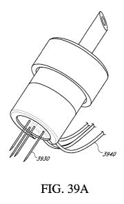
[0086] FIGS. 39A-C illustrate embodiments of an intracellular delivery
apparatus
with a Y-type configuration having a micro-hub configured for electroporation.
[0087] FIGS. 40A-B depict a small HIP (high injection pressure)
injector and a large
HIP injector.
[0088] FIG. 41 is a graphical depiction of a rabbit study five days
post immunization
with a nucleic acid encoding N53/4A, the radioactivity of cells, as counts per
minute, when
incubated with various antigens at various concentrations to show radioactive
thymidine uptake
in a T cell proliferation assay is provided.
[0089] FIG. 42 is a chart illustrating HCV 1\153-specific T cell
proliferation as a
result of immunization with the HIP injector. Proliferation is measured as
radioactivity of cells
incubated with antigen divided by the radioactivity of cells incubated with
media alone.
[0090] FIGS. 43A-C are histological evaluations of tissue at the site
of injection with
a regular 27 gauge needle (FIG. 43A), a small HIP injector (FIG. 43B), and a
large HIP injector
(FIG. 43C).
[0091] FIGS. 44A-I depict various constructs containing the N53/4A
platform and
the HBcAg with and without N53 protease cleavage sites.
[0092] FIG. 45A depicts an embodiment of a setup for measuring the
force
requirements when injecting material using one of the injection needle devices
disclosed herein.
[0093] FIG. 46A-F are top and cross-sectional views of Tests 7-9
showing dyed
water injected into chicken breast using an intracellular delivery apparatus
described herein.
[0094] FIG. 47A-F are top and cross-sectional views of Tests 25-27
showing dyed
water injected into chicken breast using an intracellular delivery apparatus
described herein.
[0095] FIG. 48A-F are top and cross-sectional views of Tests 16-18
showing dyed
water injected into chicken breast using an intracellular delivery apparatus
described herein.
[0096] FIG. 49A-F are top and cross-sectional views of Tests 34-36
showing dyed
water injected into chicken breast using an intracellular delivery apparatus
described herein.
[0097] FIG. 50A-F are top and cross-sectional views of chicken breast
having dyed
water injected by hand using an intracellular delivery apparatus described
herein.
[0098] FIG. 51A-F are top and cross-sectional views of chicken breast
having dyed
water injected by hand using a single needle.
CA 02839196 2013-12-12
WO 2012/172424 PCT/1B2012/001321
[0099] FIG. 52 shows a graph of the maximum force for injecting a
placebo fluid
with needle configurations "3Y01(72)", "3001(96)", and "6X01(144)" using a 5
mL syringe.
[0100] FIG. 53 shows a graph of the maximum force for injecting water
into air with
needle configurations "3Y01(72)", "3001(96)", and "6X01(144)" using a 10 mL
syringe.
[0101] FIG. 54 shows a graph of the maximum force for injecting water
into air with
needle configurations "3Y05(72)", "3005(72)", and "3X05(72)" using a 10 mL
syringe.
[0102] FIG. 55 shows images of dyed water injected into chicken by
hand delivery
with a standard needle, or using a Lloyd force tensometer with the 3Y05(72)",
"3005(72)",
"3X05(72)", 3Y01(72)", or "3001(96)" needle configurations.
[0103] FIGS. 56A1-B2 are example results of injection using a regular
needle and
injection using an intracellular delivery apparatus described herein coupled
with electroporation.
[0104] FIGS. 57A1-C2 are example results of injection using an
intracellular delivery
apparatus described herein at two injection force values coupled with
electroporation compared
to a conventional needle with electroporation.
[0105] FIG. 58A illustrates immunization data in a mouse using an IV1N
intracellular
delivery apparatus, without electroporation.
[0106] FIG. 58B illustrates immunization data in a mouse using an a
conventional
injection needle, without electroporation.
[0107] FIG. 58C illustrates immunization data in a mouse using an WIN
intracellular
delivery apparatus, with electroporation.
[0108] FIG. 58D illustrates immunization data in a mouse using a
conventional
injection needle with electroporation.
[0109] FIGS. 59A-B illustrate immunization data of non-immunized pigs.
[0110] FIGS. 59C-D illustrate immunization data of immunized pigs.
DETAILED DESCRIPTION
[0111] Aspects of this invention described herein concern devices and
methods for
the delivery of agents (e.g., nucleic acids) into a tissue. Some embodiments
concern an an
intracellular delivery apparatus configured to introduce agents, such as
nucleic acids, especially
DNA, into a target tissue, wherein the molecules are taken up by the cells in
a region localized to
a site near or proximal to the site of injection (e.g., within a region
defined by the area within a
needle array of an intracellular delivery apparatus described herein).
11
CA 02839196 2013-12-12
WO 2012/172424 PCT/1B2012/001321
[0112] The needle(s) of an intracellular delivery apparatus may
comprise a fitting
connector or a needle hub, which may comprise a sleeve with an internal
thread. The fitting
connector or needle hub is configured to attach the needle to the syringe or
vessel containing the
agent to be introduced. In some embodiments, the sleeve forms the attachment
means and can
be screwed onto an outer thread on an attachment part of a syringe. The
fitting connectors or
needle hubs can also comprise a press-on assembly, a snap-on assembly, or a
Luer Taper
connection, such as a Luer Lok or Luer Slip connection or a butterfly
connector.
[0113] The needle(s) of an intracellular delivery apparatus described
herein may be
attached to one or more syringe barrels (e.g., permanently affixed or
removably attached) and
said syringe barrels or the device may contain the prophylactic and/or
therapeutic agent that is to
be delivered. For example, in some embodiments, the needle(s) and attached
syringe may be pre-
loaded with a prophylactic and/or therapeutic agent, such as a nucleic acid,
protein, modified
nucleic acid, aptamer, or cell population for a single-use application. In
some embodiments, the
agent may be drawn into the syringe through apertures disposed on the
needles(s). The syringe
barrels can be of a variety of sizes (e.g., 0.3cc-100cc or more). That is, the
syringe barrels can be
greater than or equal to or any number in between 0.1, 0.3, 0.4, 0.5, 1, 2, 3,
4, 5, 6, 7, 8, 9, 10,
11, 12, 13, 14, 15, 16, 17, 18, 19, 20, 21, 22, 23, 24, 25, 26, 27, 28, 29,
30, 31, 32, 33, 34, 35, 36,
37, 38, 39, 40, 41, 42, 43, 44, 45, 46, 47, 48, 49, 50, 51, 52, 53, 54, 55,
56, 57, 58, 59, 60, 61, 62,
63, 64, 65, 66, 67, 68, 69, 70, 71, 72, 73, 74, 75, 76, 77, 78, 79, 80, 81,
82, 83, 84, 85, 86, 87, 88,
89, 90, 91, 92, 93, 94, 95, 96, 97, 98, 99, or 100 cc size. The syringe
barrels can be constructed
from a variety of materials (e.g., metal, plastic, nylon, polyethylene,
glass).
[0114] The needle(s) of an intracellular delivery apparatus described
herein may be
attached to one or more devices that facilitate delivery of prophylactic
and/or therapeutic
molecules or agents to tissue, including but not limited to gene guns,
electroporation systems,
and microneedle devices. The injection needle(s) described herein can be
modified for use with
existing technologies, including gene gun delivery systems (see e.g., U.S.
Patent Nos. 5,036,006;
5,240,855; and 5,702,384, the disclosures of which are hereby expressly
incorporated by
reference in their entireties), delivery systems using electroporation (see
e.g., U.S. Patent Nos.
6,610,044 and 5,273,525, 6,132,419, and 6,527,216, the disclosures of which
are hereby
expressly incorporated by reference in their entireties) and microneedle
delivery systems (see
e.g., U.S. Patent Nos. 6,960,193; 6,623,457; 6,334,856; 5,457,041; 5,527,288;
5,697,901;
12
CA 02839196 2013-12-12
WO 2012/172424 PCT/1B2012/001321
6,440,096; 6,743,211; and 7,226,439, the disclosures of which are hereby
expressly incorporated
by reference in their entireties).
[0115] As described herein, the intracellular delivery devices
comprising the
needle(s) may also contain a variety of prophylactic and/or therapeutic agents
(e.g. a cell
population, such as a cell population comprising stem cells, a chemical, a
compound, a
chemotherapeutic agent, a protein, a specificity exchanger, a nucleic acid
such as DNA, RNA,
other natural nucleic acid, a modified nucleic acid, or a DNA or nucleic acid
aptamer). In some
embodiments, the intracellular delivery devices comprising one or more of the
needle(s)
described herein comprise a DNA that encodes an immunogen (preferably a viral
antigen, such
as hepatitis C virus (HCV), hepatitis B virus (HBV), human immunodeficiency
virus (HIV),
influenza, Japanese encephalitis virus (JEV), human papilloma virus (HPV), or
a parasite
antigen, such as a malaria antigen, or a plant antigen, such as birch antigen,
or a bacterial
antigen, such as a staphylococcal or anthrax antigen, or a tumor antigen). In
some embodiments,
the intracellular delivery devices comprising one or more of the needles
described herein
comprise one or more of the aforementioned DNAs pre-loaded (e.g., a pre-
loaded, single use
syringe with coupled needle(s) containing a measured dose of delivered agent).
[0116] In some embodiments, the prophylactic and/or therapeutic agent
that is
delivered or contained in a syringe, needle, or injection device, as described
herein, comprises a
natural nucleic acid and in other embodiments, the prophylactic and/or
therapeutic agent that is
delivered or contained in a syringe, needle, or an intracellular delivery
device, as described
herein, comprises an unnatural nucleic acid (e.g., containing an artificial
nucleotide, universal
base, or spacer). Natural nucleic acids that can be used as the therapeutic
agent that is delivered
or contained in a syringe or injection device, as described herein, comprise a
deoxyribose- or
ribose-phosphate backbone. An artificial or synthetic polynucleotide that can
be used as the
prophylactic and/or therapeutic agent that is delivered or contained in a
syringe, needle, or
intracellular delivery device, as described herein, can comprise any
polynucleotide that is
polymerized in vitro or in a cell free system and contains the same or similar
bases but may
contain a backbone of a type other than the natural ribose-phosphate backbone.
These
backbones include: PNAs (peptide nucleic acids), phosphorothioates,
phosphorodiamidates,
morpholinos, and other variants of the phosphate backbone of native nucleic
acids. Bases that
may be included in one or more embodiments described herein include purines
and pyrimidines,
which further include the natural compounds adenine, thymine, guanine,
cytosine, uracil,
inosine, and natural analogs. Synthetic derivatives of purines and pyrimidines
that may be
13
CA 02839196 2013-12-12
WO 2012/172424
PCT/1B2012/001321
included in one or more embodiments described herein include, but are not
limited to,
modifications which place new reactive groups such as, but not limited to,
amines, alcohols,
thiols, carboxylates, and alkylhalides.
[0117]
The term "base," as used herein, encompasses any of the known base analogs
of DNA and RNA including, but not limited to, 4-acetylcytosine, 8-hydroxy-N6-
methyladenosine, aziridinylcytosine, pseudoisocytosine, 5-
(carboxyhydroxylmethyl)uracil, 5-
fluorouracil, 5-bromouracil, 5-carboxymethylaminomethy1-2-thiouracil, 5-
carboxymethyl-
aminomethyluracil, dihydrouracil, inosine, N6-isopentenyladenine, 1-
methyladenine, 1-
methylp s eudo-uracil, 1-methylguanine, 1-
methylinosine, 2,2-dimethyl-guanine, 2-
methyladenine, 2-methylguanine, 3-methyl-cytosine, 5-methylcytosine, N6-
methyladenine, 7-
methylguanine, 5-methylaminomethyluracil, 5-methoxy-amino-methyl-2-thiouracil,
beta-D-
manno sylqueo sine, 5'-methoxycarbonylmethyluracil, 5-methoxyuracil, 2-
methylthio-N6-
isopentenyladenine, uracil-5-oxyacetic acid methylester, uracil-5-oxyacetic
acid, oxybutoxosine,
pseudouracil, queosine, 2-thiocytosine, 5-methyl-2-thiouracil, 2-thiouracil, 4-
thiouracil, 5-
methyluracil, N-uracil-5-oxyacetic acid methylester, uracil-5-oxyacetic acid,
pseudouracil,
queosine, 2-thiocytosine, and 2,6-diaminopurine.
The term polynucleotide includes
deoxyribonucleic acid (DNA) and ribonucleic acid (RNA) and combinations on
DNA, RNA and
other natural and synthetic nucleotides.
[0118]
The prophylactic and/or therapeutic agent that is delivered or contained in a
syringe, needle, or an intracellular delivery device, as described herein, can
comprise DNA,
which may be in the form of cDNA, in vitro polymerized DNA, plasmid DNA, parts
of a
plasmid DNA, genetic material derived from a virus, linear DNA, vectors (P1,
PAC, BAC,
YAC, artificial chromosomes), expression cassettes, chimeric sequences,
recombinant DNA,
chromosomal DNA, an oligonucleotide, anti-sense DNA, or derivatives of these
groups. RNA
may be in the form of oligonucleotide RNA, tRNA (transfer RNA), snRNA (small
nuclear
RNA), rRNA (ribosomal RNA), mRNA (messenger RNA), in vitro polymerized RNA,
recombinant RNA, chimeric sequences, anti-sense RNA, siRNA (small interfering
RNA),
miRNA (micro RNA), ribozymes, or derivatives of these groups. The prophylactic
and/or
therapeutic agent that is delivered or contained in a syringe, needle, or an
intracellular delivery
device, as described herein, can also comprise an anti-sense polynucleotide
that is a
polynucleotide that interferes with the function of DNA and/or RNA. Antisense
polynucleotides
include, but are not limited to: morpholinos, 2'-0-methyl polynucleotides,
DNA, RNA and the
like. SiRNA comprises a double stranded structure typically containing 15 to
50 base pairs and
14
CA 02839196 2013-12-12
WO 2012/172424 PCT/1B2012/001321
preferably 21 to 25 base pairs and having a nucleotide sequence identical or
nearly identical to
an expressed target gene or RNA within the cell. Interference may result in
suppression of
expression. The polynucleotide can be a sequence whose presence or expression
in a cell alters
the expression or function of cellular genes or RNA. In addition, DNA and RNA
may be single,
double, triple, or quadruple stranded. Double, triple, and quadruple stranded
polynucleotide may
contain both RNA and DNA or other combinations of natural and/or synthetic
nucleic acids.
These polynucleotides can be delivered to a cell to express an exogenous
nucleotide sequence, to
inhibit, eliminate, augment, or alter expression of an endogenous nucleotide
sequence, or to
express a specific physiological characteristic not naturally associated with
the cell.
Polynucleotides may be coded to express a whole or partial protein, or may be
anti-sense. The
delivered polynucleotide can stay within the cytoplasm or nucleus apart from
the endogenous
genetic material. Alternatively, the polymer could recombine with or become a
part of the
endogenous genetic material. For example, the therapeutic agent that is
delivered or contained
in a syringe or an intracellular delivery device, as described herein, can
comprise a DNA that can
insert itself into chromosomal DNA by either homologous or non-homologous
recombination.
[0119] The prophylactic and/or therapeutic agent that is delivered or
contained in a
syringe, needle, or an intracellular delivery device, as described herein, can
also comprise an
RNA inhibitor, which is any nucleic acid or nucleic acid analog containing a
sequence whose
presence or expression in a cell causes the degradation of or inhibits the
function or translation
of a specific cellular RNA, usually a mRNA, in a sequence-specific manner. An
RNA inhibitor
may also inhibit the transcription of a gene into RNA. Inhibition of RNA can
effectively inhibit
expression of a gene from which the RNA is transcribed. RNA inhibitors
include, but are not
limited to, siRNA, microRNAs (miRNAs), interfering RNA or RNAi, dsRNA, RNA
Polymerase
III transcribed DNAs, ribozymes, and antisense nucleic acid, which may be RNA,
DNA, or an
artificial nucleic acid. MicroRNAs (miRNAs) also typically have a length of
between about 15-
50 nucleotides, preferably between about 20-25 nucleotides in length, and
miRNAs can be used
as post-transcriptional regulators that bind to complementary sequences on
target messenger
RNA transcripts (mRNAs), usually resulting in translational repression and
gene silencing.
Antisense polynucleotides can include, but are not limited to: morpholinos, 2'-
0-methyl
polynucleotides, DNA, RNA and the like. RNA polymerase III transcribed DNAs
can contain
promoters, such as the U6 promoter. These DNAs can be transcribed to produce
small hairpin
RNAs in the cell that can function as siRNA or linear RNAs that can function
as antisense RNA.
The RNA inhibitor may be polymerized in vitro, recombinant RNA, contain
chimeric sequences,
CA 02839196 2013-12-12
WO 2012/172424 PCT/1B2012/001321
or derivatives of these groups.
The RNA inhibitor may contain ribonucleotides,
deoxyribonucleotides, synthetic nucleotides, or any suitable combination such
that the target
RNA and/or gene are inhibited. In addition, these forms of nucleic acid may be
single, double,
triple, or quadruple stranded.
[0120]
The prophylactic and/or therapeutic agent that is delivered or contained in a
syringe, needle, or an intracellular delivery device, as described herein, can
also include a nucleic
acid that is incorporated into a vector (e.g., an expression vector). Vectors
are polynucleic
molecules originating from a virus, a plasmid, or the cell of a higher
organism into which
another nucleic fragment of appropriate size can be integrated; vectors
typically introduce
foreign DNA into host cells, where it can be reproduced. Examples are
plasmids, cosmids, and
yeast artificial chromosomes; vectors are often recombinant molecules
containing DNA
sequences from several sources.
[0121]
As used herein, term "vector" refers any DNA molecule that could include
associate molecules to transfer DNA sequences into a cell for expression.
Examples include
naked DNA, non-viral DNA complexes (e.g. DNA plus polymers [cationic or
anionic], DNA
plus transfection enhancing compounds, and DNA plus amphipathic compounds) and
viral
particles. As used herein, vector may also include a viral vector: for
example, adenovirus; DNA;
adenoassociated viral vectors (AAV) which are derived from adenoassociated
viruses and are
smaller than adenoviruses; and retrovirus (any virus in the family
Retroviridae that has RNA as
its nucleic acid and uses the enzyme reverse transcriptase to copy its genome
into the DNA of
the host cell's chromosome; examples include VSV G and retroviruses that
contain components
of lentivirus including HIV type viruses).
[0122]
The prophylactic and/or therapeutic agent that is delivered or contained in a
syringe, needle, or an intracellular delivery device, as described herein, can
also comprise one or
more compounds that enhance the uptake of the therapeutic agent (e.g., a
nucleic acid, as
described herein). The therapeutic agent that is delivered or contained in a
syringe, needle, or an
intracellular delivery device, as described herein, can comprise a polymer,
for example, which is
a molecule built up by repetitive bonding together of smaller units called
monomers. The term
"polymer" can include both oligomers, which have two to about 80 monomers and
polymers
having more than 80 monomers. The polymer can be linear, branched network,
star, comb, or
ladder types of polymer. The polymer can be a homopolymer in which a single
monomer is used
16
CA 02839196 2013-12-12
WO 2012/172424 PCT/1B2012/001321
or can be copolymer in which two or more monomers are used. Types of
copolymers include
alternating, random, block and graft.
[0123] The prophylactic and/or therapeutic agent that is delivered or
contained in a
syringe, needle, or an intracellular delivery devices, as described herein,
may also comprise a
nucleic acid-polycation complex. Cationic proteins like histones and
protamines or synthetic
polymers like polylysine, polyarginine, polyornithine, DEAE dextran,
polybrene, and
polyethylenimine are effective intracellular delivery agents. A polycation is
a polymer
containing a net positive charge, for example poly-L-lysine hydrobromide. The
polycation can
contain monomer units that are charge positive, charge neutral, or charge
negative, however, the
net charge of the polymer is desirably positive. The term "polycation" also
can refer to a non-
polymeric molecule that contains two or more positive charges. A polyanion is
a polymer
containing a net negative charge, for example polyglutamic acid. The polyanion
can contain
monomer units that are charge negative, charge neutral, or charge positive,
however, the net
charge on the polymer must be negative. The term "polyanion" can also refer to
a non-
polymeric molecule that contains two or more negative charges. The term
"polyion" includes
polycation, polyanion, zwitterionic polymers, and neutral polymers that
contain equal amounts
of anions and cations. The term "zwitterionic" refers to the product (salt) of
the reaction
between an acidic group and a basic group that are part of the same molecule.
Salts are ionic
compounds that dissociate into cations and anions when dissolved in solution.
Salts increase the
ionic strength of a solution, and consequently decrease interactions between
nucleic acids with
other cations.
[0124] The prophylactic and/or therapeutic agent that is delivered or
contained in a
syringe, needle, or an intracellular delivery device, as described herein, may
also comprise a
specificity exchanger. Several types of specificity exchangers are known and
any one or more of
these molecules can be delivered or contained in a syringe, needle, or
injection device, as
described herein. For example, U.S. Patent Nos.7,318,926; 7,019,111;
6,933,366; 6,660,842;
6,469,143; 6,245,895; 6,040,137; 5,869,232; 7,943,149; 6,417,324 describe
specificity
exchangers, the disclosure of which are expressly incorporated by reference in
their entireties.
Preferably, specificity exchangers that comprise a ligand for a receptor on a
pathogen joined to
an oligosaccharide (e.g., the gal epitope or Gala1-3Gal-131-4G1cNAc-R
(preferably synthetically
assembled specificity exchangers or glycoconjugates are used, for example,
specificity
exchangers or glycoconjugates prepared by solid phase peptide synthesis).
17
CA 02839196 2013-12-12
WO 2012/172424 PCT/1B2012/001321
[0125] Some embodiments relate to an intracellular delivery device
that comprises a
plurality of needles, which are arranged or configured to deliver a
therapeutic agent to a targeted
tissue. In some embodiments, the agent is delivered through the proximal end
of the injection
device by a syringe and the agent is delivered to the targeted tissue through
a plurality of
apertures disposed on or near the distal ends of the plurality of needle
barrels. In other
embodiments, the end of the apertures can be disposed on the proximal ends of
the needles
barrels. In some embodiments, a plurality of needles of any one or more of the
design features
described herein are provided on an injection device. Embodiments described
herein also
include a cannula that comprises a plurality of needles configured as
described above. That is, in
some embodiments the intracellular delivery device and/or cannula can
comprise, consist, or
consist essentially of 1, 2, 3, 4, 5, 6, 7, 8, 9, or 10 or more needles. The
needles can be of the
same size and length or can be of different sizes and lengths. Each needle in
embodiments that
have more than one needle can have a plurality of apertures, which can be in a
first or second
zone, as described above, or both (e.g., along the length of the band).
Preferably, the needles of
the intracellular delivery devices have a closed end, as described above.
Intracellular delivery
devices and/or cannulas that comprise, consist, or consist essentially of 2,
3, 4, 5, 6, 7, 8, 9, or 10
needles can be configured such that at least two needles have a different
amount of apertures
and/or different sizes of apertures and/or different shapes of apertures
and/or different positions
of apertures and said needles preferably have a closed end. Preferably, the
needles and apertures
are oriented such that the apertures on each needle oppose the apertures on
another needle so as
to generate an opposing field of delivery of a delivered agent or a cross-
spray pattern of delivery
of a delivered agent (e.g., in a four needle array having a central needle and
three outer needles,
the outer needles can have apertures that direct delivered agent toward
apertures present on the
central needle and the central needle has apertures in three zones, wherein
each zone opposes
apertures present on the outer needles and delivered agent exiting the central
needle is directed to
apertures present on each of the outer needles). In some embodiments of an
intracellular
delivery device, such needle arrays are connected to or disposed on a hub that
comprises at least
one electrical connector. Again, preferably, the apertures on each needle in
the array oppose the
apertures on another needle so as to generate a cross-spray pattern or a
region of opposing
delivery of a delivered agent. In this way, a region within the needle array
is overloaded locally
with the delivered agent, thereby providing a high concentration of the
delivered agent within the
region, which is particularly useful when an electric field is applied thereby
inducing uptake of
the delivered agent. The injection devices, hypodermic needle assemblies, or
intracellular
18
CA 02839196 2013-12-12
WO 2012/172424 PCT/1B2012/001321
delivery devices also preferably include an electrode (in some embodiments,
one or more of the
needles themselves are electrodes) and an electrical power supply configured
to generate an
electric field at the electrode, wherein the electrical power supply is
connected to the electrode
through the at least one electrical connector. In some embodiments, one needle
or a plurality of
needles has apertures in a first zone proximal to a closed end of the barrel
and one needle or a
plurality of needles that has apertures in a second zone that is distal to a
closed end of the needle
barrel. Additionally, some embodiments may have a first needle (e.g., a single
needle on a single
needle device, as described below, or the first needle in a device having a
plurality of needles) or
a first plurality of needles with apertures that are a size equal to, greater
than or less than 0.01,
0.02, 0.03, 0.04, 0.05, 0.06, 0.07, 0.08, 0.09, 0.1, 0.15, 0.2, 0.25, 0.3,
0.35, 0.4, 0.45, 0.5, 0.55,
0.6, 0.65, 0.7, 0.75, 0.8, 0.85, 0.9, 0.95, 1.0, 1.05, 1.10, 1.15, 1.20, 1.25,
1.30, 1.35, 1.40, 1.45,
1.50, 1.55, 1.60, 1.65, 1.70, 1.75, 1.80, 1.85, 1.90, 1.95, 2.0, 2.05, 2.10,
2.15, 2.20, 2.25, 2.30,
2.35, 2.40, 2.45, 2.50, 2.55, 2.60, 2.65, 2.70, 2.75, 2.80, 2.85, 2.90, 2.95,
3.0, 3.05, 3.10, 3.15,
3.20, 3.25, 3.30, 3.35, 3.40, 3.45, 3.50, 3.55, 3.60, 3.65, 3.70, 3.75, 3.80,
3.85, 3.90, 3.95, or 4.0
mm in its widest portion). In some embodiments, one needle or a plurality of
needles has
apertures that less than 1 pm at its widest portion. In some embodiments, one
needle or a
plurality of needles has apertures that less than about 900 nm at its widest
portion. In some
embodiments, one needle or a plurality of needles has apertures that less than
about 500 nm at its
widest portion. In some embodiments, the first needle in an intracellular
delivery device having
a plurality of needles has apertures that are smaller or substantially smaller
than a second needle
or a second plurality of needles in the device.
[0126] In some embodiments, the intracellular delivery device includes
only one
needle. The needle can include any of the design features disclosed herein.
For example the
needle can have a closed or open end. The needle may include a plurality of
apertures, for
example, at least, greater than or equal to or any number in between about 5,
10, 20, 30, 50, 70,
100, 120, 140, 160, 180, 200, 500 apertures (e.g., at least about 5-100, 10-
100, 20-100, 30-100,
40-100, 50-100 or 5-200, 10-200, 20-200, 30-200, 40-200, 50-200, 100-200, 100-
500, 140-500,
150-500, 200-500 apertures). The apertures can be evenly spaced, or randomly
distributed. The
apertures may, in some embodiments, form a regular pattern on the needle. For
example, the
apertures may form a pattern having a rotational symmetry along the axis of
the needle. The
rotational symmetry may include 2-fold, 3-fold, 4-fold, 5-fold, six-fold, or a
higher degree of
rotational symmetry. As another example, the apertures may form a pattern
having a screw axis
19
CA 02839196 2013-12-12
WO 2012/172424 PCT/1B2012/001321
symmetry. The screw axis symmetry can be include 2-fold, 3-fold, 4-fold, 5-
fold, six-fold, or a
higher degree of rotational symmetry. The translation vector for the screw
axis can, for example,
be about, at least, at least about, not more than, not more than about 0.01
mm, 0.05 mm, 0.1 mm,
0.15 mm, 0.2 mm, 0.25 mm, 0.3 mm, 0.4 mm, 0.5 mm, 0.6 mm, 0.7 mm, 0.8 mm, 0.9
mm, 1
mm, 2 mm, 3 mm, 4 mm, 5mm, 6 mm, 7 mm, 8 mm, 9 mm, 1 cm, 2 cm or 3 cm. In some
embodiments, the apertures are configured to produce a radial pattern when a
prophylactic
and/or therapeutic material is injected.
[0127] In some embodiments, the intracellular delivery device
comprises one or more
needles that are fluidly coupled to a syringe or a reservoir containing the
prophylactic and/or
therapeutic material so that the relative orientation of the one or more
needles with the syringe or
reservoir is fixed. The end(s) of the one or more needles may, in some
embodiments, be both
fluidly coupled and disposed near the syringe or a reservoir containing the
prophylactic and/or
therapeutic material. For example, an end of the one or more needles may be
about, not more
than, not more than about 1 mm, 5 mm, 1 cm, 2 cm, 3 cm, 4 cm, 5 cm, 6 cm, 7
cm, 8 cm, 9 cm,
or 10 cm apart from the syringe or a reservoir containing the prophylactic
and/or therapeutic
material. In some embodiments, the end(s) of the one or more needles are no
more than about
cm apart from the syringe or a reservoir that is fluidly coupled with the
needle(s) and contains
the prophylactic and/or therapeutic material.
[0128] Some embodiments relate to injection devices, intracellular
delivery devices,
cannulas, and needles comprising a fluid containing an agent, as described
herein (e.g., a
medicinal compound, chemical, nucleic acid, peptide, specificity exchanger,
and, in particular, a
DNA). In some embodiments, the intracellular delivery devices, cannulas, and
needles,
described herein, are for single use. That is, some embodiments comprise one
or more of the
needle designs, described herein, joined to a receptacle (preferably a sterile
container, such as a
sterilized syringe) that comprises a single application or dose of delivered
agent (e.g., medicinal
compound, chemical, nucleic acid, peptide, specificity exchanger, and, in
particular a DNA).
Accordingly, a single application or device can be conveniently packaged and
provided to
medical practitioners or end-consumers, which can administer said agent at an
appropriate site
and, following administration, the used injection device, needle, or cannula
comprising a
plurality of needles can be appropriately discarded. Some embodiments also
include methods of
making and using the aforementioned devices and methods of inducing an immune
response to a
desired antigen.
CA 02839196 2013-12-12
WO 2012/172424 PCT/1B2012/001321
[0129] In some embodiments, the intracellular delivery device is not
configured to
apply an electric field shortly after or simultaneous with the introduction of
the prophylactic
and/or therapeutic material (e.g., DNA) at the tissue around and/or through
the site of the
injection. For example, the intracellular delivery device may not include a
voltage source
coupled to the device and configured to apply an electric field to the tissue
at or near the site of
injection. In other embodiments, the intracellular delivery device is
configured to apply an
electric field shortly after or simultaneous with the introduction of the
prophylactic and/or
therapeutic material (e.g., DNA) at the tissue around and/or through the site
of the injection. For
example, the intracellular delivery device may include a voltage source
coupled to the device and
configured to apply an electric field to the tissue at or near the site of
injection.
[0130] Some embodiments disclosed herein include a method of
delivering a
prophylactic and/or therapeutic material to a subject in need thereof, where
the prophylactic
and/or therapeutic material is administered using any of the intracellular
delivery devices
disclosed herein. The prophylactic and/or therapeutic material may be any of
those materials
disclosed herein. In some embodiments, the method may also include maintaining
the one or
more needles inserted within the tissue for at least a predetermined time
after injecting the
prophylactic and/or therapeutic material but before withdrawing the one or
more needles. The
one or more needles may be maintained in the tissue, for example, at least,
greater than or equal
to 1 s, 2 s, 3 s, 4 s, 5 s, or more after injecting the therapeutic material
but before withdrawing
the one or more needles. In some embodiments, the entire dosage is delivered
in a period of
time less than about 60 seconds, 40 seconds, 30 seconds, 20 seconds, 15
seconds, 10 seconds, 5
seconds, 4 seconds, 3 seconds, 2 seconds, 1.5 seconds, 1.0 second, 0.8
seconds, 0.5 seconds, 0.4
seconds, 0.3 seconds, or 0.2 seconds.
[0131] The period of time between inserting the one or more needles
into the tissue
and removing the one or more needles into the tissue can also vary. The period
of time may,
for example, be about, at least, at least about, not more than, not more than
0.5 seconds, 1
second, 2 seconds, 3 seconds, 4 seconds, 5 seconds, 6 seconds, 7 seconds, 8
seconds, 9 seconds,
seconds, 15 seconds, 30 seconds, 45 seconds, or 60 seconds.
[0132] In some embodiments, the needle(s) and any of the devices
described herein
can be affixed to the body of a subject for greater periods of time so as to
allow for a long term
delivery of a prophylactic and/or therapeutic agent (e.g., delivery for at
least, greater than or
equal to 1, 2, 3, 4, 5, 6, 7, 8, 9, 10, 11, 12, 13, or 14 days) and such
needles and devices can be
21
CA 02839196 2013-12-12
WO 2012/172424 PCT/1B2012/001321
affixed to miniature pumps so as to administer small amounts of prophylactic
and/or therapeutic
material (e.g. a cell population, such as a cell population comprising stem
cells, chemical, a
compound, a chemotherapeutic agent, a protein, a nucleic acid, such as DNA,
RNA, other
natural nucleic acid, a modified nucleic acid, or a DNA or nucleic acid
aptamer), to said subjects
over an extended period of time.
[0133] The prophylactic and/or therapeutic material may be delivered
to the body, in
some embodiments, at high pressures through any of the intracellular delivery
devices disclosed
herein, using a pressure generation element. The pressure generation element
may be a plunger
on a syringe, and may comprise a spring or other element which depresses the
plunger to
generate a high force or pressure on the prophylactic and/or therapeutic
material. The high force
or pressure generated by the pressure generation element is applied to the
prophylactic and/or
therapeutic material to displace it through the needle (e.g., by applying
force to a plunger on a
syringe coupled to the needle(s)). The maximum force can, for example, be
about, at least, at
least about, not more than, not more than about 25 N, 40 N, 50 N, 75 N, 100 N,
150 N, 200 N, or
500 N. The maximum pressure applied to the therapeutic material can, for
example, be about, at
least, at least about, not more than, not more than 50 kPa, 100 kPa, 200 kpa,
300, kPa, 400 kPa,
500 kPa, 600 kPa, 700 kPa, 800 kPa, 900 kPa, 1000 kPa, 1100 kPa, 1200 kPa,
1300 kPa, 1400
kPa, 1500 kPa, 1600 kPa, 1700 kPa, 1800 kPa, 1900 kPa, 2000 kPa, 2100 kPa,
2200 kPa, 2300
kPa, 2400 kPa, 2500 kPa, 2600 kPa, 2700 kPa, 2800 kPa, 2900 kPa, 3000 kPa,
3100 kPa, 3200
kPa, 3300 kPa, 3400 kPa, 3500 kPa, 3600 kPa, 3700 kPa, 3800 kPa, 3900 kPa,
4000 kPa, 4100
kPa, 4200 kPa, 4300 kPa, 4400 kPa, 4500 kPa, 4600 kPa, 4700 kPa, 4800 kPa,
4900 kPa, 5000
kPa, 6000 kPa, 7000 kPa, 8000 kPa, 9000 kPa, 10 MPa, 15 MPa, 20 MPa, 25 MPa,
or 30 MPa.
[0134] In some embodiments, the prophylactic and/or therapeutic
material can be
delivered at high hole velocities (i.e., the average velocity of the fluid
exiting the apertures). The
hole velocity can be, for example, be about, at least, at least about, not
more than, not more than
about 1 mm/s, 1.5 mm/s, 2 mm/s, 2.5 mm/s, 3 mm/s, 3.5 mm/s, 4 mm/s, 5 mm/s, 6
mm/s, 7
mm/s, 8 mm/s, 9 mm/s or 10 mm/s. In a preferred embodiment, the hole velocity
can be at least
about 3 mm/s. The hole velocity may be determined, for example, by the volume
rate of
injecting the prophylactic and/or therapeutic material relative the average
size of the apertures.
The intracellular delivery devices disclosed herein may optionally be
configured to delivery at
any of the hole velocities disclosed above. For example, the intracellular
delivery device can be
coupled with a spring piston configured to inject prophylactic and/or
therapeutic material at a
volume rate sufficient to produce the desired hole velocity.
22
CA 02839196 2013-12-12
WO 2012/172424 PCT/1B2012/001321
[0135] Some aspects of the invention concern a intracellular delivery
device
comprising a needle that comprises a lumen adapted for the passage of a
prophylactic and/or
therapeutic material and a needle barrel that comprises a plurality of
apertures on the length of
the barrel, wherein said needle barrel has a closed-end; and a connector
configured to join said
needle to a pressure generation element. In some embodiments, the
intracellular delivery device
above comprises a plurality of needles as described herein and in some
embodiments, the
intracellular delivery device comprises a circular, diamond, triangular,
square, rectangle,
trapezoidal, ovoid, or otherwise shaped array of needles. Preferably, the
intracellular delivery
device is designed such that the plurality of said needles is configured such
that the apertures on
the needle barrels face each other but in some embodiments, the hypodermic
needle assembly
has a plurality of said needles that is configured such that the apertures on
the needle barrels face
away from each other. In some embodiments, the intracellular delivery device
further comprises
a pressure generation element joined to said intracellular delivery devices
and this pressure
generation element can be a syringe. The intracellular delivery devices above
of can have
apertures that have a diameter of about 10nm-4mm, 0.01 mm-4mm, 0.1 mm-4mm, 1.0
mm-
4mm, 1.5 mm-4mm, 2.0 mm-4mm, or 3.0 mm-4mm.
[0136] In some embodiments, the intracellular delivery device above
comprises a
single syringe joined to at least three of said needles. In some embodiments,
the at least three of
said needles are between about 2 and about 10 mm apart. In other embodiments,
the
intracellular delivery device above can comprise a single syringe joined to at
least four
hypodermic needles. In some embodiments, the intracellular delivery device has
at least four
hypodermic needles that are between about 3 and about 6 mm apart. A single use
intracellular
delivery device is also an embodiment and such devices preferably comprise a
plurality of
needles attached to at least one syringe, wherein the needles comprise a
plurality of apertures
distributed along the barrel of said needles and a closed end; and said at
least one syringe
comprises a single dose of a prophylactic and/or therapeutic agent. In some
embodiments, the
prophylactic and/or therapeutic agent in the intracellular delivery device is
a nucleic acid. The
prophylactic and/or therapeutic agent can be a DNA that encodes a protein. In
some
embodiments, the intracellular delivery device above comprises a single
syringe joined to at least
three hypodermic needles and in some embodiments, the at least three
hypodermic needles are
between about 2 and about 10 mm apart. In other embodiments, the intracellular
delivery device
above comprises a single syringe joined to at least four needles and in some
embodiments, the at
least four hypodermic needles are between about 3 and about 6 mm apart.
23
CA 02839196 2013-12-12
WO 2012/172424 PCT/1B2012/001321
[0137] Aspects of the invention also include methods of making and
using the
aforementioned devices. By one approach, some of the devices described herein
are used to
deliver a prophylactic and/or therapeutic agent to a subject and said methods
are practiced by
providing one of the delivery devices described herein, inserting the needles
of said device into a
tissue of a subject; and displacing the therapeutic agent from the syringe
through the needles and
into the tissue. In some embodiments, the prophylactic and/or therapeutic
agent is a nucleic
acid, the nucleic acid can encode an antigen, such as a viral antigen,
preferably, a hepatitis
antigen such as an HCV or HBV antigen such that some of the delivery devices
described herein
can be used for the purposes of inducing an immune response in a subject to an
antigen that is
delivered by said device.
[0138] Additional embodiments include an intracellular delivery device
for the
delivery of prophylactic and/or therapeutic material into tissue, the device
comprising a
connection to a pressure generation element; a lumen adapted for the passage
of a prophylactic
and/or therapeutic material; and a needle barrel, wherein the needle barrel
comprises a plurality
of apertures that extend along the length of the barrel. In some embodiments,
the therapeutic
material comprises a nucleic acid, a polypeptide, a carbohydrate, a
specificity exchanger, a
steroid, a cell population, a chemical or an immunogen. In some embodiments,
the prophylactic
and/or therapeutic agent induces the immune system.
[0139] The needle barrel can be adapted to transmit an electric
current and the device
can further comprise an electrode adapted to transmit an electromagnetic
field. In some
embodiments, the prophylactic and/or therapeutic agent enters a cell and in
others it remains
extracellular. In some embodiments, the pressure is transmitted using a fluid
medium or a gas
medium. In some embodiments, the nucleic acid comprises a sequence from a
hepatitis virus
such as a hepatitis B antigen (HBV), such as HBcAg, or a hepatitis C virus
(HCV) antigen, such
as NS3/4A, or a combination thereof, such as HBcAg from an HBV virus that
infects stork or
heron joined to NS3/4A. In other embodiments, the nucleic acid comprises a
sequence from a
human simian virus antigen. Preferably, the nucleic acid comprises a sequence
encoding an
antigen capable of generating a proliferative T-cell response and in some
embodiments, the
nucleic acid comprises a sequence from a human immunodeficiency virus.
[0140] Additional embodiments include, a intracellular delivery device
for the
delivery of prophylactic and/or therapeutic material into tissue comprising a
prophylactic and/or
therapeutic material pressure generation element; an array of needle barrels
coupled to the
24
CA 02839196 2013-12-12
WO 2012/172424 PCT/1B2012/001321
pressure generation element; wherein at least one of the needle barrels in the
array comprises a
plurality of apertures adapted to deliver a pressure transmitted from the
pressure generation
element into a tissue to cause an increase in the permeability of a cell
membrane, and at least one
of the needle barrels in the array is adapted for the passage of the
prophylactic and/or therapeutic
material as described elsewhere herein.
[0141] More embodiments, include an intracellular delivery device
having a
longitudinal axis, the device comprising a connector configured to engage a
source of
pressurized fluid; and a needle assembly, the needle assembly comprising a
stem extending from
the connector in a direction substantially parallel to the longitudinal axis
of the device, the stem
comprising a first lumen that is fluidly coupled with the connector, a first
needle barrel
extending from the stem in a direction substantially parallel to the
longitudinal axis of the
device, the first needle barrel comprising a second lumen that is fluidly
coupled with the stem
and at least one aperture that is fluidly coupled with the second lumen, and a
second needle
barrel extending from the stem in a direction substantially parallel to the
longitudinal axis of the
device, the second needle barrel comprising a third lumen that is fluidly
coupled with the stem
and at least one aperture that is fluidly coupled with the third lumen.
[0142] In some embodiments, the first needle barrel and the second
needle barrel
form an injection cavity space therebetween. In some embodiments, the
injection cavity space is
configured to receive at least a portion of a subject, such as a tissue. In
some embodiments, the
first needle barrel and second needle barrel each comprise the same number of
apertures. In
some embodiments, each aperture on the first needle barrel faces an aperture
on the second
needle barrel. In some embodiments, the first needle barrel and the second
needle barrel
comprise a pointed distal tip disposed opposite the stem. In some embodiments,
the apertures
are generally curvilinear. In some embodiments, the apertures are generally
polygonal. In some
embodiments, the apertures are evenly disposed along a line segment that is
substantially parallel
to the longitudinal axis of the device. In some embodiments, a third needle
barrel extending
from the stem in a direction substantially parallel to the longitudinal axis
of the device, the third
needle barrel comprising a fourth lumen that is fluidly coupled with the stem
and at least one
aperture that is fluidly coupled with the fourth lumen. In some embodiments,
at least one
aperture is configured to apply negative pressure to the injection cavity
space.
[0143] Still more embodiments concern an intracellular delivery device
for delivering
a prophylactic and/or therapeutic agent to subject, the device having a
longitudinal axis and
CA 02839196 2013-12-12
WO 2012/172424 PCT/1B2012/001321
comprising a plurality of syringes disposed generally parallel to the
longitudinal axis of the
device, each syringe comprising a needle with a plurality of apertures
disposed along a length of
the needle, wherein the apertures face the longitudinal axis of the device. In
these embodiments,
the at least one syringe comprises a prophylactic and/or therapeutic agent
comprising a gene. In
some embodiments, each needle comprises a tip and the tips of the plurality of
needles are
disposed on a plane that lies substantially normal to the longitudinal axis of
the device.
Additional embodiments include a hypodermic needle comprising a plurality of
apertures
distributed along the barrel of said needle, wherein the end of said needle is
closed. In some
embodiments, said closed end is blunt.
[0144] In some embodiments, the assembly further comprises a syringe
attached to
the needle. In some embodiments, said syringe comprises a prophylactic and/or
therapeutic
agent, which can be a nucleic acid such as a DNA that encodes a protein. Still
more aspects of
the invention concern an intracellular delivery device comprising a plurality
of hypodermic
needles that comprise a plurality of apertures distributed along the barrel of
said needles joined
to one or more syringes. Preferably, the end of said needles are closed. In
some embodiments,
the end of said needles are blunt. In some embodiments, said syringe comprises
a prophylactic
and/or therapeutic agent such as a DNA that encodes a protein. In some
embodiments, the
injection device above comprises a single syringe joined to at least three
hypodermic needles.
Some embodiments concern a single use intracellular delivery device comprising
a plurality of
needles attached to at least one syringe, wherein the needles comprise a
plurality of apertures
distributed along the barrel of said needles and said at least one syringe
comprises a single dose
of a prophylactic and/or therapeutic agent. In some embodiments, the end of
said needles are
closed. In some embodiments, the end of said needles are blunt. In some
embodiments, the
prophylactic and/or therapeutic agent is a nucleic acid. In some embodiments,
the nucleic acid is
a DNA that encodes a protein.
[0145] In some embodiments, the intracellular delivery device
comprises a needle
hub having a plurality of needles partially disposed therein; the plurality of
needles each
comprising a lumen adapted for the passage of a therapeutic material and a
needle barrel, the
needle barrel comprising a plurality of apertures along the length of the
barrel, wherein said
barrel has a closed-end and wherein the apertures are evenly spaced apart on
each of the needle
barrels and have an average diameter in the range of about 0.01 mm to about 6
mm; a syringe
comprising a plunger; a connector configured to fluidly couple the syringe and
needle hub, the
connector configured to form a gap between the connector and needle hub. In
some
26
CA 02839196 2013-12-12
WO 2012/172424 PCT/1B2012/001321
embodiments, the hypodermic needle comprises zones on the needle barrel,
wherein each zone is
configured to pass therapeutic material in a different direction. In some
embodiments, the
hypodermic needle comprises a slit having a first dimension corresponding to
the axis of the
needle barrel and a second dimension perpendicular to the axis of the needle
barrel wherein the
first dimension is longer than the second dimension.
[0146] Methods of using anyone or more of the aforementioned devices
are also
embodiments, including a method of delivering a nucleic acid into a cell
comprising using the
injection devices as described herein In some embodiments, the method of
delivering to a
subject a prophylactic and/or therapeutic agent comprises using
electroporation by providing a
voltage source, inserting one or more needles of an intracellular delivery
device into a tissue of a
subject, wherein the one or more needles each comprises a barrel having a
lumen and a plurality
of apertures, the barrel made from an electrically conductive material, and an
electrical
connection to the voltage source; displacing the prophylactic and/or
therapeutic agent through
the lumen of the needle and into the tissue through the plurality of
apertures, wherein the one or
more needles are joined to a syringe containing said prophylactic and/or
therapeutic agent such
that the syringe and one or more needles form a rigid or non-flexible
structure; and applying an
electric field to the tissue of the subject using the one or more needles as
electrodes. In some
embodiments, displacing the prophylactic and/or therapeutic agent is performed
prior to applying
the electric field to the tissue. In some embodiments, displacing the
prophylactic and/or
therapeutic agent is performed subsequent to applying the electric field to
the tissue. In some
embodiments, displacing the prophylactic and/or therapeutic agent is performed
prior to applying
the electric field to the tissue. In some embodiments, displacing the
prophylactic and/or
therapeutic agent is performed at about the same time as applying the electric
field to the tissue
(e.g., a prophylactic and/or therapeutic agent, such as a nucleic acid
encoding an antigen, can be
administered by passing the prophylactic and/or therapeutic agent through the
apertures on the
needle barrel before, during, or after applying an electric field to the
recipient tissue at or near
the site of injection of the prophylactic and/or therapeutic agent. In some
embodiments, the
nucleic acid is a DNA that encodes a protein, such as an antigen.
[0147] Some embodiments relate to an intracellular delivery device
comprising a
plurality of pairs of needles, each needle of the plurality of pairs of
needles comprising a closed
end, a barrel, and a plurality of apertures along the barrel, wherein the
apertures on at least one of
the needles of the plurality of pairs of needles is aligned in relation to at
least one other needle to
generate a cross-spray pattern; wherein at least one of the plurality of pairs
of needles comprises
27
CA 02839196 2013-12-12
WO 2012/172424 PCT/1B2012/001321
a pair of electrodes having opposite polarity, the electrodes configured to
apply an electric field
to a subject.
[0148] In some embodiments, said DNA encodes a viral antigen. In some
embodiments, said viral antigen is an HCV or HBV antigen. Furthermore, in some
embodiments a use of a HBcAg or a fragment thereof or a nucleic acid encoding
HBcAg or a
fragment thereof as an adjuvant. By some approaches, said HBcAg or a fragment
thereof or a
nucleic acid encoding HBcAg or a fragment thereof is a sequence selected from
the group
consisting of SEQ. ID NOs. 1-32. Methods of enhancing an immune response to an
antigen are
also embodiments and said methods can comprise providing said antigen or a
nucleic acid
encoding said antigen to a subject in mixture with or shortly after providing
said subject with
HBcAg or a fragment thereof or a nucleic acid encoding HBcAg or a fragment
thereof. In some
methods, said HBcAg or a fragment thereof or a nucleic acid encoding HBcAg or
a fragment
thereof is a sequence selected from the group consisting of SEQ. ID NOs. 1-32.
In some
methods, the DNA encodes N53/4A and/or HBcAg (e.g., an HBcAg derived from a
virus that
infects stork and heron).
[0149] In some embodiments, the intracellular delivery devices
described herein are
used to deliver an immunogenic composition to a subject in need thereof.
Preferably, the
immunogenic composition is a nucleic acid encoding an antigen (e.g., an HCV
antigen, such as
N53/4A or N55, and/or an HBV antigen, such as HBcAg, preferably from an HBV
that infects
stork or heron). In some of these approaches, the immune response to the
immunogenic
composition (e.g., the antigen encoded by the nucleic acid, such as N53/4A,
N55, or HBcAg), is
evaluated, measured, analyzed, or observed. A preferred device for delivery of
such
immunogenic compositions is one that comprises a syringe capable of connecting
to one or more
of the needles described herein such that the syringe and needle assembly is
non-flexible or rigid
(i.e., not a catheter). In some embodiments, at least about 0.3 mL of
prophylactic and/or
therapeutic material (e.g., an immunogenic composition suspended in 0.2, 0.3,
0.4, or 0.5 ml) is
delivered to said subject in less than 60, 45, 30, 15, 10, 5, or 1 second and
the pressure of
delivery is at least about 200 kpa. In some embodiments, the amount of
prophylactic and/or
therapeutic material may be at least about 1 mL.
[0150] Accordingly, aspects of the invention concern an intracellular
delivery device
that is configured for delivery of a prophylactic and/or therapeutic agent
(e.g. a cell population,
such as a cell population comprising stem cells, chemical, a compound, a
chemotherapeutic
28
CA 02839196 2013-12-12
WO 2012/172424 PCT/1B2012/001321
agent, a protein, a specificity exchanger, a nucleic acid, such as DNA, RNA,
other natural
nucleic acid, a modified nucleic acid, or a DNA or nucleic acid aptamer),
wherein said
intracellular delivery device comprises a plurality of needles that comprise a
closed or open end
and a plurality of apertures that extend along the length of each needle. In
some embodiments
the intracellular delivery device comprises one needle as described herein. In
some
embodiments, the intracellular delivery device comprises a plurality of
needles as described
herein. The needle(s) can be blunt-ended (e.g., having a closed end) or can
have a beveled,
pointed, or a sharp end, each of which may also have a closed end. The needle
can be made to a
variety of gauges ( e.g., at least, equal to or greater than 7, 8, 9, 10, 11,
12, 13, 14, 15, 16, 17, 18,
19, 20, 21, 22, 23, 24, 25, 26, 27, 28, 29, 30, 31, 32, 33, or 34 gauge).
Preferably, the needle(s)
is of a gauge that is greater than or equal to 20 (e.g., greater than or equal
to 20, 21, 22, 23, 24,
25, 26, 27, 28, 29, 30, 31, 32, 33, or 34 gauge) and more preferably, the
needle(s) is of a gauge
that is greater than or equal to 23 (e.g., 23, 24, 25, 26, 27, 28, 29, 30, 31,
32, 33, or 34 gauge)
and most preferably, the needle(s) is of a gauge that is greater than or equal
to 25 (e.g., 25, 26,
27, 28, 29, 30, 31, 32, 33, or 34 gauge). In some embodiments, the apertures
are not located at
or near the tip of the one or more needles. For example, the apertures can be
located at least 1
mm, 2 mm, 3 mm, 4 mm, 5 mm, 6 mm, 7 mm, 8 mm, 9 mm, 10 mm, 11 mm, 12 mm, 13
mm, 14
mm, 15 mm, 16 mm, 17 mm, 18 mm, 19 mm, 2 cm, 3 cm, 4 cm, or more from the tip
of the
needle.
[0151] The length of the needle(s) can vary according to the type of
delivery desired.
In order to target specific cells in the skin or particular tissues, for
example, the preferred target
depth depends on the particular cell or tissue being targeted and the
thickness of the skin of the
particular subject (e.g., to target the Langerhan's cells in the dermal space
of human skin, it is
desired that the delivery encompass, at least, in part, the epidermal tissue
depth typically ranging
from about 0.025 mm to about 0.2 mm in humans). Accordingly, in embodiments,
wherein
delivery to Langerhan's cells is desired, needle lengths can be between about
0.025mm to about
0.2mm. In some embodiments, it is desired that the therapeutic agents are
delivered at a targeted
depth just under the stratum corneum and encompassing the epidermis and upper
dermis (e.g., in
these embodiments preferred needle lengths include between about 0.025 mm to
about 2.5 mm).
In other embodiments, the therapeutic agents are delivered into the muscle
tissue or adipose
tissue (e.g., in these embodiments, it is desired that the preferred needle
lengths include between
about 0.5 cm to about 15cm). Accordingly, aspects of the invention concern
intracellular
delivery devices that comprise one or more needles and uses thereof, wherein
the length of the
29
CA 02839196 2013-12-12
WO 2012/172424 PCT/1B2012/001321
needle(s) is greater than, equal to, less than or any number in between about
0.025mm, 0.05mm,
0.075mm, 0.1mm, 0.2mm, 0.3mm, 0.4mm, 0.5mm, 0.6mm, 0.7mm, 0.8mm, 0.9mm, lmm,
5mm, lOmm, 15mm, 20mm, 25mm, 30mm, 35mm, 40mm, 45mm, 50mm, 55mm, 60mm,
65mm, 70mm, 75mm, 80mm, 85mm, 90mm, 95mm, 100mm, 125mm, 150mm, 175mm,
200mm, 225mm, 250mm, 275mm, 300mm, 325mm, 350mm, 375mm, 400mm, 425mm,
450mm, 475mm, 500mm, 525mm, 550mm, 575mm, 600mm, 625mm, 650mm, 675mm,
700mm, 725mm, 750mm, 775mm, 800mm, 825mm, 850mm, 875mm, 900mm, 925mm,
950mm, 975mm, lcm, 1.25cm, 1.5cm, 2.0cm, 2.25cm, 2.5cm, 2.75cm, 3.0cm, 3.25cm,
3.5cm,
3.75cm, 4.0cm, 4.25cm, 4.5cm, 4.75cm, 5.0cm, 5.25cm, 5.5cm, 5.75cm, 6.0cm,
6.25cm, 6.5cm,
6.75cm, 7.0cm, 7.25cm, 7.5cm, 7.75cm, 8.0cm, 8.25cm, 8.5cm, 8.75cm, 9.0cm,
9.25cm, 9.5cm,
9.75cm, 10.0cm, 10.25cm, 10.5cm, 10.75cm, 11.0cm, 11.25cm, 11.5cm, 11.75cm,
12.0cm,
12.25cm, 12.5cm, 12.75cm, 13.0cm, 13.25cm, 13.5cm, 13.75cm, 14.0cm, 15.25cm,
14.5cm,
14.75cm, or 15 cm. In some embodiments, the length of the needle(s) is greater
than 10 mm. In
some embodiments, the length of the needle(s) is greater than about 15 mm. In
some
embodiments, the length of the needle(s) is greater than about 20 mm. In some
embodiments,
the length of the needle(s) is greater than about 30 mm.
[0152] Any of the needle(s) described herein (e.g., any one or more of
the needles of
the shapes and dimensions described above) can include a plurality of
apertures of a variety of
sizes and shapes (e.g., oval, circular, slit, or ovoid shape), which can be
produced by machine
cutting or laser. The one or more needles can each comprise, for example,
greater than, equal to,
less than, or any number between 2, 3, 4, 5, 6, 7, 8, 9, 10, 11, 12, 13, 14,
15, 16, 17, 18, 19, 20,
21, 22, 25, 26, 27, 28, 29, 30, 31, 32, 33, 34, 35, 36, 37, 38, 39, 40, 50,
60, 70, 80, 90, 100, 110,
120, 130, 140, 150, 160, 170, 180, 190, 200, 300, 400, or 500 apertures. In
preferred
embodiments, the total number of apertures in all of the one or more needles
in the device can be
72, 96, or 144. In some embodiments, the apertures can be evenly spaced along
the length of the
needle. In some embodiments, the apertures can be grouped in one area (e.g.,
spaced in a first or
a second zone of the needle, (such as, wherein the two zones are demarcated by
the two sides
opposing the middle point of the length of the needle) or said apertures can
be along the length
of the needle), or unevenly spaced along the length of the needle. The
needle(s) can have a
closed or open end but a closed end is preferred, as such a design is
configured to increase the
pressure of delivery when small diameter apertures (e.g., a size equal to or
less than 0.01, 0.02,
0.03, 0.04, 0.05, 0.06, 0.07, 0.08, 0.09, 0.1, 0.15, 0.2, 0.25, 0.3, 0.35,
0.4, 0.45, 0.5, 0.55, 0.6,
0.65, 0.7, 0.75, 0.8, 0.85, 0.9, 0.95, 1.0 mm in its widest portion) are
employed. The needle(s)
CA 02839196 2013-12-12
WO 2012/172424 PCT/1B2012/001321
can be composed of surgical steel, stainless steel, or a metal alloy (e.g.,
consisting essentially of
at least about 52% Ni and at least about 48% Ti). In some embodiments, the
needle may
comprise an electrically conductive material. In some embodiments, when the
needle is attached
or coupled to a pressurized fluid reservoir, such as a syringe, for example by
a luer lock
mechanism, the syringe and needle form a rigid, non-flexible body such that
the syringe and
needle assembly is not a catheter. Similarly, many of the embodiments
described herein
comprise a syringe joined to one or more of the needles described herein such
that the syringe
and needle assembly lack an exterior outer sheath covering the needle, as is
required for
catheters. While many of the devices described herein have features similar to
that found in a
catheter, many of the embodiments of the present invention are not catheters
and many of the
devices described herein are rigid and non-flexible when the syringe is
connected to the needle
(e.g., when a syringe is directly coupled to a hub comprising one or more of
the needles
described herein.
[0153] Embodiments disclosed herein are not limited to any particular
manufacturing
process to create the barrels or apertures disclosed. The needle barrels can
be manufactured
using any of the standard needle manufacturing techniques including, by way of
example only,
die-casting, injection molding, blow molding, machine tooling, laser
fabrication and others.
Similarly, the material for the needle can be chosen from any number of well-
known needle
materials such as stainless steel, carbon steel, and various metal alloys. The
apertures on the
barrels can be created as a part of the barrel manufacturing process, or can
be added later by
drilling or laser etching. These various manufacturing methods are all well-
known in the art.
[0154] Aspects of the present invention also relate generally to
methods of
transmembrane delivery of drugs, nucleic acids, or other bioactive molecules
and compounds
using the intracellular delivery devices described above. The active
ingredients (e.g. DNA,
RNA, nucleic acids, protein, or compounds) can be formulated in a number of
solutions for
delivery through the needles described herein. In some embodiments, the active
ingredients (e.g.
DNA, RNA, nucleic acids, protein, or compounds) may be mixed in with a carrier
solution such
water, a buffer, saline, an oil emulsion, oil, or glycerin. The liquid can
then be passed through a
needle as described herein. In some embodiments the active ingredients (e.g.
DNA, RNA,
nucleic acids, protein, or compounds) can be attached to a support (e.g. a
nanoparticle, protein,
sugar, or pellet) and mixed with one or more of the aforementioned carrier
solutions (e.g. water,
a buffer, saline, an oil emulsion, oil, or glycerin) and the support bound
agent is passed through
the needles described herein. It will be understood that there exists a
variety of carrier mediums
31
CA 02839196 2013-12-12
WO 2012/172424 PCT/1B2012/001321
and supports, and using carrier mediums or supports not specifically mentioned
herein will not
depart from the spirit of the invention. For instance, the carrier medium may
be a cationic oil.
[0155] The nucleic acid contemplated for use with the intracellular
delivery devices
described herein can be nucleic acids from human, non-human primates, mice,
bacteria, viruses,
mold, protozoa, bird, reptiles, birds ¨ such as stork, and heron, mice,
hamsters, rats, rabbits,
guinea pigs, woodchucks, pigs, micro-pigs, goats, dogs, cats, humans and non-
human primates,
e.g., baboons, monkeys, and chimpanzees, as mentioned above. In certain
embodiments, the
intracellular delivery devices described herein can be used for the delivery
of nucleic acids
encoding proteins found in the hepatitis C virus (HCV). The HCV gene products
can be viruses
known to infect animals of any species, including, but not limited to,
amphibians, reptiles, birds
¨ such as stork, and heron, mice, hamsters, rats, rabbits, guinea pigs,
woodchucks, pigs, micro-
pigs, goats, dogs, cats, humans and non-human primates, e.g., baboons,
monkeys, and
chimpanzees. In certain embodiments, the intracellular delivery devices
described herein can be
used for the delivery of nucleic acids encoding proteins found in the
hepatitis B virus (HBV).
The HBV gene products can be viruses known to infect animals of any species,
including, but
not limited to, amphibians, reptiles, birds ¨ such as stork, and heron, mice,
hamsters, rats,
rabbits, guinea pigs, woodchucks, pigs, micro-pigs, goats, dogs, cats, humans
and non-human
primates, e.g., baboons, monkeys, and chimpanzees.
[0156] In certain embodiments an adjuvant is used in addition to the
active
ingredient. For instance, a pharmacologic agent can be added to a drug being
delivered by a
device described herein as needed to increase or aid its effect. In another
example, an
immunological agent that increases the antigenic response can be utilized with
a device
described herein. For instance, US Patent Number 6,680,059, which is hereby
incorporated in its
entirety by reference, describes the use of vaccines containing ribavirin as
an adjuvant to the
vaccine. However, an adjuvant may refer to any material that has the ability
to enhance or
facilitate an immune response or to increase or aid the effect of a
prophylactic and/or therapeutic
agent. In some embodiments, an adjuvant administered in conjunction with one
or more of the
antigens or nucleic acids encoding an antigen (e.g., NS3/4A) disclosed herein
is IL-12 or a
nucleic acid encoding IL-12, which may or may not be present on the same
construct as the
nucleic acid encoding the antigen of interest (e.g., N53/4A).
[0157] In certain embodiments, any nucleic acid can be used with the
device and
methods presented, for example, plasmid DNA, linear DNA, antisense DNA and
RNA. For
32
CA 02839196 2013-12-12
WO 2012/172424 PCT/1B2012/001321
instance, the nucleic acid can be a DNA expression vector of the type well
known in the art. In
some embodiments, the invention is used for the purpose of DNA or RNA
vaccination. That is,
the invention includes a method of enhancing the transmembrane flux rate of an
injected DNA
or RNA nucleic acid into the intracellular space.
[0158] In certain embodiments, the intracellular delivery device can
be used for high
pressure injection into various tissues of organisms, wherein it is desirable
to deliver a
prophylactic and/or therapeutic material. For instance, the tissue could be
skeletal muscle,
adipose tissue, an internal organ, bone, connective tissue, nervous tissue,
dermal tissue, and
others. For instance, DNA vaccines may delivered by intramuscular injection
into skeletal
muscle or by intradermal injection into the dermis of an animal. In other
embodiments, a
therapeutic material may be delivered via parenteral delivery into
subcutaneous or intraperitoneal
tissues. Depending on the target tissue and prophylactic and/or therapeutic
agent or agents being
delivered, parameters of the needles may be appropriately modified to
accommodate the desired
physical properties necessary to achieve generation of the pressure sufficient
to enhance agent
delivery.
[0159] In some embodiments, the intracellular delivery device may be
configured to
deliver a prophylactic and/or therapeutic material at a predetermined delivery
rate. For example,
the syringe may controlled by a spring-actuated device that produces a desired
stroke speed for
pressing the syringe plunger to produce a desired delivery rate. U.S. Patent
No. 6,019,747
discloses one example of such a device and is hereby incorporated by reference
in its entirety. In
some embodiments, the syringe may be controlled by a gas spring, an
electromechanical device,
compressed air, or other similar actuation mechanism. The delivery rate may,
for example, be at
least 0.1 mL/s, 0.3 mL/s, 0.5 mL/s, 0.8 mL/s, 0.9 mL/s, 1.0 mL/s, 1.1 mL/s,
1.2 mL/s, 1.3, mL/s,
1.4 mL/s, 1.5 mL/s, 2.0 mL/s, or 3.0 mL/s. The delivery rate may, for example,
be no more than
20.0 mL/s, 10.0 mL/s, 7 mL/s, 6 mL/s, 5 mL/s, 4 mL/s, 3 mL/s, or 2 mL/s. As
discussed further
below, the present application includes methods of using the injection device.
Accordingly, the
method may include delivering a therapeutic material at a predetermined rate,
such as any of the
rates disclosed above.
[0160] In some embodiments, an intracellular delivery device contains
a prophylactic
and/or therapeutic agent. The device can comprise, for example, a nucleic acid
that is
formulated for intra muscular delivery. Desirably, DNA encoding an immunogen
or a DNA-
containing immunogenic composition (e.g., a DNA vaccine) is provided in a
device comprising
33
CA 02839196 2013-12-12
WO 2012/172424 PCT/1B2012/001321
one or more of the needles described herein. However, a wide variety of
nucleic acids can be
delivered by an embodiment described herein. That is, one or more of the
embodiments
described herein can comprise one or more of a nucleic acid selected from the
group consisting
of: mRNA, tRNA, rRNA, cDNA, miRNA (microRNA), siRNA, (small interfering RNA),
RNAi
(interfering RNA), piRNA (Piwi-interacting RNA), aRNA (Antinsense RNA), snRNA
(Small
nuclear RNA), snoRNA (Small nucleolar RNA), gRNA (Guide RNA), shRNA (Small
hairpin
RNA), stRNA (Small Temporal RNA), ta-siRNA (Trans-acting small interfeing
RNA), cpDNA,
(Chloroplast DNA), gDNA (Genomic DNA), msDNA (Multicopy single-stranded DNA),
mtDNA (Mitochondrial DNA), GNA (Glycol nucleic acid), LNA (Locked nucleic
acid), PNA
(Peptide nucleic acid), TNA (Threose nucleic acid), Morpholino containing
nucleic acids, sulfur-
containing nucleic acids, 2-0-methyl nucleic acids, and nucleic acids
containing one or more
modified bases or spacers.
[0161] The concentration of the nucleic acid contained in or delivered
by a device
described herein can vary from about 0.1 ng/ml to about 50 mg/ml. In some
aspects, the nucleic
acid concentration that is contained in or delivered by a device described
herein (e.g., a suitable
dose of nucleic acid for delivery by a device described herein) is between
about 10 ng/ml to 25
mg/ml. In still other aspects, the nucleic acid concentration is between 100
ng/ml to 10 mg/ml.
In some aspects, the nucleic acid concentration contained in or delivered by a
device described
herein (e.g., a suitable dose of nucleic acid for delivery by a device
described herein) is greater
than or equal to or less than about 100 ng/ml, 150 ng/ml, 200 ng/ml, 250
ng/ml, 300 ng/ml, 350
ng/ml, 400 ng/ml, 450 ng/ml, 500 ng/ml, 550 ng/ml, 600 ng/ml, 650 ng/ml, 700
ng/ml, 750
ng/ml,, 800 ng/ml, 850 ng/ml, 900 ng/ml, 950 ng/ml, 1 jig/ml, 2 jig/ml, 3
jig/ml, 4 jig/ml, 5
jig/ml, 6 jig/ml, 7 jig/ml, 8 jig/ml, 9 jig/ml, 10 jig/ml, 11 jig/ml, 12
jig/ml, 13 jig/ml, 14 jig/ml,
15 jig/ml, 16 jig/ml, 17 jig/ml, 18 jig/ml, 19 jig/ml, 20 jig/ml, 21 jig/ml,
22 jig/ml, 23 jig/ml, 24
jig/ml, 25 jig/ml, 26 jig/ml, 27 jig/ml, 28 jig/ml, 29 jig/ml, 30 jig/ml, 31
jig/ml, 32 jig/ml, 33
jig/ml, 34 jig/ml, 35 jig/ml, 36 jig/ml, 37 jig/ml, 38 jig/ml, 39 jig/ml, 40
jig/ml, 41 jig/ml, 42
jig/ml, 43 jig/ml, 44 jig/ml, 45 jig/ml, 46 jig/ml, 47 jig/ml, 48 jig/ml, 49
jig/ml, 50 jig/ml, 55
jig/ml, 60 jig/ml, 65 jig/ml, 70 jig/ml, 75 jig/ml, 80 jig/ml, 85 jig/ml, 90
jig/ml, 95 jig/ml, 100
i.tg/ml, 150 tg/ml, 200 tg/ml, 250 tg/ml, 300 tg/ml, 350 tg/ml, 400 tg/ml, 450
tg/ml, 500
jig/ml, 550 jig/ml, 600 jig/ml, 650 jig/ml, 700 jig/ml, 750 i.tg/m1õ 800
jig/ml, 850 jig/ml, 900
jig/ml, 950 jig/ml, 1.0 mg/ml, 1.1 mg/ml, 1.2 mg/ml, 1.3 mg/ml, 1.4 mg/ml, 1.5
mg/ml, 1.6
mg/ml, 1.7 mg/ml, 1.8 mg/ml, 1.9 mg/ml, 2.0 mg/ml, 2.1 mg/ml, 2.2 mg/ml, 2.3
mg/ml, 2.4
mg/ml, 2.5 mg/ml, 2.6 mg/ml, 2.7 mg/ml, 2.8 mg/ml, 2.9 mg/ml, 3.0 mg/ml, 3.1
mg/ml, 3.2
34
CA 02839196 2013-12-12
WO 2012/172424 PCT/1B2012/001321
mg/ml, 3.3 mg/ml, 3.4 mg/ml, 3.5 mg/ml, 3.6 mg/ml, 3.7 mg/ml, 3.8 mg/ml, 3.9
mg/ml, 4.0
mg/ml, 4.1 mg/ml, 4.2 mg/ml, 4.3 mg/ml, 4.4 mg/ml, 4.5 mg/ml, 4.6 mg/ml, 4.7
mg/ml, 4.8
mg/ml, 4.9 mg/ml, 5.0 mg/ml, 5.1 mg/ml, 5.2 mg/ml, 5.3 mg/ml, 5.4 mg/ml, 5.5
mg/ml, 5.6
mg/ml, 5.7 mg/ml, 5.8 mg/ml, 5.9 mg/ml, 6.0 mg/ml, 6.1 mg/ml, 6.2 mg/ml, 6.3
mg/ml, 6.4
mg/ml, 6.5 mg/ml, 6.6 mg/ml, 6.7 mg/ml, 6.8 mg/ml, 6.9 mg/ml, 7.0 mg/ml, 7.1
mg/ml, 7.2
mg/ml, 7.3 mg/ml, 7.4 mg/ml, 7.5 mg/ml, 7.6 mg/ml, 7.7 mg/ml, 7.8 mg/ml, 7.9
mg/ml, 8.0
mg/ml, 8.1 mg/ml, 8.2 mg/ml, 8.3 mg/ml, 8.4 mg/ml, 8.5 mg/ml, 8.6 mg/ml, 8.7
mg/ml, 8.8
mg/ml, 8.9 mg/ml, 9.0 mg/ml, 9.1 mg/ml, 9.2 mg/ml, 9.3 mg/ml, 9.4 mg/ml, 9.5
mg/ml, 9.6
mg/ml, 9.7 mg/ml, 9.8 mg/ml, 9.9 mg/ml, 10.0 mg/ml, 11 mg/ml, 12 mg/ml, 13
mg/ml, 14
mg/ml, 15 mg/ml, 16 mg/ml, 17 mg/ml, 18 mg/ml, 19 mg/ml, 20 mg/ml, 21 mg/ml,
22 mg/ml,
23 mg/ml, 24 mg/ml, 25 mg/ml, 26 mg/ml, 27 mg/ml, 28 mg/ml, 29 mg/ml, 30
mg/ml, 31
mg/ml, 32 mg/ml, 33 mg/ml, 34 mg/ml, 35 mg/ml, 36 mg/ml, 37 mg/ml, 38 mg/ml,
39 mg/ml,
40 mg/ml, 41 mg/ml, 42 mg/ml, 43 mg/ml, 44 mg/ml, 45 mg/ml, 46 mg/ml, 47
mg/ml, 48
mg/ml, 49 mg/ml, 50 mg/ml, or within a range defined by, and including, any
two of these
values.
[0162] The amount of nucleic acid provided by an intracellular
delivery device
described herein can vary from about 1 ng to 10g. In some aspects, the amount
of nucleic acid
contained in the intracellular delivery device or provided by the
intracellular delivery device is
less than greater than or equal to about 1 ng, 5 ng, 10 ng, 20 ng, 30 ng, 40
ng, 50 ng, 60 ng, 70
ng, 80 ng, 90 ng, 100 ng, 150 ng, 200 ng, 250 ng, 300 ng, 350 ng, 400 ng, 500
ng, 600 ng, 700
ng, 800 ng, 900 ng, 1 [tgl jig, 2 jig, 3 jig, 4 jig, 5 jig, 6 jig, 7 jig, 8
jig, 9 jig, 10 jig, 11 jig, 12 jig,
13 jig, 14 jig, 15 jig, 16 jig, 17 jig, 18 jig, 19 jig, 20 jig, 21 jig, 22
jig, 23 jig, 24 jig, 25 jig, 26 jig,
27 jig, 28 jig, 29 jig, 30 jig, 31 jig, 32 jig, 33 jig, 34 jig, 35 jig, 36
jig, 37 jig, 38 jig, 39 jig, 40 jig,
41 jig, 42 jig, 43 jig, 44 jig, 45 jig, 46 jig, 47 jig, 48 jig, 49 jig, 50
jig, 55 jig, 60 jig, 65 jig, 70 jig,
75 jig, 80 jig, 85 jig, 90 jig, 95 jig, 100 jig, 105 jig, 110 jig, 115 jig,
120 jig, 125 jig, 130 jig, 135
jig, 140 jig, 145 [ig 150 jig, 155 jig, 160 jig, 165 jig, 170 jig, 175 jig,
180 jig, 185 jig, 190 jig,
195 [ig 200 [ig, 205 [ig, 210 [ig, 215 [ig, 220 [ig, 225 [ig, 230 [ig, 235
[ig, 240 [ig, 245 [ig 250 [ig,
255 jig, 260 jig, 265 jig, 270 jig, 275 jig, 280 jig, 285 jig, 290 jig, 295
jig, 300 jig, 305 jig, 310
jig, 315 jig, 320 jig, 325 jig, 330 jig, 335 jig, 340 jig, 345 [ig 350 jig,
355 jig, 360 jig, 365 jig,
370 jig, 375 jig, 380 jig, 385 jig, 390 jig, 395 [ig 400 jig, 405 jig, 410
jig, 415 jig, 420 jig, 425
[ig, 430 [ig, 435 [ig, 440 [ig, 445 [ig 450 [ig, 455 [ig, 460 [ig, 465 [ig,
470 [ig, 475 [ig, 480 [ig,
485 jig, 490 jig, 495 [ig 500 jig, 505 jig, 510 jig, 515 jig, 520 jig, 525
jig, 530 jig, 535 jig, 540
jig, 545 [ig 550 jig, 555 jig, 560 jig, 565 jig, 570 jig, 575 jig, 580 jig,
585 jig, 590 jig, 595 [ig 600
CA 02839196 2013-12-12
WO 2012/172424 PCT/1B2012/001321
i.tg, 605 i.tg, 610 i.tg, 615 i.tg, 620 i.tg, 625 i.tg, 630 i.tg, 635 i.tg,
640 i.tg, 645 i.ig 650 i.tg, 655 i.tg,
660 i.tg, 665 i.tg, 670 i.tg, 675 i.tg, 680 i.tg, 685 i.tg, 690 i.tg, 695
i.tg, 700 i.tg, 705 i.tg, 710 i.tg, 715
i.tg, 720 i.tg, 725 i.tg, 730 i.tg, 735 i.tg, 740 i.tg, 745 i.ig 750 i.tg, 755
i.tg, 760 i.tg, 765 i.tg, 770 i.tg,
775 jig, 780 i.tg, 785 i.tg, 790 i.tg, 795 i.tg, 800 i.tg, 805 i.tg, 810 i.tg,
815 i.tg, 820 i.tg, 825 i.tg, 830
i.tg, 835 i.tg, 840 i.tg, 845 i.ig 850 i.tg, 855 i.tg, 860 i.tg, 865 i.tg, 870
i.tg, 875 i.tg, 880 i.tg, 885 i.tg,
890 i.tg, 895 i.ig 900 i.tg, 905 i.tg, 910 i.tg, 915 i.tg, 920 i.tg, 925 i.tg,
930 i.tg, 935 i.tg, 940 i.tg, 945 i.ig
950 i.tg, 955 i.tg, 960 i.tg, 965 i.tg, 970 i.tg, 975 i.tg, 980 i.tg, 985
i.tg, 990 i.tg, 995 i.tg, 1.0 mg, 1.1
mg, 1.2 mg, 1.3 mg, 1.4 mg, 1.5 mg, 1.6 mg, 1.7 mg, 1.8 mg, 1.9 mg, 2.0 mg,
2.1 mg, 2.2 mg,
2.3 mg, 2.4 mg, 2.5 mg, 2.6 mg, 2.7 mg, 2.8 mg, 2.9 mg, 3.0 mg, 3.1 mg, 3.2
mg, 3.3 mg, 3.4
mg, 3.5 mg, 3.6 mg, 3.7 mg, 3.8 mg, 3.9 mg, 4.0 mg, 4.1 mg, 4.2 mg, 4.3 mg,
4.4 mg, 4.5 mg,
4.6 mg, 4.7 mg, 4.8 mg, 4.9 mg, 5.0 mg, 5.1 mg, 5.2 mg, 5.3 mg, 5.4 mg, 5.5
mg, 5.6 mg, 5.7
mg, 5.8 mg, 5.9 mg, 6.0 mg, 6.1 mg, 6.2 mg, 6.3 mg, 6.4 mg, 6.5 mg, 6.6 mg,
6.7 mg, 6.8 mg,
6.9 mg, 7.0 mg, 7.1 mg, 7.2 mg, 7.3 mg, 7.4 mg, 7.5 mg, 7.6 mg, 7.7 mg, 7.8
mg, 7.9 mg, 8.0
mg, 8.1 mg, 8.2 mg, 8.3 mg, 8.4 mg, 8.5 mg, 8.6 mg, 8.7 mg, 8.8 mg, 8.9 mg,
9.0 mg, 9.1 mg,
9.2 mg, 9.3 mg, 9.4 mg, 9.5 mg, 9.6 mg, 9.7 mg, 9.8 mg, 9.9 mg, 10.0 mg, 11
mg, 12 mg, 13 mg,
14 mg, 15 mg, 16 mg, 17 mg, 18 mg, 19 mg, 20 mg, 21 mg, 22 mg, 23 mg, 24 mg,
25 mg, 26
mg, 27 mg, 28 mg, 29 mg, 30 mg, 31 mg, 32 mg, 33 mg, 34 mg, 35 mg, 36 mg, 37
mg, 38 mg,
39 mg, 40 mg, 41 mg, 42 mg, 43 mg, 44 mg, 45 mg, 46 mg, 47 mg, 48 mg, 49 mg,
50 mg, 55
mg, 60 mg, 65 mg, 70 mg, 75 mg, 80 mg, 85 mg, 90 mg, 95 mg, 100 mg, 150 mg,
200 mg, 250
mg, 300 mg, 350 mg, 400 mg, 450 mg, 500 mg, 550 mg, 600 mg, 650 mg, 700 mg,
750 mg, 800
mg, 850 mg, 900 mg, 950 mg, 1g, 2g, 3g, 4g, 5g, 6g, 7g, 8g, 9g, lOg or within
a range defined
by, and including, any two of these values.
[0163] The volume of prophylactic and/or therapeutic material provided
by an
intracellular delivery device described herein can vary, for example, from
about 100 !IL to 100
mL. In some aspects, the volume of prophylactic and/or therapeutic material
contained in the
hypodermic injection pressure device or provided by the hypodermic injection
pressure device is
less than greater than or equal to about 100 !IL, 150 !IL, 200 !IL, 250 !IL,
300 !IL, 350 !IL, 400
!IL, 500 !IL, 600 !IL, 700 !IL, 800 !IL, 900 !IL, 1 mL, 2 mL, 3 mL, 4 mL, 5
mL, 6 mL, 7 mL, 8
mL, 9 mL, 10 mL, 20 mL, 30 mL, 40 mL, 50 mL, 60 mL, 70 mL, 80 mL, 90 mL, or
100 mL or
within a range defined by, and including, any two of these values.
[0164] Aspects of the invention also concern methods of making one or
more of the
aforementioned devices. By one approach, one or a plurality of the needles
described herein are
36
CA 02839196 2013-12-12
WO 2012/172424 PCT/1B2012/001321
provided and said needle(s) are attached to a syringe that contains a
therapeutic agent (e.g., a
nucleic acid such as DNA, RNA, protein, or a compound). The attachment of the
needle(s) and
the syringe can be made such that the needle cannot be removed from the
syringe (e.g., the
needle and syringe are molded together) or the attachment can be made such
that the needle and
the syringe are detachable. Preferably, the attachment of the needle(s) and
the syringe is done
prior to loading the syringe with the therapeutic agent. The needle and
syringe can be sterilized
prior to or after adding the therapeutic agent. Preferably, the needle and
syringe assembly is
sterilized prior to addition of the therapeutic agent and shortly after
sterilization, sterilized
therapeutic agent is added in a sterile fashion. Desirable manufacturing
processes are used to
produce a single use device comprising one or more of the sterilized needles
described herein,
which are attached to one or more sterilized syringes that contain a single
dose of one or more
sterilized therapeutic agents. These single use devices can be separately
sterile packaged such
that a user merely needs to tear open a package and inject the therapeutic
agent into a suitable
tissue (e.g., single use DNA vaccination by injection into muscle).
[0165] Aspects of the invention also concern methods of using one or
more of the
devices described herein. By one approach, methods of intracellular delivery
of a compound are
provided, wherein a compound contained in a device described herein is
administered to a
subject. In some embodiments, a compound (e.g., a nucleic acid, such as DNA or
protein) is
provided in a device described herein (e.g., a syringe comprising one or more
of the needles
described herein). The compound is then delivered to the subject by inserting
the needles into
tissue of the subject, deploying the plunger to provide pressure on the
solution in the syringe
thereby pressing the compound out the apertures of the needles at a desired
pressure. The
increased pressure in the tissue promotes the uptake of the compound by the
cells thereby
allowing for the intracellular delivery of the compound. Indeed, any
prophylactic and/or
therapeutic material in which it is desirable for the material to be injected
into under a high-
injection pressure can be used in conjunction with the invention, including,
but not limited to,
polypeptides, carbohydrates, microparticles, steroids, or low-molecular weight
molecules. For
instance, nucleic acid and proteins can be simultaneously or serially
introduced into an tissue
undergoing high injection pressure.
[0166] Some embodiments concern methods of expressing a protein from
DNA,
wherein a device as described herein is provided (e.g., a syringe comprising
one or more of the
needles described herein and a DNA), the needles are inserted into a tissue of
a subject (e.g.,
muscle), the DNA is introduced into the tissue by exiting the apertures under
pressure (e.g.,
37
CA 02839196 2013-12-12
WO 2012/172424 PCT/1B2012/001321
pressure exerted by deploying the plunger and pressing it toward the DNA
solution in the
syringe), and the DNA is taken up by the muscle cells. Optionally, the device
containing the
DNA is introduced or deployed in a manner that promotes an inflammatory
response (e.g.,
mobilization of or activation of cells associated with an inflammatory
response). Optionally, the
needle design (e.g., plurality of apertures) or configuration of the device
produces an
inflammatory response (e.g., mobilization of or activation of cells associated
with an
inflammatory response). Optionally, the amount of protein expression and/or
mobilization of
cells associated with an inflammatory response is measured. Such measurements
can be made
using immunology and/or histochemistry.
[0167] Accordingly, some aspects of the invention concern methods of
inducing an
immune response to a desired antigen, whereby, a device as described herein is
provided (e.g., a
syringe comprising one or more of the needles described herein and a DNA), the
needles are
inserted into a tissue of a subject (e.g., muscle), the DNA is introduced into
the tissue by exiting
the apertures under pressure (e.g., pressure exerted by deploying the plunger
and pressing it
toward the DNA solution in the syringe), and the DNA is taken up by the muscle
cells.
Subsequently, protein encoded by the DNA is made in the cells, and the immune
system
responds to the protein. Optionally, an immune response to the antigen
produced from the
introduced DNA is measured (e.g., presence of antibody, specific T cells,
reduction or clearance
of infection, or a reduction or disappearance of disease symptoms). Using
certain embodiments
of the invention, gene constructs may be administered directly into a skeletal
muscle tissue for
the uptake of the gene by a cell for the subsequent synthesis of the encoded
product. In some
methods of the invention, a high-pressure injection needle may be used to
propel a liquid that
contains DNA or RNA molecules into a subject's tissue. The liquid is propelled
at a sufficient
velocity such that upon impact with the tissue the liquid exerts a high
pressure onto the tissue,
increasing cell permeability, and causing the DNA or RNA molecule to permeate
the cells in the
area. In some embodiments, an intracellular delivery device as described
herein may be used to
deliver genetic material to tissue of other organs in order to introduce a
nucleic acid molecule to
cells of that organ. Indeed, it will be readily recognized that other gene
delivery mechanisms
well known in the art can be adapted to be used with embodiments of the
present invention,
including liposome-derived systems, artificial viral envelopes, and other
systems known in the
art (Rossi, J. J. (1995) Br. Med. Bull. 51:217-225; Boado, R. J. et al. (1998)
J. Pharm. Sci.
87:1308-1315; Morris, M. C. et al. (1997) Nucleic Acids Res. 25:2730-2736, all
of which are
hereby included in their entirety by reference). Additionally, one may use a
variety of adjuvants
38
CA 02839196 2013-12-12
WO 2012/172424 PCT/1B2012/001321
(e.g., ribavirin or IL-12 or a nucleic acid encoding IL-12), to either enhance
immunogenicity
and/or cell permeability.
[0168] For instance, by way of example only and not by way of any
limitation,
certain embodiments of the invention can be used in conjunction with the
constructs described in
U.S. Publication Number 2005-0277192 and U.S. Publication Number 2005-0124573,
the
entireties of which are hereby expressly incorporated by reference. These
references describe the
use of a nucleic acid encoding hepatitis C virus (HCV) nonstructural protein
3/4A (N53/4A) to
promote an immune response in humans. For example, it was observed that when
HCV N53/4A
gene was transfected into mammalian cells, vis a vis a eukaryotic expression
vector, appreciable
levels of expression of N53 were observed. Further, mice immunized with the
N53/4A gene
were found to have primed high levels of 1\153-specific antibodies and antigen
specific T cells.
Recently, similar constructs have been found to produce a potent immune
response in clinical
trials with patients that are infected with HCV.
[0169] Accordingly, some embodiments concern methods of treating and
preventing
HCV infection, wherein one or more of the devices described herein, which
contain one or more
of the HCV DNA constructs that have been shown to produce a potent immune
response in
humans, is provided to a patient that is infected with or who is at risk of
infection by HCV.
Optionally, an individual in need of a medicament that prevents and/or treats
HCV infection is
identified and said individual is then provided a medicament comprising one or
more of the
HCV constructs that have been found to produce a potent immune response in
humans (e.g., an
expression construct encoding N53/4A) using a high-pressure injection needle
device, as
described herein. Optionally, an immune response to N53/4A, a reduction in
viral titer, or
production anti-HCV antibodies is measured in the inoculated individual after
treatment or
during the course of treatment. However, the current invention is not limited
to antigens of HCV
for DNA immunization. Indeed, the invention can be used any time in which
expression of any
antigenic peptide within cell is desirable. For instance, some non-limiting
examples of known
antigenic peptides in relation to specific disease states include the
following:
HBV: PreS1, PreS2 and Surface env proteins, core and pol
HIV: gp120, gp40, gp160, p24, gag, pol, env, vif, vpr, vpu, tat, rev, nef
Papilloma: El, E2, E3, E4, ES, E6, E7, E8, Ll, L2
HSV: gL, gH, gM, gB, gC, gK, gE, gD, ICP47, ICP36, ICP4,
as taught in U.S. Patent Number 7,074,770 to Charo, et al., entitled "Method
of DNA
vaccination," and which is hereby incorporated by reference in its entirety.
Some of the
39
CA 02839196 2013-12-12
WO 2012/172424 PCT/1B2012/001321
embodiments described herein also include and/or administer one or more of the
nucleic acids
selected from the group consisting of: mRNA, tRNA, rRNA, cDNA, miRNA
(microRNA),
siRNA, (small interfering RNA), piRNA (Piwi-interacting RNA), aRNA (Antinsense
RNA),
snRNA (Small nuclear RNA), snoRNA (Small nucleolar RNA), gRNA (Guide RNA),
shRNA
(Small hairpin RNA), stRNA (Small Temporal RNA), ta-siRNA (Trans-acting small
interfeing
RNA), cpDNA, (Chloroplast DNA), gDNA (Genomic DNA), msDNA (Multicopy single-
stranded DNA), mtDNA (Mitochondrial DNA), GNA (Glycol nucleic acid), LNA
(Locked
nucleic acid), PNA (Peptide nucleic acid), TNA (Threose nucleic acid),
Morpholino containing
nucleic acids, sulfur-containing nucleic acids, 2-0-methyl nucleic acids, and
nucleic acids
containing one or more modified bases or spacers.
[0170] Referring to the Figures, FIG. 1A depicts a side view of an
embodiment of a
hypodermic needle device also referred to as an intracellular delivery device
having two barrels,
each barrel having five apertures for delivering a prophylactic and/or
therapeutic agent to an area
in between the barrels. Referring to FIG. 1A, the embodiment of a hypodermic
needle device
includes a proximal end 103, a distal end 101 opposite the proximal end, and a
longitudinal axis
running from the distal end 101 to the proximal end 103. In some embodiments,
an intracellular
delivery device can contain one or a plurality of needles. In some
embodiments, the an
intracellular delivery device comprises 2, 3, 4, 5, 6, 7, 8, 9, 10, 11, 12,
13, 14, 15, 16, 17, 18, 19,
20, 21, 22, 23, 24, 25, 26, 27, 28, 29, or 30 needles. The an intracellular
delivery device
comprises a standard connector 100 and a needle body 102 extending from the
connector 100.
The standard connector 100 and needle body 102 are disposed on an axis that is
substantially
parallel to the longitudinal axis. In some embodiments, the standard connector
100 is a luer lock
or similar mechanism configured to connect the device to a pressure delivery
device (not
shown), for example, a syringe or pump.
[0171] The distal tip of the needle can be blunt, beveled, tapered,
sharpened, or
pointed to permit an operator to pierce the skin of a subject (e.g., a human,
domestic animal,
such as a cat or dog, or farm animal, such as a horse, cow, pig, or chicken)
in order to reach the
underlying desired target tissue. For example, the tips 105a, 105b can
comprise a regular
medical point (e.g., a "lancet point"). In some embodiments, the tips 105a,
105b can be blunted.
In some embodiments, the distal tip of the needle is closed such that the tip
does not establish
fluid communication between the lumens of the needle barrel and the distal end
of the needle
body. In other embodiments, the distal tip is open such that the tip
establishes fluid
communication between the needle barrel and the distal end of the needle.
CA 02839196 2013-12-12
WO 2012/172424 PCT/1B2012/001321
[0172] In some embodiments, the hypodermic needle device or an
intracellular
delivery device can be configured to be a one-time disposable device, wherein
the prophylactic
and/or therapeutic agent is contained within the device and no additional
connection is required.
The needle body 102 may comprise one or more needle delivery barrels 120a and
120b that
extend from a stem or cannula 115. The stem 115 can include a central lumen or
channel. Each
needle barrel 120a, 120b also includes at least one lumen that is fluidly
connected to the stem
115 and standard connector 100. In the illustrated embodiment, the needle body
102 includes
two needle delivery barrels 120a, 120b with each needle barrel 120 including a
distal tip 105a,
105b. The lengths of the needle barrels 120a, 120b can vary. In some
embodiments, the needle
barrels 120a, 120b are each about the same length and in some embodiments, the
needle barrels
are different lengths. The needle barrels 120a, 120b can range from about 2 mm
to about 100
mm. The gauges of the needles barrels 120a and 120b can vary from device to
device or from
barrel 120a to barrel 120b on a single device.
[0173] In some embodiments, the opening created by the space between
the needle
barrels 120a, 120b at the distal end of the device is sufficiently large in
size to enable the needle
barrels 120a, 120b to surround one or more cells.
[0174] The needle barrels 120a, 120b can each comprise apertures 110a,
110b
disposed along a length of the barrels. In some embodiments, each needle
barrel 120a, 120b
comprises at least one aperture 110a, 110b. In other embodiments, at least one
needle barrel
120a, 120b does not comprise an aperture 110a, 110b. In some embodiments, the
size and shape
of each aperture 110a, 110b can vary from barrel to barrel. In some
embodiments, the length of
the needle can vary from barrel to barrel.
[0175] Each needle barrel 120a, 120b may comprise 0 to 100 apertures.
In some
embodiments, the needle has 1 or 2 apertures along the length of the needle
(e.g., a closed ended
needle having at least two apertures along the length of the needle). In other
embodiments, the
needle has a number of apertures that is exactly, less than, or greater than
2, 3, 4, 5, 6, 7, 8, 9, 10,
11, 12, 13, 14, 15, 16, 17, 18, 19, 20, 21, 22, 23, 24, 25, 26, 27, 28, 29,
30, 31, 32, 33, 34, 35, 36,
37, 38, 39, 40, 41, 42, 43, 44, 45, 46, 47, 48, 49, 50, 51, 52, 53, 54, 55,
56, 57, 58, 59, 60, 61, 62,
63, 64, 65, 66, 66, 67, 68, 69, 70, 71, 72, 73, 74, 75, 76, 77, 78, 79, 80,
81, 82, 83, 84, 85, 86, 87,
88, 89, 90, 91, 92, 93, 94, 95, 96, 97, 98, 99, or 100. The apertures can be
located near the distal
end of a barrel or anywhere along the length of the barrel. The apertures on
each barrel may
each be disposed in a plane that is substantially parallel to the longitudinal
axis. The apertures
41
CA 02839196 2013-12-12
WO 2012/172424 PCT/1B2012/001321
can also be disposed along a line segment that is substantially parallel to,
and facing, the
longitudinal axis of the device. In other embodiments, the apertures may be
disposed on one or
more planes that are not substantially parallel to the longitudinal axis of
the device. Each
aperture can face a common point, for example, a point on an axis that is
substantially parallel to
the longitudinal axis or each aperture can face a different point or
direction.
[0176] The apertures can vary in size and shape. For example,
apertures can be
circular, round, generally curvilinear, square, rectangular, triangular,
generally polygonal,
generally symmetrical, generally asymmetrical, or irregularly shaped.
Additionally, the apertures
can vary in size and shape within each barrel. For example, in some
embodiments, a first
aperture on a barrel can be generally curvilinear and have a diameter of about
1 mm and a
second aperture on the barrel can have the same shape as the first aperture
and have a diameter
of about 1.50 mm. In some embodiments, each aperture can have generally the
same shape and
same size. The apertures can vary in size and shape. For example, apertures
can be circular,
round, generally curvilinear, square, rectangular, triangular, generally
polygonal, generally
symmetrical, generally asymmetrical, or irregularly shaped. Additionally, the
apertures can vary
in size and shape within each barrel. For example, in one embodiment, a first
aperture on barrel
can be generally curvilinear and have a diameter of about 1 mm and a second
aperture on barrel
can have the same shape as the first aperture and have a diameter of about
1.50 mm. In other
embodiments, each aperture can have generally the same shape and same size.
[0177] FIG. 1B illustrates an exploded perspective view of one
embodiment of a
needle hub with four barrels for delivering a prophylactic and/or therapeutic
agent to an area in
between the barrels. Threaded luer adaptor 130 is configured to engage a
syringe (not shown)
containing a prophylactic and/or therapeutic material. Hub insert 140
comprises a plurality of
needles 150 at the distal side of hub insert 140. Needles 150 may be of
varying configuration,
including any configuration described herein. Collar 160 is configured to
engage threaded luer
adaptor 130 and secure hub insert 140. Gasket 170 may optionally be disposed
on hub insert
140 to maintain a sealed channel from a syringe to plurality of needles 150.
The needles may
optionally include a plurality of apertures (e.g., as depicted in FIG. 1A), as
discussed above.
[0178] The size, shape, and quantity of apertures can be selected in
order to
maximize the efficient delivery of injected fluid or genetic material, to
create the optimal
pressure within the injection cavity space to enhance cell membrane
permeability, or both.
Referring to FIG. 2A, an intracellular delivery device includes two needle
barrels 220a, 220b
42
CA 02839196 2013-12-12
WO 2012/172424 PCT/1B2012/001321
each including three apertures 210a, 210b and a pointed distal tip 205a, 205b.
The distal tips
205a, 205b are separated from one another by a distance 203. Moving in the
proximal direction
from the distal tips 205a, 205b the opening 203 forms an injection cavity
space 204 formed
between the needle barrels 220a, 220b. In some embodiments, the opening 203
created by the
space between needle barrels 220a, 220b between the tips 205a, 205b is
sufficiently large in size
to enable the needle barrels 220a, 220b to surround one more or cells in the
injection cavity
space 204.
[0179] In some embodiments, in order to create an intracellular
delivery device for
the delivery of a fluid containing a desired agent to targeted tissue, a
needle 220a and/or 220b
may comprise a plurality of generally curvilinear apertures 210a, 210b with
widths or diameters
ranging from about 0.01 to about 4.0 mm. In some embodiments, the width of the
apertures
210a, 210b at their widest portion is greater than, less than or equal to
about 0.01i_tm, 0.02i_tm,
0.03i_tm, 0.04i_tm, 0.05i_tm, 0.06i_tm, 0.07i_tm, 0.08i_tm, 0.09i_tm, 0.1i_tm,
0.15i_tm, 0.2i_tm,
0.25i_tm, 0.3i_tm, 0.35i_tm, 0.4i_tm, 0.45i_tm, 0.5i_tm, 0.55i_tm, 0.6i_tm,
0.65i_tm, 0.7i_tm,
0.75i_tm, 0.8i_tm, 0.85i_tm, 0.9i_tm, 0.95i_tm, 1.0i_tm, 1.5i_tm, 2.0i_tm,
2.5i_tm, 3.0i_tm, 3.5i_tm,
4.0i_tm, 4.5i_tm, 5.0i_tm, 5.5i_tm, 6.0i_tm, 6.5i_tm, 7.0i_tm, 7.5i_tm,
8.0i_tm, 8.5i_tm, 9.0i_tm,
9.5i_tm, 0.01mm, 0.02mm, 0.03mm, 0.04mm, 0.05mm, 0.06mm, 0.07mm, 0.08mm,
0.09mm,
0.1mm, 0.15mm, 0.2mm, 0.25mm, 0.3mm, 0.35mm, 0.4mm, 0.45mm, 0.5mm, 0.55mm,
0.6mm,
0.65mm, 0.7mm, 0.75mm, 0.8mm, 0.85mm, 0.9mm, 0.95mm, 1.0mm, 1.05mm, 1.10mm,
1.15mm, 1.20mm, 1.25mm, 1.30mm, 1.35mm, 1.40mm, 1.45mm, 1.50mm, 1.55mm,
1.60mm,
1.65mm, 1.70mm, 1.75mm, 1.80mm, 1.85mm, 1.90mm, 1.95mm, 2.0mm, 2.05mm, 2.10mm,
2.15mm, 2.20mm, 2.25mm, 2.30mm, 2.35mm, 2.40mm, 2.45mm, 2.50mm, 2.55mm,
2.60mm,
2.65mm, 2.70mm, 2.75mm, 2.80mm, 2.85mm, 2.90mm, 2.95mm, 3.0mm, 3.05mm, 3.10mm,
3.15mm, 3.20mm, 3.25mm, 3.30mm, 3.35mm, 3.40mm, 3.45mm, 3.50mm, 3.55mm,
3.60mm,
3.65mm, 3.70mm, 3.75mm, 3.80mm, 3.85mm, 3.90mm, 3.95 mm, or within a range
defined by,
and including, any two of these values. In other embodiments, one can select a
plurality (e.g.,
ten) generally curvilinear apertures 210a, 210b with diameters ranging from
about lOnm to about
2.0 mm.
[0180] By adjusting the size, shape, and quantity of apertures 210a,
210b and taking
into account the physical properties of the pressure transmitting medium into
which needle 220a
and 220b are inserted, the intracellular delivery device can deliver a local
pressure in the range of
about 1 to about 5000 kilopascals. The needles described herein may be
configured to deliver a
fluid at a pressure in the range of greater than, less than, equal to, 1, 5,
10, 15, 20, 25, 30, 35, 40,
43
CA 02839196 2013-12-12
WO 2012/172424 PCT/1B2012/001321
45, 50, 55, 60, 65, 70, 75, 80, 85, 90, 95, 100, 105, 110, 115, 120, 125, 130,
135, 140, 145, 150,
155, 160, 165, 170, 175, 180, 185, 190, 195, 200, 300, 400, 500, 1000, 2000,
3000, 4000, or
5000 kilopascals or any number in between these numbers. An increased local
pressure in the
tissue contained within the injection cavity space 204 alters the cell
membrane permeability
characteristics of cells within the tissue and promotes entry of an agent
(e.g., DNA) into the
cells.
[0181] The length of the needles 220a and 220b can vary from about 0.5
cm to about
15 cm. In certain embodiments, the needle is, is about, is at least, is at
least about, is not more
than, is not more than about 0.5, 0.75, 1.0, 1.25, 1.5, 2.0, 2.25, 2.5, 2.75,
3.0, 3.25, 3.5, 3.75, 4.0,
4.25, 4.5, 4.75, 5.0, 5.25, 5.5, 5.75, 6.0, 6.25, 6.5, 6.75, 7.0, 7.25, 7.5,
7.75, 8.0, 8.25, 8.5, 8.75,
9.0, 9.25, 9.5, 9.75, 10.0, 10.25, 10.5, 10.75, 11.0, 11.25, 11.5, 11.75,
12.0, 12.25, 12.5, 12.75,
13.0, 13.25, 13.5, 13.75, 14.0, 15.25, 14.5, 14.75, or 15 cm, or any number
between these values.
In some embodiments, the needle(s) have a length more than 1 cm. In some
embodiments, the
needle(s) have a length more than 15 mm. In some embodiments, the needle(s)
have a length
more than 20 mm.
[0182] Although the tips 205a, 205b are shown with the beveling
angling towards the
longitudinal axis of the device, the bevels may be angled in the opposite
direction, or different
directions (see FIG. 4), in order to spread tissue and deliver at least some
targeted tissue through
an area disposed between the needle barrels 220a, 220b and into an injection
cavity space 204
disposed therebetween. In some embodiments, each tip can include multiple
beveled edges, such
two, three, four, five, six, or more beveled edges. This can result in a tip
having generally a
rotational symmetry about its axis and may provide for uniform insertion of
each needle. In
some embodiments, tips 205a and 205b may comprise a trocar tip, or a "quadcar"
tip which has
four beveled edges which may used on one or more needles in the injection
devices disclose
herein. In some embodiments, needles 220a and 220b may similar or different
tips 205a and
205b. In some embodiments, at least one beveled edge on the needle tip faces
generally the same
direction as one or more apertures on the same needle. In some embodiments,
none of the
beveled edges on the needle tip face in generally the same direction as any of
the apertures on the
same needle.
[0183] The delivery pressure of an agent may affect the efficacy and
safety of a
treatment. For example, applying too much pressure may result in undesirable
damage to the
cell, while applying too little pressure may not yield a sufficient
permeability shift so as to allow
44
CA 02839196 2013-12-12
WO 2012/172424 PCT/1B2012/001321
for uptake of the agent. The laws of fluid dynamics and associated equations
can be used to
generate a profile of acceptable pressures in the injection cavity space 204.
For example, the
needle barrel 220a, 220b geometry and the fluid characteristics of the agent,
for example,
viscosity and density, will affect the local pressure in the injection cavity
space 204. In some
embodiments, the size and shape of the apertures 210a, 210b, the fluid and
delivered agent, as
well as the driving pressure may be selected to produce a desired local
pressure in the injection
cavity space 204. The Darcy-Weisbach equation, for example, may be used to
define the
pressure drop with regards to the velocity of flow, the viscosity of the
fluid, and the ratio of the
diameter of the barrel lumen to the pipe length. The equation is useful, among
other things, in
determining the appropriate aperture 210a, 210b size when using different
carrier medium fluids
(e.g. phosphate buffered saline, glycerin, ethanol, deionized water, filtered
water, various oils,
emulsions, etc.), as each type of fluid has its own viscosity properties.
Standard computational
fluid dynamics software can be utilized in determining the optimal physical
parameters of the
needle barrels and apertures to achieve a desired pressure drop. However, the
invention is not
limited to the use of fluid for the creation of the pressure drop, and can
utilize other types of
pressure transmitting mediums. For instance, in some embodiments, air or other
gas, such as
CO2 or N2, may be used to transmit pressure onto tissue.
[0184] FIG. 2B illustrates an embodiment of a hypodermic needle device
or an
intracellular delivery device having five apertures on each needle, the
apertures spaced equally
apart. Needles 230 each comprise five apertures 235 having a distance 240
between each
aperture, measured from the center of each aperture, or from a corresponding
edge of each
aperture. The spacing between the apertures may, in some embodiments, be the
same for all the
apertures in the needles, or in some embodiments, distance 240 may vary. The
spacing may, be
about, at least, at least about, not more than, not more than about 0.01 mm,
0.05 mm, 0.1 mm,
0.15 mm, 0.2 mm, 0.25 mm, 0.3 mm, 0.4 mm, 0.5 mm, 0.6 mm, 0.7 mm, 0.8 mm, 0.9
mm, 1
mm, 2 mm, 3 mm, 4 mm, 5mm, 6 mm, 7 mm, 8 mm, 9 mm, 1 cm, 2 cm or 3 cm.
Apertures 235
are configured such that each aperture on a first needle faces a corresponding
second aperture on
a second needle. This arrangement results in opposing fluid flow or a cross-
spray of the
therapeutic material between apertures that face each other. In some
embodiments, all of the
apertures are configured to face (or oppose) another aperture on a different
needle. In some
embodiments, at least 2, 4, 6, 8, 10, 16, 20, 30, 40, 50, or 60 of the
apertures are configured to
face (or oppose) another aperture on a different needle.
CA 02839196 2013-12-12
WO 2012/172424 PCT/1B2012/001321
[0185] FIG. 2C illustrates an embodiment of a needle hub with three
needles and
depicts dimensions of the hypodermic needle hub. Hub 245 includes three
needles 250 fluidly
coupled to a distal end of hub 245. Needles 250 each have a needle length 255
from the distal
end of hub 245 to needle tip 257. Needle length 255 may vary depending upon
the target tissue
for delivering a prophylactic and/or therapeutic material. Distance 265
between needle point 257
and the aperture 235 on the needle furthest from needle point 257 can vary.
For example,
distance 265 may be between 0.1 mm and 5 cm, such as about 1 mm, 2 mm, 3 mm, 4
mm, 5
mm, 6 mm, 7 mm, 8 mm, 9 mm, 10 mm, 11 mm, 12 mm, 13 mm, 14 mm, 15 mm, 16 mm,
17
mm, 18 mm, 19 mm, 20 mm, 21 mm, 22 mm, 23 mm, 24 mm, 25 mm, 26 mm, 27 mm, 28
mm,
29 mm 3 cm, 3.5 cm, 4 cm or more, or a range between any two of these values.
In some
embodiments, distance 265 may be greater than 10 mm. In some embodiments,
distance 265
may be greater than 11 mm. In some embodiments, distance 265 may be greater
than 15 mm. In
some embodiments, distance 265 may be greater than 20 mm. Similarly, distance
270 between
the aperture 235 closest to needle point 257 and the aperture furthest from
needle point 257 may
also vary. In some embodiments, distance 257 may be between 0.5 mm and 10 cm,
such as 1
mm, 2 mm, 3 mm, 4 mm, 5 mm, 6, mm, 7 mm, 8 mm, 9 mm, 1 cm, 2 cm, 3 cm, 4 cm, 5
cm, 6
cm, 7 cm, 8 cm, 9 cm, or more.
[0186] FIG. 2D illustrates an embodiment of a needle hub with four
needles in a
staggered configuration. Hub 275 includes four needles 280, 285, 287, and 290
fluidly coupled
to the distal end of hub 245. Needle 287 is longer than needle 290 by distance
295. Needle 280
is longer than needles 285, 290 but shorter than needle 287. Numerous other
variations of the
staggered arrangement may also be used. In some embodiments, the injection
device includes a
plurality of needles, where at least one or more needles have a first length
and one or more
needles have second length that is longer than the first length. In some
embodiments, the each
needle may have a different length. In some embodiments, distance 295 may be,
for example, at
least 0.1 mm, 0.15 mm, 0.2 mm, 0.25 mm, 0.3 mm, 0.4 mm, 0.5 mm, 0.6 mm, 0.7
mm, 0.8 mm,
0.9 mm, 1 mm, 2 mm, 3 mm, 4 mm, or 5 mm. The difference in length between the
needles
may, for example, be no more than 5 cm, 2 cm, 1 cm, 5 mm, 4 mm, 3 mm, 2 mm, or
1 mm.
[0187] FIG. 3 illustrates a side view an embodiment of a hypodermic
needle device
or an intracellular delivery device having two barrels, each barrel having ten
apertures for
delivering a therapeutic agent to an area in between the barrels. The needle
barrels 320a, 320b
include lumens that are in fluid communication with a central lumen 315. A
pressurized
46
CA 02839196 2013-12-12
WO 2012/172424 PCT/1B2012/001321
prophylactic and/or therapeutic agent can be directed through the central
lumen 315 to the needle
barrels 320a, 320b and can exit the needle barrels 320a, 320b via apertures
310a, 310b. Needle
barrels 320a, 320b each comprise ten curvilinear apertures evenly distributed
along a distal
length of the barrels. The apertures 310a, 310b are configured to direct the
pressurized agent
towards the longitudinal axis of the device and thus, the apertures 310a on
needle barrel 320a
face the apertures 310b on needle barrel 320b. In one embodiment, the
apertures can be
disposed proximally from the tips of the barrels 320a, 320b between about 1
and about 3 mm
towards the proximal ends of the barrels.
[0188] FIG. 4 illustrates a side view of an embodiment of a hypodermic
needle
device or an intracellular delivery device delivering a prophylactic and/or
therapeutic agent
including DNA into a muscle cell. The prophylactic and/or therapeutic agent
430 may comprise
a gene, a nucleic acid, protein, or other large molecule for delivery into
part of a cell 450, or into
multiple cells, as described herein. As depicted in FIG. 4, the an
intracellular delivery device has
been introduced into the muscle tissue such that the injection cavity space
404 surrounds at least
part of one muscle cell 450. A high pressure source of fluid (not shown) is
directed into the
central lumen 415 of the device and through the lumens of each of the needles
barrels 420a,
420b before it is expelled through the apertures 410a, 410b into the injection
cavity space 404.
The high pressure that exists at each aperture 410a, 410b results from
pressure applied to the
fluid as it is expelled into the tissue located in the injection cavity space
404. The resulting
increase in local pressure alters the permeability properties of the membrane
in order to enhance
uptake of the injected element. The resulting permeability change allows
pharmaceutical drugs,
nucleic acids and other compounds to gain access to the interior of the cell.
[0189] As described herein, the number of needle barrels may vary
depending on the
intended application for the injection device, the manufacturing process used
to create the
injection device, the amount of local pressure desired, and/or other factors.
In some
embodiments, the number of barrels can be equal or greater than 1, 2, 3, 4, 5,
6, 7, 8, 8, 10, or
more.
[0190] FIG. 5A illustrates a top view of an intracellular delivery
device. Needle
barrels 520a, 520b, 520c are disposed around the center of the connector or
central lumen
housing 500. The needle barrels 520a, 520b, 520c can form an equilateral,
isosceles, scalene, or
right triangle. The diameter D1 of connector 500 can vary as can the length L1
between the
needle barrels 520. In some embodiment, the diameter D1 of the connector 500
ranges from
47
CA 02839196 2013-12-12
WO 2012/172424 PCT/1B2012/001321
about 3 to about 25 mm and the length L1 between the needle barrels ranges
from about 1 to
about 8 mm, or more. L1 may be measured from the center of a barrel to the
center of an
adjacent barrel, or from the edge of one barrel to the corresponding edge of
an adjacent barrel
[0191] FIG. 5B illustrates a side view of an embodiment of a
hypodermic needle
device or an intracellular delivery device having three barrels. Barrels 520a-
c are connected to
three separate syringes 501a, 501b, 501c, respectively. The syringes can be
configured to
contain similar or different volumes of a therapeutic agent for delivery to a
patient. In one
embodiment, each syringe is configured to contain 1 mL of a prophylactic
and/or therapeutic
agent. Each syringe 501a, 501b, 501c includes a needle barrel 520a, 520b, 520c
extending
longitudinally therefrom. Each needle barrel 520 includes a plurality of
apertures 510a, 510b,
510c facing the longitudinal axis of the device. The number of apertures 510a,
510b, 510c on
each needle barrel 520a, 520b, 520c can range from one to twenty. In one
embodiment, the
apertures 510 on a barrel 520 are evenly distributed with one aperture
disposed about over
0.2mm. The volume range per length of needle barrel 520 can vary depending on
the distance
between apertures 510. In one embodiment, each millimeter of length of needle
barrel 520
corresponds to 75 ill of prophylactic and/or therapeutic agent. The three
syringes 501 can be
arranged in an equilateral triangle shape centered around the longitudinal
axis of the device with
each needle barrel 520 being about equal distance from each of the other two
needle barrels.
[0192] The distance between the needle barrels 520 can vary depending
on the
number of apertures 510. In one embodiment, each needle barrel 520 comprises
ten apertures
510 and the needles are disposed about 3.0 mm apart from one another. In
another embodiment,
each needle barrel 520 comprises 8 apertures 510 and the needles are disposed
about 2.2 mm
apart from one another. In another exemplary embodiment, each needle barrel
520 comprises six
apertures and the needles are disposed about 1.5 mm apart from one another. In
yet another
embodiment, each needle barrel 520 comprises apertures 510a-c and the needles
are disposed
about 1.0 mm apart from one another. Apertures 510a-c may be oriented in a
variety of
orientations. In some embodiments, apertures 510a-c may face an adjacent
needle. In some
embodiments, apertures 510a-c may face an opposite needle, and produce a cross-
spray pattern
as illustrated in FIG. 5C.
[0193] FIG. 5C illustrates a perspective view of the hypodermic needle
device or the
intracellular delivery device of FIG. 5B delivering a therapeutic agent to the
tissue of a subject
590.
48
CA 02839196 2013-12-12
WO 2012/172424 PCT/1B2012/001321
[0194] Referring now to FIG. 6A, a side view of an embodiment of a
hypodermic
needle device or an intracellular delivery device having two barrels, each
barrel being disposed
at an angle relative to the longitudinal axis of the device is illustrated.
The injection device in
FIG. 6A comprises two syringes 620a, 620b each disposed at an angle relative
to the longitudinal
axis of the device. A support 670 holds the syringes 620 in position relative
to one another and
is generally aligned with the longitudinal axis of the device.
[0195] FIG. 6B illustrates a perspective view of an embodiment of a
hypodermic
needle device or an intracellular delivery device having two barrels and a
connector fitting.
Needle barrels 620a, 620b are fluidly connected to a common lumen 615 that is
housed within a
housing or connector 600. Needle barrels 620a, 620b are generally parallel to
one another and
distribute a prophylactic and/or therapeutic agent to a subject that is
directed to the barrels by the
common lumen 615.
[0196] FIG. 6C illustrates a top view of the hypodermic needle device
or the
intracellular delivery device with a connector fitting. Needles barrels 620a,
620b can be
separated one another by a length L2 and the connector 600 can have a diameter
or width D2.
The diameter D2 of the connector 600 can vary as can the length L2 between the
needle barrels
620. In one embodiment, the diameter D2 of the connector 600 ranges from about
3 to about 25
mm and the length L2 between the needle barrels ranges from about 1 to about 6
mm.
[0197] FIG. 7A illustrates a perspective view of an embodiment of a
hypodermic
needle device or an intracellular delivery device having six barrels, each
barrel having a plurality
of apertures for delivering a prophylactic and/or therapeutic agent to the
tissue of a subject. Six
needle barrels 720a-f extend generally parallel to one another from a
connector 700. The
connector 700 houses a common lumen 715 that distributes a pressurized
prophylactic and/or
therapeutic agent to the needle barrels 720.
[0198] FIG. 7B illustrates a top view of the injection device of FIG.
7A. As shown in
FIG. 7B, five of the needles barrels 720 form a pentagram or five-sided
polygon centered around
the center of the connector 700. Each of these five needle barrels 720 can be
separated from a
left and right needle barrel 720 by a length L3. The sixth needle barrel 720
is disposed in the
center of the five-sided polygon and separated from the other five needle
barrels by a length L4.
The connector 700 can also have a diameter of maximum width D3. In some
embodiments, the
diameter D3 can be between about 3 and about 25 mm. The lengths L4 and L3 can
be equal to
49
CA 02839196 2013-12-12
WO 2012/172424 PCT/1B2012/001321
one another or different. In some embodiments, length L4 ranges from about 1
to about 6 mm
and length L3 ranges from about 1 to about 6 mm.
[0199] FIG. 8A illustrates a side view of an embodiment of a
hypodermic needle
device or an intracellular delivery device having four barrels. Needle barrels
820 are fluidly
connected with a common lumen 815 housed within a connector 800. Each needle
barrel 820
can include any number of inner facing apertures 810a-d, for example, six or
ten. In some
embodiments, a needle barrel 820a-d is disposed along the longitudinal axis of
the device and
includes no apertures 810a-d, or includes apertures 810a-d that face away from
the center or
longitudinal axis of the device. For example, needle barrel 820b may comprise
three zones of
apertures, where each zone includes apertures that face one needle selected
from needle 820a,
needle 820c or needle 820d. The zones may include apertures which each face a
separate needle
820a-d as compared with the other zones. The needle barrels 820a-d can extend
from the
connector 800 for a length L5 between about 3 and about 100 mm.
[0200] FIG. 8B illustrates a top view of the injection device of FIG.
8A including the
connector 800 and the needle barrels 820a-d. Three of the needle barrels 820a,
820c, and 820d
can be disposed in a triangle, centered around the longitudinal axis of the
device and sharing a
common center with the connector 800. Needle barrel 820b is located
equidistant from the
surrounding needle barrels 820a, 820c, and 820d. This needle arrangement may
be termed a "Y-
type configuration." Needle barrels 820a-d can be separated from one another
by a length L6.
This length L6 can vary between about 2 and about 12 mm. For example, L6 can
be about 3 mm
or about 6mm. The connector 800 can have a diameter or maximum width D4
dimension
ranging from about 3 to about 20 mm.
[0201] The an intracellular delivery device may comprise four needles
as depicted in
FIG. 8B. In some embodiments, L6 is 6 mm. In some embodiments, L6 may be from
about 0.1
mm to about 6 mm. The total number of apertures in the four needles is 72. The
center needle
includes 36 apertures distributed in three rows along the axis of the needle,
each row facing an
outer needle. The three outer needles each include a single row of 12
apertures facing the center
needle to produce a cross-flow between needles. The apertures may have a
diameter of 0.1 mm
and a spacing of about 0.2 mm. In a Y-type configuration, the central needle
may have apertures
positioned to provide flow in 3 directions, out from the barrel toward each of
the outer needles.
Similarly, the outer needles may have apertures disposed along the length of
the barrel such that
therapeutic agent flows from each needle barrel toward the center needle.
CA 02839196 2013-12-12
WO 2012/172424 PCT/1B2012/001321
[0202]
In some embodiments, the an intracellular delivery device includes four
needles as depicted in FIG. 8B, where L6 is 3 mm. The total number of
apertures is the four
needles is 72. The center needle includes 36 apertures distributed in three
rows along the axis of
the needle, each row facing an outer needle. The three outer needles each
include a single row of
12 apertures facing the center needle to produce a cross-flow between needles.
The apertures
may have a diameter of 0.05 mm and a spacing of about 0.2 mm.
[0203]
FIG. 8C illustrates a top view of an embodiment of an intracellular delivery
device of FIG. 8A. Needles 830a-d are arranged as the points of a square, or
any other
quadrilateral, such as a trapezoid, isosceles trapezoid, parallelogram, kite,
rhombus, or rectangle,
having a length L7 between needle 830d and needle 830b. This length L7 can
vary between
about 2 and about 12 mm, such as 3 mm or 6 mm. In some embodiments, L7 may be
from about
0.1 mm to about 6 mm or any distance therebetween.
[0204]
In some embodiments, each needle may be configured with a first zone of
apertures that face a first adjacent needle. For example, needle 830b may
include a first zone of
apertures that face needle 830a. In some embodiments, each needle may be
configured with a
second zone of apertures that oppose a second adjacent needle. For example,
needle 830b may
include a first zone of apertures that face needle 830a and a second zone of
apertures that face
needle 830c. In some embodiments, each needle may be configured with a third
zone of
apertures that oppose a third adjacent needle. For example, needle 830b may
include: a first
zone of apertures that face needle 830a, a second zone of apertures that face
needle 830c, and a
third zone of apertures that face needle 830d. In some embodiments, each
needle is configured
with the same number of zones. In some embodiments, each zone includes the
same number of
apertures. In some embodiments, each needle may comprise any one of, or a
combination of the
three zones. Needles 830 may optionally be configured to form a diamond-shape,
such as a
parallelogram or rhombus. This needle arrangement may be called an "0-type or
X-type
pattern," depending on the specific composition of aperture zones. In an 0-
type pattern, the
zones on a needle 830a-d are configured to face only adjacent needles,
resulting in a spray
pattern that resembles an "0". In an X-type pattern, the zones on a needle
830a-d are configured
to face adjacent and opposite needles, resulting in a spray pattern that, in
part, resembles an "X."
[0205]
In some embodiments, the injection device includes needles as depicted in
FIG. 8C, where L7 is 3 mm and each needle includes 18 apertures (4 times 18 is
72 total
apertures) distributed in three rows along the axis of the needle. Each row
faces one of the three
51
CA 02839196 2013-12-12
WO 2012/172424 PCT/1B2012/001321
other needles to produce a cross-flow between the needles and converging flows
at about the
center equidistant from the four needles. The apertures may have a diameter of
0.05 mm and a
spacing of about 0.2 mm.
[0206] In some embodiments, the intracellular delivery device includes
needles as
depicted in FIG. 8C, where L7 is 3 mm and each needle includes 24 apertures (4
times 24 is 96
total apertures) distributed in two rows along the axis of the needle. Each
row faces an adjacent
needle to produce a cross-flow between needles. The apertures may have a
diameter of 0.1 mm
and a spacing of about 0.2 mm.
[0207] In some embodiments, the intracellular delivery device
comprises needles as
depicted in FIG. 8C, where L7 is 6 mm and each needle includes 36 apertures (4
times 36 is 144
total apertures) distributed in three rows along the axis of the needle. Each
row faces one of the
three other needles to produce a cross-flow between the needles and converging
flows at about
the center equidistant from the four needles. The apertures may have a
diameter of 0.1 mm and a
spacing of about 0.2 mm.
[0208] In some embodiments, the intracellular delivery device
comprises needles as
depicted in FIG. 8C, where L7 is 6 mm and each needle includes 18 apertures (4
times 18 is 72
total apertures) distributed in two rows along the axis of the needle. Each
row faces an adjacent
needle to produce a cross-flow between needles. The apertures may have a
diameter of 0.05 mm
and a spacing of about 0.2 mm.
[0209] FIGS. 9-15 illustrate top views of various other embodiments of
injection
devices. Each of these injection devices includes a plurality of needle
barrels and can include
apertures disposed on the needle barrels. The apertures can be configured to
deliver a
pressurized prophylactic and/or therapeutic agent to a subject and/or apply a
negative pressure to
a subject.
[0210] FIG. 9 illustrates a top view of an embodiment of a hypodermic
needle device
or an intracellular delivery device comprising four needle barrels. Each
needle barrel 920a-d
comprises at least one inward or center facing aperture 910 configured to
deliver a pressurized
prophylactic and/or therapeutic material into an injection space 904.
[0211] FIG. 10A illustrates a top view of an embodiment of a
hypodermic needle
device or an intracellular delivery device comprising seven barrels. Six of
the needle barrels
1020 form a hexagon with the seventh needle barrel disposed near the center of
the hexagon.
52
CA 02839196 2013-12-12
WO 2012/172424 PCT/1B2012/001321
[0212] FIG. 10B illustrates a perspective view of a hypodermic needle
hub
comprising seven barrels. Needle barrels 1020a-g are disposed at least
partially within a hub
1045. Referring to FIGS. 10A and 10B, six needle barrels 1020a-c and 1020e-g
are arranged
around a seventh center needle barrel 1020d. The apertures 1010 may be
arranged in zones such
that the apertures are directed to adjacent needle barrels and the center
needle barrel. For
example, on one needle barrel 1020a in a first zone face a first adjacent
needle barrel 1020g, the
apertures in a second zone face the second adjacent needle barrel 1020b, and
the apertures in a
third zone face the center needle barrel 1020d, as depicted by the arrows on
FIG. 10A. Length
Dlo may, in some embodiments, be any distance from about 0.5 mm to about 12 mm
or any
distance therebetween. In some embodiments, D10 may be about 3 mm. In some
embodiments
Dlo may be about 6 mm.
[0213] FIG. 11 illustrates an embodiment of an intracellular delivery
device having
ten needle barrels 1120a-j with each needle barrel comprising at least one
inward or center
facing aperture 1110a-j configured to deliver a pressurized prophylactic
and/or therapeutic
material into an injection space 1104.
[0214] FIGS. 12-14 illustrate embodiments of intracellular delivery
devices wherein
a pressurized therapeutic agent is delivered asymmetrically about an injection
cavity space. This
may be desirable in some circumstances, for instance, to deliver more focused
positive pressure
on only a portion or region of the tissue, rather than on all sides.
[0215] FIG. 12 illustrates an embodiment of an intracellular delivery
device having
three needle barrels 1220a-c with two of the three needle barrels comprising
at least one inward
or center facing aperture 1210b-c configured to deliver a pressurized
prophylactic and/or
therapeutic material into an injection space 1204. The third needle barrel
1220a does not
comprise any apertures configured to deliver pressurized fluid to the
injection space 1204.
[0216] In some embodiments, a hypodermic needle device or an
intracellular delivery
device is configured to apply negative pressure via one or more apertures to
an injection cavity
space. Negative or counter-pressure can be used to deliver an optimal amount
of pressure onto a
cell membrane. In these embodiments, negative pressure is represented by
arrows directed
toward one or more of the needle barrels. Negative pressure can be applied by
connecting
certain apertures to a different lumen than other apertures.
[0217] FIG. 13 illustrates an embodiment of an intracellular delivery
device having
three needle barrels 1320a-c with two of the three needle barrels 1320b-c
comprising at least one
53
CA 02839196 2013-12-12
WO 2012/172424 PCT/1B2012/001321
inward or center facing aperture 1310b, 1310c configured to deliver a
pressurized prophylactic
and/or therapeutic material into an injection space 1304. The third needle
barrel 1320a
comprises at least two inward or center facing apertures 1310a configured to
apply a negative
pressure to the injection space 1304.
[0218] FIG. 14 illustrates an embodiment of an intracellular delivery
device having
four needle barrels 1420a-d with two of the four needle barrels 1420c-d
comprising at least one
inward or center facing aperture 1410c-d configured to deliver a pressurized
prophylactic and/or
therapeutic material to an injection space 1404. The third and fourth needle
barrels 1420a-b do
not comprise any apertures configured to deliver pressurized fluid into the
injection space 1404.
[0219] FIG. 15 illustrates an embodiment of an intracellular delivery
device having
four needle barrels 1520a-d with two of the four needle barrels 1520b, 1520d
comprising at least
one inward or center facing aperture 1510b, 1510d configured to deliver a
pressurized
prophylactic and/or therapeutic material into an injection space 1504. The
third and fourth
needle barrels 1520a, 1520c comprise apertures 1510a, 1510c configured to
apply a negative
pressure to the injection space 1504.
[0220] As described herein, the shape of each needle barrel can vary.
FIGS. 16-18
illustrate embodiments of ring shaped needle barrels that include inward or
center facing
apertures. FIG. 16 illustrates a needle barrel 1620 that is ring shaped and
includes three inward
or center facing apertures 1610. Two of the three apertures 1610 are
configured to deliver a
pressurized prophylactic and/or therapeutic material into an injection space
1604 and the third
aperture 1610 is configured to apply a negative pressure to the injection
space. The apertures
1610 can form a triangle, for example, an equilateral triangle. FIG. 17
illustrates a needle barrel
1720 that is ring shaped and includes two inward or center facing apertures
1710 that face one
another. One of the two apertures 1710 is configured to deliver a pressurized
prophylactic
and/or therapeutic material into an injection space 1704 and the other
aperture 1710 is
configured to apply a negative pressure to the injection space. FIG. 18
illustrates a needle barrel
1820 that is ring shaped and includes two inward or center facing apertures
1810 that face one
another. Both of the apertures 1820 are configured to deliver a pressurized
prophylactic and/or
therapeutic material into an injection space 1804. The apertures 1810 can
comprise any suitable
shape, for example, a generally polygonal shape.
[0221] In some embodiments, a needle barrel can comprise one lumen
that is fluidly
connected to a plurality of apertures or more than one lumen. FIG. 19
illustrates a cut-away
54
CA 02839196 2013-12-12
WO 2012/172424 PCT/1B2012/001321
view of an embodiment of a barrel including a single lumen. Needle barrel 1920
comprises a
single lumen 1935 and three apertures 1910 that are each fluidly connected
with the single lumen
1935. The lumen 1935 is used for both the transmission of pressure and the
delivery of the
prophylactic and/or therapeutic agent.
[0222] FIG. 20 illustrates a cut-away view of an embodiment of a
barrel including
two lumens. Needle barrel 2020 comprises a first lumen 2035 that is fluidly
connected with two
apertures 2010. The needle barrel 2035 also includes a second lumen 2037 that
is fluidly
connected with a third aperture 2012. This embodiment may be employed, for
example, if it
becomes desirable to use a first lumen for the delivery of a pressurized
prophylactic and/or
therapeutic agent and a second lumen for the delivery of another fluid,
prophylactic and/or
therapeutic agent, and/or the application of negative pressure, or vice versa.
[0223] In some embodiments, an adjustable delivery unit for use with
an adjustable
intracellular delivery device comprises a channel configured to receive the
hypodermic needle, a
handle which operates to enclose the syringe and needle hub within the channel
such that the
closed ends of the needle barrels protrude from the channel and are available
to engage a
biological specimen, a charging element configured to be charged by the
operation of the handle,
the charging element coupled to a trigger and the syringe such that operation
of the trigger
releases the charging element, wherein the charging element acts on the
syringe, thereby
displacing prophylactic and/or therapeutic material out of the syringe. In
some embodiments, the
adjustable delivery unit comprises a spring configured to be compressed by the
operation of the
handle and configured to decompress upon operation of the trigger.
[0224] FIG. 21A is one example of spring-actuated device that can be
used with the
needles devices described herein. Spring-actuated device 2100 includes loading
ring grip 2110
on one side and depth adjusting member 2120 on an opposite side. Depth
adjustment member
2120 may rotatably engage spring-actuated device 2100 and be configured to
adjust the depth
that needles penetrate tissue when administering to a subject. Trigger button
2130 can be
pressed to trigger the device to compress the needle plunger and inject
prophylactic and/or
therapeutic material.
[0225] Fig. 21B shows intracellular delivery device 2140 being
inserted into spring
actuated device 2100. Loading ring grip 2110 is withdrawn so that the needles
can be inserted
into a cavity of spring actuated device 2100. Fig. 21C shows an intracellular
delivery device
2140 loaded within spring actuated device 2100. Fig. 21D shows a side view of
spring-actuated
CA 02839196 2013-12-12
WO 2012/172424 PCT/1B2012/001321
device 2100 where springs 2150 are disposed along the lumen of spring-actuated
device 2120
extending along a length of intracellular delivery device 2140. Springs 2150
are configured to
extend when loading ring grip 2110 is withdrawn and compress the plunger of
syringe 2140
upon pressing trigger button 2130.
[0226] Fig. 22A is a perspective view of one embodiment of a spring-
actuated
delivery device for using with the injection needle devices of the present
application. Spring ¨
actuated trigger device 2200 includes plunger aperture 2210 configured to
receive the plunger
portion of the syringe, and barrel aperture 2220 configured to receive the
barrel portion of the
syringe. Trigger 2230 is configured so that squeezing trigger 2230 depresses
the plunger of a
syringe. Fig. 22A depicts needle device 2240 being inserted into trigger
device 2200. Needle
device 2240 may be any needle device or needle hub as described herein.
[0227] Fig. 22D is a side view of trigger device 2200 where trigger
2230 is coupled
to plunger aperture 2210 (e.g., coupled by a lever or gear) so that squeezing
trigger 2230
compressed the plunger of the needle device and injects the prophylactic
and/or therapeutic
material.
[0228] FIG. 23A illustrate an embodiment of a needle hub. Bottom-hub
component
2300 is configured to receive a plurality of needles, each needle having
needle barrel 2310 and
hub-engaging member 2320 disposed at one end of the needle. Bottom-hub
component 2300
includes apertures 2330 that receive the needle barrel 2310 and engage hub-
engaging member
2320 to maintain the needle within the hub. FIG. 23B illustrates the needles
after being inserted
into apertures 2330. The depth of apertures 2330 may vary so that the needles
are staggered
relative to each other (e.g., as depicted in FIG. 2D). FIG. 23C shows top-hub
component 2340
having aperture-engaging members 2350 that are configured to engage apertures
2330 when top-
hub component 2340 is disposed on bottom-hub component 2300. Aperture-engaging
members
2350 can secure the hub-engaging member 2320 within the hub. Bottom-hub
component 2300
and top-hub component 2340 may be secured together by, for example, welding
the two
components together.
[0229] Fig. 24 is perspective view of one embodiment of an adjustable
delivery
device or adjustable delivery unit for use with an intracellular delivery
device. Adjustable
delivery unit (ADU) 2400 includes spring piston 2410 coupled to spring trigger
2420. Spring
piston 2410 is configured to depress the plunger in syringe 2430 when spring
trigger 2420 is
actuated (e.g., by depressing). Syringe 2430 is secured within lumen 2435 of
ADU 2400 by ball-
56
CA 02839196 2013-12-12
WO 2012/172424 PCT/1B2012/001321
bearing 2440 and collar 2450. Syringe 2430 is coupled to hub 2460 including
one or more
needles for injecting a prophylactic and/or therapeutic material. Hub 2460 can
have any of the
designs disclosed herein. ADU 2400 may optionally include slot 2470 through
the body of ADU
2400 and spring trigger 2420. Slot 2400 can be configured to receive a priming
member that
compresses spring piston 2410 before delivering the prophylactic and/or
therapeutic material.
ADU 2400 may also optionally include threaded adjuster 2480 that is rotatably
coupled to spring
piston 2410 and configured to adjust the force (or pressure) applied to the
syringe when spring
trigger 2420 is actuated.
[0230] Fig. 25 illustrates a cross-sectional view of one embodiment an
adjustable
delivery unit with a priming jig. ADU 2400 may be configured to be operated
with priming
member which couples to an alignment aid 2510 and a priming jig 2520. The ADU
2400 may
be seated in alignment aid 2510 and engaged with priming jig 2520, thereby
raising spring
trigger 2420, and allowing access to lumen 2435. Syringe 2430 is inserted into
lumen 2435, and
syringe 2430 is fastened to secure the syringe within the lumen of ADU 2400.
The primed ADU
2400 can now be removed from alignment aid 2510 for use in a subject. To
dispense the
prophylactic and/or therapeutic material, spring trigger 2420 is depressed,
spring 2530 is
released, thereby operating spring piston 2410 which, in turn, operates the
plunger of syringe
2435.
[0231] In some embodiments, the trigger device and spring can be
configured to
perform any of the methods of delivering the prophylactic and/or therapeutic
material using the
needles devices disclosed herein. For example, the trigger device can
configured to deliver the
prophylactic and/or therapeutic material within a time period (e.g., about 1
second or less), apply
a maximum force or pressure to the prophylactic and/or therapeutic material
(e.g., about 50-150
N or about 50 to 5000 kPa), or deliver the therapeutic material at a maximum
rate (e.g., at least
about 2.5 mL/s).
[0232] FIG. 26 illustrates an embodiment of an intracellular delivery
device having
slit-type apertures and closed ends. Four needle barrels 2620 arranged in a Y-
type pattern. The
four needle barrels 9120 are disposed partially within hub 2645 with a center
needle and three
outer needles positioned equidistant from the center needle and from each
other. The aperture
2610 may comprise at least one elongate slit having a first dimension along
the axis of the needle
barrel and a second dimension perpendicular to the axis of the needle barrel.
57
CA 02839196 2013-12-12
WO 2012/172424 PCT/1B2012/001321
[0233] The first dimension of aperture 2610 may, for example, be
greater than, equal
to, or less than about 0.01i_tm, 0.02i_tm, 0.03i_tm, 0.04i_tm, 0.05i_tm,
0.06i_tm, 0.07i_tm,
0.08i_tm, 0.09i_tm, 0.1i_tm, 0.15i_tm, 0.2i_tm, 0.25i_tm, 0.3i_tm, 0.35i_tm,
0.4i_tm, 0.45i_tm,
0.5i_tm, 0.55i_tm, 0.6i_tm, 0.65i_tm, 0.7i_tm, 0.75i_tm, 0.8i_tm, 0.85i_tm,
0.9i_tm, 0.95i_tm,
1.0i_tm, 1.5i_tm, 2.0i_tm, 2.5i_tm, 3.0i_tm, 3.5i_tm, 4.0i_tm, 4.5i_tm,
5.0i_tm, 5.5i_tm, 6.0i_tm,
6.5i_tm, 7.0i_tm, 7.5i_tm, 8.0i_tm, 8.5i_tm, 9.0i_tm, 9.5i_tm, 10i_tm, 15i_tm,
20i_tm, 25i_tm, 30i_tm,
35i_tm, 40i_tm, 45i_tm, 50i_tm, 55i_tm, 60i_tm, 65i_tm, 70i_tm, 75i_tm,
80i_tm, 85i_tm, 90i_tm,
95i_tm, 0.1mm, 0.2mm, 0.3mm, 0.4mm, 0.5mm, 0.6mm, 0.7mm, 0.8mm, 0.9mm, lmm,
1.05mm, 1.10mm, 1.15mm, 1.20mm, 1.25mm, 1.30mm, 1.35mm, 1.40mm, 1.45mm,
1.50mm,
1.55m, 1.60mm, 1.65mm, 1.70mm, 1.75mm, 1.80mm, 1.85mm, 1.90mm, 1.95mm, or
2.0mm or
within a range defined by, and including, any two of these values.
[0234] The second dimension of aperture 2610 may, for example, be
greater than,
equal to, or less than about 0.01i_tm, 0.02i_tm, 0.03i_tm, 0.04i_tm, 0.05i_tm,
0.06i_tm, 0.07i_tm,
0.08i_tm, 0.09i_tm, 0.1i_tm, 0.15i_tm, 0.2i_tm, 0.25i_tm, 0.3i_tm, 0.35i_tm,
0.4i_tm, 0.45i_tm,
0.5i_tm, 0.55i_tm, 0.6i_tm, 0.65i_tm, 0.7i_tm, 0.75i_tm, 0.8i_tm, 0.85i_tm,
0.9i_tm, 0.95i_tm,
1.0i_tm, 1.5i_tm, 2.0i_tm, 2.5i_tm, 3.0i_tm, 3.5i_tm, 4.0i_tm, 4.5i_tm,
5.0i_tm, 5.5i_tm, 6.0i_tm,
6.5i_tm, 7.0i_tm, 7.5i_tm, 8.0i_tm, 8.5i_tm, 9.0i_tm, 9.5i_tm, 10i_tm, 15i_tm,
20i_tm, 25i_tm, 30i_tm,
35i_tm, 40i_tm, 45i_tm, 50i_tm, 55i_tm, 60i_tm, 65i_tm, 70i_tm, 75i_tm,
80i_tm, 85i_tm, 90i_tm,
95i_tm, 0.1mm, 0.2mm, 0.3mm, 0.4mm, 0.5mm, 0.6mm, 0.7mm, 0.8mm, 0.9mm, lmm,
1.05mm, 1.10mm, 1.15mm, 1.20mm, 1.25mm, 1.30mm, 1.35mm, 1.40mm, 1.45mm,
1.50mm,
1.55m, 1.60mm, 1.65mm, 1.70mm, 1.75mm, 1.80mm, 1.85mm, 1.90mm, 1.95mm, 2.0mm,
2.05mm, 2.10mm, 2.15mm, 2.20mm, 2.25mm, 2.30mm, 2.35mm, 2.40mm, 2.45mm,
2.50mm,
2.55mm, 2.60mm, 2.65mm, 2.70mm, 2.75mm, 2.80mm, 2.85mm, 2.90mm, 2.95mm, 3.0mm,
3.1mm, 3.2mm, 3.3mm, 3.4mm, 3.5mm, 3.6mm, 3.7mm, 3.8mm, 3.9mm, or 4.0mm or
within a
range defined by, and including, any two of these values.
[0235] In some embodiments, the at least one slit may have a first
dimension of
about 2.15mm and a second dimension of about 0.05mm. The elongate slits may
have any
aperture dimensions as described herein, provided that the first dimension is
greater than the
second dimension. The apertures 2610 on the outer needles 2620 may be
configured such that
they face the center needle, and the apertures on the center needle may be
configured to face
each outer needle respectively.
58
CA 02839196 2013-12-12
WO 2012/172424 PCT/1B2012/001321
[0236] FIGS. 27A-C illustrate an embodiment of an intracellular
delivery device
having a plurality of needles partially disposed within a micro-hub. The hub
of this device is a
micro-hub because of its smaller size relative to the injection devices
described elsewhere
herein.
[0237] Referring to FIG. 27A, an exploded perspective view of a micro-
hub injection
device 2700 is depicted. Referring to FIG. 27B, side cutaway view of a micro-
hub injection
device is depicted. Micro-hub injection device 2700 comprises a plurality of
needle barrels 2720
disposed within micro-hub 2745 such that the portion of the needle barrel
comprising apertures
is not disposed within the micro-hub. Needle barrels 2720 comprise a plurality
of apertures
2710 as described elsewhere herein. The apertures are disposed along the
length of the needle
barrels and are disposed on the portion of needle barrel 2720 not disposed
within micro-hub
2745. Micro-hub 2745 may be generally of cylindrical shape.
[0238] The micro-hub intracellular delivery device also comprises a
luer connector
2730. Luer connector 2730 is configured to mate with a luer fitting on a
syringe on a first end
2731 and micro-hub 2745 on a second end 2732. Luer connector forms a conduit
2735 passing
through the center of luer connector 2730. The conduit 2735 provides a flow
path for fluid
through luer connector 2735. The first end 2731 of luer connector 2730 may
mate with a luer
fitting on a syringe using a threaded connection, a friction fit, or other
suitable attaching method.
The second end 2732 of luer connector 2730 which mates with micro-hub 2745 has
an internal
hollow diameter sized to receive a first cylindrical diameter of hub 2745.
[0239] Micro-hub 2745 has a first cylindrical diameter 2746, which is
sized to
provide a surface which is in direct contact with a surface of luer connector
2730. Micro-hub
2745 has a second cylindrical diameter 2747, which is sized to fit within
second end 2732 of a
portion of luer connector 2730. The second cylindrical diameter 2747 of micro-
hub 2745 which
corresponds to the portion of mircro-hub 2745 which is disposed within second
end 2732 of luer
connector 2730 is sized such that a gap L92 is created when needle holder 2745
is fully engaged
within luer connector 2730. In some embodiments, L92 may be approximately 0.5
mm. The gap
is sized to create a chamber 2740 between an end of micro-hub 2745 and the
second end 2732 of
luer connector 2730 which allows for fluid which flows through luer connector
2730 to access
the open end 2715 of each needle barrel 2720. Chamber 2740 provides fluid
communication
between the open end 2715 of each needle barrel 2720 and chamber 2735 such
that fluid from an
attached syringe may ultimately flow through all apertures 2710 on all needle
barrels 2720.
59
CA 02839196 2013-12-12
WO 2012/172424 PCT/1B2012/001321
[0240] FIG. 27C illustrates a view of a micro-hub intracellular
delivery device having
trocar-tip needles. Trocar-tip needles have three beveled surfaces defined by
planes intersecting
at an angle relative the long axis of the needle barrel and culminating in a
point. Referring to
FIG. 27C, L92A may, in some embodiments, be from about 0.01 mm to about 3 mm.
In some
embodiments, L92A is about 1 mm. In some embodiments, L9213 may be from about
0.01 mm to
about 4 mm. In some embodiments, L9213 is about 1 mm. In some embodiments,
L92c may be
from about 1 to about 6 mm. In some embodiments, L92c is about 4 mm. Needle
barrels 2720
may, for example, have 6 apertures 2710 per needle barrel, each aperture
having a diameter of
about 0.5 mm and spaced about 0.2 mm apart. In some embodiments, the apertures
may have a
diameter of 0.05 mm.
[0241] FIGS. 28A-B illustrate perspective views of an adjustable
delivery unit being
operated with an intracellular delivery device having a micro-hub attached to
a syringe.
Referring to FIGS. 28A and 28B, the adjustable delivery unit comprises a
cavity 2850
configured to receive a syringe and micro-hub intracellular delivery device, a
locking handle
2860, and a button 2870. The syringe and micro-hub injection device are
positioned within
cavity 2850 such that the needle barrels are protruding from a first end of
the adjustable delivery
device. Once positioned within the cavity, locking handle 2850 is lowered.
Lowering handle
2860 primes a charging element within adjustable delivery device, which is
coupled to button
2870, which is disposed near a second end of the adjustable delivery device.
The charging
element may be a spring, which is compressed by the action of lowering handle
2860. Once the
handle is completely lowered, the syringe and micro-hub device are within
cavity 2850, and the
charging element is primed, the adjustable delivery device may be inserted in
to a subject or
tissue, as desired. User may press button 2870 which releases the charging
element, causing the
plunger of the syringe to be operated. Operating the plunger of the syringe
expels fluid or
therapeutic agent out of the syringe, through the luer connector, through the
needle barrels, out
the apertures, and ultimately into the tissue or subject, as described
elsewhere herein.
[0242] The needle barrels and intracellular delivery device
embodiments described
herein may be used in conjunction with other known methods and systems for
enhancing gene
delivery. Accordingly, some embodiments of the present invention utilize
control circuitry to
generate an electric current or an electromagnetic field to alter cell
permeabilities. In some
embodiments, it may be desired to utilize one or more of the needle barrels
themselves to
conduct or transmit the generated current or field into the tissue. The needle
barrels may be used
CA 02839196 2013-12-12
WO 2012/172424 PCT/1B2012/001321
in conjunction with any number of known alternative microporation methods
using optionally
one or more of sonic, electromagnetic, mechanical and thermal energy or a
chemical enhancer,
such as that disclosed in U.S. Patent Number 6,527,716 to Eppstein, which is
included by its
entirety herein.
[0243] Aspects of the invention also concern use of the aforementioned
devices as
intracellular delivery devices or in conjunction with electroporation.
Electroporation is a known
therapeutic treatment, which uses electrodes to generate electric fields in a
tissue or sample to
increase permeability of a cell's outer membrane, thus allowing greater cell
uptake of therapeutic
agents. (See U.S. Pat. No. 6,241,701, U.S. Pat. No. 6,516,223, U.S. Pat. No.
6,678,556, U.S. Pat.
No. 6,610,044, and U.S. Pat. No. 6,110,161, all of which are hereby expressly
incorporated by
reference in their entireties.)
[0244] The skilled artisan, guided by the teaching of the present
application, can
readily configure the intracellular delivery devices and adjustable delivery
units of the present
application to comprise electrodes for use in electroporation. In some
embodiments, the needles
of an intracellular delivery device described herein may comprise electrodes
(also referred to as
needle-electrodes or combined needle and electrode) configured to apply an
electric field to a
tissue or sample. The needles may be configured as electrodes for use in
electroporation. In
some embodiments, the needles of the aforementioned devices may be inserted
into a sample or
a tissue. The needles, configured as electrodes, may generate an electric
field in the tissue or
sample in order to permeabilize the cell membranes of cells in the vicinity of
the needles. The
therapeutic agent may then be delivered via the intracellular delivery devices
described herein,
and cell uptake may be improved. In some embodiments, the needles of the
aforementioned
devices may be inserted into a sample or a tissue and deliver a therapeutic
agent, as described
herein. The needles, configured as electrodes, may also be used to generate an
electric field in
the sample or tissue, which permeabilizes the cell membranes of the cells in
the vicinity of the
needles. Upon permeabilization of the cell membranes, cell uptake of the
therapeutic agent can
be improved.
[0245] In some embodiments, injection speed and pressure may be varied
by using
various delivery units. The delivery unit may have a pressure generation
element actuated by
spring, gas spring, electromechanical mechanism, compressed air, or other
similar device. By
varying the force with which the pressure generation element is actuated, the
injection speed,
and/or injection pressure may be varied. In some embodiments the intracellular
delivery
61
CA 02839196 2013-12-12
WO 2012/172424 PCT/1B2012/001321
apparatus is configured to rapidly deliver an agent to an area defined by the
needle array. The
rapid injection of prophylactic and/or therapeutic along with increased
intracellular pressure
overloads a tissue locally, thereby increasing permeability of the cell
membrane. By varying
injection pressure and/or speed, the local tissue overload can be varied,
resulting in varying
uptake of the prophylactic and/or therapeutic agent.
[0246] FIGS. 29A-C illustrate the electrical connections to one or
more needles of an
intracellular delivery device for use in electroporation. Referring to FIG.
29A, needles 2920a-d,
are constructed of an electrically conductive material, and are individually
electrically connected
via electrical connectors 2925a-d, to a voltage source, current source, or
other power source (not
depicted). In some embodiments, the voltage source can be battery powered. The
voltage
source may be configured to apply signals of varying polarity to individual
needles to achieve a
desired electric field. In some embodiments, a positive voltage may be applied
to needles 2920a,
2920b, and 2920c, and a negative voltage may be applied to needle 2920d. In
some
embodiments, a negative voltage may be applied to needles 2920a, 2920b, and
2920c, and a
positive voltage may be applied to needle 2920d. In some embodiments, a
constant voltage may
be applied to needles 2920a-d. In some embodiments, the voltage may be pulsed
or varying. In
some embodiments, the same voltage may be applied to each needle 2920a-d. In
some
embodiments, different voltages or voltage patterns may be applied to needles
2920a-d. A
person of skill in the art would understand that needles 2920a-d may have any
combination of
polarities or voltages.
[0247] Referring to FIG. 29B, needles 2930a-d are constructed of an
electrically
conductive material, and are individually electrically connected via
electrical connectors 2935a-d
to a voltage source (not depicted). In some embodiments, a positive voltage
may be applied to
needles 2930a and 2930b, and a negative voltage may be applied to needles
2930c and 2930d.
In some embodiments, a positive voltage may be applied to needles 2930a and
2930c, and a
negative voltage may be applied to needles 2930b and 2930d.
[0248] Referring to FIG. 29C, needles 2940a-f are constructed of an
electrically
conductive material, and are individually electrically connected via
electrical connectors 2945a-f
to a voltage source (not depicted). The voltage source may be configured to
apply signals of
varying polarity to individual needles. In some embodiments, a positive charge
may be applied
to needles 2940a-e and a negative voltage may be applied to needle 2940f. In
some
embodiments, a negative charge may be applied to needles 2940a-e and a
positive voltage may
62
CA 02839196 2013-12-12
WO 2012/172424 PCT/1B2012/001321
be applied to needle 2940f. In some embodiments, a positive charge may be
applied to needles
2940a, 2940c, and 2940f, and a negative voltage may be applied to needles
2940b, 2940d, and
2940e. Although specific embodiments of voltage polarity patterns are
disclosed, the skilled
artisan will appreciate that the polarity of the voltage applied to the
needles in the present
application can be varied, and, without limitation, any combination of
polarities may be applied
to the individual needles in any pattern.
[0249] FIGS. 30A-F illustrate embodiments of needle and electrode
configurations of
a microhub injection device. The microhub injection device 3001 can include
needles 3010,
electrodes 3020 and/or combined needle-electrodes 3030. Combined needle-
electrodes 3030
and/or separate needles 3010 can be utilized for delivery of a prophylactic or
therapeutic agent as
described elsewhere herein. The direction of flow of prophylactic or
therapeutic agent out of the
needles of the microhub injection device is depicted by arrows extending from
needle-electrodes
3030. Combined needle-electrodes 3030 and/or separate electrodes 3020 can be
utilized to
produce one or more electrode fields 3040. A plurality of needles 3010,
electrodes 3020, and/or
combined needle-electrodes 3030 may be configured to produce a desired
delivery type and a
desired field pattern of the electrode fields. Delivery types can include, but
are not limited to
'star' type, 'X' type and 'Y' type. Field patterns can include, but are not
limited to parallel field
pattern, '0' field pattern and 'A' field pattern. Field patterns can be
commutating. Referring to
FIG. 30A, combined needle-electrodes 3030 are configured for 'star' type
delivery and field
pattern. Referring to FIG. 30B, combined needle-electrodes 3030 are configured
for 'Y' type
delivery and field pattern. Referring to FIG. 30C, separate needles 3010 are
configured for 'Y'
type delivery and separate electrodes 3020 are configured for parallel field
pattern. Referring to
FIG. 30D, combined needle-electrodes 3030 and a central needle 3010 are
configured for 'star'
type delivery and commutating '0' field pattern. Referring to FIG. 30E,
separate needles 3010
are configured for 'Y' type delivery and commutating electrodes 3020 are
configured for 'A'
field pattern. Referring to FIG. 30F, combined needle-electrodes 3030 and
separate needles
3010 are configured for 'Y' type delivery and separate electrodes 3020 are
configured for
parallel field pattern. Polarities of needles 3010 shown in FIGS. 30A-F are
for illustrative
purposes only. The polarities may be reversed or commutated. Needle polarity
pairings and
field patterns may be different from that shown. For example, the needle
polarity pairings and
field patterns may be altered to enhance electroporation. FIGS. 30A-F also
depict the direction
of flow of a prophylactic and/or therapeutic material out of the needles
and/or the apertures
63
CA 02839196 2013-12-12
WO 2012/172424 PCT/1B2012/001321
located thereon. The direction is indicated by arrows extending from each
needle and/or needle
electrode. For example, FIGS. 30A and B depict two types of arrays of needles
having one
needle at the center and outer needles disposed in a hexagonal or triangular
shape, respectively,
around the center needle. The arrays of needles enclose an active zone or area
into which flow
of prophylactic and/or therapeutic material is directed. As depicted, the
center needle/needle-
electrode has apertures positioned along the needle barrel to direct the flow
of prophylactic
and/or therapeutic material toward each outer needle. The outer needles have
apertures
positioned along the needle barrels which direct flow toward the center
needle. In some
embodiments (for example, FIG. 33D, described later), the outer needles may
also have
apertures positioned along the needle barrels which direct flow of a
prophylactic and/or
therapeutic agent toward an adjacent needle, or to one of the outer needles,
as well as, apertures
directing flow toward the center needles. In some embodiments, the outer
needles do not have
apertures which direct flow out of the active zone defined by the area within
the needle array.
[0250] FIG. 31A-31B illustrate examples of a distal end of an
intracellular delivery
device having needle hub configurations. FIG. 31A, illustrates a non-pocket
hub, while FIG.
31B, illustrates a pocket hub having a distance between electrodes 3110, a
sleeve layer 3120 and
an isolated area of the electrode 3130. The sleeve layer 3120 may be insulated
to prevent
electroporation of patients. Non-pocket hub assumes acceptable field losses at
distal end of the
needles 3010/electrodes 3020. The isolated area of the electrode 3130 can
create a localized
field. Pocket hub assumes the increased distance between distal ends of the
needles 3010 and/or
electrodes 3020 results in acceptable system losses at distal end of the
needles 3010
and/electrode 3020. In some embodiments, the pocket hub reduces electrical
losses at the distal
end of the needles 3010.
[0251] FIGS. 32A-B illustrate examples of intracellular delivery
devices having
laminated needles 3010. Laminated needles 3010 may be used to focus charge and
minimize
losses. For example, the flow of electrical current through the drug within
the hub can be
minimized to minimize the electrical power losses. Laminated needles 3010 can
include a
conductive coating 3250 positioned between two insulated layers 3210. In some
embodiments,
the conductive coating 3250 can act as the electrode, while the needle and
drug do not have a
direct coupling to the electrical signal. In other embodiments, the laminated
needle 3010 can act
as the electrode and the inside of the needle can be coated with a non-
conductive coating 3215.
Non-conductive coating 3215 prevents the prophylactic and/or therapeutic agent
from being in
direct electrical contact with the electrodes while in the needle barrel. This
arrangement may
64
CA 02839196 2013-12-12
WO 2012/172424 PCT/1B2012/001321
prevent break-down or degradation of the prophylactic or therapeutic material
due to the
electrical field inside the needle barrel. Laminated needles 3010 may be any
needle described
herein. For example, as depicted, needles 3010 have apertures along the length
of each barrel
oriented to direct flow of therapeutic material toward an aperture on the
barrel of an adjacent or
opposite needle.
[0252] FIG. 33A illustrates an example of an intracellular delivery
device having
dual syringes. Dual syringes 3380 can include two or more syringes. The dual
syringes 3380
can be used to control needle-electrode polarity. Dual drug chambers of the
dual syringes 3380
can prevent electrical current from flowing through the drug within the hub.
The hub can
include electrical connections 3310 to the needle-electrodes 3330. The hub may
comprise one or
more electrodes which is not coupled to a reservoir containing a prophylactic
and/or therapeutic
material, but acts only as an electrode.
[0253] FIGS. 33B-D illustrate examples of delivery and field patterns
of an
intracellular delivery device having dual syringes. Each syringe 3380 of the
dual syringes may
be coupled to one or more needle electrodes 3330. The number of needle-
electrodes 3330 is not
particularly limited, and can be chosen as desired to accomplish a desired
configuration or
pattern of injection and electroporation. As discussed herein, combination
needle-electrodes
3330 can be configured to produce any desired delivery type and/or field
pattern. FIG. 33B
illustrates a hexagonal shaped delivery and field pattern, the arrows
indicating the direction of
flow of prophylactic and/or therapeutic material from the apertures on the
barrel of the needle-
electrodes 3330. FIG. 33C illustrates 'Row' type delivery and field pattern.
FIG. 33D illustrates
'0 & Y' type delivery and '0 & Y' field pattern, where the arrows indicate the
direction of flow
of prophylactic and/or therapeutic material from the apertures on the barrel
of the needle-
electrodes 3330. In a 'row' type delivery and field pattern, six needles are
needle electrodes,
configured in pairs, wherein one of each pair has a positive polarity and the
other in each pair
has a negative polarity. The pairs may be aligned across from one another as
depicted in FIG.
33C, or they may be arranged in another pattern of pairs, as depicted in FIG.
33D. In some
embodiments, the there may be one, two, three, four, or five or more pairs of
needle electrodes,
arranged to provide electroporation. In some embodiments, the electric field
can be
commutated, or, the polarity of individual needle-electrodes or needle-
electrode pairs may have
alternating polarity as the electrical signal is applied.
CA 02839196 2013-12-12
WO 2012/172424 PCT/1B2012/001321
[0254] FIG. 34A illustrates an example of an intracellular delivery
device having
isolating valves. Hub 3405 can include isolation valves 3410. The isolation
valves 3410 can
help to eliminate losses at the distal end of the needle-electrode. For
example, the isolation
valves can help to prevent electrical current from flowing through the drug
within the hub. One
or more of the needle-electrodes 3010 can include an isolation valve 3410.
Isolation valves can
be in the form of non-conductive mechanical check-valves. Isolation valves can
include, but are
not limited to, membrane, ball valve, or other. The hypodermic needle device
can include
electrical connections to the needle-electrodes 3430. The electrical
connections to needle-
electrodes 3430 can be configured to provide a desired polarity or pattern
[0255] FIGS. 34B-C illustrate examples of needle and electrode
configurations of an
intracellular delivery device having isolating valves. Isolation valves allow
any configuration of
combined needle-electrodes 3430, such as those described herein. FIG. 34B
illustrates a
commutating field in a 'star' type pattern, wherein individual needles are
commutated in pairs in
a clockwise or counter clockwise direction, leading to a commutating and
varying electric field
produced in an area bounded by the array of needles within a subject tissue.
[0256] FIG. 35A illustrates an example of an intracellular delivery
device during a
first stage of a two stage delivery. In some embodiments, the methods
described herein can
include a two stage delivery including a first stage and a second stage. The
needles can be
separate from the electrodes and may be of any type described herein. During
the first stage,
retractable needles 3510 are inserted into a subject. The prophylactic and/or
therapeutic material
is delivered substantially within an active area 3525 of the electrodes 3520.
[0257] FIG. 35B illustrates an example of a needle and electrode
configuration
during the first stage of a two stage delivery. As discussed herein, needles
3530 and electrodes
3520 can be configured to produce any desired delivery type and/or field
pattern. For example,
in the illustrated configuration, the needles 3530 are configured for 'Y'
delivery and the
electrodes 3520 are configured to provide parallel electrical field.
[0258] FIG. 35C illustrates an example of an intracellular delivery
device in a second
stage of a two stage delivery. During the second stage, the retractable
needles 3530 are removed
or retracted following delivery of the therapeutic and/or prophylactic
material, and the electrodes
3520 are energized. Energized electrodes 3520 deliver an electrical field
during electroporation.
The retractable needles 3530 can include metal parts. It will be appreciated
that retracting the
66
CA 02839196 2013-12-12
WO 2012/172424 PCT/1B2012/001321
needles 3530 removes metal parts of the needles 3530 from the electrical filed
during the
electroporation.
[0259] FIG. 35D illustrates the example needle and electrode
configuration of FIG.
35B during the second stage of a two stage delivery.
[0260] FIG. 36 illustrates a perspective view of a reusable delivery
device configured
for use with an intracellular delivery device comprising a micro-hub attached
to a syringe and
electrical connections for electroporation. Reusable delivery device 3600
comprises priming
lever 3610, an electrical supply cable 3620, a syringe receiving portion 3630,
and electrical port
3640. Reusable delivery device 3600 is configured to receive a syringe 3680.
Syringe 3680
comprises a hub 3685 and electrical connector 3690. Syringe 3680 is inserted
into syringe
receiving portion 3630 such that electrical connector 3690 mates with
electrical port 3640,
thereby creating a conductive path from an electrical power supply (not shown)
through
electrical supply cable 3620, and to the needle electrodes in hub 3685.
Priming lever 3610 is
configured to operate to retain syringe 3680 within syringe receiving portion
3630, and to actuate
a compression spring within reusable delivery device, which upon actuation is
primed to operate
syringe 3680 when delivery is desired.
[0261] FIG. 37 depicts reusable delivery device 3600 with syringe 3680
disposed
within following actuation of priming lever 3610. As depicted, priming lever
3610 actuates
compression spring 3730 for release upon operation of trigger device 3750.
Upon operation of
trigger device 3750, compression spring 3730 expands and operates either
directly, or indirectly
through one or more components, syringe 3680, forcing therapeutic and/or
prophylactic material
out of syringe 3680, through needles 3695 disposed within hub 3685, and into a
subject.
Needles 3695 may be electrode-needles or needles as described elsewhere
herein. Reusable
delivery device 3600 is electrically connected to a power supply via
electrical supply cable 3620.
Reusable delivery device 3600 may also be connected to an electric signal
generator (not shown)
controllable by an operator, to deliver a desired electroporation pattern or
signal to a subject via
electrodes or needle-electrodes disposed within hub 3685. It will be
appreciated by those skilled
in the art that the trigger device could be operated and/or released by an
electrical or
electromechanical mechanism, e.g., solenoid, motor, shape memory alloy muscle
wire, and other
similar mechanisms.
[0262] FIGS. 38A-C illustrate embodiments of an intracellular delivery
device
having a micro-hub configured for electroporation. Intracellular delivery
device 3800 comprises
67
CA 02839196 2013-12-12
WO 2012/172424 PCT/1B2012/001321
a micro-hub 3810 and syringe attachment portion 3820. Micro-hub 3810 comprises
needles
3830. In some embodiments, needles 3830 may comprise electrode-needles or non-
electrode
needles in any combination. Electrodes and needle electrodes are in electrical
contact with
electrical conduits 3840. As depicted in FIG. 38B, two needles 3830 are in
electrical contact
with connecters 3845 which are in electrical contact with electrical conduits
3840. In some
embodiments, one or more needles 3830 may be in electrical contact with one or
more electrical
connectors 3845 and arranged in any pattern or shape to provide a desirable
electrical field.
Needles 3830 may be similar to any of the needles described elsewhere herein,
for example,
needles having apertures along the barrel where the apertures are zoned for a
particular spray
pattern, or any other suitable design. For example, one or more of needles
3830 may have a
positive polarity when charged, or one or more of needles 3830 may have a
negative polarity
when charged, as described elsewhere herein.
[0263] FIG. 38C illustrates a micro-hub 3810 comprising needles 3830
and
electrodes 3835. As described elsewhere herein, one of skill in the art would
understand that
needles 3830 and electrodes 3835 could be electrically connected in various
patterns in order to
provide various electrical fields in a subject or tissue.
[0264] FIGS. 39A-C illustrate embodiments of an intracellular delivery
device with a
Y-type configuration having a micro-hub configured for electroporation. As
depicted, needles
3930 are arranged in a Y-type pattern, and have apertures along the barrel to
provide a Y-type
spray pattern as described elsewhere herein. Needles 3930 are all in
electrical contact with
electrical connectors 3945 and thus in electrical contact with electrical
conduits 3940. Micro-
hub 3910 and syringe connecting portion 3920 may be configured to provide
either a pocket
design or non-pocket design. FIG. 38C depicts a pocket design, FIG. 39C
depicts a non-pocket
design.
[0265] In some embodiments, the electric field applied to a subject
may vary based
on the voltage applied to the needle electrodes. As cell types vary, so does
the electrical field
strength required to electroporate the cells. The voltage required across a
cell is inversely
proportional to the radius of the cell. For example, muscle cells require a
lower voltage, and
liver cells require a higher voltage. Therefore, the electric field created by
the hypodermic
needle devices as described herein may be variable according to the target
cell type. The electric
field strength delivered by the hypodermic needle devices described herein may
range from a
few Volts/cm, 100 Volts/cm, 200 Volts/cm, 300 Volts/cm, 400 Volts/cm, 500
Volts/com, 600
68
CA 02839196 2013-12-12
WO 2012/172424 PCT/1B2012/001321
Volts/cm, 800 Volts/cm, 900 Volts/cm, 1000 Volts/cm, 1500 Volts/cm, 2000
Volts/cm or
greater, or any value in between the listed values. If the electric field
strength gets too high, for
example, 3000 Volts/cm, cell death becomes a possibility.
[0266] The injection systems described herein may be termed in vivo
intracellular
injection (IV1N), to achieve a highly localized injection and uptake of the
DNA. During the
injection the therapeutic and/or prophylactic agent will overload the volume
between the needles
in a subject or tissue, thereby the increase local pressure forces the
prophylactic and/or
therapeutic agent into the cells. Thus, WIN, as described herein, achieves
both a highly localized
deposit of a therapeutic and/or prophylactic agent, such as DNA, and an
improved local uptake
into target cells when compared to injection with a standard hypodermic
needle. Electroporation
(EP) devices can be combined with the technology, as has been described. For
example, WIN
may be used to locally overload a volume of muscle cells with DNA that will be
treated with in
vivo EP. Without EP, standard intra-muscular IV1N greatly improves the
efficiency of DNA
uptake and expression.
[0267] The IV1N technology may overload muscle tissue with a DNA-
containing
solution such that the cells become permeable and the DNA is taken up. With
the WIN
technology this can be achieved at a highly localized site in a muscle with
needles placed along a
circle surround the tissue and injects the DNA tissue. Thus, the tissue is
locally overloaded
resulting in both a local inflammation and an improved uptake of DNA and
antigen expression.
[0268] More importantly, in a larger muscle, here represented by the
rabbit tibialis
anterior, the IV1N shows a similar efficiency in transfecting muscle fibres in
vivo as a regular
needle injection combined with in vivo EP.
[0269] In some embodiments, when WIN is combined with in vivo EP the
uptake is
further improved without causing additional tissue damage. In some
embodiments, the in vivo
EP pulse pattern has one short high-voltage pulse to make cells permeable, and
a second longer
low-voltage pulse to promote cellular uptake of DNA. The pulse pattern has a
minimal
destructive effect on the tissue and is more tolerable than pulse patterns
also intended to cause
tissue destruction and inflammation. Thus, the combination of WIN and in vivo
EP may result in
a highly effective and tolerable mode of DNA delivery. Furthermore, the
combination of WIN
and in vivo EP results in a synergistic effect, as is shown below in Example
12.
[0270] When IV1N is combined with in vivo EP, both a significant
inflammation and
transfection is seen, as is demonstrated in the Examples below.
69
CA 02839196 2013-12-12
WO 2012/172424 PCT/1B2012/001321
[0271] The in vivo EP pulse pattern may have one short high-voltage
pulse to make
cells permeable, and a second longer low-voltage pulse to promote cellular
uptake of DNA. The
pulse pattern may have a minimal destructive effect on the tissue and may be
more tolerable than
pulse patterns also intended to cause tissue destruction and inflammation.
Thus, the combination
of WIN and in vivo EP results in a highly effective and tolerable mode of DNA
delivery.
[0272] In some embodiments sequences from the highly immunogenic
hepatitis B
core antigen (HBcAg) have been added. By doing this the immunogenicity is not
much improved
as determined in a wild-type animal. However, when the combined NS3/4A-HBAcg
vaccine is
used in a host with dysfunctional T cells to HCV, as in the chronically
infected human,
responses are greatly improved.
[0273] Accordingly, an embodiment may include HBcAg of a non-human
origin
since around 2 billion humans already have been in contact with HBV, for
example, avian
(stork) HBcAg (sHBcAg), which no human has been in contact with. In addition,
there is no
cross reactivity between human and non-human HBcAg. Ideally, four final
genotype-1 and -2
plasmids will be tested in the wild-type and NS3/4A-Tg models to identify the
most
immunogenic version, i.e. selection of the best version of sHBcAg added to
NS3/4A. The
genotype 3 and 4 NS3/4A-sHBcAg vaccines are built as copies of these since
they are expected
to behave similarly. Optimized vaccine sequences for all four NS3/4A genotype
have been
generated. Thus, the complete vaccine-mix contains HCV NS3/4A(gt1)-sHBcAg,
NS3/4A(gt2)-
sHBcAg, NS3/4A(gt3)-sHBcAg, and NS3/4A(gt4)-sHBcAg. This is then combined with
the
correct dose of IL-12 plasmid and be formulated in to a final optimal mix,
which constitutes the
final vaccine.
[0274] The following examples are given to illustrate various
embodiments of the
present invention in the field of DNA immunization, which can be delivered to
a subject in need
of an immune response to the antigen contained therein. It is to be understood
that the following
examples are not comprehensive or exhaustive of the many types of embodiments
which can be
prepared in accordance with the present invention.
EXAMPLE 1
[0275] New Zealand white rabbits weighing 3.5 Kg were injected with a
solution
containing 0.3 ml 0.9% NaC1 containing 0.9 mg of either ChronVac-C (coNS3/4A
DNA) or
coHBcAg in the tibialis anterior using either a large high injection pressure
(HIP) injector, a
CA 02839196 2013-12-12
WO 2012/172424 PCT/1B2012/001321
small HIP injector, or a regular 27 gauge needle. Rabbits were injected either
in the right tibialis
(TA) anterior, left tibialis anterior (TA), or both.
[0276] As depicted in FIG. 40A, the small HIP injector has needles 4-5
mm in
length. The small HIP injector has 4 needles. As depicted in the figure, the
three outer needles
are oriented in a triangular formation, equally spaced with approximately 3 mm
between each
needle to form an equilateral triangle. The center needle is placed in the
middle of the triangle
formed by the three outer needles. Each needle has 6 apertures. The outer
needles all have
apertures opening to the center and the center needle has apertures opening at
four directions at
90 degree angles. The large HIP injector, as depicted in FIG. 40B has needles
8-9 mm in length.
The large HIP injector has 4 needles oriented in a triangular formation,
equally spaced with 6
mm between each needle. The center needle is placed in the middle of the
equilateral triangle
formed by the three outer needles. Each needle of the large HIP injector has
10 apertures. The
outer needles all have apertures opening to the center and the center needle
has apertures
opening at four directions at 90 degree angles. The injection scheme is shown
in table 1 below:
TABLE 1
Rabbit # Needle Type Injection Site Plasmid Dose
Sacrificed
115 HIP-large Right TA coNS3/4A 0.9 mg/0.3m1 Day 5
Regular Needle Left TA coNS3/4A 0.9 mg/0.3m1
116 HIP-large Right TA coNS3/4A 0.9 mg/0.3m1 Day 5
Regular Needle Left TA coNS3/4A 0.9 mg/0.3m1
117 HIP-small Right TA coNS3/4A 0.9 mg/0.3m1 Day 5
None -- -- --
118 HIP-small Right TA coNS3/4A 0.9 mg/0.3 ml Day 5
None -- -- --
119 HIP-large Right TA coNS3/4A 0.9 mg/0.3 ml Day 10
HIP-large Left TA coHBcAg 0.9 mg/0.3 ml
120 HIP-large Right TA coNS3/4A 0.9 mg/0.3 ml Day 10
71
CA 02839196 2013-12-12
WO 2012/172424
PCT/1B2012/001321
Rabbit # Needle Type Injection Site Plasmid Dose Sacrificed
HIP-large Left TA coHBcAg 0.9 mg/0.3 ml
121 Regular Right TA coNS3/4A 0.9 mg/0.3 ml Day 10
Regular Left TA coHBcAg 0.9 mg/0.3 ml
122 None -- -- -- Day 10
none -- -- --
[0277] At day 5, rabbits 115-118 were sacrificed and peripheral blood
mononuclear
cells (PBMCs) were analyzed for T cell proliferation. The PBMCs were assayed
for in-vitro
proliferative recall responses using a standard 96h proliferation assay. (See
Lazinda et al., J.
Gen. Virol. 82:1299-1308 (2001), herein expressly incorporated by reference in
its entirety.) In
brief, microtiter plates were seeded with approximately 200,000 cells/well and
the cells were
incubated with media alone, recombinant N53 or HBcAg. PBMCs were also
incubated with
Concanavalin A (ConA) as a positive control. After 72 hours, radioactive
thymidine was added
and 16-24 hours later the cells were harvested. The radioactivity of the cells
as counts per
minute are depicted in FIG. 41 and listed in TABLE 2. The proliferation was
determined as
radioactivity of the cells as the counts per minute (cpm) of cells incubated
with the antigen
divided by the CPM of the cells incubated with the media alone (sample to
negative ration; S/N).
The results are shown in FIG. 42.
TABLE 2
itg 1 itg 0.1 itg
Rabbit media 0.01 itg NS3 1 itg HBcAg
Con-A NS3 NS3
115 14792 958 8570 14141 6816 Not
tested
116 172935 406 21595 22360 Not tested Not
tested
117 71133 3632 7465 8625 10658 Not
tested
118 32152 7632 3705 11152 7724 Not
tested
119/120 67470 191 717 Not tested Not tested
6838
[0278] The results show that rabbits immunized with the large HIP
injector show a
more robust immune response displayed through greater T cell proliferation
than rabbits
immunized with the small HIP injector. The data also provide strong evidence
that the DNA
that was introduced into the muscle tissue by the HIP injectors was
effectively transferred into
the cell, wherein it was transcribed, translated, and was used by the immune
system of the
animal to generate a potent immune response. Both the DNA encoding the HCV
antigen
72
CA 02839196 2013-12-12
WO 2012/172424 PCT/1B2012/001321
NS3/4A and the DNA encoding the HBV antigen HBcAg effectively generated a
potent immune
response in mammals demonstrating that a variety of DNAs that encode
immunogens can be
effectively introduced into mammals using a delivery device described herein
to induce an
immune response in the inoculated animal.
[0279] The injection site for each rabbit was also collected for
histological evaluation
(as described in Ahlen et al., In Vivo Electroporation Enhances the
Immunogenicity of Hepatitis
C Virus Nonstructural 3/4A DNA by Increased Local DNA Uptake, Protein
Expression,
Inflammation and Infiltration of CD3+ T Cells. J. Immunol. 2007 179(7):4741-
53, herein
incorporated by reference in its entirety). Briefly, the tissue was fixed in a
buffered 4%
formaldehyde solution, dehydrated, and embedded in paraffin. The embedded
tissues were
sectioned in 4-6 i.tm sections. The sections were mounted onto glass slides
and stained with
hematoxylin and eosin stain (H&E), or polyclonal mouse sera from a coNS3/4A
DNA-
immunized mouse, which was detected by a biotinylated goat anti-mouse
secondary antibody
and peroxidase labeled streptavidin using an insoluble peroxidase substrate.
[0280] The results are shown in FIG. 43A-C. The injection of 0.9 mg of
coNS3/4A
with both HIP injectors produced significant amounts of local inflammation,
regeneration, and
fibrosis, as indicated by the high concentration of stained immune cells that
localized to the
injection site, in particular, between the needles. The data show that the
large injector produced
a better inflammatory response than the small injector in the rabbits. The
injection of 0.9 mg of
coNS3/4A with the conventional 27 gauge needle caused very little local
inflammation,
regeneration, and fibrosis, as indicated by the almost absent stained immune
cells localized to
the injection site. Additionally, both the HIP injectors induced the cells
surrounding the
injection site to produce significant amounts of NS3 protein, as indicated by
the antibody
labeling; whereas, the conventional injection with the 27 gauge needle under
these conditions
produced no detectable NS3 protein. Accordingly, the data show that the HIP
injectors
effectively delivered DNA into the cells, wherein it was transcribed and
translated in significant
amounts, which could be detected by an antibody specific for NS3 but the
conventional injection
with the 27 gauge needle did not.
[0281] The results provided in this example demonstrate that the HIP
injectors
described herein effectively deliver an expression plasmid that encodes an
antigen into a cell of a
subject in quantities sufficient to allow for a level of protein expression
that is detectable by an
antibody directed to the antigen and in an amount that is sufficient to
generate appreciable
73
CA 02839196 2013-12-12
WO 2012/172424 PCT/1B2012/001321
amounts of antigen-specific T cells. That is, the data show that the HIP
injectors described
herein effectively deliver nucleic acids to cells of the body in an amount
sufficient to produce a
potent immune response in the subject. Thus, injecting a DNA vaccine using the
HIP injector
improves the immune response relative to standard methods of delivering
vaccines.
EXAMPLE 2
[0282] The mechanisms by which a high injection pressure (HIP) needle
improves
the potency of intramuscular DNA vaccination are characterized by using the
hepatitis C virus
nonstructural (NS) 3/4A gene. Sustained control and clearance of HCV infection
is related to an
effective immune response, in particular a T cell response targeted to the
nonstructural N53
protein. By activating T cells outside the liver via vaccination, one may
allow for the
complementing or reshaping of the existing T cell repertoire. The present
N53/4A plasmid-
based vaccine example is tested in mice. In vivo HIP needle administered
vaccine is
contemplated to increase the permeability of myocyte cell members, wherein the
plasmid is
effectively taken up in the nucleus and expressed, thereby inducing a
functional in vivo immune
response. The use of an in vivo HIP needle enhances the immunogenicity of
coNS3/4A by both
increasing protein expression levels and the duration of expression and by
enhancing the
infiltration of CD3+ T cells and a local inflammatory response at the site of
injection.
[0283] Male and female C57BL/6 mice are bred and caged at five mice
per cage.
The mice are fed a commercial diet (RM3 (p) PL IRR diet; Special Diet Service)
with free
access to food and water. All animals are at least 6 weeks of age before start
of the experiment.
The 5V40-luciferase plasmid (pGL4.13-[Luc2-5V40]; Promega) is produced in-
house by
standard technologies. The coNS3/4A plasmid is produced under Good
Manufacturing Practice
regulations.
[0284] The coNS3/4A DNA vaccine is administered by a single
intramuscular
injection (0.05 ml in mice) with a two-barrel 27-gauge HIP needle into the
right tibialis anterior
(TA) muscle. Doses range from 0.5 to 50 i.ig of DNA in mice. One two-barrel
needle is used
per injection and per animal. The procedure is repeated up to three times in
mice at monthly
intervals.
74
CA 02839196 2013-12-12
WO 2012/172424 PCT/1B2012/001321
[0285] Detection of mouse antibody to NS3 by enzyme immunoassay is
performed
using standard immunoassay techniques. Antibodies titers are determined as the
last serum
dilution giving an OD at 405 nm of three times the OD at the same dilution of
a non-immunized
animal serum. With respect to NS3 antibody levels, a dose-response
relationship is seen after
vaccination with different doses of coNS3/4A-DNA administered with or without
using the HIP
needle. The boost effect is seen after immunization. The lower dose given with
the HIP needle
induces the same mean NS3-specific antibody levels as a greater dose delivered
without the HIP
needle. In conclusion, the HIP needle makes the coNS3/4A DNA-based
immunization more
effective with respect to antibody responses, supporting the benefits of the
adjuvant effects
mediated by using a HIP needle.
EXAMPLE 3
[0286] New Zealand White rabbits weighing 2.5-3.5 kg, are purchased
from
commercial vendors. The coNS3/4A DNA vaccine is administered by a single
intramuscular
injection with a four-barrel 27-gauge HIP needle into the right tibialis
anterior (TA) muscle.
Doses range from 70 to 700 i.ig of DNA. One four-barrel needle is used per
injection and per
animal. The procedure is repeated up to five times in rabbits at monthly
intervals.
[0287] Detection of rabbit antibodies to NS3 by enzyme immunoassay is
performed
using standard immunoassay techniques. Antibodies titers are determined as the
last serum
dilution giving an OD at 405 nm of three times the OD at the same dilution of
a non-immunized
animal serum.
[0288] Proliferative responses to NS3 are determined in rabbit whole
blood. A total
of 4 ml of whole blood is obtained from the ear artery of each rabbit
immediately before the first
vaccination and 2 weeks after each vaccination and collected in heparin tubes.
Plasma and
peripheral mononuclear cells (PMBC) are isolated by gradient centrifugation.
Plasma is stored
at -80 C until the analysis of NS3-specific antibody by enzyme immunoassay.
PBMCs are
immediately assayed for in vitro proliferative recall responses using a
standard 96 hour
proliferation assay. In brief, microplates are seeded with 200,000 cells per
well and the cells are
incubated with medium alone, ConA, PHA, or rNS3. After 72 hours, radioactive
thymidine is
added and 16-24 hours later, the cells are harvested. Proliferation is
determined from the
radioactivity of the cells as the counts per minute (cpm) of cells incubated
with an antigen
CA 02839196 2013-12-12
WO 2012/172424 PCT/1B2012/001321
divided by the cpm of the cells incubated with medium alone, sample to
negative (S/N) ratio.
Groups are compared by the mean S/N ratios at each time point.
[0289] Rabbits are injected in the right TA with 0.3 ml of saline
containing the
indicated amount of coNS3/4A DNA. Antibody levels are recorded as the mean end
point titers.
Peak antibody end point titers are reached after several injections.
[0290] Data is recorded showing the dose-response relation with
respect to induction
of NS3-specific proliferative responses in PBMC in rabbits immunized using a
HIP needle.
Data is collected to indicate a proliferative result as the mean S/N of
duplicate or triplicate
determinations in the presence of rNS3 in vitro.
[0291] NS3-specific proliferation will be detectable. The mean NS3-
recalled
proliferation is consistently higher in the groups receiving higher doses of
coNS3/4A DNA as
compared with the control group. Thus, the vaccination primes in vitro
detectable T cell
responses in rabbits.
EXAMPLE 4
[0292] In a next series of experiments, the injection needle(s)
described herein are
modified for use with existing gene transfer technologies, including gene gun
delivery systems,
delivery systems using electroporation, and microneedle delivery systems. In
these experiments,
the NS3/4A-pVAX1 vector is administered to mice or rabbits via the modified
gene gun delivery
system, the modified electroporation device, or the modified microneedle
delivery system.
Purified NS3/4A-pVAX1 vector is used to immunize groups of mice or rabbits.
The plasmid is
injected directly into regenerating tibialis anterior (TA) muscle via either
the modified gene gun
delivery system, the modified electroporation device, or the modified
microneedle delivery
system. Immunization of is performed with approximately 0.25mg/kg of DNA of
plasmid DNA.
Immunizations are performed on weeks 0, 4, and 8.
[0293] Enzyme immunosorbent assays (EIAs) are used to detect the
presence of
murine NS3-specific antibodies. These assays are performed essentially as
described (Chen et
al., Hepatology 28(1): 219 (1998)). Briefly, rNS3 is passively adsorbed
overnight at 4 C to 96-
well microtiter plates (Nunc, Copenhagen, Denmark) at 1 ig/m1 in 50 mM sodium
carbonate
buffer (pH 9.6). The plates are then blocked by incubation with dilution
buffer containing PBS,
2% goat serum, and 1% bovine serum albumin for one hour at 37 C. Serial
dilutions of mouse
sera starting at 1:60 are then incubated on the plates for one hour. Bound
murine and rabbit
76
CA 02839196 2013-12-12
WO 2012/172424 PCT/1B2012/001321
serum antibodies are detected by an alkaline phosphatase conjugated goat anti-
mouse or goat
anti-rabbit IgG (Sigma Cell Products, Saint Louis, MO) followed by addition of
the substrate
pNPP (1 tablet/5m1 of 1M Diethanol amine buffer with 0.5 mM MgC12). The
reaction is stopped
by addition of 1M NaOH and absorbency is read at 405 nm.
[0294] After four and six weeks, all mice and rabbits immunized with
N53/4A-
pVAX1 will develop N53 antibodies. Similarly, all mice and rabbits immunized
with N53/4A-
pVAX1 will develop potent T cell responses. All mice and rabbits immunized
with N53/4A-
pVAX1 via either the modified gene gun delivery system, the modified
electroporation device,
or the modified microneedle delivery system will develop a potent immune
response to the
desired antigen.
EXAMPLE 5
[0295] It is well known that the exogenous capsid protein (HBcAg) of
the hepatitis B
virus (HBV) is highly immunogenic on a CD4+ T cell level in all species
tested. However,
HBcAg has not been explored as an adjuvant for genetic vaccines, and in
particular the non-
human forms of HBcAg. A key feature of using non-human HBcAg is that HBV is a
very
common infection that affects almost a third of the worlds population. Thus,
HBcAg sequences
from highly distant species should be used in order to be able to use these
vaccines also in areas
highly endemic for HBV. The use of HBcAg as a DNA vaccine adjuvant is explored
here.
HBcAg-sequences effectively improved the immunogenicity of hepatitis C virus
derived genes
supporting that HBcAg can act as a intracellular adjuvant (iac). Importantly,
the major role of
the addition of HBcAg¨sequences was seen in models mimicking the human HCV
infection.
HBcAg¨based vaccines could overcome the profound T cell tolerance in
transgenic mice co-
expressing the human leucocyte antigen (HLA)-A2 and the HCV non-structural
(NS) 3/4A
complex. Here the presence of "healthy" non-tolerized heterologous T cells
aided in the
activation of the dysfunctional HCV N53/4A-specific T cells. Thus, HBcAg
effectively acts as
an intracellular adjuvant that can help restoring a dysfunctional T cell
response in a host with
persistent presence of a viral antigen, as generally seen in chronic viral
infections.
[0296] Some embodiments include, for example, one or more of the HBcAg
nucleic
acid or protein sequences disclosed in International Patent Application
Publication Number WO
2009/130588, which designated the United States and was published in English,
the disclosure
of which is hereby expressly incorporated by reference in its entirety. Some
embodiments
77
CA 02839196 2013-12-12
WO 2012/172424 PCT/1B2012/001321
include the NS3/4A/HBcAg fusions or a nucleic acid encoding said fusion
identified in FIGS. 44
A-I, or a nucleic acid or a nucleic acid or a nucleic acid encoding a protein
described in SEQ. ID
NOS 1-32. Additional nucleic acid sequences encoding antigenic peptides, such
as those
described in WO 2009/130588 (e.g., birch antigen) and WO 2010/086743, both of
which
designated the United States and published in English, the disclosure of which
is hereby
expressly incorporated by reference in their entirety can also be joined to an
HBcAg encoding
nucleic acid sequence and said fusions can be administered to a subject in
need thereof using one
or more of the injection devices described herein. Some embodiments also
include additional
adjuvants, including but not limited to ribavirin or a CPG nucleotide e.g.,
SEQ. ID NO. 33. Any
of the aforementioned embodiments can be incorporated into one or more of the
injection
devices described herein and can be administered to a subject in need thereof.
EXAMPLE 6
[0297] The force requirements for injecting material using an
intracellular delivery
device described herein were studied. Placebo liquid was injected into open
space or chicken
breast and the applied forces were measured using a Lloyd force tensometer.
[0298] FIG. 45 is an example of the setup for measuring the force
requirements when
injecting material using one of the injection needle devices disclosed herein.
Lloyd Force Tester
4500 was used to compress syringe 4510 containing fluid 4520 at a
predetermined velocity to
measure the applied force while injecting 0.3 mL of fluid (e.g., air or
water). Support jig 4530
secured syringe 4510 during compression and high-speed camera 4540 recorded
the spray
pattern from the needles barrels 4550. Two different syringes were tested: (i)
a 3 mL syringe
requiring a plunger depth of 5.09 mm to inject 0.3 mL, and (ii) a 5 mL syringe
requiring a
plunger depth of 2.63 mm to inject 0.3 mL. An initial test studied the force
required for
injecting air into an open area (i.e., not positioned within muscle tissue).
Tests were also
completed for injecting died water into an open area or into chicken breast.
[0299] The tested injection device comprises four needles configured
with generally
the same structure depicted in FIG. 8B. The length L6 is 6 mm. Specifically,
needle 820b
includes three zones, each having 15 apertures that all face one of the
adjacent needles 820a,
820c, and 820d. That is, needle 820b include a first zone having 15 apertures
that all face needle
820a, a second zone having 15 apertures that all face needle 820b, and a third
zone having 15
apertures that all face needle 820c. Meanwhile, needles 820a, 820c, and 820d
each include one
78
CA 02839196 2013-12-12
WO 2012/172424 PCT/1B2012/001321
zone of 15 apertures that all face needle 820b. All of the apertures in a
given zone were spaced
vertically apart along the axis of the needle barrel. Each aperture was
separated by distance of
about 0.2 mm between the centers of each apertures. Needles with 0.05 mm
circular apertures or
0.1 mm circular apertures were tested.
[0300] The results are shown Table 3.
TABLE 3
Syringe Flow
Aperture Compression
Rate Fluid
Target Maximum
Test
Volume
Size (mm) (mL) (mL/s) Speed (mm/s) Material Force (N)
1 0.1 3 mL 17 1.0 Air None 2.9
2 0.1 3 mL 10.2 0.6 Air None 2.6
3 0.1 3 mL 5.1 0.3 Air None 2.1
4 0.1 3 mL 17 1.0 H20 None 16.0
0.1 3 mL 10.2 0.6 H20 None 8.5
6 0.1 3 mL 5.1 0.3 H20 None 4.0
Died
7 0.1 3 mL 17 1.0 Chicken 18.0
H20
Died
8 0.1 3 mL 10.2 0.6 Chicken 9.8
H20
Died
9 0.1 3 mL 5.1 0.3 Chicken 5.25
H20
0.1 5 mL 17 1.9 Air None 1.9
11 0.1 5 mL 10.2 1.2 Air None 1.2
12 0.1 5 mL 5.1 0.6 Air None 0.6
13 0.1 5 mL 17 1.9 H20 None 36.0
14 0.1 5 mL 10.2 1.2 H20 None 36.5
0.1 5 mL 5.1 0.6 H20 None 15.9
Died
16 0.1 5 mL 17 1.9 Chicken 46.0
H20
Died
17 0.1 5 mL 10.2 1.2 Chicken 37.0
H20
Died
18 0.1 5 mL 5.1 0.6 Chicken 16.9
H20
19 0.05 3 mL 17 1.0 Air None 2.8
0.05 3 mL 10.2 0.6 Air None 2.7
21 0.05 3 mL 5.1 0.3 Air None 2.25
22 0.05 3 mL 17 1.0 H20 None 18.25
23 0.05 3 mL 10.2 0.6 H20 None 10.1
24 0.05 3 mL 5.1 0.3 H20 None 5.0
Died
0.05 3 mL 17 1.0 Chicken 24.4
H20
Died
26 0.05 3 mL 10.2 0.6 Chicken 12.9
H20
Died
27 0.05 3 mL 5.1 0.3 Chicken 7.6
H20
28 0.05 5 mL 17 1.9 Air None 1.9
29 0.05 5 mL 10.2 1.2 Air None 1.2
0.05 5 mL 5.1 0.6 Air None 0.6
79
CA 02839196 2013-12-12
WO 2012/172424 PCT/1B2012/001321
Syringe Flow
Aperture Compression Target Maximum
Test . Volume Rate Fluid
Size (mm) (mL) (mL/s) Speed (mm/s) Material Force (N)
31 0.05 5 mL 17 1.9 H20 None 47.0
32 0.05 5 mL 10.2 1.2 H20 None 41.0
33 0.05 5 mL 5.1 0.6 H20 None 18.2
Died
34 0.05 5 mL 17 1.9 Chicken 42.0
H20
Died
35 0.05 5 mL 10.2 1.2 Chicken 47.0
H20
Died
36 0.05 5 mL 5.1 0.6 Chicken 23.0
H20
The spray patterns for water into an open area were studied using a high-speed
camera.
Generally, tests that produced a 1 mLis flow rate or higher produced a well-
defined, symmetric
spray pattern that is expected to increase pressure and may be suitable for
delivering therapeutic
material. Figs. 46-49 show top and cross-sectional views of chicken breast
after injection with
dyed water.
EXAMPLE 7
[0301] This example describes using an intracellular delivery device
disclosed herein
to inject material into a tissue sample by hand to consider the practical
pressure limits for
manually delivering material. The needles were configured the same is Example
6 and included
0.05 mm apertures with a 3 mm spacing between needles. The 3 mL syringe was
supported using
a support jig and the plunger was manually depressed as quickly as possible.
The plunger
motion was recorded using a high-speed camera and used to calculate the time
for injecting 0.3
mL of died water into the chicken breast.
[0302] The test was repeated three times and the time required for
delivering the
material was 0.48 s, 0.40 s, and 0.48 s. Therefore, the average hand delivery
speed was about
0.45 seconds. FIG. 50 shows top and cross-sectional views of chicken breast
after manual
injection with died water. FIG. 51 is a comparative example showing top and
cross-sectional
views of chicken breast after manual injection with died water using only a
single needle.
EXAMPLE 8
[0303] The force requirements for injecting material using an
intracellular delivery
device described herein were studied using generally the same procedures
described in Example
6.
CA 02839196 2013-12-12
WO 2012/172424 PCT/1B2012/001321
[0304] Several different injection devices were tested. Injection
device "3Y01(72)"
included four needles as depicted in FIG. 8B, where L6 was 3 mm. The total
number of apertures
in the four needles was 72. The center needle included 36 apertures
distributed in three rows
along the axis of the needle, each row facing an outer needle. The three outer
needles each
include a single row of 12 apertures facing the center needle to produce a
cross-flow between
needles. The apertures had a diameter of 0.1 mm and a spacing of about 0.2 mm.
[0305] Injection device "3001(96)" included needles as depicted in
FIG. 8C, where
L7 was 3 mm and each needle includes 24 apertures (4 times 24 is 96 apertures)
distributed in
two rows along the axis of the needle. Each row faces an adjacent needle to
produce a cross-
flow between needles. The apertures had a diameter of 0.1 mm and a spacing of
about 0.2 mm.
[0306] Injection device "6X01(144)" included needles as depicted in
FIG. 8C, where
L7 was 6 mm and each needle includes 36 apertures (4 times 36 is 144
apertures) distributed in
three rows along the axis of the needle. Each row faced one of the three other
needles to
produce a cross-flow between needles and also converging at about the center
equidistant from
the four needles. The apertures had a diameter of 0.1 mm and a spacing of
about 0.2 mm.
[0307] Injection device "3005(72)" had generally the same
configuration as
"3001(96)" except that each aperture was 0.05 mm, L7 was 3 mm, and each needle
included 18
apertures. Injection device "3X05(72)" had generally the same configuration as
"6X01(144)"
except that each aperture was 0.05 mm, L7 was 3 mm, and each needle included
18 apertures.
Injection device "3Y05(72)" had generally the same configuration as "3Y01(72)"
except that
each aperture was 0.05 mm and , L6 was 3 mm,.
[0308] FIG. 52 shows the maximum force at varying jet velocities (in
mm/sec.) for
the water exiting the aperture using a 5 mL syringe.
[0309] FIG. 53 shows the maximum force at varying jet velocities (in
mms/sec.) for
the water exiting the aperture using a 10 mL syringe. The 'X', '0', and 'Y'
type holes are
"6X01(144)", "3001(96)", and "3Y01(72)", respectively.
[0310] FIG. 54 shows the maximum force at varying jet velocities (in
mm/sec.) for
the water exiting the aperture using a 10 mL syringe. The 'X', '0', and 'Y'
type holes are
"3X05(72)", "3005(72)", and "3Y05(72)", respectively. The maximum injection
forces for the
six needle devices described above were tested when (i) injecting air into
open space; (ii)
injecting water into open space; and (iii) injecting water into chicken. The
maximum forces are
shown in the Table 4 below.
81
CA 02839196 2013-12-12
WO 2012/172424 PCT/1B2012/001321
[0311]
The delivery of dyed water into the chicken breast was tested. The dyed water
was injected using the Lloyd force tensometer with needles "3Y05(72)",
"3005(72)",
"3X05(72)", "3Y01(72)", and "3001(96)".
As a comparative example, dyed water was
delivered into chicken bread using a single needle with one aperture at the
end of the barrel. 0.3
mL was injected for each test and FIG. 55 shows the results. The results show
improved,
localized tissue delivery for the needles "3Y05(72)", "3005(72)", "3X05(72)",
"3Y01(72)", and
"3001(96)".
TABLE 4
Hub Number of Velocity / Hole AIR: PLACEBO: CHICKEN:
Holes /Hub (m/sec) Load @ 0.3m1 Load @ 0.3m1 Load @ 0.3m1
(N) (N) (N)
3Y01 72 5.0 38.0 44.3 53.6
3001 96 3.7 33.4 42.5 59.5
6X01 144 2.5 36.9 42.1 N/A
3Y05 72 19.9 60.4 49.1 93.7
3005 72 19.9 56.1 56.7 62.4
3X05 72 19.9 57.3 65.5 73.2
EXAMPLE 9
[0312] A
combined acute and repeated toxicology study is performed using a total of
72 rabbits (36 females and 36 males). After two weeks of acclimatization all
animals are injected
intramuscularly with either buffer, the clinical dose (amount DNA)/kg or 10 x
the clinical dose
(amount DNA)/kg intended for human use in combination with electroporation,
using needles
and methods described herein. The health status of the animals is monitored
daily after injection
of DNA. At 7 days after the first vaccination 4 animals from each group of
animals is subjected
to blood sampling for haematology and clinical chemistry analyses. The animals
are thereafter
euthanized and relevant organs are be dissected for histopathological
analyses. Of particular
interest for histopathology is the site of injection.
[0313]
To study toxicity after repeated administration the remaining 48 animals (24
males, 24 females) are vaccinated, totally 4 times with one month between
vaccinations, with
buffer, the clinical dose or 10 x the clinical dose (amount DNA)/kg intended
for human use. At 7
82
CA 02839196 2013-12-12
WO 2012/172424 PCT/1B2012/001321
days after the last injection the animals are subjected to blood sampling for
haematology and
clinical chemistry analyses. The animals are thereafter euthanized and
relevant organs are
dissected for histopathological analyses. Of particular interest for
histopathology is the site of
injection. During the period of repeated administration, blood samples are
taken to determine the
development of antibodies in response to the vaccination and to validate the
effectiveness of the
vaccine.
EXAMPLE 10
[0314] In a first study, HCV infected individuals are injected with a
solution
containing approximately 0.5 ml 0.9% NaC1 containing approximately 0.25mg/kg
bodyweight of
ChronVac-C (coNS3/4A DNA), an expression plasmid encoding codon-optimized HCV
NS3/4A, in the thigh muscle using a large high injection pressure (HIP)
injector. In a second
study, HBV infected individuals are injected with a solution containing
approximately 0.5 ml
0.9% NaC1 containing approximately 0.25mg/kg bodyweight of coHBcAg (an
expression
plasmid encoding codon-optimized HBV core antigen) in the thigh muscle using a
large HIP
injector. The large HIP injector has 4 needles oriented in a triangular
formation, equally spaced
with 6 mm between each needle. The center needle is placed in the middle of
the equilateral
triangle formed by the three outer needles. Each needle of the large HIP
injector has 10
apertures. The outer needles all have apertures opening to the center and the
center needle has
apertures opening at four directions at 90 degree angles.
[0315] At day 5 and 10 blood is drawn from the inoculated individuals,
peripheral
blood mononuclear cells (PBMCs) are isolated, and the PBMCs are analyzed for T
cell
proliferation. The PBMCs are assayed for in-vitro proliferative recall
responses using a standard
96h proliferation assay. (See Lazinda et al., J. Gen. Virol. 82:1299-1308
(2001), herein
expressly incorporated by reference in its entirety.) Microtiter plates are
seeded with
approximately 200,000 cells/well and the cells are incubated with media alone
or recombinant
N53 or HBcAg. PBMCs are also incubated with Concanavalin A (ConA) as a
positive control.
After 72 hours, radioactive thymidine is added and 16-24 hours later the cells
are harvested. The
radioactivity of the cells as counts per minute are measured. Additionally,
the presence of
antibodies specific for N53/4A and or HBcAg can be determined using standard
assays (e.g.,
ELISA). Optionally, a boost injection is provided at two or three week
intervals. The results
will show that humans immunized with the large HIP injector show appreciable
immune
response to N53/4A and/or HBcAg.
83
CA 02839196 2013-12-12
WO 2012/172424 PCT/1B2012/001321
EXAMPLE 11
[0316] Electrification of needle electrodes in various combinations
were studied to
determine the effect of electric field generation on a prophylactic and/or
therapeutic material
contained within an electrified needle electrode and to determine the effect
on the electrical
properties of electrode needles, e.g. current draw, voltage, etc. as the
injection medium and
subject changed. The combinations and the results are described in Table 5.
[0317] In Table 5, two embodiments of intracellular delivery devices
as described
herein configured with needle electrodes were tested. Probe IV is a seven
needle injection
device having a pocket hub, as described herein. Probe VII is a 4 needle, Y-
type injection device
having a pocket hub, as described herein. The configuration column depicts
which needles,
identified by number had a positive polarity, and which had a negative
polarity. Needles were
numbered in a clockwise progression with the center needle having the highest
number. Voltage
was adjusted to maintain a desired 600 V/cm electrical field created in the
subject or EP
medium. The electrical field strength at the tips of the needles changes
depending on needle
pitch.
TABLE 5
Test Probe Voltage Configuration EP Needle Max.
+ - Medium Contents Current
1 IV 240 1 7 Saline Empty 4.32
2 IV 190 1 7 Saline Empty 3.52
3 IV 190 2 7 Saline Empty 3.68
4 IV 190 3 7 Saline Empty 3.41
IV 190 4 7 Saline Empty 3.41
6 IV 190 1 3 Saline Empty 2.95
7 IV 220 1 3 Saline Empty 3.45
8 VII 190 1 4 Saline Empty 3.82
9 VII 180 1 4 Saline Empty 3.59
VII 170 1 4 Saline Empty 3.41
11 VII 170 2 4 Saline Empty 3.41
12 VII 170 2 4 Saline Saline 3.57
13 VII 170 1 4 Saline DNA 3.59
84
CA 02839196 2013-12-12
WO 2012/172424 PCT/1B2012/001321
14 VII 170 1 4 Banana DNA 0.70
15 VII 240 1 4 Banana DNA 1.27
16 VII 170 1 4 Chicken Empty 1.27
17 VII 240 1 4 Chicken Empty 1.43
18 VII 240 1 4 Chicken DNA 1.73
19 VII 170 1 4 Chicken DNA 1.07
20 VII 170 1 4 Chicken DNA 1.16
21 VII 170 1 4 Chicken DNA 1.18
22 VII 170 1 4 Chicken DNA 0.59
23 VII 170 1 4 Chicken DNA 1.27
[0318]
The testing of the present example demonstrated that using needles as
electrodes and as carriers of prophylactic and/or therapeutic material
(delivered agents) can be
accomplished without degrading the prophylactic and/or therapeutic agent, and
without
undesirably increasing the current draw.
Needle electrodes may be manufactured from
electrically conducing material. In some embodiments, the hub is constructed
from electrically
non-conductive material.
EXAMPLE 12
[0319]
As will be appreciated from the example disclosed herein, it has been found
that utilizing WIN delivery followed by electroporation in a two stage
delivery, achieves a
synergistic response over either method alone or the additive effect of both
methods. Delivery,
uptake and expression results, in larger muscles, represented by the rabbit
tibialis anterior, are
illustrated in FIGS. 56A1-B2 and FIGS. 57A1-C2.
[0320]
In the current experiment, a first group of animals was injected with N53/4A
using a regular hypodermic needle (may also be referred to as a regular
needle). A second group
of animals was injected with N53/4A using a regular needle followed by in vivo
EP. A third
group of animals was injected with N53/4A, using an IV1N device as described
herein. A fourth
group of animals was injected with N53/4A followed by in vivo EP, using the
same IV1N device.
[0321]
In the present example, the EC marked Cliniporator EP device provided by
IGEA/Kiritec was used to deliver the in vivo EP. The pulse pattern of the in
vivo EP had one
short high-voltage pulse followed by a second longer low-voltage pulse.
Without being limited
by theory, it is believed the one short high-voltage pulse helped to make
cells permeable, while
CA 02839196 2013-12-12
WO 2012/172424 PCT/1B2012/001321
the second longer low-voltage pulse helped to promote cellular uptake of DNA.
As observed,
the pulse pattern had a minimal destructive effect on the tissue and was more
tolerable than pulse
patterns also intended to cause tissue destruction and inflammation.
[0322] Following injection and in vivo EP (if any) tissue samples from
each sample
population were assayed for NS3/4A expression levels.
[0323] As shown in FIG. 56A1, moderate to no expression of NS3/4A was
observed
in the tissue sample injected with the regular needle and no in vivo EP. As
shown in FIG. 56A2,
appreciable expression levels of NS3/4A were detected in the tissue sample
injected with the
regular needle and receiving in vivo EP. Thus, as shown in FIG. 56A1 and FIG.
56A2, results
from the use of a regular needle are greatly enhanced when coupled with in
vivo EP (FIG.96A1)
than the use of a regular needle when not coupled with in vivo EP (FIG.96A2).
[0324] Similarly, as shown in FIG. 56B1-B2, results from the use of an
IV1N needle
with a 125N injection force were enhanced when coupled with in vivo EP (FIG.
56B1) than the
use of the IV1N needle with a 125N injection force when not coupled with in
vivo EP (FIG.
56B2).
[0325] Comparing FIG. 56A2 with FIG. 56B1, the WIN injection with a
125N
injection force showed a similar efficiency in transfecting muscle fibres in
vivo (FIG. 56B1) as a
regular needle injection combined with in vivo EP (FIG. 56A2).
[0326] Without being limited in theory, it is believed injection with
the WIN
technology achieved a highly localized site in the tissue since the
configuration of the hub placed
the needles along a circle fixing the tissue and injecting the N53/4A
centrally in the tissue. It is
believed the tissue was locally overloaded resulting in both a local
inflammation and an
improved uptake of DNA and antigen expression.
[0327] Further, as can be seen by comparing FIG. 56A2 and FIG. 56B1
with FIG.
56B2, the combined WIN needle with a 125N injection force and in vivo EP (FIG.
56B2),
showed both an enhanced inflammation and transfection over either the in vivo
EP (FIG. 56A2)
or the IV1N needle (FIG. 56B1) alone. Furthermore, as can be seen in FIG. 56B2
the combined
WIN needle with a 125N injection force and in vivo EP, producing a truly
synergistic effect over
the additive effect of in vivo EP (FIG. 56A2) and the WIN needle (FIG. 56B1).
[0328] FIG. 57A1-C2 are example results of injection using the
hypodermic needle
device described above with two injection force values coupled with
electroporation as
compared to a regular needle with electroporation. As shown in FIG. 57A1-A2,
results from the
86
CA 02839196 2013-12-12
WO 2012/172424 PCT/1B2012/001321
use of a regular needle are greatly enhanced when coupled with in vivo EP
(FIG. 57A1) than the
use of a regular needle when not coupled with in vivo EP (FIG. 57A2).
Similarly, as shown in
FIG. 57B1-B2, results from the use of an WIN needle with a 75N injection force
were enhanced
when coupled with in vivo EP (FIG. 57B1) than the use of the WIN needle with a
75N injection
force when not coupled with in vivo EP (FIG. 57B2). Similarly, as shown in
FIG. 57C1-C2,
results from the use of an IV1N needle with a 125N injection force were
enhanced when coupled
with in vivo EP (FIG. 57C1) than the use of the IV1N needle with a 125N
injection force when
not coupled with in vivo EP (FIG. 57C2).
[0329] As seen in FIG. 57C2, the combination of IV1N with a 125N
injection force
and in vivo EP transfection was highly effective.
[0330] FIG. 58A illustrates the number of interferon gamma producing
cells/million
splenocytes from a C57BL/6 mouse injected with 5 jig N53/4A DNA in using an
WIN
intracellular delivery apparatus into the tibialis anterior muscle, with no
electroporation.
[0331] FIG. 58B illustrates the same quantity using standard injection
methods to
deliver 5 jig N53/4A DNA in a C57BL/6 mouse.
[0332] FIG. 58C illustrates number of interferon gamma producing
cells/million
splenocytes from a C57BL/6 mouse injected with 5 jig N53/4A DNA in using an
WIN
intracellular delivery apparatus into the tibialis anterior muscle together
with electroporation.
[0333] FIG. 58D illustrates the number of interferon gamma producing
cells/million
splenocytes from a C57BL/6 mouse injected with 5 jig N53/4A DNA in using a
conventional
needle into the tibialis anterior muscle combined with electroporation.
[0334] Thus, DNA delivery using WIN results in a localized deposition
and uptake
and expression of the DNA, as well as a local inflammation. Also, when
combined with in vivo
EP the uptake and expression is further improved without causing additional
tissue damage.
Thus, the combination of IV1N and in vivo EP results in a highly effective and
tolerable mode of
DNA delivery, which can be particularly useful for delivering genetic vaccines
(e.g., nucleic acid
constructs encoding a desired antigen, such as N53/4A with or without HBcAg).
EXAMPLE 13
[0335] Outbred pigs were injected using an intracellular delivery
apparatus having an
WIN injection system to deliver N53/4A DNA in the skin (6x 50 ug) or in the
right thigh muscle
(lx 500 ug). FIG. 59A and B illustrate the number of pig-interferon gamma
producing
cells/million peripheral blood mononuclear cells of pigs receiving no N53/4A
DNA WIN
87
CA 02839196 2013-12-12
WO 2012/172424 PCT/1B2012/001321
injection (non-immunized pigs). FIGS. 59C and D illustrate the number of pig-
interferon
gamma producing cells/million peripheral blood mononuclear cells of pigs after
injection with
N534A DNA (immunized pigs). As the data shows, an appreciable immune response
was
induced after using an intracellular delivery device delivering the N53/4A
construct.
88