Note: Descriptions are shown in the official language in which they were submitted.
CA 02839200 2013-12-12
THE TITLE OF THE INVENTION
MEDICAL TISSUE-MARKER AND MANUFACTURING METHOD FOR SAME
TECHNICAL FIELD
[0001]
The present invention relates to a medical tissue-marker and a manufacturing
method for the
same.
BACKGROUND ART
[0002]
Recently, a surgical operation using an endoscope has been developed and
employed in a
method for diagnosis and a medical treatment. In a surgical operation, a
tissue-marker is
extremely useful. A tissue-marker makes a mark on region to be diagnosed or
medically
treated. A region for diagnosis and a medical treatment can be easily
identified by making a
mark.
[0003]
Techniques for a well-known tissue-marker such as indocyanine green are
disclosed, for
example, in non-patent literatures 1 to 6, and patent literatures 1 and 2
(hereinafter, referred
to as "literatures"). In literatures described below, disclosed is that a
tissue-marker fabricated
by combining indocyanine green and gelatin is used, and absorption for a
visible region is
observed by an endoscope camera.
[0004]
Technique using X-ray contrast mediums such as iodized poppy oil ethyl ester
as a tissue-
1
CA 02839200 2013-12-12
marker is disclosed in a non-patent literature 7 described below. In the
literatures described
below, disclosed is that a tissue-maker fabricated by combining the iodized
poppy oil ethyl
ester and a phospholipid is more stable than a tissue-maker in which no
phospholipid is used.
[0005]
Furthermore, in the patent literature 3, a vesicle formed by combining a
phospholipid and a
near-infrared fluorescent dye is incorporated into a hydrophilic solvent to
prepare a medical
tissue-marker having a vesicle cluster where a plurality of capsules are
formed and
aggregated by an emulsifier.
[0006]
Non-patent literature 1: edited and written by Kusano Mitsuo, All about ICG
fluorescent
Navigation Surgery, Intermedia, 2008
Non-patent literature 2: S. Yoneya et al, Investigative Ophthalmology and
Visual Science
1998; 39: 1286-1290
Non-patent literature 3: S. Ito et al, Endoscopy 2001; 33: 849-853
Non-patent literature 4: R. Ashida et al, Endoscopy 2006; 38: 190-192
Non-patent literature 5: S. Taoka et al, Digestive Endoscopy 1999; 11: 321-326
Non-patent literature 6: J. V. Frangioni, Current Opinion in Chemical Biology
2003; 7: 626-
634
Non-patent literature 7: Ahkoh Seihiro, basic research for hepatic artery
chemoembolotherapy using lipiodol emulsion mixed with lecithin, Tokyo Medical
Women's
College magazine, 1990; 60: 999-1010
Patent literature 1: Japanese Unexamined Patent Application Publication No.
2007-262062
2
CA 02839200 2013-12-12
Patent literature 2: Japanese Unexamined Patent Application Publication No.
2008-69107
Patent literature 3: Japanese Unexamined Patent Application Publication No.
2010-266295
DESCRIPTION OF THE INVENTION
Problems to be solved by the Invention
[0007]
Techniques disclosed in non-patent literatures 1 to 6, and patent literatures
1 and 2 are
useful for roughly catching a marking position for tissue. However, it is not
easy to use an
identification that precisely determines a minimum range of tissue to be
excised. Specifically,
in techniques described above, a tissue inside in an organ and a marker for
marking the
tissue can be directly observed by an endoscope. However, it is difficult to
confirm a marking
position inside in an organ by observation from the outside of an organ in
which visible light
cannot transmit, and to excise target lesion with minimum margins.
Furthermore, when ICG
is simply mixed with gelatin, identifying a marking position is difficult
because diffusion occurs
through tissues of a body in early stage.
[0008]
These problems mean that there is a room for functional improvement in a
marker. It is
difficult to find ordinary markers placed inside in an organ from the outside
of the organ. A
marker with fluorescence of a near-infrared light wavelength range can be
detected from
outside of the organ since near-infrared light can transmit through biological
tissues.
However, such marker is immediately diffused after administration, a marking
point become
blurred. As a result, an organ with target lesion is unnecessarily widely to
be excised and a
burden on a patent is increased.
3
CA 02839200 2013-12-12
[0009]
These problems described above can be solved by technique disclosed in the
patent
literature 3. However, it is not easy to catch a marking position within an
entire organ. For
example, if a marking position is easily detected from outside of an organ by
an X-ray
computed tomography (CT) and endoscope, it is expected that the information of
the marking
position can be utilized for a simulation before surgery as well as a
navigation during surgery.
[0010]
In the technique disclosed in the non-patent literature 7, iodized poppy oil
ethyl ester having
poor water-solubility is protected by a phospholipid. Thus, there are merits
that dispersibility
of the iodized poppy oil ethyl ester in water and retentivity in a body are
enhanced. However,
since the dispersion liquid has high fluidity, when an organ is marked,
fixation thereof is low
and it leaks out of the marking point.
[0011]
Thus, in order to solve the above mentioned problem, it is an object of the
present invention
to provide a medical tissue-marker and a manufacturing method for the same in
which it is
possible to identify a position from the outside of an organ, it is easy to be
locally stayed for a
long period, and it is easy to catch a marking position within an entire
organ.
Means for Solving the Problems
[0012]
4
CA 02839200 2013-12-12
A medical tissue-marker according to one aspect of the present invention to
solve the above
problem comprises a vesicle formed by combining a phospholipid and a near-
infrared
fluorescent dye, an emulsion formed by combining the phospholipid and an X-ray
contrast
medium, the vesicle and the emulsion being incorporated into a hydrophilic
solvent, and a
cluster in which a plurality of capsules are formed and aggregated by an
emulsifier.
[0013]
A method for manufacturing a medical tissue-marker according to another aspect
of the
present invention comprises adding a near-infrared fluorescent dye, an X-ray
contrast
medium and a phospholipid into a first hydrophilic solvent and stirring the
first hydrophilic
solvent, adding the first hydrophilic solvent and an emulsifier into a
hydrophobic solvent to
form suspension, and performing centrifugation by the suspension and a second
hydrophilic
solvent.
Effects of the Invention
[0014]
Thus, according to the present invention, a medical tissue-marker and a
manufacturing
method for the same can be provided. It is possible to identify a position
even in the outside
of an organ and to be stayed for a long period, and it is easy to catch a
marking position
within an entire organ.
BRIEF DESCRIPTION OF THE DRAWINGS
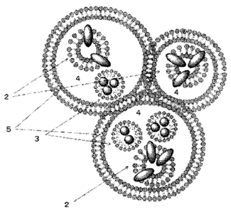
Fig. 1 shows a schematic view for clusters according to one embodiment.
CA 02839200 2013-12-12
Fig. 2 shows a schematic view for a vesicle according to one embodiment.
Fig. 3 shows a schematic view for an emulsion according to one embodiment.
Fig. 4 shows a schematic view for processes of a method for manufacturing
clusters
according to one embodiment.
Fig. 5 shows a drawing that displays processes for manufacturing clusters
according to one
embodiment.
Fig. 6 shows a schematic view of clusters according to one embodiment.
Fig. 7 shows a bright-field microscopic image of clusters according to an
example.
Fig. 8 shows a fluorescent microscopic image of clusters according to an
example.
Fig. 9 shows a bright-field microscopic image (after 27 hours) of clusters
according to an
example.
Fig. 10 shows a fluorescent microscopic image (after 27 hours) of clusters
according to an
example.
Fig. 11 shows a result for an X-ray CT image of clusters according to an
example.
Fig. 12 shows a endoscopic view of the inside of the stomach wall of a pig
when clusters are
injected according to an example.
Fig. 13 shows a laparoscopic view of the outside of stomach of a pig when
clusters are
injected according to an example.
6
CA 02839200 2013-12-12
Fig. 14 shows fluorescent laparoscopic images of the outside of stomach of a
pig when
clusters are injected according to an example.
Fig. 15 shows fluorescent laparoscopic images of the outside of stomach of a
pig when an
ICG aqueous solution is injected according to an example.
Fig. 16 shows fluorescent laparoscopic images of the outside of the stomach of
a pig when
clusters are injected (after 24 hours) according to an example.
Fig. 17 shows fluorescent images when an amount of injection is 50112
according to an
example.
Fig. 18 shows fluorescent images when an amount of injection is 100 2
according to an
example.
Fig. 19 shows fluorescent images when an amount of injection is 200g according
to an
example.
Fig. 20 shows fluorescent images when an amount of injection is 3001.11
according to an
example.
Fig. 21 is a fluorescent image of the excised stomach 32 hours after
administration of
clusters according to an example
Fig. 22 is a X-ray CT image of the excised stomach 32 hours after
administration of clusters
according to an example.
7
CA 02839200 2013-12-12
Fig. 23 shows images in which intensities of fluorescence are varied when ICG
concentration
and egg yolk lecithin concentration are changed.
Fig. 24 shows X-ray CT images when a vesicle fabricated in example 2 in
disperse liquid.
Fig. 25 shows endoscopic view of the local injection of clusters fabricated in
example 3 into
submucosal layer of the stomach wall (A), three-dimensional volumetric
reconstruction of X-
ray CT images immediately after administration of the marker (B), fluorescence
laparoscopic
view from the outside of the stomach 18 hours after administration of the
marker(C),
fluorescence laparoscopic view from the outside of the stomach 18 hours after
administration
of ICG aqueous slolution.
BEST MODE FOR CARRYING OUT THE PRESENT INVENTION
[0016]
Hereinafter, embodiments of the present invention are described with reference
to the
drawings. However, the present invention can be accomplished with different
embodiments
and is not limited to embodiments and examples described below.
[0017] (embodiment 1)
(a medical tissue-marker)
A medical tissue-marker according to the present embodiment 1 comprises a
vesicle formed
by combining a phospholipid and a near-infrared fluorescent dye, an emulsion
formed by
combining the phospholipid and an X-ray contrast medium, the emulsion being
incorporated
into a hydrophilic solvent, and has clusters in which a plurality of capsules
are formed and
8
CA 02839200 2013-12-12
aggregated by an emulsifier (hereinafter, referred to as "clusters"). Fig. 1
shows a schematic
view of clusters 1 in a medical tissue-marker according to the present
embodiment 1. Fig. 2
shows a schematic view of a vesicle 2 contained in the clusters 1, and fig. 3
shows a
schematic view of an emulsion 3 contained in the clusters 1.
[0018]
As shown in fig. 2, a vesicle 2 according to the present embodiment 1 is
formed by including
phospholipids 21 and near-infrared fluorescent dyes 22. Herein, the vesicle 2
means bag-
shaped bilayer membranes formed by self-assembled phospholipids due to
intermolecular
forces. The near-infrared fluorescent dyes 22 are combined with the
phospholipids 21 to
become component of the vesicle 2. Herein, the "combination" means a state of
forming
complex with a vesicle mainly by intermolecular interaction of hydrophobic
interaction or
means a state of being dissolved in the vesicle 2. By combining the near-
infrared fluorescent
dyes 22 with the phospholipids 21, the vesicle 2 according to the present
embodiment 1
stabilizes the near-infrared fluorescent dyes 22 to stably generate
fluorescent light in a near-
infrared region.
[0019]
The phospholipids 21 according to the present embodiment 1 are not limited as
far as a
vesicle can be formed. Examples thereof may be lecithin, phosphatidylcholine,
or mixtures
thereof. Lecithin is not limited. However, examples thereof maybe egg yolk
lecithin, soybean
lecithin, or mixtures thereof. From the viewpoint of fluorescence intensity in
a body, the
phospholipids 21 are preferably egg yolk lecithin.
[0020]
The phosphatidylcholine is not limited as far as requirement described above
is satisfied.
Example thereof may be 1-palmitoy1-2-oleoy1-3-sn-glycerophosphatidylcholine, 1-
steary1-2-
9
CA 02839200 2015-09-23
oleoy1-3-sn-glycerophosphatidylcholine, 1-palmitoy1-2-linoleate-3-sn-
glycerophosphatidylcholi
ne, 1-steary1-2-linoleate-3-sn-glycerophosphatidylcholine, 1,2-dilinoleate-3-
sn-phosphatidylch
oline, 1,2-dipalmitoy1-3-sn-glycerophosphatidylcholine, 1,2-disteary1-3-sn-
glycerophosphatidyl
choline, 1,2-dilinoleate-3-sn-glycerophosphatidylcholine, or mixtures thereof.
[0021]
In the present embodiment 1, the near-infrared fluorescent dyes 22 may be
indocyanine
green, brilliant green, Indigo Carmine or derivatives thereof. The near-
infrared fluorescent
dyes 22 means a compound in which a portion of the indocyanine green, the
brilliant green
or the Indigo Carmine is substituted with other functional group, while
maintaining main
structure and function thereof. The indocyanine green, the brilliant green and
the Indigo
Carmine are expressed by chemical formulae 1, 2, and 3, respectively.
[chemical formula 1]
(e *
( 1 )
O 03e Na
[chemical formula 2]
CA 02839200 2015-09-23
?2115 C21-15
+1
C21-15 C2115
( 2 )
11110 HSO4-
[chemical formula 3]
0
Na03S N
N
SO3Na ( 3 )
0
[0022]
Size of the vesicle 2 according to the present embodiment 1 is not
particularly limited.
Generally, the size thereof is preferably 10 nm or more and 100[1m or less,
and is more
preferably 100 nm or more and 10 m or less.
[0023]
In the vesicle 2 according to the present embodiment 1, amounts of the
phospholipids 21 and
the near-infrared fluorescent dyes 22 can be adjustable without limitation.
For example, when
amount of lecithin of the phospholipids 21 is one, amount of indocyanine green
of the near-
infrared fluorescent dyes 22 is preferably 1X10⁻⁴ or more and 1X10⁻³ or less,
and is more
preferably 4X10⁻³ or more and 6x10⁻³ or less. Within the range of 1X10⁻⁴ or
more and 1X10-
or less, it is easy to identify a marking position for a tissue in the inside
of an organ from the
outside of an organ. Within the range of 4X10⁻³ or more and 6x10⁻³ or less,
effect thereof
becomes more remarkable.
[0024]
11
CA 02839200 2013-12-12
As shown in fig 3, an emulsion 3 according to the present embodiment 1 is
formed by
including phospholipids 31 and X-ray contrast mediums 32. Herein, the emulsion
3 means pa
rticles wrapped bymolecular film formed by the phospholipids 31 being self-
assembled due
to intermolecular interaction. The X-ray contrast mediums 32 mean component of
the
emulsion 3 by combination with the phospholipids 31. Herein, the "combination"
means a
state of forming complex with the phospholipids 31 mainly by intermolecular
interaction of
hydrophobic interaction or means a state of being dissolved in the emulsion 3.
By combining
the X-ray contrast mediums 32 with the phospholipids 31, the emulsion 3
according to the
present embodiment 1 stabilizes the X-ray contrast mediums 32 to properly
capture an X-ray
CT image.
[0025]
The phospholipids 31 according to the present embodiment 1 are similar to the
phospholipids
21 in the vesicle 2.
[0026]
In the present embodiment 1, the X-ray contrast mediums 32 are not limited.
Example
thereof preferably is iodized poppy oil ethyl ester and derivative thereof,
iodobenzene and
derivative thereof, barium salt or mixtures thereof. The iodized poppy oil
ethyl ester is a
compound obtained by iodization and esterification of a poppy oil fatty acid.
Example of the
iodized poppy oil ethyl ester can be expressed by a chemical formula (4). From
the viewpoint
of X-ray absorption ratio in an organ, the X-ray mediums 32 are preferably the
iodized poppy
oil ethyl ester.
[chemical formula 4]
12 =
CA 02839200 2015-09-23
0
H H
H3C HA" ¨C ____ C--(CH2)m
( 4 )
rn is an integer of 1-10, m is an integer of 2-12]
[0027]
Size of the emulsion 3 according to the present embodiment 1 is not
particularly limited.
Generally, the size thereof is preferably 10 nm or more and 100[im or less,
and is more
preferably 100 nm or more and 10p.m or less.
[0028]
In the emulsion 3 according to the present embodiment 1, amounts of the
phospholipids 31
and the X-ray contrast mediums 32 can be adjustable without limitation. For
example, when
amount of lecithin of the phospholipids 31 is one, amount of the iodized poppy
oil ethyl ester
of the X-ray contrast mediums 32 is preferably 1X10⁻¹ or more and 1X10³ or
less, and is
more preferably 2X10⁻¹ or more and 2x101 or less. Within the range of 1X10⁻¹
or more and
1X10³ or less, it is possible sufficiently to protect surface of the emulsion
3 of the iodized
poppy oil ethyl ester with a film of the phospholipids 31. Within the range of
2X10⁻¹ or more
and 2x101 or less, effect thereof becomes more remarkable.
[0029] =
As shown in fig. 1, the clusters 1 according to the present embodiment1
includes a plurality
of capsules 5 in which a hydrophilic solvent 4 is incorporated, wherein the
plurality of
capsules 5 are formed and aggregated by an emulsifier. In the hydrophilic
solvent 4, at least
13
CA 02839200 2013-12-12
any one of the vesicle 2 and the emulsion 3 is incorporated.
[0030]
The hydrophilic solvent 4 is used for the vesicle 2 and the emulsion 3 being
stably
incorporated and is not limited as far as the above condition is satisfied.
Preferably, example
thereof is water, physiological salt water, phosphate buffer solution, TRIS
hydrochloric acid
buffer solution, HEPES buffer solution, or mixtures thereof. When phosphate
buffer solution,
TRIS hydrochloric acid buffer solution or HEPES buffer solution is used, the
range of pH 6.5
or more and 8 or less is preferable.
[0031]
In order to perform stable stay for a long period at a marking position of
tissue in a body, an
edible thickener is preferably added into the hydrophilic solution 4. Example
thereof is not
limited and may be gelatin, agar, fibrinogen, saccharide, or mixtures thereof.
[0032]
Example of the gelatin is not limited and may be collagen type I, collagen
type II, collagen
type III, collagen type V or mixtures thereof.
[0033]
Example of the agar is not limited and may be agarose, agaropectin, or
mixtures thereof, the
agarose and the agaropectin having molecular weight of from several thousands
to several
ten thousands.
[0034]
Example of the fibrinogen is not limited. For example, fibrinogen having
concentration of from
mg/mL to 50 mg/mL as a main ingredient is included, and calcium chloride,
prothrombin, or
14
CA 02839200 2013-12-12
mixtures thereof is also included.
[0035]
Example of saccharide is not limited and may be glucose, sucrose, maltose,
galactose,
arabinose, ribulose, fructose, rutose, mannose, lactose, cellobiose or
mixtures thereof.
[0036]
Amount of adding the edible thickener is not limited. When amount of the
hydrophilic solvent
4 contained in the capsules is one, amount of an edible thickener is
preferably 1X10⁻³ or
more and 10 or less, and is more preferably 1X10⁻¹ or more and 1 or less.
Within the range of
1X10⁻³ or more, it is possible to increase viscosity of the hydrophilic
solvent 3. Within the
range of1X10-1 or more, effect thereof becomes more remarkable. Within the
range of 10 or
less, a lowering of fluidity for the hydrophilic solvent 3 can be restrained,
and within the range
of 1 or less, effect thereof becomes more remarkable.
[0037]
in the present embodiment 1, weight ratio of sum of the near-infrared
fluorescent dyes, the
X-ray contrast mediums and the phospholipids with respect to the hydrophilic
solvent (weight
ratio of the vesicle and the emulsion) is not limited as far as sufficient
fluorescence intensity
can be maintained as a medical tissue-marker and an X-ray CT image are
sufficiently captur
ed. Preferably, the weight ratio may be 100:1 or more and 1:100 or less, and
more preferably,
the weight ratio may be 10:1 or more and 1:1 or less. When the ratio is 100:1
or more,
fluorescence intensity and X-ray absorption ratio of the medical tissue-marker
is higher than
those of an organ which is background. When the weight ratio is 10:1 or more,
the effect
thereof becomes remarkable. Furthermore, when the weight ratio is 1:100 or
less,
interference is restrained by X-ray absorption with respect to fluorescent
light, and when the
weight ratio is 1:1 or less, the effect thereof becomes remarkable.
CA 02839200 2013-12-12
[0038]
In the present embodiment 1, weight of the near-infrared fluorescent dyes, the
X-ray contrast
mediums and the phospholipids which are added into the hydrophilic solvent
(weight of the
vesicle and the emulsion) is not limited as far as sufficient fluorescence
intensity can be
maintained as a medical tissue-marker and an X-ray CT image are sufficiently
captured.
When weight of the hydrophilic solvent (in case of including edible thickener
and the like,
weight including the edible thickener, etc.) is one, the weight thereof is
preferably 1X10⁻⁴ or
more and 1X10⁻¹ or less, and is more preferably 1X10⁻³ or more and 1X10⁻² or
less. Within
the range of 1X10⁻⁴ or more, it is possible to enhance fluorescence intensity
and X-ray
absorption ratio, and within the range of1X10-3 or more, effect thereof
becomes more
remarkable. Furthermore, within the range of 1X10⁻¹ or less, changing into
lamella phase
instead of being the vesicle and the emulsion may be restrained in the
hydrophilic solvent
and within the range of 1X10⁻² or less, effect thereof becomes more
remarkable.
[0039]
In the present embodiment 1, the emulsifier is formed on walls of the capsules
in which the
hydrophilic solvent is contained, and the emulsifier is used for aggregation
as clusters. The
emulsifier according to the present embodiment 1 can form not only walls of
the capsules but
also epidermis covering entire clusters. Thus, a plurality of capsules can be
aggregated and
combined. Example of the emulsifier according to the present embodiment 1 is
not limited
and may be polyglyceryl polyricinoleate, polyglyceryl polyricinoleate
derivative, glycerol fatty
acid ester derivative or mixtures thereof.
[0040]
In the present embodiment 1, weight of the emulsifier added for forming the
capsules is not
limited. When weight of the hydrophilic solvent (including total weight of the
near-infrared
16
CA 02839200 2013-12-12
fluorescent dyes, X-ray contrast mediums and the phospholipids, and in the
case where
edible thickener is also included, including total weight thereof) is one, the
weight of the
emulsifier is preferably 1X10⁻³ or more and 1 or less, and is more preferably
1X10⁻² or more
and 1X10⁻¹ or less. Within the range of 1X10⁻³ or more, the emulsifier can
stably makes the
capsules of the hydrophilic solvent, and within the range of1X10-2 or more,
effect thereof
becomes more remarkable. Furthermore, within the range of 1 or less, reaction
in which the
emulsifier, hydrophobic solvent and a first hydrophilic solvent form a gel
layer is restrained,
and within the range of 1X10⁻¹ or less, effect thereof becomes more
remarkable.
[0041]
In the present embodiment 1, particle diameter is not limited as far as
function for a medical
tissue-marker is maintained. For example, the particle diameter is preferably
50p.m or more
and 5001.tm or less, and is more preferably 10010 or more and 250 m or less.
Within the
range of 50 m or more, the maker is hard to decompose and fluorescence
intensity for the
marker can be enhanced. Within the range of 100m or more, effect thereof
becomes more
remarkable. Furthermore, Within the range of 500 m or less, it is possible to
restrain that
injection needle through an endoscope is stopped. Within the range of 250[im
or less, effect
thereof becomes more remarkable.
[0042]
In the present embodiment 1, number of capsules in one cluster is not limited
as far as
function for a medical tissue-marker is maintained. For example, the number of
capsules is
preferably 1 or more and 103 or less, and is more preferably 10 or more and
102 or less.
Within the range of 1 or more, fluorescence intensity is increased and within
the range of 10
or more, effect thereof becomes more remarkable. Furthermore, within the range
of 103 or
less, strength for the capsules is increased and the marker becomes stable,
and within the
range of 102 or less, effect thereof becomes more remarkable.
17
CA 02839200 2013-12-12
[0043]
Furthermore, in order to preferably maintain clusters, a medical tissue-marker
according to
the present embodiment 1 uses, for example, a hydrophobic solvent, the
clusters being
maintained in the hydrophobic solvent. Thus, there is an effect that a
plurality of capsules are
formed and aggregated. Besides the above solvent, in order to stabilize and
strengthen the
function of the medical tissue-marker, another element such as hydrophobic
polymer and the
like can be added to cross-link.
[0044]
Hereinabove, by a medical tissue-marker according to the present embodiment 1,
it is
possible to identify a position even in the outside of an organ, to be locally
stayed for a long
period, and to catch a marking position within an entire organ.
[0045]
More specifically, a medical tissue-marker according to the present embodiment
1 can
strongly and stably generate a near-infrared fluorescent light since near-
infrared fluorescent
dyes are combined with a vesicle, and can photograph an X-ray CT image since X-
ray
contrast mediums are forming an emulsion. Furthermore, even when capsules are
contacted
with tissue liquid in a body by being drived into an organ, there is an
advantage that each
capsule is hard to be dissociated and locally to be stayed for a long period
since both the
vesicle and the emulsion are incorporated into a hydrophilic solution to form
clusters that
includes the capsules. There is also advantage that strength for a local stay
for a long period
and flexibility for injection into an organ through passage of an endoscope
are maintained
since an edible thickener is used for the capsules of the clusters.
[0046] (A method for manufacturing clusters)
Herein, an example of a method for manufacturing a medical tissue-marker
(hereinafter,
18
CA 02839200 2013-12-12
referred to as "the present manufacturing method") is described in detail.
Fig. 4 is a
schematic view of the present manufacturing method.
[0047]
As shown in fig. 4, the present manufacturing method is characterized by
comprising a first
step of adding a near-infrared fluorescent dye, an X-ray contrast medium and a
phospholipid
into a first hydrophilic solvent and stirring the first hydrophilic solvent, a
second step of
adding the first hydrophilic solvent and an emulsifier into a hydrophobic
solvent to form
suspension, and a third step of performing centrifugation by the suspension
and a second
hydrophilic solvent.
[0048]
By the first step of adding a near-infrared fluorescent dye, an X-ray contrast
medium and a
phospholipid into a first hydrophilic solvent and stirring the first
hydrophilic solvent, a vesicle
including the phospholipid combined with the near-infrared fluorescent dye and
an emulsion
including the phospholipid combined with the X-ray contrast medium can be
formed. In the
present embodiment 1, there is an advantage that this operation can be
performed at one
time, and large device is unnecessary. The step of forming the vesicle and the
step of
forming the emulsion are separately performed. Then each solvent may be mixed
to be one
solvent. In this case, the step of forming the vesicle and the step of forming
the emulsion are
not limited to the step of adding the phospholipid for stirring. The step of
removing solvent
may be used by decompression process after the phospholipid is mixed with an
organic
solvent or supercritical fluid. The step of performing filter treatment or
ultrasonic treatment by
adding the phospholipid may also be used. However, from the viewpoints of
enhancement
for biocompatibility by including no organic solvent and stability of the
phospholipid, the step
of adding the phospholipid for stirring is preferable.
19
CA 02839200 2013-12-12
[0049]
In the first step described above, from the viewpoint of the capsules 4 being
easily formed,
the first hydrophilic solvent preferably employs the same as the hydrophilic
solvent which
exists in the capsules. That is to say, example of the first hydrophilic
solvent is preferably
water, physiological salt water, phosphate buffer solution, TRIS hydrochloric
acid buffer
solution, HEPES buffer solution, or mixtures thereof.
[0050]
The first hydrophilic solvent preferably includes an edible thickener. The
edible thickener may
be gelatin, agar, fibrinogen, saccharide, or mixtures thereof.
[0051]
Amount of the near-infrared fluorescent dyes, the X-ray contrast mediums and
the
phospholipids with respect to the first hydrophilic solvent is not limited.
The same range
preferably applied to in relation to the near-infrared fluorescent dyes, the X-
ray contrast
mediums and the phospholipids in the hydrophilic solvent which exists in the
capsules. That
is to say, when weight (in case of including an edible thickener and the like,
including weight
thereof) of the first hydrophilic solvent is one, weight thereof is preferably
1X10⁻⁴ or more and
1X10⁻¹ or less, and is more preferably 1X10⁻³ or more and 1X10⁻² or less.
Within the range of
1X10⁻⁴ or more, intensity of fluorescent light is increased and an X-ray CT
image is
sufficiently captured, and within the range of 1X10⁻³ or more, effect thereof
becomes more
remarkable. Furthermore, within the range of 1X10⁻¹ or less, changing into
lamellar phase
instead of the vesicle and the emulsion in the hydrophilic solvent is
restrained, and within the
range of 1X10⁻² or less, effect thereof becomes more remarkable.
[0052]
Temperature for performing the first step is not limited as far as the vesicle
and an emulsion
CA 02839200 2013-12-12
can be formed. Example thereof is preferably 4 C or more and 80 C or less, and
more
preferably is room temperature for a convenience. A time for stirring the
first hydrophilic
solvent is also not limited as far as the vesicle and the emulsion can be
formed. Example
thereof is preferably 5 minutes or more and 1 hour or less, and more
preferably is 10 minutes
or more and 30 minutes or less.
[0053]
By the second step for forming suspension by adding the first hydrophilic
solvent and the
emulsifier into the hydrophobic solvent, the emulsifier can be boarded on
around the first
hydrophilic solvent in which the vesicle and the emulsion are incorporated,
and a plurality of
capsule-shaped emulsion in the hydrophobic solvent can be formed.
[0054]
The hydrophobic solvent in the second step is not limited as far as the
capsule-shaped
emulsion is formed at a temperature of 4 C or more and 80 C or less. Example
of the
hydrophobic solvent is kerosene, hexane, decane, dodecane, heptane, squalene,
squalane,
liquid paraffin, mineral oil or mixtures thereof.
[0055]
In the present embodiment 1, weight of the hydrophobic solvent is not limited.
When weight
of the hydrophilic solvent is one, the weight of the hydrophobic solvent is
preferably 1 or
more and 100 or less, and is more preferably 5 or more and 10 or less. Within
the range of 1
or more, it is restrained that capsule-shaped emulsion is transferred to gel
phase, and within
the range of 5 or more, effect thereof becomes more remarkable. Furthermore,
within the
range of 100 or less, the clusters stably maintain a particle diameter of
capsule-shaped
emulsion, and within the range of 10 or less, effect thereof becomes more
remarkable.
[0056]
21
CA 02839200 2013-12-12
In the second step, the emulsifier described above can be employed.
[0057]
In the second step, amount of the first hydrophilic solvent can be properly
adjusted without
limitation. For example, when weight amount of the hydrophobic solvent is one,
the amount
of the first hydrophilic solvent is preferably 1X10⁻³ or more and 1 or less,
and is more
preferably 1X10⁻² or more and 1X10⁻¹ or less. Within the range of 1X10⁻³ or
more, intensity of
fluorescent light of a marker is enhanced by increasing number of capsules per
cluster, and
within the range of 1X10⁻² or more, effect thereof becomes more remarkable.
Furthermore,
within the range of 1 or less, phase separation between the hydrophilic
solvent and the
hydrophobic solvent is restrained, and within the range of 1X10⁻¹ or less,
effect thereof
becomes more remarkable.
[0058]
In the second step, amount of the emulsifier can be properly adjusted without
limitation. For
example, when weight amount of the hydrophilic solvent is one, the emulsifier
is preferably
1X10⁻³ or more and 1 or less, and is more preferably 1X10⁻² or more and 1X10⁻¹
or less.
Within the range of 1X10⁻³ or more, the emulsifier stably generates the
capsules of the
hydrophilic solvent, and within the range of 1X10⁻² or more, effect thereof
becomes more
remarkable. Furthermore, within the range of 1 or less, reaction in which the
emulsifier, the
hydrophobic solvent and the first hydrophilic solvent form a gel layer is
restrained, and within
the range of 1X10⁻¹ or less, effect thereof becomes more remarkable.
[0059]
In the present embodiment 1, the third step of performing centrifugation by
the suspension
and a second hydrophilic solvent is a method in which a hydrophobic solvent
layer and a
hydrophilic solvent layer is phase-separated for an arrangement and the
capsule-shaped
22
CA 02839200 2013-12-12
emulsion existing in the hydrophobic solvent is precipitated in the
hydrophilic solvent by
centrifugation. In fig. 5, the schematic view is shown. As a result, the
emulsion on the
interface between the hydrophobic solvent layer and the hydrophilic solvent
layer can form
clusters from capsules.
[0060]
A second hydrophilic solvent may be used for centrifugation without
limitation. For example,
water, physiological salt water, phosphate buffer solution, TRIS hydrochloric
acid buffer
solution, HEPES buffer solution, or mixtures thereof is preferably used.
[0061]
Amount of a second solvent is not limited. For example, when amount of
suspension is one,
the second solvent is preferably 1 or more and 1000 or less, and more
preferably 10 or more
and 100 or less. Within the range of 1 or more, phase separation between the
hydrophobic
solvent layer and the hydrophilic solvent layer can be stabilized, and within
the range of 10 or
more, effect thereof becomes more remarkable. Furthermore, within the range of
1000 or
less, the lowering of viscosity can be restrained, and within the range of 100
or less, effect
thereof becomes more remarkable.
[0062]
As a result, a medical tissue-marker can be configured.
[0063] (embodiment 2)
(a medical tissue-marker)
A medical tissue-marker according to the present embodiment 2 is almost the
same as the
embodiment 1 except that when suspension is formed by adding a first
hydrophilic solvent
and an emulsifier to a hydrophobic solvent, X-ray contrast mediums is added
into the
23
CA 02839200 2013-12-12
hydrophobic solvent. Difference therebetween is described below.
[0064]
Fig. 6 is a schematic view for clusters 1 in a medical tissue-marker according
to the present
embodiment 2. As shown in fig. 6, a medical tissue-marker according to the
present
embodiment 2 is characterized by including the X-ray contrast mediums even in
the outside
of the capsules 5. Thus, lots of the X-ray contrast mediums can be included.
It is also
possible to include the X-ray contrast mediums located in near distance with
the outside of
the clusters 1. Thus, sensitivity thereof is enhanced.
[0065]
(a method for manufacturing clusters)
Herein, a method for manufacturing a medical tissue-marker according to the
present
embodiment 2 is described. A method for manufacturing clusters according to
the present
embodiment 2 is almost the same as the embodiment 1 except that suspension is
formed by
adding a first hydrophilic solvent and an emulsion into a hydrophobic solvent
in the second
step of the embodiment 1. Specifically, when suspension is formed by adding
the first
hydrophilic solvent and the emulsifier into the hydrophobic solvent, adding X-
ray contrast
mediums differs from the embodiment 1.
[0066]
The X-ray contrast mediums added in the second step is the same as in the
embodiment 1.
Concentration of the X-ray contrast mediums is not specially limited. For
example, amount of
the first hydrophilic solvent is one, amount of the X-ray contrast mediums is
preferably 0.01
or more and 10 or less, and more preferably 0.1 or more and 1 or less. Within
the range of
0.01 or more, sensitivity for the X-ray contrast image is increasing, and
within the range of
0.1 or more, effect thereof becomes more remarkable. Furthermore, within the
range of 10 or
24
CA 02839200 2013-12-12
less, when clusters is fabricated, precipitation due to its weight is
restrained, and within the
range of 1 or less, effect thereof becomes more remarkable.
[0067]
Hereinabove, by a medical tissue-marker according to the present embodiment 2,
it is
possible to identify a position from the outside of an organ even marked on
the inside of an
organ, and to be locally stayed for a long period. It is also possible to
catch a marking
position within an entire organ. Especially, by a marker according to the
present embodiment
2, the X-ray contrast mediums can be included even in the outside of capsules.
Thus,
sensitivity thereof is increased.
Examples
[0068]
Herein, a medical tissue-marker was specifically fabricated and effects of the
present
invention were confirmed. Hereinafter, the details are described below.
[0069] (example 1)
In the present example 1, TRIS hydrochloric acid buffer solution as a first
hydrophilic solvent,
indocyanine green (hereinafter, referred to as "ICG") as a near-infrared
fluorescent dye,
iodized poppy oil ethyl ester (hereinafter, referred to as "LPD") as a X-ray
contrast medium,
and egg yolk lecithin as a phospholipid were employed, respectively. Sucrose
as a thickener
was employed.
[0070]
TRIS buffer solution of 1 mL was prepared to be 50 mM and pH 7.8 in a glass
tube at room
temperature. Then, ICG of 2X10⁻² mM, LPD of 20 mM, and egg yolk lecithin of 30
mM were
added thereto for stirring. A vesicle and an emulsion were formed.
CA 02839200 2013-12-12
[0071]
Then, polyglyceryl polyricinoleate (PGPR) of 15w/w% was dissolved into
squalene of a
hydrophobic solvent of 15 mL. The solution of 1mL including the vesicle and
the emulsion
fabricated was added thereto. Suspension including emulsion by PGPR (PGPR
emulsion)
was prepared. In the present example 1, LPD of 4 mM was added even into the
hydrophobic
solvent. Thus, the LPD existed in the PGPR emulsion or surrounding the PGPR
emulsion.
[0072]
Then, TRIS buffer solution of 5 mL having 50 mM and pH7.7 was prepared as a
second
hydrophilic solution. Suspension of 10 mL including the PGPR emulsion was
added into the
second hydrophilic solution from upper side by using glucose as thickener. Oil
phase
(squalene phase) and aqueous phase (TRIS buffer solution phase) were contacted
each
other and rotated at a speed of 3500 rpm for 30 minutes at room temperature to
form
clusters of PGPR.
[0073]
Fig. 7 is a bright-field microscopic image of clusters of PGPR. Fig. 8 is a
fluorescent
microscopic image. From fig. 7 and fig. 8, existing clusters and generation of
fluorescent light
were confirmed. Fig. 9 is a bright-field microscopic image when clusters of
PGPR were
fabricated and 27 hours were passed. Fig. 10 is a fluorescent microscopic
image when
clusters of PGPR were fabricated and 27 hours were passed. As a result, even
after one day
or more was passed, it was confirmed that shape and function of clusters were
stable.
[0074]
Fig. 11 shows a result of an X-ray computed tomography (CT) in a state of
fluid dispersion
with respect to the clusters. From fig. 11, it was confirmed that X-ray
absorption (CT
numbers) of the clusters were sufficiently higher than that of the stomach
wall itself.
26
CA 02839200 2013-12-12
[0075]
Then, clusters fabricated were injected into biological tissue and result
thereof was confirmed.
Specifically, submucosal layer of the stomach wall of a pig was a most
suitable target of the
marker administration. The fluid dispersion of 300p.e including clusters was
administrated on
four points surrounding a metal clip placed inside the stomach by local
injection. Fig. 12
shows an endoscopic view of the inside of the stomach. Fig. 13 shows a
laparoscopic view of
the outside of the stomach.
[0076]
Fig. 14 shows fluorescent images of the outside of stomach. Left and middle
panels show
conventional and fluorescent laparoscopic images immediately after local
injection,
respectively. Right panel showed a fluorescent image 6 hours after injection.
As shown in fig.
14, even after 6 hours, four injection points were clearly identified. It was
confirmed that
clusters were sufficiently stayed at the injection points. Fig. 15 shows
fluorescent images
when ICG aqueous solution was locally injected to the positions similar to
those as indicated
in fig. 14. In this case, exact injection positions were unclear immediately
after injection as
well as 6 hours after injection.
[0077]
Herein, stability was confirmed again 24 hours after local injection of the
marker.
Submucosal layer of the stomach wall was a most suitable target of the marker
administration. The fluid dispersion of 300 2 including clusters was
administrated at two
points surrounding a metal clip by local injection. The pig was recovered from
general
anesthesia after the administration. Twenty-four hours later, laparoscopy was
performed
again under general anesthesia. Fluorescent laparoscope view of the outside of
the stomach
revealed sufficient fluorescent intensity at the injection points as indicated
in fig.16.
27
CA 02839200 2013-12-12
[0078]
Optimum injection amount of the marker was verified. When the concentration of
the clusters
according to the example 1 was 1042 or more, injection points could be
confirmed. The
concentration thereof was preferably 200p.¾ or more, and is more preferably
nog or more.
Fluorescent images of the injection site with various amount of the marker are
shown in figs.
17 to 20. In fig. 17, 501.te was used. In fig. 18, wog was used. In fig. 19,
200p.g was used.
In fig. 20, 300 2 was used.
[0079]
Then, stomach of the pig was excised, and fluorescence and X-ray CT imaging
was
performed 32 hours after marker paw each at four points around a metal clip)
administration. Fluorescence imaging with the use of a near-infrared LED light
showed four
spots of individually distinguishable fluorescence on the marker injection
site (pyloric side of
the stomach, i.e., right side on the image of the stomach, in fig. 21.) and
broad diffusion of
the fluorescence on the site of ICG aqueous solution injection site (cardia
side of the
stomach, i.e., left side on the image of the stomach, in fig 21.)
[0080]
Furthermore, a volumetric reconstruction of the X-ray CT images of the stomach
was
performed to allow three-dimensional analysis of the location of the injection
sites. Fig. 22
shows three-dimensional reconstructed CT images. Injection sites of the marker
on the
three-dimensional image were observed in exactly the same location with that
revealed
under the fluorescent laparoscope observation.
[0081]
Hereinabove, according to the example 1, it was confirmed that a medical
tissue-marker and
a manufacturing method therefor were provided. This medical tissue-marker
could be
28
CA 02839200 2013-12-12
detected by X-ray CT as well as fluorescent imaging. Furthermore, injection
site of the
marker could be identified from the outside of an organ, and to be locally
stayed for a long
period, and it is easy to catch a marking location within the entire organ.
[0082]
In the example 1, for example, the concentration of ICG was 3.2X10⁻²mM and the
concentration of ICG can be adjustable. In fig. 23(a), changes of fluorescence
intensity are
shown when the concentration of ICG combined with the vesicle was varied (egg
yolk lecithin
of 30mM). In fig. 23(b), changes of fluorescence intensity were shown when the
concentration of egg yolk lecithin combined with the vesicle was varied (the
concentration of
ICG aqueous solution of 3.2X10⁻²mM). It was confirmed that the concentration
of ICG was
preferably 3.2X10⁻¹mM or more and 1.6X10⁻¹mM or less, and the concentration of
egg yolk
lecithin was 5mM or more and 40mM or less.
[0083] (example 2)
In the present example 2, a medical tissue-marker was fabricated by using the
same material
and method used in the example 1 except that LPD was only added to a first
hydrophilic
solvent. Difference therebetween is mainly described below.
[0084]
TRIS buffer solution of 1 mL was prepared to be 50 mM and pH 7.8 in a glass
tube at room
temperature. Then, ICG of 3.2X10⁻²mM, LPD of 20 mM, egg yolk lecithin of 30 mM
were
added thereto for stirring. A vesicle and an emulsion were formed.
[0085]
Then, polyglyceryl polyricinoleate (PGPR) of the emulsifier having 15w/w% was
dissolved
into squalene of a hydrophobic solvent of 15 mL. The solution of 1mL including
the vesicle
29
CA 02839200 2013-12-12
and the emulsion fabricated was added thereto. Suspension including emulsion
by PGPR
(PGPR emulsion) was prepared. In the present example 2, no LPD was added into
the
hydrophobic solvent.
[0086]
Then, IRIS buffer solution of 5 mL having 50 mM and pH7.7 was prepared as a
second
hydrophilic solution. Suspension of 10 mL including the PGPR emulsion was
added into the
second hydrophilic solution from upper side by using glucose as a thickener.
Oil phase
(squalene phase) and aqueous phase (TRIS buffer solution phase) were contacted
each
other and rotated at a speed of 3500 rpm for 30 minutes at room temperature to
form
clusters of PGPR.
[0087]
Fig. 24 shows X-ray CT images of clusters fabricated in fluid dispersion. Even
in clusters, X-
ray absorption (CT numbers) of it is sufficiently higher than that of the
stomach wall itself.
[0088] (Example 3)
In the present example 3, a medical tissue-marker using ICG-8 shown in
chemical formula
(5) of ICG derivative was fabricated in the following steps. TRIS buffer
solution of 1 mL was
prepared to be 50 mM and pH 7.8 in a glass tube at room temperature. Then, ICG-
8 of
3.2X10⁻²mM, LPD of 40mg/mL, egg yolk lecithin of 30 mM were added thereto for
stirring. A
vesicle and an emulsion were formed.
[chemical formula 5]
CA 02839200 2015-09-23
4
,
/
0
SO3
(5)
[0089]
Then, polyglyceryl polyricinoleate (PGPR) of the emulsifier having 15w/w% and
[PD of
160mg/mL were dissolved into squalene of a hydrophobic solvent of 15 mL. The
solution of
1mL including the vesicle and the emulsion fabricated was added thereto.
Suspension
including emulsion by PGPR (PGPR emulsion) was prepared.
[0090]
Then, TRIS buffer solution of 5 mL having 50 mM and pH7.7 was prepared as a
second
hydrophilic solution. Suspension of 10 mL including the PGPR emulsion and LH)
were
added into the second hydrophilic solution from upper side by using glucose as
a thickener.
Oil phase (squalene phase) and aqueous phase (TRIS buffer solution phase) were
contacted
each other and rotated at a speed of 3500 rpm for 30 minutes at room
temperature to form
clusters of PGPR.
=
[0091]
31
CA 02839200 2013-12-12
Fig. 25 shows results that (A) the present medical tissue-marker was injected
with every 300
into four points on a circumference of a circle for submucosal layer of the
stomach wall of
a pig under general anesthesia, (B) it was possible to clearly identify the
four points of the
injection in three-dimensional reconstruction images of X-ray CT immediately
after the
administration, (C) the medical tissue-marker locally injected at four points
closely located
each other on the stomach wall of the pig were individually distinguishable by
fluorescent
laparoscope even 18 hours after administration, and (D) it was impossible to
distinguish four
points of locally injected ICG aqueous solution with the same manner as the
medical tissue-
marker because of broad blurring of the solution through tissues.
[0092]
Thus, according to the present examples, it was confirmed that a medical
tissue-marker and
a manufacturing method therefor were provided. With the use of the medical
tissue-marker
and the manufacturing method therefor, it was possible to obtain marking point
images with
X-ray CT as well as a fluorescent endoscope, it was also possible to identify
the marking
positions from the outside of the organ even administered inside in an organ
and to be locally
stayed for a long period, and it is easy to catch an accurate marking
positions within an entire
organ.
Industrial Applicability
[0093]
The present invention is industrially applicable a medical tissue-marker and a
manufacturing
method therefor.
32