Note: Descriptions are shown in the official language in which they were submitted.
CA 02839261 2013-12-12
WO 2012/174056 PCT/US2012/042165
MITIGATION OF CUTANEOUS INJURY WITH IL-12
CROSS-REFERENCE TO RELATED APPLICATIONS
[0001] This application claims priority to U.S. Provisional Application Serial
Number
61/496,472, filed June 13, 2011, and U.S. Provisional Application Serial
Number 61/528,053,
filed August 26, 2011, each of which are incorporated herein by reference in
their entirety,
including all figures and tables.
BACKGROUND
[0002] Cutaneous wounds represent a major cause of morbidity in diabetics.
Every year, 2-
3% of diabetics will develop foot ulcers. The lifetime probability of
development of a
diabetic foot ulcer is between 10 and 15%. In wound injuries that result in
limb amputation,
up to 90% began with a foot ulcer.
[0003] In addition, burn injury is a common cause of morbidity and mortality
in the United
States, with approximately 100,000 cases of moderate to severe burn injuries
requiring
hospitalization and 5000 patients dying of burn-related complications each
year (Church et
al., Clin Microbiol Rev., 19(2):403-34 (2006)).
[0004] Reduction of the healing time for cutaneous wounds is highly desirable,
as this can
reduce the chances of infection and other complications. However, healing of
cutaneous
injuries is a complex process and can be slowed or interrupted by a variety of
other factors,
including diabetes, venous or arterial disease, old age, and infection,
leading to chronic
wounds.
SUMMARY OF INVENTION
[0005] In one aspect, the present invention relates to methods for treating a
cutaneous
wound in a subject, including administrating IL-12 to the cutaneous wound. In
some
embodiments, the administration is topical, subcutaneous, intramuscular, or
any combination
thereof In some embodiments the subject is human.
[0006] In some embodiments of the foregoing aspect, the IL-12 is administered
topically.
In some embodiments the IL-12 is administered topically in a dosage of from
about 1 ng/mL
up to about 10 [tg/mL. In some embodiments, the IL-12 is administered
topically in a dosage
of from about 10 ng/mL up to about 5 [tg/mL. In some embodiments, the IL-12 is
administered topically at a dosage of about 100 ng/mL.
1
CA 02839261 2013-12-12
WO 2012/174056 PCT/US2012/042165
[0007] In some embodiments of the foregoing aspect, the IL-12 is administered
subcutaneously. In some embodiments, the IL-12 is administered subcutaneously
in a dosage
of from about 10 ng/kg to about 500 ng/kg. In some embodiments, the IL-12 is
administered
subcutaneously in a dosage of about 80 ng/kg.
[0008] In some embodiments, administration of IL-12 results in at least about
a 5% increase
in wound healing as measured by wound closure, as compared to wound closure
observed in
the absence of IL-12 administration. In some embodiments, administration of IL-
12 results
in at least about a 20% increase in wound healing as measured by wound
closure, as
compared to wound closure observed in the absence of IL-12 administration. In
some
embodiments, administration of IL-12 results in at least about a 50% increase
in wound
healing as measured by wound closure, as compared to wound closure observed in
the
absence of IL-12 administration. In some embodiments, administration of IL-12
results in at
least about a 75% increase in wound healing as measured by wound closure, as
compared to
wound closure observed in the absence of IL-12 administration. In some
embodiments,
administration of IL-12 results in at least about a 95% increase in wound
healing as measured
by wound closure, as compared to wound closure observed in the absence of IL-
12
administration.
[0009] In some embodiments, of the foregoing aspect, the subject has been
exposed to
radiation, which results in a cutaneous wound. In some embodiments, the IL-12
is
administered from about 1 hour up to about 24 hours following exposure to
radiation
resulting in a cutaneous wound.
[0010] In some embodiments, the subject is a member of a patient population
characterized
by an impediment to normal cutaneous wound healing. In some embodiments, the
subject is
diabetic, elderly, or is an HIV/AIDS patient. In some embodiments, the
cutaneous wound is
a burn wound. In some embodiments, the cutaneous wound is present at a
surgical site. In
some embodiments, the IL-12 is administered in conjunction with a skin graft.
In some
embodiments, the IL-12 is administered in conjunction with acellular or
cellular dermal
matrices. In some embodiments, the IL-12 is emulsified in a gel matrix. In
some
embodiments, the gel matrix is isotonic 4% carboxymethylcellulose.
[0011] The foregoing general description and following brief description of
the drawings
and the detailed description are exemplary and explanatory and are intended to
provide
further explanation of the invention as claimed. Other objects, advantages,
and novel features
will be readily apparent to those skilled in the art from the following
detailed description of
the invention.
2
CA 02839261 2013-12-12
WO 2012/174056 PCT/US2012/042165
BRIEF DESCRIPTION OF THE DRAWINGS
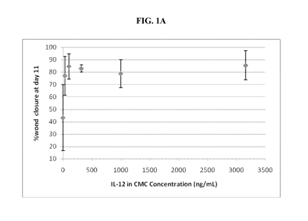
[0012] FIG. lA shows a plot of percent closure of full thickness wounds on day
11 of
rMuIL-12 treatment for several concentrations of rMuIL-12. FIG. 1B shows a
graph of
changes in wound area over time (in days) for mice treated with various
concentrations of
rMuIL-12. FIG. 1C shows part of the data in FIG. 1B along with error bars,
including
control, 31.6 ng/mL, 100 ng/mL, and 3160 ng/mL dosages.
[0013] FIG. 2A shows a photomicrograph of trichrome-stained tissue sections of
wound
sites at 40x, from full-thickness injuries treated with vehicle alone. The
large area of
granulation tissue is noted in the photomicrograph, as well as the location of
the wound
margin and dermis. FIGS. 2B and 2C show photomicrographs (40x) of trichrome-
stained
tissue sections of wound sites from mice treated with vehicle and 31.6 ng/mL
rMuIL-12.
[0014] FIG. 3A shows a photomicrograph of IL-12R132 expression in the dermis
of a
cutaneous wound from an irradiated mouse (3 days post-injury). M =
macrophages, PMN =
polymorphonuclear leukocytes, F = fibroblasts. FIG. 3B shows a photomicrograph
of IL-
12R132 expression in the sebaceous gland (SEB) and basal epidermis (BE) from a
cutaneous
wound of an irradiated mouse. FIG. 3C shows a photomicrograph of IL-12RI32
expression in
the stratum basale (BE), cuboidal cells (Cu) of the stratum spinosum, and
squamous cells
(Sq) in the stratum granulosum.
[0015] FIG. 4A is a graph of changes in wound area over time (in days) for
irradiated mice
treated with various concentrations of rMuIL-12 (R M = male mice; R F = female
mice).
FIG. 4B is graph of data from FIG. 4A, only for irradiated mice that received
100 ng/mL of
rMuIL-12. FIG. 4C shows a series of photomicrographs of trichrome-stained
sections of skin
wounds from mice treated with vehicle only, as well as the corresponding wound
seen on the
mouse itself (1x) (Ep = epidermis, D = dermis). Each row is a tissue section
from a single
animal, with the left photomicrograph taken at 40x, the middle photomicrograph
taken at
400x, and the corresponding wound taken at lx magnification. FIG. 4D shows
photomicrographs from mice that received 100 ng/mL rMuIL-12.
[0016] FIG. 5A is a graph of changes in the percentage of wound size over time
(in days)
for irradiated mice treated with rMuIL-12 topically and/or subcutaneously
about 24 hours
following creation of the wound. FIG. 5B shows a series of photographs (at lx)
of wounds in
vehicle-treated and rMuIL-12-treated mice following 9 days of healing.
[0017] FIG. 6A shows a graph of wound area over days 0-9 for diabetic Zucker
rats treated
with vehicle only, 100 ng/mL of rMuIL-12, and 3160 ng/mL of rMuIL-12. FIG. 6B
shows
3
CA 02839261 2013-12-12
WO 2012/174056 PCT/US2012/042165
trichrome-stained photomicrographs of mice treated with vehicle alone at day 9
of treatment
(left side), along with corresponding lx photographs of the wound (right
side). FIG. 6C
shows trichrome-stained photomicrographs of mice treated with 100 ng/mL rMuIL-
12 at day
9 of treatment (left side), along with corresponding lx photographs of the
wound (right side).
DETAILED DESCRIPTION OF THE INVENTION
[0018] The present invention is directed to methods of treating cutaneous
wounds
comprising administration of IL-12 administered to the wound. Administration
of the IL-12
is preferably topical, subcutaneous, intramuscularly, or any combination
thereof IL-12 can
be used in conjunction with treating any cutaneous wound. Examples of
cutaneous wounds
include, but are not limited to, cutaneous wounds associated with burns,
cutaneous wounds in
a patient population characterized by an impediment to normal cutaneous wound
healing,
such as diabetics ,the elderly, and patients with HIV/AIDS, and cutaneous
wounds associated
with radiation exposure. While not intended to be bound by any theory, the
data described
herein suggests that IL-12 may contribute to wound healing by stimulation of
stem cells
within sebaceous glands.
[0019] In particular, the present invention is directed to the surprising
discovery that IL-12
can accelerate closure of full-thickness skin injuries in a full-thickness
injury model of wound
healing. This is significant as this type of injury closely mimics the state
of a burn wound
following surgical debridement and is highly relevant to the use of IL-12 as a
therapeutic to
enhance wound closure following burn injury.
[0020] Moreover, IL-12 can be administered, for example topically and/or
subcutaneously,
to skin wounds treated with a full-thickness, split-thickness, or composite
skin grafts to
enhance engraftment and accelerate fusion of the graft to the recipient site
and accelerate
healing and resolution of the donor site.
[0021] IL-12 can be administered topically and/or subcutaneously in
conjunction with
acellular or cellular dermal matrices. IL-12 stimulates the differentiation
and migration of
keratinocytes and fibroblasts across a wound bed exposed by a full-thickness
cutaneous
injury. An acellular dermal matrix over the wound site can provide a
structural scaffold for
migration and attachment of IL-12 stimulated keratinocytes and fibroblasts.
[0022] IL-12 can also be used in conjunction an acellular dermal matrix to
treat a tendon or
ligament injury, such as rotator cuff injuries. For example, a patient with a
full-thickness
infraspinatus tendon tear of the rotator cuff can have the defect bridged by
placement and
suture of an acellular dermal matrix; IL-12 can be applied to the surgical
site by methods
4
CA 02839261 2013-12-12
WO 2012/174056 PCT/US2012/042165
such as direct injection, infusion of the matrix with IL-12 before
implantation, or
implantation of a second biodegradable matrix that can deliver IL-12 near the
surgical site.
[0023] In one embodiment, the methods of the invention, comprising
administering IL-12
to a cutaneous wound, result in an about 5% increase in wound healing as
measured by
wound closure over a specified time period, e.g., such as over a 1, 2, 3, 4,
5, 6, 7, 8, 19, 10,
11, 12, 13, 14, 15, 16, 17, 18, 19, 20, 21, 22, 23, 24, 25, 26, 27, 28, 29, or
30 day time period.
In other embodiments, the methods of the invention result in an about 10%,
about 15%, about
20%, about 25%, about 30%, about 35%, about 40%, about 45%, about 50%, about
55%,
about 60%, about 65%, about 70%, about 75%, about 80%, about 85%, about 90%,
about
75%, about 100% improvement in wound healing, as measured by wound closure
over a
specified time period.
I. Definitions
[0024] As used herein, the term "about" will be understood by persons of
ordinary skill in
the art and will vary to some extent depending upon the context in which it is
used. If there
are uses of the term which are not clear to persons of ordinary skill in the
art given the
context in which it is used, "about" will mean up to plus or minus 10% of the
particular term.
[0025] As used herein, except where the context requires otherwise, the term
"comprise"
and variations of the term, such as "comprising," "comprises" and "comprised"
are not
intended to exclude other additives, components, integers or steps.
[0026] As used herein, "Interleukin-12 (IL-12)" refers to any IL-12 molecule
that results in
improved cutaneous wound healing, including native IL-12 molecules, variant 11-
12
molecules and covalently modified IL-12 molecules, now known or to be
developed in the
future, produced in any manner known in the art now or to be developed in the
future.
Generally, the amino acid sequences of the IL-12 molecule used in embodiments
of the
invention are derived from the specific mammal to be treated by the methods of
the
invention. Thus, for the sake of illustration, for humans, generally human IL-
12, or
recombinant human IL-12, would be administered to a human in the methods of
the
invention, and similarly, for felines, for example, the feline IL-12, or
recombinant feline IL-
12, would be administered to a feline in the methods of the invention. Also
included in the
invention, however, are certain embodiments where the IL-12 molecule does not
derive its
amino acid sequence from the mammal that is the subject of the therapeutic
methods of the
invention. For the sake of illustration, human IL-12 or recombinant human IL-
12 may be
utilized in a feline mammal. Still other embodiments of the invention include
IL-12
CA 02839261 2013-12-12
WO 2012/174056 PCT/US2012/042165
molecules where the native amino acid sequence of IL-12 is altered from the
native sequence,
but the IL-12 molecule functions to yield the hematopoietic properties of IL-
12 that are
disclosed herein. Alterations from the native, species-specific amino acid
sequence of IL-12
include changes in the primary sequence of IL-12 and encompass deletions and
additions to
the primary amino acid sequence to yield variant IL-12 molecules. An example
of a highly
derivatized IL-12 molecule is the redesigned IL-12 molecule produced by
Maxygen, Inc.
(Leong SR, et al., Proc Natl Acad Sci USA., 100(3): 1163-8 (Feb. 4, 2003)),
where the variant
IL-12 molecule is produced by a DNA shuffling method. Also included are
modified IL-12
molecules are also included in the methods of invention, such as covalent
modifications to
the IL-12 molecule that increase its shelf life, half-life, potency,
solubility, delivery, etc.,
additions of polyethylene glycol groups, polypropylene glycol, etc., in the
manner set forth in
U.S. Pat. Nos. 4,640,835; 4,496,689; 4,301,144; 4,670,417; 4,791,192 or
4,179,337. One
type of covalent modification of the IL-12 molecule is introduced into the
molecule by
reacting targeted amino acid residues of the IL-12 polypeptide with an organic
derivatizing
agent that is capable of reacting with selected side chains or the N- or C-
terminal residues of
the IL-12 polypeptide. Both native sequence IL-12 and amino acid sequence
variants of IL-
12 may be covalently modified. Also as referred to herein, the IL-12 molecule
can be
produced by various methods known in the art, including recombinant methods.
Since it is
often difficult to predict in advance the characteristics of a variant IL-12
polypeptide, it will
be appreciated that some screening of the recovered variant will be needed to
select the
optimal variant. A preferred method of assessing a change in the hematological
stimulating
or enhancing properties of variant IL-12 molecules is via the lethal
irradiation rescue protocol
disclosed below. Other potential modifications of protein or polypeptide
properties such as
redox or thermal stability, hydrophobicity, susceptibility to proteolytic
degradation, or the
tendency to aggregate with carriers or into multimers are assayed by methods
well known in
the art.
[0027] The term "IL-12 receptor" is defined herein as a heterodimeric,
membrane-bound
receptor for the IL-12 ligand. The IL-12 receptor heterodimer subunits are
beta 1 (01) and
beta 2 (I32). In accordance with the present invention, the IL-12 receptor may
also bind the
IL-12 homodimer and the IL-12 monomer, as defined herein, to form a multimer
complex
comprising the IL-12 ligand/IL-12 receptor pair and the homodimer and/or the
monomer. In
the present invention, the multimer complex would further activate the IL-12
ligand/IL-12
receptor pair or may modify the activity of the ligand/receptor pair. In
accordance with the
present invention, the IL-12 receptor protein is defined to be in its
endogenous state as
6
CA 02839261 2013-12-12
WO 2012/174056
PCT/US2012/042165
isolated from the IL-12 selected stem cell taken from a donor or a patient. As
such, the IL-12
receptor may contain polymorphisms distinct from the canonical amino acid
sequence of the
131 and 132 subunits.
[0028] The term "One or more therapeutically effective dose(s) of IL-12"
refers to any dose
administered for any time intervals and for any duration that can improve
healing of a
cutaneous wound.
[0029] The term "therapeutically effective amount or dose" is defined herein
as a dose of a
substance that produces effects for which it is administered. The exact dose
of IL-12 will
depend on the purpose of the treatment, the timing of administration of IL-12,
certain
characteristics of the subject to be treated, and the severity of the
cutaneous wound, and is
ascertainable by one skilled in the art using known techniques (see, e.g.,
Lieberman,
Pharmaceutical Dosage Forms (vols. 1-3, 1992); Lloyd, The Art, Science and
Technology of
Pharmaceutical Compounding (1999); Pickar, Dosage Calculations (1999); and
Remington:
The Science and Practice of Pharmacy, 20th Edition, 2003, Gennaro, Ed.,
Lippincott,
Williams & Wilkins).
[0030] Generally, a dose of a therapeutic agent, according to the methods and
compositions
of the present invention, can be expressed in terms of the total amount of
drug to be
administered, (i.e., ng, g, or mg). The dose can be expressed as a weight
amount of drug
administered to a subject (e.g., 20 ng), or as a ratio of the weight amount of
drug per volume
unit of carrier (e.g., ng/mL), along with the volume of drug and carrier
administered (e.g., 1
mL). Alternatively, the dose can be expressed as a ratio of drug to be
administered to weight
or surface area of subject receiving the administration (i.e., ng/kg, g/kg,
ng,/m2, or g/m2).
When referring to a dose in terms of the mass to be administered per mass of
subject (i.e.,
ng/kg), it will be understood that doses are not equivalent between different
animals, and thus
conversion factors will need to be used to ensure that one animal receives the
same dose
equivalent as another animal. Suitable factors for the conversion of a mouse
"dose
equivalent" for intraperitoneal (i.p.) injection of IL-12 to a "dose
equivalent" of a different
animal are given in Table 1 below.
Table 1 ¨ Conversion Factors and Equivalent IL-12 Doses for Several Animals
Species Wei2ht Total Dose Dose (n2/k2) Dose Conversion
ILigl gl (n2/m2) Factor
Human 65 25655.82 394.7 15,000 0.0794
Mouse 0.02 99.47 4973.44 15,000 1.0000
Hamster 0.03 130.2 4339.87 15,000 0.8726
Rat 0.15 381.12 2540.8 15,000 0.5109
7
CA 02839261 2013-12-12
WO 2012/174056 PCT/US2012/042165
Table 1 ¨ Conversion Factors and Equivalent IL-12 Doses for Several Animals
Species WeiOtt Total Dose Dose (n2/k2) Dose Conversion
ILigl gl (n2/m2) Factor
Guinea Pig 1.00 1335 1335 15,000 0.2684
Rabbit 2.0 2381.1 1190.65 15,000 0.2394
Cat 2.5 2956.44 1182.57 15,000 0.2376
Monkey 3.0 3681.75 1227.25 15,000 0.2468
Dog 8.0 6720 840 15,000 0.1689
Thus, in one embodiment, doses are given in terms of mass to surface area
(i.e., ng/m2 or
g/m2), which are equivalent for all animals. The following basic conversion
factors can be
used to convert ng/kg to ng/m2: mouse = 3.0, hamster = 4.1, rat = 6.0, guinea
pig = 7.7,
human = 38.0 (Cancer Chemother Repts., 50(40):219(1966)).
[0031] The term "cutaneous wound" refers to an injury to the skin of a
subject. The wound
may be a full-thickness wound, a partial thickness wound, or a wound of only
the epidermis.
A cutaneous wound may be due to a burn, physical trauma, or surgical trauma.
II. Cutaneous Wound Hea1in2 Processes
A. Overview
[0032] Wound healing in the skin is a complex phenomenon roughly divided into
three
phases of inflammation, proliferation, and maturation. Innate immune cells,
particularly
macrophages, play an essential role in the inflammatory and proliferative
stages of wound
healing. In fact, depletion of macrophages impairs the rate of wound closure
(Brancato et al.,
Am. J. Pathol., 178(1):19-25, 2011).
[0033] The inflammatory phase begins with platelet-mediated induction of
hemostasis.
Platelets secrete several proinflammatory factors that act locally and act as
chemoattractants
for neutrophils, monocytes, and fibroblasts. It is during this phase that
monocytes mature
into macrophages which debride the wound and stimulate fibroblasts to
synthesize collagen
and ground substance of granulation tissue. Macrophages also stimulate the
influx of
keratinocytes to cover the new skin and endothelial cells for
neovascularization.
[0034] In the proliferative phase, two to three days after injury, macrophages
secrete factors
(bFGF, TGFb, and PDGF) to stimulate fibroblasts to begin migrating from the
wound edge to
contract the wound. Fibroblasts also stabilize and remodel the wound site by
organizing
collagen molecules into fibers, thereby increasing tensile strength. The
fibrin clot begins to
resolve leading to a decrease in migration and proliferation of fibroblasts.
8
CA 02839261 2013-12-12
WO 2012/174056 PCT/US2012/042165
[0035] During the maturation phase, type II collagen is replaced by type I
collagen and
epithelial cells cover the wound site until they are contact inhibited.
Finally, fibroblasts have
differentiated into actin containing myofibroblasts, leading to further
contraction of the
wound.
B. Cutaneous Wounds in Diabetic Patients
[0036] Wound healing in non-diseased individuals results as a consequence of
an
overlapping and complex interplay between connective tissue formation,
cellular proliferation
and differentiation, and growth factors. The normal process of wound healing
is impeded in
diabetics because of deficiencies in the initiation and maintenance of the
inflammatory phase
of wound healing, ultimately resulting in an impaired cellular proliferation
and migration
over the wound site. Upregulation of matrix metalloproteinases in diabetic
wounds leads to
diminishment of growth factors. Finally, diabetics exhibit decreased collagen
synthesis and
deposition.
[0037] Skin wounds on diabetic subjects can be treated using topical and/or
subcutaneous
dosages of IL-12 to improve wound healing. Preferred topical dosages of IL-12
include 31.6
ng/mL, 100 ng/mL, 316 ng/mL, 1000 ng/mL, and 3160 ng/mL. Such topical doses
can be
applied at least up to 24 hours following creation of the wound. Subcutaneous
dosages of IL-
12 can also be applied, either alone or in addition to topical administration
of IL-12.
Preferred subcutaneous dosages of IL-12 are about 80 ng/kg.
III. Burn Injury
[0038] Burns are classified according to five degrees involving the depth of
tissue damage
and the total body surface area (TBSA) occupied by the wound (Minor = <15%
TBSA,
Moderate = 15-20%TBSA, Major =20% TBSA and above). First degree burns are the
least
serious generally requiring little more than analgesia for comfort, and
typically resolve within
a few days. Second degree burns, frequently classified as superficial partial
thickness burns,
involve damage to the dermis and extends through the epidermis and into the
papillary
dermis. Second degree burns are typified by the presence of fluid filled
blisters. Treatment
generally involves the use of analgesics, antibiotics, and bandaging. A more
severe second
degree burn would extend through the epidermis and into the deep reticular
dermis. These
types of burns can progress to a third degree status. They often require
extensive clinical
intervention, have an extended recovery period, and can require debridement
and grafting to
facilitate healing. Third degree burns extend throughout the entire depth of
the dermis.
9
CA 02839261 2013-12-12
WO 2012/174056 PCT/US2012/042165
These burns are severe life threatening injuries that require extensive
clinical management
involving surgical debridement followed by full or partial thickness skin
grafting. Scarring is
extensive and recovery is lengthy and requires arduous physical therapy to
recover function
in affected tissues. Finally, fourth degree burns, the most severe, damage
extends through the
dermis and into the muscle and bone. These injuries frequently require
amputation. Because
of the loss of the protective epithelial barrier, death from third and fourth
degree burns often
results from infection.
[0039] Topical and subcutaneous dosages of IL-12 can be applied to full-
thickness
cutaneous wounds, such as severe burn wounds, to improve healing of the
wounds. Preferred
topical dosages of IL-12 include 31.6 ng/mL, 100 ng/mL, 316 ng/mL, 1000 ng/mL,
and 3160
ng/mL. Such topical doses can be applied at least up to 24 hours following
creation of the
wound. Subcutaneous dosages of IL-12 can also be applied, either alone or in
addition to
topical administration of IL-12. Preferred subcutaneous dosages of IL-12 are
about 80 ng/kg.
A. Second Intention Burn Treatment
[0040] While the preferred treatment options for major burns necessitate the
use of
aggressive use of skin grafting, in minor and moderate second or third degree
burns, a
preferred method to wound resolution is surgical debridement followed by
closure through
second intention healing. In this scenario, the wound is managed and the
body's natural
capacity to heal the wound employed to slowly resolve the wound over time. The
protracted
time frame required for this method however renders the patient susceptible to
infection and
can exacerbate scarring.
[0041] The examples described herein demonstrate that IL-12 can accelerate
closure of full-
thickness skin injuries in a full-thickness injury model of wound healing.
This type of injury
closely mimics the state of a burn wound following surgical debridement and is
highly
relevant to the use of IL-12 as a therapeutic to enhance wound closure
following burn injury.
[0042] For example, a patient with minor second or third degree burns may
receive surgical
debridement of the burn to remove damaged tissue, followed by supportive care.
Surgical
debridement includes dissection and removal of damaged wound edge epithelium
to expose
healthy epidermis and dermis. IL-12 is then administered to the wound
topically, or
subcutaneously at or near the wound site. The wound is covered with a clear
permeable
bandage to keep it aseptic (i.e., TegadermTm) and monitored for closure.
CA 02839261 2013-12-12
WO 2012/174056 PCT/US2012/042165
B. Use of IL-12 with Skin Grafts
[0043] Full-thickness skin grafts are indicated for coverage of deep wounds
extending
through the dermis such as might be seen in third degree burns and diabetic
ulcers,
necrotizing fasciitis, etc. Full-thickness grafts consist of the epidermis and
much of the
underlying dermis. In a typical application, the wound is debrided, an
autologous full-
thickness graft harvested from healthy tissue and the graft sutured into
place. A full-
thickness graft is a serious surgical procedure and often not indicated if the
patient is
seriously impaired from injury or accompanying disease. Full-thickness grafts
bring the
advantage of an intact wound barrier with accompanying progenitor populations,
hair,
vasculature, collagen, etc. Disadvantages of using full-thickness skin grafts
include
insufficient vascular perfusion leading to poor engraftment or rejection if
patient condition
necessitated the use of an allograft.
[0044] Split thickness grafts are frequently used for coverage and healing of
burn wounds
and other difficult to heal cutaneous injuries. The tissue comprising these
grafts are derived
from donor sites containing healthy tissue. The graft is harvested by use of a
dermatome and
comprised of epidermal and some dermal tissue containing keratinocyte
progenitors and
collagen-producing fibroblasts. The advantage of autologous split-thickness
grafts is they
can be processed through a meshing apparatus and expanded up to nine times to
cover large
areas. Re-epithelialization occurs by outgrowth of keratinocytes into the
exposed dermis.
[0045] Composite grafts are typically smaller grafts that include skin and
underlying
cartilage tissue. They are often indicated when cartilaginous tissue (nose,
ears) have been
damaged or destroyed as a consequence of injury. The term is sometimes used to
describe
human skin equivalents (HSEs) like Apligraf'TM, which is composed of
keratinocytes and
fibroblasts on a collagen support matrix.
[0046] IL-12 can be administered topically and/or subcutaneously to skin
wounds treated
with a full-thickness, split-thickness, or composite skin grafts to enhance
engraftment and
accelerate fusion of the graft to the recipient site and accelerate healing
and resolution of the
donor site.
C. Use of IL-12 with Acellular and Cellular Dermal Matrices
[0047] One approach to the treatment of large cutaneous injuries, such as burn
injuries, has
been the application of an acellular dermal matrix (e.g. GraftjacketTM,
IntegraTM,
SkinTempTm). These grafts can either serve as a scaffold to support outgrowth
of epidermal
11
CA 02839261 2013-12-12
WO 2012/174056 PCT/US2012/042165
keratinocytes or, after implantation be overlayed with an autologous
keratinocyte graft that
has been generated in vitro.
[0048] Cellular dermal matrix, also known as living skin substitute, consists
of cultured
epidermal autografts or more commonly as "Human Skin Equivalents" (HSEs). HSEs
are
typically composed of a collagen support matrix overlayed with living
allogeneic fibroblasts
and/or keratinocytes derived from male foreskin donors. Exemplary products
include
Apligrafrm (Organogenesis, Inc., Canton MA), DermagraftTM (Advanced
BioHealing,
Westport, CT), GintuitTM (Organogenesis, Inc., Canton MA), and OrcelTM
(Forticell
Bioscience, Englewood Cliffs, NJ). Composited HSEs such as Apligraf'TM have
been
successfully used under compassionate use for critically burned patients.
Another example of
living skin substitutes includes EpicelTM (Genzyme Biosurgery), which is made
up of sheets
of autologous keratinocytes cultured ex vivo (2 to 8 cell layers thick). These
HSEs are used
to treat diabetic skin ulcers, burns, and to infill receded gum lines.
[0049] IL-12 can be administered topically and/or subcutaneously in
conjunction with
acellular or cellular dermal matrices. IL-12 stimulates the differentiation
and migration of
keratinocytes and fibroblasts across a wound bed exposed by a full-thickness
cutaneous
injury. An acellular dermal matrix over the wound site can provide a
structural scaffold for
migration and attachment of IL-12 stimulated keratinocytes and fibroblasts.
[0050] IL-12 can also be used in conjunction an acellular dermal matrix to
treat rotator cuff
injuries. Adams et al., Arthroscopy, 22(7): 700-709 (2006). For example, a
patient with a
full-thickness infraspinatus tendon tear of the rotator cuff can have the
defect bridged by
placement and suture of an acellular dermal matrix; IL-12 can be applied to
the surgical site
by methods such as direct injection, infusion of the matrix with IL-12 before
implantation, or
implantation of a second biodegradable matrix that can deliver IL-12 near the
surgical site.
[0051] The following examples are given to illustrate the present invention.
It should be
understood, however, that the invention is not to be limited to the specific
conditions or
details described in these examples.
D. Enhanced Cosmetic Remodeling
[0052] Delayed healing can result in scarring when collagen is deposited in
symmetrically
cross-linked fibers rather than in the basket weave pattern associated with
non-injured skin
(Dallon et al., Mathmatical modeling of extracellular matrix dynamics using
discrete cells:
Fiber orientation and tissue regeneration, J. Theor. Biol. 199:449-471, 1999).
In almost all
instances, scarring is a normal consequence of healing of skin injuries.
Accelerated wound
12
CA 02839261 2013-12-12
WO 2012/174056 PCT/US2012/042165
closure diminishes scar formation by limiting the time required for aberrant
collagen
deposition. The pattern of aligned collagen deposition results in diminished
wound strength
and blocks the repopulation of the injured tissue with new sweat glands and
hair follicles. In
addition, scars can present cosmetic challenges when they manifest in exposed
area such as
the face. The type and severity of injury, coupled with underlying genetics of
the individual,
can lead to the development of hypertrophic (raised), atrophic (sunken), or
keloid (large,
benign, tumorous) scars. Patients with wounds that have closed via secondary
intention
healing often manifest hypertrophic or keloid scars as a result of delayed
healing. Current
therapies concentrate on the use of post-scarring surgical interventions such
as chemical
peals, dermabrasion, fillers (ArtefillTM, i.e., bovine collagen and
polymethylmethalcrylate,
RadiesseTM, DermatixTM (silicone gel)), lasers, and radiation (reduction of
keloid scars).
Only 1 injectible drug (corticosteroid) is approved for the treatment of
appearance of keloid
scars. Clinical trials are under underway for the assessment of an
interventional drug for the
suppression or prevention of scars. Avotermin (JuvistaTM, rTGFI33) is a drug
being
investigated for the suppression of split-thickness graft scarring and other
surgical scars
(Occleston, et al., 2011; So et al., 2011; Durani, et al., 2008).
[0053] Topical or subcutaneous administration of IL-12 in patients with
cutaneous injuries
diminishes scar formation, enhances wound strength and UV resistance, and
improves
cosmetic appearance by accelerating the rate of wound closure. In addition, IL-
12 diminishes
delayed wound closure by suppression of cutaneous infections. Thus, IL-12 may
be
administered subcutaneously in the range of about 15,000 ng/m2 to diminish
delays in wound
closure.
IV. Timin2 of IL-12 Administration
[0054] Advantageously, as provided by the methods of the present invention,
administration of IL-12 may occur during any suitable time period following a
cutaneous
wound.
[0055] In one embodiment, where the cutaneous wound is associated with
exposure to
radiation, IL-12 can be administered any time after radiation exposure up to
and including
about a week after exposure. Although the total dose of radiation will factor
into the time
period in which IL-12 should be administered, according to one embodiment, IL-
12 may be
administered at any time up to about 120 hours following exposure to radiation
resulting in a
cutaneous injury. In other embodiments, IL-12 can be administered at any time
up to about
13
CA 02839261 2013-12-12
WO 2012/174056 PCT/US2012/042165
96 hours post-irradiation, up to about 72 hours post-irradiation, or at a time
up to about 60
hours, about 48 hours, about 36 hours, about 24 hours, about 18 hours, about
12 hours, about
8 hours, about 6 hours, or less following exposure to radiation resulting in a
cutaneous injury.
In one specific embodiment, IL-12 is administered to a subject in need thereof
between a
range of about 1 hour to about 72 hours after exposure to ionizing radiation.
In another
embodiment, IL-12 is administered between a range of about 1 hour and about 24
hours after
exposure, or between a range of about 6 hours and about 24 hours following
exposure to an
acute dose of whole body ionizing radiation.
[0056] IL-12 can be administered at any time point after radiation exposure
resulting in a
cutaneous wound. In one embodiment of the invention, IL-12 is administered at
least about
24 hours or more following exposure to radiation. Other time points for
administration
following radiation exposure resulting in a cutaneous wound include about 1
hour, about 2
hours, about 3 hours, about 4 hours, about 5 hours, about 6 hours, about 7
hours, about 8
hours, about 9 hours, about 10 hours, about 11 hours, about 12 hours, about 13
hours, about
14 hours, about 15 hours, about 16 hours, about 17 hours, about 18 hours,
about 19 hours,
about 20 hours, about 21 hours, about 22 hours, about 23 hours, about 24
hours, about 1 day,
about 1.5 days, about 2 days, about 2.5 days, about 3 days, about 3.5 days,
about 4 days,
about 4.5 days, about 5 days, about 5.5 days, about 6 days, about 6.5 days,
about 7 days, or
about any combination thereof with multiple IL-12 administrations (e.g., at 12
hours and 48
hours.
V. IL-12 Dosin2 and Dosnes
[0057] Generally the IL-12 doses used in the methods for treating cutaneous
wounds will
be high enough to be effective for the treatment of a cutaneous wound, but low
enough to
mitigate negative side effects associated with IL-12 administrations,
including for example,
radiosensitivity of the GI tract (associated with radiation exposure) and IFN-
y up-regulation.
[0058] In one aspect, a single dose of IL-12 is sufficient to confer improved
cutaneous
wound healing. In other aspects, IL-12 may be administered in more than one
dose, such as
about 2, about 3, about 4, about 5 or more doses.
[0059] Accordingly, in one aspect, the present invention provides a method for
treating
cutaneous wounds, including improving mitigation of cutaneous wounds,
comprising the
administration of a dose of IL-12 to a subject having cutaneous wound. In one
embodiment,
the dose of IL-12 is less than about 100 [tg/m2. In another embodiment, the
dose of IL-12 is
less than about 75 [tg/m2, or less than about 400 ng/kg (15 [tg/m2). In
another embodiment,
14
CA 02839261 2013-12-12
WO 2012/174056 PCT/US2012/042165
the dose can be between about 1 [tg/m2 and about 100 [tg/m2. Other exemplary
IL-12
dosages include less than 1 [tg/m2or about 1 [tg/m2, less than about 3
[tg/m2or about 3 [tg/m2,
less than about 4 [tg/m2or about 4 [tg/m2, less than about 5 [tg/m2 or about 5
[tg/m2, less than
about 6 [tg/m2or about 6 [tg/m2, less than about 7 [tg/m2 or about 7 [tg/m2,
less than about 8
[tg/m2or about 8 [tg/m2, less than about 9 [tg/m2or about 9 [tg/m2, less than
about 10 [tg/m2 or
about 10 [tg/m2, less than about 11 [tg/m2 or about 11 [tg/m2, less than about
12 [tg/m2or
about 12 [tg/m2, less than about 15 [tg/m2 or about 15 [tg/m2, less than about
20 [tg/m2or
about 20 [tg/m2, less than about 25 [tg/m2 or about 25 [tg/m2, less than about
30 [tg/m2or
about 30 [tg/m2, less than about 35 [tg/m2 or about 35 [tg/m2, less than about
40 [tg/m2or
about 40 [tg/m2, less than about 45 [tg/m2 or about 45 [tg/m2, less than about
50 [tg/m2or
about 50 [tg/m2, less than about 55 [tg/m2 or about 55 [tg/m2, less than about
60 [tg/m2or
about 60 [tg/m2, less than about 65 [tg/m2 or about 65 [tg/m2, less than about
70 [tg/m2or
about 70 [tg/m2, less than about 75 [tg/m2 or about 75 [tg/m2, less than about
80 [tg/m2or
about 80 [tg/m2, less than about 85 [tg/m2 or about 85 [tg/m2, less than about
90 [tg/m2or
about 90 [tg/m2, less than about 95 [tg/m2 or about 95 [tg/m2, less than about
100 [tg/m2or
about 100 [tg/m2, less than about 900 ng/m2 or about 900 ng/m2, less than
about 800 ng/m2 or
about 800 ng/m2, less than about 700 ng/m2 or about 700 ng/m2, less than about
600 ng/m2 or
about 600 ng/m2, less than about 500 ng/m2 or about 500 ng/m2, less than about
400 ng/m2 or
about 400 ng/m2, less than about 300 ng/m2 or about 300 ng/m2, less than about
250 ng/m2 or
about 250 ng/m2, less than about 200 ng/m2 or about 200 ng/m2, less than about
100 ng/m2 or
about 100 ng/m2, and all doses in-between.
[0060] In one embodiment of the invention, the dosage of IL-12 is between
about 1 ng/mL
and about 10 [tg/mL. In another embodiment, the dosage of IL-12 is between
about 10
ng/mL and about 5 [tg/mL. In other embodiments of the invention, the dosage of
IL-12 is
about 10, about 20, about 30, about 40, about 50, about 60, about 70, about
80, about 90,
about 100, about 110, about 120, about 130, about 140, about 150, about 160,
about 170,
about 180, about 190, about 200, about 210, about 220, about 230, about 240,
about 250,
about 260, about 270, about 280, about 290, about 300, about 310, about 320,
about 330,
about 340, about 350, about 360, about 370, about 380, about 390, or about 400
ng/mL.
[0061] In one embodiment of the invention, the dosage of IL-12 is between
about 10 ng/kg
and about 500 ng/kg. In other embodiments of the invention, the dosage of IL-
12 is about 10,
about 20, about 30, about 40, about 50, about 60, about 70, about 80, about
90, about 100,
about 110, about 120, about 130, about 140, about 150, about 160, about 170,
about 180,
about 190, about 200, about 210, about 220, about 230, about 240, about 250,
about 260,
CA 02839261 2013-12-12
WO 2012/174056 PCT/US2012/042165
about 270, about 280, about 290, about 300, about 310, about 320, about 330,
about 340,
about 350, about 360, about 370, about 380, about 390, or about 400 ng/kg.
[0062] When administered in multiple doses, i.e. two, three, four, or more,
the first IL-12
dose and subsequent IL-12 dose(s) can be equivalent doses, or they can be
different dose
amounts. For example, in certain embodiments, subsequent dose(s) can be
administered at
about 90% of the initial dose, or at about 80%, about 75%, about 70%, about
60%, about
50%, about 40%, about 30%, about 25%, about 20%, or about 10% or less of the
original
dose.
VI. IL-12 Compositions
[0063] For general descriptions relating IL-12, see U.S. Pat. Nos. 5,573,764,
5,648,072,
5,648,467, 5,744,132, 5,756,085, 5,853,714 and 6,683,046. Interleukin-12 (IL-
12) is a
heterodimeric cytokine generally described as a proinflamatory cytokine that
regulates the
activity of cells involved in the immune response (Fitz et al., J. Exp. Med.,
170: 827-45
(1989)). Generally IL-12 stimulates the production of interferon-y (IFN-y)
from natural killer
(NK) cells and T cells (Lertmemongkolchai et al., J. of Immunology, 166: 1097-
105 (2001);
Cui et al., Science, 278:1623-6 (1997); Ohteki et al., J. Exp. Med., 189:1981-
6 (1999);
Airoldi et al., J. of Immunology, 165: 6880-8 (2000)), favors the
differentiation of T helper 1
(TH1) cells (Hsieh et al., Science, 260: 547-9 (1993); Manetti et al., J. Exp.
Med., 177: 1199-
1204 (1993)), and forms a link between innate resistance and adaptive
immunity. IL-12 has
also been shown to inhibit cancer growth via its immuno-modulatory and anti-
angiogenesis
effects (Brunda et al., J. Exp. Med., 178: 1223-1230 (1993); Noguchi et al.,
Proc. Natl. Acad.
Sci. U.S.A., 93: 11798-11801 (1996); Giordano et al., J. Exp. Med., 194: 1195-
1206(2001);
Colombo et al., Cytokine Growth factor, Rev.13: 155-168 (2002); Yao et al.,
Blood, 96:
1900-1905 (2000)). IL-12 is produced mainly by dendritic cells (DC) and
phagocytes
(macrophages and neutrophils) once they are activated by encountering
pathogenic bacteria,
fungi or intracellular parasites (Reis et al., J. Exp. Med., 186:1819-1829
(1997); Gazzinelli et
al., J. Immunol., 153: 2533-2543 (1994); Dalod et al., J. Exp. Med., 195: 517-
528 (2002)).
The IL-12 receptor (IL-12 R) is expressed mainly by activated T cells and NK
cells (Presky
et al., Proc. Natl. Acad. Sci. U.S.A., 93: 14002-14007 (1996); Wu et al., Eur.
J. Immunol., 26:
345-50 (1996)).
[0064] Generally the production of IL-12 stimulates the production of IFN-y,
which, in
turn, enhances the production of IL-12, thus forming a positive feedback loop.
In in vitro
systems, it has been reported that IL-12 can synergize with other cytokines
(IL-3 and SCF for
16
CA 02839261 2013-12-12
WO 2012/174056 PCT/US2012/042165
example) to stimulate the proliferation and differentiation of early
hematopoietic progenitors
(Jacobsen et al., J. Exp. Med., 2: 413-8 (1993); Ploemacher et al., Leukemia,
7: 1381-8
(1993); Hirao et al., Stem Cells, 13: 47-53 (1995)).
[0065] In certain embodiments, the IL-12 is a mammalian IL-12, recombinant
mammalian
IL-12, murine IL-12 (mIL-12), recombinant murine IL-12 (rmIL-12), human IL-12
(hIL-12),
recombinant human IL-12 (rhIL-12), canine IL-12 or rIL-12, feline IL-12 or rIL-
12, bovine
IL-I2 or rIL-I2, equine IL-I2 or rIL-I2, or biologically active variants or
fragments thereof. In
one specific embodiment, the rh1L-I2 is HemaMaxTm (Neumedicines Inc.). In
certain
embodiments, the IL-12 can be modified in a fashion so as to reduce the
immunogenicity of
the protein after administration to a subject. Methods of reducing the
immunogenicity of a
protein are well known in the art and include, for example, modifying the
protein with one or
water soluble polymers, such as a PEG, a PEO, a carbohydrate, a polysialic
acid, and the like.
[0066] It is well known that solutions of proteins that are formulated at low
concentrations
are susceptible to loss of a significant fraction of the protein prior to
administration. One
major cause of this problem is adsorption of the protein on the sides of
tubes, vials, syringes,
and the like. Accordingly, in certain aspects, when administered at low or
ultralow doses, it
will be beneficial to administer IL-12 along with a suitable carrier molecule
or bulking agent.
In one embodiment, the carrier agent may be a protein suitable for
pharmaceutical
administration, such as albumin. Generally, the carrier molecule or protein
will be present in
the formulation in excess of IL-12 to minimize the amount of IL-12 lost prior
to
administration. In certain embodiments, the carrier will be present at a
concentration of at
least about 2 times the concentration of IL-12, or at a concentration of at
least about 3, at least
about 4, at least about 5, at least about 6, at least about 7, at least about
8, at least about 9, at
least about 10, at least about 25, at least about 50, at least about 100, or
more times the
concentration of IL-12 in the formulation.
[0067] IL-12 composition provided herein and used according to the methods of
the
invention can be formulated for administration via any known method, but
preferably
topically, subcutaneously, or intramuscularly. Further, an efficacious dose of
IL-12 may
differ with different routes of administration.
[0068] In some embodiments, the formulations provided herein further comprise
one or
more pharmaceutically acceptable excipients, carriers, and/or diluents. In
addition, the
formulations provided herein may further comprise other medicinal agents,
carriers,
adjuvants, diluents, tissue permeation enhancers, solubilizers, and the like.
Methods for
preparing compositions and formulations for pharmaceutical administration are
known to
17
CA 02839261 2013-12-12
WO 2012/174056 PCT/US2012/042165
those skilled in the art (see, for example, REMINGTON'S PHARMACEUTICAL
SCIENCES, 18TH ED., Mack Publishing Co., Easton, PA (1990)). Formulations used
according to the methods of the invention may include, for example, those
taught in U.S.
Patent No. 5,744,132, which is hereby incorporated by reference in its
entirety for all
purposes.
EXAMPLES
Example 1: Topical Administration of rMuIL-12 to Full-Thickness Cutaneous
Wounds
[0069] To study the effect of IL-12 on full-thickness skin wounds, mice with
full thickness
skin wounds were treated topically with different amounts of rMuIL-12. Full-
thickness
cutaneous injuries, which are equivalent to a third degree burn, are a
challenging and
commonly used model for the study of the mechanisms of wound healing, as well
as the
examination of potential treatments for accelerating or enhancing resolution
of normal or
diseased wounds.
Materials & Methods
[0070] 18 mice were divided into six treatment groups of 3 animals each. The
treatment
groups are listed in Table 2 below.
Table 2 ¨ Treatment Groups for Topical IL-
12 Administration
GroupTreatment
1 CMC (4%) Alone
2 CMC (4%) + 31.6 ng/mL rMuIL-12
3 CMC (4%) + 100 ng/mL rMuIL-12
4 CMC (4%) + 316 ng/mL rMuIL-12
CMC (4%) + 1000 ng/mL rMuIL-12
6 CMC (4%) + 3160 ng/mL rMuIL-12
*cmc = carboxymethylcellulose
[0071] A circular 8.0 mm diameter wound was induced in anesthetized mice using
an 8 mm
biopsy punch and the epidermal layer was removed to expose the underlying
tissue.
Recombinant murine IL-12 (rMuIL-12) (SBH Biosciences) was emulsified in a
sterile
isotonic gel matrix consisting of 4% carboxymethylcellulose (CMC) in
Dulbecco's Phosphate
Buffered Saline (DPBS). Benzoin tincture was applied peripherally around the
wound site
and a TegadermTm dressing was applied over the wound site. The wound site for
each mouse
was then filled to capacity with control (CMC alone) or CMC + rMuIL-12 gel
matrix using
an 18 gauge syringe needle (approximately 150 [iL). A 1.0mL syringe (Terumo,
Slip-Tip
18
CA 02839261 2013-12-12
WO 2012/174056 PCT/US2012/042165
#SS-01T) was used for delivery of the CMC and CMC/rMuIL-12. The syringe is
graduated
in 10 1 increments. Delivered volumes are apparent by noting level on
graduations. No
external dressing was applied over the TegadermTm dressing. The gel matrix
under the
dressing was replenished at 2 days and 7 days following wound creation and
initial
application of the gel matrix.
[0072] Wound areas were quantified by overlaying the wound site with a glass
slide and
tracing the wound margin onto the slide with a black pen. The slides were
scanned as a JPEG
image and imported into Photoshop CSTM. The number of pixels comprising the
wound area
was determined and converted into an area of square millimeters (calculated
area of wound =
50.24 mm2, 8mm diameter full-thickness wound, Tcr2). Wound area was measured
five times,
at two to three day intervals.
[0073] On the eleventh day of the study, blood samples were collected and
examined for
lymphocyte counts and levels of IL-12 and IFN-k. In addition, the wound site
was collected
at Day 11 and stained with Mallory's trichrome for histologic examination.
Trichrome
staining is the standard for visualization of stage of remodeling of cutaneous
wounds
following injury.
Results
[0074] Maximal healing 11 days following wound creation and topical rMuIL-12
application occurred at a dose of 100 ng/mL, and did not increase with
increasing doses of
rMuIL-12 (FIG. 1A). All groups treated with CMC + rMuIL-12 showed a greater
percentage
of wound closure as compared with CMC control matrix by days 9 and 11 (FIG.
1B). Data
for the control and only three of the dosages in FIG. 1B (31.6 ng/mL, 100
ng/mL, and 3160
ng/mL) are shown in FIG. 1C. Table 3 below shows p-values of wound area
compared with
control for days 2, 4, 7, 9, and 11 (bolded numbers are p < 0.05). , The 100
ng/mL rMuIL-12
group showed a statistically significant reduction in wound area at days 7, 9,
and 11 (by t-
test; p<0.05) compared to the control group. Statistically significant
reductions in wound
area were also observed for the 31.6 ng/mL dose at day 11, and for the 3160
ng/mL dose at
days 9 and 11. Without being held to any particular theory, the lack of
statistical significance
from control at doses of 316 ng/mL and 1000 ng/mL are likely due to the small
number of
mice (three) in each of the groups.
19
CA 02839261 2013-12-12
WO 2012/174056 PCT/US2012/042165
Table 3 - Statistical Significance of Wound Area Treated with IL-12 vs.
Control
Day rMuIL-12 rMuIL-12 rMuIL-12 rMuIL-12 rMuIL-12
(31.6 ng/mL) (100 ng/mL) (316 ng/mL) (1000 ng/mL) (3160 ng/mL)
2 0.1215 0.381627 0.55888 0.34026 0.428741
4 0.342364 0.437305 0.263721 0.059673 0.496804
7 0.246345 0.012142 0.326977 0.262902 0.086662
9 0.101027 0.018461 0.121607 0.057338 0.012446
11 0.035108 0.046694 0.181913 0.10688 0.022395
[0075] No changes in blood cell counts for animals treated with CMC + rMuIL-12
were
observed, although a trend towards increases in lymphocytes was observed.
ELISA for IL-12
and IFN-k showed no detection of either cytokine in plasma of treated or
control mice.
[0076] FIG. 2A shows contiguous micrographs of trichrome-stained tissue
sections of
wound sites at 40x, from full-thickness injuries treated with vehicle alone.
The large area of
granulation tissue is noted in the micrograph, as well as the location of the
wound margin and
dermis. FIGS. 2B and 2C show micrographs (40x) of trichrome-stained tissue
sections of
wound sites from mice treated with vehicle and 31.6 ng/mL rMuIL-12. The
presence of rete
ridges is indicated in FIG. 2B, indicative of maturing epidermis. New
epidermis is indicated
in FIG. 2C, along with some granulated tissue.
Example 2: Expression of IL-12RI32 in Wounds of Irradiated Mice
[0077] A study was undertaken to examine the level, distribution, and timing
of expression
of IL-12RI32 in full-thickness cutaneous injuries topically treated with
vehicle alone or
vehicle + rMuIL-12.
Materials and Methods
[0078] Thirty-six mice received 500 cGy of total body irradiation in a
Gammacell 40
irradiator. The mice were then anesthetized and given a circular 10.0 mm
diameter wound
using a biopsy punch. The mice were then split into two groups, with one group
treated with
4% CMC alone, and the second group receiving 4% CMC + 100 ng/mL of rMuIL-12.
Wound treatment and measurement are as described in Example 1 (calculated area
of wound
= 78.5 mm2, lOmm diameter full-thickness wound, cr2).
[0079] Mice were then sacrificed and their wounds removed, sectioned in
paraffin, and
fixed in formalin at various time points following wound initiation and
treatment with
CA 02839261 2013-12-12
WO 2012/174056 PCT/US2012/042165
rMuIL-12. The fixed tissue was deparaffinized with xylene, hydrated in
ethanol, and washed
in water.
[0080] For antigen retrieval and staining, the tissue sections were immersed
in 1X HIER
buffer (heat induced epitope retrieval) and heated in a pressure cooker for 10
minutes.
Peroxidases and non-specific proteins were then blocked using 0.3% hydrogen
peroxide and
Background SniperTM reagent (Biocare Medical, Concord, CA), respectively. The
treated
tissue sections were then stained with rabbit anti-human IL-12RI32, detected
using a
horseradish peroxidase-based secondary antibody detection system (ImmPRESS
anti-rabbit
IgG and ImmPact AEC substrate; Vector Laboratories, Burlingame, CA). The
tissue sections
were also counterstained with hematoxylin.
Results
[0081] Examination of skin wounds from irradiated mice showed that IL-12RI32
expression
is upregulated regardless of exposure to rMuIL-12, a unique finding not
previously described.
There is no apparent effect of IL-12 treatment on expression of IL-12RI32 in
irradiated skin.
IL-12RI32, upregulated as a consequence of injury, exists in a ready state
waiting to trigger a
wound healing cascade in response to endogenous or exogenous administration of
IL-12.
[0082] In dermis, the majority of IL-12RI32 expression is in macrophages,
consistent with
observations of other researchers. FIG. 3A shows a photomicrograph of IL-
12RI32
expression in the dermis of a cutaneous wound from an irradiated mouse (3 days
post-injury;
M = macrophages, PMN = polymorphonuclear leukocytes, F = fibroblasts).
Polymorphonuclear leukocytes (PMNs) and fibroblasts (identified
morphologically) also
express IL-12RI32 but these cell types are in the minority at 3 days after
injury.
[0083] The majority of IL-12RI32 expression in the epidermis is in the cells
comprising the
basal membrane, as well as in the sebaceous glands of wounded skin (see FIG.
3B). Stem
cells known to be contained in sebaceous glands have been described as
contributing to re-
epithelialization of wounded tissue (Ghazizadeh and Taichman, EMBO 20(6):1215-
22
(2001); Blanpain, Nature 464:686-7 (2010)). Given the high level of IL-12RI32
expression in
the sebaceous glands of wounded skin, IL-12 may contribute to wound healing by
stimulation
of stem cells within sebaceous glands. In fact, multipotent stem cells derived
from sebaceous
glands are being used to rapidly create human skin substitutes for wound
grafts. FIG. 3C
shows a photomicrograph of IL-12RI32 expression in the stratum basale (BE),
cuboidal cells
(Cu) of the stratum spinosum, and squamous cells (Sq) in the stratum
granulosum.
21
CA 02839261 2013-12-12
WO 2012/174056 PCT/US2012/042165
Example 3: Mitigation of Skin Wounds in Irradiated Mice using IL-12
[0084] A study was undertaken to examine the healing of full-thickness
cutaneous injuries
in irradiated mice that were topically treated with vehicle alone or vehicle +
rMuIL-12.
Materials and Methods
[0085] Eighteen mice received 500 cGy of total body irradiation in a Gammacell
40
irradiator, as described in Example 2. The mice were then anesthetized and
given a circular
10.0 mm diameter wound using a biopsy punch. The mice were then divided into
three
treatment groups: Group 1 was treated with 4% CMC alone; Group 2 was treated
with 4%
CMC + 100 ng/mL of rMuIL-12; and Group 3 was treated with 4% CMC + 3160 ng/mL
of
rMuIL-12. Wounds were covered with Tegaderm as described in Example 1.
Approximately
150 ul of CMC was delivered to each wound. This volume represents an
approximate
rMuIL-12 wound dosage of 15 ng (for 100 ng/mL concentration) or 474 ng (for
3160 ng/mL
concentration). Wound measurement are as described in Example 1 (calculated
area of
wound = 78.5 mm2, lOmm diameter full-thickness wound, TCr2).
Results
[0086] Wound closure of all treated and control mice is shown in the graph of
FIGS. 4A
and 4B. 50% wound closure (T50) in rMuIL-12-treated (100 ng/mL) irradiated
male and
female mice was observed at days 5-6 and 75% wound closure (T75) was seen at
days 8-9.
T50 for CMC-treated (no rMuIL-12) irradiated female and male mice was days 11-
12 and 12-
13 respectively. Full wound closure was achieved for all rMuIL-12-treated mice
by days 10-
13. All treated groups show accelerated healing over time relative to control.
Vehicle-treated
irradiated female mice healed at a faster rate relative to vehicle-treated
irradiated male mice.
However, this sex-specific difference in healing was not observed in mice that
received
rMuIL-12.
[0087] At the 100 ng/mL rMuIL-12 dosage, the epidermal layer is composed of
columnar,
cuboidal, and squamous cells as well as a layer of keratinized epithelial
tissue. In addition,
many rMuIL-12-treated wounds showed the presence of advanced tertiary
development as
evidenced by the presence of sebaceous glands and hair follicles within the
newly developed
epidermis/dermis. FIG. 4C shows a series of photomicrographs of trichrome-
stained sections
of skin wounds from mice treated with vehicle only, as well as the
corresponding wound seen
on the mouse itself (1x) (Ep = epidermis, D = dermis). Each row is a tissue
section from a
single animal, with the left photomicrograph taken at 40x, the middle
photomicrograph taken
22
CA 02839261 2013-12-12
WO 2012/174056
PCT/US2012/042165
at 400x, and the corresponding wound taken at lx magnification. Delayed and
incomplete
wound closure can be seen in these animals. FIG. 4D shows photomicrographs
from mice
that received 100 ng/mL rMuIL-12. These mice showed enhanced wound closure
relative to
controls.
[0088] Thus, rMuIL-12 accelerated wound healing in a radiation combined injury
model.
100 ng/mL was sufficient to induce accelerated wound closure, and no increased
rate of
wound closure using 3160 ng/mL rMu-IL-12 was observed.
Example 4: Mitigation of Skin Wounds in Irradiated Mice using IL-12 at 24
Hours
Post-Injury
[0089] A study was undertaken to examine the healing of full-thickness
cutaneous injuries
in irradiated mice, where rMuIL-12 is topically and/or subcutaneously
administered to the
wound 24 hours after the wound is created.
Materials and Methods
[0090] Thirty mice received 500 cGy of total body irradiation in a Gammace110
40
irradiator, and then given a 10.0 mm diameter wound using a biopsy punch, as
described in
Examples 2 and 3. The mice were then split into 5 treatment groups of 6
animals each:
Group 1 was treated with 4% CMC alone; Group 2 was treated with 4% CMC + 100
ng/mL
of rMuIL-12 immediately after receiving the wound; Group 3 was treated with 4%
CMC +
100 ng/mL of rMuIL-12 about 24 hours post-injury; Group 4 was treated with 4%
CMC +
100 ng/mL of rMuIL-12 and a subcutaneous injection of 20 ng/mL of rMuIL-12
about 24
hours post-injury; and Group 5 was treated with only a subcutaneous injection
of 100 [L1 (20
ng) a 200 ng/mL solution of rMu-IL-12 about 24 hours post-injury.
[0091] Approximately 150 [L1 of CMC was delivered to each wound. This volume
represents an approximate rMuIL-12 wound dosage of 15 ng (for 100 ng/mL
concentration).
Wound measurement is described in Example 1 (calculated area of wound = 78.5
mm2,
lOmm diameter full-thickness wound, cr2)
Results
[0092] Topically-treated wounds from mice who received a combined
radiation/full-
thickness skin injury closed at a faster rate relative to vehicle-treated
controls. FIG. 5A
shows a graph of wound size as a percentage of that measured on Day 1 of the
study (24
hours after the wounds were administered). FIG. 5B shows photographs of wounds
at lx for
23
CA 02839261 2013-12-12
WO 2012/174056 PCT/US2012/042165
each of the treatment regimens. Co-administration of topical rMuIL-12 and a
single
subcutaneous injection of 20 ng rMuIL-12 afforded no significant boost in
wound healing
relative to animals receiving topical application of rMuIL-12. Animals who
received a single
subcutaneous administration of 20 ng rMuIL-12 at 24 hours post
injury/irradiation healed at
the same rate as topically-treated animals. Thus, rMuIL-12 administered even
24 hours
following injury (topically and/or subcutaneously) results in increased rate
of wound closure.
Example 5: Mitigation of Skin Wounds in Diabetic Rats using IL-12
[0093] A study was undertaken to examine the healing of full-thickness
cutaneous injuries
in diabetic rats that were topically treated with vehicle alone or vehicle +
rMuIL-12.
Materials and Methods
[0094] Eighteen male Zucker rats were anesthetized and given a circular 10.0
mm diameter
wound using a biopsy punch as described in Example 1. The rats were then
divided into
three treatment groups: Group 1 was treated with 4% CMC alone; Group 2 was
treated with
4% CMC + 100 ng/mL of rMuIL-12; and Group 3 was treated with 4% CMC + 3160
ng/mL
of rMuIL-12. Wounds were covered with Tegaderm as described in Example 1.
Approximately 150 [L1 of CMC was delivered to each wound. This volume
represents an
approximate rMuIL-12 wound dosage of 15 ng (for 100 ng/mL concentration) or
474 ng (for
3160 ng/mL concentration). Wound measurements are as described in Example 1
(calculated
area of wound = 78.5 mm2, lOmm diameter full-thickness wound, cr2)
Results
[0095] Mice treated with rMuIL-12 healed faster relative to vehicle-treated
controls. FIG.
6A shows a graph of wound area over days 0-9 for each of the treated groups.
The wound
response to rMuIL-12 seems to be somewhat dose-dependent. 50% wound closure
(T50) in
rMuIL-12-treated (100 ng/mL) diabetic rats was observed at approximately day
6. T50 in
rMuIL-12-treated (3160 ng/mL) diabetic rats was observed at approximately day
5. T50 for
CMC-treated diabetic rats was observed at approximately day 7. Full wound
closure was
achieved for all rMuIL-12-treated mice by days 10-13. All treated groups show
accelerated
healing over time relative to control. FIG. 6B shows trichrome-stained
photomicrographs of
mice treated with vehicle alone at day 9 of treatment (left side), along with
corresponding lx
photographs of the wound (right side). FIG. 6C shows trichrome-stained
photomicrographs
24
CA 02839261 2013-12-12
WO 2012/174056 PCT/US2012/042165
of mice treated with 100 ng/mL rMuIL-12 at day 9 of treatment (left side),
along with
corresponding lx photographs of the wound (right side).
[0096] Thus, rMuIL-12 accelerated wound healing in a rat model of type II
diabetes. A
dosage of 100 ng/mL rMuIL-12 was sufficient to induce accelerated wound
closure, and a
dosage of 3160 ng/mL rMuIL-12 displayed an enhanced response relative to the
100 ng/mL
group.
******
[0097] The above examples are given to illustrate the present invention. It
should be
understood, however, that the spirit and scope of the invention is not to be
limited to the
specific conditions or details described in these examples. All publicly
available documents
referenced herein, including but not limited to U.S. patents, are specifically
incorporated by
reference.
[0098] It will be apparent to those skilled in the art that various
modifications and
variations can be made in the methods and compositions of the present
invention without
departing from the spirit or scope of the invention. Thus, it is intended that
the present
invention cover the modifications and variations of this invention provided
they come within
the scope of the appended claims and their equivalents.