Note: Descriptions are shown in the official language in which they were submitted.
CA 02839796 2013-10-30
WO 2013/003632 PCT/US2012/044714
1
TARGET IDENTIFICATION TOOL FOR INTRA-LUMENAL LOCALIZATION
RELATED APPLICATIONS
[0001]
This application claims priority to U.S. Provisional Application Serial No.
61/501,931 filed June 28, 2011 entitled Target Identification Tool For Intra-
Lumenal
Localization, which is hereby incorporated herein by reference in its
entirety.
BACKGROUND OF THE INVENTION
[0002]
In the case of a suspected lung mass in a high risk patient for lung cancer,
it
is the current standard of care to send the patient for radical removal of the
mass.
Certain portions of these surgeries are made by Video Assisted Thoracotomy
Surgery
(VATS), which is a minimally invasive surgery, and invasive Thoracic Surgery.
Obtaining
accurate diagnosis in the least invasive means possible as quickly as possible
is
essential. During VATS, it is often very hard to recognize the suspected small
lung
masses during the procedure. VATS success is limited by the ability to
visualize and
palpate the nodule if it is less than 10 mm in size and if it is more than 5mm
from a
pleural surface. Historically, in 63% to 82% of cases there is an inability to
visualize or
palpate a detected nodule. (1. Burdine, et al. CHEST 2002; 122:1467, 2.
Suzuki, et al.
CHEST 1999; 115:563). Minimally invasive surgery is becoming more and more
popular
and holds similar challenges to those seen in VATS when used in the abdominal
cavity,
the urogenital system or other parts of the body.
[0003]
A lung mass (solitary pulmonary nodules (SPN) or other) in the periphery of
the lungs that is identified by X-ray machine or CT must also be physically
identified by
the surgeon for removal. However, visual identification of the mass may often
be
difficult due to tissue obstructions, such as, when the nodule is buried deep
in the lung
tissue.
[0004]
Lack of visual identification creates problems. In some instances, surgeons
discover lesions during surgery that were not earlier identified by a
referring physician or
radiologist. In this case, the surgeon needs to decide which of the lesions is
suspected
to be cancerous. Therefore, to avoid mistakes, the surgeon typically removes a
larger
CA 02839796 2013-10-30
WO 2013/003632 PCT/US2012/044714
2
portion of the tissue, ensuring the entire lesion is removed but also
increasing tissue
trauma, the possibility of complications, patient suffering, and so forth. In
other cases,
lack of visual identification results in the excision of healthy tissue rather
than the
targeted lesion.
[0005] In other body cavities similar challenges are encountered since
visibility and
the means to identify specific pre-planned lesions as were identified by
medical imaging
is often limited.
[0006] Most current methods for identifying masses and other such lesions
and
tissues may best be characterized as "from the outside to the inside," and are
often
rather complex, invasive and risky. Such methods include, for example, manual
identification (e.g., finger palpation through the rib cage), intrathorascopic
ultrasound,
transthoracic placement of an external wire, injecting solidifying liquids,
dye injection,
TC-99 injection, radiopaque markers such as barium or injectable coils,
guidance by CT,
intrathorascopic ultrasound, fluoroscopy-assisted thoracoscopic resection,
etc.
[0007] There are current challenges with external beam radiation delivery
due to the
inability to see the tumor during treatment. Accurate alignment of sterotactic
planning
onto the patient, before the procedure, is required for accurate real-time
tracking of the
tumor. Additionally, tumor position in the lungs is changing as a result of
the normal
respiratory cycle, unpredictable baseline shifts and variable amplitude of
respiratory
rates. Consequently, an insufficient dose of radiation may be delivered due to
its toxic
effects on surrounding healthy lung tissue and may lead to failure to control
tumor
growth. Because of these challenges, fiducial markers are often used in soft
tissue to
guide focused-beam radiation treatment.
[0008] One of the major drawbacks to fiducial marker placement is delivery
of the
marker transthoracically. This approach can lead to pneumothorax or collapsed
lungs
because often the patients already have compromised lung function. In addition
to the
risk of pneumothorax there is also the complication of marker migration.
Unlike the
relatively static, homogeneous tissue of the prostate, the lung tissue moves
significantly
with the breathing cycle and is also porous and interlaced with airways. As a
result, an
implanted seed is prone to migrate, typically out of the channel formed during
CA 02839796 2013-10-30
WO 2013/003632 PCT/US2012/044714
3
placement, and fall down an airway. Once in the airway, the seed will either
settle in a
distal portion of the lungs, or be coughed out.
[0009] If an inert or active marker seed or temporary catheter migrates,
the target is
lost. If the therapy vehicle is expectorated, the treatment ends prematurely.
Even
worse, if the delivery vehicle migrates away from the target, therapy is
administered to
healthy tissue instead of a tumor, thereby damaging the healthy tissue and
sparing the
tumor.
[0010] In many cases, it is desired to mark a location inside a lumen, such
as the
airway, rather than inside tissue adjacent an airway. Placing a marker within
the airway
is particularly difficult. The walls of the airways expand and contract with
every breath,
are filled with a moving medium which, during events like coughing and
sneezing, can
become violently forceful, and are lined with mucous. Additionally, in order
to prevent
migration and/or restriction of the airway, which can result in coughing or
collapsing of
the lung downstream of the marker location, the marking device must have a
profile that
is non-restrictive, while still presenting a bright imaging profile.
[0011] There is a need for an improved identification device or marking
device and
method of introducing this device into the body. More specifically, there is a
need for an
identification device or marking device that can be placed within the airway
without
migration. Preferably, the marker device would have minimal impact on the
tissue of the
airways.
SUMMARY OF THE INVENTION
[0012] In view of the foregoing, one aspect of the present invention is to
provide an
identification or marking device and method that overcomes the limitations of
the prior
art.
[0013] Another aspect of the present invention is to provide an
identification or
therapeutic device that may be placed permanently or semi-permanently
(removable
only with excision of the surrounding tissue) or removably (without
significant trauma to
the surrounding tissue).
CA 02839796 2013-10-30
WO 2013/003632 PCT/US2012/044714
4
[0014] Another aspect of the present invention is to provide an
identification or
therapeutic device that may be pre-, intra- or post operatively activated and
implanted in
the location of interest or adjacent to the location of interest within the
body (for
example, at or near a mass and surrounding tissues desired for extraction).
[0015] Yet another aspect of the invention provides a marking device that
may be
securely placed within the airways, with minimal risk of migration.
[0016] Another aspect of the invention provides a marking device that may
be
securely placed within the proximal airways without traumatizing the airway
tissue.
[0017] Another aspect of the invention provides a marking device that may
be
securely placed within the distal airways that promotes a healing response by
the airway
tissue, which then secures the device in place.
[0018] Still another aspect of the invention provides a marking device that
may be
securely placed within the airways, has a bright imaging profile, but does not
impede
airflow significantly.
BRIEF DESCRIPTION OF THE DRAWINGS
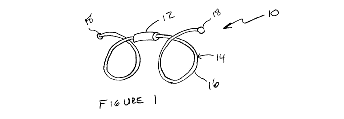
[0019] Figure 1 is a perspective view of an embodiment of the device of the
present
invention;
[0020] Figure 2 is a perspective view of the embodiment of the device of
the present
invention;
[0021] Figure 3a is a perspective view of an embodiment of the device of
the
present invention in an unconstrained state;
[0022] Figure 3b is a perspective view of the device of Figure 3a
constrained within
an airway;
[0023] Figure 4a is a perspective view of an embodiment of the device of
the
present invention in an unconstrained state;
CA 02839796 2013-10-30
WO 2013/003632 PCT/US2012/044714
[0024] Figure 4b is a perspective view of the device of Figure 4a
constrained within
an airway;
[0025] Figure 5a is an end view of an embodiment of the device of the
present
invention;
[0026] Figure 5b is a profile view of the device of Figure 5a;
[0027] Figure 6a is an end view of an embodiment of the device of the
present
invention;
[0028] Figure 6b is a profile view of the device of Figure 6a;
[0029] Figure 7a is an end view of an embodiment of the device of the
present
invention; and,
[0030] Figure 7b is a profile view of the device of Figure 7a.
DETAILED DESCRIPTION OF THE INVENTION
[0031] In general, the present invention includes an identification or
therapeutic
device comprising a body portion and an anchoring portion, which is
introducible into an
intra-body structure (e.g., a mass or lesion) and/or an anatomical space to
mark a
location of interest (e.g., a tissue layer and/or lumen of a body cavity). The
identification
device of the present invention may include a power source, either external to
the body
or internally at or near the body portion or some combination thereof. It is
understood
that any of the various anchoring portions described below may be used with
any of the
body portions. It is also understood that the body portions may give off
energy, such as
light energy (i.e. glow-in-the-dark materials, LEDs, incandescent devices,
etc.), thermal
energy, radiation, RF energy, acoustic energy, or cryoenergy.
[0032] Furthermore, the various embodiments of the body portions may be
constructed of various application-specific materials. For example, the body
portions
may be loaded with chemicals or dyes that enhance localization. Non-limiting
examples
include: Ba504, bismuth, copper, gold, and platinum. Also, the body portions
could be
loaded with drugs and/or chemotherapy agents for treatment and have features
such as
CA 02839796 2013-10-30
WO 2013/003632 PCT/US2012/044714
6
controlled elution and diffusion rates. Non-limiting examples of these agents
include
antineoplastics, antiobiotics and others.
[0033] One embodiment of the present invention is shown in Figure 1, which
illustrates an identification or therapeutic device 10, including a body
portion 12 and
anchoring portion 14. The body portion 12 may be any energy source or simply a
marker or a focusing element for RF energy, as described above. If an energy
source is
used, it is understood that appropriate additional equipment will be used in
order to
receive and identify the energy being transmitted. One embodiment provides a
body
portion 12 that is highly radiopaque, such as gold. Due to the high
radiopacity of gold,
the gold body portion 12 may be sized small enough that it poses little to no
restriction
on airflow, while still being highly visible by an imaging device, such as CT
or
fluoroscopy.
[0034] The embodiment shown in Figure 2 includes a similar anchoring
portion 14
with multiple body portions 12a, 12b, 12c, and so on. Providing multiple body
portions
provides a bright imaging profile while still maintaining a low resistance to
airflow.
Additionally, multiple body portions provide information during imaging as to
the
orientation of the device 10. For example, if the alignment of the body
portions 12
changes relative to each other over time, it may be indicative that migration
is occurring.
Information may also be gleaned as to whether the airways distal of the body
portion are
remaining open. If the body portions can be seen moving rhythmically with
inhalation
and exhalation, it is indicative that air is flowing past the body portions
12. However, if
the body portions 12 are not moving despite inhalation and exhalation, it may
indicate
that air is not flowing past the body portions.
[0035] Conversely, the embodiment shown in Figure 1 includes a single body
portion 12. The single body portion design allows a larger quantity of
radiopaque
material, such as gold, to be concentrated in a single location without
impeding airflow.
A single mass of gold provides a brighter single point in imaging than the
smaller body
portions used in the multiple body portion embodiments. It is noted that all
of the various
anchor designs shown in the Figures and described herein, may be used with a
single
body portion 12 or multiple body portions 12a, 12b, 12c, etc.
CA 02839796 2013-10-30
WO 2013/003632 PCT/US2012/044714
7
[0036] The anchoring portion 14 is constructed and arranged to be placed
into a
small delivery catheter and to expand upon exit from the catheter into a shape
that
secures the device 10 within an airway. Several examples are shown in the
figures.
The anchoring portion 14 shown in Figures 1 and 2 comprises a coil 16 that is
preferably
constructed of a material, such as Nitinol, that is biocompatible and has
shape memory
qualities. The anchoring portion 14 may also include blunt end caps 18 that
prevent
then ends from penetrating the lung tissue during delivery, and also ensure
that the
device will be able to slide through the delivery catheter. The anchoring
portion 14 is
capable of being straightened and contained in a delivery catheter for
extended periods
while still reassuming a deployed shape when released from the catheter. The
anchoring portion shown in Figures 1 and 2 has a spiral or coil shape when
released.
This coil shape places a gentle outward force on the airways, and also expands
and
contracts with the airways to maintain the desired, implanted location.
[0037] Figures 3a and 3b show an anchoring portion 14 that is suitable for
implantation in a large airway. The anchoring portion 14 includes a single
bend 20 that
can be as much as 180 degrees when unconstrained. Figure 3a shown the device
10 in
an unconstrained configuration. Figure 3b shows the device 10 in a deployed
configuration within an airway 1, shown in phantom lines.
[0038] Figures 4a and 4b also show an anchoring portion 14 that is
constructed of a
resilient material, such as a memory metal, that can be elongated and placed
into a
small delivery catheter. Upon release from the catheter, the anchoring portion
14
expands to form a star shape having a plurality of points 20. Figure 4a shows
the
device 10 in an unconstrained configuration. Figure 4b shows the device 10
within an
airway 1. Like the coil shape, the star shape places gentle outward force on
the
airways, and expands and contracts with the airways to maintain the desired,
implanted
location. However, the star shape also expands and contracts in such a manner
that
the contact points of the star, those points 16 contacting tissue, do not
slide as a result
of expansion and contraction. Hence, wear on the tissue contacted by the
device is
minimized.
[0039] Figures 5a and 5b show a device 10 having an anchoring portion 14
that is
constructed and arranged with coils 16 at either end of the device and a
center portion
CA 02839796 2013-10-30
WO 2013/003632 PCT/US2012/044714
8
22 that extends between the two coils 16. The center portion 22 holds the body
portion
12 in the center of the airway. Like the other embodiments, the ends of the
anchoring
device include blunt end caps 18.
[0040] Figures 6a and 6b show a device 10 having an anchoring portion 14
that is
constructed and arranged with coils 16 at either end of the device and a
center portion
22 that extends between the two coils 16. The center portion 24 holds the body
portion
12 along a wall of the airway. Like the other embodiments, the ends of the
anchoring
device include blunt end caps 18. This embodiment maximizes the amount of
airflow
allowed to continue to flow through the airway.
[0041] Figures 7a and 7b show a device 10 having an anchoring portion 14
that is
constructed and arranged with a coil 16 at one end of the device and an axial
portion 26
the other end of the device. The axial portion 26 holds the body portion 12 in
the center
of the airway. Because the axial portion 26 is at the end of the device 10, a
user may
find it easier to predictably place the body portion in a desired location.
Like the other
embodiments, the ends of the anchoring device include blunt end caps 18.
[0042] The method of the present invention is thus described as a method of
marking a location within a lumen of a patient that includes placing an
intralumenal
marker, having an anchoring portion and a body portion attached to said
anchoring
portion, within a catheter in an elongated configuration; navigating said
catheter to a
target location within a lumen of a patient; deploying said intralumenal
marker from said
catheter into said lumen; and allowing said anchoring portion of said marker
to expand
within said lumen, thereby placing atraumatic pressure on walls of said lumen
such that
said body portion is fixed within said lumen.
[0043] The step of allowing said anchoring portion of the marker to expand
within
the lumen, thereby placing atraumatic pressure on walls of the lumen such that
the body
portion is fixed within the lumen, can include allowing the anchoring portion
of the
marker to expand within the lumen, thereby placing atraumatic pressure on
walls of the
lumen such that the body portion is axially centered within the lumen.
[0044] The step of allowing the anchoring portion of the marker to expand
within the
lumen, thereby placing atraumatic pressure on walls of the lumen such that the
body
CA 02839796 2013-10-30
WO 2013/003632 PCT/US2012/044714
9
portion is fixed within the lumen, can include allowing the anchoring portion
of the
marker to expand within the lumen, thereby placing atraumatic pressure on
walls of the
lumen such that the body portion is adjacent a sidewall of the lumen.
[0045] Although the invention has been described in terms of particular
embodiments and applications, one of ordinary skill in the art, in light of
this teaching,
can generate additional embodiments and modifications without departing from
the spirit
of or exceeding the scope of the claimed invention. Accordingly, it is to be
understood
that the drawings and descriptions herein are proffered by way of example to
facilitate
comprehension of the invention and should not be construed to limit the scope
thereof.