Note: Descriptions are shown in the official language in which they were submitted.
CA 02839919 2013-12-18
WO 2012/178157 PCT/US2012/043984
METHOD FOR ANALYZING BIOLOGICAL SPECIMENS BY SPECTRAL IMAGING
Related Application
10001] This application claims the benefit of U.S. Patent Application No.
13/067,777
titled "METHOD FOR ANALYZING BIOLOGICAL SPECIMENS BY SPECTRAL
IMAGING" filed on June 24, 2011. This application is related to
PCT/U52011/041884 titled "METHOD FOR ANALYZING BIOLOGICAL
SPECIMENS BY SPECTRAL IMAGING" filed on June 24, 2011, and U.S.
Provisional Patent Application No, 61/358,606 titled "DIGITAL STAINING OF
HISTOPATHOLOGICAL SPECIMENS VIA SPECTRAL HISTOPATHOLOGY" filed
June 25, 2010. The entirety of each of the foregoing applications is hereby
incorporated by reference herein.
Field of the Invention
[0002] Aspects of the invention relate to a method for analyzing biological
specimens
by spectral imaging to provide a medical diagnosis, prognostic and/or
predictive
classification. The biological specimens may include medical specimens
obtained by
surgical methods, biopsies, and cultured samples.
Background
[00031 Various pathological methods are used to analyze biological specimens
for
the detection of abnormal or cancerous cells. For example, standard
histopathology
involves visual analysis of stained tissue sections by a pathologist using a
microscope. Typically, tissue sections are removed from a patient by biopsy,
and
the samples are either snap frozen and sectioned using a cryo-microtome, or
they
are formalin-fixed, paraffin embedded, and sectioned via a microtome. The
tissue
sections are then mounted onto a suitable substrate. Paraffin-embedded tissue
CA 02839919 2013-12-18
WO 2012/178157 PCT/US2012/043984
sections are subsequently deparaffinized. The tissue sections are stained
using, for
example, an hemotoxylin-eosin (H&E) stain arid are coverslipped.
[0004] The tissue samples are then visually inspected at a high resolution
visual
inspection, for example, 10x to 40x magnification. The magnified cells are
compared
with visual databases in the pathologist's memory. Visual analysis of a
stained
tissue section by a pathologist involves scrutinizing features such as nuclear
and
cellular morphology, tissue architecture, staining patterns, and the
infiltration of
immune response cells to detect the presence of abnormal or cancerous cells.
[0005] If early metastases or small clusters of cancerous cells measuring from
less
than 0.2 to 2 mm in size, known as micrometastases, are suspected, adjacent
tissue
sections may be stained with an immuno-histochemical (IHC) agent/counter stain
such as cytokeratin-specific stains. Such methods increase the sensitivity of
histopathology since normal tissue, such as lymph node tissue, does not
respond to
these stains. Thus, the contrast between unaffected and diseased tissue can be
enhanced.
[0006] The primary method for detecting micrometastases has been standard
histopathology. The detection of micrometastases in lymph nodes, for example,
by
standard histopathology is a formidable task owing to the small size and lack
of
distinguishing features of the abnormality within the tissue of a lymph node.
Yet, the
detection of these micrometastases is of prime importance to stage the spread
of
disease because if a lymph node is found to be free of metastatic cells, the
spread of
cancer may be contained. On the other hand, a false negative diagnosis
resulting
from a missed micrometastasis in a lymph node presents too optimistic a
diagnosis,
and a more aggressive treatment should have been recommended.
2
CA 02839919 2013-12-18
WO 2012/178157 PCT/US2012/043984
[0007] Although standard histopathology is well-established for diagnosing
advanced
diseases, it has numerous disadvantages. In
particular, variations in the
independent diagnoses of the same tissue section by different pathologists are
common because the diagnosis and grading of disease by this method is based on
a
comparison of the specimen of interest with a database in the pathologist's
memory,
which is inherently subjective. Differences in diagnoses particularly arise
when
diagnosing rare cancers or in the very early stages of disease. In addition,
standard
histopathology is time consuming, costly and relies on the human eye for
detection,
which makes the results hard to reproduce. Further, operator fatigue and
varied
levels of expertise of the pathologist may impact a diagnosis.
[0008] In addition, if a tumor is poorly differentiated, many
immunohistochemical
stains may be required to help differentiate the cancer type. Such staining
may be
performed on multiple parallel cell blocks. This staining process may be
prohibitively
expensive and cellular samples may only provide a few diagnostic cells in a
single
cell block.
[0009] To overcome the variability in diagnoses by standard histopathology,
which
relies primarily on cell morphology and tissue architectural features,
spectroscopic
methods have been used to capture a snapshot of the biochemical composition of
cells and tissue. This makes it possible to detect variations in the
biochemical
composition of a biological specimen caused by a variety of conditions and
diseases.
By subjecting a tissue or cellular sample to spectroscopy, variations in the
chemical
composition in portions of the sample may be detected, which may indicate the
presence of abnormal or cancerous cells. The application of spectroscopy to
infrared cytopathology (the study of diseases of cells) is referred to as
"spectral
3
CA 02839919 2013-12-18
WO 2012/178157 PCT/US2012/043984
cytopathology" (SCP), and the application of infrared spectroscopy to
histopathology
(the study of diseases of tissue) as "spectral histopathology" (SHP).
[0010] SCP on individual urinary tract and cultured cells is discussed in B.
Bird et al.,
Vibr. Spectrosc., 48, 10 (2008) and M. Romeo et al., Biochim Biophys Acta,
1758,
915 (2006). SCP based on imaging data sets and applied to oral mucosa and
cervical cells is discussed in WO 2009/146425. Demonstration of disease
progression via SCP in oral mucosal cells is discussed in K. Papamarkakis et
al.,
Laboratory Investigations , 90, 589 (2010). Demonstration of sensitivity of
SCP to
detect cancer field effects and sensitivity to viral infection in cervical
cells is
discussed in K. Papamarkakis et al., Laboratory Investigations, 90, 589,
(2010).
[0011] Demonstration of first unsupervised imaging of tissue using SHP of
liver
tissue via hierarchical cluster analysis (HCA) is discussed in M. Diem et al.,
Biopolymers, 57, 282 (2000). Detection of metastatic cancer in lymph nodes is
discussed in M. J. Romeo et al., Vibrational Spectrosc.. 38, 115 (2005) and M.
Romeo et al., Vibrational Microspectroscopy of Cells and Tissues, Wiley-
Interscience, Hoboken, NJ (2008). Use of neural networks, trained on HCA-
derived
data, to diagnose cancer in colon tissue is discussed in P. Lasch et at.,
J.Chemometrics, 20, 209 (2007). Detection of micro-metastases and individual
metastatic cancer cells in lymph nodes is discussed in B. Bird et al., The
Analyst,
134, 1067 (2009), B. Bird et al., BMC J. Clin. Pathology, 8, 1 (2008), and B.
Bird et
al., Tech. Cancer Res. Treatment, 10, 135 (2011).
[0012] Spectroscopic methods are advantageous in that they alert a pathologist
to
slight changes in chemical composition in a biological sample, which may
indicate an
early stage of disease. In contrast, morphological changes in tissue evident
from
standard histopathology take longer to manifest, making early detection of
disease
4
CA 02839919 2013-12-18
WO 2012/178157 PCT/US2012/043984
more difficult. Additionally, spectroscopy allows a pathologist to review a
larger
sample of tissue or cellular material in a shorter amount of time than it
would take the
pathologist to visually inspect the same sample. Further, spectroscopy relies
on
instrument-based measurements that are objective, digitally recorded and
stored,
reproducible, and amenable to mathematical/statistical analysis. Thus, results
derived from spectroscopic methods are more accurate and precise then those
derived from standard histopathological methods.
[0013] Various techniques may be used to obtain spectral data. For example,
Raman spectroscopy, which assesses the molecular vibrations of a system using
a
scattering effect, may be used to analyze a cellular or tissue sample. This
method is
described in N. Stone et al, Vibrational Spectroscopy for Medical Diagnosis,
J.Wiley
& Sons (2008), and C.Krafft, et al., Vibrational Spectrosc. (2011).
[0014] Raman's scattering effect is considered to be weak in that only about 1
in
101a incident photons undergoes Raman scattering. Accordingly, Raman
spectroscopy works best using a tightly focused visible or near-1R laser beam
for
excitation. This, in turn, dictates the spot from which spectral information
is being
collected. This spot size may range from about 0.3 pm to 2 pm in size,
depending
on the numerical aperture of the microscope objective, and the wavelength of
the
laser utilized. This small spot size precludes data collection of large tissue
sections,
since a data set could contain millions of spectra and would require long data
acquisition times. Thus, SHP using Raman spectroscopy requires the operator to
select small areas of interest. This approach negates the advantages of
spectral
imaging, such as the unbiased analysis of large areas of tissue.
[0015] SHP using infrared spectroscopy has also been used to detect
abnormalities
in tissue, including, but not limited to brain, lung, oral mucosa, cervical
mucosa,
5
CA 02839919 2013-12-18
WO 2012/178157 PCT/US2012/043984
thyroid, colon, skin, breast, esophageal, prostate, and lymph nodes. Infrared
spectroscopy, like Raman spectroscopy, is based on molecular vibrations, but
is an
absorption effect, and between 1% and 50% of incident infrared photons are
likely to
be absorbed if certain criteria are fulfilled. As a result, data can be
acquired by
infrared spectroscopy more rapidly with excellent spectral quality compared to
Raman spectroscopy. In addition, infrared spectroscopy is extremely sensitive
in
detecting small compositional changes in tissue. Thus, SHP using infrared
spectroscopy is particularly advantageous in the diagnosis, treatment and
prognosis
of cancers such as breast cancer, which frequently remains undetected until
metastases have formed, because it can easily detect micro-metastases. It can
also
detect small clusters of metastatic cancer cells as small as a few individual
cells.
Further, the spatial resolution achievable using infrared spectroscopy is
comparable
to the size of a human cell, and commercial instruments incorporating large
infrared
array detectors may collect tens of thousands of pixel spectra in a few
minutes.
[0016] A method of SHP using infrared spectroscopy is described in Bird et
al.,
"Spectral detection of micro-metastates in lymph node histo-pathology", J.
Biophoton. 2, No. 1-2, 37-46 (2009), (hereinafter "Bird"). This method
utilizes
infrared micro-spectroscopy (IRMSP) and multivariate analysis to pinpoint
micro-
metastases and individual metastatic cells in lymph nodes.
[0017] Bird studied raw hyperspectral imaging data sets including 25,600
spectra,
each containing 1650 spectral intensity points between 700 and 4000 cm-1.
These
data sets, occupying about 400 MByte each, were imported and pre-processed.
Data preprocessing included restriction of the wavenumber range to 900-1800 cm-
1
and other processes. The "fingerprint" infrared spectral region was further
divided
into a "protein region" between 1700 and 1450 cm-1, which is dominated by the
6
CA 02839919 2013-12-18
WO 2012/178157 PCT/US2012/043984
amide I and amide II vibrational bands of the peptide linkages of proteins.
This
region is highly sensitive to different protein secondary and tertiary
structure and can
be used to stage certain events in cell biology that depend on the abundance
of
different proteins. The lower wavenumber range, from 900 to 1350 crn'l, the
6 "phosphate region", contains several vibrations of the phosphodiester
linkage found
in phospholipids, as well as DNA and RNA.
[0018] In Bird, a minimum intensity criterion for the integrated amide I band
was
imposed to eliminate pixels with no tissue coverage. Then, vector
normalization and
conversion of the spectral vectors to second derivatives was performed.
Subsequently, data sets were subjected individually to hierarchical cluster
analysis
(HCA) using the Euclidean distance to define spectral similarity and Ward's
algorithm
for clustering. Pixel cluster membership was converted to pseudo-color
spectral
images.
[0019] According to Bird's method, marks are placed on slides with a stained
tissue
section to highlight areas that correspond to areas on the unstained adjacent
tissue
section that are to be subjected to spectral analysis. The resulting spectral
and
visual images are matched by a user who aligns specific features on the
spectral
image and the visual image to physically overlay the spectral and visual
images.
[0020] By Bird's method, corresponding sections of the spectral image and the
visual
image are examined to determine any correlation between the visual
observations
and the spectral data. In particular, abnormal or cancerous cells observed by
a
pathologist in the stained visual image may also be observed when examining a
corresponding portion of the spectral image that overlays the stained visual
image.
Thus, the outlines of the patterns in the pseudo-color spectral image may
correspond
to known abnormal or cancerous cells in the stained visual image. Potentially
7
CA 02839919 2013-12-18
WO 2012/178157 PCT/US2012/043984
abnormal or cancerous cells that were observed by a pathologist in a stained
visual
image may be used to verify the accuracy of the pseudo-color spectral image.
(0021] Bird's method, however, is inexact because it relies on the skill of
the user to
visually match specific marks on the spectral and visual images. This method
is
often imprecise. in addition, Bird's method allows the visual and spectral
images to
be matched by physically overlaying them, but does not join the data from the
two
images to each other. Since the images are merely physically overlaid, the
superimposed images are not stored together for future analysis.
(0022] Further, since different adjacent sections of tissue are subjected to
spectral
and visual imaging, Bird's overlaid images do not display the same tissue
section.
This makes it difficult to match the spectral and visual images, since there
may be
differences in the morphology of the visual image and the color patterns in
the
spectral image.
(00231 Another problem with Bird's overlaying method is that the visual image
is not
in the same spatial domain as the infrared spectral image. Thus, the spatial
resolution of Bird's visual image and spectral image are different. Typically,
spatial
resolution in the infrared image is less than the resolution of the visual
image. To
account for this difference in resolution, the data used in the infrared
domain may be
expanded by selecting a region around the visual point of interest and
diagnosing the
region, and not a single point. For every point in the visual image, there is
a region
in the infrared image that is greater than the point that must be input to
achieve
diagnostic output. This process of accounting for the resolution differences
is not
performed by Bird. Instead, Bird assumes that when selecting a point in the
visual
image, it is the same point of information in the spectral image through the
overlay,
CA 02839919 2013-12-18
WO 2012/178157 PCT/US2012/043984
and accordingly a diagnostic match is reported. While the images may visually
be
the same, they are not the same diagnostically.
[0024] To claim a diagnostic match, the spectral image used must be output
from a
supervised diagnostic algorithm that is trained to recognize the diagnostic
signature
of interest. Thus, the spectral image cluster will be limited by the algorithm
classification scheme to driven by a biochemical classification to create a
diagnostic
match, and not a user-selectable match. By contrast, Bird merely used an
"unsupervised HCA image to compare to a "supervised" stained visual image to
make a diagnosis. The HCA image identifies regions of common spectral features
that have not yet been determined to be diagnostic, based on rules and limits
assigned for clustering, including manually cutting the dendrogram until a
boundary
(geometric) match is visually accepted by the pathologist to outline a cancer
region.
This method merely provides a visual comparison.
[0025] Other methods based on the analysis of fluorescence data exist that are
generally based on the distribution of an external tag, such as a stain or
label, or
utilize changes in the inherent fluorescence, also known as auto-fluorescence.
These methods are generally less diagnostic, in terms of recognizing
biochemical
composition and changes in composition. In addition, these methods lack the
fingerprint sensitivity of techniques of vibrational spectroscopy, such as
Raman and
infrared,
[0026] A general problem with spectral acquisition techniques is that an
enormous
amount of spectral data is collected when testing a biological sample. As a
result,
the process of analyzing the data becomes computationally complicated and time
consuming. Spectral data often contains confounding spectral features that are
frequently observed in microscopically acquired infrared spectra of cells and
tissue,
9
CA 02839919 2013-12-18
WO 2012/178157 PCT/US2012/043984
such as scattering and baseline artifacts. Thus, it is helpful to subject the
spectral
data to pre-processing to isolate the cellular material of interest, and to
remove
confounding spectral features.
[0027] One type of confounding spectral feature is Mie scattering, which is a
sample
morphology-dependent effect. This effect interferes with infrared absorption
or
reflection measurements if the sample is non-uniform and includes particles
the size
of approximately the wavelength of the light interrogating the sample. Mie
scattering
is manifested by broad, undulating scattering features, onto which the
infrared
absorption features are superimposed,
[0028] Mie scattering may also mediate the mixing of absorptive and reflective
line
shapes. In principle, pure absorptive line shapes are those corresponding to
the
frequency-dependence of the absorptivity, and are usually Gaussian, Lorentzian
or
mixtures of both. The absorption curves correspond to the imaginary part of
the
complex refractive index. Reflective contributions correspond to the real part
of the
complex refractive index, and are dispersive in line shapes. The dispersive
contributions may be obtained from absorptive line shapes by numeric KK-
transform,
or as the real part of the complex Fourier transform (FT).
[0029] Resonance Mie (RMie) features result from the mixing of absorptive and
reflective band shapes, which occurs because the , refractive index undergoes
anomalous dispersion when the absorptivity goes through a maximum (i.e., over
the
profile of an absorption band). Mie scattering, or any other optical effect
that
depends on the refractive index, will mix the reflective and absorptive line
shapes,
causing a distortion of the band profile, and an apparent frequency shift.
[0030] Figure 1 illustrates the contamination of absorption patterns by
dispersive
band shapes observed in both SCP and SHP. The bottom trace in Figure 1 depicts
CA 02839919 2013-12-18
WO 2012/178157 PCT/US2012/043984
a regular absorption spectrum of biological tissue, whereas the top trace
shows a
spectrum strongly contaminated by a dispersive component via the RMie effect.
The
spectral distortions appear independent of the chemical composition, but
rather
depend on the morphology of the sample. The resulting band intensity and
frequency shifts aggravate spectral analysis to the point that uncontaminated
and
contaminated spectra are classified into different groups due to the presence
of the
band shifts. Broad, undulating background features are shown in Figure 2. When
superimposed on the infrared micro-spectroscopy (IR-MSP) patterns of cells,
these
features are attributed to Mie scattering by spherical particles, such as
cellular
nuclei, or spherical cells.
[0031] The appearance of dispersive line shapes in Figure 1 superimposed on IR-
MSP spectra was reported along with a theoretical analysis in M. Romeo, et
al.,
Vibrational Spectroscopy. 38, 129 (2005) (hereinafter "Romeo 2005"). Romeo
2005
indentifies the distorted band shapes as arising from the superposition of
dispersive
(reflective) components onto the absorption features of an infrared spectrum.
These
effects were attributed to incorrect phase correction of the instrument
control
software. In particular, the acquired raw interferogram in FTIR spectroscopy
frequently is "chirped" or asymmetric, and needs to be symmetrized before FT.
This
is accomplished by collecting a double sided interferogram over a shorter
interferometer stroke, and calculating a phase correction to yield a symmetric
interferogram.
[0032] In Romeo 2005, it was assumed that this procedure was not functioning
properly, which causes it to yield distorted spectral features. An attempt was
made
to correct the distorted spectral features by calculating the phase between
the real
and imaginary parts of the distorted spectra, and reconstructing a power
spectrum
11
CA 02839919 2013-12-18
WO 2012/178157 PCT/US2012/043984
from the phase corrected real and imaginary parts. Romeo 2005 also reported
the
fact that in each absorption band of an observed infrared spectrum, the
refractive
index undergoes anomalous dispersion. Under certain circumstances, various
amounts of the dispersive line shapes can be superimposed, or mixed in, with
the
absorptive spectra.
[0033] The mathematical relationship between absorptive and reflective band
shapes is given by the Kramers-Kronig (KK) transformation, which relates the
two
physical phenomena. The mixing of dispersive (reflective) and absorptive
effects in
the observed spectra was identified, and a method to correct the effect via a
procedure called "Phase Correction" (PC) is discussed in Romeo 2005. Although
the cause of the mixing of dispersive and absorptive contributions was
erroneously
attributed to instrument software malfunction, the principle of the
confounding effect
was properly identified. Due to the incomplete understanding of the underlying
physics, however, the proposed correction method did not work properly.
[0034] P. Bassan et al., Analyst, 134, 1586 (2009) and P. Bassan et al.,
Analyst,
134, 1171 (2009) demonstrated that dispersive and absorptive effects may mix
via
the "Resonance Mie Scattering" (RMieS) effect. An algorithm and method to
correct
spectral distortion is described in P. Bassan et al., "Resonant Mie Scattering
(RMieS)
correction of infrared spectra from highly scattering biological samples",
Analyst,
135, 268-277 (2010). This method is an extension of the "Extended
Multiplicative
Signal Correction" (EMSC) method reported in A. Kohler et al., Appl.
Spectrosc., 59,
707 (2005) and A. Kohler et al., Appl. Spectrosc., 62, 259 (2008).
[0035] This method removes the non-resonant Mie scattering from infrared
spectral
datasets by including reflective components obtained via KK-transform of pure
absorption spectra into a multiple linear regression model. The method
utilizes the
12
CA 02839919 2013-12-18
WO 2012/178157 PCT/US2012/043984
raw dataset and a "reference" spectrum as inputs, where the reference spectrum
is
used both to calculate the reflective contribution, and as a normalization
feature in
the EMSC scaling. Since the reference spectrum is not known a priori, Bassan
et al.
use the mean spectrum of the entire dataset, or an "artificial" spectrum, such
as the
spectrum of a pure protein matrix, as a "seed" reference spectrum. After the
first
pass through the algorithm, each corrected spectrum may be used in an
iterative
approach to correct all spectra in the subsequent pass. Thus, a dataset of
1000
spectra will produce 1000 RMieS-EMSC corrected spectra, each of which will be
used as an independent new reference spectrum for the next pass, requiring
1,000,000 correction runs. To carry out this algorithm, referred to as the
"RMieS-
EMSC" algorithm, to a stable level of corrected output spectra required a
number of
passes (-10), and computation times that are measured in days.
[0036] Since the RMieS-EMSC algorithm requires hours or days of computation
time, a fast, two-step method to perform the elimination of scattering and
dispersive
line shapes from spectra was developed, as discussed in B. Bird, M. Miljkovio
and M.
Diem, "Two step resonant Mie scattering correction of infrared micro-spectral
data:
human lymph node tissue", J. Biophotonics, 3 (8-9) 597-608 (2010). This
approach
includes fitting multiple dispersive components, obtained from KK-transform of
pure
absorption spectra, as well as Mie scattering curves computed via the van
Huist
equation (see H. C. Van De Hu1st, Light Scattering by Small Particles, Dover,
Mineola, NY, (1981)), to all the spectra in a dataset via a procedure known as
Extended Multiplicative Signal Correction (EMSC) (see A. Kohler et al.,
Appl.Spectrosc., 62, 259 (2008)) and reconstructing all spectra without these
confounding components.
13
CA 02839919 2013-12-18
WO 2012/178157 PCT/US2012/043984
[0037] This algorithm avoids the iterative approach used in the RMieS-EMSC
algorithm by using uncontaminated reference spectra from the dataset. These
uncontaminated reference spectra were found by carrying out a preliminary
cluster
analysis of the dataset and selecting the spectra with the highest amide I
frequencies
in each cluster as the "uncontaminated" spectra. The spectra were converted to
pure reflective spectra via numeric KK transform and used as interference
spectra,
along with compressed Mie curves for RMieS correction as described above. This
approach is fast, but only works well for datasets containing a few spectral
classes.
[0038] In the case of spectral datasets containing many tissue types, however,
the
extraction of uncontaminated spectra can become tedious. Furthermore, under
these conditions, it is unclear whether fitting all spectra in the dataset to
the most
appropriate interference spectrum is guaranteed. In addition, this algorithm
requires
reference spectra for correction, and works best with large datasets.
[0039] In light of the above, there remains a need for improved methods of
analyzing
biological specimens by spectral imaging to provide a medical diagnosis.
Further,
there is a need for an improved pre-processing method that is based on a
revised
phase correction approach, does not require input data, is computationally
fast, and
takes into account many types of confounding spectral contributions that are
frequently observed in microscopically acquired infrared spectra of cells and
tissue.
Summary
[0040] One aspect of the invention relates to a method for analyzing
biological
specimens by spectral imaging to provide a medical diagnosis. The method
includes
obtaining spectral and visual images of biological specimens and registering
the
images to detect abnormalities in the biological specimen, such as, but not
limited to,
cell abnormalities, pre-cancerous cells, and cancerous cells. This
method
14
CA 02839919 2013-12-18
WO 2012/178157 PCT/US2012/043984
overcomes the obstacles discussed above, among others, in that it eliminates
the
bias and unreliability of diagnoses and prognosis that are inherent in
standard
histopathological, cytological, and other spectral methods.
[00411 Another aspect of the invention relates to a method for correcting
confounding spectral contributions that are frequently observed in
microscopically
acquired infrared spectra of cells and tissue by performing a phase correction
on the
spectral data. This phase correction method may be used to correct various
kinds of
absorption spectra that are contaminated by reflective components.
[0042] According to aspects of the invention, a method for analyzing
biological
specimens by spectral imaging includes acquiring a spectral image of the
biological
specimen, acquiring a visual image of the biological specimen, and registering
the
visual image and spectral image.
(00431 A method of developing a data repository according to aspects of the
invention includes identifying a region of a visual image displaying a disease
or
condition, associating the region of the visual image to spectral data
corresponding
to the region, and storing the association between the spectral data and the
corresponding disease or condition.
[0044] A method of providing a medical diagnosis according to aspects of the
invention includes obtaining spectroscopic data for a biological specimen,
comparing
the spectroscopic data for the biological specimen to data in a repository
that is
associated with a disease or condition, determining any correlation between
the
repository data and the spectroscopic data for the biological specimen, and
outputting a diagnosis associated with the determination.
(0045] A system for providing a medical diagnosis and prognosis, according to
aspects of the invention includes a processor, a user interface functioning
via the
CA 02839919 2013-12-18
WO 2012/178157 PCT/US2012/043984
processor, and a repository accessible by the processor, where spectroscopic
data
of a biological specimen is obtained, the spectroscopic data for the
biological
specimen is compared to repository data that is associated with a disease or
condition, any correlation between the repository data and the spectroscopic
data for
the biological specimen is determined; and a diagnosis associated with the
determination is output.
[0046] A computer program product according to aspects of the invention
includes a
computer usable medium having control logic stored therein for causing a
computer
to provide a medical diagnosis. The control logic includes a first computer
readable
program code means for obtaining spectroscopic data for a biological specimen,
second computer readable program code means for comparing the spectroscopic
data for the biological specimen to repository data that is associated with a
disease
or condition, third computer readable program code means for determining any
correlation between the repository data and the spectroscopic data for the
biological
16 specimen, and fourth computer readable program code means for outputting
a
diagnosis and/or or a prognostic determination associated with the
determination.
[0047] Description of the Drawings
[0048] Figure 1 illustrates the contamination of absorption patterns by
dispersive
band shapes typically observed in both SCP and SHP;
[0049] Figure 2 shows broad, undulating background features typically observed
on
IR-MSP spectral of cells attributed to Mie scattering by spherical particles;
[0050] Figure 3 is a flowchart illustrating a method of analyzing a biological
sample
by spectral imaging according to aspects of the invention;
[0051] Figure 3A is a flowchart illustrating steps in a method of acquiring a
spectral
image according to aspects of the invention;
16
CA 02839919 2013-12-18
WO 2012/178157 PCT/US2012/043984
[0052] Figure 3B is a flowchart illustrating steps in a method of pre-
processing
spectral data according to aspects of the invention;
[0053] Figures 4A and 4B are a flowchart illustrating an example method of
performing image registration on a spectral image and a visual image in
accordance
with aspects of the present invention;
[0054] Figure 4C illustrates an example slide holder in accordance with
aspects of
the present invention;
[0055] Figure 5A is a flowchart illustrating an example method of refining
image
registration in accordance with aspects of the present invention;
[0056] Figure 5B is an example a graphical user interface (GUI) interface for
setting
a threshold value in accordance with aspects of the present invention;
[0057] Figure 5C is an example GUI interface illustrating an example
optimization
window in accordance with aspects of the present invention;
[0058] Figure 6A shows a typical spectrum, superimposed on a linear background
according to aspects of the invention;
[0059] Figure 6B shows an example of a second derivative spectrum according to
aspects of the invention;
[0060] Figure 7 shows a portion of the real part of an interferogram according
to
aspects of the invention;
[0061] Figure 8 shows that the phase angle that produces the largest intensity
after
phase correction is assumed to be the uncorrupted spectrum according to
aspects of
the invention;
[0062] Figure 9A shows that absorption spectra that are contaminated by
scattering
effects that mimic a baseline slope according to aspects of the invention;
17
CA 02839919 2013-12-18
WO 2012/178157 PCT/US2012/043984
[0063] Figure 9B shows that the imaginary part of the forward FT exhibits
strongly
curved effects at the spectral boundaries, which will contaminate the
resulting
corrected spectra according to aspects of the invention;
[0064] Figure 10A is H&E-based histopathology showing a lymph node that has
confirmed breast cancer micro-metastases under the capsule according to
aspects
of the invention;
[0065] Figure 10B shows data segmentation by Hierarchical Cluster Analysis
(HCA)
carried out on the lymph node section of Figure 10A according to aspects of
the
invention;
[0066] Figure 10C is a plot showing the peak frequencies of the amide I
vibrational
band in each spectrum according to aspects of the invention; =
[0067] Figure 10D shows an image of the same lymph node section of Figure 10A
after phase-correction using RMieS correction according to aspects of the
invention;
[0066] Figure 11A shows the results of HCA after phase-correction using RMieS
correction of Figure 10D according to aspects of the invention;
[0069] Figure 11B is H&E-based histopathology of the lymph node section of
Figure
11A according to aspects of the invention;
[0070] Figure 12A is a visual microscopic image of a section of stained
cervical
image;
[0071] Figure 12B is an infrared spectral image created from hierarchical
cluster
analysis of an infrared dataset collected prior to staining the tissue
according to
aspects of the invention;
[0072] Figure 13A is a visual microscopic image of a section of an H&E-stained
axillary lymph node section according to aspects of the invention;
18
CA 02839919 2013-12-18
WO 2012/178157 PCT/US2012/043984
[00731 Figure 138 is an infrared spectral image created from a Multilayer
Perceptron
Networks analysis of an infrared dataset collected prior to staining the
tissue
according to aspects of the invention;
[0074] Figure 14A is a visual image of a small cell lung cancer tissue
according to
aspects of the invention.
[0076] Figure 14B is an HCA-based spectral image of the tissue shown in Figure
14A according to aspects of the invention;
[0076] Figure 14C is a registered image of the visual image of Figure 14A and
the
spectral image of Figure 14B, according to aspects of the invention;
[0077] Figure 140 is an example of a graphical user interface (GUI) for the
registered image of Figure 14C according to aspects of the invention;
[0078] Figure 15A is a visual microscopic image of H&E-stained lymph node
tissue
section according to aspects of the invention;
[0079] Figure 158 is a global digital staining image of section shown in
Figure 15A,
distinguishing capsule and interior of lymph node according to aspects of the
invention;
[0080] Figure 15C is a diagnostic digital staining image of the section shown
in
Figure 15A, distinguishing capsule, metastatic breast cancer, histiocytes,
activated
B-lymphocytes, and T -lymphocytes according to aspects of the invention;
[0081] Figure 16 is a schematic of relationship between global and diagnostic
digital
staining according to aspects of the invention;
[0082] Figure 17A is a visual image of H&E-stained tissue section from an
axillary
lymph node according to aspects of the invention;
[00831 Figure 17B is a SHP-based digitally stained region of breast cancer
micrometastasis according to aspects of the invention;
19
CA 02839919 2013-12-18
WO 2012/178157 PCT/US2012/043984
[0084] Figure 17C is a SHP-based digitally stained region occupied by B-
lymphocyes according to aspects of the invention;
[0085] Figure 17D is a SHP-based digitally stained region occupied by
histocytes
according to aspects of the invention.
[0086] Figure 18 illustrates the detection of individual cancer cells, and
small clusters
of cancer cells via SHP according to aspects of the invention;
[0087] Figure 19A shows raw spectral data sets comprising cellular spectra
recorded
from lung adenocarcinoma, small cell carcinoma, and squamous cell carcinoma
cells
according to aspects of the invention;
[0088] Figure 19B shows corrected spectral data sets comprising cellular
spectra
recorded from lung adenocarcinoma, small cell carcinoma, and squamous cell
carcinoma cells according to aspects of the invention;
[0089] Figure 19C shows standard spectra for lung adenocarcinoma, small cell
carcinoma, and squamous cell carcinoma according to aspects of the invention;
[0090] Figure 190 shows KK transformed spectra calculated from spectra in
Figure
19C;
[0091] Figure 19E shows PCA scores plots of the multi class data set before
EMSC
correction according to aspects of the invention;
[0092] Figure 19F shows PCA scores plots of the multi class data set after
EMSC
correction according to aspects of the invention;
[0093] Figure 20A shows mean absorbance spectra of lung adenocarcinoma, small
cell carcinoma, and squamous carcinoma, according to aspects of the invention;
[0094] Figure 20B shows second derivative spectra of absorbance spectra
displayed
in Figure 20A according to aspects of the invention;
CA 02839919 2013-12-18
WO 2012/178157 PCT/US2012/043984
[0096] Figure 21A shows 4 stitched microscopic R&E-stained images of 1 mm x 1
mm tissue areas comprising adenocarcinoma, small cell carcinoma, and squamous
cell carcinoma cells, respectively, according to aspects of the invention;
[0096] Figure 21B is a binary mask image constructed by performance of a rapid
reduced RCA analysis upon the 1350 cm-1 - 900 cm-1 spectral region of the 4
stitched raw infrared images recorded from the tissue areas shown in Figure
21A
according to aspects of the invention;
[0097] Figure 21C is a 6-cluster RCA image of the scatter corrected spectral
data
recorded from regions of diagnostic cellular material according to aspects of
the
invention;
[0098] Figure 22 shows various features of a computer system for use in
conjunction
with aspects of the invention; and
[0099] Figure 23 shows a computer system for use in conjunction with aspects
of the
invention;
[00100] The file of this patent contains at least one drawing executed in
color.
Copies of this patent with color drawing(s) will be provided by the Patent and
Trademark Office upon request and payment of the necessary fee.
Detailed Description
[00101] Unless otherwise defined, all technical and scientific terms used
herein have
the same meaning as commonly understood by one of ordinary skill in the art to
which aspects of this invention belong. Although methods and materials similar
or
equivalent to those described herein can be used in the practice or testing,
suitable
methods and materials are described below. All publications, patent
applications,
patents, and other references mentioned herein are incorporated by reference
in
their entirety. In case of conflict, this specification, including
definitions, will control.
21
CA 02839919 2013-12-18
WO 2012/178157 PCT/US2012/043984
In addition, the materials, methods, and examples are illustrative only and
not
intended to be limiting.
[00102] One aspect of the invention relates to a method for analyzing
biological
specimens by spectral imaging to provide a medical diagnosis. The biological
specimens may be medical specimens obtained by surgical methods, biopsies, and
cultured samples. The method includes obtaining spectral and visual images of
biological specimens and registering the images to detect cell abnormalities,
pre-
cancerous cells, and cancerous cells. The biological specimens may include
tissue
or cellular samples, but tissue samples are preferred for some applications.
This
method identifies abnormal or cancerous and other disorders including, but not
limited to, lymph node, thyroid, breast, uterine, renal, testicular, ovarian,
or prostate
cancer, small cell lung carcinoma, non-small cell lung carcinoma, and
melanoma, as
well as non-cancerous effects including, but not limited to, inflammation,
necrosis,
and apoptosis.
[001031 One method in accordance with aspects of the invention overcomes the
obstacles discussed above in that it eliminates or generally reduces the bias
and
unreliability of diagnoses, prognosis, predictive, and theranostics that are
inherent in
standard histopathological and other spectral methods. In addition, it allows
access
to a spectral database of tissue types that is produced by quantitative and
reproducible measurements and is analyzed by an algorithm that is calibrated
against classical histopathology. Via this method, for example, abnormal and
cancerous cells may be detected earlier than they can be identified by the
related
art, including standard histopathological or other spectral techniques.
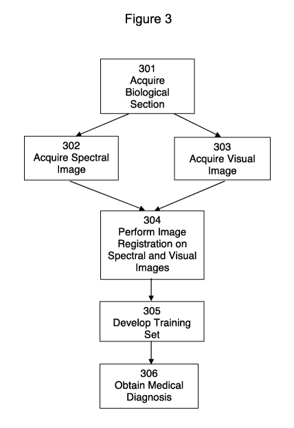
[00104] A method in accordance with aspects of the invention is illustrated in
the
flowchart of Figure 3. As shown in Figure 3, the method generally includes the
steps
22
CA 02839919 2013-12-18
WO 2012/178157 PCT/US2012/043984
of acquiring a biological section 301, acquiring a spectral image of the
biological
section 302, acquiring a visual image of the same biological section 303, and
performing image registration 304. The registered image may optionally be
subjected to training 305, and a medical diagnosis may be obtained 306.
[00106] Bjolpgical Section
[00106] According to the example method of the invention shown in Figure 3,
the
step of acquiring a biological section 301 refers to the extraction of tissue
or cellular
material from an individual, such as a human or animal. A tissue section may
be
obtained by methods including, but not limited to core and punch biopsy, and
excising. Cellular material may be obtained by methods including, but not
limited to
swabbing (exfoliation), washing (lavages), and by fine needle aspiration
(FNA).
[00107] A tissue section that is to be subjected to spectral and visual image
acquisition may be prepared from frozen or from paraffin embedded tissue
blocks
according to methods used in standard histopathology. The section may be
mounted on a slide that may be used both for spectral data acquisition and
visual
pathology. For example, the tissue may be mounted either on infrared
transparent
microscope slides comprising a material including, but not limited to, calcium
fluoride
(CaF2) or on infrared reflective slides, such as commercially available "low-
e" slides.
After mounting, paraffin-embedded samples may be subjected to
deparaffinization.
[00108] Spectral Image
[00109] According to aspects of the invention, the step of acquiring a
spectral image
of the biological section 302 shown in Figure 3 may include acquiring spectral
data
from the biological section 308, performing data pre-processing 310,
performing
multivariate analysis 312, and creating a grayscale or pseudo-color image of
the
biological section 314, as outlined in the flowchart of Figure 3A.
23
CA 02839919 2013-12-18
WO 2012/178157 PCT/US2012/043984
[00110] Spectral Data
[00111] As set forth in Figure 3A, spectral data from the biological section
may be
acquired 401. Spectral data from an unstained biological sample, such as a
tissue
sample, may be obtained to capture a snapshot of the chemical composition of
the
sample. The spectral data may be collected from a tissue section in pixel
detail,
where each pixel is about the size of a cellular nucleus. Each pixel has its
own
spectral pattern, and when the spectral patterns from a sample are compared,
they
may show small but recurring differences in the tissue's biochemical
composition.
[00112] The spectral data may be collected by methods including, but not
limited to
infrared, Raman, visible, terahertz, and fluorescence spectroscopy.
Infrared
spectroscopy may include, but is not limited to, attenuated total reflectance
(ATR)
and attenuated total reflectance Fourier transform infrared spectroscopy (ATR-
FTIR).
In general, infrared spectroscopy may be used because of its fingerprint
sensitivity,
which is also exhibited by Raman spectroscopy. Infrared spectroscopy may be
used
with larger tissue sections and to provide a dataset with a more manageable
size
than Raman spectroscopy. Furthermore, infrared spectroscopy data may be more
amenable to fully automatic data acquisition and interpretation. Additionally,
infrared
spectroscopy may have the necessary sensitivity and specificity for the
detection of
various tissue structures and diagnosis of disease.
[00113] The intensity axis of the spectral data, in general, express
absorbance,
reflectance, emittance, scattering intensity or any other suitable measure of
light
power. The wavelength may relate to the actual wavelength, wavenumber,
frequency or energy of electromagnetic radiation.
[00114] Infrared data acquisition may be carried out using presently available
Fourier transform (FT) infrared imaging microspectrometers, tunable laser-
based
24
CA 02839919 2013-12-18
WO 2012/178157 PCT/US2012/043984
imaging instruments, such as quantum cascade or non-linear optical devices, or
other functionally equivalent instruments based on different technologies. The
acquisition of spectral data using a tunable laser is described further in
U.S. Patent
Application Serial No. 13/084,287 titled, "Tunable Laser-Based Infrared
Imaging
System and Method of Use Thereof, which is incorporated herein in its entirety
by
reference.
[00116] According to one method in accordance with aspects of the invention, a
pathologist or technician may select any region of a stained tissue section
and
receive a spectroscopy-based assessment of the tissue region in real-time,
based on
the hyperspectral dataset collected for the tissue before staining. Spectral
data may
be collected for each of the pixels in a selected unstained tissue sample.
Each of
the collected spectra contains a fingerprint of the chemical composition of
each of
the tissue pixels. Acquisition of spectral data is described in WO
2009/146425,
which is incorporated herein in its entirety by reference.
[00116] In general, the spectral data includes hyperspectral datasets, which
are
constructs including N = n = m individual spectra or spectral vectors
(absorption,
emission, reflectance etc.), where n and m are the number of pixels in the x
and y
dimensions of the image, respectively. Each spectrum is associated with a
distinct
pixel of the sample, and can be located by its coordinates x and y, where
1sx5n, and
15ysm. Each vector has k intensity data points, which are usually equally
spaced in
the frequency or wavenumber domain.
[00117] The pixel size of the spectral image may generally be selected to be
smaller
than the size of a typical cell so that subcellular resolution may be
obtained. The
size may also be determined by the diffraction limit of the light, which is
typically
about 5 pm to about 7 pm for infrared light. Thus, for a 1 mm2 section of
tissue,
CA 02839919 2013-12-18
WO 2012/178157 PCT/US2012/043984
about 1402 to about 2002 individual pixel infrared spectra may be collected.
For each
of the N pixels of a spectral "hypercube", its x and y coordinates and its
intensity
vector (intensity vs. wavelength), are stored.
[00118] Pre-Procesinq
[00119] Subjecting the spectral data to a form of pre-processing may be
helpful to
isolate the data pertaining to the cellular material of interest and to remove
confounding spectral features. Referring to Figure 3A, once the spectral data
is
collected, it may be subjected to such pre-processing 310.
[00120] Pre-processing may involve creating a binary mask to separate
diagnostic
from non-diagnostic regions of the sampled area to isolate the cellular data
of
interest. Methods for creating a binary mask are disclosed in WO 2009/146425,
which is incorporated by reference herein in its entirety.
[00121] A method of preprocessing, according to another aspect of the
invention,
permits the correction of dispersive line shapes in observed absorption
spectra by a
"phase correction" algorithm that optimizes the separation of real and
imaginary
parts of the spectrum by adjusting the phase angle between them. This method,
which is computationally fast, is based on a revised phase correction
approach, in
which no input data are required. Although phase correction is used in the pre-
processing of raw interferograms in FTIR and NMR spectroscopy (in the latter
case,
the interferogram is usually referred to as the "free induction decay, FID")
where the
proper phase angle can be determined experimentally, the method of this aspect
of
the invention differs from earlier phase correction approaches in that it
takes into
account mitigating factors, such as Mie. RMie and other effects based on the
anomalous dispersion of the refractive index, and it may be applied to
spectral
datasets retroactively,
26
CA 02839919 2013-12-18
WO 2012/178157 PCT/US2012/043984
[00122] The pre-processing method of this aspect of the invention transforms
corrupted spectra into Fourier space by reverse FT transform. The reverse FT
results in a real and an imaginary interferogram. The second half of each
interferogram is zero-filled and forward FT transformed individually. This
process
yields a real spectral part that exhibits the same dispersive band shapes
obtained via
numeric KK transform, and an imaginary part that includes the absorptive line
shapes. By recombining the real and imaginary parts with a correct phase angle
between them, phase-corrected, artifact-free spectra are obtained.
[00123] Since the phase required to correct the contaminated spectra cannot be
determined experimentally and varies from spectrum to spectrum, phase angles
are
determined using a stepwise approach between -90 and 90 in user selectable
steps. The "best" spectrum is determined by analysis of peak position and
intensity
criteria, both of which vary during phase correction. The broad undulating Mie
scattering contributions are not explicitly corrected for explicitly in this
approach, but
they disappear by performing the phase correction computation on second
derivative
spectra, which exhibit a scatter-free background.
[00124] According to aspects of the invention, pre-processing 310 as shown in
Figure 3A may include selecting the spectral range 316, computing the second
derivative of the spectra 318, reverse Fourier transforming the data 320, zero-
filling
and forward Fourier transforming the interferograms 322, and phase correcting
the
resulting real and imaginary parts of the spectrum 324, as outlined in the
flowchart of
Figure 3B. =
[00125] Spectral Range
[00126] In 316, each spectrum in the hyperspectral dataset is pre-processed to
select the most appropriate spectral range (fingerprint region). This range
may be
27
CA 02839919 2013-12-18
WO 2012/178157 PCT/US2012/043984
about 800 to about 1800 cm-1, for example, which includes heavy atom
stretching as
well as X-H (X: heavy atom with atomic number 12) deformation modes. A typical
example spectrum, superimposed on a linear background, is shown in Figure 6A.
[00127] Second Derivative of Spectra
[00128] The second derivative of each spectrum is then computed 318 as shown
in
the flowchart of Figure 3B. Second derivative spectra are derived from
original
spectral vectors by second differentiation of intensity vs. wavenumber. Second
derivative spectra may be computed using a Savitzky-Golay sliding window
algorithm, and can also be computed in Fourier space by multiplying the
interferogram by an appropriately truncated quadratic function.
[00129] Second derivative spectra may have the advantage of being free of
baseline
slopes, including the slowly changing Mie scattering background. The second
derivative spectra may be nearly completely devoid of baseline effects due to
scattering and non-resonant Mie scattering, but still contain the effects of
RMieS.
The second derivative spectra may be vector normalized, if desired, to
compensate
for varying sample thickness. An example of a second derivative spectrum is
shown
in Figure 6B.
[00130] Reverse Fourier Transform
[00131] As shown in 320 of the flowchart of Figure 38, each spectrum of the
data set
is reverse Fourier transformed (FT). Reverse FT refers to the conversion of a
spectrum from intensity vs. wavenumber domain to intensity vs. phase
difference
domain. Since FT routines only work with spectral vectors the length of which
are an
integer power of 2, spectra are interpolated or truncated to 512, 1024 or 2048
(NFT)
data point length before FT. Reverse FT yields a real (RE) and imaginary (IM)
28
CA 02839919 2013-12-18
WO 2012/178157 PCT/US2012/043984
interferogram of NFT/2 points. A portion of the real part of such an
interferogram is
shown in Figure 7.
[00132] Zero-Fill and Forward Fourier Transform
[00133] The second half of both the real and imaginary interferogram for each
spectrum is subsequently zero-filled 322. These zero-filled interferograms are
subsequently forward Fourier transformed to yield a real and an imaginary
spectral
component with dispersive and absorptive band shapes, respectively.
[00134] Phase Correction
[00136] The real (RE) and imaginary (1M) parts resulting from the Fourier
analysis
are subsequently phase corrected 324, as shown in the flowchart of Figure 3B.
This
yields phase shifted real (RE') and imaginary (1M') parts as set forth in the
formula
below:
cos(4) sin(41) I RE
I Lin (0 cosii0
where q) is the phase angle.
[00136] Since the phase angle q) for the phase correction is not known, the
phase
angle may be varied between -rr/2 5. q) .1-rr/2 in user defined increments,
and a
spectrum with the least residual dispersive line shape may be selected. The
phase
angle that produces the largest intensity after phase correction may be
assumed to
be the uncorrupted spectrum, as shown in Figure 8. The heavy trace marked with
the arrows and referred to as the "original spectrum" is a spectrum that is
contaminated by RMieS contributions, The thin traces show how the spectrum
changes upon phase correction with various phase angles. The second heavy
trace
is the recovered spectrum, which matches the uncontaminated spectrum well. As
29
CA 02839919 2013-12-18
WO 2012/178157 PCT/US2012/043984
indicated in Figure 8, the best corrected spectrum exhibits the highest amide
I
intensity at about 1655 cm-1. This peak position matches the position before
the
spectrum was contaminated.
[00137] The phase correction method, in accordance with aspects of the
invention in
316-324, works well both with absorption and derivative spectra. This approach
even solves a complication that may occur if absorption spectra are used, in
that if
absorption spectra are contaminated by scattering effects that mimic a
baseline
slope, as shown schematically in Figure 9A, the imaginary part of the forward
FT
exhibits strongly curved effects at the spectral boundaries, as shown in
Figure 9B,
which will contaminate the resulting corrected spectra. Use of second
derivative
spectra may eliminate this effect, since the derivation eliminates the sloping
background; thus, artifact-free spectra may be obtained. Since the ensuing
analysis
of the spectral data-set by hierarchical cluster analysis, or other
appropriate
segmenting or diagnostic algorithms, is carried out on second derivative
spectra
anyway, it is advantageous to carry out the dispersive correction on second
derivative spectra, as well. Second derivative spectra exhibit reversal of the
sign of
spectral peaks. Thus, the phase angle is sought that causes the largest
negative
intensity. The value of this approach may be demonstrated from artificially
contaminated spectra: since a contamination with a reflective component will
always
decrease its intensity, the uncontaminated or "corrected" spectrum will be the
one
with the largest (negative) band intensity in the amide I band between 1650
and
1660 cm-1.
[00138] Example 1 - Operation of Phase Co:ection Algorkthin
[00139] An example of the operation of the phase correction algorithm is
provided in
Figures 10 and 11. This example is based on a dataset collected from a human
CA 02839919 2013-12-18
WO 2012/178157 PCT/US2012/043984
lymph node tissue section. The lymph node has confirmed breast cancer micro-
metastases under the capsule, shown by the black arrows in Figure 10A. This
photo-micrograph shows distinct cellular nuclei in the cancerous region, as
well as
high cellularity in areas of activated lymphocytes, shown by the gray arrow.
Both
these sample heterogeneities contribute to large RMieS effects.
(001401 When data segmentation by hierarchical cluster analysis (HCA) was
first
carried out on this example lymph node section, the image shown in Figure 10B
was
obtained. To distinguish the cancerous tissue (dark green and yellow) from the
capsule (red), and the lymphocytes (remainder of colors), 10 clusters were
necessary, and the distinction of these tissue types was poor. In Figure 10B,
the
capsule shown in red includes more than one spectral class, which were
combined
into 1 cluster.
[00141] The difficulties in segmenting this dataset can be gauged by
inspection of
Figure 10C. This plot depicts the peak frequencies of the amide I vibrational
band in
each spectrum. The color scale at right of the figure indicates that the peak
occurs
between about 1630 and 1665 cm-1 of the lymph node body, and between 1635 and
1665 cm-1 for the capsule. The spread of amide I frequency is typical for a
dataset
heavily contaminated by RMieS effects, since it is well-known that the amide I
frequency for peptides and proteins should occur in the range from 1650 to
1660 cm
1, depending on the secondary protein structure. Figure 10D shows an image of
the
same tissue section after phase-correction based RMieS correction. Within the
body
of the lymph node, the frequency variation of the amide I peak was reduced to
the
range of 1650 to 1654 cm-1, and for the capsule to a range of 1657 to 1665 cm-
1
(fibro-connective proteins of the capsule are known to consist mostly of
collagen, a
protein known to exhibit a high amide I band position).
31
CA 02839919 2013-12-18
WO 2012/178157 PCT/US2012/043984
[00142] The results from a subsequent HCA are shown in Figure 11. In Figure
11A,
cancerous tissue is shown in red; the outline of the cancerous regions
coincides well
with the H&E-based histopathology shown in Figure 11B (this figure is the same
as
10A). The capsule is represented by two different tissue classes (light blue
and
purple), with activated B-lymphocytes shown in light green. Histiocytes and T-
lymphocytes are shown in dark green, gray and blue regions. The regions
depicted
in Figure 11A match the visual histopathology well, and indicate that the
phase
correction method discussed herein improved the quality of the spectral
histopathology methods enormously. In an aspect, narrow band normalization may
also be used to enhance and/or improve the quality of the image, which may be
helpful for image registration accuracy. The narrow band normalization may
select
features and/or subsets of features within the broad band spectral region and
apply a
weighting to the selected features.
[00143] The advantages of the pre-processing method in accordance with aspects
of
the invention over previous methods of spectral correction include that the
method
provides a fast execution time of about 5000 spectra/second, and no a priori
information on the dataset is required. In addition, the phase correction
algorithm
can be incorporated into spectral imaging and "digital staining" diagnostic
routines for
automatic cancer detection and diagnosis in SCP and SHP. Further, phase
correction greatly improves the quality of the image, which is helpful for
image
registration accuracy and in diagnostic alignment and boundary
representations.
[00144] Further, the pre-processing method in accordance with aspects of the
invention may be used to correct a wide range of absorption spectra
contaminated
by reflective components. Such contamination occurs frequently in other types
of
spectroscopy, such as those in which band shapes are distorted by dispersive
line
32
CA 02839919 2013-12-18
WO 2012/178157 PCT/US2012/043984
shapes. such as Diffuse Reflectance Fourier Transform Spectroscopy (DRIFTS),
Attenuated Total Reflection (ATR), and other forms of spectroscopy in which
mixing
of the real and imaginary part of the complex refractive index, or dielectric
susceptibility, occurs to a significant extent, such as may be present with
Coherent
Anti-Stokes Raman Spectroscopy (CARS).
[00145] Multivariate Analysis
[00146] Multivariate analysis may be performed on the pre-processed spectral
data
to detect spectral differences, as outlined in 312 of the flowchart of Figure
3A. In
certain multivariate analyses, spectra are grouped together based on
similarity. The
number of groups may be selected based on the level of differentiation
required for
the given biological sample. In general, the larger the number of groups, the
more
detail that will be evident in the spectral image. A smaller number of groups
may be
used if less detail is desired. According to aspects of the invention, a user
may
adjust the number of groups to attain the desired level of spectral
differentiation.
[00147] For example, unsupervised methods, such as HCA and principal component
analysis (PCA), supervised methods, such as machine learning algorithms
including,
but not limited to, artificial neural networks (ANNs), hierarchical artificial
neural
networks (hANN), support vector machines (SVM), and/or "random forest"
algorithms
may be used. Unsupervised methods are based on the similarity or variance in
the
dataset, respectively, and segment or cluster a dataset by these criteria,
requiring no
information except the dataset for the segmentation or clustering. Thus, these
unsupervised methods create images that are based on the natural similarity or
dissimilarity (variance) in the dataset. Supervised algorithms, on the other
hand,
require reference spectra, such as representative spectra of cancer, muscle,
or
33
CA 02839919 2013-12-18
WO 2012/178157 PCT/US2012/043984
bone, for example, and classify a dataset based on certain similarity criteria
to these
reference spectra.
[00148] HCA techniques are disclosed in Bird (Bird et al., "Spectral detection
of
micro-metastates in lymph node histo-pathology", J. Biophoton. 2, No. 1-2, 37-
46
(2009)), which is incorporated herein in its entirety. PCA is disclosed in WO
2009/146425, which is incorporated by reference herein in its entirety.
[00149] Examples of supervised methods for use in accordance with aspects of
the
invention may be found in P. Lasch et al. "Artificial neural networks as
supervised
techniques for FT-IR microspectroscopic imaging" J. Chemometrics 2006
(hereinafter "Lasch"); 20: 209-220, M. Miljkovic et al., "Label-free imaging
of human
cells: algorithms for image reconstruction of Raman hyperspectral datasets"
(hereinafter "Miljkovic"), Analyst, 2010, xx, 1-13, and A. Dupuy et al.,
"Critical
Review of Published Microarray Studies for Cancer Outcome and Guidelines on
Statistical Analysis and Reporting", JNCI, Vol. 99, Issue 2 I January 17, 2007
(hereinafter "Dupuy"), each of which is incorporated by reference herein in
its
entirety.
[00150] Grayscale or Pseudo-Color Spectral Image
[00151] Similarly grouped data from the multivariate analysis may be assigned
the
same color code. The grouped data may be used to construct "digitally stained"
grayscale or pseudo-color maps, as set forth in 314 of the flowchart of Figure
3A.
Accordingly, this method may provide an image of a biological sample that is
based
solely or primarily on the chemical information contained in the spectral
data.
[00152] An example of a spectral image prepared after multivariate analysis by
HCA
is provided in Figures 12A and 12B. Figure 12A is a visual microscopic image
of a
section of stained cervical image, measuring about 0.5 mm x 1 mm. Typical
layers
34
CA 02839919 2013-12-18
WO 2012/178157 PCT/US2012/043984
of squamous epithelium are indicated. Figure 12B is a pseudo-color infrared
spectral image constructed after multivariate analysis by HCA prior to
staining the
tissue. This image was created by mathematically correlating spectra in the
dataset
with each other, and is based solely on spectral similarities; no reference
spectra
were provided to the computer algorithm. As shown in Figure 12B, an HCA
spectral
image may reproduce the tissue architecture visible after suitable staining
(for
example, with a H&E stain) using standard microscopy, as shown in Figure 12A.
In
addition, Figure 12B shows features that are not readily detected in Figure
12A,
including deposits of keratin at (a) and infiltration by immune cells at (b).
[00153] The construction of pseudo-color spectral images by HCA analysis is
discussed in Bird.
[00154] An example of a spectral image prepared after analysis by ANN is
provided
in Figures 13A and 13B. Figure 13A is a visual microscopic image of a section
of an
H&E-stained axillary lymph node section. Figure 13B is an infrared spectral
image
created from ANN analysis of an infrared dataset collected prior to staining
the tissue
of Figure 13A.
[001551 Visual Image
[00156] A visual image of the same biological section obtained in 302 may be
acquired, as indicated by 303 as shown in Figure 3. The biological sample
applied
to a slide in step 301 described above may be unstained or may be stained by
any
suitable well-known method used in standard histopathology, such as by one or
more H&E and/or IHC stains, and may be coverslipped. Examples of visual images
are shown in Figures 12A and 13A.
[00157] A visual image of a histopathological sample may be obtained using a
standard visual microscope, such as one commonly used in pathology
laboratories.
CA 02839919 2013-12-18
WO 2012/178157 PCT/US2012/043984
The microscope may be coupled to a high resolution digital camera that
captures the
field of view of the microscope digitally. This digital real-time image is
based on the
standard microscopic view of a stained piece of tissue, and is indicative of
tissue
architecture, cell morphology and staining patterns. The digital image may
include
many pixel tiles that are combined via image stitching, for example, to create
a
photograph. According to aspects of the invention, the digital image that is
used for
analysis may include an individual tile or many tiles that are stitched
combined into a
photograph. This digital image may be saved and displayed on a computer
screen.
[00158] Registration of Spectral and Visual Images
[00159] According to one method in accordance with aspects of the invention,
once
the spectral and visual images have been acquired, the visual image of the
stained
tissue may be registered with a digitally stained grayscale or pseudo-color
spectral
image, as indicated in 304 of the flowchart of Figure 3. In general, image
registration
is the process of transforming or matching different sets of data into one
coordinate
system. Image registration involves spatially matching or transforming a first
image
to align with a second image. The pixels in the first image and the pixels in
the
second image may coincide to the same points in the coordinate system. The
images may contain different types of data, and image registration allows the
matching or transformation of the different types of data. In an aspect, the
transformation may include a scaled rigid body transformation. It should be
noted
that the transformation may include warping if staining the sample made the
sample
shrink non-uniformly. Example transformation equations that the computing
system
may use include the following:
u = u0 4- scale * (x * cos(9) y sin(8))
V = VO + scale * (x * sin(e) y * COS(e))
36
CA 02839919 2013-12-18
WO 2012/178157 PCT/US2012/043984
[00160] where (u0,v0) is a shift of the origin, 8 is a rotation angle in
radians and
scale is the scale factor, (x,y) are coordinates in the HCA image, and (u,v)
are
coordinates in the H&E (visual image).
[00161] In accordance with aspects of the invention, image registration may be
performed in a number of ways. For example, a common coordinate system may be
established for the visual and spectral images. If establishing a common
coordinate
system is not possible or is not desired, the images may be registered by
point
mapping to bring an image into alignment with another image. In point mapping,
control points on both of the images that identify the same feature or
landmark in the
images are selected. Based on the positions of the control points, spatial
mapping of
both images may be performed. For example, at least two control points may be
used. To register the images, the control points in the visible image may be
correlated to the corresponding control points in the spectral image and
aligned
together.
[00162] In an aspect, at least two control points may be used to determine the
transformation parameters of the scaled body transformation. The
transformation
parameters may be selected to minimize error between the mapped control points
in
the registered images (e.g., the overlapped images). For example, when two
control
points are used to determine the transformation parameters, two solutions for
the
transformation may be generated by the computing system. The computing system
may select one of the two solutions generated based upon, for example, the
orientation of the image. However, when three control points are used to
determine
the transformation parameters, a unique solution for the transformation may be
generated by the computing system. Thus, more than two control points may be
used by the computing system to determine the parameters of the scaled body
37
CA 02839919 2013-12-18
WO 2012/178157 PCT/US2012/043984
transformation. In addition, as the number of control points increase, the
accuracy of
the transformation may also increase and/or improve.
[00163] In one variation according to aspects of the invention, control points
may be
selected by placing reference marks on the slide containing the biological
specimen.
Reference marks may include, but are not limited to, ink, paint, and a piece
of a
material, including, but not limited to polyethylene. The reference marks may
have
any suitable shape or size, and may be placed in the central portion, edges,
or
corners of the side, as long as they are within the field of view. The
reference mark
may be added to the slide while the biological specimen is being prepared. If
a
in material having known spectral patterns, including, but not limited to a
chemical
substance, such as polyethylene, and a biological substance, is used in a
reference
mark, it may be also used as a calibration mark to verify the accuracy of the
spectral
data of the biological specimen.
[00164] In another variation according to aspects of the invention, a user,
such as a
pathologist, may select the control points in the spectral and visual images.
The
user may select the control points based on their knowledge of distinguishing
features of the visual or spectral images including, but not limited to, edges
and
boundaries. For biological images such as cells and tissue, control points may
be
selected from any of the biological features in the image. For example, such
biological features may include, but are not limited to, clumps of cells,
mitotic
features, cords or nests of cells, sample voids, such as alveolar and bronchi,
and
irregular sample edges. The user's selection of control points in the spectral
and
visual images may be saved to a repository that is used to provide a training
correlation for personal and/or customized use. This approach may allow
subjective
best practices to be incorporated into the control point selection process.
38
CA 02839919 2013-12-18
WO 2012/178157 PCT/US2012/043984
[00165] In another variation according to aspects of the invention, software-
based
recognition of distinguishing features in the spectral and visual images may
be used
to select control points. The software may detect at least one control point
that
corresponds to a distinguishing feature in the visual or spectral images. For
example, control points in a particular a cluster region may be selected in
the
spectral image. The cluster pattern may be used to identify similar features
in the
visual image. The control points may be used to digitally correlate the pixels
from
the spectral image with the pixels from the visual image. In another aspect,
the
software may use morphological (e.g., shape) features in the images to select
the
control points. The morphological features may relate, for example, to the
shape of
the specimen, the shape of the spaces between the tissues, and/or the shape of
stained regions within the tissue (e.g., as a result of staining the
biological sample,
for example, with an IHC agent). Thus, any shape that may occur in the visual
image that also occurs in the spectral image may be used to select the control
points.
[00166] The features in both images may be aligned by translation, rotation,
and
scaling. Translation, rotation and scaling may also be automated or semi-
automated, for example, by developing mapping relationships or models after
selecting the features selection. Such an automated process may provide an
approximation of mapping relationships that may then be resampled and
transformed to optimize registration, for example. Resampling techniques
include,
but are not limited to nearest neighbor, linear, and cubic interpolation.
[00167] Once the control points are aligned, the pixels in the spectral image
having
coordinates P1 (xi, Yi) may be aligned with the corresponding pixels in the
visual
image having coordinates P2 (x2, y2). This alignment process may be applied to
all or
39
CA 02839919 2013-12-18
WO 2012/178157 PCT/US2012/043984
a selected portion of the pixels in the spectral and visual images. Once
aligned, the
pixels in each of the spectral and visual images may be registered together.
By this
registration process, the pixels in each of the spectral image and visual
images may
be digitally joined with the pixels in the corresponding image. Since the
method in
accordance with aspects of the invention allows the same biological sample to
be
tested spectroscopically and visually, the visual and spectral images may be
registered accurately.
[00168] An identification mark such as a numerical code, bar code, may be
added to
the slide to verify that the correct specimen is being accessed. The reference
and
identification marks may be recognized by a computer that displays or
otherwise
stores the visual image of the biological specimen. This computer may also
contain
software for use in image registration.
[00169] An example of image registration according to an aspect of the
invention is
illustrated in Figures 14A-14C. Figure 14A is a visual image of a small cell
lung
cancer tissue sample, and Figure 14B is spectral image of the same tissue
sample
subjected to HCA. Figure 14B contains spectral data from most of the upper
right-
hand section of the visual image of Figure 14A. When the visual image of
Figure
14A is registered with the spectral image of Figure 14B, the result is shown
in Figure
14C. As shown in Figure 14C, the circled sections containing spots and
contours 1-
4 that are easily viewable in the spectral image of Figure 14B correspond
closely to
the spots and contours visible in the microscopic image of Figure 14A.
[00170] Once the coordinates of the pixels in the spectral and visual images
are
registered, they may be digitally stored together. The entire images or a
portion of
the images may be stored. For example, the diagnostic regions may be digitally
CA 02839919 2013-12-18
WO 2012/178157 PCT/US2012/043984
stored instead of the images of the entire sample. This may significantly
reduce data
storage requirements.
[00171] A user who views a certain pixel region in either the spectral or
visual image
may immediately access the corresponding pixel region in the other image. For
example, a pathologist may select any area of the spectral image, such as by
clicking a mouse or with joystick control, and view the corresponding area of
the
visual image that is registered with the spectral image. Figure 14D is an
example of
a graphical user interface (GUI) for the registered image of Figure 14C
according to
aspects of the invention. The GUI shown in Figure 14D allows a pathologist to
toggle between the visual, spectral, and registered images and examine
specific
portions of interest.
[00172] In addition, as a pathologist moves or manipulates an image, he/she
can
also access the corresponding portion of the other image to which it is
registered.
For example, if a pathologist magnifies a specific portion of the spectral
image,
he/she may access the same portion in the visual image at the same level of
magnification.
(00173] Operational parameters of the visual microscope system, as well as
microscope magnification, changes in magnification etc., may be also stored in
an
instrument specific log file, The log file may be accessed at a later time to
select
annotation records and corresponding spectral pixels for training the
algorithm.
Thus, a pathologist may manipulate the spectral image, and at a later time,
the
spectral image and the digital image that is registered to it are both
displayed at the
appropriate magnification. This feature may be useful, for example, since it
allows a
user to save a manipulated registered image digitally for later viewing or for
electronic transmittal for remote viewing.
41
CA 02839919 2013-12-18
WO 2012/178157 PCT/US2012/043984
[00174] Image registration may be used with a tissue section, a cell section,
and/or
any other biological sample having a known diagnosis, prognosis, and/or
predictive
use to extract training spectra during a training step of a method in
accordance with
aspects of the invention. During the training step, a visual image of stained
tissue
may be registered with an unsupervised spectral image, such as from HCA, Image
registration may also be used when making a diagnosis, prognosis, and/or
predictive
use on a tissue section. For example, a supervised spectral image of the
tissue
section may be registered with its corresponding visual image. Thus, a user
may
obtain a diagnosis, prognosis, and/or predictive use based on any point in the
registered images that has been selected.
[00175] Image registration according to aspects of the invention provides
numerous
advantages over prior methods of analyzing biological samples. For example, it
allows a pathologist to rely on a spectral image, which reflects the highly
sensitive
biochemical content of a biological sample, when making analyzing biological
material. As such, it provides significantly greater accuracy in detecting
small
abnormalities, pre-cancerous, or cancerous cells, including micrometastates,
than
the related art. Thus, the pathologist does not have to base his/her analysis
of a
sample on his/her subjective observation of a visual image of the biological
sample.
Thus, for example, the pathologist may simply study the spectral image and may
easily refer to the relevant portion in the registered visual image to verify
his/her
findings, as necessary.
[00176] In addition, the image registration method in accordance with aspects
of the
invention provides greater accuracy than the prior method of Bird (Bird et
al.,
"Spectral detection of micro-metastates in lymph node histo-pathology", J.
Biophoton. 2, No. 1-2, 37-46 (2009)) because it is based on correlation of
digital
42
CA 02839919 2013-12-18
WO 2012/178157 PCT/US2012/043984
data, i.e. the pixels in the spectral and visual images. Bird does not
correlate any
digital data from the images, and instead relies merely on the skill of the
user to
visually match spectral and visual images of adjacent tissue sections by
physically
overlaying the images. Thus, the image registration method in accordance with
aspects of the invention provides more accurate and reproducible diagnoses
with
regard to abnormal or cancerous cells. This may be helpful, for example, in
providing accurate diagnosis in the early stages of disease, when indicia of
abnormalities and cancer are hard to detect.
(00177] In an aspect, image registration may automatically occur between a
spectral
image and a visual image. For example, a computing system may automatically
register a spectral image and a visual image based on features of the images,
as
illustrated in Figures 5A-5C. In addition, a computing system may
automatically
register a spectral image and a visual image based on coordinates that are
independent of image features, as illustrated in Figures 4A and 4B.
[00178] Referring now to Figures 4A and 4B, illustrated is an example
automated
method 400 for performing image registration based on coordinates that are
independent of image features, in accordance with an aspect of the present
invention. For example, the method may be used when the spectral image and
visual image are captured using the same slide holder, such as a stage plate
used
with microscope stages. The slide holder may allow the biological sample to be
placed with spatial accuracy and precision in each microscope.
[001791 The method may include receiving coordinate positions of a plurality
of
reticles on a slide holder with a biological sample in a visual collection
apparatus
402. The visual collection apparatus may include, but is not limited to, a
microscope
that is capable of capturing an image of the biological sample. In addition,
the slide
43
CA 02839919 2013-12-18
WO 2012/178157 PCT/US2012/043984
holder may include a plurality of reticles marking a coordinate location on
the slide
holder, as illustrated in Figure 4C.
[00180] Referring now to Figure 4C, illustrated is an example slide holder 426
in
accordance with an aspect of the present invention. Slide holder 426 may
include a
slot 428 where the biological sample may be inserted. Biological samples may
include, but are not limited to, cells and tissues. In addition, slide holder
426 may
also include a plurality of reticles 430, 432, and 434 marking a position on
the slide
holder 426. In another aspect, the plurality of reticles may be placed
directly on the
slide, marking a position on the slide instead of the slide holder. Reticles
430, 432,
and 434 may each have a coordinate location, e.g., an (x,y) coordinate. The
coordinates from each of reticles 430, 432, and 434 may define a coordinate
system
that may be used during data acquisition of the biological sample. It should
be noted
that, in one example implementation, at least two reticles may be used to
determine
the coordinate system. For example, when the coordinates of two reticles are
used,
two solutions for the coordinate systems may be generated by the computing
system. The computing system may select one of the two solutions generated,
based upon the orientation of the biological sample. For example, the
computing
system may select the solution based upon the assumption that the biological
sample is not turned upside down and/or flipped. When three reticles are used
to
determine the coordinate system, a unique solution for the coordinate system
may
be generated by the computing system. Thus, as the number of reticles
increase,
the accuracy of the transformation may also increase and/or improve
[00181] Referring back to Figure 4A, the coordinate locations of each of the
reticles
on the slide holder may be received from the visual collection apparatus. In
an
aspect, a computing system in communication with the visual collection
apparatus
44
CA 02839919 2013-12-18
WO 2012/178157 PCT/US2012/043984
may receive the coordinate positions of the plurality of reticles on the slide
holder.
For example, the visual collection apparatus (e.g., a microscope) may be
programmed to locate each of the reticles on the slide holder by moving the
microscope and/or the slide holder until the reticle comes into view and
transmitting
6 the coordinate locations of the reticles to the computing system. In
another aspect, a
user may enter the coordinate locations of the reticles into the computing
system.
For example, the user may move the microscope and/or the slide holder until
each
reticle comes into view (and may become aligned with indicators, like
crosshaiis,
within the microscope), and the user may enter the coordinates displayed on
the
microscope into the computing system. Thus, it should be noted that a variety
of
mechanisms, automated or otherwise, may be used to capture the coordinate
position of the reticles on the slide holder and send the coordinate
information to the
computing system.
[00182] The method may also include receiving a visual image of the biological
sample from the visual image collection apparatus 404. For example, the visual
image collection apparatus may transmit the visual image of the biological
sample
captured by the visual image collection apparatus to the computing system.
[00183] In addition, the method may include associating the coordinate
positions of
the plurality of reticles on the slide holder with the visual image 406 and
storing the
visual image coordinate positions and the visual image 408. In an aspect, the
computing system may associate the coordinate positions of the reticles
received
with the visual image received and store the visual image coordinate positions
and
the visual image, for example, in a data repository. In an aspect, the
computing
system may associate the file that stores the received visual image
coordinates with
the file that stores the received visual image.
CA 02839919 2013-12-18
WO 2012/178157 PCT/US2012/043984
[00184] The method may further include receiving coordinate positions of the
plurality of the reticles on the slide holder with the biological sample in a
spectral
image collection apparatus 410. It should be noted that the same slide holder
with
the biological sample that is used in the visual image collection apparatus
may also
be used in the spectral image collection apparatus. The computing system may
also
be in communication with the spectral image collection apparatus and may
receive
the coordinate positions of each of the plurality of the reticles on the slide
holder
directly from the spectral image collection apparatus and/or through a user of
the
spectral image collection apparatus. For example, the spectral collection
apparatus
may be programmed to locate each of the reticles on the slide holder by moving
the
spectral collection apparatus and/or the slide holder until the reticle comes
into view
and sending the coordinate locations of the reticles to the computing system.
A user
may also enter the coordinate locations of the reticles into the computing
system.
[00185] In addition, the method may include receiving a spectral image of the
biological sample from the spectral image collection apparatus 412. The
spectral
image collection apparatus may transmit the captured spectral image of the
biological sample to the computing system.
[00186] The method may also include associating the coordinate positions of
the
plurality of reticles on the slide holder with the spectral image 414 and
storing the
spectral image coordinates positions and the spectral image 416. The computing
system may associate the received spectral image coordinates with the received
spectral image. For example, the computing system may apply a label to the
file
storing the spectral image coordinates associating the file to the spectral
image. It
should also be noted that the spectral image coordinates may be stored in the
same
file as the spectral image.
46
CA 02839919 2013-12-18
WO 2012/178157 PCT/US2012/043984
[00187] The method may further include aligning or otherwise associating the
received visual image coordinates with the received spectral image coordinates
418.
The computing system may automatically map the spectral image coordinates to
the
visual image coordinates to create a common coordinate system between the
visual
image and the spectral image.
(00188] The method may additionally include generating a registered image
aligning
the received spectral image and the received visual image based upon the
alignment
of the visual image coordinates and the spectral image coordinates 420. For
example, the computing system may overlay the spectral image on the visual
image
using the alignment of the visual image coordinates with the spectral image
coordinates and automatically generate a registered image. Thus, the computing
system may automatically register the spectral image with the visual image by
using
coordinates that are independent of the features from the spectral image and
the
visual image.
[001891 The method may optionally include storing the registered image 422.
The
computing system may store the registered image in a data repository so that a
user
of the computing system may access the registered image and/or make changes to
the registered image.
(001903 In addition, the method may optionally include optimizing the
registered
image 424. For example, the computing system may apply one or more
optimizations to find the best rigid body transforms that will cause the
visual image
coordinate points and the spectral image coordinate points to align or
correspond.
The computing system may use one or more optimizations to improve the accuracy
of a registered image by attempting to further align the images.
47
CA 02839919 2013-12-18
WO 2012/178157 PCT/US2012/043984
[00191] One optimization may include minimizing the distance between the
spectral
image and the visual images in the overlaid images that are mapped in the same
coordinate system. The distance may be a measure of the grayscale pixel-by-
pixel
errors summed over the whole image. For example, the optimization may include:
min(p) J = sum D(p,I1,12)2
D = 12(p) - 11
[00192] where p is the same scaled rigid body transformation used for the
select
points or reticle-based registration, D(p,.,.) is the distance measure (which
is applied
pixel-by-pixel), and 12(p) is the 12 image transformed by p into the same
space as 11.
11 and 12 images may be created by applying a series of transformations to the
spectral and visual images in order to get the images into the same grayscale
space
because the visual pixel values may not be directly comparable to the HCA or
spectral pixel values.
[00193] Alternatively, in another aspect, the computing system may perform an
optimization to minimize a registration distance function between all the
points in the
registered images. The optimization may be more specifically defined as
follows:
,2
rain,/
[00194] where image 7 is based on the visual image, /(p), is based on the
spectral
image further transformed by the scaled rigid body transform p = [u v A 0] and
interpolated into the same coordinate frame as T. The sum is over all pixels
(i,j) in
the images. While this optimization uses the grayscale distance function D =
/(p)
T, it should be noted that other distance functions may be used in the
optimization,
such as the normalized gradient field. The images 2" and I are interpolated,
filtered
and converted versions of the visual and spectral images, as required by the
48
CA 02839919 2013-12-18
WO 2012/178157 PCT/US2012/043984
particular distance function being used. Different distance functions may
require
different conversions and filtering of the visual and spectral images.
[00195] Another optimization may include minimizing the least squared error
among
pairs of points selected in the two images (e.g., the visual image and the
spectral
image). In an aspect, the computing system may perform an optimization to
minimize the least squared error between pairs of points selected in the two
images
(e.g., the visual image and the spectral image). For example, the optimization
may
include:
min/ =Dx, xj)2 + (yi ¨ yi)2
[00196] where (xi, y,) are selected reference points in the visual image, (xj,
yi) are the
reference points from the spectral image after they are mapped to the visual
image,
and p = [u v A 91 are the registration parameters.
[00197] It should be noted that various optimization settings may be used by
the
computing system to provide optimization limits on the optimizations being
performed by the computing system. For example, optimization limits may
include,
but are not limited to, a maximum number of function evaluations, convergence
tolerances, and/or an upper and a lower bound on the transformation
parameters.
The upper and lower bounds may be advantageous in preventing the optimization
from venturing too far outside of a desired solution.
[00198] Referring now to Figure 5A, illustrated is an example method 500 for
refining
image registration based on image features in accordance with an aspect of the
present invention. The computing system may refine an image registration when
the
overlay of the visual image with the spectral image does not correspond well.
For
example, the overlay of the images may display the features of the biological
sample
out of alignment between the visual image and the spectral image. In an
aspect, the
49
CA 02839919 2013-12-18
WO 2012/178157 PCT/US2012/043984
computing system may automatically perform the method for refining the image
registration to align the image features from the spectral image with the
visual image
as precisely as possible. It should be noted that the computing system may
also
switch between a first spectral image level and a different spectral image
level, for
example, if the first spectral image level does not contain sufficient useful
information
in the spectral image when the registration occurs.
[00199] The method may include scaling the spectral image 502 and scaling the
visual image 504. Scaling may be performed so that the morphological (e.g.,
shape)
features of interest are approximately the same size in each image. Scaling
may be
based upon the ratios between the spectral image and the visual images. For
example, the visual image may have a higher resolution than the spectral
image, and
therefore, the scaling may include setting an upper and lower bound on the
images
to scale the higher resolution image to a lower resolution image. Example
scaling
equations that the computing system may use include the following:
xli&E = u f X * (xvicA*cose - yucA*sinel
Yll&E = V + X * (ylicA*sine -+ ylicA*c0s0)
[002001 where (u,v) represents a translation, A a scale factor and e a
rotation. The
scaling may be applied to selected spectral image reference points (e.g.,
reticle
coordinates and/or registration points selected by a user) and to map the
selected
spectral image reference points to the visual image reference points (e.g.,
reticle
coordinates and/or registration points selected by the user).
[00201] The method may optionally include normalizing the spectral image 505.
In
an aspect, the computing system may normalize the spectral image to improve
the
image features of the spectral images. For example, when the normalized
spectral
image is compared with the visual image, the features in the normalized
spectral
CA 02839919 2013-12-18
WO 2012/178157 PCT/US2012/043984
image may appear sharper and provide a more accurate representation of the
image
features.
[00202] In an aspect, the computing system may apply a weighted normalization
to
the spectral image. Using a weighted normalization on the spectral image may
be
beneficial, for example, because the infrared absorption spectrum of a cell or
tissue
pixel is dominated by the protein vibrations. Since proteins contribute over
60% of a
cell's dry mass, whereas nucleic acids (DNA and RNA) contribute about 20% or
less
of a cell's dry mass. Therefore, the vibrations of proteins (observed
predominantly in
the amide I and ll regions, between 1700 and 1500 cm-1) may be much more
prominent in the spectra than the features of nucleic acids, which may be
observed
mostly in the symmetric (ca. 1090 cm-1) and antisymmetric (ca. 1230 cm-1)
phosphodiester stretching vibrations. Since changes in nucleic acid
vibrational
bands are frequently observed with the onset of cancerous disease, it may be
advantageous to utilize normalization procedures that emphasize the low
wavenumber spectral region of spectra in a data set. This approach may be
advantageous, for example, when carrying out hierarchical cluster analysis
(HCA) for
the initial partition of the spectral data set.
[00203] In an aspect, the weighted normalization may include a ramp function
with a
value of 1 at the low wavenumber limit of the spectrum (typically 778 cm-1)
and a
value of 0 at the high wavenumber limit (typically 1800 cm-1) multiplied by
the
spectral vector after standard vector normalization (e.g., "the ramp method").
The
product of this function may include a weighted spectral vector in which the
importance of the protein region is suppressed.
[00204] In another aspect, the weighted normalization may include region
normalization. In region normalization, the spectrum S is divided into two or
more
51
CA 02839919 2013-12-18
WO 2012/178157 PCT/US2012/043984
(for example 2 or 3) regions, such that the protein and nucleic acid spectral
features
fall within different regions (for example: region 1 from 1800 to 1480 cm-1,
and region
2 from 1478 to 778 cm-1), The two (or more) regions may be vector normalized
separately, adding more weight to the low intensity spectral regions. Although
"region normalization" may cause a discontinuity (for example, between 1478
and
1480 cm-1) in the spectra, this method may result in better discrimination of
normal
and cancerous regions, for example, as measured by the number of clusters
required in HCA for the discrimination of normal and cancerous regions.
[00205] The computing system may also perform the weighted normalization on
the
spectral image to improve the features of the spectral image to aid in the
refinement
of the registered image.
[00206] The method may also include applying a threshold value to the spectral
image and the visual image 506 and generating a binary spectral image and a
binary
visual image based on the applied threshold value 508. The computing system
may
automatically select a threshold value to apply to the spectral image and a
threshold
value to apply to the visual image. In addition, the computing system may
receive
the threshold values from a user of the computing system.
[00207] Referring now to Figure 5B, illustrated is an example GUI interface
that
allows a user to select a threshold value for the visual image 520a and the
spectral
image 520b. For example, the user may use sliders to set the threshold values.
As
the user moves the sliders, the tissues in both images may change color (e.g.,
black
to white or white to black) and the shapes in or other aspects of the images
may
become more or less visible or distinct, for example. The user may select a
threshold value for each of the spectral image and the visual image when the
tissues
in both images become the same color/shade (e.g., black or white) and common
52
CA 02839919 2013-12-18
WO 2012/178157 PCT/US2012/043984
shapes in each image may become visible without a lot of noise in the image.
It
should be noted that the threshold values for the spectral image and the
visual
image may be the same number and/or may be a different number.
00208] Referring back to Fig. 5A, the computing system may receive the
selected
threshold values and generate a binary spectral image and a binary visual
image
based on the applied threshold values. For example, each pixel with a number
above the threshold value may be converted to white, while each pixel with a
number
below the threshold value may be converted to black. The computing system may
map all the pixels in the spectral image and the visual image into black and
white
using the threshold values for each respective image. By generating a binary
image
(e.g., a black and white image), the interstitial spaces between the tissues
may be
highlighted, as well as the basic structure of the biological sample, any
morphological (e.g., shape) features in the biological sample, and/or the
shape of
stained regions within the tissue (e.g., as a result of staining the
biological sample,
for example, with an IHC agent). Thus, any shape that may occur in the visual
image that also occurs in the spectral image could be highlighted.
[00209] In addition, the computing system may display a difference image
illustrating
the difference between the spectral binary image and one or both of the visual
binary
image and the registered image. Referring now to Figure 5C, illustrated is an
example GUI screen with a difference image illustrated and a graph with points
illustrating the progress in minimizing the error in the difference image. The
difference image provides a visual indication of the accuracy of the fit of
the
registered images. In an aspect, the difference image may be as black in color
as
possible. If the difference image includes frequent white spaces, this result
may
indicate the presence of error in the registered image (e.g., the overlay of
the images
53
CA 02839919 2013-12-18
WO 2012/178157 PCT/US2012/043984
illustrates image features out of alignment). As illustrated in the graph,
multiple
iterations of the threshold selection may occur before the difference image
illustrates
minimum error (e.g., a mostly black image) in the threshold selections, It
should be
noted that the iteration process may terminate if a maximum number of
iterations is
reached before the error is minimized in the difference image.
[00210] Referring back to Figure 5A, the computing system may continue to
apply
various threshold values until a minimum error value is reached for the binary
spectral image and the visual spectral image and/or until a maximum number of
iterations are reached, whichever occurs first,
[00211] The method may also include applying a morphological closure to the
binary
spectral image and the binary visual image 510. The morphological closure may
remove noise from the binary images by smoothing any tiny dots that may appear
in
the binary images into white areas. For example, the computing system may
apply
the morphological closure to the binary images by adding a boundary to the
images
and converting the dots to white into white areas and/or removing small black
or
white dots within larger white or black areas, respectively.
[00212] In addition, the method may include softening the edges in the binary
spectral image and the binary visual image 512. In an aspect, the computing
system
may apply a Gaussian filter to blur the ramps between the black and white
edges in
the binary images. For example, the computing system may smooth across the
edges to blur the edges and make them softer, in order to improve convergence
of
the optimization.
[00213] The method may further include minimizing the grayscale difference
between the binary spectral image and the binary visual image 514. For
example,
the computing system may apply one or more of the optimizations discussed
above
54
CA 02839919 2013-12-18
WO 2012/178157 PCT/US2012/043984
in 502-512 to minimize the grayscale difference between the spectral image and
the
binary image in order to obtain registration parameters with a better fit. It
should be
noted that the optimization process may repeat until the structures in the
spectral
image and the visual image are aligned as close as possible in the registered
image.
[00214] Training
[00215] A training set may optionally be developed, as set forth in step 305
in the
method provided in the flowchart of Figure 3. According to aspects of the
invention,
a training set includes spectral data that is associated with specific
diseases or
conditions, among other things. The association of diseases or conditions to
spectral data in the training set may be based on a correlation of classical
pathology
to spectral patterns based on morphological features normally found in
pathological
specimens. The diseases and conditions may include, but are not limited to,
cellular
abnormalities, inflammation, infections, pre-cancer, and cancer.
(00216] According to one aspect in accordance with the invention, in the
training
step, a training set may be developed by identifying a region of a visual
image
containing a disease or condition, correlating the region of the visual image
to
spectral data corresponding to the region, and storing the association between
spectral data and the corresponding disease or condition. The training set may
then
be archived in a repository, such as a database, and made available for use in
machine learning algorithms to provide a diagnostic algorithm with output
derived
from the training set. The diagnostic algorithm may also be archived in a
repository,
such as a database, for future use.
(00217] For example, a visual image of a tissue section may be registered with
a
corresponding unsupervised spectral image, such as one prepared by HCA. Then,
a
user may select a characteristic region of the visual image. This region may
be
CA 02839919 2013-12-18
WO 2012/178157 PCT/US2012/043984
classified and/or annotated by a user to specify a disease or condition. The
spectral
data underlying the characteristic region in the corresponding registered
unsupervised spectral image may be classified and/or annotated with the
disease or
condition.
[00218] The spectral data that has been classified and/or annotated with a
disease
or condition provides a training set that may be used to train a supervised
analysis
method, such as an ANN. Such methods are also described, for example, in
Lasch,
Miljkovic Dupuy. The trained supervised analysis method may provide a
diagnostic
algorithm.
[00219] A disease or condition information may be based on algorithms that are
supplied with the instrument, algorithms trained by a user, or a combination
of both.
For example, an algorithm that is supplied with the instrument may be enhanced
by
the user.
[00220] An advantage of the training step according to aspects of the
invention is
16 that the registered images may be trained against the best available,
consensus-
based "gold standards", which evaluate spectral data by reproducible and
repeatable
criteria. Thus, after appropriate instrument validation and algorithm
training,
methods in accordance with aspects of the invention may produce similar
results
worldwide, rather than relying on visually-assigned criteria such as normal,
atypical,
low grade neoplasia, high grade neoplasia, and cancer. The results for each
cell
may be represented by an appropriately scaled numeric index or the results
overall
as a probability of a classification match. Thus, methods in accordance with
aspects
of the invention may have the necessary sensitivity and specificity for the
detection
of various biological structures, and diagnosis of disease.
56
CA 02839919 2013-12-18
WO 2012/178157 PCT/11S2012/043984
[00221] The diagnostic limitation of a training set may be limited by the
extent to
which the spectral data are classified and/or annotated with diseases or
conditions.
As indicated above, this training set may be augmented by the user's own
interest
and expertise. For example, a user may prefer one stain over another, such as
one
or many INC stains over an H&E stain. In addition, an algorithm may be trained
to
recognize a specific condition, such as breast cancer metastases in axillary
lymph
nodes, for example. The algorithm may be trained to indicate normal vs.
abnormal
tissue types or binary outputs, such as adenocarcenoma vs. not-adenocarcenoma
only, and not to classify the different normal tissue types encountered, such
as
capsule, B- and T-lymphocytes. The regions of a particular tissue type, or
states of
disease, obtained by SHP, may be rendered as "digital stains" superimposed on
real-time microscopic displays of the tissue sections.
[00222] Diagnosis, Prognosis. Predictive, Thernostic
[00223] Once the spectral and visual images have been registered, they may be
used make a medical diagnosis, as outlined in step 306 in the flowchart of
Figure 3.
The diagnosis may include a disease or condition including, but not limited
to,
cellular abnormalities, inflammation, infections, pre-cancer, cancer, and
gross
anatomical features. In a method according to aspects of the invention,
spectral
data from a spectral image of a biological specimen of unknown disease or
condition
that has been registered with its visual image may be input to a trained
diagnostic
algorithm, as described above. Based on similarities to the training set that
was
used to prepare the diagnostic algorithm, the spectral data of the biological
specimen
may be correlated to a disease or condition. The disease or condition may be
output
as a diagnosis.
57
CA 02839919 2013-12-18
WO 2012/178157 PCT/US2012/043984
[00224] For example, spectral data and a visual image may be acquired from a
biological specimen of unknown disease or condition. The spectral data may be
analyzed by an unsupervised method, such as HCA, which may then be used along
with spatial reference data to prepare an unsupervised spectral image. This
unsupervised spectral image may be registered with the visual image, as
discussed
above. The spectral data that has been analyzed by an unsupervised method may
then be input to a trained supervised algorithm. For example, the trained
supervised
algorithm may be an ANN, as described in the training step above. The output
from
the trained supervised algorithm may be spectral data that contains one or
more
labels that correspond to classifications and/or annotations of a disease or
condition
based on the training set.
[00225] To extract a diagnosis based on the labels, the labeled spectral data
may
used to prepare a supervised spectral image that may be registered with the
visual
image and/or the unsupervised spectral image of the biological specimen. For
example, when the supervised spectral image is registered with the visual
image
and/or the unsupervised spectral image, through a GUI, a user may select a
point of
interest in the visual image or the unsupervised spectral image and be
provided with
a disease or condition corresponding to the label at that point in the
supervised
spectral image. As an alternative, a user may request a software program to
search
the registered image for a particular disease or condition, and the software
may
highlight the sections in any of the visual, unsupervised spectral, and
supervised
spectral images that are labeled with the particular disease or condition.
This
advantageously allows a user to obtain a diagnosis in real-time, and also
allows the
user view a visual image, which he/she is familiar with, while accessing
highly
sensitive spectroscopically obtained data.
58
CA 02839919 2013-12-18
WO 2012/178157 PCT/US2012/043984
[00226] The diagnosis may include a binary output, such as an "is/is not" type
output, that indicates the presence or lack of a disease or condition. In
addition, the
diagnosis may include, but is not limited to an adjunctive report, such as a
probability
of a match to a disease or condition, an index, or a relative composition
ratio.
[00227] In accordance with aspects of the method of the invention, gross
architectural features of a tissue section may be analyzed via spectral
patterns to
distinguish gross anatomical features that are not necessarily related to
disease.
Such procedures, known as global digital staining (GDS), may use a combination
of
supervised and unsupervised multivariate methods. GDS may be used to analyze
anatomical features including, but not limited to, glandular and squamous
epithelium,
endothelium, connective tissue, bone, and fatty tissue.
[00228] In GDS, a supervised diagnostic algorithm may be constructed from a
training dataset that includes multiple samples of a given disease from
different
patients. Each individual tissue section from a patient may be analyzed as
described
above, using spectral image data acquisition, pre-processing of the resulting
dataset,
and analysis by an unsupervised algorithm, such as HCA. The HCA images may be
registered with corresponding stained tissue, and may be annotated by a
pathologist.
This annotation step, indicated in Figures 15A-C, allows the extraction of
spectra
corresponding to typical manifestation of tissue types or disease stages and
states,
or other desired features. The resulting typical spectra, along with their
annotated
medical diagnosis, may subsequently be used to train a supervised algorithm,
such
as an ANN, that is specifically suited to detect the features it was trained
to
recognize.
[00229] According to the GDS method, the sample may be stained using classical
stains or immuno-histochemical agents. When the pathologist receives the
stained
59
CA 02839919 2013-12-18
WO 2012/178157 PCT/US2012/043984
sample and inspects it using a computerized imaging microscope, the spectral
results may be available to the computer controlling the visual microscope.
The
pathologist may select any tissue spot on the sample and receive a
spectroscopy-
based diagnosis. This diagnosis may overlay a grayscale or pseudo-color image
onto the visual image that outlines all regions that have the same spectral
diagnostic
classification.
[00230] Figure 15A is a visual microscopic image of H&E-stained lymph node
tissue
section. Figure 15B shows a typical example of global discrimination of gross
anatomical features, such as capsule and interior of lymph node. Figure 15B is
a
global digital staining image of section shown in Figure 15A, distinguishing
capsule
and interior of lymph node.
[00231] Areas of these gross anatomical features, which are registered with
the
corresponding visual image, may be selected for analysis based on more
sophisticated criteria in the spectral pattern dataset. This next level of
diagnosis may
be based on a diagnostic marker digital staining (DMDS) database, which may be
solely based on SHP results, for example, or may contain spectral information
collected using immuno-histochemical (NC) results. For example, a section of
epithelial tissue may be selected to analyze for the presence of spectral
patterns
indicative of abnormality and/or cancer, using a more diagnostic database to
scan
the selected area. An example of this approach is shown schematically in
Figure
15C, which utilizes the full discriminatory power of SHP and yields details of
tissue
features in the lymph node interior (such as cancer, lymphocytes, etc.), as
may be
available only after immune-histochemical staining in classical
histopathology.
Figure 15C is a DMDS image of section shown in Figure 15A, distinguishing
capsule,
metastatic breast cancer, histiocytes, activated B-lymphocytes and T -
lymphocytes.
CA 02839919 2013-12-18
WO 2012/178157 PCT/US2012/043984
[00232] The relationship between GDS and DMDS is shown by the horizontal
progression marked in dark blue and purple, respectively, in the schematic of
Figure
16, Both GDS and DMDS are based on spectral data, but may include other
information, such as IHC data. The actual diagnosis may also be carried out by
the
same or a similarly trained diagnostic algorithm, such as a hANN. Such a hANN
may first analyze a tissue section for gross anatomical features detecting
large
variance in the dataset of patterns collected for the tissue (the dark blue
track).
Subsequent "diagnostic element" analysis may be carried out by the hANN using
a
subset of spectral information, shown in the purple track. A multi-layer
algorithm in
binary form may be implemented, for example. Both GDS and DMDS may use
different database subsections, shown as Gross Tissue Database and Diagnostic
Tissue Database in Figure 16, to arrive at the respective diagnoses, and their
results
may be superimposed on the stained image after suitable image registration.
[00233] According to an example method in accordance with aspects of the
invention, a pathologist may provide certain inputs to ensure that an accurate
diagnosis is achieved. For example, the pathologist may visually check the
quality of
the stained image. In addition, the pathologist may perform selective
interrogation to
change the magnification or field of view of the sample.
[00234] The method according to aspects of the invention may be performed by a
pathologist viewing the biological specimen and performing the image
registration.
Alternatively, since the registered image contains digital data that may be
transmitted
electronically, the method may be performed remotely.
[00235] Methods may be demonstrated by the following non-limiting examples.
[00236] Example 2 ¨ Lymph Node Section
61
CA 02839919 2013-12-18
WO 2012/178157 PCT/US2012/043984
[00237] Figure 17 shows a visual image of an H&E-stained axillary lymph node
section measuring 1 mm x 1 mm, containing a breast cancer micrometastasis in
the
upper left quadrant. Figure 17B is a SHP-based digitally stained region of
breast
cancer micrometastasis. By selecting, for example, by clicking using a cursor
controlled mouse, in the general area of the micrometastasis, a region that
was
identified by SHP to be cancerous is highlighted in red as shown in Figure
17B.
Figure 17C is a SHP-based digitally stained region occupied by B-Iymphocyes.
By
pointing toward the lower right corner, regions occupied by B-lymphocyte are
marked
in light blue, as shown in Figure 17C. Figure 17D is a SHP-based digitally
stained
region that shows regions occupied by histocytes, which are identified by the
arrow.
[00238] Since the SHP-based digital stain is based on a trained and validated
repository or database containing spectra and diagnoses, the digital stain
rendered
is directly relatable to a diagnostic category, such as "metastatic breast
cancer," in
the case of Figure 17B. The system may be first used as a complementary or
auxiliary tool by a pathologist, although the diagnostic analysis may be
carried out by
SHP. As an adjunctive tool, the output may be a match probability and not a
binary
report, for example. Figure 18 shows the detection of individual and small
clusters of
cancer cells with SHP.
[00239] Example 3¨ Fine Needle Aspirate Sample of Lung Section
[00240] Sample sections were cut from formalin fixed paraffin embedded cell
blocks
that were prepared from fine needles aspirates of suspicious legions located
in the
lung. Cell blocks were selected based on the criteria that previous
histological
analysis had identified an adenocarcinoma, small cell carcinoma (SCC) or
squamous
cell carcinoma of the lung. Specimens were cut by use of a microtome to
provide a
thickness of about 5 pm and subsequently mounted onto low-e microscope slides
62
CA 02839919 2013-12-18
WO 2012/178157 PCT/US2012/043984
(Keyley Technologies, Ohio, USA). Sections were then deparaffinized using
standard protocols. Subsequent to spectroscopic data collection, the tissue
sections
were hematoxylin and eosin (H&E) stained to enable morphological
interpretations
by a histopathologist.
[00241] A Perkin Elmer Spectrum 1/ Spotlight 400 Imaging Spectrometer (Perkin
Elmer Corp, Shelton, CT, USA) was employed in this study. Infrared micro-
spectral
images were recorded from 1 mm x 1 mm tissue areas in transflection
(transmission/reflection) mode, with a pixel resolution of 6,25 pm x 6.25 pm,
a
spectral resolution of 4 cm-1. and the co-addition of 8 interferograms, before
Norton-
Beer apodization (see, e.g., Naylor, et al. J Opt. Soc. Am., A24:3644-3648
(2007))
and Fourier transformation. An appropriate background spectrum was collected
outside the sample area to ratio against the single beam spectra. The
resulting
ratioed spectra were then converted to absorbance. Each 1 mm x 1 mm infrared
image contains 160 x 160, or 25,600 spectra.
[00242] Initially, raw infrared micro-spectral data sets were imported into
and
processed using software written in MATLAB (version R2009a, Mathworks, Natick,
MA, USA). A spectral quality test was performed to remove all spectra that
were
recorded from areas where no tissue existed, or displayed poor signal to
noise. All
spectra that pass the test were then baseline off-set normalized (subtraction
of the
minimal absorbance intensity across the entire spectral vector), converted to
second
derivative (Savitzy-Golay algorithm (see, e.g., Savitzky, et al. Anal. Chem.,
36:1627
(1964)), 13 smoothing points), cut to only include intensity values recorded
in the
1350 cm"1- 900 cm"' spectral region, and finally vector normalized.
[00243] Processed data sets were imported into a software system and HCA
performed using the Euclidean distance to define spectral similarity, and
Ward's
83
CA 02839919 2013-12-18
WO 2012/178157 PCT/US2012/043984
algorithm (see, e.g., Ward, J Am. Stat. Assoc., 58:236 (1963)) for clustering.
Pseudo-color cluster images that describe pixel cluster membership, were then
assembled and compared directly with H&E images captured from the same sample.
HCA images of between 2 and 15 clusters, which describe different clustering
structures, were assembled by cutting the calculated HCA dendrogram at
different
levels. These cluster images were then provided to collaborating pathologists
who
confirmed the clustering structure that best replicated the morphological
interpretations they made upon the H&E-stained tissue.
[00244] Infrared spectra contaminated by underlying base line shifts,
unaccounted
signal intensity variations, peak position shifts, or general features not
arising from or
obeying LambertBeer law were corrected by a sub-space model version of EMSC
for
Mie scattering and reflection contributions to the recorded spectra (see B.
Bird, M.
Miljkovio and M. Diem, "Two step resonant Mie scattering correction of
infrared
micro-spectral data: human lymph node tissue", J. Biophotonics, 3 (8-9) 597-
608
(2010)). Initially, 1000 recorded spectra for each cancer type were pooled
into
separate data sets from the infrared images presented in Figure 19A -19F.
[00245] These data sets were then searched for spectra with minimal scattering
contributions, a mean for each cancer type was calculated to increase signal
to
noise, and KK transforms were calculated for each cell type, as shown in
Figure 19A
and Figure 19B. Figure 19A shows raw spectral data sets comprising cellular
spectra recorded from lung adenocarcinoma, small cell carcinoma, and squamous
cell carcinoma cells. Figure 19B shows corrected spectral data sets comprising
cellular spectra recorded from lung adenocarcinoma, small cell carcinoma, and
squamous cell carcinoma cells, respectively. Figure 19C shows standard spectra
for
lung adenocarcinoma, small cell carcinoma, and squamous cell carcinoma.
64
CA 02839919 2013-12-18
WO 2012/178157 PCT/US2012/043984
[002461 A sub space model for Mie scattering contributions was constructed by
calculating 340 Mie scattering curves that describe a nuclei sphere radius
range of 6
pm - 40 pm, and a refractive index range of 1.1 - 1.5, using the Van de Hu1st
approximation formulae (see. e.g., Brussard, et al., Rev. Mod. Phys., 34:507
(1962)).
The first 10 principal components that describe over 95% of the variance
composed
in these scattering curves, were then used in a addition to the KK transforms
for
each cancer type, as interferences in a 1 step EMSC correction of data sets.
The
EMSC calculation took approximately 1 sec per 1000 spectra. Figure 190 shows
KK
transformed spectra calculated from spectra in Figure 19C. Figure 19E shows
PCA
scores plots of the multi class data set before EMSC correction. Figure 19F
shows
PCA scores plots of the multi class data set after EMSC correction. The
analysis
was performed on the vector normalized 1800 cm-1 - 900 cm-1 spectral region.
(00247] Figure 20A shows mean absorbance spectra of lung adenocarcinoma, small
cell carcinoma, and squamous carcinoma, respectively. These were calculated
from
1000 scatter corrected cellular spectra of each cell type. Figure 20B shows
second
derivative spectra of absorbance spectra displayed in Figure 20A. In general,
adenocarcinoma and squamous cell carcinoma have similar spectral profiles in
the
low wavenumber region of the spectrum. However, the squamous cell carcinoma
displays a substantially low wavenumber shoulder for the amide I band, which
has
been observed for spectral data recorded from squamous cell carcinoma in the
oral
cavity (Papamarkakis, et al. (2010), Lab. Invest., 90:589-598). The small cell
carcinoma displays very strong symmetric and anti-symmetric phosphate bands
that
are shifted slightly to higher wavenumber, indicating a strong contribution of
phospholipids to the observed spectra.
CA 02839919 2013-12-18
WO 2012/178157 PCT/US2012/043984
[002481 Since the majority of sample area is composed of blood and non-
diagnostic
material, the data was pre-processed to only include diagnostic material and
correct
for scattering contributions. In addition, HCA was used to create a binary
mask and
finally classify the data. This result is shown in Figures 21A-21C. Figure 21A
shows
4 stitched microscopic R&E-stained images of 1 mm x 1 mm tissue areas
comprising
adenocarcinoma, small cell carcinoma, and squamous cell carcinoma cells,
respectively. Figure 21B is a binary mask image constructed by performance of
a
rapid reduced RCA analysis upon the 1350 cm-1 - 900 cm-1 spectral region of
the 4
stitched raw infrared images recorded from the tissue areas shown in Figure
21A.
The regions of diagnostic cellular material and blood cells are shown. Figure
21C is
a 6-cluster RCA image of the scatter corrected spectral data recorded from
regions
of diagnostic cellular material. The analysis was performed on the 1800 cm-1 -
900
-
cm1 spectral region. The regions of squamous cell carcinoma, adenicarcinoma,
small cell carcinoma, and diverse desmoplastic tissue response are shown.
Alternatively, these processes can be replaced with a supervised algorithm,
such as
an ANN.
(00249] The results presented in the Examples above show that the analysis of
raw
measured spectral data enables the differentiation of SCC and non-small cell
carcinoma (NSCC). After the raw measured spectra are corrected for scattering
contributions, adenocarinoma and squamous cell carcinoma according to methods
in
accordance with aspects of the invention, however, the two subtypes of NSCC,
are
clearly differentiated. Thus, these Examples provide strong evidence that this
spectral imaging method may be used to identify and correctly classify the
three
main types of lung cancer.
66
CA 02839919 2013-12-18
WO 2012/178157 PCT/US2012/043984
[00250] Figure 22 shows various features of an example computer system 100 for
use in conjunction with methods in accordance with aspects of invention,
including,
but not limited to image registration and training. As shown in Figure 22, the
computer system 100 may be used by a requestor 101 via a terminal 102, such as
a
personal computer (PC), minicomputer, mainframe computer, microcomputer,
telephone device, personal digital assistant (PDA), or other device having a
processor and input capability. The server module may comprise, for example, a
PC, minicomputer, mainframe computer, microcomputer, or other device having a
processor and a repository for data or that is capable of accessing a
repository of
data. The server module 106 may be associated, for example, with an accessible
repository of disease based data for use in diagnosis,
[00251] Information relating to a diagnosis, for example, via a network, 110,
such as
the Internet, for example, may be transmitted between the analyst 101 and the
server module 106. Communications may be made, for example, via couplings 111,
113, such as wired, wireless, or fiberoptic links.
[00252] Aspects of the invention may be implemented using hardware, software
or a
combination thereof and may be implemented in one or more computer systems or
other processing systems. In one variation, aspects of the invention are
directed
toward one or more computer systems capable of carrying out the functionality
described herein. An example of such a computer system 200 is shown in Figure
23.
[00253] Computer system 200 includes one or more processors, such as processor
204. The processor 204 is connected to a communication infrastructure 206
(e.g., a
communications bus, cross-over bar, or network). Various software aspects are
described in terms of this exemplary computer system. After reading this
67
CA 02839919 2013-12-18
WO 2012/178157 PCT/US2012/043984
description, it will become apparent to a person skilled in the relevant
art(s) how to
implement the aspects of invention using other computer systems and/or
architectures.
[00264] Computer system 200 can include a display interface 202 that forwards
graphics, text, and other data from the communication infrastructure 206 (or
from a
frame buffer not shown) for display on the display unit 230. Computer system
200
also includes a main memory 208, preferably random access memory (RAM), and
may also include a secondary memory 210. The secondary memory 210 may
include, for example, a hard disk drive 212 and/or a removable storage drive
214,
representing a floppy disk drive, a magnetic tape drive, an optical disk
drive, etc.
The removable storage drive 214 reads from and/or writes to a removable
storage
unit 218 in a well-known manner. Removable storage unit 218, represents a
floppy
disk, magnetic tape, optical disk, etc., which is read by and written to
removable
storage drive 214. As will be appreciated, the removable storage unit 218
includes a
16 computer usable storage medium having stored therein computer software
and/or
data.
[00255] In alternative variations, secondary memory 210 may include other
similar
devices for allowing computer programs or other instructions to be loaded into
computer system 200. Such devices may include, for example, a removable
storage
unit 222 and an interface 220. Examples of such may include a program
cartridge
and cartridge interface (such as that found in video game devices), a
removable
memory chip (such as an erasable programmable read only memory (EPROM), or
programmable read only memory (PROM)) and associated socket, and other
removable storage units 222 and interfaces 220, which allow software and data
to be
transferred from the removable storage unit 222 to computer system 200.
68
CA 02839919 2013-12-18
WO 2012/178157 PCT/US2012/043984
(00256] Computer system 200 may also include a communications interface 224.
Communications interface 224 allows software and data to be transferred
between
computer system 200 and external devices. Examples of communications interface
224 may include a modem, a network interface (such as an Ethernet card), a
communications port, a Personal Computer Memory Card International Association
(PCMCIA) slot and card, etc. Software and data transferred via communications
interface 224 are in the form of signals 228, which may be electronic,
electromagnetic, optical or other signals capable of being received by
communications interface 224. These signals 228 are provided to communications
to interface 224 via a communications path (e.g., channel) 226. This path
226 carries
signals 228 and may be implemented using wire or cable, fiber optics, a
telephone
line, a cellular link, a radio frequency (RF) link and/or other communications
channels. In this document, the terms "computer program medium" and "computer
usable medium" are used to refer generally to media such as a removable
storage
drive 214, a hard disk installed in hard disk drive 212, and signals 228.
These
computer program products provide software to the computer system 200. Aspects
of the invention are directed to such computer program products.
(00257] Computer programs (also referred to as computer control logic) are
stored in
main memory 208 and/or secondary memory 210. Computer programs may also be
received via communications interface 224. Such computer programs, when
executed, enable the computer system 200 to perform the features in accordance
with aspects of the invention, as discussed herein. In particular, the
computer
programs, when executed, enable the processor 204 to perform such features.
Accordingly, such computer programs represent controllers of the computer
system
200.
69
CA 02839919 2013-12-18
WO 2012/178157 PCT/US2012/043984
[00258] In a variation where aspects of the invention are implemented using
software, the software may be stored in a computer program product and loaded
into
computer system 200 using removable storage drive 214, hard drive 212, or
communications interface 224. The control logic (software), when executed by
the
processor 204, causes the processor 204 to perform the functions as described
herein. In another variation, aspects of the invention are implemented
primarily in
hardware using, for example, hardware components, such as application specific
integrated circuits (ASICs). Implementation of the hardware state machine so
as to
perform the functions described herein will be apparent to persons skilled in
the
relevant art(s).
[00259] In yet another variation, aspects of the invention are implemented
using a
combination of both hardware and software.