Note: Descriptions are shown in the official language in which they were submitted.
CA 02840690 2014-01-27
=
1 TITLE OF THE INVENTION
2 BIOLOGICAL
TISSUE CLOSURE DEVICE AND METHOD
3 This
application is a divisional application of co-pending application Serial
No. 2,573,065 filed June 30, 2005.
4
BACKGROUND OF THE INVENTION
6 [0001] The present invention relates to the field of closing openings in
biological tissue
7 and methods of performing the same.
8 [00021 A number of diagnostic and interventional vascular procedures are
now
9 performed translumenally, where a catheter is introduced to the vascular
system at a
convenient access location - such as the femoral, brachial, or subclavian
arteries - and
11 guided through the vascular system to a target location to perform
therapy or diagnosis.
12 When vascular access is no longer required, the catheter and other
vascular access
13 devices must be removed from the vascular entrance and bleeding at the
puncture site
14 must be stopped.
[0003] One common approach for providing hemostasis is to apply external force
near
16 and upstream from the puncture site, typically by manual compression.
This method is
17 time-consuming, frequently requiring one-half hour or more of
compression before
18 hemostasis. This procedure is uncomfortable for the patient and
frequently requires
19 administering analgesics. Excessive pressure can also present the risk
of total occlusion
of the blood vessel, resulting in ischemia and/or thrombosis.
21 [0004] After hemostasis is achieved by manual compression, the patient
is required to
22 remain recumbent for six to eighteen hours under observation to assure
continued
23 hemostasis, During this time bleeding from the vascular access wound can
restart,
CA 02840690 2014-01-27
1 potentially resulting in major complications. These complications may
require blood
2 transfusion and/or surgical intervention.
3 [0005] Bioabsorbable fasteners have also been used to stop bleeding.
Generally, these
4 approaches rely on the placement of a thrombogenic and bioabsorbable
material, such as
collagen, at the superficial arterial wall over the puncture site. This method
generally
6 presents difficulty locating the interface of the overlying tissue and
the adventitial surface
7 of the blood vessel. Implanting the fastener too far from the desired
location can result in
8 failure to provide hemostasis. If, however, the fastener intrudes into
the vascular lumen,
9 thrombus can form on the fastener. Thrombus can embolize downstream
and/or block
normal blood flow at the thrombus site. Implanted fasteners can also cause
infection and
11 auto-immune reactions/rejections of the implant.
12 [0006] Suturing methods are also used to provide hemostasis after
vascular access. The
13 suture-applying device is introduced through the tissue tract with a
distal end of the
14 device located at the vascular puncture. Needles in the device draw
suture through the
blood vessel wall on opposite sides of the punctures, and the suture is
secured directly
16 over the adventitial surface of the blood vessel wall to close the
vascular access wound.
17 [00071 To be successful, suturing methods need to be performed with a
precise control.
18 The needles need to be properly directed through the blood vessel wall
so that the suture
19 is well anchored in tissue to provide for tight closure. Suturing
methods also require
additional steps for the surgeon.
21 [0008] In U.S. Patent No. 6,56,136 to Weng et al., a hemostatic seal is
attempted by the
22 use of high intensity forced ultrasound (HIFU). In commercialized
devices utilizing
23 acoustic energy to create hemostasis seals, an acoustic transducer is
held near an
2
=
CA 02840690 2014-01-27
1 arteriotomy, and acoustic energy is transmitted to the target location to
heat-seal the
2 opening. All other surgical devices are removed from the arteriotomy
before application
3 of the acoustic energy. Due to the lack of definite aiming of the
acoustic transducer at the
4 arteriotomy, the acoustic energy from the transducer can fail to seal the
target
arteriotomy, and/or can unintentionally effect surrounding tissue. In
addition, the
6 arteriotomy is in the approximate shape of a cylinder, increasing the
possibility that walls
7 of the arteriotomy will be too far apart to seal together during energy
application.
8 [00091 Due to the deficiencies of the above methods and devices, a need
exists for a more
9 reliable vascular closure method and device. There also exists a need for
a vascular
closure device and method that does not implant a foreign substance and is
self-sealing.
11 There also exists a need for a vascular closure device and method
requiring no or few
12 extra steps to close the vascular site. Furthermore, there exists a need
for a vascular
13 closure device using energy to create a hemostatic seal, where the
energy is precisely
14 aimed at the vascular site. Additionally, there exists a need for a
vascular closure device
using energy to create a hemostatic seal for a vascular opening, where the
walls of the
16 vascular opening are brought together before application of the energy.
17
18 BRIEF SUMMARY OF THE INVENTION
19 [0010] A device for closing an opening in biological tissue is
disclosed. The device has a
tensioner and a seal applier. The tensioner is configured to tension the
opening. The
21 tensioner can have a first elongated member and a second elongated
member. The first.
22 elongated member can be configured to bias away from the second
elongated member.
3
CA 02840690 2014-01-27
1 The second elongated member is configured to bias away from the first
elongated
2 member.
3 [0011] The seal applier can have an RF transducer, an acoustic (e.g.,
ultrasound)
4 transducer, a resistive heater, a microwave heater, an inductive heater,
a. hole (e.g., a
microscopic pore), a web, or combinations thereof. The web can have a first
fiber and a
6 second fiber. The first fiber can cross the second fiber. The web can be
made from a
7 bioabsorbable material. The web can be removably attached to the device.
8 [0012] Furthermore, a vascular closure device is disclosed. The vascular
closure device
9 uses energy to create a hemostatic seal. The device is configured to
deliver energy to an
arteriotomy. The device is configured to precisely aim the energy at the
arteriotomy.
11 [0013] A device for closing an opening in biological tissue is also
disclosed. The
12 opening has an internal wall. The device has a wall manipulator, and a
seal applier. The
13 wall manipulator is configured to bring a first part of the wall
adjacent to a second part of
14 the wall.
[0014] A method for closing an opening in a biological tissue is disclosed.
The opening
16 has an internal wall. The method includes tensioning the opening and
applying a sealer
17 to the opening. Tensioning the opening can include bringing a first part
of the wall
18 adjacent to a second part of the wall. The first part of the wall can be
brought to less than
19 about 0.51 mm away from the second part of the wall. The first part of
the wall can be
brought to less than about 0.38 mm away from the second part of the wall. The
first part
21 of the wall can be brought to more than about 0.25 mm away from the
second part of the
22 wall. The sealer can include energy, such as acoustic energy (e.g.,
ultrasound), RF
23 energy, conductive heat energy, a liquid adhesive, or combinations
thereof.
4
CA 02840690 2014-01-27
1 [0015] The method can also include aiming the sealer at the opening.
Aiming can
2 include deploying an aiming device into the opening. The aiming device
can be on or
3 adjacent to the skin surface. The method can also include deploying a web
into the
4 opening. The method can also include leaving the web in the opening at
least until the
web is entirely bioabsorbed.
6 [0016] Also disclosed is a method for closing an opening in a biological
tissue. The
7 opening has an internal wall. The method includes bringing a first part
of the wall
8 adjacent to a second part of the wall and applying a sealer to the
opening.
9
BRIEF DESCRIPTION OF THE DRAWINGS .
11 [0017] Figure 1 illustrates an embodiment of the distal end of the
closure device.
12 [0018] Figure 2 illustrates a close-up view of Figure 1 centered around
the second
13 expander wire.
14 [0019] Figures 3-6 illustrate various embodiments of the distal end of
the closure device.
[0020] Figure 7 illustrates an embodiment of the distal end of the closure
device in a
16 retracted configuration.
17 [0021] Figures 8 and 9 are see-through views of an embodiment of the
closure device in
18 a retracted configuration.
19 [0022] Figure 10 is a see-through view of an embodiment of the closure
device in a
retracted configuration.
21 [0023] Figures 11 through 13 illustrate a method of changing an
embodiment of the
22 closure device from a retracted configuration to an extended
configuration.
5
CA 02840690 2014-01-27
1 [0024] Figure 14 illustrates a close-up view of the distal end of the
closure device of
2 Figure 11.
3 [0025] Figure 15 illustrates a close-up view of the distal end of the
closure device of
4 Figure 13.
[0026] Figures 16 and 17 illustrate a method for deploying the expander wires
into an
6 arteriotomy in a see-through portion of the lumen wall.
7 [0027] Figure 18 illustrates a distant view of the method for deploying
the expander
8 wires into an arteriotomy in a see-through portion of the lumen wall of
Figure 17.
9 [0028] Figures 19 through 22 illustrate close-up views of various
embodiments for
methods of using the closure device in an arteriotomy in a see-through portion
of the
11 lumen wall.
12 [0029] Figure 23 illustrates a cut-away view of an embodiment for a
method of using the
13 closure device with an external transducer.
14 [0030] Figure 24 illustrates a cut-away perspective view of the
embodiment of Figure 23.
16 DETAILED DESCRIPTION
17 [0031] Figure 1 illustrates in an extended (i.e., expanded)
configuration, a closure device
18 2 for biological tissue closure, for example to create hemostasis across
an arteriotomy.
19 Figure 2 illustrates a close-up of the distal end of the closure device
of Figure 1.
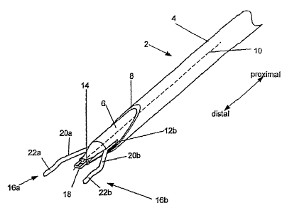
[0032] The closure device 2 can have a delivery guide 4. The delivery guide 4
can be a
21 tubular member, such as a catheter or sheath on the outer radial side of
the closure device
22 2. The delivery guide 4 can be hollow. In one configuration, the
delivery guide 4 can be
23 on the proximal end of the closure device 2. In another configuration,
the delivery guide
6
CA 02840690 2014-01-27
1 4 can be the entire length of the closure device 2. The delivery guide 4
can have a low-
2 friction inner surface. The delivery guide 4 can be configured to receive
an inner
3 member 6. The delivery guide 4 can have a distal port 8 at the distal end
of the delivery
4 guide 4.
[0033] The delivery guide 4 can have a proximally-located handle (not shown).
The
6 handle can facilitate manipulation of the delivery guide 4 and the inner
member 6, and
7 operation of the closure device 2.
8 [0034] The closure device 2 can have the inner member 6. The inner member
6 can be
9 configured to sfidably or fixedly attach to the inside of the delivery
guide 4. The inner
member 6 can have a member longitudinal axis 10. The distal port 8 of the
delivery
11 guide 4 can be at a non-perpendicular angle with respect to the member
longitudinal axis
12 10.
13 [0035] The inner member 6 can have a first wire port (not shown) and a
second wire port
14 12b. The wire ports 12a and 12b can be channels along entire length
(e.g., from the distal
end to the handle at the proximal end) of the member longitudinal axis 10. The
wire
16 ports 12a and 12b can have an opening at or near the distal end of the
inner member 6.
17 [0036] The inner member 6 can have a sealer channel (not shown). The
sealer channel
18 can have an energy conduit and/or a fluid conduit. The sealer channel
can be configured
19 to deliver energy (e.g., for tissue adhesion and/or for enhanced cell
growth and/or
denaturing and recoagulation of the proteins, such as adventitial proteins
and/or collagen)
21 and/or a liquid sealant (e.g., a hemostatic agent and/or tissue adhesive
and/or volume
22 filler, such as polyethylene glycol (PEG)) to a sealer port 14 at a
distal tip of the inner
7
CA 02840690 2014-01-27
1 member 6, and/or to one or more elongated members, such as first and/or
second
2 expander wires 16a and/or 16b.
3 [0037] A supplemental sealer delivery device 18 can be attached to the
sealer port 14. A
4 natural seal can occur due to natural healing of the tissue of the
arteriotomy from being in
proximity with itself. Supplemental sealing can be any sealing action in
addition to the
6 natural seal, including methods to facilitate, maximize, and/or increase
the efficiency of
7 the natural sealing. The supplemental sealer delivery device 18, or other
delivery device,
8 can be configured to deliver a sealer, for example energy, such as
acoustic or radio
9 frequency (RF) energy, microwave energy, and/or a biocompatible adhesive
liquid. The
supplemental sealer delivery device 18 can be an acoustic transducer, such as
a high
11 intensity focus ultrasound (H1FU) transducer or image-guided HIFU. The
supplemental
=
12 sealer delivery device 18 can be from a loop extending from, and
returning to, the sealer
13 port 14. The supplemental sealer delivery device 18 can be a spout (not
shown) for
14 delivering-the liquid sealer. The supplemental sealer delivery device 18
can be a
combination of various individual supplemental sealer delivery devices 18
(e.g., an
16 acoustic transducer and a spout).
17 [0038] The first expander wire 16a and the second expander wire 16b can
be slidably,
18 and/or rotatably, and/or fixedly attached to the first wire port 12a and
second wire port
19 12b, respectively. The expander wires 16a and 16b can distally extend
from the wire
ports 12a and 12b, respectively. The first and second expander wires 16a and
16b can
21 have first and second expander wire extensions 20a and 20b,
respectively, and first and
22 second expander wire tips 22a and 22b, respectively.
8
CA 02840690 2014-01-27
1 [0039] As exemplarily shown on the second expander wire 16b in Figure 2,
the expander
2 wire extensions 20a and 20b can extend radially away from the member
longitudinal axis
3 10. First and second expander wire tips 22a and 22b can extend at angles
from the first
4 and second expander wire extensions 20a and 20b, respectively. The first
and second
expander wire tips 22a and 22b can have tip longitudinal axes 24a and 24b. The
first and
6 second tip longitudinal axes 24a and 24b can be substantially parallel
with the member
7 longitudinal axis 10. The distal ends of the first and second expander
wire tips 22a and
8 22b can have first (not shown) and second feet 26b, respectively. The
feet 26a and 26b
9 can extend radially further from the member longitudinal axis 10 than a
main portion of
the expander wire tips 22a and 22b.
11 [0040] The expander wires 16a and 16b can have wire diameters 28. The
wire diameters
12 28 can be transverse (i.e.., about perpendicular) to the tip
longitudinal axes 24a and 24b.
13 The wire diameters 28 can be from about 0.1 mm (0.005 in.) to about 1.2
mm (0.050 in.),
14 for example about 0.38 mm (0.015 in.).
[0041] The distance from about the member longitudinal axis 10 to about the
radially
16 outer side of the expander wire tips 22a or 22b can be a sealing radius
30. The sealing
17 radius 30 can be from about 0.51 mm (0.020 in.) to about 5.08 mm (0.200
in.), for
18 example about 2.0 mm (0.080 in.).
19 [00421 The expander wire tips 22a and 22b can have tip lengths 32. The
tip lengths 32
can be from about 0.51 mm (0.020 in.) to about 25 mm (1.0 in.), for example
about 4.06
21 nun (0.160 in.).
9
CA 02840690 2014-01-27
1 100431 The expander wire extensions 20a and 20b can have extension
lengths 34. The
2 extension lengths 34 can be from about 2.54 mm (0.100 in.) to about 25
nun (1.0 in.), for
3 example about 9.65 mm (0.380 in.).
4 [0044] Figure 3 illustrates the closure device 2 that can have the
supplemental sealer
delivery device 18 that can extend from the sealer port 14. The supplemental
sealer
6 delivery device 18 can extend along the member longitudinal axis 10 to
about the same
7 distance as the distal ends of the first and/or second expander wires 16a
and/or 16b are
8 located parallel to the member longitudinal axis 10.
9 [0045] The supplemental sealer delivery device 18 can be configured to
transmit RF
energy. For example, the supplemental sealer delivery device 18 can be in
electrical
11 communication with a conductive wire (e.g., from inside the inner
member). The first
12 and/or second expander wires 16a and/or 16b can be configured to
transmit RF energy.
13 For example, the first and/or second expander wires 16a and/or 16b can
be in electrical
14 communication with a conductive wire (e.g., from inside the inner member
6).
[0046] The supplemental sealer delivery device 18 can be configured to
transmit
16 microwave energy. For example, the supplemental sealer delivery device
18 can be in
17 electrical communication with a wave guide (e.g., from inside the inner
member). The
18 first and/or second expander wires 16a and/or 16b can be configured to
transmit
19 microwave energy. For example, the first and/or second expander wires
16a and/or 16b
can be in electrical communication with a wave guide (e.g., from inside the
inner member
21 6).
22 [0047] Figure 4 illustrates the closure device 2 that can have no
supplemental sealer
23 delivery device 18. The first and/or second expander wires 16a and/or
16b can be
CA 02840690 2014-01-27
1 configured to transmit one or more sealers, for example energy. The first
and/or second
2 expander wires 16a and/or 16b can be attached to an acoustic energy
actuator, for
3 example inside the inner member 6. The first and/or second expander wires
16a and/or
4 16b can be in electrical communication with a single conductive wire or
conductive wires
for each expander wire 16a and 16b.
6 (0048] Figure 5 illustrates the closure device 2 that can have no
supplemental sealer
7 delivery device 18. The first and/or second expander wires 16a and/or 16b
can be
8 configured to transmit a physical sealer, for example a liquid adhesive
sealant. The first
9 and/or second expander wires 16a and/or 16b can be hollow. The first
and/or second
expander wires 16a and/or 16b can have delivery holes 36 (e.g., microscopic
pores and/or .
11 macroscopic openings) on the surface thereof, for example to delivery
liquid adhesive
12 sealant, or any anti-biotic, anesthetic, vaso-restrictors, PEG, or any
other agent listed
13 supra or combinations thereof. The delivery holes 36 can be on the first
and/or second
14 expander wire tips 22a and/or 22b. The delivery holes 36 can be on the
sides of the first
and/or second expander wires 16a and/or 16b facing the member longitudinal
axis 10.
16 The delivery holes 36 can be arranged along a line, for example parallel
to the member
17 longitudinal axis 10. The first and/or second expander wires 16a and/or
16b can be
18 attached, and in fluid communication, at a proximal end to a reservoir,
and/or pump,
19 and/or valve holding and/or delivering a sealer, for example a liquid
adhesive sealant.
[0049] Figure 6 illustrates the closure device 2 that can have a web 38 that
can be
21 attached to the first and second expander wires 16a and 16b. The web 38
can be fixedly
22 or removably attached to the first and second expander wire tips 22a and
22b. The web
23 38 can be two or more crossed fibers or wires of material. The web 38
can be a mesh.
11
CA 02840690 2014-01-27
1 The web 38 can be a porous surface. The web 38 can be made from a metal,
and/or a
2 conductive polymer. The web 38 can be made from a resorbable polymer. The
web 38
3 can be configured to transmit RF energy, or can be inductively heated.
For example, the
4 web 38 can be in electrical communication with a conductive wire (e.g.,
via the first
and/or second expander wire tip 22a and/or 22b from inside the inner member 6
and/or
6 from along the outside of the delivery guide 4 and/or from another tool
not a part of the
7 closure device 2), and/or have current induced therein (e.g., from an
external induction
8 coil). The web 38 can be in fluid communication with the delivery holes
36, as shown in
9 Figure 5. The fibers or wires of the web 38 can be hollow and/or have
holes or pores (not
shown). The web 38 can be configured to transmit a physical sealer, for
example a liquid
11 adhesive sealant.
12 [0050] Figure 7 illustrates the closure device 2 that can have a
retracted (i.e.,
13 compressed) configuration. The inner member 6 (not shown) can be
retracted into the
14 delivery guide 4. The first expander wire 16a and/or the second expander
wire 16b can
be retracted into the delivery guide 4. The distal ends of the first expander
wire 16a
16 and/or the second expander wire 16b can be proximal to the distal port.
17 [0051] Figures 8 and 9 illustrate, in a retracted configuration, the
closure device 2 that
18 can check for fluid pressure at the distal port 8. The closure device 2
can have a pressure
19 check port 40 in the delivery guide 4 and/or inner member 6 (not shown).
The pressure
check port 40 can be distal to the expander wire tips 22a and 22b when the
expander wire
21 tips 22a and 22b are in a retracted configuration. The pressure check
port 40 can be in
22 fluid communication with the distal port 8 when the closure device 2 is
in a retracted
23 position. The pressure check port 40 can be in fluid communication with
an outer wall of
12
CA 02840690 2014-01-27
1 the delivery guide 4. The pressure check port 40 can be in fluid
communication with a
2 pressure check lumen 42. The pressure check lumen 42 can be in fluid
communication
3 with a sensor or port on or near the handle (not shown) of the delivery
guide 4, such as an
4 external lumen where blood flow can be observed, for example flow from
the end of an
external tube or port and/or through a transparent or translucent window.
6 [0052] Figure 10 illustrates, in a retracted configuration, the closure
device 2 that can
7 have the pressure check port 40 aligned with the distal ends of the
expander wire tips 22a
8 and 22b, for example, even with a distal alignment line 43.
9 [0053] When the closure device 2 is used, the distal end of the delivery
guide 4 can be
inserted across the wall of a vessel until a "flash" of blood enters the
pressure check port
11 40, flows up the pressure check lumen 42, and can then be observed by
the sensor or port
12 on or near the handle. Once the blood "flash" is observed, the delivery
guide 4 can be
13 moved slowly in the proximal direction until the "flash" stops. The
"flash" stopping can
14 be an indication of the distal location of the delivery guide (i.e., the
pressure check port
40 can be blocked by the lumen wall 54).
16 [0054] Any or all elements of the closure device 2 and/or other devices
or apparatuses
17 described herein can be made from, for example, a single or multiple
stainless steel
18 alloys, nickel titanium alloys (e.g., Nitinol), cobalt-chrome alloys
(e.g., ELGILOY from
19 Elgin Specialty Metals, Elgin, IL; CONICITR.OME from Carpenter Metals
Corp.,
Wyomissing, PA), molybdenum alloys (e.g., molybdenum TZM alloy, for example as
21 disclosed in International Pub. No. WO 03/082363 A2, published 9 October
2003, which
22 is herein incorporated by reference in its entirety), tungsten-rhenium
alloys, for example,
23 as disclosed in International Pub. No. WO 03/082363, polymers such as
polyester (e.g.,
13
CA 02840690 2014-01-27
1 DACRON from E. I. Du Pont de Nemours and Company, Wilmington, DE),
2 polypropylene, polytetrafluoroethylene (PTFE), expanded PTFE (ePTFE),
polyether
3 ether ketone (PEEK), nylon, peilyether-block co-polyamide polymers (e.g.,
PEBAX
4. from ATOFINA, Paris, France), aliphatic polyether polyurethanes (e.g.,
.TECOFLEX
. 5 from Thermedics Polymer Products, Wilmington, MA), polyvinyl chloride
(PVC),
6 polyurethane, thermoplastic, fluorinated ethylene propylene (PEP),
absorbable or
7 .resorbable polymers such as polyglycolic acid (PGA), pdylactic acid
(PLA),
8 polydioxanone, and pseudo-polyarnino tyrosine-based acids, extruded
collagen, silicone,
9 zinc, echogenic, radioactive, radiopaque materials or combinations
thereof. Examples of
radiopaque materials are barium sulfate, zinc oxide, titanium, stainless
steel, nickel-
11 titanium alloys, tantalum and gold.
=
12 [0055] Any or all elements of the closure device 2 and/or other devices
or apparatuses
13 described hereinean tie or have a matrix for cell ingrowth or used with
a fabric, for
14 example a covering (not shown) that acts as a matrix for cell ingrowth.
The matrix
and/or fabric cante, for example, polyester (e.g., DACRON from E. I. Du Pont
de
16 Nemours and Company, Wilmington, DE), polypropylene, PTFE, ePTFE, nylon,
17 extruded collagen, silicone or combinations thereof.
18 [00561 The elements of closure device 2 and/or other
devices or apparatuses
19 described herein and/or the fabric can be filled and/or coated with an
agent delivery
2?) matrix known to one having ordinary skill in the art and/or a
therapeutic and/or
21 diagnostic agent. The agents within these matrices can include
radioactive materials;
22 radiopaque materials; cytogenic agents; cytotoxic agents; cytostatic
agents; thrombogenic
23 agents, for example polyurethane, cellulose acetate polymer mixed with
bismuth trioxide,
14
CA 02840690 2014-01-27
1 and ethylene vinyl alcohol; lubricious, hydrophilic materials; phosphor
cholene; anti-
2 inflammatory agents, for example non-steroidal anti-inflammatories
(NSAIDs) such as
3 cyclooxygenase-1 (COX-1) inhibitors (e.g., acetylsalicylic acid, for
example ASPIRIN
4 from Bayer AG, Leverkusen, Germany; ibuprofen, for example ADVIL from
Wyeth,
Collegeville, PA; indomethacin; mefenamic acid), COX-2 inhibitors (e.g., VIOXX
6 from Merck & Co., Inc., Whitehouse Station, NJ; CELEBREX from Pharmacia
Corp.,
7 Peapack, NJ; COX-1 inhibitors); immunosuppressive agents, for example
Sirolimus
8 (RAPAMME , from Wyethõ Collegeville, PA), or matrix metalloproteinase
(MMP)
9 inhibitors (e.g., tetracycline and tetracycline derivatives) that act
early within the
pathways of an inflammatory response. Examples of other agents are provided in
Walton
11 et al, Inhibition of Prostoglandin Fa Synthesis in Abdominal Aortic
Aneurysms,
12 Circulation, July 6, 1999, 48-54; Tambiah et al, Provocation of
Experimental Aortic
13 Inflammation Mediators and Chlamydia Pneumoniae, Brit. .1 Surgery 88(7),
935-940;
14 Franklin et al, Uptake of Tetracycline by Aortic Aneurysm Wall and Its
Effect on
Inflammation and Proteolysis, Brit. J Surgery 86(6), 771-775; Xu et al, Spl
Increases
16 Expression of Cyclooxygenase-2 in Hypoxic Vascular Endothelium, J
Biological
17 Chemistry 275 (32) 24583-24589; and Pyo et al, Targeted Gene Disruption
of Matrix
18 Metalloproteinase-9 (Gelatinase B) Suppresses Development of
Experimental Abdominal
19 Aortic Aneurysms, J Clinical Investigation 105 (11), 1641-1649.
21
22 METHOD OF MANUFACTURE
CA 02840690 2014-01-27
1 [0057] The elements of the closure device 2 can be directly attached by,
for example,
2 melting, screwing, gluing, welding, soldering, abrasing, or use of an
interference fit or
3 pressure fit such as crimping, snapping, or combining methods thereof.
The elements can
4 be integrated, for example, molding, die cutting, laser cutting,
electrical discharge
machining (EDM) or stamping from a single piece or material. Any other methods
can
6 be used as known to those having ordinary skill in the art.
7 [0058] Integrated parts can be made from pre-formed resilient materials,
for example
8 resilient alloys (e.g., Nitinol, ELGILOYe) that are preformed and biased
into the post-
9 deployment shape and then compressed into the deployment shape as known
to those
having ordinary skill in the art.
11 [0059] The expander wires 16a and 16b can be made from pre-formed
resilient materials,
12 for example resilient alloys (e.g., Nitinol, ELGILOYO) that are
preformed and biased
13 into the post-deployment shape and then compressed into the deployment
shape. The
14 post-deployment shape can be the configuration shown in Figure 2 and
elsewhere herein.
[0060] Any elements of the closure device 2, or the closure device 2 as a
whole after
16 assembly, can be coated by dip-coating, brush-coating or spray-coating
methods known
17 to one having ordinary skill in the art. For example, the expander wires
16a and 16b can
18 be spray coated, dip-coated or brushed-coated.
19 [0061] One example of a method used to coat a medical device for
vascular use is
provided in U.S. Patent No. 6,358,556 by Ding et al.
21 Time release coating methods known to one having ordinary
22 skill in the art can also be used to delay the release of an agent in
the coating, for example
23 the coatings on the expander wires 16a and 16b.
16
CA 02840690 2014-01-27
1
2 METHOD OF USE
3 [0062] Figures 11 through 13 illustrate a method for changing the closure
device 2 from a
4 first configuration to a second configuration. Figures 14 and 15 also
show close-up
views of distal ends of the closure device 2 of Figures 11 and 13,
respectively. As shown
6 in Figures 11 and 14, the closure device 2 can be in a fully retracted
configuration. The
7 inner member 6 and the expander wires 16a and 16b can be concealed within
the delivery
8 guide 4.
9 [0063] As shown in Figure 12, the closure device 2 can be in a partially
deployed
configuration. The inner member 6 can be pushed or pulled to be translated, as
shown by
11 arrow 44, distally relative to the delivery guide 4, and/or the delivery
guide 4 can be
12 pushed or pulled to be translated, as shown by arrow 46, proximally
relative to the
13 delivery guide 4.
14 [0064] The delivery guide 4 can restrict (e.g., by interference fit) the
expander wires 16a
and 16b from expanding away from the member longitudinal axis 10. The expander
16 wires 16a and 16b, can move distally, as shown by arrows 47, relative to
the delivery
17 guide 4. The expander wires 16a and 16b can be attached to the inner
member 6, such
18 that the inner member 6 pushes the expander wires 16a and 16b when the
inner member 6
19 is pushed.
[0065] As shown in Figures 13 and 15, the closure device 2 can be in a fully
deployed
21 configuration. The inner member 6 can be pushed or pulled to be
translated, as shown by
22 arrow 48, distally relative to the delivery guide 4, until the inner
member 6 reaches a stop
23 (not shown) with respect to the supplemental sealer delivery device. The
stop can be an
17
CA 02840690 2014-01-27
1 interference fit between the delivery guide 4 and the inner member 6. The
delivery guide
2 4 can be pushed or pulled to be translated, as shown by arrow 50,
proximally relative to
3 the delivery guide 4, until the delivery guide 4 reaches the stop.
4 [0066] The expander wires 16a and 16b, can move distally, as shown by
arrows 51,
relative to a location at which the expander wires 16a and 16b exit the wire
ports 12a and
6 12b. The location at which the expander wires 16a and 16b exit the
respective wire ports
7 12a and 12b can move beyond the distal port 8 and the delivery guide 4.
The expander
8 wires 16a and 16b can expand radially, as shown by arrows 51, away from
the member
9 longitudinal axis 10.
[0067] Figure 16 illustrates that the closure device 2 can be inserted, as
shown by arrow,
11 into an opening in tissue, for example the arteriotomy 52 in a lumen
wall 54. The closure
12 device 2 can be in the retracted configuration when the closure device 2
is inserted into
13 the arteriotomy 52. After inserting the closure device 2, the distal end
of the closure
14 device 2 can be located in or outside and distal to the arteriotomy 52.
The lumen wall 54
can have an inner lumen wall surface 56, and can surround a lumen 57.
16 [0068] The arteriotomy 52 can have an arteriotomy diameter 58. The
arteriotomy
17 diameter 58 can be from about 0.5 mm (0.020 in.) to about 40 mm (1.5
in.), yet a
18 narrower range from about 1.0 mm (0.040 in.) to about 10.2 mm (0.400
in.), for example
19 about 2.54 mm (0.100 in.). When in the retracted configuration, the
closure device 2 can
have a diameter smaller than the arteriotomY diameter 58.
21 [0069] The lumen wall 54 can have a lumen wall thickness 60. The lumen
wall thickness
22 60 can be from about 0.51 mm (0.020 in.) to about 5.08 mm (0.200 in.),
for example
23 about 1.0 mm (0.040 in.).
18
CA 02840690 2014-01-27
1 100701 Figures 17 and 18 illustrate expanding the closure device 2 after
the closure
2 device 2 has been inserted into the arteriotomy 52. The delivery guide 4
can be moved
3 proximally relative to the inner member 6. The expander wires 16a and 16b
can expand,
4 as shown by arrows 62, away from the member longitudinal axis 10. The
expander wire
tips 22a and 22b can be located inside the arteriotomy 52. The expander Wire
tips 22a
6 and 22b can expand, for example laterally, against the arteriotomy 52.
The arteriotomy
7 52 can change shape in response to tensioning forces applied by the
expander wire tips
8 22a and 22b, for example, during expansion. The feet 26a and 26b can
pressure and/or
9 interference fit with the arteriotomy 52 and/or the inner lumen wall
surface 56.
[0071] The arteriotomy 52 can have an arteriotomy width 64 and an arteriotomy
height
11 66. The arteriotomy width 64 can be about half the circumference of the
arteriotomy 52.
12 The arteriotomy width 64 can be from about 1.0 mm (0.040 in.) to about
10.2 mm (0.400
13 in.), for example about 4.06 mm (0.160 in.).
14 [00721 The arteriotomy height 66 can be about the wire diameter 28. The
arteriotomy
height 66 can be less than about 0.51 mm (0.020 in.), more narrowly, less than
about 0.38
16 mm (0.015 in.). The arteriotomy height 66 can be from about 0.1 mm
(0.005 in.) to about
17 1.3 mm (0.050 in.), for example about 0.38 mm (0.015 in.). The
arteriotomy height 66
18 can be small enough to enable cell growth, blood clotting, acoustic
sealing, heat sealing,
19 gluing, enhanced self-sealing and combinations thereof across the
arteriotomy 52.
[0073] Figure 19 illustrates a method for applying energy (e.g., acoustic) to
the tensioned
21 arteriotomy 52. The closure device 2 can have the supplemental sealer
delivery device
22 18. The supplemental sealer delivery device 18 can be an acoustic (e.g.,
ultrasound)
23 transducer. For example, because the expander wires 16a and 16b can
produce opposite
19
CA 02840690 2014-01-27
1 forces on opposite sides of the inside of the arteriotomy 52, the
supplemental sealer
2 delivery device 18 can be automatically aimed and automatically centered
(e.g., aligned
3 along the member longitudinal axis 10 with about the center of the
arteriotomy 52). The
4 supplemental sealer delivery device 18 can transmit acoustic energy to
the arteriotomy
52.
6 [00741 Figure 20 illustrates a method for applying energy (e.g., RF or
microwave) to the
7 tensioned arteriotomy 52. The supplemental sealer delivery device 18 can
extend into
8 about the center of the arteriotomy 52. The supplemental sealer delivery
device 18 can
9 be an RF transducer. The first and/or second expander wires 16a and/or
16b can be RF or
microwave transducers (e.g., microwave antenna). For example, the first and/or
second
11 expander wires 16a and/or 16b can be first RF poles, and the
supplemental sealer delivery
12 device 18 can be a second RF pole.
13 [0075] Figure 21 illustrates a method for applying the sealer (e.g.,
energy or liquid) into
14 the tensioned arteriotomy 52 using the web 38. The web 38 can be an RF
transducer,
and/or a resistive heater, and/or an inductive heater and/or microwave heater.
The web
16 38 can be hollow and have holes or pores (not shown). The web 38 can be
in fluid
17 communication with a hollow first and/or second expander wires 16a
and/or 16b. The
18 web 38 can transfer a liquid, for example a sealer, into the arteriotomy
52.
19 [00761 Once the web 38 applies the sealer to the tensioned arteriotomy
52, the web can
be removed from the expander wire tips 22a and 22b, and left in the
arteriotomy 52 when
21 the remainder of the closure device 2 is removed. The web 38 can be
absorbed by the
=
22 tissue surrounding the arteriotomy 52.
CA 02840690 2014-01-27
1 [0077] Figure 22 illustrates a method for applying liquid sealer into the
tensioned
2 arteriotomy 52. The liquid sealer can flow, as shown by arrows, from the
delivery holes
3 36 into the arteriotomy 52. Liquid sealers (e.g., biocompatible
adhesives, biocompatible
4 epoxies, PEG) for filling and sealing arteriotomies 52 are known to those
having ordinary
skill in the art. The sealer can act as an adhesive. The adhesive can act as a
filler, for
6 example PEG. The sealer can be bioabsorbable.
7 [00781 The arteriotomy 52 can be partially or completely sealed by the
energy. Fluid
8 flow can be substantially and/or completely stopped (i.e., hemostasis).
Fluid flow
9 through the arteriotomy 52 can be partially or completely sealed by the
energy.
[0079] The supplemental sealer delivery device 18, and/or the web 38, and/or
the
11 expander wire tips 22a and 22b can be electrical resistive heater
elements. The sealer can
12 be direct heat transferred through conduction, and/or convection, and/or
radiative heating.
13 The supplemental sealer delivery device 18 can heat the arteriotomy
directly through
14 conduction.
[0080] Any combination of energies, in any proportion, can be applied to the
arteriotomy
16 52. For example, RF or other heating energy can initially be applied to
the tensioned
17 arteriotomy 52. The RF or other heating energy can then be stopped and
acoustic energy
18 can be applied to the tensioned arteriotomy 52.
19 [0081] Resistive heat energy (i.e., conducted heat generated by
electrical resistors) and
acoustic energy can be applied simultaneously and in any proportion to the
arteriotomy
21 52. RF energy and resistiN4 heat energy can be applied simultaneously
and in any
22 proportion to the arteriotomy 52. Acoustic energy and RF energy can be
applied
23 simultaneously and in any proportion to the arteriotomy 52. Acoustic
energy and
21
CA 02840690 2014-01-27
1 inductive energy can be applied simultaneously and in any proportion to
the arteriotomy
2 52. Resistive heat energy, acoustic energy, RF energy, inductive energy
and/or
3 microwave energy can be applied simultaneously and in any proportion to
the
4 arteriotomy 52.
[0082] Figures 23 and 24 illustrate a treatment area that can have skin 68
separated from
6 the vessel 70 by subcutaneous tissue 72 (e.g., fat, muscle, other
vessels). An external
7 transducer 74 can be in contact with or adjacent to the skin 68. A gel or
other contact
8 supplement known to one having ordinary skill in the art can be sued to
improve energy
9 conduction between the external transducer 74 and the skin 68.
[0083] After the arteriotomy is substantially sealed, the holes in the lumen
wall 54 from
11 which the expander wires 16a and 16b and/or the supplemental sealer
delivery device 18
12 are removed can be inconsequentially small so that bleeding from the
holes can be
13 negligible. Sealing (e.g., heating) can be performed as the closure
device 2 is removed
14 from the arteriotomy 52 so as to close an holes in the lumen wall 54
formed by the
removal of the closure device 2.
16 [0084] The external transducer 74 can be an acoustic transducer, such as
an ultrasonic
17 imager, HIFU, image guided HIFU; a radiological transducer, such as an x-
ray imager; a
18 magnetic imager, such as a magnetic resonance imager (MRI); therapeutic
versions of the
19 aforementioned imagers, or combinations thereof.
[0085] The external transducer 74 can be used to send energy waves 76 to the
21 arteriotomy 52. The energy waves 76 can reflect from, transmit through,
and/or resonate
22 from the arteriotomy 52 and/or the expander wire tips 22a and 22b.
Reflected and/or
23 transmitted and/or resonated energy waves 76 can be received by the
external transducer
22
CA 02840690 2014-01-27
1 74 and used to detect the condition (e.g., morphology, structure) and
location of the
2 arteriotomy 52 and the expander wire tips 22a and 22b. The external
transducer 74 can
3 track the location of the arteriotomy 52 and the expander wire tips 22a
and 22b.
4 [0086] The expander wire tips 22a and 22b can have a material or
configuration that
enhances the ability of the external transducer 74 to detect the expander wire
tips 22a and
6 22b. For example, the expander wire tips 22a and 22b can have an
echogenic and/or
7 radiopaque material and/or configuration, such as radiopaque materials
listed supra. The
8 first and second expander wire tips 22a and 22b can frame the arteriotomy
52 location
9 and provide a target got an image-guided external transducer 74 (e.g.,
image guided
H1FU). The energy waves 76 can be therapeutic energy, for example used to seal
the
11 arteriotomy 52. The energy waves 76 can be focused on the arteriotomy
52, and can
12 transmit minimal energy into surrounding tissue. For example, the energy
waves 76 can
13 be therapeutic ultrasound broadcast from a phased array such that a node
of the energy
14 waves 76 is located at the arteriotomy 52.
[0087] The closure device 2 can be removed from the arteriotomy 52. The
closure
16 device 2 can be directly withdrawn from the arteriotomy, for example in
a parallel
17 direction with the tip longitudinal axes 24a and 24b. The closure device
2 can be
18 withdrawn from the arteriotomy 52 while the first and second expander
wires 16a and
19 16b are in an expanded configuration.
[0088] Before the closure device is withdrawn from the arteriotomy 52, and/or
21 subcutaneous tissue track, the inner member 6 can be retracted into the
delivery guide 4,
22 with or without fully retracting the expander wires 16a and 16b into the
first and second
23 wire ports 12a and 12b. The delivery guide 4 can be moved distally
relative to the inner
23
CA 02840690 2014-01-27
1 member 6, reversing the method shown in Figures 11 through 16, and
changing the
2 closure device 2 into a retracted configuration. The closure device 2 can
then be
3 removed from the arteriotomy 52 with the expander wires 16a and 16b in an
expanded
4 or retracted configuration.
[0089] If the arteriotomy 52 was created by a surgical procedure using a
hollow
6 member, such as a catheter, or there is otherwise a catheter in the
arteriotomy 52 prior
7 to performing the methods described herein, the already-deployed catheter
can be used
8 as the delivery guide 4, or as a sheath for the delivery guide 4.
9 [0090] The closure devices and methods shown and described herein can be
used
integrally and/or in other combinations with access and closure devices and
methods
11 shown and described in U.S. Patent Publication 2005/267520. For example,
the
12 arteriotomy 52 can be at an angle with respect to the lumen, wherein the
angle can be
13 from about 20 to about 90 , more narrowly from about 30 to about 60 ,
for example
14 about 45 , or otherwise described in U.S. Patent Publication
2005/267520. Also for
example, the arteriotomy 52 can have a shape as described by U.S. Patent
16 Publication 2005/267520. The devices and methods described herein can be
used in
17 combination with the supplemental closure devices, such as tensioners,
clips, toggles,
18 sutures, and combinations thereof, described by U.S. Patent Publication
2005/267520.
19 [0091] It is apparent to one skilled in the art that various changes and
modifications
can be made to this disclosure. Elements shown with any embodiment are
exemplary
21 for the specific embodiment and can be used on other embodiments within
this
22 disclosure. The scope of the claims should not be limited by the
embodiments set out
23 herein but should be given the broadest interpretation consistent with
the description as
24 a whole.
24