Note: Descriptions are shown in the official language in which they were submitted.
CA 02841601 2014-01-14
WO 2013/013370 PCT/CN2011/077506
ECG ELECTRODE CONNECTOR
Technical Field of the Invention
[00011
The present disclosure relates to biomedical electrodes, and in
particular, to a radiolucent biomedical electrode connector and radiolucent
lead
wires for performing biomedical monitoring of a patent during imaging
procedures.
Back2round of the Invention
[0002] Electrocardiograph (ECG) monitors are widely used to obtain medical
(i.e. biopotential) signals containing information indicative of the
electrical
activity associated with the heart and pulmonary system. To obtain medical
signals, ECG electrodes are applied to the skin of a patient in various
locations.
The electrodes, after being positioned on the patient, connect to an ECG
monitor
by a set of ECG lead wires. The distal end of the ECG lead wire, or portion
closest to the patient, may include a connector which is adapted to operably
connect to the electrode to receive medical signals from the body. The
proximal
end of the ECG lead set is operably coupled to the ECG monitor and supplies
the
medical signals received from the body to the ECG monitor.
[0003]
A typical ECG electrode assembly may include an electrically
conductive layer and a backing layer, the assembly having a patient contact
side
and a connector side.
The contact side of the electrode pad may include
biocompatible conductive gel or adhesive for affixing the electrode to a
patient's
body for facilitating an appropriate electrical connection between a patient's
body and the electrode assembly. The connector side of the pad may incorporate
1
CA 02841601 2014-01-14
WO 2013/013370 PCT/CN2011/077506
a metallic press stud having a bulbous profile for coupling the electrode pad
to
the ECG lead wire. In use, the clinician removes a protective covering from
the
electrode side to expose the gel or adhesive, affixes the electrode pad to the
patient's body, and attaches the appropriate ECG lead wire connector to the
press stud by pressing or "snapping" the lead wire connector onto the bulbous
press stud to achieve mechanical and electrical coupling of the electrode and
lead wire. Alternatively, ECG connectors that engage via manipulation of a
lever or other mechanical locking device may be employed. After use, a
clinician then removes the ECG lead wire connector from the pad by pulling or
"unsnapping" the connector from the pad or by releasing the lever or other
locking mechanism.
[0004] Placement of the electrodes on a patient has been established by
medical protocols. A common protocol requires the placement of the electrodes
in a 5-lead configuration: one electrode adjacent each clavicle bone on the
upper
chest and a third electrode adjacent the patient's lower left abdomen, a
fourth
electrode adjacent the sternum, and a fifth electrode on the patient's lower
right
abdomen.
[0005] During certain procedures it may be necessary to monitor biological
(e.g., ECG) parameters of a patient that is undergoing imaging, such as CT-
scan
or MRI. Use of conventional ECG connectors and lead wire sets typically
associated therewith may have drawbacks in these applications, since they tend
to interfere with the imaging systems. In one example, certain components of
the ECG connectors and/or lead wires may be detected by the imaging apparatus
and consequently may obfuscate the visual images upon which clinicians and
surgeons rely. In another example, ferrous and/or magnetic components
commonly found in ECG connectors, such as in springs and clips, may be
7
CA 02841601 2015-11-30
potentially hazardous when used within the intense magnetic field of an MRI
scanner.
Summary of the Invention
[0006] According to an aspect, there is provided an ECG connector assembly,
comprising: a housing having an interior recessed surface having disposed
therein
an opening dimensioned to operably receive the press stud of an ECG electrode
pad; a radiolucent conductor disposed on at least a portion of the interior
recessed
surface; a radiolucent lead wire conductor extending from a proximal end of
the
housing and operably coupled to the radiolucent conductor; an engagement
member pivotably disposed upon the interior recessed surface and having an
engaging face and a pivot, wherein the engagement member is pivotable between
a
first position whereby the engaging face is closer to a top portion of the
opening
and a second position whereby engaging face is further from a top portion of
the
opening; an arcuate stiffener disposed between the engaging face and pivot of
the
engagement member; and a radiolucent resilient member configured to bias the
engagement member towards the first position; wherein the arcuate stiffener is
spaced apart from the engaging face.
Brief Description of the Drawings
[0007] The accompanying drawings are not intended to be drawn to scales. In
the drawing, each identical or nearly identical component that is illustrated
in
various figures is represented by a like numeral. For purposes of clarity, not
every
component may be labeled in every drawing. Various embodiments of the present
disclosure are described hereinbelow with references to the drawings, wherein:
[0008] Fig. 1 is an exploded view of a conventional ECG electrode
connector;
[0009] Fig. 2 is a schematic diagram of the conventional ECG electrode
connector of Fig. 1;
3
CA 02841601 2015-11-30
[0010] Fig. 3A is a view of an embodiment of a radiolucent ECG electrode
connector in an engaged configuration in accordance with the present
disclosure;
[0011] Fig. 3B is a view of the Fig. 3A embodiment in a disengaged
configuration in accordance with the present disclosure;
[0012] Fig. 3C is a detail view of a press stud opening of the Fig. 3A
embodiment of a radiolucent ECG electrode connector in accordance with the
present disclosure
[0013] Fig. 4A is a view of another embodiment of a radiolucent ECG
electrode
connector in an engaged configuration in accordance with the present
disclosure;
[0014] Fig. 4B is a view of the Fig. 4A embodiment in a disengaged
configuration in accordance with the present disclosure; and
[0015] Fig. 5 is a view of another embodiment of a radiolucent ECG
electrode
connector in accordance with the present disclosure.
Detailed Description of the Preferred Embodiments
[0016] This invention is not limited in its application to the details of
construction and the arrangement of components set forth in the following
description or illustrated in the drawings. The invention is capable of other
embodiments and of being practiced or of being carried out in various ways.
Also,
the phraseology and terminology used herein is for the purpose of
4
CA 02841601 2014-01-14
WO 2013/013370 PCT/CN2011/077506
description and should not be regarded as limiting. The use of "including,"
"comprising," "having," "continuing," or "involving" and variations thereof
herein, is meant to encompass the items listed thereafter and equivalents
thereof
as well as additional items.
[0017] Particular embodiments of the present disclosure are described
hereinbelow with reference to the accompanying drawings; however, the
disclosed embodiments are merely examples of the disclosure, which may be
embodied in various forms. Well-known functions or constructions and
repetitive matter are not described in detail to avoid obscuring the present
disclosure in unnecessary or redundant detail. Therefore, specific structural
and
functional details disclosed herein are not to be interpreted as limiting, but
merely as a basis for the claims and as a representative basis for teaching
one
skilled in the art to variously employ the present disclosure in virtually any
appropriately detailed structure.
[0018] In the drawings and in the descriptions that follow, the term
"proximal," as is traditional, shall refer to the end of the instrument that
is closer
to a user, while the term "distal" shall refer to the end that is farther from
a user.
In addition, as used herein, terms referencing orientation, e.g., "top",
"bottom",
"up", "down", "left", "right", "clockwise", "counterclockwise", and the like,
are
used for illustrative purposes with reference to the figures and features
shown
therein. Embodiments in accordance with the present disclosure may be
practiced in any orientation without limitation.
[0019] The present invention is directed to an electrode connector suitable
for
use during patient imaging, such as during a CT-scan or MRI. Commonly
available electrode connectors have components which may be detected on the
CA 02841601 2014-01-14
WO 2013/013370 PCT/CN2011/077506
image and/or may become dangerous when exposed to a particular field, such as
a magnetic field.
[0020] One embodiment of a conventional electrode connector 1320 is shown
in Fig. 1 and Fig. 2 which includes a housing 1322 having an upper member
1324 and a lower member 1326, and defining an internal cavity 1328
therebetween. Housing 1322 is fabricated from a non-conducting material, e.g.,
an injection molded polymer which electrically insulates the subject from the
conductive element(s) therewithin. Upper member 1324 and lower member
1326 are separate components attached to each other by any suitable method of
bonding, such as without limitation, adhesive, ultrasonic welding, or heat
welding. Upper member 1324 and lower member 1326 form a non-conductive
element of the housing 1322.
[0021] Housing 1322 of the conventional electrode connector includes a lead
wire terminal 1330 which is electrically connected to a respective end of lead
wire 1304 by any suitable method of connection, including without limitation,
crimping, soldering, or welding. Lead wire terminal 1330 is formed of a
conductive material, typically a metal such as stainless steel. Housing 1322
supports a contact member 1332 also formed of a conductive material that is
electrically connected to a lead wire. In one embodiment, the lead wire is
formed of a conductive metals such as tinned copper. In another embodiment,
the conductive material of the contact member is a metal such as stainless
steel.
Contact member 1332 and lead wire terminal 1330 may be integrally formed.
Contact member 1332 defines a contact opening 1334 formed therein and in
communication with internal cavity 1328 of housing 1322. Contact opening
1334 includes first contact opening portion 1334a and second contact opening
portion 1334b. First contact opening portion 1334a defines an internal
6
CA 02841601 2014-01-14
WO 2013/013370 PCT/CN2011/077506
dimension or diameter which is greater than the corresponding internal
dimension or diameter of second contact opening portion 1334b.
[0022] Housing 1322 of conventional electrode connector further includes a
lever 1340 pivotably connected thereto. Lever 1340 includes an actuating end
1336. Lever 1340 is biased to a first position by a biasing member 1338, as
shown in Fig. 2. Biasing member 1338 is formed of a resilient metal, such as
stainless steel. Lever 1340 includes an engaging region 1336a projecting
therefrom so as to extend across first contact opening portion 1334a of
contact
opening 1334 when lever 1340 is in the first position. In use, lever 1340 is
actuatable to a second position wherein engaging region 1336a thereof does not
obstruct or extend across first contact opening portion 1334a of contact
opening
1334. For example, a clinician may apply finger pressure to actuating end 1336
that is sufficient to overcome the biasing force of biasing member 1338,
thereby
causing engaging region 1336a to move to a second position as herein
described.
[0023] Conventional ECG electrode connector 1320 is adapted for connection
to a conventional snap-type biomedical electrode (not explicitly shown). A
typical snap-type biomedical electrode incorporates an electrode flange or
base
and male press stud or terminal extending in transverse relation to the
electrode
base. The male press stud terminal may have a bulbous head whereby an upper
portion of the terminal has a greater cross-sectional dimension than a lower
portion of the terminal. Accordingly, in use, when lever 1340 of electrode
connector 1320 is in the second position, the head of the male press stud
terminal of the snap-type biomedical electrode may be inserted into first
contact
opening portion 1334a of contact opening 1334 and actuating end 1336, and
thus,
lever 1340, may be released so that biasing member 1338 moves engaging
region 1336a of lever 1340 against the head of the male press stud (not
explicitly
7
CA 02841601 2014-01-14
WO 2013/013370 PCT/CN2011/077506
shown) to push or force the lower portion of the press stud into a second
contact
opening portion 1334b of contact opening 1334. The biasing force of biasing
member 1338 helps to maintain the press stud within second contact opening
portion 1334b of contact opening 1334 and thus inhibits removal or
disconnection of the biomedical electrode from ECG connector 1320. However,
because lead wire terminal 1330, contact member 1332 and biasing member
1338 are metallic, one or more of these components may be detected in the
image and/or become dangerous when exposed to a magnetic filed.
[0024] Accordingly, one aspect of the present invention provides an
electrode
connector which may be used during patient imaging. One embodiment of an
ECG electrode connector of the present invention is shown in Figs. 3A, 3B, and
3C. In view thereof, and so as not to obscure the present disclosure with
redundant information, only those features distinct to ECG electrode connector
1400 will be described hereinafter.
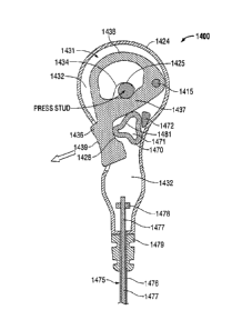
[0025] ECG electrode connector 1400 is configured to facilitate the
monitoring
of ECG and other biological parameters while the subject patient is undergoing
an imaging procedure, such as without limitation, MRI, CT, PET, and the like.
Connector 1400 includes a housing 1424 having an interior recessed surface
1431 that includes an opening 1434 defined therein that opens to a patient-
facing
surface of the housing. Opening 1434 is dimensioned to accept the insertion of
a
head of a press stud of a patient electrode. Housing 1424 may be formed from
any suitable non-conductive material, including polymeric material. The
connector 1400 includes an engagement member 1436 having an actuation
surface 1439, which may be a contoured pushbutton, and an engaging face 1437.
Engagement member 1436 is pivotable about a pivot 1415 to enable the
engaging face 1437 to move from a first position whereby engaging face 1437 is
8
CA 02841601 2014-01-14
WO 2013/013370 PCT/CN2011/077506
closer to a top portion 1425 of opening 1434 and a second position whereby
engaging face 1437 is further from a top portion 1425 of opening 1434. By this
arrangement, the bulbous head of a press stud that has been introduced into
opening 1434 may be captured in opening 1434 between engaging face 1437 and
a sidewall of opening 1434. Engagement member 1436 includes a stiffener 1438,
that may have an arcuate shape, disposed between engaging face 1437 and pivot
1415.
[0026] The interior recessed surface 1431 of housing 1424 includes a
radiolucent conductor 1432 that facilitates the conduction of biological
signals
between a press stud captured within opening 1434 and a lead wire conductor
1477. Radiolucent conductor 1432 may be included with surface 1431 by any
suitable manner, including without limitation, as a conductive coating and/or
a
conductive material incorporated within housing 1424 or associated portions
thereof In some embodiments, radiolucent conductor 1432 may be formed by
dispersing conductive carbon powder over interior recessed surface 1431. The
conductive carbon powder is then fused via the application of heat and/or
pressure to the polymeric material that forms interior recessed surface 1431.
In
some embodiments, radiolucent conductor 1432 may be formed by the
application of radiolucent conductive ink to interior recessed surface 1431.
In
other embodiments, the radiolucent conductor 1432 may comprise a carbon fiber
wire fixed to the recessed surface 1431. As shown in Fig. 3C, radiolucent
conductor 1432 may extend onto at least a portion of a sidewall 1441 of
opening
1434.
[0027] ECG electrode connector 1400 includes a lead wire 1475 extending
from a proximal (e.g., bottom) end thereof Lead wire 1475 includes an outer
insulator 1476 coaxially disposed about a conductor 1477. Conductor 1477 is
9
CA 02841601 2014-01-14
WO 2013/013370 PCT/CN2011/077506
formed from radiolucent electrically conductive material, such as conductive
carbon or conductive carbon monofilament wire. In some embodiments,
conductor 1477 is formed from one or more carbon fibers. A distal portion of
the outer insulator is stripped thus exposing a distal portion of conductor
1477'.
The exposed portion 1477' of conductor 1477 is operatively joined to
radiolucent conductor 1432 of interior recessed surface 1431. Conductor 1477'
may be joined by any suitable manner, including without limitation a crimping
element 1478 and/or by radiolucent electrically conductive adhesive. In some
embodiments, the exposed portion 1477' of conductor 1477 and radiolucent
conductor 1432 are integrally formed. A strain relief 1479 surrounds a portion
of lead wire 1475 where lead wire 1475 exits the housing 1424
[0028] A resilient member 1470 biases engagement member 1436 towards a
first position whereby engaging face 1437 is closer to a top portion 1425 of
opening 1434. Lobed resilient member 1470 is positioned between a recess
1428 defined in engagement member 1436 and a saddle 1472 provided by
housing 1424. Resilient member 1470 may be formed from a radiolucent
elastomer, including without limitation, silicone. Resilient member 1470 may
have any shape to provide sufficient force to allow the desired movement of
the
engagement member 1436. The resilient member 1470 may have any regular or
irregular shape, including circle, square, triangle, and clover. In one, one
embodiment, resilient member 1470 is a lobed member. In the embodiment
shown in Figs. 3A and 3B, lobed resilient member 1470 includes a three-lobe
profile having each lobe evenly spaced at about 120 apart, however, a lobed
resilient member 1470 in accordance with the present disclosure may include
fewer than three lobes, or more than three lobes. Additionally or
alternatively,
lobed resilient member 1470 may include lobes that are not evenly spaced
and/or
CA 02841601 2014-01-14
WO 2013/013370 PCT/CN2011/077506
irregularly placed. The resilient member may be solid throughout, or comprise
one or more openings. Lobed resilient member 1470 includes a center opening
1471 defined therein and having a shape that generally corresponds to the
contour of the perimeter (e.g., the lobe profile) of lobed resilient member
1470,
and/or that may include one or more interior projections 1481. The ratio of
the
size of opening 1471 to the overall size of the lobed resilient member 1470
determines, at least in part, the resiliency of lobed resilient member 1470
and
may facilitate tactile feedback to a user during the actuation/compression and
release/extension of the combination of lobed resilient member 1470 and
engagement member 1436. For example, and without limitation, cooperative
interference between one or more interior projections 1481 as resilient member
1470 is compressed and/or released may generate one or more vibrations that
may, in turn, be sensed as tactile feedback by a user's fingertip via
actuating
surface 1439 and/or via housing 1424.
[0029] During use, a user may apply force to actuating surface 1439 using,
e.g.,
a fingertip, thereby overcoming the biasing force of resilient member 1470 to
cause engagement member 1436 to rotate slightly counterclockwise about pivot
1415. In turn, engaging face 1437 moves further from a top surface 1425 of
opening 1434 which provides sufficient clearance to enable the introduction of
a
bulbous head of a press stud into opening 1434. Once the press stud is
inserted
into opening 1434, the user may remove finger pressure from actuating surface
1439, whereupon the biasing force of resilient member 1470 causes engagement
member 1436 to rotate slightly clockwise about pivot 1415, thereby
electromechanically engaging the press stud with a portion of opening 1434 and
thus, electrically coupling the press stud with radiolucent conductor 1432 and
conductor 1477.
11
CA 02841601 2014-01-14
WO 2013/013370 PCT/CN2011/077506
[0030] Yet another embodiment of a radiolucent ECG electrode connector
1500 is shown in Figs. 4A and 4B. In view thereof, and so as not to obscure
the
present disclosure with redundant information, only those features distinct to
ECG electrode connector 1500 will be described hereinafter. Radiolucent
electrode connector 1500 includes an engagement member 1536 having an
actuation surface 1539, which may be a contoured pushbutton, and an engaging
face 1537. Engagement member 1536 is pivotable about a pivot 1515 to enable
the engaging face 1537 to move from a first position whereby engaging face
1537 is closer to a top portion 1525 of opening 1534 and a second position
whereby engaging face 1537 is further from a top portion 1525 of opening 1534.
By this arrangement, the bulbous head of a press stud that has been introduced
into opening 1534 may be captured between engaging face 1537 and opening
1534.
[0031] A resilient member 1570 biases engagement member 1536 towards a
first position whereby engaging face 1537 is closer to a top portion 1525 of
opening 1534. Resilient member 1570 may have any shape to provide sufficient
force to allow the desired movement of the engagement member 1536. The
resilient member 1570 may have any regular or irregular shape, including
circle,
square, triangle, and clover, and may, but need not be solid throughout. In
some
embodiments resilient member 1570 has a generally spherical shape. Spherical
resilient member 1570 is positioned between a recess 1528 defined in
engagement member 1536 and a saddle 1572 provided by a housing 1524.
Spherical resilient member 1570 may be formed from a radiolucent elastomer,
including without limitation, silicone. In the embodiment shown in Figs. 4A
and
4B, spherical resilient member 1470 may include surface or internal features,
such as without limitation, ribs, voids, and/or textures that may facilitate
tactile
12
CA 02841601 2014-01-14
WO 2013/013370 PCT/CN2011/077506
feedback to a user during the actuation/compression and release/extension of
the
combination of spherical resilient member 1570 and engagement member 1536.
In some embodiments resilient member 1570 may have a generally cylindrical
shape, a generally ovoid shape, and/or or a compound shape that may include,
e.g., a combination spherical, cylindrical, and/or ovoid shape. In some
embodiments, resilient member 1570 may be hollow.
[0032] Fig. 5 shows in another embodiment of the present invention similar
to
the electrode connector shown in Figs. 3A, 3B, and 3C. In view thereof, and so
as not to obscure the present disclosure with redundant information, only
those
features distinct to ECG electrode connector 1600 will be described
hereinafter.
As seen in Fig. 5, opening 1634 which is dimensioned to accept the insertion
of
a head of a press stud of a patient electrode is bounded on at least one side
by a
conductor 1677. Conductor 1677 may have any size and shape as long as at
least a portion of the conductor extend into opening 1634 along at least a
portion
of sidewall 1634. In one embodiment, conductor 1677 extends through opening
1634 to completely cover at least apportion of the circumference of the
opening
1634. Conductor 1677 may be made of a radiolucent conductive material such
as a conductive polymer or a conductive carbon. A radiolucent leadwire (not
shown) formed of a conductive carbon may be positioned in a passageway 1699
of the connector housing and joined to conductor 1677. In use, once an
electrode stud is positioned in opening 1634 and engagement member 1636 is
released, engagement face 1637 captures the electrode stud between the
engagement face 1637 and a portion of conductor 1677.
[0033] According to one aspect of the invention, the radiolucent electrode
connectors of the present invention are advantageous because they need not be
removed from a patient before imaging reducing the time required to administer
13
CA 02841601 2015-11-30
often critical procedures. The radiolucent electrode connectors of the present
invention may also increase patient safety by reducing or eliminating the
dangers
associated with imaging conventional electrode connectors. Moreover, the
radiolucent electrode connectors of the present invention may allow ECG
patient
monitoring during imaging.
[0034] It
will be understood that various modifications, alterations, and
improvements will readily occur to those skilled in the art. Such
modifications,
alterations, and improvements are intended to be part of this disclosure.
Further
variations of the above-disclosed and other features and functions, or
alternatives
thereof, may be desirably combined into many other different systems,
instruments
and applications. Various presently unforeseen or unanticipated alternatives,
modifications, variations or improvements therein may be subsequently made by
those skilled in the art, which are also intended to be encompassed by the
following
claims. Accordingly, the foregoing description and drawing are by way of
example
only. The invention, rather, is defined by the claims.
14