Note: Descriptions are shown in the official language in which they were submitted.
CA 02841747 2014-01-14
WO 2013/010138
PCT/US2012/046789
DENTAL IMPLANTATION SYSTEM AND METHOD USING MAGNETIC
SENSORS
[0001] This application claims priority from U.S. Provisional Patent
Application No.
61/507,956 filed July 14, 2011 and titled "Dental Implantation System and
Method Using
Magnetic Sensors", the entire disclosure of which is hereby incorporated by
reference herein for
all purposes.
BACKGROUND
[0002] Dental implant surgery involves placing a prosthetic device such as one
or more
artificial replacement teeth in the mouth of a patient. Such prosthetic
devices must be precisely
placed in the mouth for the best aesthetic and functional results. Precise
placement of the
prosthetic device requires suitable preparation of the implant site with
respect to surrounding
tissue and bone. The prosthetic device typically comprises a tooth implant
abutment, a pontic
attached thereto, and a tooth implant fixture that extends from the abutment
and is received into
an implant shaft drilled into the patient's bone with a drilling tool (e.g.,
dental handpiece).
During the drilling of bone to create the implant shaft, great care must be
taken to avoid causing
injury to the patient. Injury may be caused by, for example, inadvertent entry
into the
mandibular nerve canal, inadvertent entry into the sinuses, perforation of the
cortical plates,
damage to adjacent teeth, or other damage known in the art.
[0003] Systems that provide real-time imaging of implant sites can be helpful
to the implant
practitioner in avoiding injury to patients and in more accurately preparing
the bone and implant
site, and preparing of the shaft for receiving the implant. Conventional
systems that provide
such imaging can be cumbersome, complicated, and difficult to use. Moreover,
the images
provided by systems that rely on optical (viewable) images can be limited by
images that are
obscured by fluids, including blood and water found at the implant site during
drilling. In
addition, some computer-assisted imaging systems are not especially accurate
in determining
location of anatomical structures and instruments, nor are they especially
accurate in updating
such location information in real-time during the drilling procedure.
[0004] Improved real-time imaging would assist the implant practitioner with
precise location
of the drilling tool during the procedure and would benefit the patient by
reducing the risk of
1
CA 02841747 2014-01-14
WO 2013/010138
PCT/US2012/046789
injury and helping to provide an effective implant. Such techniques could also
be used in a
variety of procedures, beyond the dental field, including, for example, other
health practices and
non-medical procedures.
BRIEF SUMMARY
[0005] According to one aspect, a system for indicating the location of a
dental drill comprises
a dental handpiece including the dental drill, and a plurality of sensors that
detect a magnetic
field. The sensors produce a set of respective sensor outputs, and the sensor
outputs are usable at
least in part to indicate the location of the dental drill.
[0006] According to another aspect, a method of indicating the location of a
dental drill
comprises reading outputs produced by a set of sensors, wherein the sensors
detect a magnetic
field, and wherein the sensor outputs are usable to detect the location of a
dental drill in relation
to the sensors. The method further comprises processing the sensor outputs to
produce an
indication of the spatial relationship of the dental drill to a patient's
dentition, and displaying the
indication of the spatial relationship of the dental drill to the patient's
dentition.
[0007] According to another aspect, a workpiece guide comprises a dental arch
portion that
conforms to the dentition of a particular patient, and a set of sensors fixed
in relation to the
dental arch portion. Each sensor is capable of producing an output that
indicates at least one
characteristic of a magnetic field.
[0008] According to another aspect, a method comprises fabricating a workpiece
guide of a
configuration to engage the dentition of a particular patient having an
implant site, and placing a
set of fiducial references on the workpiece guide. The method further
comprises fixing a sensor
to the workpiece guide. The sensor is capable of, when the sensor is exposed
to a magnetic field,
producing an output indicating an aspect of the magnetic field.
[0009] According to another aspect, a computerized controller comprises an
image processor
that receives a radiographic image of a patient's dentition, and a location
system that receives
outputs from one or more sensors. The sensors detect at least one aspect of a
magnetic field, and
the sensor outputs change as the spatial relationship of the magnetic field
and the sensors
changes due to changes in the location of a dental handpiece that includes a
dental drill. The
location system processes the sensor outputs to determine the location of the
dental drill in
relation to the patient's dentition. The computerized controller further
includes a viewing system
that generates a display image at a computer display such that the generated
display image
2
CA 02841747 2014-01-14
WO 2013/010138
PCT/US2012/046789
comprises the image of the patient's dentition and a depiction of the location
of the dental drill
relative to the patient's dentition as determined by the location system.
[0010] According to another aspect a computerized controller comprises a
processor, a data
input interface, a display, and a computer-readable memory. The computer
readable memory
holds instructions that, when executed by the processor, cause the
computerized controller to
read outputs produced by a set of sensors. The sensors detect a magnetic field
and the sensor
outputs are usable to characterize the spatial relationship of a dental drill
to the sensors. The
instructions, when executed by the processor, further cause the computerized
controller to
process the outputs to produce an indication of the spatial relationship of
the dental drill to a
patient's dentition, and display the indication of the spatial relationship of
the dental drill to the
patient's dentition.
[0011] According to another aspect, a calibration station comprises a body
defining a
receptacle. The receptacle is of a shape and size to receive a dental drill.
The calibration station
further includes a plurality of sensors surrounding the receptacle, each
sensor capable of
producing an output when the sensor is exposed to a magnetic field associated
with a dental drill
placed in the receptacle.
[0012] According to another aspect, a non-transitory computer readable medium
holds
computer instructions adapted to be executed to implement a method of
indicating the location of
a dental drill. The method includes reading outputs produced by a set of
sensors. The sensors
detect a magnetic field, and the sensor outputs are usable to detect the
location of a dental drill in
relation to the sensors. The method also includes processing the sensor
outputs to produce an
indication of the spatial relationship of the dental drill to a patient's
dentition, and displaying the
indication of the spatial relationship of the dental drill to the patient's
dentition.
[0013] According to another aspect, a sensing device includes a carrier having
circuit traces,
the carrier defining a through hole. The sensing device also includes a
plurality of electronic
sensors mounted to the carrier around the through hole. Each sensor is
sensitive to a magnetic
field and configured to produce an output indicating an aspect of the magnetic
field. The sensing
device is of a size and shape for the sensors to fit within the mouth of a
dental patient.
[0014] According to another aspect, a kit includes a sensing device. The
sensing device
includes a carrier having circuit traces, the carrier defining a through hole,
and a set of electronic
sensors mounted to the carrier around the through hole. Each sensor is
sensitive to a magnetic
field and configured to produce an output indicating an aspect of the magnetic
field. The sensing
3
CA 02841747 2014-01-14
WO 2013/010138
PCT/US2012/046789
device is of a size and shape for the sensors to fit within the mouth of a
dental patient. The kit
further includes a non-transitory computer readable medium holding computer
instructions
adapted to be executed to implement a method of indicating the location of a
dental drill. The
method includes reading outputs produced by the set of sensors, wherein the
sensors detect a
magnetic field, and wherein the sensor outputs are usable to detect the
location of a dental drill in
relation to the sensors. The method further includes processing the sensor
outputs to produce an
indication of the spatial relationship of the dental drill to a patient's
dentition, and displaying the
indication of the spatial relationship of the dental drill to the patient's
dentition.
BRIEF DESCRIPTION OF THE DRAWINGS
[0015] FIG. 1 illustrates a system in accordance with an embodiment of the
invention, for
indicating the location of a dental drill.
[0016] FIG. 2 illustrates a block diagram of an exemplary controller.
[0017] FIG. 3 is a block diagram illustrating the interaction of components of
a system, in
accordance with embodiments.
[0018] FIG. 4 illustrates a step in the fabrication of a workpiece guide, in
accordance with
embodiments.
[0019] FIG. 5 illustrates one simplified example interactive user interface by
which a dental
professional may determine and specify a desired implant shaft.
[0020] FIG. 6 illustrates the example workpiece guide of FIG. 4, in a later
stage of fabrication.
[0021] FIG. 7 illustrates an example calibration station, according to
embodiments of the
invention.
[0022] FIG. 8 is a block diagram of a system in accordance with other
embodiments.
[0023] FIG. 9 illustrates an example arrangement of components that may reside
in a patient's
mouth during an implant procedure.
[0024] FIGS. 10A-10C illustrate an example magnetizer/calibration station, in
accordance with
embodiments.
[0025] FIG. 11 illustrates one example technique for determining the location
of a drill with
respect to sensors, and thus with respect to the patient's dentition.
4
CA 02841747 2014-01-14
WO 2013/010138
PCT/US2012/046789
[0026] FIG. 12 illustrates a system in accordance with another embodiment of
the invention,
for indicating the location of a dental drill.
[0027] FIG. 13A illustrates a workpiece guide and a sensor assembly, in
accordance with
embodiments of the invention.
[0028] FIG. 13B shows the sensor assembly of FIG. 13A in more detail.
[0029] FIG. 13C shows the sensor assembly of FIG. 13A engaged with alignment
pins on a
workpiece guide, in accordance with embodiments of the invention.
[0030] FIG. 13D shows the sensor assembly of FIG. 13A engaged with different
alignment
pins.
[0031] FIG. 14A illustrates a workpiece guide and a sensor assembly, in
accordance with other
embodiments of the invention.
[0032] FIG. 14B shows the sensor assembly of FIG. 14A in place over the
workpiece guide.
[0033] FIG. 15 shows the relationship of a magnetic field with sensors in a
"dual quad"
arrangement, in accordance with embodiments of the invention.
[0034] FIG. 16 illustrates a coordinate system useful in describing sensor
behavior.
[0035] FIG. 17 illustrates an orthogonal view of the interaction of the field
and sensors of FIG.
15, in more detail.
[0036] FIG. 18 shows an approximate representation of a field angle.
[0037] FIG. 19 is a flowchart of a method according to an example embodiment.
DETAILED DESCRIPTION
[0038] Unless expressly defined, the terms used herein have meanings as
customarily used in
the dental and medical arts.
[0039] The terms "implant," "dental implant" and the like (noun), refer in the
customary sense
to a permanently placed (e.g., non-removable or difficult to remove)
prosthetic device which
includes an artificial tooth root replacement. In some embodiments, the
implant includes an
implant fixture which is embedded in bone and undergoes integration (i.e.,
osseointegration) to
form a stable integrated structure capable of supporting an artificial tooth
or providing support
5
CA 02841747 2014-01-14
WO 2013/010138
PCT/US2012/046789
for another dental structure including, for example but not limited to, an
implant-support bridge
or implant-supported denture, as known in the art. The implant fixture is
joined to an implant
abutment, typically near the gingival surface, to which implant abutment can
be affixed a
replacement tooth (i.e., pontic). The term "implant" (verb) refers in the
customary sense to the
placement of a dental implant. "Implant fixture" refers to that portion of a
dental implant which
is embedded in bone or other hard tissue or material and which serves to
anchor the implant, as
known in the art.
[0040] The term "patient" refers to a recipient of dental attention, care, or
treatment. In some
embodiments, a patient is a mammal, for example a human, but a patient may
also be an animal
other than a human.
[0041] The term "dentition" refers to the arrangement of teeth in the mouth.
An image of a
patient's dentition may show all or part of the patient's dentition, and need
not depict all of the
patient's teeth.
[0042] "Workpiece guide" refers in the customary sense to a removable
prosthetic guide
capable of being rigidly affixed within the mouth of a patient to the upper or
lower dental arch.
A workpiece guide may have one or more radiopaque markers affixed thereto. A
workpiece
guide may be formed on an impression of the patient's dentition and/or other
structural features
of the mouth by methods well known in the art. A workpiece guide may be
fabricated from a
variety of materials, including but not limited to, thermosetting and light-
setting plastics, acrylic,
and the like, as known in the art.
[0043] The terms "radiopaque marker," "radiopaque fiducial marker," "fiducial
marker" and
the like refer in the customary sense to a deposit of radiopaque material on
and/or within, for
example, a radiographic guide, capable of being located in a radiographic
image. A "fiducial
reference" is a reference locator for a part, and may be, for example, a
radiopaque fiducial
marker or a mechanical datum.
[0044] "Implant site" refers to an oral site capable of receiving, or having
received, an implant.
[0045] "Implant drill shaft," "implant shaft" and the like in the context of
dental implantation
refer to a hole which is formed to receive an implant fixture. Such a hole may
also be referred to
as an "osteotomy site" in the art. "Desired implant shaft," "proposed implant
shaft" and the like
refer to the location (i.e., position, depth and angular orientation relative
to anatomical structures
of the patient identified e.g., in a 3-D scan image) of an implant shaft to be
drilled.
6
CA 02841747 2014-01-14
WO 2013/010138
PCT/US2012/046789
[0046] "Handpiece" and "dental handpiece" refer in the customary sense to a
dental drilling
device suitable for drilling dental tissue. In some embodiments, a dental
handpiece may include
a handle, a handpiece head, a drill engine contained therein, and a drill
attached to the drill
engine.
[0047] "Drill" refers in the customary sense to a dental drill having a drill
shaft, optionally a
drill shaft extension, and a drill tip. Types of drill tip include burr,
conical, twist and the like, as
known in the art. In one embodiment, a drill shaft extension is non-magnetic.
In one
embodiment, a drill shaft extension is magnetic, preferably having the same
magnetic properties
as the drill tip to which it is attached.
[0048] Additional information may be found in co-pending International Patent
Application
PCT/US11/22290, filed January 24, 2011 and titled "Dental Implantation System
and Method",
the entire disclosure of which is hereby incorporated by reference herein for
all purposes.
[0049] FIG. 1 illustrates a system 100 in accordance with an embodiment of the
invention, for
indicating the location of a dental drill. For the purposes of this
disclosure, the term "location"
encompasses angular orientation as well as translational position.
[0050] In example system 100, a dental handpiece 101 includes a handpiece head
102, which
may house a motor or other drill engine, which in turn drives drill 103
mounted to dental
handpiece 101. A magnetized element 104 is fixed to drill 103, and generates a
magnetic field
105. In the example shown, magnetized element 104 is toroidal in shape and
generates a lobed
magnetic field, but it is contemplated that other kinds of magnetized elements
and field shapes
may be used. For example, in some embodiments, the drill 103 itself may be
magnetized and
serve as the magnetized element. In other embodiments, magnetic field 105 may
be transverse to
drill 103, and in some embodiments may have multiple poles. Many field shapes
are possible.
In any event, the generated magnetic field and drill 103 should remain in a
fixed spatial
relationship with respect to each other, so that as dental handpiece 101 and
drill 103 are moved,
the magnetic field moves with them.
[0051] A workpiece guide 106 is also provided. Workpiece guide 106 is molded
to conform to
the dentition of a particular implant patient, and may be made of any suitable
material such as a
thermosetting or light setting polymer. Workpiece guide 106 preferably
conforms to at least part
of an upper or lower dental arch of the patient, and may encompass an implant
site where an
implant is to be placed. In some embodiments, workpiece guide 106 conforms to
the entire
dental arch, and in other embodiment, workpiece guide 106 conforms to only
part of the dental
7
CA 02841747 2014-01-14
WO 2013/010138
PCT/US2012/046789
arch. Workpiece guide 106 preferably is removable from and replaceable onto
the patient's
dentition, but conforms tightly to the patient's teeth so that when replaced,
it returns repeatably
enough to the same location that any errors introduced by the removal and
replacement are
negligible. Workpiece guide 106 may conveniently include a relatively flat
surface 107 over the
implant site, but this is not a requirement.
[0052] Affixed to workpiece guide 106 are sensors 108a, 108b, and 108c. While
a
constellation of three sensors 108a-108c is shown, workable systems may be
envisioned having
more sensors (e.g. 4, 5, 6, 7, 8, or even more sensors) or fewer sensors (e.g.
2 sensors). For the
purposes of this disclosure, a "constellation" of elements is a set of
elements in an arrangement
fixed in relation to each other. Each of sensors 108a-108c detects at least
one aspect of magnetic
field 105, and produces an output (also referred to as a sensor output) that
changes as the spatial
relationship between the sensor and magnetized element 104 changes due to
changes in the
location of dental handpiece 101 and consequent changes in the location of
magnetic field 105.
Each of sensors 108a-108c may be, for example, a model HMC5883L 3-Axis Digital
Compass
integrated circuit available from Honeywell International Inc., of Morristown,
New Jersey, USA.
When exposed to a magnetic field, such a sensor provides output that describes
the strength of
the local magnetic field, and the direction of the magnetic field in relation
to the axes of the
sensor.
[0053] In other embodiments, the positions of the sensors and magnetized
element may be
reversed. For example, a magnetized element may be fixed to workpiece guide
106, and a set of
sensors fixed to handpiece 101.
[0054] The shape of magnetic field 105 is known, and the spatial relationship
of magnetic field
105 to drill 103 may be characterized ahead of time. A sufficient number of
sensors, which may
be one or more sensors, is provided that the location of magnetic field 105
with respect to the
sensors can be determined given the sensor outputs and knowledge of the shape
of magnetic field
105. That is, the sensor outputs characterize the location of the magnetic
field in relation to the
sensors. The "location" of the magnetic field may be conceptualized as the
collective locations
in space of the field lines of the magnetic field. In some embodiments,
redundant sensors may
be provided. For example, if two sensors are sufficient to characterize the
location of magnetic
field 105, three sensors may be provided so that if the location determined
from the outputs of
any pair sensors differs from the location determined from the outputs of any
other pair, it may
8
CA 02841747 2014-01-14
WO 2013/010138
PCT/US2012/046789
be assumed that an error has occurred and the user of the system may be
alerted to avoid possible
injury to the patient.
[0055] Once the location of magnetic field 105 is determined from the sensor
outputs, the
location of drill 103 in relation to sensors 108a-108c can be determined from
the previously-
[0056] Preferably, the spatial relationship of sensors 108a-108c to the
patient's dentition has
also been previously characterized (as is explained in more detail below), and
therefore the
location of drill 103 with respect to the patient's dentition can be computed.
In example system
100, a computerized controller 109 receives the sensor outputs 110a-110c.
Controller 109 also
[0057] During use of system 100, controller 109 may repeatedly read sensor
outputs 110a-
110c and compute the spatial relationship between drill 103 and the patient's
dentition. The
relationship is preferably presented to the user in a graphical representation
on a visual display
111. Display 111 may be, for example, a cathode ray tube, a liquid crystal
display, or another
[0058] In the example shown, display 111 shows previously-recorded images 112a
and 112b,
which are pictorial representations of the patient's dentition. For example,
images 112a and
112b may be digitized x-ray images or may be derived from CT scan images.
Superimposed on
images 112a and 112b are arrows 113a and 113b, which represent the current
location of drill
of handpiece 101 is not significantly compromised by measurement or processing
delays. In
9
CA 02841747 2014-01-14
WO 2013/010138
PCT/US2012/046789
some embodiments, the measurement and processing delays may be imperceptible.
Because the
sensing used to determine the drill position is done magnetically, it is
typically insensitive to
liquids or biological particulates that may be present at the implant site and
that might obscure
direct viewing of the implant site.
[0059] While example images 112a and 112b show front and side views of the
patient's
dentition, other appropriate views may be utilized. In some embodiments, a
three-dimensional
model of the patient's dentition may be used, and the user of the system may
be able to rotate or
otherwise reorient the displayed model to obtain a more convenient view. Any
representations
of the drill location such as arrows 113a and 113b would be simultaneously
redrawn so as to
show their correct locations in the displayed model.
[0060] Also shown in the example display 111 are indications 114a and 114b of
the spatial
relationship of drill 103 with a previously-specified desired implant shaft
115. Determination of
the desired implant shaft is described in more detail below. Controller 109
utilizes the
specification of desired implant shaft 115 and the computed location of drill
103 to generate
indications 114a and 114b. Controller 109 may also alert the user if the
location of drill 103
departs from desired implant shaft 115 more than a predetermined amount. For
example,
controller 109 may alert the user if the location of the tip of drill 103
departs from the centerline
of desired implant shaft by more than 0.1 millimeters, 0.3 millimeters, 0.5
millimeters, 1.0
millimeter, or another predetermined amount. In some embodiments, controller
109 may alert
the user if the angular orientation of drill 103 departs from the centerline
of desired implant shaft
115 by more than 0.2 degrees, 0.5 degrees, 1 degrees, 2 degrees, 3 degrees, or
by another
predetermined amount. Many other techniques for measuring departure of the
location of drill
103 from desired implant shaft 115 are possible.
[0061] To alert the user of a departure from desired implant shaft 115,
controller 109 may
generate a warning signal such as visual signal, an audio signal, both a
visual signal and an audio
signal, or a signal of another kind. For example, an alarm may sound to warn
the user of a
departure, and in some embodiments, the pitch or volume of the alarm may be
varied to indicate
the severity of the departure. In other embodiments, some part of display 111
may be altered to
visually indicate a departure. For example, desired implant shaft 115 could be
depicted in red
when a departure occurs, and could be depicted in green when drill 103 is
properly located with
respect to desired implant shaft 115. Many other kinds of warning signals are
possible.
CA 02841747 2014-01-14
WO 2013/010138
PCT/US2012/046789
[0062] FIG. 2 illustrates a block diagram of an exemplary controller 109. It
should be noted
that FIG. 2 is meant only to provide a generalized illustration of various
components, any or all
of which may be utilized as appropriate. FIG. 2, therefore, broadly
illustrates how individual
system elements may be implemented in a relatively separated or relatively
more integrated
manner.
[0063] Controller 109 is shown comprising hardware elements that can be
electrically coupled
via a bus 226 (or may otherwise be in communication, as appropriate). The
hardware elements
can include one or more central processor units (CPUs) 202, including without
limitation one or
more general-purpose processors and/or one or more special-purpose processors
or processor
cores. The hardware elements can further include one or more input devices
204, such as a
computer mouse, a keyboard, a touchpad, and/or the like for providing user
input to the CPU
202; and one or more output devices 206, such as a flat panel display device,
a printer, visual
projection unit, and/or the like. Data input interface 230 preferably also
includes an interface for
receiving sensor outputs 110a-110c from sensors 108a-108c. For example, sensor
outputs 110a-
110c may be analog signals that are converted to digital signals by controller
109, or may be
digital signals communicating numerical values. Sensor outputs 110a-110c may
be received
over a wire or cable in some embodiments. In other embodiments, sensor outputs
110a-110c
may be received over a wireless link, for example via a Bluetooth interface, a
Zigbee interface,
or other kind of standard or proprietary wireless interface.
[0064] Controller 109 may further include (and/or be in communication with)
one or more
storage devices 208, which can comprise, without limitation, local and/or
network accessible
storage and/or can include, without limitation, a disk drive, a drive array,
an optical storage
device, solid-state storage device such as a random access memory ("RAM"),
and/or a read-only
memory ("ROM"), which can be programmable, flash-updateable, and/or the like.
[0065] Controller 109 can also include a communications subsystem 214, which
can include
without limitation a modem, a network card (wireless or wired), an infra-red
communication
device, a wireless communication device and/or chipset (such as a Bluetooth
device, an 802.11
device, a WiFi device, a WiMax device, cellular communication facilities,
etc.), and/or the like.
The communications subsystem 214 may permit data to be exchanged with other
computers,
with a network via a network interface, and/or any other external devices
described herein. In
many embodiments, controller 109 will further include a working memory 218,
which can
include RAM and/or ROM devices, as described above.
11
CA 02841747 2014-01-14
WO 2013/010138
PCT/US2012/046789
[0066] Controller 109 also may include software elements, shown as being
located within the
working memory 218. The software elements can include an operating system 224
and/or other
code, such as one or more application programs 222, which may comprise
computer programs
that are supported by the operating system for execution, and/or may be
designed to implement
methods described herein and/or configure systems as described herein. Merely
by way of
example, one or more procedures described with respect to the method(s)
discussed above might
be implemented as code and/or instructions executable by a computer (and/or a
processor within
a computer) such as controller 109. A set of these instructions and/or code
might be stored on a
computer readable storage medium 210b. In some embodiments, the computer
readable storage
medium 210b is the storage device(s) 208 described above. In other
embodiments, the computer
readable storage medium 210b might be incorporated within a computer system.
In still other
embodiments, the computer readable storage medium 210b might be separate from
the computer
system (i.e., it could be a removable medium, such as a compact disc, optical
disc, flash memory,
etc.), and or provided in an installation package, such that the storage
medium can be used to
program a general purpose computer with the instructions/code stored thereon.
These
instructions might take the form of executable code, which is executable by
controller 109 and/or
might take the form of source and/or installable code, which, upon compilation
and/or
installation on controller 109 (e.g., using any of a variety of generally
available compilers,
installation programs, compression/decompression utilities, etc.), then takes
the form of
executable code. In these embodiments, the computer readable storage medium
210b may be
read by a computer readable storage media reader 210a of controller 109.
[0067] The various components of controller 109 communicate with each other
via a system
bus 226. Optional processing acceleration 216 may be included in the computer
system, such as
digital signal processing chips or cards, graphics acceleration chips or
cards, and/or the like.
Such processing acceleration may assist the CPU 202 in performing the
functions described
herein with respect to providing the display images.
[0068] It will be apparent to those skilled in the art that substantial
variations may be made in
accordance with specific requirements. For example, customized hardware might
also be used,
and/or particular elements might be implemented in hardware, software
(including portable
software, such as applets, etc.), or both. Further, connection to other
computing devices such as
network input/output devices may be employed.
12
CA 02841747 2014-01-14
WO 2013/010138
PCT/US2012/046789
[0069] In some embodiments, one or more of the input devices 204 may be
coupled with a
data input interface 230. For example, the data input interface 230 may be
configured to directly
interface with sensors 108a-108c, whether physically, optically,
electromagnetically, or the like.
Further, in some embodiments, one or more of the output devices 206 may be
coupled with data
output interface 232. The data output interface 232 may be configured, for
example, to produce
data suitable for controlling tools or processes associated with the implant
procedure, such as
CAD/CAM systems or device manipulation and control systems.
[0070] In one embodiment, some or all of the display functions described
herein are performed
by controller 109 in response to the CPU 202 executing one or more sequences
of one or more
instructions (which might be incorporated into the operating system 224 and/or
other code, such
as an application program 222) contained in the working memory 218. Such
instructions may be
read into the working memory 218 from another machine-readable medium, such as
one or more
of the storage device(s) 208 (or 210). Merely by way of example, execution of
the sequences of
instructions contained in the working memory 218 might cause the processor(s)
202 to perform
one or more procedures of the methods described herein.
[0071] The terms "machine readable medium" and "computer readable medium," as
used
herein, refer to any medium that participates in providing data that causes a
machine to operate
in a specific fashion. In an embodiment implemented using controller 109,
various machine-
readable media might be involved in providing instructions/code to
processor(s) 202 for
execution and/or might be used to store and/or carry such instructions/code
(e.g., as signals). In
many implementations, a computer readable medium is a physical and/or tangible
storage
medium. Such a medium may take many forms, including but not limited to, non-
volatile media,
volatile media, and transmission media. Non-volatile media includes, for
example, optical or
magnetic disks, such as the storage device(s) (208 or 210). Volatile media
includes, without
limitation, dynamic memory, such as the working memory 218. Transmission media
includes
coaxial cables, copper wire, and fiber optics, including the wires that
comprise the bus 226, as
well as the various components of the communication subsystem 214 (and/or the
media by
which the communications subsystem 214 provides communication with other
devices).
[0072] Common forms of physical and/or tangible computer readable media
include, for
example, a floppy disk, a flexible disk, hard disk, magnetic tape, or any
other magnetic medium,
a CD-ROM, any other optical medium, punchcards, papertape, any other physical
medium with
patterns of holes, a RAM, a PROM, an EPROM, a FLASH-EPROM, any other memory
chip or
13
CA 02841747 2014-01-14
WO 2013/010138
PCT/US2012/046789
cartridge, a carrier wave as described hereinafter, or any other medium from
which a computer
can read instructions and/or code. A "non-transitory computer readable medium"
is a medium in
which data can reside more than fleetingly. A non-transitory computer readable
medium may
require that power be supplied to it. Examples of non-transitory computer
readable media
include, without limitation, ROM, RAM, machine registers, EPROM, FLASH-EPROM,
various
kinds of disk and tape storage, and the like.
[0073] Various forms of machine-readable media may be involved in carrying one
or more
sequences of one or more instructions to the CPU 202 for execution. Merely by
way of example,
the instructions may initially be carried on a magnetic disk and/or optical
disc of a remote
computer. A remote computer might load the instructions into its dynamic
memory and send the
instructions as signals over a transmission medium to be received and/or
executed by controller
109. These signals, which might be in the form of electromagnetic signals,
acoustic signals,
optical signals, and/or the like, are all examples of carrier waves on which
instructions can be
encoded, in accordance with various embodiments of the invention.
[0074] The communications subsystem 214 (and/or components thereof) generally
will receive
the signals, and the bus 226 then might carry the signals (and/or the data,
instructions, etc.
carried by the signals) to the working memory 218, from which the processor(s)
202 retrieves
and executes the instructions. The instructions received by the working memory
218 may
optionally be stored on a storage device 208 either before or after execution
by the CPU 202.
[0075] FIG. 3 is a block diagram illustrating the interaction of components of
a system 300, in
accordance with embodiments. A CT scanner 301 may be used to capture a
radiologic image of
the patient's dentition and a workpiece guide such as workpiece guide 106. The
image may be
passed to an image processor 302 for storage and analysis. Magnetized element
104 attached to
dental handpiece 101 generates magnetic field 105, which is sensed by sensors
108a-108c.
Sensors 108a-108c produce outputs 110a-110c, which pass to a location system
303 of controller
109. Location system 303 may also receive information from image processor
302, and
computes an indication of the spatial relationship of drill 103 to the
patient's dentition.
Information from image processor 302 and location system 303 is passed to
viewing system 304,
which may construct a composite image showing the patient's dentition and a
representation of
the location of drill 103. The composite image may then be displayed on
display 111. The
system may further include a calibration station 305 usable to characterize
the spatial
relationship of magnetic field 105 and drill 103, as will be discussed in more
detail below.
14
CA 02841747 2014-01-14
WO 2013/010138
PCT/US2012/046789
[0076] The sequence of events leading to the placement of a dental implant
follows a path
determined by the professional judgment and practice of the implant
practitioner. A typical
sequence is described below.
[0077] Presentation. A patient in need of an implant would present for
evaluation to a dental
practitioner trained in the art of implantology (i.e., "an implant
practitioner"). The terms
"implantology" and the like refer in the customary sense to the practice of
dentistry related to
placing dental implants. Typically, the patient will have been referred by a
general dentist,
prosthodontist, restorative dentist, periodontist, or other practitioner as
the result of a perceived
need for an implant. A variety of needs for an implant are recognized in the
art, including but
not limited to replacing one or more teeth, providing an abutment to anchor a
dental prosthesis,
and in the extreme case of an edentulous patient, actually providing the sole
anchoring means for
a denture, bridge, or other dental prosthesis.
[0078] Evaluation. Patient evaluation determines whether a patient is a
candidate for an
implant. Evaluation considerations, in the professional judgment of the dental
practitioner,
include a variety of factors, including but not limited to, the general and
oral health of the
patient, medications currently taken by the patient, the site of the implant,
proximity to adjacent
teeth, and the positioning and morphology of adjacent anatomical landmarks
including, but not
limited to, the sinus and nasal passages and the floors thereof, other bony
and nervous system
features of the mandible or maxilla, the mental foramen, adjacent teeth, and
available bone. The
term "available bone" as used herein refers to tissue into which an implant
may be placed.
Available bone may include only naturally occurring bone, or may include
additional material
placed by a dentist to enhance the stability of an implant. A variety of
methods for enhancing
available bone are known in the art, including but not limited to, sinus
lifting and bone grafting.
Very high accuracy is required in dental implantology, where even a fraction
of a millimeter of
excess penetration, for example of the maxillary or mandibular tissue, or a
small angular
misalignment can mean the difference between a successful and an unsuccessful
procedure.
[0079] Patient evaluation can include acquiring and analyzing one or more
conventional X-ray
images (i.e., "screening X-rays"), as known in the art. Due to the limitations
of 2-dimensional
screening X-rays, the amount of available bone may not be known to the implant
practitioner
upon viewing only the screening X-rays. Those skilled in the art will know
that multiple X-ray
scans comprising a 3-dimensional radiographic scan, such as a CT scan, can
provide a 3-
dimensional view of anatomical structures. Accordingly, a 3-dimensional
radiographic scan of
CA 02841747 2014-01-14
WO 2013/010138
PCT/US2012/046789
the patient is desirable for at least the purpose of evaluation with respect
to, for example, the
amount of available bone.
[0080] Fabrication of workpiece guide. In an initial step, workpiece guide 106
is fabricated
as shown in FIG. 4. The fabrication of workpiece guide 106 may be done
according to known
methods. For example, a cast of the patient's dentition may be made and the
guide molded to the
cast. Workpiece guide 106 conforms to an upper or lower dental arch of the
patient, and may
encompass the implant site. Additional methods for the fabrication of a
workpiece guide are
known in the art including, but not limited to, computer assisted
manufacturing processes based
on a previously obtained 3-dimension radiographic scan. The initial
radiographic workpiece
guide is preferably sufficiently sturdy to resist flexing under operation of
the handpiece during
dental surgery including implant placement.
[0081] At least three radiopaque fiducial markers, e.g., 401a, 401b, and 401c,
may be fixed to
workpiece guide 106. In the example of FIG. 4, fiducial markers 401a-401c are
shown as fixed
or embedded in relatively flat surface 107 over the implant site, but this is
not a requirement.
The fiducial markers, e.g. 401a-401c, must be non-collinear in order to define
a plane in 3-
dimensional space, but otherwise can be placed in any convenient locations on
workpiece guide
106. In other embodiments, fiducial references other than radiopaque fiducial
markers may be
used. For example, workpiece guide 106 may include a set of mechanical datums
sufficient to
define the locations of features of workpiece guide 106.
[0082] Sensors 108a-108c may be placed on workpiece guide 106 at this stage,
or may be
placed at a later time. Preferably, sensors 108a-108c are positioned to
receive adequate signals
from a magnetic field such as magnetic field 105 during drilling. While
sensors 108a-108c are
shown without interconnecting wires for clarity of illustration, in actual
embodiments, sensors
108a-108c may be fixed to a printed circuit board or flex circuit that is in
turn fixed to workpiece
guide 106, such as by an adhesive, to hold sensors 108a-108c in fixed
relationship to workpiece
guide 106. Sensors 108a-108c are preferably positioned in a known relationship
to the fiducial
references of workpiece guide 106, for example radiopaque fiducial markers
401a-401c, and that
relationship is characterized for future reference. In some embodiments,
sensors 108a-108c may
serve as the radiopaque fiducial markers.
[0083] Three-dimensional imaging. Workpiece guide 106 is then engaged with the
patient's
dentition, and a radiographic image of the workpiece guide and the patient's
dentition is obtained
while the patient is wearing the workpiece guide. For example, the
radiographic image may be
16
CA 02841747 2014-01-14
WO 2013/010138
PCT/US2012/046789
obtained by a CT scan, and preferably shows details of the patient's
dentition, as well as of
workpiece guide 106. Fiducial markers 401a-401c are radiopaque, and will show
clearly in the
radiographic image. Because of the repeatable fit of workpiece guide 106 with
the patient's
dentition and the fact that fiducial markers 401a-401c are fixed to workpiece
guide 106, fiducial
markers 401a-401c (or other fiducial references) may serve as an anchor
reference in relation to
the patient's dentition.
[0084] Determining implant location. A dental professional, for example the
implant
practitioner, then determines the desired location of the implant shaft. This
may be done, for
example, by examining a three-dimensional model of the patient's dentition and
bone structure
derived from the CT scan. The dental professional specifies the location of
the desired implant
shaft, including its position, angular orientation, and depth, in relation to
the patient's dentition,
and therefore in relation to fiducial markers 401a-401c or other fiducial
references.
[0085] FIG. 5 illustrates one simplified example interactive user interface by
which a dental
professional may determine and specify the desired implant shaft. In the
example of FIG. 5, a
computer system, possibly controller 109 or another computer system has
constructed a three-
dimensional model from CT scan data, and displayed portions of the model,
including teeth 501
and 502, bone 503, and workpiece guide 106. Radiopaque fiducial markers 401a-
401c are also
visible. The model and display may be similar to those commonly used in
computer aided
design (CAD) systems that perform three-dimensional modeling. The system also
superimposes
a representation of an implant shaft 504. The different structures such as
bone 503, teeth 501
and 502, a visible gumline 505, and workpiece guide 106, may be distinguished
in the display by
different colors, textures, degrees of opacity, or other means, for example
according to their
relative density or opacity to x-ray radiation. The dental professional can
then manipulate the
implant shaft representation 504 using keyboard or mouse clicks to translate
and rotate the
implant shaft representation 504 and adjust its depth, until a location is
reached that, in the
judgment of the dental professional, will most likely result in a successful
implant.
[0086] In some embodiments, additional views or controls may be provided for
viewing and
magnifying different portions of the patient's dentition, for changing the
angle of view displayed,
or for other functions that may assist the dental professional in locating a
desired implant shaft
location. Views need not be displayed orthogonally. Many other suitable user
interfaces may be
envisioned.
17
CA 02841747 2014-01-14
WO 2013/010138
PCT/US2012/046789
[0087] Once the dental professional is satisfied, he or she may "select" the
location, or
otherwise indicate that the displayed implant shaft representation 504 is in
the desired position.
The computer system may then record the mathematical description of the shaft
location. The
locations of radiopaque fiducial markers 401a-401c are also determined from
the three-
dimensional model, and thus the spatial relationship of the desired implant
shaft and the
radiopaque fiducial markers 401a-401c can be mathematically characterized.
[0088] In some embodiments, a pilot hole 601 may then be formed in workpiece
guide 106, as
shown in FIG. 6. Preferably, pilot hole 601 has a centerline that will be
substantially collinear
with the desired implant shaft when workpiece guide 106 is engaged with the
patient's dental
arch. For example, workpiece guide 106 may be placed in a fixture that aligns
workpiece guide
using its fiducial references, and pilot hole 601 drilled based on the
specification of the desired
implant shaft in relation to the fiducial references. Pilot hole 601 may be
helpful to the dental
professional in starting the drilling process.
[0089] FIG. 6 also illustrates that sensors 108a-108c may be mounted on a flex
circuit 602
having traces that provide power and control signals to sensors 108a-108c, and
also bring sensor
output signals 110a-110c out of the patient's mouth via ribbon cable 603 for
communication to
controller 109. Sensors 108a-108c may be encapsulated in a protective and
waterproof coating.
Many other mounting and signal carrying methods are possible.
[0090] In other embodiments, the outputs of sensors 108a-108c may be
transmitted wirelessly,
rather than through a wired connection such as flex circuit 602. In that case,
a wireless
transmitter such as a Bluetooth transmitter may be incorporated onto workpiece
guide 106, and
may receive outputs from sensors 108a-108c and relay the outputs to controller
109.
[0091] Calibration of drill and sensor data. In some embodiments, a
calibration may be
performed to characterize the relationship between the data provided by
sensors 108a-108c and
the location of drill 103. This relationship may depend on several factors, at
least some of which
may not be determined until the time of drilling. For example, different
magnetized elements
104 may generate fields of different strengths, and there may be some
variation in the pattern of
magnetic flux generated by one particular magnetized element as compared with
another. As
drills are changed during preparation of an implant shaft, it may be necessary
to recalibrate with
each new drill. Additionally, the system may be used with dental handpieces of
differing
designs, and magnetic field 105 may be affected differently by the presence of
different dental
handpiece models.
18
CA 02841747 2014-01-14
WO 2013/010138
PCT/US2012/046789
[0092] FIG. 7 illustrates an example calibration station 305, according to
embodiments of the
invention. Calibration station 305 includes a base having a second set of
sensors 701a, 701b,
and 701c arranged around a hole 702. Hole 702 may have a fixed depth, so that
when handpiece
101 is brought to calibration station 305 and drill 103 is inserted into hole
702 to its full depth,
the distal tip of drill 103 is then in a fixed position in relationship to
sensors 701a-701c. The
relationship is determined by the particular design of calibration station
305. Hole 702 may be
sized to permit the insertion of drill 103 with minimal play. In some
embodiments, hole 702
may be fitted with a centering mechanism to accommodate drills of different
sizes. Magnetized
element 104 produces magnetic field 105, which is sensed by sensors 701a-701c.
Sensors 701a-
701c produce output signals, which may be sent via a cable 703 or another kind
of interface to a
computer system such as controller 109 for processing. The output signals are
analyzed to
characterize the shape and strength of magnetic field 105, and to characterize
the spatial
relationship between magnetic field 105 and drill 103. While the example shown
in FIG. 7
characterizes magnetic field 105 generated by magnetized element 104 and
associated with drill
103 by virtue of the relationship between magnetized element 104 and drill
103, the invention is
not so limited. For example, a calibration station such as calibration station
305 may be used to
characterize a magnetic field associated with drill 103 by virtue of drill 103
itself being
magnetized.
[0093] In some embodiments, the sensors used in calibration station 305 may be
of the same
number and positioning as sensors used on workpiece guide 106. In other
embodiments, more or
fewer sensors may be used on calibration station 305. For example, more
sensors may enable a
more detailed characterization of magnetic field 105, which may enable more
accurate
determination of the location of drill 103 during drilling.
[0094] The characterization of the spatial relationship between magnetic field
105 and drill
103 is stored for later use.
[0095] Real-time display during drilling. Once the necessary spatial
relationships have been
determined, whether by design or calibration, and the characterizations stored
in controller 109,
controller 109 has sufficient information to compute the location of drill 103
in relation to the
patient's dentition and to generate a display indicating the relationship, as
shown in FIG. 1.
When handpiece 101 and drill 103 are brought into proximity with sensors 108a-
108c, the
sensors generate outputs 110a-110c, which are read by controller 109.
Controller 109 has
already stored a description of the previously-characterized spatial
relationship between sensors
19
CA 02841747 2014-01-14
WO 2013/010138
PCT/US2012/046789
108a-108c and the patient's dentition. For example, this relationship may be
computed from the
relationship of the sensors to the fiducial references of workpiece guide 106
and the relationship
of the fiducial references to the patient's dentition as determined from the
three-dimensional scan
data. The spatial relationship of magnetic field 105 to drill 103 may have
been characterized by
specification or by calibration, as described above.
[0096] Controller 109 reads the sensor outputs 110a-110c and processes the
outputs according
to the stored relationships to determine the location of drill 103, and to
produce an indication of
the spatial relationship of the drill to the patient's dentition.
[0097] FIG. 8 is a block diagram of a system 800 in accordance with other
embodiments.
System 800 may include several components in common with system 300 shown in
FIG. 3, and
like components are given like reference numbers. In system 800, an
intermediate device 801 is
disposed between sensors 108a-108c and controller 109. Sensor outputs 110a-
110c are
communicated to intermediate device 801, rather than directly to controller
109. Intermediate
device may format sensor outputs 110a-110c for transmission over an interface
802, which may
be a proprietary interface, but is preferably a standard interface such as a
universal serial bus
(USB) interface. Intermediate device 801 may also exchange signals with a
magnetizer/calibration station 803, as is described in more detail below.
[0098] Intermediate device 801 may include a microprocessor, memory, and
input/output
circuitry, and may thus be considered to be computerized, but in some
embodiments may not
include such items as a keyboard or display, and may be small enough to
conveniently reside
near the patient and within the reach of the implant practitioner. In this
way, flexibility is
provided in the placement of system components. It will be recognized that
sensor outputs 110a-
110c may be communicated wirelessly to intermediate device 801, and interface
802 may be a
wireless interface, providing further convenience. Suitable wireless
interfaces may include
Bluetooth, Zigbee, IEEE 802.11, or another kind of standard or proprietary
interface. In some
embodiments, intermediate device 801 may serve as an electrical isolation
point, for example
providing galvanic isolation between controller 109 and any electronics in
contact with the
patient. Intermediate device 801 may also serve as a convenient connection
point to separate
disposable patient-contacting system components from reusable system
components.
[0099] FIG. 9 illustrates an example arrangement of components that may reside
in the
patient's mouth when a wireless interface is used to transmit sensor outputs
110a-110c, whether
to an intermediate device such as intermediate device 801, or directly to a
controller such as
CA 02841747 2014-01-14
WO 2013/010138
PCT/US2012/046789
controller 109. In the embodiment of FIG. 9, workpiece guide 106 has been
prepared as
described previously. Sensors 108a-108c are attached to a carrier 901, which
may be a printed
circuit board, flex circuit, or other suitable kind of carrier fixed to
workpiece guide 106, for
example by an adhesive or other suitable means. Each of sensors 108a-108c
provides its outputs
to circuitry 902, which may include, for example, a highly miniaturized
processor system, as
well as a wireless interface such as a Bluetooth interface. Power for the in-
mouth circuitry may
be provided by a battery 903. An antenna (not shown) may also be provided, for
example as a
trace on carrier 901, enabling transmission of wireless signals 904 between
circuitry 902 and
controller 109, intermediate device 801, or another receiver. Other power
sources may be used
for powering sensors 108a-108c. For example, power may be transferred to
sensors 108a-108c
by optical, acoustic, radio frequency, thermal, kinetic, or other means.
[0100] FIGS. 10A-10C illustrate an example magnetizer/calibration station 803,
in accordance
with embodiments. Magnetizer/calibration station 803 may be especially useful
when drill 103
itself serves as the magnetized element. During an implant surgery, multiple
drills may be used,
for example drills of different diameters as the implant shaft enlarges. It is
desirable to
magnetize each drill to a known magnetization strength and pattern compatible
with the system.
For example, the magnetization strength should be high enough to provide
robust signals from
sensors 108a-108c, but low enough so that the sensors are not saturated. And
because the
presence of handpiece 101 may affect the magnetic field generated by a
magnetized drill 103, it
may be important to re-characterize the magnetic field after each drill
change.
[0101] Magnetizer/calibration station 803 preferably performs both functions,
although the
magnetization and calibration functions could be separated and performed by
different devices if
desired. First, as shown in FIG. 10A, drill 103 is inserted into a receptacle
1001 in
magnetizer/calibration station 803, for magnetizing drill 103. For example, a
coil within
magnetizer/calibration station 803 may surround drill 103 and be driven with
an electric current,
causing drill 103 to be magnetized. In some embodiments, drill 103 may be
drawn through
magnetizer/calibration station 803, for additional uniformity of
magnetization. Such a system
may include additional sensing means for measuring the depth of drill 103, to
provide depth vs.
field data. Depth information may be provided by a motion control system that
controls the
position of drill 103 during magnetization. In other embodiments, drill 103
may be magnetized
while it is mounted in handpiece 101. The magnetization process may include
demagnetizing
any existing remanence from drill 103 as an initial step. FIG. 10B illustrates
a representation of
21
CA 02841747 2014-01-14
WO 2013/010138
PCT/US2012/046789
drill 103 after magnetization, including an approximate representation of the
shape of magnetic
field 105 generated by the magnetized drill 103.
[0102] After magnetization, drill 103 may be mounted to handpiece 101 and
inserted into
receptacle 1002 as shown in FIG. 10C. Receptacle 1002 is surrounded by a
number of sensors,
in this example eight sensors 1003a-1003h. More or fewer sensors may be used,
the
arrangement of which may or may not be co-planar. In some embodiments, drill
103 may be
drawn through the plane of sensors 1003a-1003h and the sensors repeatedly read
to provide
strength and direction readings for magnetic field 105 at a number of
positions in three-
dimensional space. In other embodiments, more sensors may be provided in
additional planes,
so that the strength and direction of magnetic field 105 is measured in many
three-dimensional
positions at once. The result is a map characterizing the strength and
direction of magnetic field
105. The sensor readings may be stored in a numerical array, and the array
used as the
characterization of magnetic field 105. In some embodiments, the sensor
readings may be
analyzed to create a formula describing the strength and direction of magnetic
field 105 as a
function of spatial position within the field. In FIG. 10C, no attempt has
been made to depict the
effect of handpiece 101 on the shape of magnetic field 105, but it will be
recognized that the
technique depicted accommodates distortion of the field caused by the presence
of handpiece
101.
[0103] In some embodiments, the sensors used during drilling, such as sensors
108a-108c,
may also be used for calibration. For example, when drill 103 is changed,
carrier 901 may be
removed from the patient's mouth and placed on a calibration station similar
to
magnetizer/calibration station 803, such that sensors 108a-108c are placed in
a known location
with respect to receptacle 1002. Drill 103 may then be passed through
magnetizer/calibration
station 803, and the outputs of sensors 108a-108c recorded for each of several
axial locations of
drill 103. The sensor outputs would be stored to provide a characterization of
magnetic field
105. Once magnetic field 105 is characterized, carrier 901 would be placed
back in the patient's
mouth and referenced to its original location with respect to workpiece guide
106. Sensors
108a-108c would then be utilized as described above to aid in guiding the
drilling process. This
kind of calibration process may eliminate a potential source of error arising
from differences in
readings taken with different sensor sets.
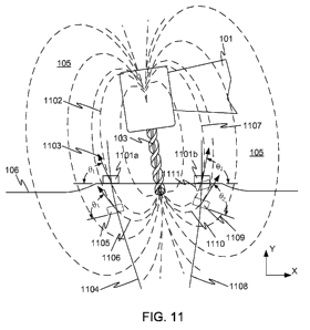
[0104] FIG. 11 illustrates one example technique for determining the location
of drill 103 with
respect to sensors 1101a and 1101b, and thus with respect to the patient's
dentition. The
22
CA 02841747 2014-01-14
WO 2013/010138
PCT/US2012/046789
example of FIG. 11 depicts only two dimensions for ease of explanation, but it
will be
recognized that the technique may be generalized to a three-dimensional
system. In FIG. 11,
handpiece 101 and drill 103 are shown in a particular location with respect to
sensors 1101a and
1101b, which are fixed to workpiece guide 106. This example utilizes drill 103
as the
magnetized element. A particular flux line 1102 of magnetic field 105 passes
through sensor
1101a, at an incident angle 01, and with a strength represented by the length
of vector 1103. The
output of sensor 1101a indicates the field strength and direction of magnetic
field 105 as seen by
sensor 1101a ¨ that is the output indicates the field strength and 01.
[0105] The output of sensor 1101a alone is not sufficient to characterize the
location of sensor
1101a within magnetic field 105. For example, sensor 1101a could be at any
position along
isomagnetic locus 1104, which is the locus of all points within magnetic field
105 having the
same magnetic field strength as the point at which sensor 1101a happens to
reside. (Only
portions of the isomagnetic loci in FIG. 11 are illustrated. In practice, each
isomagnetic locus
will be a closed curve.) Given the field strength reading from sensor 1101a,
isomagnetic locus
1104 may be determined from the previous characterization of magnetic field
105, for example
by interpolating within a numerical array describing the field, or
formulaically if the field has
been described by a mathematical formula. Another possible location of sensor
1101a within
magnetic field 105 is shown at location 1105. If sensor 1101a and magnetic
field 105 were in a
relationship that placed sensor 1101a at location 1105, at an angle of 01 with
respect to field line
1106, sensor 1101a would give an identical output. More information is needed
to determine the
relationship of magnetic field 105 to the sensors.
[0106] Similarly, sensor 1101b is crossed by field line 1107 at an angle 02.
Thus, the system
can determine that sensor 1101b is located somewhere on isomagnetic locus
1108, but given
only the output of sensor 1101b, cannot determine where on isomagnetic locus
1108. For
example, sensor 1101b could be at location 1109, oriented at an angle of 02
with respect to field
line 1110.
[0107] By combining the information from both sensor outputs with previously
determined
information about the orientation of sensors 1101a and 1101b, it is possible
to uniquely
determine the locations of sensors 1101a and 1101b within magnetic field 105.
In some
embodiments, it is known how far apart sensors 1101a and 1101b actually are on
workpiece
guide 106. Given that information and a hypothetical location of one sensor,
it is possible to
calculate the expected position of the other sensor, and test whether the two
locations fit the
23
CA 02841747 2014-01-14
WO 2013/010138
PCT/US2012/046789
measured data. For example, if it is assumed that sensor 1101a is at location
1105, then sensor
1101b would be expected to be at location 1111. While position 1111 is quite
close to the actual
X-Y position of sensor 1101b, hypothetical location 1111 is oriented
incorrectly with respect to
the local field lines, and cannot be the actual position of sensor 1101b.
Thus, location 1105
cannot be the correct location of sensor 1101a. Potential locations for sensor
1101a along
isomagnetic locus 1104 may be searched until the predicted location of sensor
1101b matches
the actual angular data from sensor 1101b. Once a matching pair of locations
is found, the
locations of sensors 1101a and 1101b within magnetic field 105 is ascertained.
From that
information, it is straightforward to calculate the orientation of magnetic
field 105 with respect to
workpiece guide 106, and accordingly with respect to the patient's dentition.
And because the
location of drill 103 is known with respect to magnetic field 105, the
location of drill 103 can be
calculated with respect to the patient's dentition. From that relationship and
the previously-
stored radiographic image, the system can generate the display graphically
illustrating the
location of drill 103 with respect to the patient's dentition. Similarly,
because the location of the
desired implant shaft is also known, the system can generate the indication of
the location of drill
103 with respect to the desired implant shaft.
[0108] FIG. 12 illustrates a system 1200 in accordance with another embodiment
of the
invention, for indicating the location of a dental drill. System 1200 includes
some components
similar to components shown in FIG. 1, and like components are given like
reference numbers.
In the system of FIG. 1, magnetic element 104 is fixed to drill 103, and
sensors 108a-108c are
fixed to workpiece guide 106. System 1200 reverses that arrangement.
[0109] In system 1200, a magnetized element 1201 is fixed to workpiece guide
106, and
generates a magnetic field 105. Sensors 108a-108c are fixed in relation to
handpiece 101, and
consequently in relation to drill 103. As handpiece 101 is moved, sensors 108a-
108c are
exposed to different parts of magnetic field 105, and produce different
outputs 110a-110c.
Sensor outputs 110a-110c are provided to controller 109, for example via a
flexible cable 1202
(shown in only a partial view), or via a wireless connection. An intermediate
device similar to
intermediate device 801 may also be present. Controller 109 processes sensor
outputs 110a-
110c to provide an indication of the location of drill 103 in relation to the
dentition of a patient
wearing workpiece guide 106. For example, the strength and shape of magnetic
field 105 and its
spatial relationship to the patient's dentition may be characterized, and the
spatial relationship of
sensors 108a-108c to drill 103 may be characterized, and this information
supplied to controller
109, which then processes sensor outputs 110a-110c according to these
previously-characterized
24
CA 02841747 2014-01-14
WO 2013/010138
PCT/US2012/046789
relationships to determine the location of drill 103 with respect to the
patient's dentition. As in
the embodiments described above, location may be determined by interpolating
within a
numerical array describing the field, or formulaically if the field has been
described by a
mathematical formula.
[0110] To characterize the relationship between magnetic field 105 and the
patient's dentition,
the relationship of magnetic field 105 to magnetized element 1201 may first be
characterized.
For example, a set of sensors similar to those on calibration station 305 or
magnetizer/calibration
station 803 may be used. Magnetized element 1201 may be placed in a known
relationship to
the sensors, and readings produced by the sensors used to characterize
magnetic field 105. In
other embodiments, magnetized element 1201 may be supplied from the factory
with a data file
describing magnetic field 105.
[0111] Magnetized element 1201 may then be placed in a known location with
respect to
workpiece guide 106 (whose relationship to the patient's dentition is known
from the process of
fabricating workpiece guide 106). For example, surface 107 may be a planar
surface coincident
with the plane defined by radiopaque fiducial markers 401a-401c. A pilot hole
601 is formed in
workpiece guide, a pin (which may preferably be a stepped pin) may be placed
in pilot hole 601
and magnetized element 1201 slipped over the pin until magnetized element 1201
touches
surface 107 of workpiece guide 106. Magnetized element 1201 may then be fixed
to workpiece
guide 106, for example using an epoxy or other adhesive. This process
completely defines the
location of magnetized element 1201 with respect to the patient's dentition
(once workpiece
guide 106 is replaced in the patient's mouth).
[0112] The relationship of sensors 108a-108c to drill 103 may be characterized
by
mechanically positioning drill at a predetermined location with respect to
sensors 108a-108c.
For example, a fixture may be utilized to set the depth of insertion of drill
103 into handpiece
101 such that the distance from the bottom of sensor mounting plate 1203 to
the tip of drill 103 is
set consistently to a predetermined value, even when drill 103 is changed
during the implant
procedure. In other embodiments, a calibration fixture having a previously-
characterized
magnetic field could be used.
[0113] FIG. 13A illustrates a workpiece guide 1301 and a sensor assembly 1302,
in
accordance with embodiments of the invention. Workpiece guide 1301 and sensor
assembly
1302 are adapted for performing two implants in a single treatment session,
although it will be
CA 02841747 2014-01-14
WO 2013/010138
PCT/US2012/046789
recognized that certain features of the system are applicable to single-
implant embodiments, or
to embodiments adapted for three or more implants.
[0114] Example workpiece guide 1301 is configured for performing implants at
two adjacent
tooth locations. Using the techniques described previously, a dental
professional has selected the
locations of two implant shafts. Workpiece guide 1301 has been fabricated to
conform to the
patient's dentition, and includes three fiducial markers 1303a-1303c affixed
to surface 1304.
Two pilot holes 1305a and 1305b have been formed in workpiece guide 1301,
preferably aligned
with the two desired implant shafts. While workpiece guide 1301 is configured
for performing
two implants, it will be recognized that in other embodiments a workpiece
guide may be
configured for performing more implants, including implants at non-adjacent
tooth locations.
Also, a different number of fiducial markers could be used. For example, each
implant site could
use its own respective set of fiducial markers.
[0115] Also positioned near each pilot hole 1305a, 1305b is a set of alignment
pins. For
example, alignment pins 1306a and 1306b are positioned near pilot hole 1305a,
and alignment
pins 1306c and 1306d are positioned near pilot hole 1305b. The alignment pins
may be placed
in known relationship to the other features of workpiece guide 1301. For
example, at the time
pilot holes 1305a and 1305b are formed, holes for receiving alignment pins
1306a-1306d may
be formed. Alignment pins 1306a-1306d can then be inserted into the prepared
holes, for
example by press fitting. Alignment pins 1306a-1306d may be made of any
suitable material,
but may preferably be made of a polymer such as polycarbonate or acrylonitrile
butadiene
styrene (ABS), a non-magnetic metal such as titanium, or another material that
will have little or
no effect on magnetic fields in the area.
[0116] Example sensor assembly 1302 includes a circuit board 1307 having
alignment holes
1308a and 1308b, spaced for engagement with the respective sets of alignment
pins 1306a-
1306d. Thus, sensor assembly 1302 can be engaged with a first set of alignment
pins to aid in
drilling an implant shaft for a first implant, and then moved to engage a
different set of
alignment pins for drilling a different implant shaft for a second implant.
For example, sensor
assembly 1302 may be engaged with alignment pins 1306a and 1306b for assisting
in drilling an
implant shaft associated with pilot hole 1305a, and then moved to engage with
alignment pins
1306c and 1306d for assisting in drilling an implant shaft associated with
pilot hole 1305b.
[0117] FIG. 13B shows example sensor assembly 1302 in more detail. Circuit
board 1307
may be a double sided printed circuit board or flex circuit or another kind of
circuit carrier, and
26
CA 02841747 2014-01-14
WO 2013/010138
PCT/US2012/046789
may have multiple layers. Besides alignment holes 1308a and 1308b, circuit
board 1307
includes a clearance opening 1309, allowing clearance for a drill to reach the
appropriate pilot
hole. Circuit board 1307 also carries eight sensors 1310a-1310h in this
example. Four sensors
1310a-1310d are mounted on the top surface of circuit board 1306, and four
additional sensors
1310e-1310h (shown in broken lines) are mounted to the bottom surface of
circuit board 1307.
While sensors 1310e-1310h are shown as being mounted directly below sensors
1310a-1310d,
this is not a requirement. The sensors also need not be mounted symmetrically
around clearance
opening 1309. This "dual quad" arrangement having two layers of four sensors
each may
provide improved accuracy in determining the position of a drill as compared
with a single-layer
arrangement of sensors. Signals from sensors 1310a-1310h are carried by traces
1311 in circuit
board 1307 (the traces are shown in simplified form) to a connector 1312, and
then to a cable
1313 for communicating the signals to a controller such as controller 109, or
to an intermediate
device such as intermediate device 801.
[0118] Many different variations and system architectures are possible. For
example, if a flex
circuit is used, no connector 1312 may be necessary. Or in a wireless
arrangement similar to the
arrangement of FIG. 9, no cable 1313 may be necessary. In other embodiments,
different
numbers of sensors may be used. For example, a "dual triad" arrangement may be
used, with
three sensors on top of circuit board 1307 and three sensors on the bottom
side of circuit board
1307.
[0119] FIG. 13C shows sensor assembly 1302 engaged with alignment pins 1306a
and 1306b,
for aiding in drilling an implant shaft associated with pilot hole 1305a.
Alignment pins 1306a
and 1306b assist in holding sensor assembly 1302 in a first fixed position in
relation to
workpiece guide 1301. Many other alignment mechanisms may be envisioned for
enabling a
sensor assembly such as sensor assembly 1302 to be moved from one implant
location to
another. For example, a sleeve could be placed in each pilot hole and the
sensor assembly
aligned with the sleeve to center over the pilot hole. Or a raised shape may
be formed in
workpiece guide 1301 near each pilot hole and clearance opening 1309 of sensor
assembly 1302
placed over the raised shape to register sensor assembly 1302 to workpiece
guide 1301. The
raised shape may have a polygonal shape, for example square or trapezoidal,
and clearance
opening 1309 may have a complementary shape, to prevent rotation of sensor
assembly 1302.
By disengaging sensor assembly 1302 from alignment pins 1306a and 1306b,
sensor assembly
1302 can be moved to a second fixed position with respect to workpiece guide
1301. FIG. 13D
27
CA 02841747 2014-01-14
WO 2013/010138
PCT/US2012/046789
shows sensor assembly 1302 engaged with alignment pins 1306c and 1306d, for
aiding in
drilling an implant shaft associated with pilot hole 1306a.
[0120] FIG. 14A illustrates a workpiece guide 1401 and a sensor assembly 1402,
in
accordance with other embodiments of the invention. Workpiece guide 1401 and
sensor
assembly 1402 are adapted for performing two implants in a single treatment
session, although it
will be recognized that certain features of the system are applicable to
single-implant
embodiments, or to embodiments adapted for performing three or more implants.
Using
workpiece guide 1401 in a manner similar to that described above, pilot holes
1405a and 1405b
may be placed in line or approximately in line with desired implant shafts
previously specified
by the dental professional. Fiducial markers 1403a-1403c may be used in the
process of
determining the positions of pilot holes 1405a and 1405b. Sleeves 1406a and
1406b are placed
in pilot holes 1405a and 1405b. Sleeves 1406a and 1406b are preferably made of
a suitable
radiopaque, non-magnetic material such as an acrylic doped with barium
sulfate. Circuit board
1407 of sensor assembly 1402 includes an alignment hole 1408 sized to fit
snugly over one of
sleeves 1406a or 1406b. A tab 1409 is sized to fit within a gap or keyway 1410
formed in each
sleeve.
[0121] FIG. 14B shows sensor assembly 1402 in place over workpiece guide 1401.
Tab 1409
prevents rotation of sensor assembly 1402 about the axis of the sleeve with
which it is engaged.
The positions of the sleeve keyways may be determined with a second
radiographic
characterization of workpiece guide 1401, or by other means. Alternatively,
sleeves 1406a and
1406b may be placed in approximate locations before the radiographic
characterization of
workpiece guide 1401 in the patient's mouth, and may serve as fiducial
references instead of or
in addition to fiducial markers 1403a-1403c.
[0122] FIGS. 15-19 illustrate certain component relationships and an alternate
technique for
determining the position of drill 103 in relation to the patient's dentition,
in embodiments of the
invention. For example, FIG. 15 shows the relationship of example magnetic
field 105 with
sensors in a "dual quad" arrangement, such as sensors 1310a-1310h, in
accordance with example
embodiments. Magnetic field 105 is represented in FIG. 15 by four lobes, but
it will be
recognized that in this example, magnetic field 105 may be generally
rotationally symmetric
about the axis of drill 103. It is assumed that sensors 1310a-1310h have been
placed in a known
fixed relationship with the patient's dentition, for example using the
techniques described above.
28
CA 02841747 2014-01-14
WO 2013/010138
PCT/US2012/046789
In other embodiments, magnetic field 105 may not be rotationally symmetric,
for example if the
body of a handpiece holding the drill affects the field significantly.
[0123] FIG. 16 illustrates a coordinate system useful in describing the
behavior of sensors. In
this example, each sensor 1310a-1310h is a model HMC5883L 3-Axis Digital
Compass
integrated circuit available from Honeywell International Inc., and has its
own local coordinate
system (Xõ,Yõ,Z.), while the overall system is conveniently described using
radial coordinates (Z
,R, (I)). Each sensor of this type produces three outputs, indicating the
strength of the magnetic
field in each of the three coordinate axes.
[0124] FIG. 17 illustrates an orthogonal view of the interaction of field 105
with the sensors in
more detail. Using sensor 1310c as an example, in the position shown, a
particular flux line
1701 passes through the measurement location of sensor 1310c, at an angle of .
The angle 0
can be determined from the sensor outputs as atan(V3xN3z)*180/7r. If circuit
board 1307 were to
be positioned at Z=0, it can be seen that the flux lines are nearly vertical,
so the angle 0 would
be essentially 0 degrees. At bottom end 1702 of drill 103, the flux lines
emanate nearly
horizontally from drill 103, so if circuit board 1307 were to be positioned at
the bottom of drill
103 (but still held horizontal as shown), the angle 0 would be about 90
degrees. At top end 1703
of drill 103, the flux lines converge nearly horizontally toward drill 103, so
if circuit board 1307
were to be positioned at the top of drill 103 (but still held horizontal as
shown), the angle 0
would be about -90 degrees.
[0125] FIG. 18 shows an approximate representation of angle 0 as a function of
position
along the Z direction (as sensor 1310c traverses dashed path 1704), for a
drill having a length
L=40 mm. The exact relationship of angle 0 to Z position will depend on the
particular field
shape, but for a magnetized drill, may generally be a monotonic function over
much of the length
of drill 103. For the simple case where the drill is centered among the
sensors and perpendicular
to the plane of the sensors, the drill depth could be determined from the
angle 0 measured at any
one of the sensors.
[0126] However, it may be desirable to average the readings of the sensors, to
reduce noise
and to at least partially cancel the effects of tilt and de-centering of the
drill. For example, de-
centering of the drill within the sensor constellation will tend to reduce the
angles 0 measured by
sensors toward which the drill is moved, and will tend to increase the angles
0 measured by
sensors away from which the drill is moved. Similarly, tilt of the drill will
tend to increase the
angles measured on one side of the drill and reduce the angles measured on the
other side of the
29
CA 02841747 2014-01-14
WO 2013/010138
PCT/US2012/046789
drill. By averaging the angles 0 measured at all of the sensors (eight sensors
in the example of
FIGS. 15-17), these effects are at least approximately canceled, and a
reasonably accurate
estimate of drill depth can be obtained from a calibration curve similar to
FIG. 18. It will be
recognized that the readings from the sensors on the bottom of the circuit
board may require a
sign reversal before averaging.
[0127] The depth estimate obtained in this way may greatly simplify the
remaining
determination of drill location as a function of the sensor readings. Once the
depth is
approximately known, the required range of search within the calibration data
may be greatly
reduced, as compared with trying to locate the drill from an arbitrary set of
sensor readings.
[0128] It has also been observed that the portion of the calibration curve of
FIG. 18
corresponding to the length of the drill (-20 to +20 in the example of FIG.
18) can be
substantially linearized by multiplying the ratio of the sensor outputs by a
constant prior to
applying the arctangent function. That is, a plot of atan(k*V3xN3z)*180/n will
be nearly a
straight line in the region of interest, for an appropriate value of k. The
value of k will depend
on the particular system geometry and other implementation-specific factors,
and can be easily
chosen by plotting the calibration curve with different values of k until a
nearly-linear curve is
obtained. A linearized calibration curve may further simplify the
determination of drill location.
[0129] Another aspect that may simplify the determination of drill location is
that during
drilling, circuit board 1307 will be positioned between the ends of drill 103.
Thus only the
monotonic range of a calibration curve similar to FIG. 18 need be considered.
In FIG. 18, the
monotonic range includes values of Z from about -20 to about +20. The dental
professional may
assure that location estimation begins only after the end of the drill has
passed through circuit
board 1307, for example by signaling to the system that the drill has been
inserted into the
appropriate pilot hole. In some embodiments, the starting of the drill may
signal to the system
that location estimation is to begin, and the dental professional may simply
wait until the drill is
positioned within the pilot hole before starting the drill.
[0130] Once the drill depth has been estimated, other relationships in sensor
output may be
exploited to further refine the estimate of drill location. For example,
translation of drill 103
may cause sensors toward which drill 103 is moved to register stronger field
readings than
sensors from which drill 103 is moved away. Similarly, tilt of drill 103 may
cause some sensors
to read steeper field angles and other sensors to read field angles that are
less steep. Non-zero
readings of field components in the Y directions of the sensors indicate that
the drill is angled.
CA 02841747 2014-01-14
WO 2013/010138
PCT/US2012/046789
[0131] Techniques such as these may be combined into a method of establishing
the drill
location from the sensor readings. FIG. 19 is a flowchart of a method 1900
according to one
example embodiment. In step 1901, the magnetic field is characterized, for
example using a
fixture and methods as described above in relation to FIGS. 10A-10C. The
characterization of
the magnetic field may take the form of a table of measured sensor values at
different locations
within the field. In other embodiments, the sensor values may be fit to a
formulaic description of
the field, from which field strengths and angles can be computed as a function
of location within
the field.
[0132] In step 1902, a depth calibration curve is established. For example,
the depth
calibration curve may be similar to the curve shown in FIG. 18, showing the
field angle
measured by a sensor when the drill is centered within the sensor
constellation. The depth
calibration curve may be based on average readings taken by multiple sensors
during the
calibration process. In step 1903, the drill is placed in position for
drilling, with the circuit board
holding the sensors positioned between the ends of the drill. In step 1904, an
initial set of sensor
readings is taken, and an average field angle reading is computed. It will be
recognized that the
readings from sensors on the bottom of the circuit board may be reversed in
sign before the
averaging.
[0133] In step 1905, the average field angle reading is used to determine an
initial depth
estimate from the depth calibration curve. This estimate assumes that the
drill is perpendicular
to the average plane of the sensors, and is centered within the constellation
of sensors. In step
1906, a figure of merit is computed, indicating how well the assumed position
of the sensors
agrees with the initial sensor readings. For example, predicted sensor
readings may be computed
based on the assumed positions of the sensors within the characterized
magnetic field, and
compared with the actual initial sensor readings. The figure of merit could
be, for example, the
sum of the squares of the differences between the respective predicted and
actual sensor
readings, although other figures of merit may be envisioned. For example,
absolute value
differences could be summed, different sensor readings could be weighted
differently, or other
variations may be used. When eight sensors are used, each producing three
readings, the
computation of the figure of merit may include up to 24 differences between
predicted and actual
readings. In some embodiments, the estimation of drill position may be
performed using
multiple subsets of the sensors, so that if the estimates disagree, it may be
assumed that an error
has occurred, and drilling can be stopped.
31
CA 02841747 2014-01-14
WO 2013/010138
PCT/US2012/046789
[0134] In step 1907, the positions of the sensors are mathematically adjusted
to minimize the
figure of merit. For example, the assumed position of circuit board 1307, and
consequently the
assumed positions of sensors 1310a-1310h, may be mathematically moved to a new
location in
space. The movement may include translation in depth, two lateral translations
(perpendicular to
drill 103), and rotations in at least two degrees of freedom having rotational
axes in the sensor
plane, for a total of up to five degrees of freedom. If it is assumed that the
magnetic field is not
rotationally symmetric, the movement may also include rotation around the
longitudinal axis of
drill 103 as well, resulting in six degrees of freedom. It will be recognized
that step 1907 is
highly simplified in FIG. 19, and may involve many trial mathematical
positionings of the
sensors and computations of the figure of merit at each trial position. Any
suitable mathematical
technique may be used, for example a gradient descent algorithm, the simplex
algorithm, or
another optimization algorithm. Once the figure of merit is minimized, the
relationship of the
sensors and the magnetic field is known. That is, the transformation required
for the assumed
sensor positions to produce predicted sensor readings that agree with the
actual sensor readings
is known. The reverse of this transformation is applied to the assumed drill
position in step
1908, and the resulting measured drill location is reported in step 1909. The
measured drill
location may then be used to construct a display such as the display shown in
FIG. 1 or FIG. 12,
showing the measured position of the drill in relation to the patient's
dentition, a desired implant
shaft, or both.
[0135] Some of the steps of method 1900 may then be repeated, so that the
display can be
updated, preferably substantially in real time. For example, a new set of
sensor readings is taken
in step 1910, and control may be passed to step 1906 for a new computation of
the figure of
merit, and a new evaluation of the drill location.
[0136] Many variations are possible. For example, in other numerical
embodiments, the
location of the magnetic field may be mathematically perturbed rather than the
locations of the
sensors. In other embodiments, where the magnetic field has been characterized
using a formula,
it may be possible to backsolve the formula to obtain the location of the
drill. It is to be
understood that all workable combinations of the features and element
disclosed herein are also
considered to be disclosed.
[0137] The embodiments disclosed above are exemplary and are not to be
construed as
limiting the scope of the invention. Many variations of the methods and
devices described
herein are available to the skilled artisan without departing from the scope
of the invention.
32
CA 02841747 2014-01-14
WO 2013/010138
PCT/US2012/046789
EMBODIMENTS
[0138] Embodiment 1. A system for indicating the location of a dental drill,
the system
comprising: a dental handpiece comprising the dental drill; and a plurality of
sensors that detect a
magnetic field and produce a set of respective sensor outputs, the sensor
outputs usable at least in
part to indicate the location of the dental drill.
[0139] Embodiment 2. The system of embodiment 1, further comprising a magnetic
element
that is fixed in relation to the dental drill and generates the magnetic
field.
[0140] Embodiment 3. The system of embodiment 1, wherein the dental drill is
magnetized
and generates the magnetic field.
[0141] Embodiment 4. The system of embodiment 1, further comprising a magnetic
element
that is fixed in relation to the dentition of a patient, and wherein the
sensors are fixed in relation
to the dental handpiece.
[0142] Embodiment 5. The system of any one of the embodiments 1 to 3, further
comprising a
workpiece guide registered to a patient's dentition, wherein the sensors are
fixed in relation to
the workpiece guide.
[0143] Embodiment 6. The system of embodiment 5, wherein the sensors are
movable from a
first fixed position in relation to the workpiece guide to a second fixed
position in relation to the
workpiece guide.
[0144] Embodiment 7. The system of any one of the embodiments 1 to 6, further
comprising a
carrier on which the sensors are mounted, at least three of the sensors
mounted to a first surface
of the carrier, and at least three of the sensors mounted to a second surface
of the carrier.
[0145] Embodiment 8. The system of embodiment 7, wherein four of the sensors
are mounted
to a first surface of the carrier, and four of the sensors are mounted to a
second surface of the
carrier.
[0146] Embodiment 9. The system of any one of the embodiments 1 to 8, further
comprising a
controller that receives the sensor outputs and processes the outputs to
produce an indication of
the spatial relationship of the dental drill to a patient's dentition.
[0147] Embodiment 10. The system of embodiment 9, wherein the controller
processes the
sensor outputs according to a spatial relationship between the sensors and the
patient's dentition
and according to a spatial relationship between the magnetic field and the
dental drill.
33
CA 02841747 2014-01-14
WO 2013/010138
PCT/US2012/046789
[0148] Embodiment 11. The system of any one of the embodiments 9-10, further
comprising
an intermediate device that receives the sensor outputs and relays the sensor
outputs to the
controller.
[0149] Embodiment 12. The system of any one of the embodiments 9-11, further
comprising a
wireless interface by which the sensor outputs are transmitted to reach the
controller.
[0150] Embodiment 13. The system of any one of the embodiments 9-12, wherein
the
controller repeatedly updates the indication of the spatial relationship of
the dental drill to the
patient's dentition, substantially in real time.
[0151] Embodiment 14. The system of any one of the embodiments 1-13, further
comprising
an electronic display, and wherein the indication of the spatial relationship
of the dental drill to
the patient's dentition is pictorially represented on the electronic display.
[0152] Embodiment 15. The system of any one of the embodiments 1-14, wherein
the
indication of the spatial relationship of the dental drill to the patient's
dentition comprises: a
pictorial representation of the patient's dentition; and a representation of
the location of the
dental drill location superimposed on the pictorial representation of the
patient's dentition.
[0153] Embodiment 16. The system of any one of the embodiments 14-15, wherein
the
pictorial representation of the patient's dentition is derived from a
radiographic image of the
patient's dentition.
[0154] Embodiment 17. The system of any one of the embodiments 14-16, wherein
the
pictorial representation of the patient's dentition is a representation of a
three-dimensional model
of the patient's dentition.
[0155] Embodiment 18. The system of any one of the embodiments 9-17, wherein
the
controller further produces an indication of the spatial relationship of the
dental drill to a
previously-specified implant shaft within the patient's dentition.
[0156] Embodiment 19. The system of embodiment 18, wherein the controller
further
produces a warning signal when the dental drill departs from the previously-
specified implant
shaft by at least a predetermined amount.
[0157] Embodiment 20. The system of any one of the embodiments 1-19, further
comprising a
calibration station that further includes: a receptacle for the dental drill;
and a second plurality of
sensors fixed in relation to the receptacle, each of the second plurality of
sensors producing an
34
CA 02841747 2014-01-14
WO 2013/010138
PCT/US2012/046789
output, and wherein the outputs of the second plurality of sensors are usable
to characterize the
spatial relationship of the magnetic field to the dental drill when the dental
drill is placed in the
receptacle.
[0158] Embodiment 21. A method of indicating the location of a dental drill,
the method
comprising: reading outputs produced by a set of sensors, wherein the sensors
detect a magnetic
field, and wherein the sensor outputs are usable to detect the location of a
dental drill in relation
to the sensors; processing the sensor outputs to produce an indication of the
spatial relationship
of the dental drill to a patient's dentition; and displaying the indication of
the spatial relationship
of the dental drill to the patient's dentition.
[0159] Embodiment 22. The method of embodiment 21, wherein processing the
outputs
comprises processing the outputs according to a spatial relationship between
the sensors and the
patient's dentition and according to a spatial relationship between the
magnetic field and the
dental drill.
[0160] Embodiment 23. The method of any one of the embodiments 21-22, wherein
displaying an indication of the spatial relationship of the dental drill to
the patient's dentition
comprises repeatedly updating the display of the indication of the spatial
relationship of the
dental drill to the patient's dentition, substantially in real time.
[0161] Embodiment 24. The method of any one of the embodiments 21-23, wherein
reading
the outputs of a set of sensors comprises reading the outputs of the sensors
via a wireless
interface.
[0162] Embodiment 25. The method of any one of the embodiments 21-24, further
comprising, indicating on the display the location of the dental drill in
relation to a previously-
specified implant shaft.
[0163] Embodiment 26. The method of any one of the embodiments 21-25, further
comprising: comparing the location of the dental drill with the previously-
specified implant
shaft; and producing a warning signal when the dental drill departs from the
previously-specified
implant shaft by at least a predetermined amount.
[0164] Embodiment 27. The method of embodiment 26, wherein the warning signal
comprises
one or more signals selected from the group consisting of a visual cue and a
sound cue, alone or
in any combination.
CA 02841747 2014-01-14
WO 2013/010138
PCT/US2012/046789
[0165] Embodiment 28. A workpiece guide, comprising: a dental arch portion
that conforms
to the dentition of a particular patient; and a set of sensors fixed in
relation to the dental arch
portion, each sensor capable of producing an output that indicates at least
one characteristic of a
magnetic field.
[0166] Embodiment 29. The workpiece guide of embodiment 28, wherein the dental
arch
portion defines a pilot hole located, when the workpiece guide is engaged with
the dental arch of
the particular patient, substantially at the centerline of a desired implant
shaft.
[0167] Embodiment 30. The workpiece guide of any one of the embodiments 28-29,
further
comprising at least three non-collinear radiopaque fiducial markers on the
workpiece guide.
[0168] Embodiment 31. The workpiece guide of any one of the embodiments 28-30,
wherein
the sensors are movable from a first fixed position in relation to the
workpiece guide to a second
fixed position in relation to the workpiece guide.
[0169] Embodiment 32. A method, comprising: fabricating a workpiece guide of a
configuration to engage the dentition of a particular patient having an
implant site; placing a set
of fiducial references on the workpiece guide; and fixing a sensor to the
workpiece guide, the
sensor capable of, when the sensor is exposed to a magnetic field, producing
an output indicating
an aspect of the magnetic field.
[0170] Embodiment 33. The method of embodiment 32, further comprising:
engaging the
workpiece guide with the dental arch of the patient; obtaining a radiographic
image of the
workpiece guide and the patient's dental arch, the radiographic image
depicting the fiducial
references; determining from the radiographic image the location of a desired
implant shaft for
placing an implant at the implant site; and characterizing the location of the
desired implant shaft
with respect to the locations of the fiducial references.
[0171] Embodiment 34. The method of embodiment 33, further comprising: forming
a pilot
hole in the radiographic workpiece guide, wherein the centerline of the pilot
hole will be
substantially collinear with the centerline of the implant shaft when the
radiographic workpiece
guide is engaged with the patient's dental arch.
[0172] Embodiment 35. The method of any one of the embodiments 33-34, further
comprising: bringing a dental handpiece comprising a dental drill into
proximity with the sensor,
wherein an element fixed to the handpiece produces a magnetic field, such that
the sensor detects
the magnetic field; obtaining an output from the sensor; processing the sensor
output to
36
CA 02841747 2014-01-14
WO 2013/010138
PCT/US2012/046789
determine the spatial relationship between the dental drill and the patients'
dentition; and
displaying, on a visual display, an indication of the spatial relationship of
the dental drill to the
patient's dentition.
[0173] Embodiment 36. The method of embodiment 35, further comprising
calibrating the
spatial relationship between the magnetic field and the dental drill.
[0174] Embodiment 37. The method of any one of the embodiments 33-36, further
comprising
simultaneously displaying, on the visual display, an indication of the spatial
relationship of the
dental drill to the desired implant shaft.
[0175] Embodiment 38. The method of embodiment 37, further comprising
producing a
warning signal when the dental drill departs from the previously-specified
implant shaft by at
least a predetermined amount.
[0176] Embodiment 39. A computerized controller, comprising: an image
processor that
receives a radiographic image of a patient's dentition; a location system that
receives outputs
from one or more sensors, wherein the sensors detect at least one aspect of a
magnetic field, and
the sensor outputs change as the spatial relationship of the magnetic field
and the sensors
changes due to changes in the location of a dental handpiece that includes a
dental drill, and
wherein the location system processes the sensor outputs to determine the
location of the dental
drill in relation to the patient's dentition; and a viewing system that
generates a display image at
a computer display such that the generated display image comprises an image of
the patient's
dentition and a depiction of the location of the dental drill relative to the
patient's dentition as
determined by the location system.
[0177] Embodiment 40. The computerized controller of embodiment 39, wherein
the location
system receives updated sensor outputs and determines based at least in part
on the updated
sensor outputs an updated location of the handpiece in relation to the
patient's dentition, and the
viewing system adjusts the generated display image to show the updated
location of the dental
drill relative to the patient's dentition.
[0178] Embodiment 41. The computerized controller of any one of the
embodiments 39-40
wherein the generated display image further comprises a depiction of the
location of the dental
drill relative to a desired implant shaft.
37
CA 02841747 2014-01-14
WO 2013/010138
PCT/US2012/046789
[0179] Embodiment 42. The computerized controller of any one of the
embodiments 39-41,
further comprising a computer processor that performs operations of the
location system and
image processor.
[0180] Embodiment 43. A computerized controller, comprising: a processor; a
data input
interface; a display; and a computer-readable memory, the computer readable
memory holding
instructions that, when executed by the processor, cause the computerized
controller to read
outputs produced by a set of sensors, wherein the sensors detect a magnetic
field and the sensor
outputs are usable to characterize the spatial relationship of a dental drill
to the sensors; process
the outputs to produce an indication of the spatial relationship of the dental
drill to a patient's
dentition; and produce a display of the indication of the spatial relationship
of the dental drill to
the patient's dentition.
[0181] Embodiment 44. The computerized controller of embodiment 43, wherein
the
instructions, when executed by the processor, further cause the computerized
controller to
repeatedly update the display of the indication of the spatial relationship of
the dental drill to the
patient's dentition, substantially in real time.
[0182] Embodiment 45. The computerized controller of any one of the
embodiments 43-44,
wherein the instructions, when executed by the processor, further cause the
computerized
controller to indicate on the display the location of the dental drill in
relation to an implant shaft.
[0183] Embodiment 46. The computerized controller of any one of the
embodiments 43-45,
wherein the instructions, when executed by the processor, further cause the
computerized
controller to: compare the location of the dental drill with the implant
shaft; and produce a
warning signal when the dental drill departs from the implant shaft by at
least a predetermined
amount.
[0184] Embodiment 47. The computerized controller of embodiment 46, wherein
the warning
signal comprises one or more signals selected from the group consisting of a
visual cue and a
sound cue, alone or in any combination.
[0185] Embodiment 48. A calibration station, comprising: a body defining a
receptacle,
wherein the receptacle is of a shape and size to receive a dental drill; and a
plurality of sensors
surrounding the receptacle, each sensor capable of producing an output when
the sensor is
exposed to a magnetic field associated with a dental drill placed in the
receptacle.
38
CA 02841747 2014-01-14
WO 2013/010138
PCT/US2012/046789
[0186] Embodiment 49. The calibration station of embodiment 48, wherein the
sensors are
positioned such that their outputs are capable of characterizing the shape and
strength of the
magnetic field.
[0187] Embodiment 50. A non-transitory computer readable medium holding
computer
instructions adapted to be executed to implement a method of indicating the
location of a dental
drill, the method comprising: reading outputs produced by a set of sensors,
wherein the sensors
detect a magnetic field, and wherein the sensor outputs are usable to detect
the location of a
dental drill in relation to the sensors; processing the sensor outputs to
produce an indication of
the spatial relationship of the dental drill to a patient's dentition; and
displaying the indication of
the spatial relationship of the dental drill to the patient's dentition.
[0188] Embodiment 51. A sensing device, comprising: a carrier having circuit
traces, the
carrier defining a through hole; and a plurality of electronic sensors mounted
to the carrier
around the through hole, each sensor being sensitive to a magnetic field and
configured to
produce an output indicating an aspect of the magnetic field; wherein the
sensing device is of a
size and shape for the sensors to fit within the mouth of a dental patient.
[0189] Embodiment 52. The sensing device of embodiment 51, further comprising
flexible
electrical conductors configured to carry the sensor outputs outside the
patient's mouth.
[0190] Embodiment 53. The sensing device of any one of the embodiments 51-52,
further
comprising a wireless transmitter configured to transmit the sensor outputs
outside the patient's
mouth.
[0191] Embodiment 54. The sensing device of embodiment 53, further comprising
a battery
that powers the sensors and the wireless transmitter.
[0192] Embodiment 55. The sensing device of any one of the embodiments 51-54,
wherein the
plurality of sensors comprises at least six sensors, at least three of the
sensors mounted to a first
surface of the carrier, and at least three of the sensors mounted to a second
surface of the carrier.
[0193] Embodiment 56. The sensing device of any one of the embodiments 51-55,
wherein
the plurality of sensors comprises eight sensors, four of the sensors mounted
to a first surface of
the carrier, and four of the sensors mounted to a second surface of the
carrier.
[0194] Embodiment 57. A kit, comprising: a sensing device including: a carrier
having circuit
traces, the carrier defining a through hole; and a set of electronic sensors
mounted to the carrier
39
CA 02841747 2014-01-14
WO 2013/010138
PCT/US2012/046789
around the through hole, each sensor being sensitive to a magnetic field and
configured to
produce an output indicating an aspect of the magnetic field; wherein the
sensing device is of a
size and shape for the sensors to fit within the mouth of a dental patient;
and a non-transitory
computer readable medium holding computer instructions adapted to be executed
to implement a
method of indicating the location of a dental drill, the method including:
reading outputs
produced by the set of sensors, wherein the sensors detect a magnetic field,
and wherein the
sensor outputs are usable to detect the location of a dental drill in relation
to the sensors;
processing the sensor outputs to produce an indication of the spatial
relationship of the dental
drill to a patient's dentition; and displaying the indication of the spatial
relationship of the dental
drill to the patient's dentition.
[0195] Embodiment 58. The kit of embodiment 57, further comprising a
calibration station
including: a body defining a receptacle, wherein the receptacle is of a shape
and size to receive a
dental drill; and a second set of sensors surrounding the receptacle, each
sensor in the second set
capable of producing an output when the sensor is exposed to a magnetic field
associated with a
dental drill placed in the receptacle.
[0196] Embodiment 59. The kit of any one of the embodiments 57-58, further
comprising an
intermediate device configured to receive the sensor outputs and to relay the
sensor outputs to a
controller.