Note: Descriptions are shown in the official language in which they were submitted.
REDUCED-PRESSURE WOUND DRESSINGS
[0001]
BACKGROUND
[0002] The present disclosure relates generally to medical treatment systems
and, more
particularly, but not by way of limitation, to reduced-pressure dressings
having a drain adapter
and related systems, and methods for treating incisions.
[0003] Clinical studies and practice have shown that providing a reduced
pressure in
proximity to a tissue site can augment and accelerate the growth of new tissue
at the tissue
site. The applications of this phenomenon are numerous, but application of
reduced pressure
has been particularly successful in treating wounds. This treatment
(frequently referred to in
the medical community as "negative pressure wound therapy," "reduced-pressure
therapy," or
"vacuum therapy") provides a number of benefits, which may include faster
healing and
increased formulation of granulation tissue. Typically, reduced pressure is
applied to tissue
through a wound dressing assembly that includes a porous pad or other manifold
device. The
porous pad distributes reduced pressure to the tissue and channels fluids that
are drawn from
the tissue.
1
CA 2841860 2018-08-01
CA 02841860 2014-01-06
WO 2013/019438
PCMJS2012/047742
VAC.1012PCT
SUMMARY
[0004] According to an illustrative embodiment, a reduced-pressure system for
treating
a tissue site having a linear wound includes a dressing bolster formed from a
medical bolster
material. The dressing bolster is for placing on a patient's epidermis and is
substantially sized
to overlay the linear wound. The system further includes an over-drape for
providing a fluid
seal over the dressing bolster and a portion of the patient's epideimis, a
reduced-pressure
source, and a first reduced-pressure interface fluidly coupled to the dressing
bolster and the
reduced-pressure source. The first reduced-pressure interface is for
delivering reduced
pressure to the dressing bolster. The system also includes a reduced-pressure
delivery conduit
for fluidly coupling the reduced-pressure source and the first reduced-
pressure interface. The
system further includes a second reduced-pressure interface coupled to the
over-drape,
wherein the second reduced-pressure interface is sized and configured to
receive a
subcutaneous delivery conduit and to form a fluid seal about the subcutaneous
delivery
conduit.
[0005] According to another illustrative embodiment, a wound dressing assembly
for
treating a tissue site having a linear wound includes a dressing bolster
having a first surface
and a second, inward facing surface for deploying over a patient's epidermis
and substantially
sized to overlay the linear wound; an over-drape for providing a fluid seal
over the dressing
bolster and a portion of the patient's epidermis; and a first reduced-pressure
interface operable
to receive a reduced-pressure supply conduit. The assembly further includes an
inner layer
having a first surface and a second, inward-facing surface, and formed within
a treatment-area
aperture. The first surface of the inner layer is coupled at least in part to
the second surface of
the dressing bolster. A second-reduced-pressure interface includes an
interface body formed
with an aperture and having a first side and a second, patient-facing side.
The aperture is sized
to receive a subcutaneous delivery conduit and to form a fluid seal therewith.
The second
reduced-pressure interface is adapted to allow the subcutaneous delivery
conduit to pass from
a subcutaneous tissue site to an external site through the aperture. The
subcutaneous delivery
conduit is routed through the treatment-area aperture and the dressing bolster
to the second
interface.
[0006] According to another illustrative embodiment, a method of treating a
tissue site
having a linear wound includes applying a wound dressing assembly to the
tissue site. The
wound dressing assembly includes a dressing bolster foliated from a medical
bolster material
that is shaped for placing on a patient's epidermis and substantially sized to
overlay the linear
CA 02841860 2014-01-06
WO 2013/019438
PCMJS2012/047742
VAC.1012PCT
wound, an over-drape for providing a fluid seal over the dressing bolster and
a portion of the
patient's epidermis, a first reduced-pressure interface fluidly coupled to the
dressing bolster
for delivering reduced-pressure to the dressing bolster, and a second reduced-
pressure
interface coupled to the over-drape, wherein the second reduced-pressure
interface is sized and
.. configured to receive a subcutaneous delivery conduit and to form a fluid
seal between the
subcutaneous delivery conduit and the wound dressing assembly. The method
further includes
fluidly coupling a reduced-pressure delivery conduit to a reduced-pressure
source and the first
reduced-pressure interface, delivering reduced pressure to the reduced-
pressure delivery
conduit, fluidly coupling a subcutaneous delivery conduit to the second
interface, and
delivering reduced pressure to the subcutaneous delivery conduit.
[0(8)7] According to another illustrative embodiment, a method of
manufacturing a
wound dressing assembly for treating damaged subcutaneous damaged tissue
includes
providing a dressing bolster formed from a medical bolster material. The
dressing bolster is
for placing on a patient's epidermis and is substantially sized to overlay a
linear wound. The
method further includes providing an over-drape for providing a fluid seal
over the dressing
bolster and a portion of the patient's epidermis, providing a reduced-pressure
source,
providing a first reduced-pressure interface for delivering reduced pressure
to the dressing
bolster, providing a second reduced-pressure interface, and coupling the
second reduced-
pressure interface to the over-drape, wherein the second reduced-pressure
interface is sized
and configured to receive a subcutaneous delivery conduit and to form a fluid
seal
therebetween.
[0008] Other features and advantages of the illustrative embodiments will
become
apparent with reference to the drawings and the detailed description that
follow.
3
CA 02841860 2014-01-06
WO 2013/019438
PCMJS2012/047742
VAC.1012PCT
BRIEF DESCRIPTION OF THE DRAWINGS
[0009] FIGURE 1 is a schematic perspective view, with a portion shown in cross-
section, of an illustrative reduced-pressure system for treating a tissue site
having a linear
wound that includes a first reduced-pressure interface and a second reduced-
pressure interface;
[0010] FIGURE 2 is a schematic, cross-sectional view of the illustrative
reduced-
pressure system of FIGURE 1;
[0011] FIGURE 3A is a schematic, perspective view of a portion of an
illustrative
dressing bolster that foinis a part of a reduced-pressure system;
[0012] FIGURE 3B is a schematic, perspective view of a portion of an
illustrative
.. dressing bolster that foinis a part of a reduced-pressure system;
[0013] FIGURE 4 is a schematic, perspective view of a portion of a wound
dressing
assembly for treating a tissue site having a linear wound;
[0014] FIGURE 5 is a schematic, perspective view of a portion of a wound
dressing
assembly for treating a tissue site having a linear wound;
[0015] FIGURE 6 is a partially-exploded, schematic perspective view, with a
portion
in cross-section, of an illustrative embodiment of a wound dressing assembly
having a second
reduced-pressure interface;
[0016] FIGIJRE 6A is a detail of a portion of the wound dressing assembly of
Fig. 6;
[0017] FIGURE 7 is a schematic, side cross-sectional view of an illustrative
reduced-
pressure system for treating a tissue site having a linear wound and providing
a subcutaneous
delivery conduit to a subcutaneous tissue site;
[0018] FIGURE 8 is a schematic, exploded, perspective view of an illustrative
embodiment of a wound dressing assembly that includes a second reduced-
pressure interface;
and
[0019] FIGURE 9 is a schematic, perspective view of an illustrative embodiment
of a
wound dressing assembly having a first reduced-pressure interface and a second
reduced-
pressure interface.
4
CA 02841860 2014-01-06
WO 2013/019438
PCMJS2012/047742
VAC.1012PCT
DETAILED DESCRIPTION OF ILLUSTRATIVE EMBODIMENTS
[0020] In the following detailed description of the illustrative embodiments,
reference
is made to the accompanying drawings that form a part hereof. These
embodiments are
described in sufficient detail to enable those skilled in the art to practice
the invention, and it is
understood that other embodiments may be utilized and that logical structural,
mechanical,
electrical, and chemical changes may be made without departing from the spirit
or scope of the
invention. To avoid detail not necessary to enable those skilled in the art to
practice the
embodiments described herein, the description may omit certain information
known to those
skilled in the art. The following detailed description is, therefore, not to
be taken in a limiting
sense, and the scope of the illustrative embodiments are defined only by the
appended claims.
[0021] As a result of a surgery or other medical condition, a patient may have
a
subcutaneous wound that is located near or beneath a linear wound on the
patient's epideitnis.
For example, after a surgery to remove subcutaneous tissue from a patient, the
patient may
have a linear wound as a result of the surgical incision. The patient may also
have an area
beneath their skin that will need to heal as a result of the surgery, i.e., a
subcutaneous wound
or defect.
[0022] In such a case, the subcutaneous wound may exude or collect fluids
during the
healing process, and a drain may be inserted at or near the end of the linear
wound in order to
collect exudates and prevent the collection of unwanted fluids at the tissue
site of the
subcutaneous wound. The drain may be connected to a drain tube, which may be
referred to
as a subcutaneous delivery conduit, that is configured to exit the patient's
body at one end of
the linear wound. The drain tube, which is a type of subcutaneous delivery
conduit, may be
connected to a drain for the purposes of collecting wound exudates in a
sanitary fashion.
Contemporaneously, a reduced-pressure treatment method may be applied to treat
the linear
wound that was caused during the surgery. The reduced-pressure treatment
method involves
the formation of a fluid seal over the treatment area that includes the linear
wound.
[0023] The fluid seal is typically an important and fragile feature of a
system that
delivers reduced pressure to a wound site. Any breach in the fluid seal may
cause a leak that
may shut down or otherwise compromise a reduced-pressure delivery system
because such
systems tend to be highly intolerant of leaks. One method for providing the
fluid seal may be
to provide a wound dressing assembly that includes a pliable drape, or over-
drape, that seals
against the epidermis of the patient. By forming the fluid seal, the over-
drape may preserve a
pressure differential between the treatment area and the ambient environment,
which may also
5
CA 02841860 2014-01-06
WO 2013/019438
PCMJS2012/047742
VAC.1012PCT
be referred to as the external environment. As used throughout this document,
the term "of'
does not require mutual exclusivity. In cases where a drain tube is present,
application of a
reduced-pressure wound dressing assembly may be difficult because the drain
tube may
prevent the over-drape from sealing against the patient's epidermis. In cases
where a
treatment provider attempts to seal the treatment area by applying the over-
drape over the
drain tube, a leak may result. Further, even if a fluid seal can be obtained,
movement of the
drain tube may entirely disrupt the seal and cause a loss of reduced pressure
at the treatment
site, thereby compromising the ability to maintain the desired amount of
reduced pressure at
the tissue site. Thus, it is desirable to have a dressing assembly or
interface for allowing a
drain tube, or subcutaneous delivery conduit, to pass through a reduced-
pressure treatment
area (from a drain to a drain collection area) without disrupting the fluid
seal between the
treatment site and the ambient environment.
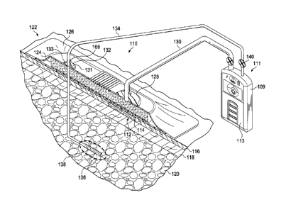
[0024] Referring now primarily to FIGURE 1, an illustrative embodiment of a
reduced-pressure treatment system 110 for treating a tissue site 112 that
includes a linear
wound 114, while simultaneously collecting wound exudates through a drain 136,
is presented.
The tissue site 112 is related to a linear wound 114. The tissue site 112 may
be the bodily
tissue of any human, animal, or other organism, including bone tissue, adipose
tissue, muscle
tissue, dermal tissue, vascular tissue, connective tissue, cartilage, tendons,
ligaments, or any
other tissue. 'I' reatment of the tissue site 112 may include removal of
fluids, e.g., exudate.
The tissue site 112 is shown at epidermis 116, but in some cases may also
involve the dermis
118, and subcutaneous tissue 120. The reduced-pressure treatment system 110
may also be
used with other tissue sites.
[0025] The reduced-pressure treatment system 110 includes a wound dressing
assembly 122 and a reduced-pressure subsystem 111. While the reduced-pressure
treatment
system 110 is shown in the context of a reduced-pressure wound dressing
assembly 122 placed
over a linear wound 114, it should be understood that the reduced-pressure
treatment system
110 may be used on other tissue sites, including open wounds. The wound
dressing assembly
122 includes a dressing bolster 124 that functions as a manifold, an over-
drape 126, and a first
reduced-pressure interface 128 to accommodate a reduced-pressure delivery
conduit 130. The
reduced-pressure delivery conduit 130 is fluidly coupled to the reduced-
pressure subsystem
111.
[0026] Functionally, the reduced pressure developed by the reduced-pressure
subsystem 111 is delivered through the reduced-pressure delivery conduit 130
to the first
reduced-pressure interface 128. In one illustrative embodiment, the first
reduced-pressure
6
CA 02841860 2014-01-06
WO 2013/019438
PCMJS2012/047742
VAC.1012PCT
interface 128 is a T.R.A.C. Pad or Sensa T.R.A.C. Pad available from KCI of
San Antonio,
Texas. The first reduced-pressure interface 128 allows the reduced pressure to
be delivered to
the dressing bolster 124. In another embodiment, no reduced-pressure interface
128 is used.
Instead, a lumen (or conduit) is placed through the over-drape 126 directly
into the dressing
bolster 124.
[0027] The wound dressing assembly 122 provides a fluid seal over an area that
includes a tissue site 112 that is to be treated with reduced pressure. A
fluid seal is a seal
adequate to maintain reduced pressure at a desired site given the particular
reduced-pressure
source(s) or subsystem involved.
[0028] The wound dressing assembly 122 also includes a second reduced-pressure
interface 132. The second reduced-pressure interface 132 is fastened to the
over-drape 126 by
an adhesive, bond, weld (e.g., an ultrasonic, thermal, or RF weld), or cement
(not shown). The
second reduced-pressure interface 132 maintains the integrity of the fluid
seal over the tissue
site 112 while allowing a subcutaneous delivery conduit 134 to pass through
the dressing
.. bolster 124 and over-drape 126. In an embodiment, the second reduced-
pressure interface 132
comprises a molded plastic component that is welded or adhered to the over-
drape 126.
[0029] The second reduced-pressure interface 132 of FIGURE 1 is configured so
that a
user or treatment provider can exert a force against an outer surface of the
second reduced-
pressure interface 132 to route a subcutaneous delivery conduit 134 through
the wound
dressing assembly 122. At the same time, the user can press a subcutaneous
delivery conduit
134 against the over-drape 126 at the location of the second reduced-pressure
interface 132
with sufficient force to breach the over-drape 126. The second reduced-
pressure interface 132
reinforces the over-drape 126 so that it can be breached without compromising
the ability of
the over-drape 126 to provide a fluid seal outside of the second reduced-
pressure interface
.. 132. In one embodiment, an aperture is formed in the over-drape at the
second reduced-
pressure interface 132 to facilitate a pathway through the wound dressing
assembly 122.
[0030] The second reduced-pressure interface 132 may take any number of shapes
and
sizes. For example, the second reduced-pressure interface 132 may be shaped
substantially as
shown in FIGURE 1, where the second reduced-pressure interface 132 includes
flat top and
bottom surfaces and an aperture 168. The aperture 168 may he sized to generate
a radial
compressive force against a subcutaneous delivery conduit 134 when the
subcutaneous
delivery conduit 134 is inserted through the second reduced-pressure interface
132. For
example, an interference fit may be formed between the aperture 168 and the
subcutaneous
delivery conduit 134. According to one illustrative embodiment, the second
reduced-pressure
7
CA 02841860 2014-01-06
WO 2013/019438
PCMJS2012/047742
VAC.1012PCT
interface 132 may comprise a nipple 131 formed from an interface body 133 with
the aperture
168 having an interior diameter Di. The subcutaneous delivery conduit 134 may
have an
external diameter 1)2. The diameters are related by the expression Di <1)2. Di
is slightly less
than D2 whereby the fluid seal is formed by an interference fit. In another
embodiment, Di
may be equal to D2.
[0031] The second reduced-pressure interface 132 may also he tapered such that
the
thickness of the second reduced-pressure interface 132 is increased at the
aperture and
gradually decreased to a minimal thickness at the outer edge. The thickness of
the second
reduced-pressure interface 132 may gradually increase from the edge to the
boundary of the
aperture 168 in a linear or curved manner. The second reduced-pressure
interface 132 may
also have a rounded, or dome-typed shape. Other shapes are possible as well.
[0032] The subcutaneous delivery conduit 134 functions to allow wound exudates
to
flow from a subcutaneous tissue site 138 from a drain 136. The subcutaneous
delivery conduit
134 may fluidly couple to the reduced-pressure subsystem Ill or a second
reduced-pressure
subsystem (not shown). Thus, both the reduced-pressure delivery conduit 130
and
subcutaneous delivery conduit 134 provide reduced pressure and may couple to
the reduced-
pressure subsystem 111. Here, the reduced-pressure delivery conduit 130 is
coupled to the
reduced-pressure subsystem 111 to apply reduced-pressure therapy to a tissue
site 112. The
subcutaneous delivery conduit 134 may also couple to the reduced-pressure
subsystem 111, or
to a separate reduced-pressure subsystem (not shown) to evacuate fluids, or
exudates, from a
drain 136.
[0033] Reduced pressure is a pressure less than the ambient pressure at a
tissue site
that is being subjected to treatment. In most cases, this reduced pressure
will be less than the
atmospheric pressure at which the patient is located. Alternatively, the
reduced pressure may
be less than a hydrostatic pressure at the tissue site. Unless otherwise
indicated, values of
pressure stated herein are gauge pressures. The reduced pressure delivered may
be constant or
varied (patterned or random) and may be delivered continuously or
intermittently. Consistent
with the use herein, unless otherwise indicated, an increase in reduced
pressure or vacuum
pressure typically refers to a relative reduction in absolute pressure.
[0034] In some embodiments, a different amount of reduced pressure may be
desired
at the tissue site 112 of the linear wound 114 than at the subcutaneous tissue
site 138. In such
embodiments, the subcutaneous delivery conduit 134 and reduced-pressure
delivery conduit
130 may be fluidly coupled to different reduced-pressure sources and may
include different
fluid reservoirs associated with each. Alternatively, the delivery conduits
130, 134 may he
8
CA 02841860 2014-01-06
WO 2013/019438
PCMJS2012/047742
VAC.1012PCT
coupled to the same reduced-pressure subsystem 111 through one or more
pressure regulators
140 that allow a single reduced-pressure subsystem 111 to supply different
amounts of
reduced pressure to each conduit. In some embodiments, the same amount of
reduced
pressure may be desired at the tissue site 112 of the linear wound 114 as the
subcutaneous
tissue site 138. In these embodiments, the subcutaneous delivery conduit 134
and reduced-
pressure delivery conduit 130 may he fluidly coupled to the same reduced-
pressure source 109
without the need for pressure regulators.
[0035] Referring now primarily to FIGURE 2, a side cross section of the wound
dressing assembly 122 of FIGURE 1 is presented. The wound dressing assembly
122 as
shown includes an optional inner comfort layer 142 that may be coupled to the
dressing bolster
124 and that lies between the dressing bolster 124 material and the epideimis
116 of a patient.
The wound dressing assembly 122 may also include (latitudinal or longitudinal)
flexibility
notches 144 in order to add flexibility to the dressing bolster 124.
Additionally, the over-drape
126 of wound dressing assembly 122 may include folds 146 or notches or ridges
to add
flexibility to the wound dressing assembly 122.
[0036] In addition to the first reduced-pressure interface 128 that is fluidly
coupled to
the reduced-pressure delivery conduit 130 and dressing bolster 124, the wound
dressing
assembly 122 also includes the second reduced-pressure interface 132. The
second reduced-
pressure interface 132 functions to accommodate the presence of a subcutaneous
delivery
conduit 134. The subcutaneous delivery conduit 134 functions as a drain tube
that is fluidly
coupled to a drain 136 without disturbing the ability of the reduced-pressure
treatment system
110 to apply reduced pressure to a tissue site 112. The second reduced-
pressure interface 132
fluidly couples a reduced pressure source to the drain 136 without causing a
leak that hinders
or stops the reduced-pressure therapy at tissue site 112.
[0037] The dressing bolster 124 of the wound dressing assembly 122 has a first
side
148 and a second, inward-facing side 150. The dressing bolster 124 may be
formed from any
bolster material or manifold material that provides a vacuum space, or
treatment space, such as
a porous and permeable foam or foam-like material, a member formed with
pathways, a graft,
or gauze. As a more specific, non-limiting example, the dressing bolster 124
may be a
reticulated, open-cell polyurethane or polyether foam that allows good
permeability of wound
fluids while under a reduced pressure. One such foam material that has been
used is a VAC
GranuFoam material available from Kinetic Concepts, Inc. (KCI) of San
Antonio, Texas.
Any material or combination of materials may be used for the manifold material
provided that
the manifold material is operable to distribute the reduced pressure.
9
CA 02841860 2014-01-06
WO 2013/019438
PCMJS2012/047742
VAC.1012PCT
[0038] A manifold is generally a substance or structure that is provided to
assist in
applying reduced pressure to, delivering fluids to, or removing fluids from a
tissue site. A
manifold typically includes a plurality of flow channels or pathways. The
plurality of flow
channels may be interconnected to improve distribution of fluids provided to
and removed
from the area of tissue around the manifold. Examples of manifolds may
include, without
limitation, devices that have structural elements arranged to form flow
channels, cellular foam,
such as open-cell foam, porous tissue collections, and liquids, gels, and
foams that include or
cure to include flow channels. In some embodiment, the manifold may be formed
by a
plurality of layers or substrates. Moreover, in some embodiments of the
manifold with
multiple layers, the layer that is closest to the patient during use may be
the least hydrophilic
and the most hydrophobic material.
[0039] The reticulated pores of the GranuFoam material are helpful in
carrying out
the manifold function, but again other materials may be used. A material with
a higher, or
lower, density (smaller pore size) than GranuFoam0 material may be desirable
in some
situations. Among the many possible materials, the following may be used:
Grant'Foam
material, Foamex0 technical foam (www.foamex.com), gauze, a flexible channel-
containing
member, a graft, etc. In some instances it may be desirable to add ionic
silver to the foam in a
micro-bonding process or to add other substances to the material, such as
antimicrobial agents.
[0040] The manifold typically includes a plurality of flow channels or
pathways that
distribute fluids provided to and removed from the tissue site around the
manifold. Here, the
manifold may be a biocompatible material that is capable of being placed in
contact with the
subcutaneous tissue site and distributing reduced pressure to the subcutaneous
tissue site. The
manifold material may include a bioresorbable material that may remain in a
patient's body
following the reduced-pressure treatment. Generally, the bioresorbable
material is a material
that enzymatically or chemically degrades into a simple chemical species in
vivo, and which
may be removed from the body by excretion of metabolism. Suitable
bioresorbable materials
may include, without limitation, a polymeric blend of polylactic acid (PLA)
and polyglycolic
acid (PGA). The polymeric blend may also include without limitation
polycarbonates,
polyfumarates, and capralactones. The manifold material may further serve as a
scaffold for
new cell-growth, or a scaffold material may be used in conjunction with the
manifold material
to promote cell-growth. A scaffold is a substance or structure used to enhance
or promote the
growth of cells or formation of tissue, such as a three-dimensional porous
structure that
provides a template for cell growth. Illustrative examples of scaffold
materials include
CA 02841860 2014-01-06
WO 2013/019438
PCMJS2012/047742
VAC.1012PCT
calcium phosphate, collagen, PLA/PGA, coral hydroxy apatites, carbonates, or
processed
allograft materials.
[0041] In one illustrative embodiment, the dressing bolster 124 is
manufactured as
follows. A foam block of Granufoam0 material, e.g., 1.21 meter x 1.8 meter x
0.5 meter
block, is cut to have a 19 mm height, and a saw is used to foim lateral
grooves, or lateral
flexibility notches as shown in FIGURES 2 and 6. Then, a dry layer, which may
be the inner
comfort layer 142, is laminated or attached onto the second, or bottom,
surface. The foam
block is then cut using a die cut to fotin individual dressing bolsters 124.
[0042] The optional inner comfort layer 142 has a first side 152 and a second,
inward-
facing side 154. The first side 152 of the optional inner comfort layer 142
may be coupled, for
example, by a heat bond or any other technique, to the second, inward-facing
side 150 of the
dressing bolster 124. The inner comfort layer 142 typically provides for
patient comfort when
the dressing bolster 124 is placed adjacent to the patient's epidermis 116.
The inner comfort
layer 142 may be any material that helps prevent skin irritation and
discomfort while allowing
fluid transmission through the inner comfort layer 142. As non-limiting
examples, a woven,
elastic material or a polyester knit textile substrate may be used. As another
non-limiting
example, an InterDryTm textile material from Milliken Chemical, a division of
Milliken &
Company, Inc. of Spartanburg, South Carolina, may be used. The inner comfort
layer 142
may include anti-microbial substances, such as silver, and may be made like a
breathable, dry
layer.
[0043] The dressing bolster 124 may include a plurality of flexibility notches
144 or
recesses that may be lateral cuts in the dressing bolster 124 on the first
side 148. In addition,
the flexibility notches 144 may be one or more longitudinal notches, or
longitudinal cuts, or
other cuts. The cuts may be made using a saw (or notched blade), a hot knife,
or other device.
The flexibility notches enhance flexibility of the dressing bolster 124. The
enhanced
flexibility may be particularly useful when the wound dressing assembly 122 is
applied over a
patient's joint area or another area of movement. For example, if the dressing
bolster 124 is
used on a knee, the dressing bolster 124 may need to flex or extend as much as
100 % or more
and the flexibility notches help provide the desired flexibility. The
flexibility notches may
also take various shapes, such as hexagons, slits, or squares.
[0044] The dressing bolster 124 may be foliated with lateral edges that are
orthogonal
with respect to the second, inward-facing side 150 of the dressing bolster
124. The lateral
edges may also be formed with a beveled edge or angled edge. The angled or
beveled edge
may distribute shear stress between the dressing bolster and the patient's
epidermis 116. The
11
CA 02841860 2014-01-06
WO 2013/019438
PCMJS2012/047742
VAC.1012PCT
dressing bolster 124 may also have rounded sides. The dressing bolster 124 may
have a small
aperture, or cut, formed through the bolster to lead the subcutaneous delivery
conduit 134
through the dressing bolster 124 with relatively less force.
[0045] A sealing member, which is shown as the over-drape 126, provides a
fluid seal
over the dressing bolster 124 and at least a portion of the patient's
epidermis 116. As such, the
over-drape 126 may be formed from any material that allows for a fluid seal.
The over-drape
126 may be sealed against epidermis 116 or against a gasket material by a
sealing apparatus,
such as a pressure-sensitive adhesive.
[0046] The sealing apparatus may take numerous forms, such as an adhesive
sealing
tape, or drape tape or strip; double-side drape tape; pressure-sensitive
adhesive; paste;
hydrocolloid; hydrogel; or other sealing means. As discussed herein, the
sealing member is
commonly an over-drape 126. If a tape is used, the tape may be formed of the
same material
as the over-drape 126 with a pre-applied, pressure-sensitive adhesive. The
pressure-sensitive
adhesive may he applied on a second, inward-facing side 158 of the over-drape
126 or portion
thereof. "[he pressure-sensitive adhesive helps provide a fluid seal between
the over-drape 126
and the epidermis 116. As used herein, the fluid seal may also include a
gasket against the
epidermis 116. Before the sealing member is secured to the epidermis,
removable strips, or
release liners, covering the pressure-sensitive adhesive may be removed.
[0047] The sealing member, or over-drape 126, may be an elastomeric material
or any
material or substance that provides a fluid seal. Examples of elastomers may
include, but are
not limited to, natural rubbers, polyisoprene, styrene butadiene rubber,
chloroprene rubber,
polybutadiene, nitrile rubber, butyl rubber, ethylene propylene rubber,
ethylene propylene
diene monomer, chlorosulfonated polyethylene, polysulfide rubber,
polyurethane, EVA film,
co-polyester, and silicones. Further still, sealing member materials may
include a silicone
drape, 3M Tegademi drape, acrylic drape such as one available from Avery
Dennison, or an
incise drape.
[0048] The sealing member, or over-drape 126, may include a first sealing
member or
drape portion 160 and a second sealing member or drape portion 162. The first
sealing drape
portion 160 extends over the first side 148 of the dressing bolster 124. The
over-drape 126
extends further to form a sealing member flange, or sealing member extension,
which has a
first side and a second, inward-facing side (not explicitly shown). An
aperture (not explicitly
shown but analogous to 559 in FIG. 8) is formed on a portion of the over-drape
126 to allow
fluid communication with a first reduced-pressure interface 128, which is
fluidly coupled to
the reduced-pressure subsystem 11l.
12
CA 02841860 2014-01-06
WO 2013/019438
PCMJS2012/047742
VAC.1012PCT
[0049] The second, inward-facing side of the over-drape extension is placed on
a first
side (top side for the orientation of FIG. 1) of the second sealing drape
portion 162 and
coupled, such as by an adhesive, bond, welding (e.g., ultrasonic, thermal or
RP' welding), or
cements (not shown). Alternatively, the first sealing drape portion 160 and
second sealing
drape portion 162 may be integrally formed. The first sealing drape portion
160 may include a
plurality of folds 146, or stretch zones. The folds 146 allow additional drape
material to
become available, to stretch, or to move, if needed. For example, if the wound
dressing
assembly 122 is used on a joint, when the joint is flexed, additional drape
material may be to
useful accommodate movement of the joint. The folds 146 facilitate such
movement.
[0050] Prior to application, one or more release members (not shown but
analogous to
581 and 583 in FIG. 8) may be releasably coupled to the first side of the
second sealing drape
portion 162. The release members provide stiffness and help during deployment
of the wound
dressing assembly 122. The release members are typically either casting paper
or a film held
on the first side of the second sealing drape portion 162.
[0051] The reduced-pressure subsystem 111 includes at least one reduced-
pressure
source 109, which can take many different forms. The reduced-pressure source
109 provides
reduced pressure as a part of the reduced-pressure treatment system 110. The
reduced-
pressure source 109 is fluidly coupled to the first reduced-pressure interface
128 by the
reduced-pressure delivery conduit 130.
[0052] The reduced-pressure subsystem 111 may have one or more of a reservoir
regions 113 or canister regions. The reservoir region 113 or canister region
may include one
or more filters to protect the pneumatic system from the ingress of liquids
from the tissue site
112 or subcutaneous tissue site 138. An interposed membrane filter, such as a
hydrophobic or
oleophobic filter, may be interspersed between the reduced-pressure delivery
conduit 130 and
.. the reduced-pressure subsystem 111. One or more devices may be fluidly
coupled to the
reduced-pressure delivery conduit 130 in addition to the reduced-pressure
subsystem 111. For
example, another fluid reservoir or collection member to hold exudates and
other fluids
removed, a pressure-feedback device, a volume detection system, a blood
detection system, an
infection detection system, a flow monitoring system, or a temperature
monitoring system may
be coupled to the reduced-pressure delivery conduit 130. Such devices may be
included in or
formed integrally to the reduced-pressure subsystem 111.
[0053] The reduced-pressure subsystem 111 may be any device for supplying a
reduced pressure, such as a vacuum pump, wall suction, or other source. While
the amount
and nature of reduced pressure applied to a tissue site will typically vary
according to the
13
CA 02841860 2014-01-06
WO 2013/019438
PCMJS2012/047742
VAC.1012PCT
application, the reduced pressure will typically be between -5 mm Hg (-667 Pa)
and -500 mm
Hg (-66.7 kPa) and more typically between -75 mm Hg (-9.9 kPa) and -300 mm Hg
(-39.9
kPa). For example, and not by way of limitation, the pressure may be -12, -
12.5, -13, -14, -
14.5, -15, -15.5, -16, -16.5, -17, -17.5, -18, -18.5, -19, -19.5, -20, -20.5, -
21, -21.5, -22, -22.5, -
.. 23, -23.5, -24, -24.5, -25, -25.5, -26, -26.5 kPa or another pressure.
[0054] The reduced pressure developed by reduced-pressure subsystem 111 is
delivered through the reduced-pressure delivery conduit 130 to the first
reduced-pressure
interface 128. The first reduced-pressure interface 128 allows the reduced
pressure to be
delivered through the over-drape 126 to the dressing bolster 124.
[0055] In providing treatment with the reduced-pressure treatment system 110,
it may
be desirable to know that reduced pressure of at least a certain threshold
level is being
delivered to the tissue site 112. A dressing reduced-pressure indicator
coupled to the reduced-
pressure source can accomplish this task. The dressing reduced-pressure
indicator may also be
a separate unit fluidly coupled to the over-drape 126 such that pressure from
within the sealed
space of the over-drape 126 reaches the dressing reduced-pressure indicator or
may be
associated with the first reduced-pressure interface 128 as part of the
reduced-pressure
subsystem 111. When adequate reduced pressure is present, the reduced-pressure
indicator
may assume a collapsed position and when inadequate reduced pressure is
present the
reduced-pressure indicator may assume a non-collapsed position.
[0056] In some embodiments, the dressing bolster 124 is pre-formed to
accommodate
the presence of a subcutaneous delivery conduit 134 that may or may not be
present. As
shown in FIGURE 3A, a dressing bolster 224 may be pre-formed to optionally
receive a
subcutaneous delivery conduit by the formation of a relieved area such as a
perforated cut 264
or perforated cylinder of bolster material. To allow for easy passage of the
conduit through
the dressing bolster, the perforated cylinder of bolster material can be
removed or punched out
of the dressing bolster 224 by hand when a drain is to be applied.
Additionally, it may be
desirable to perforate the bolster material with multiple cuts of various
diameters so that a
piece of bolster material that is approximately the size of any foreseeable
drain tube size can
be removed by hand.
[0057] Alternatively, where slightly more deformation in the dressing bolster
224 is
tolerable, the dressing bolster 224 may have a relieved area in the form of a
cross-cut 266
made in the bolster material as shown in FIGURE 3B to allow for the passage of
a drain tube.
The perforated cut 264 may be any variety of perforated shapes or sizes
depending on the type
of conduit or tubing to be routed through the dressing bolster 224. In cases
where the dressing
14
CA 02841860 2014-01-06
WO 2013/019438
PCMJS2012/047742
VAC.1012PCT
bolster 224 is assembled with an optional comfort layer, such as inner comfort
layer 142 of
FIGURE 2, the perforation or cross-cut may also he formed in the inner comfort
layer 142 to
complement the perforated cut 264 or cross-cut 266 in the dressing bolster
material.
[0058] Referring now primarily to FIGURE 4 a wound dressing assembly 222 that
incorporates a dressing bolster 224 having a relieved area is shown. The wound
dressing
assembly 222 components may have a perforated cut 264 or cross-cut 266 (FIG.
3B) to allow
for the easy removal of a cylinder of dressing bolster material in order for a
subcutaneous
delivery conduit to pass through the dressing bolster 224. In such
embodiments, the
perforated cut 264 or cross-cut 266 may extend through only part of the wound
dressing
assembly so that a fluid seal is maintained at a tissue site whether or not a
subcutaneous
delivery conduit is present. The wound dressing assembly includes a second
reduced-pressure
interface 232 to form a fluid seal at the boundary of the second reduced-
pressure interface 232
and subcutaneous delivery conduit if one is present. The second reduced-
pressure interface
232 may function to reinforce the seal provided by the over-drape 226 between
a tissue site
and the ambient environment. Here, the second reduced-pressure interface 232
also includes
an aperture 268 for allowing a section of conduit to be routed through the
second reduced-
pressure interface 232.
[0059] Referring now primarily to FIGURE 5, the second reduced-pressure
interface
232 may be molded using a material with elastomeric properties, i.e., an
elastomer, as
discussed above. The elasticity of the material of the second reduced-pressure
interface 232
enables the aperture 268 in the second reduced-pressure interface 232 to
expand to allow a
section of conduit, or subcutaneous delivery conduit 234 to pass through the
aperture 268. In
one embodiment, the aperture 268 may be of a slightly smaller diameter than
the diameter of
the conduit that may route through the second reduced-pressure interface 232.
This slight
overlap in sizing may achieve an interference fit that causes the aperture 268
to undergo a
small amount of deformation when a subcutaneous delivery conduit 234 is routed
through the
second reduced-pressure interface 232.
[0060] As a result of the deformation. elastomeric properties (i.e.,
elasticity) of the
second reduced-pressure interface 232 may generate a radial compressive force
270 between
the surface of the aperture 268 and the subcutaneous delivery conduit 234. The
radial
compressive force 270 may thereby form a fluid seal at the boundary of the
second reduced-
pressure interface 232 and subcutaneous delivery conduit 234, thereby
preserving the pressure
differential between the tissue site and the ambient environment. In some
cases, additional
materials may be used to form or strengthen this sealed boundary of the second
reduced-
CA 02841860 2014-01-06
WO 2013/019438
PCMJS2012/047742
VAC.1012PCT
pressure interface 232 and the subcutaneous delivery conduit 234. For example,
adhesives or
one or more gaskets (for example, an 0-ring) may be installed between the
surface of the
aperture 268 and the outer surface of the subcutaneous delivery conduit 234 to
enhance the
strength of the fluid seal.
[0061] Referring now primarily to FIGURE 6, another embodiment of a reduced-
pressure system 300 for treating a tissue site having a linear wound (not
shown) is presented.
The reduced pressure system 300 includes second reduced-pressure interface 332
that is
incorporated into a wound dressing assembly 322 to be applied to a tissue site
(not shown).
The tissue site is proximate to a first conduit segment 335 of the
subcutaneous delivery
conduit 334. In some cases, it may be desirable to allow the first conduit
segment 335 of
subcutaneous delivery conduit 334 to remain installed in subcutaneous tissue
320 and to apply
a wound dressing assembly 322 over the inserted conduit while disturbing the
surrounding
tissue as little as possible. Here, a more modular solution is shown for
applying a wound
dressing assembly 322 to deliver reduced pressure to a linear wound (not
shown) that is
located near the point in the patient's epidermis where the subcutaneous
delivery conduit 334
exits the patient's epidemiis or skin.
[0062] The subcutaneous delivery conduit 334 may include a first conduit
segment 335
that extends only a small distance (e.g., about 10 centimeters) above a
patient's epidermis 316
and that is subsequently coupled to additional sections of conduit, e.g.,
second segment 376 or
.. adapter 397. The combined conduit segments 335, 397, and 376 (or in some
embodiments,
335 and 376) are used to evacuate collected fluids. The first conduit segment
335 of the
subcutaneous delivery conduit 334 may remain installed in the patient and be
sized, trimmed,
or otherwise configured to protrude from the epidermis 316 enough to allow a
sealed coupling
between an end portion 396 of the first conduit segment 335 and an end portion
374 of a
second conduit segment 376. The second conduit segment 376 may be fluidly
coupled to a
reduced-pressure source and drainage collection area (not shown). The second
conduit
segment 376 of the subcutaneous delivery conduit 334 may be removed and
replaced without
removing the first conduit segment 335.
[0063] The coupling between the first conduit segment 334 and second conduit
segment 376 may occur in a number of ways. For example, an effective coupling
may be
achieved by providing an interference fit between the end portion 335 of the
first conduit
segment 334 to the second conduit segment 376. This coupling may be a direct
coupling or a
coupling made using the adapter 397, or coupler, that includes an intermediate
conduit
segment. If an adapter 397 is not used, the first conduit segment may be sized
to protrude
16
CA 02841860 2014-01-06
WO 2013/019438
PCMJS2012/047742
VAC.1012PCT
through the wound dressing assembly 322 and the second reduced-pressure
interface 332, and
the end portion (or distal end) 374 of the second conduit segment 376 may he
sized to fit
snugly over the end portion 396 of the first conduit segment 335. The overlap
of conduit
segments (335 and 376) foul's a liquid-tight seal.
[0064] The adapter 397 may be a coupler having tapered conduit ends adapted to
form
fluid seals with both the first conduit segment 335 and second conduit segment
376. Any
sealing means used to couple the adapter 397 to the first conduit segment 335
and second
conduit segment 376 may alternatively be used to couple the first conduit
segment 335 directly
to the second conduit segment 376. For example, overlapping portions of the
first conduit
segment 335 and second conduit segment 376 may be forced together to form the
coupling.
Additionally, the second conduit segment 376 may receive a grooved conduit
segment 380 on
the end segment 378 of the adapter 397, as suggested in FIGURE 6A, or a
grooved segment of
the first conduit segment 335. The end portions of the conduit segments may
also include
other physical features to aid in forming and preserving a seal. For example,
the outer surface
of a smaller conduit portion may be angled, or tapered and the inner surface
of a larger
diameter conduit portion may have complementary grooves or a taper to receive
and seal
against the opposing conduit segment.
[0065] A securing member 384 may be optionally included to secure the first
conduit
segment 335 relative to the epidermis 316 to minimize irritation of the
surrounding tissue
during the application of the dressing assembly 322. As such, a securing
member 384 may be
of a low hardness material that is very flexible and transmissive to moisture
to substantially
eliminate or reduce irritation and damage to the underlying dermis. The
securing member 384
material may be porous material, such as a sintered rubber or silicone polymer
member. A
securing member 384 may adhere to the epidermis 316 using an adhesive or other
attachment
device. The securing member 384 helps secure the first conduit segment 335 in
order to
prevent unwanted movement of the first conduit segment 335. The securing
member 384 may
take the shape of a ring, or may have a flanged shape as shown in FIGURE. 6.
In such an
arrangement, the securing member 384 may function as an intermediate conduit
segment and
form a sealed coupling between the first conduit segment 334 and second
conduit segment 376
or may be a pass through device that only secures the conduit segments 335 and
376.
[0066] Referring now primarily to FIGURE 7, another reduced-pressure system
400
for treating a tissue site having a linear wound is presented that includes a
second reduced-
pressure interface 432. A wound dressing assembly 422 is analogous to the
wound dressing
assembly 122 of FIGURE 2 in many respects. However, the over-drape 426 is more
loose
17
CA 02841860 2014-01-06
WO 2013/019438
PCMJS2012/047742
VAC.1012PCT
fitting and can be fotined over the second reduced-pressure interface 432 and
secured against
the subcutaneous delivery conduit 434 with an 0-ring 486 or clamp. The second
reduced-
pressure interface 432 may include a nipple 431 formed from an interface body
433.
[0067] Referring now primarily to FIGURE 8, an exploded perspective view of a
portion of a wound dressing assembly 522 for application in treating a tissue
site with reduced
pressure is presented. The tissue site may be a subcutaneous tissue site, a
linear wound, area
wound, or other wound or graft. The wound dressing assembly 522 presented in
FIGURE 8 is
shown in a pre-deployment state and in an exploded view. The wound dressing
assembly 522
is analogous in most respects to the wound dressing assembly 122 and of
FIGURES 1 and 2.
To indicate corresponding parts, the reference numerals have been indexed by
100 and may
not be further mentioned.
[0068] The wound dressing assembly 522 includes a dressing bolster 524, which
in
turn may include flexibility notches 544 in both the lateral and longitudinal
directions relative
to the surface of the dressing holster 524. The first side 548 of the dressing
holster 524 is
covered by an over-drape 526, which may include a first drape portion 560 and
a second drape
portion 562. The first drape portion 560 includes folds 546 and a drape
aperture 559. The
second drape portion 562 is formed with a treatment area aperture 588 that
provides an
opening for at least a portion of the dressing bolster 524 or an inner comfort
layer 542 to be
applied directly against a patient's epidermis. The second drape portion 562
has first side 587
and has an adhesive 589 applied on a portion of the first side 587. The
adhesive 589 is used
primarily during manufacture to hold the dressing bolster 524 (or inner
comfort layer 542 if
present) against the second drape portion 562 during assembly and also used to
help hold the
dressing bolster 524 during use. Before applying the dressing bolster 524 or
inner comfort
layer 542 against the adhesive 589, the adhesive 589 is covered by a center
releasable member
585. Outboard of the adhesive 589 on the first side 587 are releasable members
583 that
provide stiffness to the over-drape 526 during deployment.
[0069] The second, inward-facing side (not explicitly shown but opposite side
of the
first side 587) of the second drape portion 562 may be covered with an
adhesive. In the pre-
deployment state, this adhesive is covered by a bottom release member 590 and
side release
members 583.
[0070] Once assembled, the wound dressing assembly 522 may resemble the
dressing
assembly 122 of FIGURE 1. The use and design may vary, but in one illustrative
embodiment, the wound dressing assembly 522 may deployed as described below. A
subcutaneous delivery conduit (not shown) may be inserted into the patient and
then a distal
18
CA 02841860 2014-01-06
WO 2013/019438
PCMJS2012/047742
VAC.1012PCT
end placed through the wound dressing assembly 522 from a second side to the
first side. In
one embodiment, the subcutaneous delivery conduit is pushed through preformed
cuts 566 in
the comfort layer 542 (if present) and optionally through the dressing bolster
524.
[0071] The bottom release liner 590 is removed and exposed adhesive on the
second,
.. inward-facing side of the second drape portion 562 is placed against a
portion of the patient's
epidermis beginning at one end and the wound dressing assembly 522 may be
placed over a
linear wound. After smoothly applying the second drape portion 562, the side
release
members 583 are removed. The release members 581 on the first side 587 of the
over-drape
526 are removed. A first reduced-pressure interface 528 is coupled to the
drape aperture 559
in the first drape portion 560. A second reduced-pressure interface 532 may
also be adhered to
the drape (if not already applied during manufacture) at a location that is
coaxial with the
center of pre-foimed cuts 566 made in the dressing bolster 524 and inner
comfort layer 542 (if
present) to accommodate the presence of a drain. The center release member 585
would have
been removed during manufacture.
[0072] A user or treatment provider may apply the wound dressing assembly 522,
that
includes the second reduced-pressure interface 532 with the drain or other
subcutaneous
delivery tube within the footprint of the wound dressing assembly 522. In one
embodiment,
the second reduced-pressure interface is installed in the wound dressing
assembly during
manufacture. In another embodiment, the second reduced-pressure interface 532
is a separate
item that may be added at the time of use. For example, a drain tube may
protrude from the
end of a surgical wound and therefore a user would recognize that the second
reduced-pressure
interface 532 is desired and apply the second reduced-pressure interface 532.
[0073] In the application of one embodiment, after selecting the wound
dressing
assembly, the user would orient the wound dressing assembly 522 such that pre-
formed cuts
566 in the dressing bolster are aligned with the subcutaneous delivery conduit
of the drain.
The user would then press the conduit through the dressing bolster until the
user felt the
resistance of the over-drape 526. The user would apply a force to the molded
reduced-
pressure interface to push the conduit through the drape, which breaches when
subjected to a
critical force. In order to apply the critical force, the reduced-pressure
interface may need to
be of an adequate site for a user to grip the material that forms the
interface and press it
against the conduit. For example, the second reduced-pressure interface may
need to extend
for at least one centimeter radially past the aperture through which the
conduit passes. The
user would then draw a sufficient amount of conduit through the wound dressing
assembly
522 to allow for the placement of the wound dressing assembly 522 before
connecting the
19
CA 02841860 2014-01-06
WO 2013/019438
PCMJS2012/047742
VAC.1012PCT
conduit to a receptacle. This method of application should result in both the
second reduced-
pressure interface and the breached over-drape exerting radial compression
about the conduit
to form a fluid seal at the border of the conduit and wound dressing assembly
522.
[0074] In a similar embodiment, the second reduced-pressure interface 532 may
be
added at the time of deployment. In cases where a subcutaneous delivery
conduit is present
near the wound site, the conduit may be routed through pre-formed cuts 566, or
through
similar newly foimed cuts. Similarly, a distal end of the conduit may be
pressed through a
portion of the over-drape 526 that is designed to be punctured or through a
newly formed
incision in the drape. The second reduced-pressure interface 532 may be placed
over the distal
end of the subcutaneous delivery conduit, slid over the subcutaneous delivery
conduit, and
adhered to the outer surface of the over-drape 526. The distal end of the
subcutaneous conduit
may then be coupled to a reduced pressure source. A reduced-pressure delivery
conduit may
couple first reduced-pressure interface 528 to a reduced-pressure source. The
reduced-
pressure source may be the same for both interfaces, or different reduced-
pressure sources may
be attached to each interface as desired.
[0075] Referring now primarily to FIGURE 9, a dressing assembly 622 having an
alternative shape is shown. The dressing assembly 622 may be sized and
configured to apply
reduced pressure to different portions of the body and may vary in size and
shape accordingly.
Similarly, the size of the dressing assembly 622 may vary in accordance with
the anticipated
wound size. Here, the dressing assembly 622 is shown having many of the
features and
attributes discussed above, including dressing bolster 624, a first reduced-
pressure interface
628, a first reduced-pressure conduit 630, a second reduced-pressure interface
632, and a
subcutaneous delivery conduit 634.
[0076] In addition to facilitating a pathway for a drain tube, the second
reduced-
pressure interface 632 may also function to deliver additional treatment and
diagnostic
applications to the wound site. For example, in addition to accommodating a
drain, the second
subcutaneous delivery conduit 634 may be used to monitor the wound site using
a small wire
sensor attached to an external diagnostic device. Similarly, the second
reduced-pressure
interface 632 may allow for the application of reduced-pressure therapy at a
closely located
subcutaneous wound site. In such an application, the subcutaneous delivery
conduit 634 can
be used to deliver a manifold material to a subcutaneous tissue site such as
bone injury, to
which the manifold material and subcutaneous delivery conduit can be fluidly
coupled.
[0077] After delivering the manifold material, the subcutaneous delivery tube
634 may
be coupled to a reduced-pressure source (not shown) to deliver reduced
pressure to the
CA 02841860 2014-01-06
WO 2013/019438
PCMJS2012/047742
VAC.1012PCT
subcutaneous tissue site, such as a bone or spinal injury. As such, the
addition of the second
reduced-pressure interface 632 provides for the delivery of reduced-pressure
therapy to two
distinct tissue sites using a single wound dressing assembly and reduced-
pressure source.
[0078] Although the present invention and its advantages have been disclosed
in the
context of certain illustrative, non-limiting embodiments, it should be
understood that various
changes, substitutions, permutations, and alterations can be made without
departing from the
scope of the invention as defined by the appended claims. It will be
appreciated that any
feature that is described in connection to any one embodiment may also be
applicable to any
other embodiment.
[0079] It will be understood that the benefits and advantages described above
may
relate to one embodiment or may relate to several embodiments. It will further
be understood
that reference to "an" item refers to one or more of those items.
[0080] The steps of the methods described herein may be carried out in any
suitable
order, or simultaneously where appropriate.
[0081] Where appropriate, aspects of any of the examples described above may
be
combined with aspects of any of the other examples described to form further
examples
having comparable or different properties and addressing the same or different
problems.
[0082] It will be understood that the above description of preferred
embodiments is
given by way of example only and that various modifications may be made by
those skilled in
the art. The above specification, examples and data provide a complete
description of the
structure and use of exemplary embodiments of the invention. Although various
embodiments
of the invention have been described above with a certain degree of
particularity, or with
reference to one or more individual embodiments, those skilled in the art
could make
numerous alterations to the disclosed embodiments without departing from the
scope of the
claims.