Note: Descriptions are shown in the official language in which they were submitted.
CA 02842027 2014-01-15
WO 2013/012739
PCT/US2012/046723
SYSTEMS AND METHODS FOR THE PHYSIOLOGICAL
ASSESSMENT OF BRAIN HEALTH AND THE REMOTE
QUALITY CONTROL OF EEG SYSTEMS
CROSS REFERENCE TO RELATED APPLICATIONS
[0001] This application claims priority under 35 U.S.C. 119(e) to U.S.
Provisional
Application No. 61/508,638, filed on July 16, 2011, which is incorporated
herein by reference in
its entirety.
BACKGROUND OF THE INVENTION
Technical Field
[0002] The invention relates to diagnosis and analysis of brain health through
the use of activated tasks and stimuli in a system to dynamically assess one's
brain
state and function.
Description of Related Art
[0003] Normal functioning of the brain and central nervous system is critical
to
a healthy, enjoyable and productive life. Disorders of the brain and central
nervous
system are among the most dreaded of diseases. Many neurological disorders
such as
stroke, Alzheimer's disease, and Parkinson's disease are insidious and
progressive,
becoming more common with increasing age. Others such as schizophrenia,
depression, multiple sclerosis and epilepsy arise at younger age and can
persist and
progress throughout an individual's lifetime. Sudden catastrophic damage to
the
nervous system, such as brain trauma, infections and intoxications can also
affect any
individual of any age at any time.
[0004] Most nervous system dysfunction arises from complex interactions
between an individual's genotype, environment and personal habits and thus
often
presents in highly personalized ways. However, despite the emerging importance
of
preventative health care, convenient means for objectively assessing the
health of one's
own nervous system have not been widely available. Therefore, new ways to
monitor
the health status of the brain and nervous system are needed for normal health
1
CA 02842027 2014-01-15
WO 2013/012739
PCT/US2012/046723
surveillance, early diagnosis of dysfunction, tracking of disease progression
and the
discovery and optimization of treatments and new therapies.
[0005]
Unlike cardiovascular and metabolic disorders, where personalized
health monitoring biomarkers such as blood pressure, cholesterol, and blood
glucose
have long become household terms, no such convenient biomarkers of brain and
nervous system health exist. Quantitative neurophysiological assessment
approaches
such as positron emission tomography (PET), functional magnetic resonance
imaging
(fMRI) and neuropsychiatric or cognition testing involve significant operator
expertise,
inpatient or clinic-based testing and significant time and expense. One
potential
1.0
technique that may be adapted to serve a broader role as a facile biomarker of
nervous
system function is electroencephalography (EEG), which measures the brain's
ability to
generate and transmit electrical signals. However, formal lab-based EEG
approaches
typically require significant operator training, cumbersome equipment, and are
used
primarily to test for epilepsy.
[0006] Alternate and innovative biomarker approaches are needed to provide
quantitative measurements of personal brain health that could greatly improve
the
prevention, diagnosis and treatment of neurological and psychiatric disorders.
Unique
tests and biomarkers of Alzheimer's disease, along with the ability to
remotely calibrate
and quality control EEG systems is a pressing need.
BRIEF SUMMARY OF THE INVENTION
[0007] The systems and methods of the present invention relate to calibrating
and conducting quality control assessments of EEG systems remotely without a
trained
technician involved and using the calibrated EEG systems to assess the brain
health of
a subject by measuring EEG responses to a variety of stimuli and processing
the
responses to develop indicators of personalized physiological brain health. In
particular,
a system for calibrating and/or verifying system performance of a remote
portable EEG
system having at least one EEG sensor is provided that has at least one ground
electrode, a signal generator producing at least one channel of reference
signals, a
wired cable assembly that connects the signal generator output to the at least
one EEG
2
CA 02842027 2014-01-15
WO 2013/012739
PCT/US2012/046723
sensor and ground electrode, and a programmed processor that generates test
reference signals and collects responses generated by the EEG sensor to the
test
reference signals to confirm system calibration and and/or verify system
performance of
the remote portable EEG system.
[0008] In exemplary embodiments, the signal generator includes a sound card
assembled into a microprocessor based device. The signal generator generates
reference signals including linear combinations of sine, square, and triangle
waves of
varying frequency and amplitude. The reference signals also may include a
short circuit
between the reference signal and ground enabling a short circuit noise
assessment.
Generally, the programmed processor is programmed with software algorithms
that
enable the coordination of the generation of reference signals and the data
collection of
such reference signals for automated system verification and validation.
[0009] In further exemplary embodiments, the wired cable assembly contains a
voltage divider to diminish test reference signal amplitudes to
physiologically relevant
levels. In one embodiment, the wired cable assembly contains a removable
voltage
divider to diminish test reference signal amplitudes to physiological levels
when in place
or to calibrate reference signal amplitudes on an individual device by device
level when
removed from the wired cable assembly.
[0010]
The scope of the invention also includes systems and methods for
assessing the state or function of a subject's brain. In such embodiments, a
portable
EEG sensing device acquires a subject's EEG signal data during cognitive or
sensory
testing and a feature extraction system processes the subject's EEG signal
data to
establish a noninvasive biomarker in the brain that enables the
classification, prognosis,
diagnosis, monitoring of treatment, or response to therapy applied to the
brain by
measuring an extracted EEG feature or EEG features from a measured EEG signal
when conducting a predetermined cognitive or sensory task. The feature
extraction
system may also measure changes in the extracted EEG feature or EEG features
over
time, among multiple states, or compared to a normative database.
[0011] In exemplary embodiments, the feature extraction system establishes a
biomarker by assessing each block of EEG signal data from the subject to
create a list
3
CA 02842027 2014-01-15
WO 2013/012739
PCT/US2012/046723
of features, variables or metrics extracted from each block of EEG signal data
collected
during an individual cognitive task, the list of features, variables or
metrics including at
least one of: relative and absolute delta, theta, alpha, beta and gamma sub-
bands, the
theta/beta ratio, the delta/alpha ratio, the (theta+delta) / (alpha+beta)
ratio, the relative
power in a sliding two Hz window starting at 4 Hz and going to 60 Hz, the 1-
2.5 Hz
power, the 2.5-4 Hz power, the peak or mode frequency in the power spectral
density
distribution, the median frequency in the power spectral density, the mean or
average
(1st moment) frequency of the power spectral density, the standard deviation
of the
mean frequency (square root of the variance or 2nd moment of the
distribution), the
skewness or 3rd moment of the power spectral density, and the kurtosis or 4th
moment
of the power spectral density. The EEG feature or EEG features extracted by
the
feature extraction system may further include the relative power spectral
density within
the 18 <= f <= 20 Hz frequency range of a measured EEG signal when conducting
the
predetermined cognitive or sensory task, the feature extraction system further
establishing a cut-point between 0 and 100 percent for the relative power
spectral
density across the 18-20 Hz range. In a particular embodiment of the
invention, the
non-invasive biomarker comprises statistically significant EEG features of
Alzheimer's
Disease based on the p-value of a statistical significance test applied to the
subject.
[0012]
In further exemplary embodiments, the predetermined cognitive or
sensory task further includes at least one of a resting state Eyes Open task,
a resting
state Eyes Closed task, a Fixation task, a CogState Attention task, a CogState
Identification task, a CogState One Card Learning task, a CogState One Card
Back
task, a Paced Arithmetic Serial Auditory Task (PASAT), a King-Devick
Opthalmologic
task, a neuro-opthalmologic task, a monaural beat auditory stimulation task, a
binaural
beat auditory stimulation task, an isochronic tone auditory stimulation task,
a photic
stimulation task, an ImPACT task, a SCAT2 task, a BESS task, a vestibular eye
tracking
task, or a dynamic motor tracking task.
[0013]
In additional exemplary embodiments of the invention, the feature
extraction system further diagnoses a disease state of a brain and nervous
system of a
subject by acquiring EEG signal data of the subject during a resting state
task using the
portable EEG sensing device, measuring the relative power spectral density of
the
4
CA 02842027 2014-01-15
WO 2013/012739
PCT/US2012/046723
subject's EEG signal data in a designated frequency sub-band, applying a
predetermined cut-point to dichotomize the power spectral density results into
one or
more biomarker states or classes, and determining which biomarker class a
subject
belongs to based on the subject's individual power spectral density
measurement
relative to the predetermined cut-point.
[0014] In other exemplary embodiments of the invention, the feature extraction
system extracts an EEG feature or EEG features by applying discrete or
continuous
wavelet transformation analysis to the subject's EEG signal data to identify
statistically
meaningful features.
BRIEF DESCRIPTION OF THE DRAWINGS
[0015] Embodiments of the invention can be better understood with reference
to the following drawings, of which:
[0016] FIG. 1 is a schematic diagram illustrating the remote calibration and
quality control system of the invention.
[0017] FIG. 2 is a schematic diagram illustrating a two channel calibration
cable for remote quality control of an EEG system.
[0018] FIG. 3 is a schematic diagram illustrating a one channel calibration
cable for remote quality control of an EEG system.
[0019] FIG. 4 is a graph showing the frequency response of a EEG system
including six Fast Fourier Transformed (FFT) Power Spectral Density (PSD)
traces
calculated from a raw EEG signal collected with a NIST traceable signal
generator at
from 5 to 30 Hz in 5 Hz steps.
[0020] FIG. 5 is a graph showing the amplitude response of an EEG system as
the amplitude is reduced by 50% steps at 15 Hz showing a well behaved 4-fold
reduction in power across a large amplitude range from 80 V down to 1.25 V
after
stepping down thru a 104 voltage divider as illustrated in FIG. 2 or FIG. 3.
5
CA 02842027 2014-01-15
WO 2013/012739
PCT/US2012/046723
[0021] FIG. 6 is a table of Signal to Noise Ratios (SNR) in either the time
domain of voltage or frequency domain of frequency for four different
experiments with
input sine wave of 15 Hz from a NIST traceable function generator.
[0022] FIG. 7 is a two trace graph comparing an expensive NuAmps 10-20
reference EEG system by Compumedics to the inexpensive and portable Cerora
MindScope system. The data were collected simultaneously but show good
agreement
in frequency and amplitude response.
[0023] FIG. 8A is a graph showing an EEG signal with several artifacts which
are being detected by pre-processing artifact detection software.
[0024] FIG. 8B is a table showing the detection efficiency of the pre-
processing
artifact detection software algorithms.
[0025] FIG. 9A is a 3 dimensional Power Spectral Density plot over time of a
noise signal with an inset of the time averaged power spectral density.
[0026] FIG. 9B is a 3 dimensional Power Spectral Density plot over time of a
linear combination of four equal amplitude sine waves constructed in silico
with an inset
of the time averaged power spectral density.
[0027] FIG. 9C is a 3 dimensional Power Spectral Density plot over time of a
linear combination of four unequal amplitude sine waves constructed in silico
with an
inset of the time averaged power spectral density.
[0028] FIG. 9D is a table showing the power in the spectral sub-bands of seven
artificially constructed signals, providing verification and validation of the
spectral
analysis code used in the present invention.
[0029] FIG. 10 is a table showing the demographics of the participants in the
Palm Drive Pilot Alzheimer's disease study.
[0030] FIG. 11 is a table listing the clinical protocol of tasks that the Palm
Drive
Pilot study participants experienced while EEG data was collected using the
system and
methods of the present invention.
6
CA 02842027 2014-01-15
WO 2013/012739
PCT/US2012/046723
[0031] FIG. 12 is a graph showing a two-second interval of a resting Eyes
Open (EO) EEG signal recorded from an Alzheimer's disease participant in the
pilot
study.
[0032] FIG. 13 is a graph of the relative power spectral density (PSD) of the
full
two minute block of resting EO EEG data shown in part in FIG. 11.
[0033] FIG. 14A is a graph comparing the relative theta spectral sub-band
power in each of the N=13 Control (CTL) subjects relative to the N=10
Alzheimer's
disease subjects in the pilot study, showing an increase in relative theta
power in
Alzheimer's disease subjects.
[0034] FIG. 14B is a graph comparing the relative beta spectral sub-band
power in each of the N=13 Control (CTL) subjects relative to the N=10
Alzheimer's
disease subjects in the pilot study, showing a decrease in relative beta power
in
Alzheimer's disease subjects.
[0035] FIG. 15A is a graph comparing the mean frequency in Hertz (Hz) across
the entire 1-30 Hz PSD in each of the N=13 Control (CTL) subjects relative to
the N=10
Alzheimer's disease subjects in the pilot study, showing an overall decrease
in mean
frequency in Alzheimer's disease subjects.
[0036] FIG. 15B is a graph comparing the relative power in the unique
biomarker signature comprised of the relative power in the 18-20 Hz portion of
the beta
sub-band in each of the N=13 Control (CTL) subjects relative to the N=10
Alzheimer's
disease subjects in the pilot study, showing an overall decrease in relative
18-20 Hz
power in Alzheimer's disease subjects.
[0037] FIG. 16 is 2 by 2 diagnostic table showing the clinical performance of
the relative 18-20 Hz power biomarker using a 0.27 cut-point to classify those
who are
control versus those with mild Alzheimer's disease. The sensitivity,
specificity, Positive
Predictive Value and Negative Predictive Value are calculated to the bottom
and right of
the 2 by 2 data table. Receiver Operator Characteristic (ROC) curve analysis
shows an
area under the curve of 0.85 in JMP software.
7
CA 02842027 2014-01-15
WO 2013/012739
PCT/US2012/046723
[0038] FIG. 17 is a table listing possible tasks to include in a clinical
protocol
that sports concussion athletes and mild traumatic brain injury patients could
be
assessed with while EEG data was collected using the system and methods of the
present invention.
[0039] FIG. 18 is an example of a raw EEG signal of a subject (Subject 11)
before (top) and after (bottom) artifact detection.
[0040] FIG. 19 is a diagram showing the discrete wavelet transformation
decomposition scheme with 5 levels of decomposition, where D1¨ D5 and A5
represent
the signal.
[0041] FIG. 20 is a series of traces showing the discrete wavelet
transformation
decomposition of an individual subject's EEG signal (top trace) into the
various
component signals D1 (2nd from top), D2 (3rd from top), D3 (4th from top), D4
(5th from
top), D5 (6th from top) and A5 (bottom).
[0042] FIG. 21 is a diagram showing the discrete wavelet transform decision
tree analysis results for resting states only, where x1 is the standard
deviation of the
{D4}, corresponding to the 0 frequency sub-band, of the second Eyes Open state
(E04),
and x2 is the mean power value of the {D2}, corresponding to the 13 sub-band,
for the
second Eyes Open state (E04).
[0043] FIG. 22 is a diagram showing the discrete wavelet transform decision
tree analysis results for active states only, where x1 is the minimum value of
{D4},
corresponding to the 0 sub-band, of auditory binaural beat stimulation at A =
12 Hz
(AS2), x2 is the maximum value of {D3}, corresponding to the a sub-band, of
auditory
binaural beat stimulation at A = 6 Hz (AS1), and x3 is the skewness of the
{D3},
corresponding to the a sub-band, of the CogState One Card Back task (CG4).
[0044] FIG. 23 is a diagram showing the discrete wavelet transform decision
tree analysis results for all states, where x1 is the skewness {D5},
corresponding to the
upper 8 band, of the fourth Eyes-Closed state (EC7), x2 is the mean power
value of {D2},
8
CA 02842027 2014-01-15
WO 2013/012739
PCT/US2012/046723
corresponding to the 13 band, for PASAT 2.0 (s) interval task, and x3 is the
mean power
value of the {D2}, corresponding to the 13 band, of the first Eyes-Open state
(E02).
[0045] FIG. 24 is a diagram showing the continuous wavelet transform decision
tree analysis results for resting states only, where x is the absolute mean
power of
wavelet scales in the scale range 13-26, corresponding to 0 frequency sub-
band, during
the Eyes Open E04 task.
[0046] FIG. 25 is a screenshot of the output from a successful quality control
procedure which includes diminishing amplitude and changing frequency output
from
the sound card hardwired to the headset.
DETAILED DESCRIPTION OF THE INVENTION
[0047] The invention will be described in detail below with reference to
Figures
1-25. Those skilled in the art will appreciate that the description given
herein with
respect to those figures is for exemplary purposes only and is not intended in
any way
to limit the scope of the invention. All questions regarding the scope of the
invention
may be resolved by referring to the appended claims.
Definitions
[0048] By "electrode to the scalp" we mean to include, without limitation,
those
electrodes requiring gel, dry electrode sensors, contactless sensors and any
other
means of measuring the electrical potential or apparent electrical induced
potential by
electromagnetic means.
[0049] By "monitor the brain and nervous system" we mean to include, without
limitation, surveillance of normal health and aging, the early detection and
monitoring of
brain dysfunction, monitoring of brain injury and recovery, monitoring disease
onset,
progression and response to therapy, for the discovery and optimization of
treatment
and drug therapies, including without limitation, monitoring investigational
compounds
and registered pharmaceutical agents, as well as the monitoring of illegal
substances
9
CA 02842027 2014-01-15
WO 2013/012739
PCT/US2012/046723
and their presence or influence on an individual while driving, playing
sports, or
engaged in other regulated behaviors.
[0050] A "medical therapy" as used herein is intended to encompass any form
of therapy with potential medical effect, including, without limitation, any
pharmaceutical
agent or treatment, compounds, biologics, medical device therapy, exercise,
biofeedback or combinations thereof.
[0051] By "EEG data" we mean to include without limitation the raw time series
of voltage as a function of time, any spectral properties determined after
Fourier
transformation, any nonlinear properties after non-linear analysis, any
wavelet
1.0 properties, any summary biometric variables and any combinations
thereof.
[0052] A "sensory and cognitive challenge" as used herein is intended to
encompass any form of sensory stimuli (to the five senses), cognitive
challenges (to the
mind), and other challenges (such as a respiratory CO2 challenge, virtual
reality
balance challenge, hammer to knee reflex challenge).
[0053] A "sensory and cognitive challenge state" as used herein is intended to
encompass any state of the brain and nervous system during the exposure to the
sensory and cognitive challenge.
[0054] An "electronic system" as used herein is intended to encompass,
without limitation, hardware, software, firmware, analog circuits, DC-coupled
or AC-
coupled circuits, digital circuits, FPGA, ASICS, visual displays, audio
transducers,
temperature transducers, olfactory and odor generators, or any combination of
the
above.
[0055] By "spectral bands" we mean without limitation the generally accepted
definitions in the standard literature conventions such that the bands of the
PSD are
often separated into the Delta band (f < 4 Hz), the Theta band (4 < f < 7 Hz),
the Alpha
band (8 < f < 12 Hz), the Beta band (12 < f < 30 Hz), and the Gamma band (30 <
f <
100 Hz). The exact boundaries of these bands are subject to some
interpretation and
are not considered hard and fast to all practitioners in the field. These are
also called
sub-bands by some practitioners.
CA 02842027 2014-01-15
WO 2013/012739
PCT/US2012/046723
[0056] By "calibrating" we mean the process of putting known inputs into the
system and adjusting internal gain, offset or other adjustable parameters in
order to
bring the system to a quantitative state of reproducibility.
[0057] By "conducting quality control" we mean conducting assessments of the
system with known input signals and verifying that the output of the system is
as
expected. Moreover, verifying the output to known input reference signals
constitutes a
form of quality control which assures that the system was in good working
order either
before or just after a block of data was collected on a human subject.
[0058] By "biomarker" we mean an objective measure of a biological or
physiological function or process.
[0059] By "biomarker features or metrics" we mean a variable, biomarker,
metric or feature which characterizes some aspect of the raw underlying time
series
data. These terms are equivalent for a biomarker as an objective measure and
can be
used interchangeably.
[0060] By "non-invasively" we mean lacking the need to penetrate the skin or
tissue of a human subject.
[0061] By "diagnosis" we mean any one of the multiple intended use of a
diagnostic including to classify subjects in categorical groups, to aid in the
diagnosis
when used with other additional information, to screen at a high level where
no a priori
reason exists, to be used as a prognostic marker, to be used as a disease or
injury
progression marker, to be used as a treatment response marker or even as a
treatment
monitoring endpoint.
[0062] By "statistical predictive model" we mean the method of analysis where
input variables and factors are assembled and analyzed according to
predescribed rules
or functions to either classify a subject into a category (state A, state B or
state C) or to
predict an continuous outcome variable, such as the probability to progress to
a state B
from a state A or the likelihood of disease in any one individual given their
input factors
or variables. Any of the methods of the book The Elements of Statistical
Learning: Data
Mining, Inference, and Prediction (Second Edition) by Trevor Hastie, Robert
Tibshirani
and Jerome Friedman (2009), are non-limiting examples of predictive
statistical models.
11
CA 02842027 2014-01-15
WO 2013/012739
PCT/US2012/046723
[0063] By "multiple states" we mean any one of the non-limiting variety of
brain
states that can be assessed, such as before versus after administration of a
therapy,
before versus after a putative injury, before versus after a putative disease
state.
[0064] By "diagnostic EEG feature" we mean any one individual variable or
derived characteristic of the many possible nominal, ordinal or continuous
variables that
can be derived from the raw EEG data which was stored or analyzed as voltage
as
function of time raw data. These can be uni-variate in nature or multi-
variate, assembled
from two or more individual features or characteristics used in combination.
These
features can be used in any statistical predictive model or decision tree,
either logistic or
1.0 regressive in nature, as an input variable or input factor.
Systems and methods for calibrating and conducting quality control of EEG
systems
remotely without a trained technician
[0065] The systems and methods of the present invention comprise cables and
reference signals which can easily be delivered locally to calibrate an EEG
hardware /
software system remotely without formal training or additional equipment. It
is often
necessary to insure the integrity and good calibration of electronic equipment
controlled
by software. Often trained operators and engineers conduct detailed and
extensive
calibration procedures with scientific instruments traceable to a reference
standard like
a National Institutes of Standards and Testing (NIST) traceable standard.
Certificates of
Analysis often link a local calibration to a known reference standard. The
same needs to
be true for portable and remotely used functional EEG systems and methods,
similar to
those disclosed in PCT patent application PCT/U52010/038560 to the present
assignee. However, if a portable or disposable system is moved outside of a
clinic or
hospital setting, it is equally important for mobile health devices to remain
calibrated in
electrical and mechanical properties. Unfortunately, often problems emerge
like an
intermittent contact or a complete disruption of an electrical conductor or
contact. Often
electrical components can fail and an operator or subject may not know that
everything
is not working.
12
CA 02842027 2014-01-15
WO 2013/012739
PCT/US2012/046723
[0066]
A solution to this problem includes a remote calibration and quality
control system which is a part of the hardware/software system to collect the
remote
EEG signals. Typically, a remote EEG data collection device includes a
microprocessor
with a wired or wireless data communication protocol like USB or Bluetooth
which
interfaces to the EEG sensor data stream in one direction with a high
bandwidth
connection to a communication network, such as a mobile cellular
telecommunications
network, Wi-Fi internet network, or satellite network connection in the other
direction. In
many common instances, the microprocessor will be part of a portable device
such as
laptop personal computer, net book, Bluetooth enabled smart or feature phone,
iPod
touch, Android device or other dedicated hardwire device, as non-limiting
examples. In
each case, a signal generator or sound card is typically available within the
device. This
is true for many of the available microprocessor based consumer based devices;
in
particular this is true for laptop PCs, net books, smart or feature phones,
the iPod touch
and Android devices.
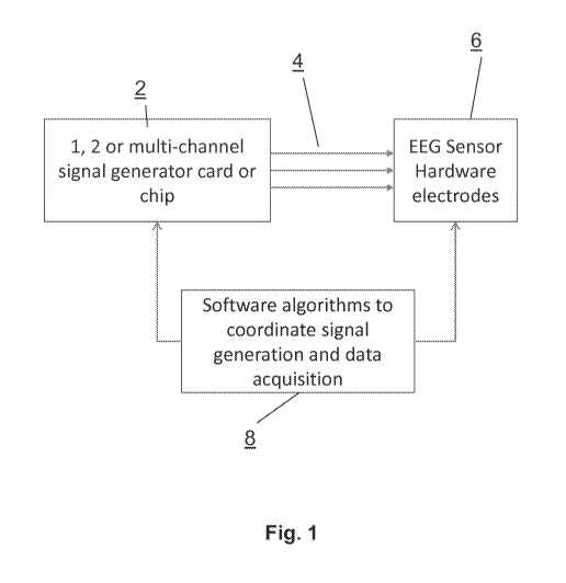
[0067] As illustrated in FIG. 1, the systems and methods of the present remote
calibration and quality control invention include (i) a signal generator card
or chip 2,
often including a sound card or other audio signal generator, to generate test
or
reference signals, (ii) a cable 4 to hardwire the sound card output (typically
from a
headphone jack with a 2.5 mm or 3.5 mm male connector) to the electrodes 6 of
the
remote and portable EEG hardware, and (iii) custom software 8 built into the
data
acquisition software program that is able initiate reference signal generation
from the
signal generator or sound card in a specified fashion to calibrate the
frequency
response and amplitude response of the EEG data acquisition system. If there
is more
than one channel of EEG data to record, then a multi-channel calibration
signal can
calibrate the phase relationship between any two channels of the data
acquisition
information streams as well. Typically, a sound card or sound chip outputs
stereo
signals with two channels of output, although monophonic sound cards or chips
with
one channel or 5.1 or 7.1 surround-sound cards or chips can equally be used
within the
system and methods of the present invention.
[0068] An example of a stereo two-channel calibration cable is shown in FIG.
2. As illustrated, the male jack pin has a first conductor (e.g. L left
channel) 10 which is
13
CA 02842027 2014-01-15
WO 2013/012739
PCT/US2012/046723
passed thru to pin 18, while a second signal conductor (e.g. R right channel)
12 is
passed thru to pin 16. The ground electrode 14 is attached to the shield 20 of
the cable
assembly. Wired into the cable assembly is a voltage divider consisting of
upper resistor
26 and lower resistor 28 for channel 1 while upper resistor 22 and lower
resistor 24
make up the voltage divider for channel 2. Wire 30 carries the 103 to 104
voltage
reduced signal for channel 1 to connector 36 which is attached to an electrode
on the
EEG recording device by an alligator clip or other mechanically and
electrically stable
means. Moreover, wire 32 carries the voltage reduced channel 2 signal to
connector 38.
The ground of the jack pin is connected via wire 34 and is wrapped around the
two
1.0
signal wires to shield the signals and is attached to the ground and/or
reference
electrode on the EEG recording device by connector 40.
[0069]
Another embodiment of a calibration system in accordance with the
present invention would be a single channel cable assembly as shown in FIG. 3.
In this
case, pin connector 50 passes thru to pin 56, while insulator 52 separates
ground
conductor 54 which is electrically continuous with cable shield 62 and
shielding wire or
signal wrap 64. A voltage divider is created between upper resistor 58 and
lower
resistor 60 to step down the reference signals by 103 to 104, although it may
only be
necessary to step down 102 or as much as 105. The voltage divided signal is
passed
along signal wire 68 to connector 70. The ground shield wire or foil 64 is
connected via
connector 66 to the ground or reference electrodes in the data acquisition
system.
[0070] An example of a frequency response output can be seen after Fourier
Transform in FIG. 4. Measured power spectral density (PSD) traces at 5 Hz
(74), 10 Hz
(75), 15 Hz (76), 20 Hz (77), 25 Hz (78) and 30 Hz (79) can be seen aligning
well with
expectation. Alternatively, an amplitude scan can be automated by the signal
generating
software and signals at a fixed or mixed frequency collected at varying
amplitude (see
FIG. 5). One sees a two-fold reduction in input signal amplitude along the x-
axis
corresponding to an expected 25% reduction in power. This power law model fit
tracks
very well demonstrating excellent amplitude response.
[0071]
Depending on the switching capabilities of the signal generating card,
one can possibly conduct a signal to noise ratio in real time by shorting
signal and
ground outputs and recording noise levels compared to physiologically
referenced
14
CA 02842027 2014-01-15
WO 2013/012739
PCT/US2012/046723
levels. Such a test could produce an output table as shown in FIG. 6 where the
SNR is
shown in both the time and frequency domains.
[0072] The system and methods of the present invention show comparable
frequency response and PSD as an expensive reference system as shown in FIG.
7.
FIG. 7 is a two trace graph comparing an expensive NuAmps 10-20 reference EEG
system (signal 86) by Compumedics to the inexpensive and portable Cerora
MindScope
system (signal 94). The data were collected simultaneously but show good
agreement
in frequency and amplitude response. However, because the two systems were
connected in parallel, there was interaction between the two systems which
lead to an
artifact at 25 Hz. Nonetheless, it was observed equivalently in both systems.
[0073] The output from a ramped 30 second passage of test reference signals
can be observed in FIG. 25, showing the reference output above relative to the
measured signal from the system below. Here both the frequency and the
amplitude are
changing in time to assess both the calibration and quality of the system
across a range
of control parameters, such as frequency and amplitude.
Systems and methods to verify and validate analytic software modules
[0074] The test signals of the present invention can be used to
verify and
validate analytic software modules written to achieve explicit purposes.
Preferred
embodiments enable the verification and validation of pre-processing artifact
detection
algorithms. In particular, if the signal generator chip has the capability to
stream digitally
synthesized artifacts or stored artifact signals, then the pre-processing
analysis
algorithms can be verified and validated for use. An example of this can be
seen in FIG.
8A where various artifacts 88, 90 and 92 were flagged and excluded from the
epochs of
artifact free EEG data. In FIG. 8B, a table of naturally occurring or
synthetically doped in
silico artifacts versus those detected by the pre-processing software was
constructed
showing an excellent efficiency at detecting known or doped artifacts of the
eye blink,
flat (no variation over many samples), or extreme (saturated signal) types.
[0075] Moreover, synthetically created signals in the signal generator card
can
be constructed with varying linear combinations of amplitudes and frequencies
to verify
CA 02842027 2014-01-15
WO 2013/012739
PCT/US2012/046723
and validate that the data acquisition system is performing as expected and is
within
calibration specification before additional human clinical data is gathered
and/or stored
for analysis. This ability provides a very important quality control and
assurance to the
human clinical data remotely collected by a patient or subject without a
trained operator
or technician present to confirm in an automated fashion, proper and
calibrated
collection of the EEG data. FIG. 9A shows white noise in both a 3-dimensional
100 and
2 dimensional time average 101 PSD plot. When four sine waves of equal
amplitude are
combined into a single artificial test waveform in FIG. 9B, they are detected
as equal
amplitude in both a 3-dimensional 102 and 2-dimensional time average 103 PSD
plot.
Finally, if the linear combination of reference signals does not have equal
amplitude, as
shown in FIG. 90, then the 3-dimensional 104 or 2-dimensional time averaged
105 PSD
can be shown to have the proper relative power in each of the intended sub-
bands, as
documented in FIG. 9D in the lower triangle of sub-band power values 106.
Moreover,
by allowing convenient remote monitoring using the present invention, the
remote
calibration and quality control and assurance activities can be automated and
can be
undertaken much less expensively than the present status.
Biomarkers and methods to diagnose brain disease (e.g. Alzheimer's disease)
[0076] The
system and methods of the present invention also relate to the
ability to non-invasively measure with a lightweight, portable and user-
friendly system,
EEG-derived biomarker features or metrics extracted from the raw time series
traces of
EEG data. These features can then be placed into a summary data table
alongside
other available data and information to enable statistical predictive models
using as
many co-variates as possible that can be constructed during the statistical
analysis
phase. Moreover, multi-variate methods such as linear discriminant analysis,
tree based
methods such as Random Forest method, and other multi-variate statistical
methods
can be conducted to create multi-variate composite biomarkers that can
demonstrate
better analytical and clinical performance to screen, classify, diagnose,
prognose,
monitor brain or disease progression, or monitor drug response. All of these
methods
16
CA 02842027 2014-01-15
WO 2013/012739
PCT/US2012/046723
fall into the general term diagnose as alternative intended uses of the
systems, markers
and methods of the present invention.
[0077] In one exemplary embodiment of the present invention, subjects would
get enrolled after either (i) IRB approval as an Investigation Device or (ii)
after FDA
510(k) clearance or (iii) after FDA Pre-market Approval (PMA). Demographic
data would
be collected on each subject included their handedness, gender, age,
education,
concomitant medications, blood pressure, diabetes and smoking history, along
with any
other imaging or biomarker data available to establish either standard of
truth or other
possible co-variates in the analysis. See FIG. 10 for an example of the
collected data.
[0078] Once ready to conduct the diagnostic procedure using the system and
methods of the present invention, a clinical assessment protocol beginning
with both
resting state Eyes Closed (EC) and resting state Eyes Open (EO) conditions
would be
initiated (see FIG. 11 for an example). This would alternate for three
successive cycles
for a total of six blocks of resting state data in one embodiment, or
alternatively consist
of one, two or four cycles of EC and EO resting states. From there, the
computer
acquisition system would begin the physiologically focused cognitive or
sensory
stimulation tasks while recording EEG signals. In one particular embodiment,
EEG
signals would be recorded while a cognitive or sensory visualization series of
tasks
were initiated. In one embodiment, the CogState Brief Battery was conducted,
including
the Detection, Identification, "One Card Back", and "One Card Learning" tasks
for a total
of 4 additional blocks of data taking roughly an additional 12 minutes. Other
non-
limiting tasks would include the ImPACT neurological assessment, the Cantab
battery
or other visualization tasks or ANAM.
[0079] Next, the software would present to the subject an auditory cognitive
or
sensory task probing the auditory cortex and requiring speech responses. One
such
embodiment could include the PASAT task starting at the slowest speed of 2.4
seconds
between trials, then begin again at the next faster speed of 2.0 seconds
between trials,
and if the subject agreed, conducted for a third and final time at the 1.6
seconds
between trial speeds. Alternatively, a verbal task such as the King-Devick
Test
developed in ophthalmology could be used to assess speech and visual acuity.
After
this, the device sound card would be hooked up to iPod like ear-buds or other
audio
17
CA 02842027 2014-01-15
WO 2013/012739
PCT/US2012/046723
transducer on the subject and would begin to output auditory stimulation to
probe the
auditory cortex with direct sounds and tones. In one preferred embodiment, a
binaural
beat frequency would be setup through differentiated left and right ear
frequencies. In
one particular embodiment, the tones would be centered in a pitch range
between 40-
400 Hz with differential delta beat frequency varying from 1 to 30 Hz. In a
more
particular embodiment, a central frequency of 400 Hz would be used with a
binaural
beat delta frequency of 6 Hz, then 12 Hz, then 18 Hz, each block recording
from 15
seconds to two minutes of EEG signals. Other center frequency and beat
frequency
combinations could be equally contemplated. Alternative auditory stimulations
could
include monoaural beats and isochronic tones. An opportunity to include photic
stimulation of the subject with eye lids closed could be conducted according
to the
methods of the present invention. The frequency of photic stimulation could be
varied
from 1 to 2 Hz on the slow side through 30 to 40 Hz on the fast side. The
appearance of
primary driving frequency signals as well as the presence of first harmonic
signals could
be monitored and used a biomarker signature to help in the diagnosis protocol.
[0080]
The existence of either the primary driving frequency or the first
harmonic or higher harmonics could be a nominal or ordinal variable output.
Moreover,
continuous output variables such as the amplitude of the driving frequency
peak, first
harmonic peak amplitude, or ratio to a resting state comparator could be used
as a
diagnostic EEG feature. Also possible, the continuous output variable relative
or
absolute power in the driving frequency or the harmonics could be used as a
diagnostic
EEG feature. Pain stimuli in the form of a thermal grill or an ice cube to the
hand could
be implemented to assess the coupling of peripheral circuits to the central
nervous
system and frontal or other cortical areas. Finally the activation/stimulation
battery of
cognitive and sensory tasks would end with a resting state EC/EO sequence for
a block
of data each of duration 2 minutes.
[0081]
EEG time series would be recorded into the various data blocks as
described above. FIG. 12 shows an example two second time series sampled at
128
Samples/sec with 10-bit ADC sample resolution. This could then go through the
pre-
processing artifact detection algorithms and those epochs that were not
flagged as
artifact would be Fast Fourier transformed into the frequency domain and
either plotted
18
CA 02842027 2014-01-15
WO 2013/012739
PCT/US2012/046723
without normalization as an absolute power spectral density or could
alternatively be
normalized to overall power of unity and represented as the relative power
spectral
density (PSD). An example relative PSD trace 130 can be seen in FIG. 13, where
the
various sub-bands have been indicated by the vertical lines on the plot. The
slowest
frequency sub-band known as delta, typically from 1-4 Hz, can be seen at 135,
followed
by the theta sub-band from 4-8 Hz at 136, followed by the alpha sub-band from
8-12 Hz
at 137, followed by the beta sub-band from 12-30 Hz shown at 138. Not shown on
the
plot in FIG. 13 is the gamma sub-band from 30-60 Hz because with a sampling
frequency of only 128 samples/sec, one can choose to not go all the way up to
the
Nyquist frequency but more rigorously require at least 4 samples per unit
cell. If one
uses a 256 samples/sec or 512 samples/sec ADC, then meaningful gamma sub-band
information can be ascertained.
[0082]
It should be noted that the algorithms and "processing means"
described herein are preferably implemented in software that runs on a
processor of the
processing unit (which is presumably part of the portable EEG sensing device).
[0083] Once the spectral analysis code has transformed each epoch of artifact
free time series EEG data, a feature extraction algorithm can assess each
block of
transformed data to create a list of features or variables or biomarkers
extracted from
each block of EEG data conducted during an individual task. This list of
variables or
metrics can include not only the relative and absolute delta, theta, alpha,
beta and
gamma sub-bands, but can include literature derived markers such as the
theta/beta
ratio, the delta/alpha ratio, the (theta+delta) / (alpha+beta) ratio, the
relative power in a
sliding two Hz window starting at 4 Hz and going to 60 Hz, the 1-2.5 Hz power,
the 2.5-4
Hz power, the peak or mode frequency in the PSD distribution, the median
frequency in
the PSD, the mean or average (1st moment) frequency of the PSD, the standard
deviation of the mean frequency (square root of the variance or 2nd moment of
the
distribution), the skewness or 3rd moment of the PSD, the kurtosis or 4th
moment of the
PSD. In addition to these spectrally derived metrics or features for each
block of EEG
data, non-spectral signal analysis could be conducted.
[0084] In an
exemplary embodiment of the present invention, a non-linear
dynamics module would calculate the largest Lyaponov exponent of the block of
EEG
19
CA 02842027 2014-01-15
WO 2013/012739
PCT/US2012/046723
data, the fractal dimension D of the EEG signal and the entropy S of the EEG
signal, as
non-limiting non-linear dynamical systems extracted EEG features. In an
alternate
embodiment, a wavelet transform signal analysis module could be applied to an
all
artifact free EEG epoch on a block by block basis. This analysis could include
both the
discrete wavelet transform (DWT) as well as continuous wavelet transform
(CWT).
More particularly, these advanced signal analysis routines would be applied to
blocks of
EEG data acquired during either cognitive or sensory stimulation to enhance
diagnostic
discriminatory power.
[0085] As a non-limiting example, a two group plot of the relative theta power
during a resting Eyes Open task between N=10 Alzheimer's subjects and N=13
Control
subjects averaged over three blocks of two minutes each can be seen in FIG.
14A. In
FIG. 14B, one can visualize the decreased relative beta sub-band in AD
relative to CTL,
again in resting EO. In both plots, the false positive rate t-Test p-value is
shown to be
statistically meaningful when not correcting for multiple comparisons.
Moreover, as can
be seen in FIG. 15A, the mean frequency is meaningfully reduced from
approximately
11 Hz in CTL subjects to around 8 Hz in AD subjects, again with a
statistically
meaningful t-Test p-value.
[0086]
Alternatively, additional features or metrics beyond those described in
the literature can be extracted from the artifact free EEG blocks of data. In
FIG. 15B, the
relative 18-20 Hz window of power averaged over three blocks of resting EO can
be
seen to have excellent diagnostic discriminatory power. In fact, if one
conducts a
nominal logistic regression to perform Receiver Operator Characteristic (ROC)
curve
analysis, an optimal cut-point to dichotomize the groups is observed at
relative 18-20 Hz
power of 2.7. When pivoting on this cut-point as shown in FIG. 16, the
preliminary
clinical performance of the relative 18-20 Hz power as a biomarker or
classifier can be
seen to correctly identify 11 of 13 CTL subjects and 9 of 10 AD subjects,
based on a
clinical diagnosis as standard of truth. This marker thus has a derived 85 %
sensitivity
with 90 % specificity, while the Positive Predictive Value (PPV) is 92 % and
the
Negative Predictive Value is 82 %.
[0087]
Alternatively, one can conduct either discrete or continuous wavelet
transformation analysis of the EEG blocks of data. In a discrete wavelet
analysis, as
CA 02842027 2014-01-15
WO 2013/012739
PCT/US2012/046723
described in Example 14 below, one can see the statistically meaningful
results with
false positive rate p<0.05 shown in Table 1. More results can be found within
FIG. 21
through FIG. 24.
[0088]
These results indicate that any one of the following extracted EEG
feature by task combinations can be used alone or in combination as an input
or factor
to a statistical predictive model for Alzheimer's disease:
1. mean power D2 during an Eyes Open (EO) task,
2. mean power D3 during an EO task,
3. mean power D4 during an EO task,
1.0 4. mean power D5 during an EO task,
5. minimum D2 during an EO task,
6. minimum D3 during an EO task,
7. minimum D5 during an EO task,
8. maximum D2 during an EO task,
9. maximum D4 during an EO task,
10. maximum D5 during an EO task,
11 .Standard Deviation (STD) D2 during an EO task,
12.STD D3 during an EO task,
13.STD D4 during an EO task,
14.STD D5 during an EO task,
15. Kurtosis D5 during an EO task,
16. mean power D3 during an Eyes Closed (EC) task,
17. mean power D4 during an EC task,
18. minimum D3 during an EC task,
19. minimum D4 during an EC task,
20. maximum D4 during an EC task,
21 .STD D3 during an EC task,
22.STD D4 during an EC task,
23.Skewness D5 during an EC task,
24.Ajlasdfj,
25. minimum D2 during an One Card Learning task,
21
CA 02842027 2014-01-15
WO 2013/012739
PCT/US2012/046723
26. minimum D3 during an One Card Back task,
27.skewness D3 during a One Card Back task,
28.skewness D2 during a D = 6 Hz binaural beat auditory stimulation task,
29. minimum value of {D4} during an auditory binaural beat stimulation at A =
12 Hz (AS2),
30. maximum value of {D3} during an auditory binaural beat stimulation at A =
6 Hz (AS1),
31.skewness of the {D3} during a CogState One Card Back task (CG4),
32.skewness {D5} during an EC task ,
1.0 33. mean power value of {D2} during a PASAT 2.0 (s) interval task,
34. mean power value of the {D2} during a EO task, and
35. absolute mean power of wavelet scales in the scale range 13-26 during an
EO task.
= ,,,, cc, = = = =
...L .. = U2
.
CA= C3, - -
"Vr = - .. ... = - = - =
= = .. ... = = = =
=
tTQ
C=5
`22 =!ZZ1 - - =!2Z
!kW,.
- -
&Vint
Fur,.
-
= . ..... === .
22
CA 02842027 2014-01-15
WO 2013/012739
PCT/US2012/046723
Table 1. Statistically significant task by extracted EEG features from the
Discrete
Wavelet Transformation analysis and Kruskal-Wallis test.
[0089]
In a continuous wavelet analysis, as described in Example 15 below,
one can see the statistically meaningful results with false positive rate
p<0.05 shown in
Table 2. More results can be found within FIG. 21 through FIG. 24.
[0090]
These results indicate that any one of the following extracted EEG
feature by task combinations can be used alone or in combination as an input
or factor
to a statistical predictive model for Alzheimer's disease:
1.0 1. relative power du during an Eyes Open (EO) task,
2. relative power ql during an Eyes Open (EO) task,
3. relative power ql during an Eyes Closed (EC) task,
4. relative power au during an Eyes Open (EO) task,
5. relative power au during an Eyes Closed (EC) task,
6. relative power bl during an Eyes Open (EO) task,
7. relative power bl during an Eyes Closed (EC) task,
8. relative power bu during an Eyes Open (EO) task
9. absolute power 8u during an Eyes Open (EO) task,
10. absolute power Olduring an Eyes Open (EO) task,
11. absolute power Olduring an Eyes Closed (EC) task,
12. absolute power 0, during an Eyes Open (EO) task,
13. absolute power 0, during an Eyes Closed (EC) task,
14. absolute power au during an Eyes Open (EO) task,
15. absolute power au during an Eyes Closed (EC) task,
16. absolute power piduring an Eyes Open (EO) task,
17. absolute power piduring an Eyes Closed (EC) task, and
18. absolute power 13, during an Eyes Open (EO) task.
23
CA 02842027 2014-01-15
WO 2013/012739
PCT/US2012/046723
---------------------------------------------------------------------- ,
Po
RA 0 0 1 0 0 ij f) i3
E.02 0 0 1 0 09 . t . ,
1. p =1_
046 , , i=-
+
0.046
EC3 - - 0 0 ¨ 1 0 0 0 . 0
0
. ,
0.000010---' 1 0 j 0 1. p = 0.007 i
1.. p = ) ON 1 , p= O. Ot.:4
EC5 0 1 , ,c., ¨ 0.024 1 0 01 ,
p ¨ 0025 1 1 , p ¨ 0, 02.,, ' 0
ECM 0 1 , ,c., ¨ 0.026 1 0 0 0 1 1
0,0M
Attscif c.;1(, Pce,w,-.3
: _____________________________________________________________________
Ff 1 0 0 1 0 0ij c.: 0
E02 0 0 j i . f?-.= 0.0i9 ' 0 c.: 0
0
. .
EC. 0 0 0 1 i . l'.?-.= 0.019 : 0
0 0
E04 1 , p= 0.0005 1 , )L= = 1i .3 x 10¨ j 1 , p =
0.007 i') 1 , .,,=.4 = 0 026 1 1 , p= 00C( ' I , p = 0 .005
EC.5 0 1 ...p = 0.047 1 1 , p = 0,029 i) i , ;.4
= 0 015 j 1 , p= 00i( 0
1106 1 , .,,=.4 = 0.040 1 ...p = 0.016 1 0 i)
0 j 1 , p¨ 00-(fì 1
Table 2. Statistically significant task by extracted EEG features from the
Continuous Wavelet Transformation analysis and Kruskal-Wallis test.
EXAMPLES
[0091] While the above description contains many specifics, these
specifics
should not be construed as limitations on the scope of the invention, but
merely as
exemplifications of the disclosed embodiments. Those skilled in the art will
envision
1.0 many other possible variations that are within the scope of the
invention. The following
examples will be helpful to enable one skilled in the art to make, use, and
practice the
present invention.
Example 1. Creation of a remote calibration cable assembly for remote Quality
Control
purposes
[0092] Using a soldering iron, resistors, stereo jack pin, wire and alligator
clips,
a calibration and quality control cable was constructed. The voltage divider
consisted of
an upper 1/4 watt resistor of 100 ohms (52) and a lower 1/4 watt resistor of
1,000,000
ohms or 1 M52 to divide the reference signals down by a factor of 104 from 1
volt to 100
pm and 50 mV to 5 V. These stepped down signals are thus within the typical
physiological range of a 1 V to 100 A/ and thus useful for assessment and
calibration
of EEG systems. If desired, metal film resistors with tighter tolerances could
be used.
24
CA 02842027 2014-01-15
WO 2013/012739
PCT/US2012/046723
Example 2. Download human EEG data and create a dummy brain setup
[0093] Publically available EEG data was downloaded from the UCSD website
(http://sccn .ucsd .ed u/¨arno/fam2data/pu bl icly_ava ilable_E EG_data .html)
and stored
locally on computers. The various .tar.gz data files were unzipped using
BitZipper
software and then the .tar files were unpacked into individual files using
Astrotite
software. Various individual proprietary format, Neuroscan .cnt files (in
particular
cba1ff01+cba1ff02, cba2ff01+cba2ff02, ega1ff01+ega1ff02, ega2ff01+ega2ff02)
were
converted into ASCII comma-separated values (CSV) files using the biosig
package for
Matlab (http://biosig.sourceforge.net/), which were then viewed and loaded
into Excel.
Sequentially matched EEG data files (based on the UCSD documentation) were
concatenated to create samples streams in excess of 65K samples.
[0094] An Agilent AT-33220A Function Generator/Arbitrary
Waveform
Generator ("Arb") and an Agilent AT-34410A 6.5 digit Digital Multi-Meter (DMM)
were
rented for use. Each instrument was successfully configured to work with PCs
using the
Agilent I/0 Suite 15.5 libraries and Agilent Connect software with a USB cable
(Arb) or
Ethernet cable (DMM). EEG data in ASCII format were copied into, and
completely
filled, one of the 65,536 sample non-volatile buffers available within the Arb
hardware
using Agilent's "Waveform Editor" software. In total, each of the four
concatenated
downloaded EEG files (cba1, cba2, ega1, ega2) was stored in the four separate
memory buffers on the Arb. These data provided output EEG signal streams of
just over
65 seconds, and as a result, the Arb was able to hold 65,536 samples. The UCSD
data
was recorded at 1,000 Samples/sec according to the documentation. Upon setting
the
Arb to a frequency of 15.259 mHz (based on 1000 Samples/sec divided by 65,536
samples in the non-volatile buffer = 15.258789 sec-1). Waveform amplitude
varied,
often set between -1.0 V and + 1.0 V to yield a voltage resolution of 0.123
millivolts with
the 14 bit dynamic range of the Arb. For visual confirmation, output from each
of the
four non-volatile Arb buffers was observed on a Tektronix digital
oscilloscope. The
traces appeared to replicate the original downloaded signal shapes as observed
in the
Waveform Editor software before transfer to the Arb.
CA 02842027 2014-01-15
WO 2013/012739
PCT/US2012/046723
Example 3. Characterization of the frequency and amplitude response
[0095] A one channel calibration and quality control cable was built according
to Example 1 as shown in FIG. 3. An Agilent AT-33220A Function
Generator/Arbitrary
Waveform Generator ("Arb") and an Agilent AT-34410A 6.5 digit DMM were used.
Each
instrument was successfully configured to work with laboratory PCs using the
Agilent
I/0 Suite 15.5 libraries and Agilent Connect software with a USB cable (Arb)
or Ethernet
cable (DMM). Downloaded UCSD EEG data in ASCII format were copied to, and
completely filled, one of the 65,536 sample non-volatile buffers available
within the Arb
hardware using Agilent's "Waveform Editor" software. In total, each of the
four
concatenated downloaded EEG files (cba1, cba2, ega1, ega2) was stored in the
four
separate memory buffers on the Arb. These data provided an output EEG signal
streams of just over 65 seconds, and as a result of the Arb can hold 65,536
samples.
The UCSD data was recorded at 1,000 Samples/sec. Upon setting the Arb to a
frequency of 15.259 mHz (based on 1000 Samples/sec divided by 65,536 samples
in
the non-volatile buffer = 15.258789 sec-1), the Arb was able to successfully
output
publically available EEG data as a dummy brain setup. Output amplitude was set
to
vary between -1.0 V and + 1.0 V to yield a voltage resolution of 0.123
millivolts with the
14 bit dynamic range of the Arb. For visual confirmation, output from each of
the four
non-volatile Arb buffers was observed on a digital oscilloscope. The traces
appeared to
replicate the original downloaded signal shapes as observed in the Waveform
Editor
software before transfer to the Arb.
[0096]
Additionally, sine wave output from the NIST traceable Arb was
hardwired into the EEG headset beginning at 5 Hz and ending at 30 Hz in 5 Hz
intervals
with modest input amplitude of approximately 25 V. Each block of independent
data
was analyzed by pre-processing artifact detection algorithms and then spectral
sub-
band analysis. The output PSD for each of the six traces can be seen in FIG.
4. As
expected, the pure sine waves exhibit excellent spectral peak widths.
Furthermore, the
frequency of the reference sine wave was fixed at 15 Hz and the input sine
wave
amplitude to the voltage divider was reduced from 800 mVpp to 12.5 mVpp in a 2
fold
serial reduction (e.g. 800, 400, 200, 100, 50, 25, 12.5). After the 104voltage
divider, the
26
CA 02842027 2014-01-15
WO 2013/012739
PCT/US2012/046723
input voltage amplitudes to the EEG sensor were 80, 40, 20, 10, 5, 2.5, and
1.25 Vpp,
covering well the physiological range. The results of the study can be seen in
FIG. 5
where a two-fold reduction in amplitude leads to a 4 fold reduction in power
as
expected. The linearity of the response looks excellent.
Example 4. Assessment of the open circuit Signal to Noise Ratio (SNR)
[0097] While experiments were conducted under closed circuit
conditions as
well as under both open circuit and short circuit conditions to assess the
signal to noise
ratio of the MindScope hardware and recording system. There are primarily two
types of
noise: short circuit noise when the differential input to the differential
operational
amplifier are shorted together and open circuit noise due to intermittent
pickup of
spurious signals when there is no signal presented to the sensor. Relevant
literature
suggested open circuit noise levels are larger than short circuit noise levels
so we
began our investigation with open circuit noise assessments in the headsets
compared
to hardwired signals from the Arb. The literature also suggested that signals
more than
three standard deviations are more than 99% probably meaningfully different
than noise
(assuming Gaussian noise). Thus, SNR greater than 10*log (32/12) = 9.5 db
represent
real signals with p<0.01. A proposed threshold criterion of 20 db is thus
highly
conservative. Multiple experiments were conducted to determine the SNR from
the data
comparing open circuit to close circuit conditions, confirming the reports in
the literature.
[0098] The SNR data were analyzed both in the voltage-time domain as well as
spectral domain. In each case, the log transformed ratio of signal to noise
was
calculated to determine the SNR in decibels (db). In addition to time-voltage
domain
SNR measurements,
[0099] The average spectral power was measured around 0.1 V2 equivalents
averaging across two measurements. This experiment was conducted with multiple
trials within each of N=2 separate days. SNR in decibels (db) is defined as
ten times the
log base 10 ratio of the signal squared divided by the noise squared, where
the values
are root-mean squared (rms), centered at 15 Hz with a bandwidth from zero to
30 Hz (P.
Horowitz and W. Hill, The Art of Electronics, 2nd Edition, Cambridge
University Press: 1989, p
27
CA 02842027 2014-01-15
WO 2013/012739
PCT/US2012/046723
434.) The data summarized both in the time domain before transformation (RMS)
and
after transformation (Spectral) are shown in FIG. 6. Thus, we find that for
pure 15 Hz
sine wave signals, we calculate a SNR = 10 log (210/0.1) = 33 db in the
spectrally
transformed space. In the un-transformed time domain, input signals above 20
mV
before the voltage divider or 2 V input to the EEG sensor will have the
necessary 20
db SNR.
Example 5. Show Equivalence to a reference system
[0100] Four
data files were recorded from signals produced by the Agilent
33220A 20 MHZ Function/Arb Waveform Generator (Arb) simultaneously on the EEG
system of the present invention and a Compumedics system (Neuroscan NuAmps
amplifier and gel-based electrodes). The Arb was limited to four non-volatile
memory
buffers for storing UCSD downloaded EEG human data so analysis was limited to
these
four UCSD EEG data traces (CDA1_1, CDA2_2, EGA1_3, EGA2_4). Compumedics
Neuroscan SCAN 4.5 analysis software was successfully installed on laboratory
computers. The 1000 Samples/sec NuAmps data files were imported into SCAN 4.5
software in the Compumedics .CNT file format. They were then transformed into
"Epoch" files of approximately 1, 2, or 4 seconds in duration and contained
1024, 2048,
or 4096 samples. Once broken into epochs, the data were Fast Fourier
Transformed
yielding either amplitude ( V) or power ( V2) measures and plotted from 0 to
30 Hz.
Sensitivity of the PSD was assessed by examining all three epoch lengths
(1024, 2048,
and 4096 sample lengths) indicating no major deviations in sensitivity were
observed.
Individual power spectra were exported as ASCII data files for direct
comparison
between power spectra of Compumedics and the MindScope system. A comparison of
the overall relative power spectra (peak normalized) revealed agreement (FIG.
7).
However, spectral deviation at the low end delta sub-band was observed.
[0101]
Additionally, both systems identify an artifactual spectral peak around
25 Hz as a function of output from the Arb. This artifact was seen throughout
the
experiments conducted with the Arb. As such, the response of the two systems
was
very comparable with the exception of the frequency response below 3 Hz.
28
CA 02842027 2014-01-15
WO 2013/012739
PCT/US2012/046723
[0102]
One issue was revealed during the data analysis. There was an
apparent interaction between the NuAmps and MindScope systems, due to the
periodic
injection of electrical current by the EEG headset to test the signal quality
of the
electrode contact to the human subject. As illustrated in FIG. 7, this
periodic injection
dramatically affected the PSD observed, but did so equivalently in both the
NuAmps
and MindScope data.
Example 6.
Verification and validation of the pre-processing artifact detection
algorithms.
[0103]
Pre-processing artifact detection provides a standardized series of
detection routines, but additionally permits the user to select from these
routines.
Artifact detection and removal is critical to EEG signal processing to
maximize the
accuracy and precision of spectral estimates as well as other measurements
used to
determine cognitive or sensory state-dependent changes. The developed artifact
detection routines assess the EEG for invalid data in the following manner:
1) Determination of samples acquired during periods of time when the EEG
signal was poor. Poor Signal occurs during intervals that the headset is not
placed upon or properly seated on the head. This is disclosed by a value
reported by the EEG wireless headset as a value ranging from 0 to 200. We
have determined that signals acquired with a Poor Signal value greater than
26 are not precise enough to use for effective analysis. (defined in the
BCI_ParameterFile as params.Artifact.minSignalStrength = 26).
2) Determination of Flat Segments (samples acquired during periods of no
frequency information - DC only). Flat Segments occur when BluetoothTM
communication to the EEG wireless headset is lost or other conditions such
as electromagnetic interference render the signal unusable, including
saturation of the amplification circuitry to the limits of the input power
supply
voltage. We have determined that Flat Segments longer than 100
29
CA 02842027 2014-01-15
WO 2013/012739
PCT/US2012/046723
milliseconds produce significant deviations in spectral estimation. (defined
in
BCI_ParameterFile as params.Artifact.minFlatSigLength = 0.1).
3) Determination of Excessive Signals (samples that exceed three standard
deviations of the signal mean). Excessive Signal segments occur during eye
blinks, interference of cardiac electrical activity (heart beat), or non-
physiological electrical noise including movement of the EEG dry electrode or
electromagnetic interference. (defined in BCI_ParameterFile as
params.Artifact.maxSignalSTDmultiplier = 3).
1.0
4) Determination of Excessive Av/At Segments (series of samples that exceed a
predetermined instantaneous frequency). These Excessive Av/At Segments
occur as a result of non-physiological electrical noise including movement of
the EEG dry electrode or electromagnetic interference. We have determined
that a change of 1.5 standard deviations from the signal mean over 3 samples
is sufficient to detect these non-physiological signals. (defined in
BCI_ParameterFile as params.Artifact. dvValMultiplier= 0.5 and
params.Artifact.MaxDT = 3).
[0104] The
performance of the artifact detection software module was
measured to provide quality control and assurance benchmarks. Five separate
signals
from UCSD data files CBA1ff01 were extracted, down-sampled to 128 Hz, band
passed
filtered (0.5 ¨ 50 Hz), and formatted for use. These signals were analyzed
visually for
known artifacts and eye blinks were counted manually while scanning the data
file. No
other major artifact was observed. To test each aspect of the artifact
detection
algorithm, each signal was incrementally seeded with 100 artifacts. Synthetic
artifact
segments were generated at sub¨threshold and super-threshold values that
contained:
1) flat signal (i.e. representing dropped signal or amplifier/ADC saturation)
or 2) extreme
values (i.e. representing electrical noise or other non-physiological signal).
Under
generic and non-optimized settings (values reported above for each detector
parameter), our artifact detection algorithm initially detected 342 of 344
artifacts that
CA 02842027 2014-01-15
WO 2013/012739
PCT/US2012/046723
existed as part of the original UCSD data sets (t-test
.(total vs detected) p = 0.870). No sub-
threshold synthetic artifacts of any type were detected by our artifact
detection software
module (t-test
.(total vs detected) p < 0.0001) demonstrating the lack of false positive
detection
events. However, threshold synthetic artifacts were detected with nearly 100%
accuracy
(t-teat(totai vs detected) P = 0.495). Overall, 1382 events were detected of
1344 pre-existing
and synthetic artifacts with a false detection rate of 2.8 percent. An example
can be
seen in FIG. 8A and the summary of this analysis is provided in FIG. 8B.
Example 7. Verification and validation of the spectral sub-band analysis
software
[0105] The spectral analysis module was designed to accept cleaned data from
the artifact detection software module, window the data with a Bartlet
windowing
function, and then spectrally transform the data using the MATLAB FFT()
function. In
addition to these standardized analysis routines, the spectral analysis module
permitted
the user to select other windowing functions (i.e. Hann, Hamming, etc.) as
well as other
spectral estimation techniques, including multi-taper spectral estimation
using Slepian
sequences, to minimize spectral leakage.
[0106]
Furthermore, the spectral analysis module automatically generated
Power Spectral Density (PSD) plots from recorded EEG data as well as summary
Comma-Separated Value (CSV) files of the spectral analysis results. The PSD
plots
were additionally sent to the Microsoft PowerPoint program for further report
generation
automatically by the spectral analysis module. Summary CSV files provide a
general
data format for the spectral analysis results that can be further analyzed in
JMP
(statistics package from SAS) or used for more complex scientific graphing in
KaleidaGraph (purchased from Synergy Software).
[0107]
An additional software analysis module was created to generate FFT
spectral sub-band metrics as a part of our signal analysis suite. This module
has the
ability to generate sub-band metrics from the spectral analysis module output
that
include:
i) Spectral power within each 8, 0, a, and 13 EEG frequency sub-
bands;
31
CA 02842027 2014-01-15
WO 2013/012739
PCT/US2012/046723
ii) Arithmetic and Geometric means of each sub-band for the eyes-closed
and eyes-open conditions; and
iii) Ratios of Arithmetic and Geometric means of each sub-band for the eyes-
closed and eyes-open conditions.
The spectral sub-band metric module automatically generated plots of the
Arithmetic
and Geometric means in addition to ratios of those means. Results from this
analysis
were plotted and sent to Microsoft PowerPoint for further report generation as
well as
written to CSV files for further analysis.
1.0 [0108] Testing and validation of the spectral analysis module was
completed
as follows. Timestamp data was extracted from the UCSD data files CBA1ff01,
down-
sampled to 128 Hz, and formatted for use. This timestamp array was used to
generate
seven synthetic analog signals. These seven in silico signals are illustrated
in FIG. 9
and included:
1) White Noise with a Gaussian distribution (mean = 0 mV, StDev = 0.1 mV);
2) Cosine wave at 2.0 Hz (Delta Band; mean = 0 mV, StDev = 1 mV) + White
Noise (from 1);
3) Cosine wave at 5.5 Hz (Theta Band; mean = 0 mV, StDev = 1 mV) + White
Noise (from 1) + Delta;
4) Cosine wave at 10 Hz (Alpha Band; mean = 0 mV, StDev = 1 mV) + White
Noise (from 1) + Delta + Theta;
5) Cosine wave at 21 Hz (Beta Band; mean = 0 mV, StDev = 1 mV) + White
Noise (from 1) + Delta + Theta + Alpha;
6) Cosine wave at 40 Hz (Gamma Band; mean = 0 mV, StDev = 1 mV) + White
Noise (from 1) + Delta + Theta + Alpha + Beta; and
7) Fractional summation of White Noise and all cosine waves that included 0.1*
White Noise + 0.25*Delta + 0.33*Theta + 0.5*Alpha + 0.1*Beta.
[0109] The spectral analysis module was tested by running the spectral
analysis code against each of these traces. Spectrograms, illustrating the
evolution of
32
CA 02842027 2014-01-15
WO 2013/012739
PCT/US2012/046723
the power spectrum over time, and power spectra of the entire files were
generated
(FIG. 9A, 9B, 90). The spectral analysis module successfully identified the
spectral
power of each frequency contained in each data trace. Spectral leakage was
nominal
(0.25 Hz), such that shoulder frequency bins (e.g. 9.75, and 10.25 Hz bins
surrounding
the 10 Hz bin) contained a very small portion of the spectral power generated
by the 10
Hz cosine waveform. Calculations were identical between spectrogram time
frames as
well as across runs. Attenuated input waveforms (seventh synthetic trace) were
appropriately calculated as fractional relative power measurements across
frequencies
and sub-band quantifications (see FIG. 9D).
Example 8. Alzheimer's disease pilot study recruitment and clinical protocol
[0110] A clinical protocol was written and approved by an
independent
Institutional Review Board. Subjects were enrolled based on Mini-Mental State
Exam
performance into either an Alzheimer's disease group (with MMSE less than 28
but
greater than 20) or healthy normals enrolled to a Control (CTL) group. A total
of N=14
CTLs and N=10 AD subjects were enrolled with the demographics of 13 CTLs shown
in
Fig. 10. History of seizure or epilepsy was among the exclusion criteria the
lead
neurologist established.
Example 9. Collection of time series EEG data
[0111] The study coordinator established Informed Consent with each subject
according to the IRB approved clinical protocol. Moreover, she collected
anywhere from
10 to 18 blocks of EEG data according to the task protocol shown in FIG. 11.
An
example time series EEG trace is shown in FIG. 12 covering a two-second
period.
Traces were sampled at 128 sam/sec with a 10-bit ADC in a NeuroSky MindSet Pro
headset coupled via Bluetooth to a Dell Inspiron 1545 laptop PC using
NeuroView
software.
33
CA 02842027 2014-01-15
WO 2013/012739
PCT/US2012/046723
Example 10. Pre-processing artifact detection followed by Fourier spectral
signal
analysis to produce a summary feature table for each subject for each block
task of data
[0112]
Each block of EEG data was pre-processed according to the system
and methods of the present invention and then spectrally transformed and time
averaged with a sliding 8 sec (1024 sample) window to produce a time averaged
PSD
like the one shown in FIG. 13. All signal analysis was conducted blinded to
subject
clinical disease diagnosis so as to remove any chance for bias. The feature
extracted
data table had roughly 120 variables.
Example 11. Univariate statistical analysis and predictive model building for
classifiers
of disease state (AD vs CTL) in the pilot study data
[0113] The blinded table of extracted features or markers was passed from the
signal analyst to the statistician for uni-variate statistical analysis. Using
JMP 8.0
software, each of the roughly 120 variables for each task for each subject was
analyzed
for statistical significance across the diagnostic group AD vs CTL. As shown
in the
literature, the AD brain exhibits a spectral slowing relative to CTL subjects.
As shown in
FIG. 14A, the lower frequency theta sub-band in AD subjects exhibits an
elevation
relative to CTL subjects. Complementary to this observation, one observes the
relative
beta sub-band suppressed with less power in AD subjects compared to CTL (FIG.
14B)
as the power has shifted to the slower theta sub-band. Also consistent with
the
literature, the overall mean frequency of the relative PSD has shifted from an
average of
just above 11 Hz in CTL subjects to around 8 Hz on average in the AD subjects.
T-Test
False Positive Rate p-values are calculated and shown as insets for each
graph.
[0114]
Interesting, new signatures or classifiers have begun to emerge from
the uni-variate analysis to include the relative power in the 18-20 Hz band.
This marker
provides preliminary excellent diagnostic performance, where a nominal
logistical
regression in JMP 8.0 determined that a cut point around 0.27 would optimally
dichotomize the diagnostic group designation. ROC curve analysis was conducted
to
34
CA 02842027 2014-01-15
WO 2013/012739
PCT/US2012/046723
derive sensitivity and specificity of 85%/90%, with PPV of 92% and NPV of 82%
respectively. This can be seen in the 2x2 diagnostic table of FIG. 16.
Example 12. Prophetic: conduct multi-variate predictive model building to find
better
composite classifiers.
[0115] Moreover, using established multi-variate predictive
statistical methods,
one can conduct multi-variate statistical analysis to build predictive
statistical models
that include from 2 to 10 variables from among the various tasks and features
extracted
in a given clinical protocol. It is well known that linear discriminant
analysis, random
forest, shrunken-centroids and other multi-variate approaches to construct
composite
signatures that classify subjects could be used on the summary feature data
table in
addition the uni-variate signatures and analysis conducted.
Example 13. Prophetic: conduct sports concussion or mTBI protocol consisting
of
cognitive assessment, vestibular/balance, auditory, and visual stimulation
[0116] Alternatively, one could tailor a brain assessment battery towards
sports
concussion diagnosis and monitoring by combining simultaneous EEG recording
with
various tasks focused on sports concussion and mild traumatic brain injury. An
prophetic example of such a battery can be seen in FIG. 17 where a subject
would
undergo resting state EC and EO conditions, cognitive elements of the SCAT2,
vestibular and balance tasks from the SCAT2, the PASAT task, the King-Devick
test,
the ImPACT testing, binaural beats or auditory stimulation to assess tinnitus,
photic
stimulation to assess photo hypersensitivity and finally resting state EC and
EO. It
should be clear that not all tasks need be included and could simply just be a
single task
or a minimal combination of the statistically important ones. Moreover,
elements from
the Standard Concussion Assessment Test version 2 or SCAT2 could be used as
tasks,
including the Graded Symptom Checklist (GSC), Standard Assessment of
Concussion
(SAC), Balance Error Scoring System (BESS) or other symptom, cognitive, or
vestibular
CA 02842027 2014-01-15
WO 2013/012739
PCT/US2012/046723
tasks or challenges. Ideally, one would use passive tasks that do not require
the
cooperation of the subject.
Example 14. Feature extraction with Discrete Wavelet Transformation (DWT) and
statistical testing on the Alzheimer's disease pilot study
Discrete Wavelet Transform EEG Feature Extraction
[0117] FIG. 18 is an example of a raw EEG signal of a subject
(Subject 11)
before (top) and after (bottom) artifact detection. Discrete Wavelet
Transformation
1.0 analyzes such a signal at different resolutions through its
decomposition into several
successive frequency bands by utilizing a scaling function q(t) and a wavelet
function
w(t), associated with low-pass and high-pass filters, respectively. A useful
property of
these functions is that they can be obtained as a weighted sum of the scaled
(dilated)
and shifted versions of the scaling function itself:
05(t) =
1P(t) = n10(2t (2)
The coefficients (weights) h[n] and g[n] that satisfy (1) and (2) constitute
the impulse
responses of the low-pass and high-pass filters and define the type of the
wavelet. The
original EEG signal x(t) forms the discrete time signal x[n], which is first
passed through
a half-band high-pass filter (g[n]) and a low-pass filter (h[n]). Filtering
followed by sub-
sampling constitutes one level of decomposition and can be expressed as
follows:
kl ............... F *Loth - , (3)
= = E..zeini.h[2k (4)
where di and ai are level 1 detail and approximation coefficients,
respectively, yhigh[k]
and ylow[k] are the outputs of the high-pass and low-pass filters after the
sub-sampling.
36
CA 02842027 2014-01-15
WO 2013/012739
PCT/US2012/046723
DWT Table 3: DWT sub-band frequencies and the corresponding EEG frequency
bands. Sub band Frequency Range Corresponding EEG (Hz) frequency band (Hz).
nNIZMIey Ra#3.W COrr(sSpOilding .EEC
(HZ') frNo.eney hand (IiK)
''''''
7,;; 0 (4 S1
C.1.875 3 I
?;=(0 2)
[0118] This procedure, called sub-band coding, is repeated for further
decomposition as many times as desired or until no more sub-sampling is
possible. At
each level, it results in half the time resolution (due to sub-sampling) and
double the
frequency resolution (due to filtering), allowing the signal to be analyzed at
different
frequency ranges with different resolutions. FIG. 19 is a diagram showing the
discrete
wavelet transformation decomposition scheme with 5 levels of decomposition,
where D1
¨ D5 and A5 represent the signal. In this analysis, we went through five
levels of
decompositions resulting in D1 (approximately related to y frequency band)
through D5
(approximately related to upper 8 frequency band) and A1 through A5
(approximately
related to lower 8 frequency band). FIG. 20 shows these five levels of
decomposition for
the EEG signal. D1 through D5 sub-bands along with the A5 sub-band consist the
DWT
representation of the EEG signal. DWT Table 3 shows these sub-bands with their
frequency ranges and their corresponding EEG major frequency bands. However,
not
all these sub-bands are useful and reliable. Since the effective sampling rate
of our
EEG recording device is G = 125 Hz, we considered frequencies above 30 Hz
(approximated half of Nyquist) unreliable. Hence, D1 sub-band (y frequency
band) was
not used in the subsequent analysis. Moreover, the analog filters employed in
the
headset had a cutoff of approximately 1 to 2 Hz. Those filters have
undisclosed
properties making the signal in 0-2 Hz unreliable. Hence, A5 sub-band (lower 8
frequency band) features were also removed from the analysis. As a result, the
effective
sub-bands used in this study were D2 ¨ D5.
37
CA 02842027 2014-01-15
WO 2013/012739
PCT/US2012/046723
[0119] Having the
sub-bands of EEG signals, we can extract the common
statistical features from DWT analysis. In this study, we selected the
minimum,
maximum, standard deviation (STD), skewness and kurtosis values as well as
average
power of the wavelet coefficients as the candidate statistical features. These
values
were computed at each level of DWT decomposition separately for each recording
state
of the subjects. Note that, we did not consider the mean values since we had
subtracted
the mean before processing the data. DWT Table 4 lists all extracted features
for the
sub-bands and those selected for the analysis.
[0120] Choosing a suitable mother wavelet function is the most important
factor
in a reliable wavelet transform analysis. Daubechies family of mother
wavelets,
especially Daubechies2 (db2), has been reported to have a better accuracy
compared
to most other mother wavelet functions. Hence, we started our analysis with
db2
wavelet and then moved on to four other wavelets from the Daubechies family
(db4 ¨
db10) to extract EEG features. The number of statistically significant EEG
features of
AD patients compared to Control subjects, identified by the five different
wavelets, are
shown in DWT Table 5. It can be seen that db6 and db8 yield more features and
are
thus are the best choices for mother wavelet functions.
DWT Table 4: Statistical features of all the sub-bands and the statistical
features under
study.
Ail Fri-NUM:4:T Bar.id SAN:ted.
--- ,
Min-imam Dr, , D2 11),
MaxiniurE3 b.% , A%
1" =;-µ
,
38
CA 02842027 2014-01-15
WO 2013/012739
PCT/US2012/046723
DWT Table 5: Number of features derived by different Daubechies family of
wavelets.
(i11)4)
However, we used db6 due to more reliable statistically significant features
as indicated
by lower p-values (not listed here). As an example, DWT FIG. 20 shows the EEG
signal
from subject 22 during the One Card Learning recording task after each level
of
decomposition by db6 wavelet function.
Discriminating Features: Statistical Testing
[0121]
Initially, a two-tailed t-test was employed, a simple and common
statistical testing method, to compare the signals from 10 AD patients with
the 14
Control (CN) subjects. However, a t-test requires normal distribution of data
which was
not a valid assumption for some of the data in the AD pilot study. Therefore,
the
Kruskal-Wallis test, a non-parametric test based on Chi-squared distribution,
was
utilized to improve the suitability of the approach. The Kruskal-Wallis one-
way analysis
of variance by ranks is a method for testing whether samples originate from
the same
distribution. Since it is a non-parametric method, the Kruskal-Wallis test
does not
assume a normal distribution. This method has been used for comparing more
than two
samples that are independent, or not related. The parametric equivalence of
the
Kruskal-Wallis test is the one-way analysis of variance (ANOVA). The factual
null
hypothesis is that the populations from which the samples originate have the
same
median. When the Kruskal-Wallis test leads to significant results, then at
least one of
the samples is different from the other samples. The test statistics of
Kruskal-Wallis is
defined as:
12 .
+ 47.4 rg,:
= =
39
CA 02842027 2014-01-15
WO 2013/012739
PCT/US2012/046723
where n, is the number of observations from sample i (i = 1, 2, = = = , k), nT
is the
combined (total) sample size (nT =
n,) and T, denotes the sum of the ranks for the
measurement in sample i after the combined sample measurements have been
ranked.
The test does not identify where the differences occur or how many differences
actually
occur.
[0122]
The result of Kruskal-Wallis statistical testing method and the
corresponding p-values related to the significant features are shown in DWT
Table 6.
According to this table, the second eyes-open state (E04) yields the most
number of
statistically significant features followed by the third eyes-open state (E06)
and the third
eyes-closed state (EC5). The min {D2} (3 frequency band) of the One Card
Learning
cognitive task, maximum {D3} (a frequency band) of the One Card Back cognitive
task,
skewness {D3} (a frequency band) of the One Card Back cognitive task, and the
skewness {D2} (3 frequency band) of the auditory binaural beat stimulation at
A = 6 Hz
were the statistically significant features in the activated states.
40
CA 02842027 2014-01-15
WO 2013/012739
PCT/US2012/046723
DWT Table 6: Statistically significant EEG features of AD based on Kruskal-
Wallis test
and their p-value. Any task / feature combination with a value less than 0.05
is
considered statistically significant and thus a candidate EEG feature for use
in statistical
predictive models.
E:so A S A E,
k 2 2 2 I 2 ;
- - - - - - - - .. -
D2
'
Pcw.23- kk=:.t. - - -
tt- +
Mmsgt -- - - - - - - - -
in .:iV83
, ¨+-
-
::Vran E)1.E.E;
- - - - - ¨ - -
=
- - -
Ds.
-
LEA
1
A53..i.ITENS!ffi - - -
1->e,
- - - -
Dt2.
+
A.211iiSafal: - - -
EAN
DEZ
-Nb.ximuirs - - - - - - - - - - -
1)2
l'Acrthut.:113 - - -
f.
, õ
t
STD
- - - ¨ -= - ¨ - -
0,2 2
Frfl
- - -
D2
Si..AefP; 11M. - - - -
Skqp;r:m. - - -
-t
- - - - - - -
Ds:
- - - - - - - - - - - -
r),2
lan-tct3, - - - - - - - - - - - - - -
- - -
D3
- - - _ _ _ _ _ _ _ _
- - - -
i =r":
41
CA 02842027 2014-01-15
WO 2013/012739
PCT/US2012/046723
Decision Tree
[0123]
Since several significant features were identified in our study from
Kruskal-Wallis statistical method, we choose to investigate further using an
algorithm to
determine the most dominant and reliable discriminating feature of AD
patients.
Therefore, we applied a widely used classification method or predictive
statistical model
called the decision tree analysis. Decision tree analysis holds several
advantages over
traditional supervised methods, such as maximum likelihood classification. It
does not
depend on assumptions of distributions of the data and therefore is a non-
parametric
method. Another valuable advantage of decision tree is its ability to handle
missing
values, which is a very common problem in dealing with the biomedical data.
[0124] A tree T is made up of nodes and branches. A node t is designated as
either an internal or a terminal node. Internal nodes can split into two
children (ti_ for the
left branch and tR for the right branch) while the terminal nodes cannot. The
most
important aspect of a decision tree induction strategy is the split criteria,
which is the
method of selecting an attribute test that determines the distribution of
training objects
into sub-sets upon which sub-trees are built consequently.
[0125]
In this study, we used two well-known split criteria: Gini and Twoing
index. Each of the splitting rules attempts to segregate data using different
approaches.
The Gini index is defined as:
Ginif,t1 =
where pi is the relative frequency of class i at node t, and node t represent
any node at
which a given split of the data is performed. pi is determined by dividing the
total
number of observations of the class by the total number of observations. The
Twoing
index is defined as:
POp ¨
Twaiw(t) = ) ) 1) (7)
where L and R refer to the left and right sides of a given split respectively,
and p(ilt) is
the relative frequency of class i at node t.
[0126]
Initially, we applied the decision tree with Twoing index to the resting
state (EC1 ¨ E06) extracted EEG features, as shown in FIG. 21. The algorithm
42
CA 02842027 2014-01-15
WO 2013/012739
PCT/US2012/046723
identified the standard deviation {D4} (0 frequency band, 4-8 Hz) of the
second eyes-
open state (E04), as the first and most dominant discriminating feature of AD
patients.
The second discriminating feature was the power mean {D2} (3 frequency band,
13-30
Hz) of the second eyes-open state (E04). These results indicate that if the
standard
deviation value of the {D4}, corresponding to 0 frequency band, of E04 state
of a
subject is greater than 2.053, then that subject is identified as an AD
patient. Otherwise,
if the {D2} power mean value, corresponding to the 13 band, in the E04 state
is less than
0.158 (following the red line in decision tree), then the subject is again
identified as an
AD patient. These discriminating features were also determined to be
statistically
significant by Kruskal-Wallis testing method, as shown in DWT Table 6.
[0127] Next, we applied the decision tree algorithm with Twoing index to
active
state recordings only with the result shown in FIG. 22. According to these
results, min
{D4} (0 frequency band, 4-8 Hz) of the auditory binaural beat stimulations
with A = 12
Hz (AS2) was the first and most dominant discriminating feature of AD
patients. The
max {D3} (a frequency band, 8-13 Hz) of the auditory binaural beat
stimulations at A = 6
Hz (AS1) was the next feature followed by the skewness {D3} (a frequency band,
8-13
Hz) of the One Card Back cognitive task (CG4). These results indicate that if
the
minimum value of the {D4}, corresponding to the 0 frequency band, during the
binaural
beat auditory stimulations at A = 12 Hz of a subject is less than -8.81, then
that subject
is identified as an AD patient. Otherwise, if the {D3} band maximum value,
corresponding to the a frequency band, during binaural beat auditory
stimulations at A =
6 Hz of the subject is greater than or equal to 5.21 and the skewness {D3}
distribution,
corresponding to the a band, during the One Card Back cognitive task of the
subject is
greater than or equal to 0.047, then the subject is identified as an AD
patient. The last
discriminating feature was also determined to be statistically significant by
Kruskal-
Wallis testing method, as shown in DWT Table 6.
[0128] Combining all recording states together, we applied the
decision tree
algorithm to all features, as shown in FIG. 23. We used the Gini index in this
case
because the Twoing index did not yield a convincing result. The algorithm
identified the
skewness {D5} (upper 8 frequency band, 2-4 Hz) of fourth Eyes-Closed state
(EC7) as
43
CA 02842027 2014-01-15
WO 2013/012739
PCT/US2012/046723
the first and most dominant discriminating feature of AD patients. The next
feature was
the power mean {D2} (3 frequency band, 13 - 30 Hz) of the PASAT with the 2.0
(s)
intervals task followed by the power mean {D2} (3 frequency band, 13-30 Hz) of
the first
eyes-open (E02) state. These results indicate that if the distribution of 8
frequency band
during EC7 of has skewness greater than or equal to -0.018 and the power mean
value
of 13 frequency band of PASAT 2.0 (s) interval of the subject is also greater
than or
equal to 0.049 and the mean power value of the 13 frequency band of the E02 of
that
subject is less than 1.042, then the subject is identified as an AD patient.
The first and
third of these features were also determined to be statistically significant
by Kruskal-
Wallis testing method, as shown in DWT Table 6. It should be noted that any
task /
feature combination that appears in the Decision Tree analysis or DWT Table 6
are
candidate EEG features to be used in predictive statistical models.
Example 15. Feature extraction with Continuous Wavelet Transformation (CWT)
and
statistical testing on the Alzheimer's pilot study
Continuous Wavelet Transformation EEG Feature Extraction
[0129] If x(t) is a square integrable function of time, t, then
its CWT is defined
as:
=
x(t)
Avi a
where a, b c R, a # 0, and R is the set of real numbers, a is the dilation
parameter called
'scale' and b is the location parameter of the wavelet, y(t) is the wavelet
function called
the "mother wavelet", superscript "*" denotes the complex conjugate of the
function, and
1 Ala is used to normalize the energy such that it stays at the same level for
different
values of a and b.
[0130] In this study, a commonly used complex-valued wavelet Morlet function
was selected:
44
CA 02842027 2014-01-15
WO 2013/012739
PCT/US2012/046723
t-
11# (t ---- el(Ltt e¨ T (2)
where y(t) is the wavelet function that depends on a non-dimensional time
parameter t,
and i denotes the imaginary unit. This wavelet function forms two exponential
functions
modulating a Gaussian envelope of unit width, where the parameter wo is the
non-
dimensional frequency parameter, here taken to be 5 to satisfy the
admissibility
condition and have a zero average. The relationship between CWT scales and
frequency is roughly of inverse form such that low scale corresponds to high
frequency
and vice versa. The Wavelet Toolbox of MATLAB (the MathWorks) uses the
following
formula to map between a scale and a pseudo-frequency:
-------- = (3)
a.L\
1.0
where a is a CWT scale, A is the sampling period (1 fs), F, is the center
frequency of the
wavelet function (0.8125 Hz for Morlet), and Fa is the pseudo-frequency
corresponding
to scale a and given as:
104
F _________________
- (4 )
45
CA 02842027 2014-01-15
WO 2013/012739 PCT/US2012/046723
CWT Table 7. Ranges for Wavelet Scales and Their Corresponding Pseudo-
Frequency and Brain Bands.
scale - Pseudo-Frequen.cy Corresponcling
Range Ra.nge (Hz) EEG Band
[1.5 3.5] [30 60]
[3,5 5] [20 301Eu
[3.5 8,5] [13 301
[8,5 ---- 10] [1.0 131 ctu
[10 131 [8 10]
[8,5 - 13] [8 - 13J
[18 - 261 [4 - 6]
[26 - 801 [1 - 4]
[0131] We calculated the coefficients of CWT from Eq. (1) for the scale range
of [1.5-80] with a scale-step of 0.1 for all subjects in the pilot Alzheimer's
study. Next,
we computed the geometric mean power spectrum of the wavelet coefficients of
each
phase:
¨ ) µi ¨
,
11. ,
where xis are the computed coefficients of the signal at each scale and 786 is
the total
number of scales. The powers are then averaged over time through the
calculation of
their geometric means.
[0132]
In this study, we analyzed the major brain frequency bands, .5, 0, a, [3,
and 7 and their upper and lower ranges. CWT Table 7 shows different scale
ranges and
46
CA 02842027 2014-01-15
WO 2013/012739
PCT/US2012/046723
their corresponding pseudo-frequency range, according to Eq. (4), and
corresponding
EEG major frequency bands. Hence, the mean value of geometric means at each
scale
range gives us their corresponding absolute power, P
- band. We also calculated the
relative powers within each scale range normalized based on the scale range's
total
power.
Statistical Testing
[0133] We initially used a two-tailed t-test to compare the
signals from AD
patients with those of controls and determine the statistically significant
discriminant
EEG features. However, t-test requires normal distribution of data which was
not always
a valid assumption in our study. Hence, we used the Kruskal-Wallis method, a
non-
parametric statistical test based on Chi-squared distribution, to ensure
reliability. Both
Kruskal-Wallis and t-test determined similar statistically significant
features. There were,
however, some additional features identified by t-test which were deemed
unreliable
and discarded. The results of the Kruskal-Wallis testing method are shown in
CWT
Table 8 with the corresponding false positive rate p-values. These results
represent the
statistically significant discriminating features of AD patients under
sequential resting
eyes-closed and eyes-open states. The results show that the highest number of
statistically significant features for both relative and absolute powers are
observed in the
second eyes-open state (E04). The second highest number of statistically
significant
features are observed during the third eyes closed (EC5) and eyes-open (E06),
while
there are very few statistically significant features during EC1 through EC3.
Note that,
since 131 and 13,, of both relative and absolute powers in E04 and E06 are
statistically
significant features, the full 13 band relative and absolute mean powers are
also
significant features. These features indicate that the relative and absolute
mean 13
powers are significantly lower for AD patients when compared to control
subjects.
Similarly, the 0 band absolute powers in E04 and EC5 states demonstrate
statistically
significant features at both lower and upper 0 ranges. In this case, the
features are
significantly higher for AD patients when compared to control subjects. Note
that, these
results are consistent with other reported FFT results in the literature.
47
CA 02842027 2014-01-15
WO 2013/012739
PCT/US2012/046723
Decision Tree
[0134]
There are many features identified, as shown in CWT Table 8 which
require further validation through more clinical studies and data collection.
Hence, an
algorithm to determine and classify the most reliable features identifying AD
patients is
desirable for the current study. We applied a decision tree algorithm to
determine the
most significant and dominant discriminating feature of AD patients. The tree
is made
up of nodes and branches. A node t is designated as either an internal or a
terminal
node. Internal nodes can split into two children while the terminal nodes
cannot. Unlike
the statistical testing methods, which use data distribution for comparison of
different
groups, decision tree attempts to segregate data using different splitting
criteria. In this
study, we used a well-known splitting criterion called the Gini index which is
defined as:
Gini(t) I (6)
where p, is the relative frequency of class i at node t, and node t represent
any node at
which a given split of the data is performed. p, is determined by dividing the
total
number of observations of the class by the total number of observations.
[0135] The top line result of the decision tree algorithm for comparing the AD
and control subjects in this study is shown in FIG. 24. The algorithm clearly
indicates
(with 100% confidence) that absolute power of CWT coefficients in the scale
range
corresponding to the 0 major brain frequency band (4 ¨ 8 Hz) of the second
eyes-open
state (E04) in the sequential EEG recordings is the most significant
discriminating
feature and the best identifier of AD patients. This implies that if the
absolute power of
CWT coefficients of the 0 frequency band from the E04 of a subject is greater
than
3.71, in arbitrary units, then the subject is identified as an AD subject.
This result shows
that the absolute 0 band mean power is significantly higher for AD patients
when
compared to control subjects and is consistent with the reported results in
the literature.
Note that, this feature was also determined to be statistically significant by
Kruskal-
Wallis and t-test statistical testing methods.
48
CA 02842027 2014-01-15
WO 2013/012739
PCT/US2012/046723
CWT Table 8. Significant Discriminating Features Based on Kruskal-Wallis
Results.
st,,lo. Reiw iv e Pov;,4.1µ.:
. ,
0 . 9 1 : = 9.946 1, p = 0.046
:
9 : 9 9 9
E.04 1. p= 03.0% 3, p = 8.7 .,: 10¨'' 9 9 1 p =
9.9C47 1, p = 0.994
j=:};-14 9 0 I . p = t-1.91; I . p= 9.925 ;;
9 0 6--
..ztJy>-)3;s3, Pov,,cny,
a:1 0 0 9 9 0 9
E02 9 0 I . p= 9.919 9 I 9 9 9
1:1C3 0 I , p= 1-3,919 9 1 9 9 9
E04 I . ;:., = 0 601.15 I . p= 5,.". ,,, 10' i , p = 0 007 0 1
i = p = 0,02G ' 1 = I:, = 0 ,r;i)1 1 , ,ci = 9.905
EC5 ' 9 1 , p = 0 047 i , p = 0 ON 9 I 1 . p ¨
0,015, 1 . p= 0,013 I 0
E06 ' i , p = 0 04iu 1 . p = 0 (}16 9 0 i 9 3 . p= 0.949 1 1
, tc;= 9.919
[0136] Insubstantial changes from the claimed subject matter as viewed by a
person with ordinary skill in the art, now known or later devised, are
expressly
contemplated as being equivalently within the scope of the claims. Therefore,
obvious
substitutions now or later known to one with ordinary skill in the art are
defined to be
within the scope of the defined elements.
49