Note: Descriptions are shown in the official language in which they were submitted.
CA 02842070 2014-01-14
WO 2013/074486
PCT/US2012/064765
DEVICES AND METHODS FOR OCCLUDING ABNORMAL OPENINGS
IN A PATIENT'S VASCULATURE
BACKGROUND
I. Field of the Invention:
[0001] Embodiments of the present invention relate generally to medical
devices
for treating certain vascular abnormalities. In particular, embodiments are
directed to
medical devices and methods for occluding vascular abnormalities in which an
end of the
medical device is in the path of blood flow, such as closure of the Left
Atrial Appendage
(LAA), Atrial and Ventricular Septal Defects (ASD, VSD), and Patent Ductus
Arteriosus
(PDA) and the like.
II. Description of the Related Art:
[0002] A wide variety of intravascular devices are used in various medical
procedures. Certain intravascular devices, such as catheters and guidewires,
are generally
used simply to deliver fluids or other medical devices to specific locations
within a
patient's body, such as a selective site within the vascular system. Other,
frequently more
complex, devices are used in treating specific conditions, such as devices
used in
removing vascular occlusions or for treating septal defects and the like.
[0003] In certain circumstances, it may be necessary to occlude an abnormal
opening in a patient's vessel, such as an abnormal opening between chambers of
the
heart, a channel, a hole, a cavity, or the like, so as to stop blood flow
therethrough. For
example, atrial fibrillation may result in the formation of a blood clot in
the left atrial
appendage (LAA), which may become dislodged and enter the blood stream. By
occluding the LAA, the release of blood clots from the LAA may be
significantly
reduced, if not eliminated. Various techniques have been developed to occlude
the LAA.
For instance, balloon-like devices have been developed that are configured to
be
implanted completely within the cavity of the LAA, while surgical techniques
have also
been developed where the cavity of the LAA is inverted and surgically closed.
CA 02842070 2014-01-14
WO 2013/074486
PCT/US2012/064765
[0004] Despite these techniques, it would be advantageous to provide an
improved occlusion device that offers an improved surface configuration to
enhance
tissue coverage or tissue in-growth, particularly on surfaces adjacent flowing
blood, as
well as increased flexibility, improved retention, improved thrombogenicity,
and easier
deployment and retrieval, thereby overcoming the shortcomings of conventional
solutions
for occluding abnormal openings within a patient's vasculature.
SUMMARY OF THE INVENTION
[0005] Embodiments therefore provide a medical device for occluding
abnormal
openings in a patient's vasculature. In general, the medical device is
configured such that
an end feature of the device is recessed within a tapered transition portion
formed at a
respective end of the medical device. In this way, only an end surface of the
end feature
(e.g., a proximal end surface of the end feature at the proximal end of the
medical
device), or a portion of this surface, is exposed to the flow of blood through
the body
lumen, and tissue in-growth over the end of the device may be enhanced and
facilitated.
[0006] In one embodiment, a device is provided that is configured to self-
expand
from a contracted state when constrained within a delivery device toward an
expanded
state when deployed from the delivery device for delivery to a target site
within the body
lumen. The medical device may include a tubular structure and a first end
feature. The
tubular structure may comprise a plurality of braided strands, with each
braided strand
comprising a proximal strand end and a distal strand end. The first end
feature may
define a proximal end and a distal end, and the first end feature may be
configured to
receive and secure the proximal strand ends via the proximal end of the first
end feature.
The tubular structure may comprise an expanded volume portion proximate to the
first
end feature and a tapered transition portion extending between the expanded
volume
portion and the proximal end of the first end feature. In the expanded state,
the expanded
volume portion of the tubular structure may define an expanded volume
diameter.
Moreover, in the expanded state, the tapered transition portion may define a
first
transition diameter proximate the expanded volume portion and a second
transition
diameter proximate the proximal end of the first end feature. The first
transition diameter
may be greater than the second transition diameter, smaller than the expanded
volume
- 2 -
CA 02842070 2014-01-14
WO 2013/074486
PCT/US2012/064765
diameter, and disposed between the second transition diameter and the expanded
volume
diameter. In addition, the second transition diameter may be substantially
equal to a
diameter of the first end feature. In some cases, the second transition
diameter may be
sized to facilitate tissue growth over a proximal end of the medical device.
[0007] Embodiments of the medical device may also include a second end
feature
configured to receive and secure the distal strand ends of the plurality of
braided strands.
The medical device may define a central axis extending between the first end
feature and
the second end feature, and the expanded volume portion may define at least
one surface
that is substantially perpendicular to the central axis. In some cases, the
expanded
volume portion may define two surfaces that are substantially perpendicular to
the central
axis. The second end feature may define a proximal end and a distal end, and
the second
end feature may be configured to receive and secure the distal strand ends via
the distal
end of the second end feature.
[0008] In some cases, the expanded volume portion may be a first expanded
volume portion and the tapered transition portion may be a first tapered
transition portion.
The tubular structure may further include a second expanded volume portion
displaced
from the first expanded volume portion and proximate the second end feature
and a
second tapered transition portion extending between the second expanded volume
portion
and the distal end of the second end feature. The expanded volume portion may
be disk
shaped.
[0009] The expanded volume portion may be a first expanded volume portion,
and the tubular structure may further comprise a second expanded volume
portion
proximate the second end feature. The first expanded volume portion may be
disk
shaped, and the second expanded volume portion may be cylindrically shaped.
The first
expanded volume portion and the second expanded volume portion may be
connected by
a flexible connector such that the first and second expanded volume portions
can
articulate with respect to each other.
[00010] The second expanded volume portion may, in some cases, comprise a
cone shaped end surface affixed to the connector. In addition, a plurality of
hooks may
be disposed on and may extend radially and axially outward from the second
expansion
- 3 -
CA 02842070 2014-01-14
WO 2013/074486
PCT/US2012/064765
volume portion. The hooks may be configured to engage body tissue when the
device is
moved along a central axis of the medical device in a proximal direction.
[00011] At least one of the first and second expanded volume portions may
comprise a polymer fabric disposed therein, and at least a portion of the
polymer fabric
may extend substantially perpendicularly to the axis. The polymer fabric may
be secured
to a respective one of the first and second expanded volume portions.
[00012] In some embodiments, the medical device may define a proximal end
and
a distal end, and the proximal end of the first end feature may substantially
coincide with
the proximal end of the medical device. The medical device may be configured
to
occlude a vessel, cavity, hole, septal defect, or lumen in a body. For
example, the
medical device may be configured to occlude the left atrial appendage of the
heart and to
prevent thrombus from escaping therefrom.
[00013] In some cases, the tubular structure may be a first tubular
structure, and
the medical device may further comprise a second tubular structure comprising
a second
plurality of braided strands. The second plurality of braided strands may be
comprised of
a metal or polymer. The braided strands may comprise a metal having elastic
properties,
and/or the braided strands may comprise a shape memory alloy. The expanded
volume
portion may be heatset in a mold to memorize its expanded state.
[00014] The medical device may further comprise a polymer fabric disposed
within the expanded volume portion, and the polymer fabric may be polyester.
[00015] In other embodiments, a medical device may be provided that is
configured to self-expand from a contracted state when constrained within a
delivery
device toward an expanded state when deployed from the delivery device for
delivery to
a target site within the body lumen. The medical device may comprise a tubular
structure
and a first end feature. The tubular structure may comprise a plurality of
braided strands,
and each braided strand may comprise a proximal strand end and a distal strand
end. The
first end feature may have a proximal end and a distal end, and the first end
feature may
be configured to receive and secure the proximal strand ends via the proximal
end of the
first end feature. Moreover, the proximal end of the first end feature may
comprise a
proximal end surface, a distal end surface, and a circumferential surface
extending
between the proximal and distal end surfaces. The tubular structure may
comprise an
- 4 -
CA 02842070 2014-01-14
WO 2013/074486
PCT/US2012/064765
expanded volume portion proximate to the first end feature and a tapered
transition
portion extending between the expanded volume portion and the proximal end of
the first
end feature. In the expanded state, the proximal strand ends may be secured to
the first
end feature such that the transition portion substantially surrounds the
circumferential
surface of the first end feature and only the proximal end surface of the
first end feature
or a portion of the proximal end surface is exposed to fluid flow through the
body lumen.
[00016] In still other embodiments, a medical device may be provided that
is
configured to self-expand from a contracted state when constrained within a
delivery
device toward an expanded state when deployed from the delivery device for
delivery to
a target site within the body lumen. The medical device may include a tubular
structure
comprising a plurality of braided strands, and each braided strand may
comprise a
proximal strand end and a distal strand end. The medical device may further
include a
first end feature having a proximal end and a distal end, where the first end
feature is
configured to receive and secure the proximal strand ends via the proximal end
of the
first end feature. The tubular structure may comprise an expanded volume
portion
proximate to the first end feature and a tapered transition portion extending
between the
expanded volume portion and the proximal end of the first end feature. In the
expanded
state, the proximal strand ends may be secured to the first end feature such
that the
proximal strand ends are at least partially inverted at the proximal end of
the first end
feature.
[00017] In still other embodiments, a method of making a medical device for
placement in a body lumen is provided. The method includes braiding a
plurality of
strands defining proximal strand ends to form a tubular structure and
attaching a first end
feature defining a proximal end and a distal end to the proximal strand ends
via the
proximal end of the first end feature. The medical device may be configured to
self-
expand from a contracted state when constrained within a delivery device
toward an
expanded state when deployed from the delivery device for delivery to a target
site within
the body lumen. The tubular structure may comprise an expanded volume portion
proximate to the first end feature and a tapered transition portion extending
between the
expanded volume portion and the proximal end of the first end feature. In the
expanded
state, the expanded volume portion of the tubular structure may define an
expanded
- 5 -
CA 02842070 2014-01-14
WO 2013/074486
PCT/US2012/064765
volume diameter. Furthermore, in the expanded state, the tapered transition
portion may
define a first transition diameter proximate the expanded volume portion and a
second
transition diameter proximate the proximal end of the first end feature. The
first
transition diameter may be greater than the second transition diameter,
smaller than the
expanded volume diameter, and disposed between the second transition diameter
and the
expanded volume diameter. The second transition diameter may be substantially
equal to
a diameter of the first end feature.
[00018] In still other embodiments, a method of delivering a medical device
is
provided. The method includes providing a medical device configured to self-
expand
from a contracted state when constrained within a delivery device toward an
expanded
state when deployed from the delivery device for delivery to a target site
within the body
lumen, where the medical device is configured as described above. The medical
device
may be advanced through a body lumen toward the target site and deployed at
the target
site.
BRIEF DESCRIPTION OF THE DRAWINGS
[00019] The foregoing features and advantages of embodiments of the
invention
will become apparent to those skilled in the art from the following detailed
description of
a preferred embodiment, especially when considered in conjunction with the
accompanying drawings in which like numerals in the several views refer to
corresponding parts.
[00020] FIG. 1 illustrates a conventional medical device that includes
protruding
end clamps;
[00021] FIG. 2 is a schematic side view of a medical device in an expanded
state
according to an exemplary embodiment;
[00022] FIG. 3 illustrates tissue growth over the proximal end of an
implanted
medical device according to an exemplary embodiment;
[00023] FIG. 4 is a schematic perspective view of the medical device of
Fig. 2 in
an expanded state from the proximal end according to an exemplary embodiment;
[00024] FIG. 5 is a schematic side view of the medical device of Fig. 2 in
a
contracted state according to an exemplary embodiment;
- 6 -
CA 02842070 2014-01-14
WO 2013/074486
PCT/US2012/064765
[00025] FIG. 6 is a simplified cross-sectional view of the medical device
of Fig. 2
according to an exemplary embodiment;
[00026] FIG. 7 is a cross-sectional view of the medical device of Fig. 2
according
to an exemplary embodiment;
[00027] FIG. 8 is a close-up cross-sectional view of a first end feature of
the
medical device according to an exemplary embodiment;
[00028] FIG. 9 illustrates a medical device that includes a polymer fabric
in each
of the first and second expanded volume portions according to an exemplary
embodiment;
[00029] FIG. 10 is a simplified cross-sectional view of the medical device
of Fig. 6
in exploded form according to an exemplary embodiment;
[00030] FIG. 10A is a perspective cross-sectional view of the first end
feature of
the medical device of Fig. 10 according to an exemplary embodiment;
[00031] FIG. 11 illustrates the proximal end of a medical device according
to an
exemplary embodiment;
[00032] FIG. 12 illustrates the distal end of a medical device according to
an
exemplary embodiment;
[00033] FIG. 13 is a schematic illustration of a medical device including
first and
second tubular structures (an inner and an outer layer) according to an
exemplary
embodiment;
[00034] FIG. 14 illustrates a flowchart for a method for making a medical
device
for occluding an abnormal opening in the body lumen;
[00035] FIG. 15A is a schematic illustration of a delivery device in a
first position
according to an exemplary embodiment;
[00036] FIG. 15B is a schematic illustration of a delivery device in a
second
position according to an exemplary embodiment;
[00037] FIG. 16 is a schematic illustration of the delivery device of Figs.
15A and
15B showing the guide member disposed within the lumen of a delivery catheter;
[00038] FIG. 17 is a schematic illustration of the delivery device of Fig.
15A
engaged to the proximal end of the medical device according to an exemplary
embodiment; and
- 7 -
[00039] FIG. 18 illustrates a flowchart for a method for delivering a
medical
device.
DETAILED DESCRIPTION
[00040] Embodiments of the present invention now will be described more
fully
hereinafter with reference to the accompanying drawings, in which some, but
not all
embodiments of the invention are shown. Indeed, the invention may be embodied
in
many different forms and should not be construed as limited to the embodiments
set
forth herein; rather, these embodiments are provided so that this disclosure
will satisfy
applicable legal requirements. Like numbers refer to like elements throughout.
[00041] In general, embodiments of a medical device are described that
provide
an end feature that is recessed within a tapered transition portion formed at
a respective
end of the medical device, such that the end feature does not protrude from
the
respective end of the device. In this way, the medical device may be safely
and easily
deployed at target sites in certain locations of the patient's vasculature
where blood
flow may occur across one or both ends by providing a surface configuration
that
facilitates tissue coverage while reducing the risk of a thrombotic embolism.
For
example, in conventional devices, such as the device 10 shown in Fig. 1, an
end clamp
20 may be provided at a proximal end of the medical device that protrudes
outwardly
from the braided structure 30. As a result, blood flow in the direction of the
arrow 40
may be disrupted in the area of the end clamp 20. Because of the smooth
surface of the
clamp and its location in the stream of blood flow, tissue growth on the clamp
may not
occur as quickly as it occurs over other surfaces of the device. There is
always a risk of
clot formation over device surfaces prior to tissue incorporation, so anti-
clotting
medications may be prescribed to protect the patient during a period of time
until tissue
coverage is complete. Embolisms may form and dislodge from uncovered surfaces
and
may travel through the patient's vasculature, putting the patient at risk, so
it is
preferable to have device surfaces where tissue coverage is facilitated.
Further
examples of medical devices are provided in U.S. Publication No. US
2009/0171386
titled "Percutaneous Cather Directed Intravascular Occlusion Devices" and
filed on
December 28, 2007.
- 8 -
CA 2842070 2018-11-19
CA 02842070 2014-01-14
WO 2013/074486
PCT/US2012/064765
[00042] Accordingly, embodiments of the medical device 100, such as shown
in
Fig. 2, are configured such that one or both ends 110, 120 of the device
encourage
formation of tissue across substantially the entire area of the respective end
that is
exposed to the blood flow, such that the risk of a thrombotic embolism may be
minimized. An illustration of a device that has been placed in the body lumen
at a target
site for a period of time, showing the growth of tissue 50 over at least the
proximal end
110 of the device, is provided in Fig. 3. Moreover, embodiments of the present
invention
provide for attachment of the end feature(s) in such a way that radial
expansion and
contraction of the medical device as the medical device is moved between
contracted and
expanded states is facilitated as compared to conventional devices, as
described in greater
detail below.
[00043] It is understood that the use of the term "target site" is not
meant to be
limiting, as the medical device may be configured to treat any target site,
such as an
abnormality, a vessel, an organ, an opening, a chamber, a channel, a hole, a
cavity, or the
like, located anywhere in the body. The term "vascular abnormality," as used
herein is
not meant to be limiting, as the medical device may be configured to bridge or
otherwise
support a variety of vascular abnormalities. For example, the vascular
abnormality could
be any abnormality that affects the shape of the native lumen, such as an LAA,
an atrial
septal defect, a lesion, a vessel dissection, or a tumor. Embodiments of the
medical
device may be useful, for example, for occluding an LAA, ASD, VSD, or PDA, as
noted
above. Furthermore, the term "lumen" is also not meant to be limiting, as the
vascular
abnormality may reside in a variety of locations within the vasculature, such
as a vessel,
an artery, a vein, a passageway, an organ, a cavity, or the like. For case of
explanation,
the examples used herein refer to the occlusion of an LAA. As used herein, the
term
"proximal" refers to a part of the medical device or the delivery device that
is closest to
the operator, and the term "distal" refers to a part of the medical device or
the delivery
device that is farther from the operator at any given time as the medical
device is being
delivered through the delivery device.
[00044] According to one embodiment of the present invention for forming
the
medical device 100, a plurality of strands may be braided together to form a
tubular
structure. Although the strands are described as being braided, it is
understood that
- 9 -
CA 02842070 2014-01-14
WO 2013/074486
PCT/US2012/064765
according to additional embodiments of the present invention, the medical
device 100
may be formed by braiding, interweaving, knitting, or otherwise combining
filamentary
materials together, such as by using a conventional braiding machine. These
filamentary
materials may include, for example, fibers, thread, yarn, cable, metallic
wires, polymer
monofilament or multifilament strands, and combinations of these materials,
any of
which are referenced herein as "strands," and such terms may be used
interchangeably.
The strands may be comprised of any material, such as natural materials,
polymers,
metals, metallic alloys, or combinations of the same. The strands may be
braided to have
a predetermined pick and pitch to define openings or fenestrations so as to
vary the
impedance of blood flow therethrough.
[00045] In some cases, other techniques may be used to form the tubular
structure.
For example, the tubular structure could be etched or laser cut from a tube
such as to
form an interstice geometry, or the tubular structure could comprise an
occlusion material
coupled to a scaffolding structure or a plurality of slices of a tubular
member coupled
together, such as via gluing. Moreover, it is understood that the medical
device 100 may
comprise one or more layers of occluding material such that the medical device
may
include a variety of occluding materials capable of at least partially
inhibiting blood flow
therethrough in order to facilitate the formation of thrombus.
[00046] According to one embodiment, the occluding material of the tubular
structure 130, shown in Fig. 4, is a metal fabric including a plurality of
strands 135, such
as two sets of essentially parallel generally helical strands, with the
strands of one set
having a "hand," or a direction of rotation, opposite that of the other set.
[00047] The pitch of the strands 135 (the angle defined between the turns
of the
strands and the axis of the braid) and the pick of the fabric (the number of
wire strand
crossovers per unit length) may be adjusted as desired for a particular
application. The
wire strands of the metal fabric used in one embodiment of the present method
may be
formed of a material that is both resilient and can be heat treated to
substantially set a
desired shape. Materials which may be suitable for this purpose include a
cobalt-based
low thermal expansion alloy referred to in the field as Elgiloy, nickel-based
high
temperature high-strength "superalloys" commercially available from Haynes
International under the trade name Hastelloy, nickel-based heat treatable
alloys sold
- 10-
under the name Incoloy by International Nickel, and a number of different
grades of
stainless steel. An important consideration in choosing a suitable material
for the wires
strands is that the wires retain a suitable amount of the deformation induced
by the
molding surface (as described below) when subjected to a predetermined heat
treatment
and elastically return to said molded shape after substantial deformation.
[00048] One class of materials which meets these qualifications is so-
called
shape memory alloys. One particular shape memory alloy that may be used is
Nitinol.
Nitinol alloys are also highly elastic and are said to be "superelastic," or
"pseudoelastic." This elasticity may allow the device to return to a preset
expanded
configuration for deployment following passage in a distorted form through a
delivery
catheter. Moreover, other suitable materials include those that are compatible
with
magnetic resonance imaging (MRI), as some materials may cause heat or torque
resulting from performing MRI, and some materials may distort the MRI image.
Thus,
metallic and/or non-metallic materials that reduce or eliminate these
potential problems
resulting from using MRI may be employed. Further examples of materials and
manufacturing methods for medical devices with shape memory properties are
provided
in U.S. Publication No. 2007/0265656 titled "Multi-layer Braided Structures
for
Occluding Vascular Defects" and filed on June 21, 2007.
[00049] In some embodiments, one or more layers of fabric may be
employed to
form a medical device, as described in greater detail below. For example, two
layers of
metal fabric could be separately woven into tubular structures, with one
tubular
structure coaxially disposed within the second tubular structure. For further
discussion
regarding a multi-layer braided device and techniques for fabricating such a
device, see
U.S. Patent Appl. Publ. No. 2007/0168019 to Amplatz et al.
[00050] The tubular structure 130 used to fabricate medical devices 100
according to one embodiment of the present invention may use wire strands
ranging in
diameter from 0.0015 in. to 0.005 in., preferably in the range of 0.003 to
0.0045 in.
The number of wires in the tubular braid may vary from 36 to 144 but
preferably is in
the range of 72 to 144. The pick count of the braid may vary from 30 to 100.
The
fabric may thus have an
- 11 -
CA 2842070 2018-11-19
CA 02842070 2014-01-14
WO 2013/074486
PCT/US2012/064765
average area between supporting fibers of between approximately 0.0016 sq. cm.
and
0.25 sq. cm.
[00051] Once an appropriately sized tubular structure is obtained, the
fabric may
be deformed to generally conform to a surface of a molding element. For
example, the
tubular structure 130 may be deformed to define one or more expanded volume
portions
180, 185, as shown in Fig. 4. Deforming the fabric will reorient the relative
positions of
the wire strands of the metal fabric from their initial order to a second,
reoriented
configuration. The shape of the molding element should be selected to deform
the fabric
into substantially the shape of the desired medical device when unconstrained.
Once the
molding element is assembled with the metal fabric generally conforming to a
molding
surface of that element, the fabric can be subjected to a heat treatment while
it remains in
contact with that molding surface. After the heat treatment, the fabric may be
removed
from contact with the molding element and should substantially retain its
shape in a
deformed state.
[00052] In this way, a medical device 100 may be formed that is configured
to
self-expand from a contracted state when constrained within a delivery device
(such as a
catheter, represented by dashed lines in Fig. 5) toward an expanded state when
deployed
from the delivery device for delivery to a target site within the body lumen
(shown in Fig.
2). In the contracted state, the medical device 100 may define a length Le,
and in the
expanded state the medical device may define a length Le. The medical device
100 may
be moved to the contracted state, for example, when the ends 110, 120 of the
device are
pulled away from each other and/or a radial constraint is applied to the
device. In other
words, as shown in Fig. 5, the application of a tensile force F on the ends of
the device
100 may serve to collapse the overall outer diameter De of the device such
that it may
achieve a reduced diameter De, allowing the device to be received within a
lumen of a
delivery device in the contracted state (Fig. 5) for delivery to the target
site. Thus, in this
example, the delivery device (e.g., a catheter) applies the radial constraint
to maintain the
medical device 100 in the contracted state.
[00053] The medical device 100 may be configured, however, such that, when
the
radial constraint is removed, the device can self-expand to the expanded state
shown in
Fig. 2. For example, as the medical device 100 is unsheathed from the delivery
device,
- 12-
CA 02842070 2014-01-14
WO 2013/074486
PCT/US2012/064765
portions of the medical device that are no longer constrained by the delivery
device may
self-expand and freely return to the expanded state, and once the medical
device has been
fully deployed from the delivery device proximate the target site, the medical
device will
at least partially assume the expanded state. For example, the vessel diameter
or the
diameter of the opening in which the medical device 100 is inserted may limit
complete
return to the expanded state.
[00054] Thus, a medical device having a predetermined shape may be
collapsed by
longitudinally stretching the medical device (as illustrated in Fig. 5) for
inserting the
device into the lumen of a delivery device (e.g., a guide catheter or delivery
sheath). The
delivery device may then be positioned and advanced in a patient's body such
that the
distal end of the delivery device is adjacent to the target site (e.g.,
straddling the abnormal
opening). The medical device 100 may be advanced through the delivery device
such
that the distal end of the medical device is near the distal end of the
delivery device.
Thus, as the medical device is deployed from the distal end of the delivery
device, the
diameter of the medical device is allowed to self-expand at the target site,
e.g., to occlude
the abnormal opening in the patient's vasculature.
[00055] A simplified cross-section of one embodiment of the medical device
is
shown in Fig. 6, with a more detailed cross-section depicting the braided
strands from
which the tubular structure is formed shown in Fig. 7. A simplified exploded
view of the
medical device 100 is shown in Fig. 10. With reference to Figs. 4, 6, 7, and
10,
embodiments of the medical device 100 comprise a tubular structure 130
comprising a
plurality of braided strands 135 including proximal strand ends 137. A first
end feature
140 is provided that includes a proximal end 142 and a distal end 144. The
first end
feature 140 may be configured to receive and secure the proximal strand ends
137 via the
proximal end of the first end feature. In some cases, as shown in Figs. 8 and
10, the first
end feature 140 may include an inner bushing 160 (e.g., a stainless steel
bushing) in
which an inner lumen 162 is formed, and internal threads 164 may be defined on
an inner
surface of the threaded inner bushing 160 for receiving corresponding threads
of a pusher
wire of a delivery system, for example. The plurality of strands 135 may be
placed
around the threaded inner bushing 160, as shown, and another, larger diameter
outer
bushing 170 (e.g., a platinum/iridium alloy bushing) may be disposed around
the inner
- 13 -
CA 02842070 2014-01-14
WO 2013/074486
PCT/US2012/064765
bushing 160, such that the proximal ends 137 of the braided strands 135 are
secured (e.g.,
tightly wedged) between the inner and outer bushings 160, 170. The proximal
ends 137
of the braided strands 135 may then be secured, e.g., by laser welding such
that a weld
165 is created to fuse both the inner and outer bushings 160, 170 to the
strands 135. The
braid may then be inverted (e.g., by reversing the braiding direction) so that
when the
medical device 100 is formed, the first end feature 140 is recessed from the
proximal end
110 of the device, as shown, and is internal to the medical device.
[00056] With reference to Fig. 10, the first end feature 140 may comprise a
marker
band 172 on an external surface of the outer bushing 170 that is configured to
facilitate
placement of the medical device 100 at the target site. For example, the
marker band 172
may include a radiopaque material, such as a platinum iridium alloy, to allow
a medical
practitioner to view the location of the medical device 100 (and, more
particularly, the
location of the proximal end 110 of the medical device) within the body using
radio
fluoroscopy to facilitate proper delivery and positioning of the device. In
some cases,
however, the outer bushing 170 itself is made of a radiopaque material, such
that the
bushing serves as the marker band.
[00057] Referring again to Figs. 4, 6, 7, and 10, the tubular structure 130
may
comprise an expanded volume portion 180 proximate the first end feature and a
tapered
transition portion 190 extending between the expanded volume portion 180 and
the
proximal end 142 of the first end feature 140. For example, the inversion of
the braid at
the proximal end 142 of the first end feature 140 (Fig. 8) may serve to create
the tapered
transition portion 190. In the expanded state (e.g., shown in Fig. 2), the
expanded
volume portion 180 of the tubular structure may have an expanded volume
diameter De,
and the tapered transition portion 190 may define a first transition diameter
D1 proximate
the expanded volume portion and a second transition diameter D2 proximate the
proximal
end 142 of the first end feature 140. As shown, the first transition diameter
D1 may be
greater than the second transition diameter D2, but smaller than the expanded
volume
diameter De. Moreover, the first transition diameter D1 may be disposed
between the
second transition diameter D2 and the expanded volume diameter De, and the
second
transition diameter D2 may be substantially equal to a diameter of the first
end feature
140.
- 14 -
CA 02842070 2014-01-14
WO 2013/074486
PCT/US2012/064765
[00058] The tapered transition portion 190 may be configured such that the
second
transition diameter D2 is sized to allow tissue growth over the proximal end
110 of the
medical device 100, as shown in Fig. 3. Said differently, because the proximal
end 142
of the first end feature 140 substantially coincides with the proximal end 110
of the
medical device 100, the proximal end 110 of the medical device has a small
smooth
surface (e.g., as compared to the protruding end clamp 20 of the conventional
medical
device 10 shown in Fig. 1) that allows and facilitates rapid tissue growth
over the surface
to minimize the chance of a thrombotic embolus being released from the device.
For
example, as shown in Fig. 10A, the first end feature 140 may include a
proximal end
surface 145, a distal end surface (not visible), and a circumferential surface
146
extending between the proximal and distal surfaces. Because of the way the
proximal
strand ends are secured to the first end feature (as depicted in the figures),
the transition
portion 190 substantially surrounds the circumferential surface, such that
only the
proximal end surface (or a portion of the proximal end surface) of the first
end feature
140 is exposed to fluid flow through the body lumen.
[00059] With reference to Figs. 6 and 7, the medical device 100 may further
comprise a second end feature 150 that is configured to receive and secure
distal strand
ends 138 (Fig. 6) of the plurality of braided strands. The second end feature
150 may, for
example, define a proximal end 152 and a distal end 154, and the proximal end
152 of the
second end feature 150 may define an opening at least partially therethrough.
The
opening at the proximal end 152 of the second feature 150 may be configured to
receive
the distal strand ends 138 and may secure them together and/or to the second
end feature
150. For example, the second end feature 150 may be configured to hold the
distal strand
ends together 138 by clamping, welding, soldering, brazing, or otherwise
adhering them
to each other and/or to the second end feature 150. The second end feature 150
may also
include a marker band 173 (Fig. 10) to help locate the distal end 120 of the
medical
device 100, as noted above with respect to the first end feature 140. Views of
the
proximal and distal ends 110, 120 of the medical device are shown in Figs. 11
and 12.
[00060] In some embodiments, however, not shown, the second end feature 150
may be configured similarly to the first end feature 140, in that the second
end feature
150 may be configured to receive and secure the distal strand ends 138 via the
distal end
- 15 -
CA 02842070 2014-01-14
WO 2013/074486
PCT/US2012/064765
154 of the second end feature. Thus, the distal end 154 of the second end
feature 150
may substantially coincide with the distal end 120 of the medical device 100,
which may
allow tissue to grow over the surface of the distal end 120 without creating
thrombus as
noted above with respect to the first end feature 140. Such a configuration
for both the
first and second end features 140, 150 may be especially useful in cases in
which both the
proximal and distal ends 110, 120 of the medical device 100 are to be exposed
to
transverse blood flow.
[00061] The medical device 100 may have various configurations depending on
factors such as the type of abnormality to be occluded, the location of the
target site, the
condition of the patient's vasculature, and the practitioner's preferences.
For example, in
the depicted embodiment of Fig. 7, the medical device 100 has an expanded
volume
portion 180 proximate the first end feature that defines at least one surface
(in this case,
two surfaces 182, 184) that are substantially perpendicular to a central axis
A extending
between the first end feature 140 and the second end feature 150. Moreover,
the
expanded volume portion 180 may be a first expanded volume portion, and a
second
expanded volume portion 185 may be provided proximate the second end feature
150 that
is displaced axially from the first expanded volume portion 180. In some
cases, as noted
above, the tapered transition portion 190 may be a first transition portion,
and a second
transition portion may be defined that extends between the second expanded
volume
portion 185 and the second end feature 150 (not shown).
[00062] As depicted in Fig. 7, the expanded volume portion 180 may be
generally
disk shaped, for example, to facilitate maintaining the medical device 100 in
position at
the target site, as described in greater detail below. The second expanded
volume portion
185 may, in some cases, be a generally cylindrically shaped portion that is
axially
disposed toward the second end feature 150 (e.g., distally from the first end
feature 140
and/or the first expanded volume portion 180). In some cases, the second
expanded
volume portion 185 may be sized to be somewhat larger in diameter (e.g., about
10-30%),
than the inside diameter of the vessel, cavity, or lumen to be occluded. This
sizing may
be intended to facilitate anchoring the device to prevent dislodgement.
[00063] At the same time, the first expanded volume portion 180 of the
device 100
may have a diameter that is intended to abut the adjacent wall surrounding the
abnormal
- 16-
CA 02842070 2014-01-14
WO 2013/074486
PCT/US2012/064765
aperture to prevent device movement toward the second expanded volume portion
185
and to assist in sealing the aperture. For example, the first expanded volume
portion 180
may be oversized so as to be capable of overlying the ostium or opening of the
LAA and
lying adjacent to, and in flush contact with, the wall of the atrium. The
diameter of the
second expanded volume portion may be less than the diameter of the first
volume
portion so as to fit in the LAA. The first expanded volume portion 180 may
also be
flexible so as to be capable of conforming to the curvature of the wall of the
atrium in
LAA applications or other vascular structures in other applications. Although
one
configuration of the first and second expanded volume portions 180, 185 is
described
above and shown in the figures, various other configurations and sizes may be
used
depending on the particular application or condition to be treated. For
example, one or
both expanded volume portions 180, 185 may be flat disks or disks having a
convex
distal end, or the device may include a smaller diameter central cylindrical
portion
between two larger diameter disks. Moreover, the depth or thickness of the
first and/or
second expanded volume portions may depend on the thickness and number of
layers
used to make the medical device 100.
[00064] In some embodiments, the tubular structure 130 may further include
a
flexible connecting portion 188 that extends between and connects the first
expanded
volume portion 180 and the second expanded volume portion 185. The flexible
connecting portion 188 may define, for example a narrower connecting diameter
D3 (Fig.
2) with respect to the diameters of the first and second expanded volume
portions 180,
185, such that the first expanded volume portion 180 is allowed to articulate
(e.g., pivot)
relative to the second expanded volume portion 185. In this way, the relative
positions of
the first and second expanded volume portions 180, 185 may be adjustable to
accommodate different target sites and configurations (e.g., size and
location) of
abnormal openings to be occluded. Furthermore, the second expanded volume
portion
185 may define a conical surface 189 from which the flexible connecting
portion 188
extends, as shown in Fig. 7. The conical surface may allow the distance
between the first
expanded volume portion 180 and the second expanded volume portion 185 to vary
and
may thus provide a way of creating tension between the expanded volume
portions and
- 17-
CA 02842070 2014-01-14
WO 2013/074486
PCT/US2012/064765
retention hooks (described below) to maintain the first expanded volume
portion 180 over
the ostium and keep the device in place.
[00065] Referring now to Figs. 2 and 4, in some embodiments, the medical
device
100 may include retention hooks 200. The retention hooks 200 may be fabricated
from
Nitinol wire that is heat set into a hook shape at each end and has a bend 205
(Fig. 2),
e.g., a bend of less than about 180 degrees, in the mid length segment of the
wire so as to
create two interconnected hooks. The hooks 200 may also at least partially
extend within
the medical device 100 in some cases (not shown). In the depicted embodiments,
the
hooks 200 are disposed on the second expanded volume portion 185, and the ends
210 of
the hooks 200 extend radially out from the second volume portion and are
oriented
toward the first expanded volume portion 180. For example, the hooks 200 may
be
sutured, woven, fastened, or otherwise attached to the braided fabric forming
the second
expanded volume portion 185.
[00066] According to one embodiment, the wires of the hooks 200 may be
about
0.003-0.007 inches in diameter and 2-10 mm in length and may be flexible
enough to be
back loaded into a delivery catheter or forward loaded if introduced in a
straightened-out
configuration. The medical device 100 may have any number of hooks 200, and in
some
cases three to twelve pairs of hooks may be provided, such as eight pairs of
hooks. The
hooks 200 may thus be configured to assist in the retention of the medical
device 100 by
resisting motion of the device in the vessel in a direction that would cause
the hooks to
engage the tissue. In other words, the hooks 200 are configured to engage body
tissue
when the medical device 100 is moved along its axis A in the proximal
direction. In the
depicted embodiment, the hooks 200 do not have barbs so that the engagement
with the
tissue is reversible by movement of the medical device 100 in a distal
direction.
Moreover, in LAA applications, for example, the hooks 200 may be configured to
penetrate the wall of the LAA, but would not extend completely through the
wall of the
LAA. Thus, the hooks 200 may reduce the incidence of effusion by not
puncturing
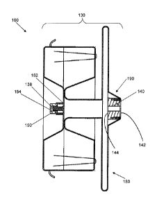
through the wall of the LAA.
[00067] In some embodiments, the hooks 200 may be integral to the medical
device 100, such as when individual strands of the braided tubular structure
130 are
isolated, cut, and a short portion of the wire adjacent the cut formed into an
outward
- 18-
CA 02842070 2014-01-14
WO 2013/074486
PCT/US2012/064765
projecting hook. Such a configuration may provide for a medical device 100
that has a
significantly lower profile as no added material (e.g., no separate hooks)
contributes to
the collapsed overall diameter D, (Fig. 5) of the medical device during
passage through a
delivery catheter. In addition, through the use of integral hooks 200 there
are no added
suture materials or suture knots that are needed to attach the hooks to the
braided tubular
structure, which also translates into a reduced profile of the medical device.
[00068] As noted above, the second expanded volume portion 185 may be
oversized so that it will engage the lumen of the vessel, body organ, or the
like to be
occluded. The medical device 100 may then be held in place by the combination
of the
radial engagement of the second expanded volume portion 185 with the lumen of
the
vessel, body organ, or the like and the engagement of the hooks 200 with the
vessel wall.
Over a relatively short period of time, thrombi will form in and on the
medical device
100 and occlude the lumen. Although the first and second expanded volume
portions
180, 185 may be various sizes, the first expanded volume portion may be at
least about
10% larger in diameter than the second expanded volume portion according to
one
embodiment.
[00069] For example, in the case of a medical device 100 that is implanted
within
the LAA, the medical device 100 may be positioned such that the first expanded
volume
portion 180 overlies the ostium of the LAA, while the second expanded volume
portion
185 is positioned within the LAA. Thus, the first expanded volume portion 180
may be
sized and configured to ensure that the first expanded volume portion 180 is
implanted to
a predetermined depth within the LAA. The second expanded volume portion 185
may
in turn be sized and configured to self expand and engage the wall of the LAA,
and the
hooks 200 may be configured to penetrate into the wall of the LAA, as
explained below.
Over time, thrombi will form in and on the first and second expanded volume
portions
180,185 to occlude the LAA.
[00070] In some embodiments, in order to speed up the occlusion of the
medical
device 100, the medical device may be at least partially coated with a
suitable
thrombogenic agent, filled with a fiber (e.g., a polymer fabric), braided with
an increased
number of strands, or include multiple layers of braided strands. For example,
the
medical device 100 may include one or more layers of polymer fabric 220
positioned
- 19-
CA 02842070 2014-01-14
WO 2013/074486
PCT/US2012/064765
within the first and/or second expanded volume portions 180, 185, as shown in
Fig. 9. In
particular, one or more layers of polymer fabric 220 may be sized and
configured to be
positioned within each of the first and second expanded volume portions 180,
185, such
that the polymer fabric extends substantially perpendicularly to the axis A of
the medical
device 100. Each piece of polymer fabric 220 may be sutured circumferentially
about its
periphery and about the inner circumference of the first and second expanded
volume
portions 180, 185, respectively. The polymer fabric 220 may be flexible and
may be
easily collapsed with the medical device 100 for delivery through a catheter.
In this way,
the interwoven fiber (which in some embodiments may be polyester) may attach
to a clot
to retain the clot firmly within the device as it forms the occlusion.
[00071] Although the embodiments depicted in Figs. 2-12 show a medical
device
having a single layer of braided fabric (e.g., a single tubular structure
130), in some cases
a second plurality of strands may be braided to form a second tubular
structure, such that
medical device includes an inner and outer layer. Referring Fig. 13, for
example, the
medical device 100 may include an inner layer 250 and an outer layer 260. The
inner
layer 250 may be disposed adjacent to the outer layer 260, and in some cases
the inner
layer may have a different shape than the outer layer. The first and second
expanded
volume portions 180, 185 and the connecting portion 188 may be integrally
formed from
the same tubular structure.
[00072] In some embodiments, the pick count, or the number of strand
crossings
per unit length of the layers 250, 260, may be set at the same or different
predetermined
values. For example, the inner layer 250 may define a first pick count, and
the outer
layer 260 may define a second pick count, where the second pick count is
different from
the first pick count. Although the first pick count, as braided, may be
different from the
second pick count, as braided, the first and second pick counts may be
selected such that
the relationship between the reduction in diameter and the elongation of the
inner layer
250 is substantially the same as the relationship between the reduction in
diameter and
the elongation of the outer layer 260 as the medical device 100 is moved
between the
expanded and contracted states. For example, a ratio of the decrease in
diameter of the
inner layer 250 to the increase in length of the inner layer 250 may be
substantially the
same as a ratio of the decrease in diameter of the outer layer 260 to the
increase in length
- 20 -
CA 02842070 2014-01-14
WO 2013/074486
PCT/US2012/064765
of the outer layer 260. Thus, adjacent portions of the inner and outer layers
250, 260 may
remain in their relative adjacent positions as the medical device 100 is moved
between
the expanded and contracted states. In this way, the inner layer 250 and the
outer layer
260 of the medical device 100 may cooperatively collapse and expand at
generally the
same rate, which enhances the stability of the medical device and facilitates
its delivery
into the vessel lumen and subsequent self-expansion. In the case where the
inner and
outer layers have different shapes from one another, the pick count of each
layer may be
selected such that in the elongated, contracted state each layer is
substantially the same
length.
[00073]
Furthermore, the helix angle of the strands (e.g., the angle formed between
the strand and the longitudinal axis of the braid mandrel as the strand is
applied to the
mandrel) used to braid the plurality of strands of the inner and outer layers
250, 260 may
be the same or different. The helix angles may be selected such that the
plurality of
strands of the inner layer 250 is braided at a first helix angle, and the
plurality of strands
of the outer layer 260 is braided at a second helix angle to ensure that the
relationship
between the reduction in diameter and the elongation of the inner layer is
substantially
the same as the relationship between the reduction in diameter and the
elongation of the
outer layer as the at least one layer is moved between the expanded state and
the
contracted state. In the case where the inner and outer layers have different
shapes from
one another, the helix angle of each layer may be selected such that in the
elongated,
contracted state each layer is substantially the same length.
[00074] As noted
above, the uniform movement that results between the inner and
outer layers 250, 260 may thus reduce the risk of bunching or gathering of the
layers
within the medical device 100, which would otherwise reduce the effectiveness
of the
medical device by increasing its delivery profile and/or generating gaps
between the
various layers of material that may cause leaks.
[00075] The
plurality of strands forming the second tubular structure may be made
of the same or different material as the strands forming the first tubular
structure,
described above. Thus, the strands of the second tubular structure may be
comprised of
metal or polymer material. For example the second tubular structure may be
made of
stainless steel, other metallic alloys, highly elastic alloys, and/or shape
memory alloys,
-21 -
CA 02842070 2014-01-14
WO 2013/074486
PCT/US2012/064765
which are both resilient and can be heat treated to substantially set a
desired shape, as
noted above with respect to the first tubular structure. In addition,
polymeric materials
may be combined with other materials in the formation of tubular structures
for certain
applications. For example, the medical device 100 may include a combination of
polyester strands and stainless steel wire. Thus, in some embodiments, the
plurality of
braided strands of the inner layer 250 may include Nitinol, and the plurality
of braided
strands of the outer layer 260 may include a polymer, or vice versa.
[00076] A method for making a medical device for placement in a body lumen
as
described above is summarized in Fig. 14. The method includes braiding a
plurality of
strands defining proximal strand ends to form a tubular structure at Block 300
and
attaching a first end feature defining a proximal end and a distal end to the
proximal
strand ends via the proximal end of the first end feature at Block 310. As
described
above with reference to the figures, the tubular structure may define a molded
and heat
set resilient expanded volume portion proximate to the first end feature and a
tapered
transition portion extending between the expanded volume portion and the
proximal end
of the first end feature. In the expanded state, the expanded volume portion
of the tubular
structure may define an expanded volume diameter, and the tapered transition
portion
may define a first transition diameter proximate the expanded volume portion
and a
second transition diameter proximate the proximal end of the first end
feature. As
described above and illustrated in the referenced figures, the first
transition diameter may
be greater than the second transition diameter, smaller than the expanded
volume
diameter, and disposed between the second transition diameter and the expanded
volume
diameter. The second transition diameter may be substantially equal to a
diameter of the
first end feature. In this way, the first end feature may be substantially
surrounded by the
tapered transition portion, such that the proximal end of the first end
feature substantially
coincides with the proximal end of the medical device.
[00077] As noted above, a second end feature defining a proximal end and a
distal
end may be attached to the distal strand ends. Block 320. In some cases, the
second end
feature may receive the distal strand ends via the proximal end of the second
feature, as
shown in the figures, whereas in other cases the second end feature may
receive the distal
strand ends via the distal end of the second end feature similar to the first
end feature,
- 22 -
CA 02842070 2014-01-14
WO 2013/074486
PCT/US2012/064765
thereby also keeping the second end feature from protruding from the distal
end of the
medical device. The medical device may be modified and configured in various
other
ways, such as by attaching retention hooks to the tubular structure (e.g., to
the outside of
second expanded volume portion) (Block 330), including a polymer fabric in one
or more
of the expanded volume portions (Block 340), and/or coating the device with a
thrombogenic agent (Block 350), as described in greater detail above.
[00078] Referring now to Figs. 15A, 15B, 16, and 17, a delivery device 400
may
be provided for deploying embodiments of the medical device 100 described
above. The
delivery device 400 may include an inner pusher wire 410 with a distal end 415
defining
external threads. The external threads of the pusher wire 410 may be
configured to
engage corresponding internal threads 164 of the first end feature 140 (shown
in Fig. 8)
so as to releasably attach the medical device 100 to the delivery device 400
for delivery
to and deployment at the target site, as illustrated in Fig. 17.
[00079] The delivery device 400 may further include an outer member 420
defining a lumen through which the inner pusher wire 410 is slideably
received. In other
words, the inner pusher wire 410 may be axially moveable within the outer
member 420,
such that the inner pusher wire may be moved between the position shown in
Fig. 15A
and Fig. 15B, for example. A distal end of the outer member 420 may include a
guide
member 430 configured to guide the proximal end 110 of the medical device into
a distal
end of a delivery sheath 440. In some cases, the guide member 430 may be made
of a
polymer material. The guide member 430 may have a tapered external surface,
such that
the diameter Dd of the guide member at its distal end is approximately the
same as (e.g.,
slightly less than) the inner diameter of the delivery sheath 440 and the
diameter Dp of the
guide member at its proximal end is approximately the same as (e.g., slightly
greater
than) the outer diameter of the outer member 420. Moreover, as shown in Fig.
17, the
distal diameter Dd of the guide member may approximate the second transition
diameter
D2 of the transition portion 190, such that the taper of the transition
portion 190 and the
guide member generally correspond to one another. In some cases, as shown, the
outer
surface of the guide member 430 may include grooves or concavities 431, which
may
serve to prevent the taper of the guide member from acting as a plunger that
draws air
into the delivery sheath 440 from the proximal end as the medical device 100
is advanced
- 23 -
CA 02842070 2014-01-14
WO 2013/074486
PCT/US2012/064765
to toward the distal end 445. In other words, the concavities 431 may allow
fluid to flow
through the delivery sheath 440, such that a positive blood pressure exists in
the delivery
sheath with respect to the pressure outside the body.
[00080] The function of the guide member 430 may be illustrated by the
following
example. When accessing a tortuous path (e.g., a vessel that includes one or
more small
radius curves), the pusher wire 410 and/or the outer member 420 may be biased
to one
side of the delivery sheath 440 once the medical device 100 has been deployed
(e.g., is
outside the delivery sheath 440, but still attached to the pusher wire 410).
In some cases,
the medical device 100 must be recaptured within the delivery sheath 440, for
example,
to reposition the medical device at the target site or to replace the device
for one of a
different size. As the medical device 100 is moved proximally (closer) to the
distal end
445 of the delivery sheath 440 during recapture, the medical device 100 may
not be
axially aligned with the lumen of the delivery sheath (e.g., as a result of
the curvature of
the vessel within which the delivery sheath is disposed). The guide member
430, by
virtue of its tapered shape, may thus bring the proximal end 110 of the
medical device
100 into closer axial alignment with the lumen of the delivery sheath to allow
for easier
recapture and to minimize the risk of damaging the medical device during
recapture.
[00081] Accordingly, in Fig. 18, a method for delivering a medical device
as
described above is summarized. The method includes providing a medical device
configured as described above in connection with one or more of Figs. 2-17.
Block 500.
For example, the medical device may include a tubular structure comprising a
plurality of
braided strands defining proximal strand ends and a first end feature defining
a proximal
end and a distal end, where the first end feature is configured to receive and
secure the
proximal strand ends via the proximal end of the first end feature. As
described above,
the tubular structure may define one or more expanded volume portions and at
least one
tapered transition portion.
[00082] The method of delivery may further include advancing the medical
device
through the body lumen toward the target site (Block 510) and deploying the
medical
device at the target site (Block 520). In some cases, as described above, the
method may
further include recapturing the medical device within the delivery sheath
(Block 530),
repositioning a distal end of the delivery device (Block 540), and redeploying
the medical
- 24 -
CA 02842070 2014-01-14
WO 2013/074486
PCT/US2012/064765
device (Block 550). Once the medical device is positioned at a desired
location, the
delivery device may be disengaged from the medical device (e.g., via
unthreading the
medical device from the pusher wire) and withdrawn from the body lumen,
leaving the
medical device in place at the target site. Block 560.
[00083] The method depicted in Fig. 14 and described above represents only
one
method for making a medical device for placement in a body lumen. Similarly,
the
method depicted in Fig. 18 and described above represents only one method for
delivering a medical device. In some embodiments, certain ones of the steps
described
above may be modified or further amplified. Furthermore, in some embodiments,
additional optional steps may be included, some examples of which are shown in
dashed
lines in Figs. 14 and 18. Modifications, additions, or amplifications to the
steps above
may be perfoimed in any order and in any combination. The particular methods
of
manufacturing and delivery will depend on the desired configuration of the
medical
device, the patient's anatomy, the condition and location of the target site,
the preferences
of the practitioner, and/or other considerations.
[00084] This invention has been described herein in considerable detail in
order to
comply with the Patent Statutes and to provide those skilled in the art with
the
information needed to apply the novel principles and to construct and use
embodiments
of the example as required. However, it is to be understood that specifically
different
devices can carry out the invention and that various modifications can be
accomplished
without departing from the scope of the invention itself. For example, options
shown for
one embodiment could easily be applied to other embodiments, as desired for a
particular
application, without departing from the scope of this invention.
- 25 -