Note: Descriptions are shown in the official language in which they were submitted.
i
,
,
-1-
MINIMAL INCISION REMOVABLE BONE SCREW, DRIVER, AND METHOD OF
USE
TECHNICAL FIELD
The present invention relates to a bone screw, a driver for the screw, and a
method of applying and removing the screw. The system may be used as a
removable fixation system for any osteotomy or fracture requiring lag
compression.
The screw has particular application for interfragmentary internal fixation of
small
bones, such as those of the foot, hand, and ankle, but is also useful with
osteotomies
or fractures of large bones and spine.
BACKGROUND ART
When a bone is fractured, either by deliberately cutting it (osteotomy) or by
trauma, it heals better if the bone fragments are pressed firmly together.
Compression of the fragments increases the contact area across the fracture
and
increases stability of the bone at the fracture. It also decreases stress on
any
orthopedic implant.
Internal fixation of a bone fracture using bone screws is now common practice.
The screw is applied across the fracture, preferably at nearly a right angle
to the
fracture, although the nature of the bone and of the fracture frequently
dictates other
angles. The distal end of the screw crosses the fracture, and when the head of
the
bone screw engages the proximal fragment, further rotation of the screw draws
the
distal fragment of the bone against the proximal fragment. Any screw that is
used
to achieve interfragmental compression is termed a lag screw. The two most
common types of lag screws are cortical and cancellous screws. Cortical screws
have fine threads on their shaft and are designed to anchor in cortical bone.
I CA 2842324 2017-07-14
CA 02842324 2014-01-16
WO 2013/013218
PCT/US2012/047743
-2-
Cancellous screws tend to have coarser threads and are designed to anchor in
the
softer cancellous bone.
Both types of lag screw generally include a threaded distal end and a proximal
head. Although the screw may be threaded nearly to the head, this design
requires
that the proximal bone fragment be pre-bored to permit the threads to pass
smoothly
through the proximal fragment. More commonly, the threads on the distal end
extend only far enough to ensure a positive grip in the distal fragment but
not so far
as to engage the proximal fragment when the screw is applied, the shaft
between the
threads and the head being smooth and sized no larger than the minor diameter
of
the threads (the maximum diameter of the thread groove). The distal side of
the
head, facing the shaft, is usually symmetrically convex, preferably
hemispherical,
and the proximal face of the proximal bone fragment is frequently lightly
countersunk, in order to spread stresses in the screw and the bone most
efficiently,
to reduce the risk of creating a stress fracture, and to minimize the
protrusion of the
screw head from the face of the bone. The upper, proximal, side of the screw
head
is generally flat or gently rounded to permit the head to lie as close to
level with the
proximal bone surface as possible. The head is provided with a slot, spaced
holes, a
hexagonal socket, or other depression to accept the blade or tip of a drive
tool or
screwdriver designed to be inserted into it.
In order to eliminate the need for pre-drilling bone and tapping the distal
bone
fragment, bone screws are now frequently made to be self-tapping. To aid
further in
the placement of bone screws, the screws are frequently cannulated, having a
hollow shaft and head. Cannulated screws may be placed more precisely than non-
cannulated screws. The surgeon first drills a small Kirschner wire (K-wire)
across
the fracture, generally under fluoroscopic control. The wire may sometimes be
inserted through the skin without the need of an incision. If necessary, the K-
wire
can be withdrawn and replaced with minimal trauma to the bone in order to
place it in
optimal position across the fracture. A small incision may then be made
through the
skin to enable the surgeon to minimize tissue trauma while placing the bone
screw
and to permit countersinking the bone around the point of insertion of the
screw.
CA 02842324 2014-01-16
WO 2013/013218
PCT/US2012/047743
-3-
The cannulated screw is then placed over the wire and slid down to the bone
surface. A special cannulated driving tool then allows the screw to be driven
into the
bone along the shaft of the K-wire. The K-wire is then withdrawn and the wound
over the screw is closed.
The construction and use of bone screws has become standardized to a great
extent. There are of course, many variations on the details of the
construction of
bone screws, including for example, the use of a break-away driven element as
shown in Patterson et al, U.S. Patent No. 8,221,478.
Bone screws may also be designed merely as an anchor for attaching an
external stabilizing device. Pedicle screws, such as illustrated in Mazda et
al., U.S.
published application US 2004/0116932 Al, are examples of such external
fixation
screws. The present invention is not principally concerned with such screws,
although some aspects of the invention may be applicable to them.
Internal fixation bone screws may be left in the body after implantation.
However, surgeons are increasingly removing fixation for a number of
justifiable
reasons. Irritation/inflammation, allergic reaction, and infection are common
reasons
to remove hardware at appropriate times. Although rare, implant rejection may
occur. Furthermore, the long-term deleterious effects of a metal such as
stainless
steel or a titanium alloy implanted in the body are not fully understood. It
is not
uncommon for barometric pressure and changes in ambient temperature to cause
rheumatic or osteoarthritic flare-ups. The
possibility of a causal relationship
influenced by hardware left in and around these areas exists. Finally, because
of
growing concerns over electro-magnetic radiation caused by cell phone use and
other exposure, it is ideal that conductive metals be removed from the body if
possible. MRI and other present electromagnetic technologies are influenced by
conductive, metallic implants. Future technologies may depend on the body
being
free of conductive elements. Therefore, particularly when a deleterious effect
is
noted, a bone screw is sometimes removed, thereby allowing bone regeneration
in
the volume formerly occupied by the bone screw. Such removal, however,
requires
considerable effort and risk, as suggested by patents such as Bonati et al.,
U.S.
CA 02842324 2014-01-16
WO 2013/013218
PCT/US2012/047743
-4-
Patent No. 7,090,680, Steffee, U.S. Patent No. 4,854,311, Vasta et al., U.S.
Patent
No. 7,582,093, or Lindemann et al., U.S. published application US 2007/0270880
Al. Special screw removal kits including multiple instruments are commercially
available.
When faced with having to remove a screw of the prior art, a surgeon must
deal with creeping fibrosis, meaning soft tissues that creep into the screw
threads
making it difficult to access the screw without an incision into periosteal
structures
and more trauma. Fibrosis, in a worst-case scenario will make it necessary for
a full
screw extraction set to be utilized thereby completely bypassing the
conventional
methodologies of placing a screwdriver into a head. This usually requires
cutting the
screw or severe countersinking.
In order to overcome these problems, some bone screws are made of
absorbable materials. These screws, however, are not as strong as metal
screws,
require drilling and tapping with metal instruments, and are transparent to x-
rays.
Hybrid metal and polymer screws are disclosed in Fischer et al., U.S. Patent
No.
4,711,232 and in TenHuisen et al., U.S. Patent No. 6,916,321, but these screws
add
complexity and do not solve all of the problems with leaving metal in the
body.
Another solution has been the use of screws made of compatible bone, as in
Reed,
U.S. Patent No. 5968047. This approach is costly and has not been entirely
satisfactory.
BRIEF SUMMARY OF THE INVENTION
Briefly stated, the present invention provides a bone screw which is easily
placed and easily removed, usually after bone healing. The bone screw may be
placed and removed using a simple screwdriver in accordance with the
invention.
In accordance with one embodiment, a bone screw comprises a thread at its
distal end, a head at its proximal end, and a bone-engaging compression member
spaced distally from the head. The screw may be made of any biocompatible
material, but is preferably made of stainless steel, titanium, or titanium
alloy.
Preferably, the thread does not extend as far as the compression member.
Preferably, the thread is self-tapping. In some situations it may be self-
drilling,
CA 02842324 2014-01-16
WO 2013/013218
PCT/US2012/047743
-5-
although this is not presently preferred. A thread which is neither self-
tapping nor
self-drilling is also useable. Many types of threads are known and are
useable. Both
cortical and cancellous screw threads may be used. If the threads extend to or
nearly to the compression member, the proximal bone fragment must be pre-
drilled
to a diameter as great as the thread major diameter to allow the compression
member to create a lag effect on the fracture. The depth and shape of the
threads,
and their length may be established in accordance with known parameters.
Preferably, the bone-engaging compression member is convex on its distal
side and is circular in cross-section. Preferably the compression member is
convex
on its proximal side and has a smoothly curved outline without edges to permit
easy
extraction through an incision and to provide a neck between the head and the
compression member. A ball having a diameter from 10% to 70% greater than the
screw diameter is preferred. A ball diameter of 110% to 140% of the maximum
major thread diameter or a diameter of 125% to 160% of the shaft diameter is
particularly suitable.
The head is preferably non-circular as viewed in top plan elevation (end-on),
so that its periphery may be drivingly engaged by a driver. Also preferably,
at least
two opposed sides of the head are curved inward toward both the proximal end
of
the screw and the distal end of the screw to enable a hollow head of a screw
driver
to engage the screw head to transmit sufficient torque to drive screw into or
out of
bone when the driver is tilted at angles of up to fifteen degrees or possibly
twenty
degrees from the axis of the screw (+/- 15 -20 articulation). The inwardly
curved
distal faces of the head also permit the screw to be drawn out of the bone and
incision when the driver is articulated away from the axis of the screw. In a
preferred
embodiment, the head is in end view a regular polygon having an even number of
sides, preferably four or six. With the polygonal configuration, it is
convenient for the
distance between opposite sides to be about equal to the major thread
diameter,
5%.. Thus, for example, a two millimeter screw (having a major thread diameter
of
two millimeters) may have a two millimeter square head, and a five millimeter
screw
may have a five millimeter square head. The height of the head, in accordance
with
CA 02842324 2014-01-16
WO 2013/013218
PCT/US2012/047743
-6-
the geometry of its sides, is generally about the same as its width, typically
on the
order of 80% to 100% of its width, usually 90% to 95% of the major thread
diameter.
The spacing distance between the base of the head and the widest part of the
compression member may be chosen to suit the use of the screw. For some
applications, the head is placed just below the skin, without causing the skin
to "tent",
so it can be found easily by subcutaneous palpation and removed easily with
minimal trauma to surrounding tissue. For these applications, a screw having a
spacing distance of about three to nine millimeters may be used. Because the
screw
will generally be removed in a few weeks, accessibility of the screw for ease
of
removal dictates that in many situations a screw having a long spacing be
placed so
that the head extends into a concavity below the surface of the epidermis. For
other
applications, patient comfort or anatomy demands a head that is closer to the
bone
surface. For these applications a spacing distance of one to three millimeters
may
be used, and if the screw cannot be found by palpation, it may be found by
mechanical or machine means, such as by fluoroscopy.
In most instances, the axial distance from the top (proximal end) of the head
to the widest part of the compression member is greater than the largest
diameter of
the head.
In preferred embodiments, the head and compression member are formed
with no sharp edges. All edges are rounded as much as is consistent with
maintaining a non-circular head which may be driven by a socket extending over
the
head and engaging its periphery, so as to maintain the lowest practical
coefficient of
friction (interaction) between the screw and surrounding soft tissue including
nerves
and blood vessels after the driver is removed. The top (proximal end) of the
head
may be flat to minimize height above the working sides of the head, or it may
be
convex, even spherical, to minimize friction with surrounding tissue and
nerves
(neuropraxia).
The screw may be either solid or cannulated. When cannulated, the size of
the cannula may be chosen in accordance with the size of the wire or pin. K-
wire
sizes from about 0.7 mm to about 1.6 mm are common. Illustrative screws of the
CA 02842324 2014-01-16
WO 2013/013218
PCT/US2012/047743
-7-
present invention have cannulae which typically range from about 0.75 mm for a
two
millimeter diameter screw to about 1.4 mm for a seven millimeter diameter
screw.
A set of screws of the present invention may include several families of
screws of different diameters, for example 2.0, 2.5, 3.0, 3.5, 4.0, 5.0, 6.0,
and 7.0
mm. Each family may include screws of different lengths, measured from the
distal
end of the screw to the maximum diameter of the compression member, ranging,
say, from 8 mm to 60 mm in two millimeter increments. Each length of each
family
may in turn include different compression member-to-head base dimensions, say
1.0
mm, 3.5 mm, 5.0 mm, and 8.0 mm. Because the size of the screw head in each
family is constant, the overall length of the screws of a given nominal length
may
vary based on the length of the compression member to head spacing. It is
anticipated that screws of the invention will be packaged and sold in sets
including at
least different compression member-to-head base dimensions.
The screw driver of the invention may comprise a simple open box socket at
the distal end of a driver shaft axially aligned with a handle. The inside
walls of the
socket may be parallel and form a shape complementary to the outside
dimensions
of the screw head, with dimensions just sufficiently larger than those of the
exterior
of the head to enable the socket to slip easily over the head while
maintaining a
positive contact with the exterior faces of the head. A spacing one half
percent to
ten percent larger than the head width at its greatest width between parallel
faces is
suitable. The depth of the socket is preferably equal to the depth of the head
10%.
The exterior of the socket is smooth, and the wall thickness of the socket is
as thin
as is consistent with strength, to minimize the amount an incision must be
spread to
accommodate the driver. The exterior of the socket may be round to minimize
interference with surrounding tissue as the driver is rotated, or the corners
of a
polygonal exterior may be rounded. The handle and socket may be formed as a
single piece, or they may be separate pieces which are permanently or
removably
connected to each other. The socket and its shaft are preferably cannulated to
accommodate cannulated screws. The socket and the shaft of the driver are
CA 02842324 2014-01-16
WO 2013/013218
PCT/US2012/047743
-8-
preferably made of stainless steel, although titanium, titanium alloys, and
other
materials are useable.
Where the handle and socket are not removably connected, a kit of
screwdrivers would consist of a screwdriver for each diameter of screw,
illustratively
a 2.0, 2.5, 3.0, 3.5, 4.0, 5.0, 6.0, and 7.0 mm size. Where they are removably
connected, a single handle and appropriate sockets make up the kit. It will be
understood that the shaft of the screwdriver could be made a part of the
handle or a
part of the removable socket, preferably of the socket.
In accordance with the method of the invention, a screw of the invention is
inserted transfragmentally through a fracture, preferably at an angle to the
fracture
chosen for acceptably compressing the bone fragments in accordance with
standard
practice, and in order to place the head in an accessible position that will
provide
minimum patient discomfort. The screw is tightened into the distal bone
fragment
using the screwdriver of the invention, to place the compression member in
contact
with the proximal bone fragment and to draw the bone fragments into snug
contact.
Unlike many previously-known screws which are not specifically designed for
removal, the screw of the present invention may, if desired, be driven through
and
beyond the margin of the distal bone fragment. This exit site is generally
spaced far
enough from the epidermis to prevent the production of a secondary wound.
Screwing the internal fixation bone screw through the distal bone ensures that
the
screw is secured in cortical bone and is therefore less likely to lose
purchase.
The tissue over the insertion site is then closed. When soft tissue closure
over the screw involves capsule and/or subcutaneous layers, the capsule and
layers
can be closed over the screw head, a small incision made, and the layers
pushed
down below the head and over the compression member before closing the
epidermis. On removal of the screw, only the epidermis needs to be incised to
expose the screw head for removal of the screw. In most cases, further
incision of
the subcutaneous layers is not needed. This ease of removal is a major
advantage
of the present invention.
CA 02842324 2014-01-16
WO 2013/013218
PCT/US2012/047743
-9-
Previously, those concerned with internal fixation bone screws have believed
that minimizing the protrusion of the head from the surface of the bone to
which it is
affixed is of great importance. See for example Zang, U.S. Patent No.
5,556,225. In
accordance with the present invention, accessibility of the head and ease of
removing an internal fixation screw have been found to be more important. An
additional advantage to having removable hardware that is easy to remove is
that it
is equally easy to replace. This means that should a screw lose purchase, a
larger
screw can serve as a replacement screw without the necessity of removal
through a
large incision and reevaluation of the exact size.
After the bone fracture has healed sufficiently, in accordance with
radiographic and clinical evidence (frequently two to eight weeks after the
procedure), the head of the screw is located either by palpation or by other
means
such as needle or fluoroscopy, and a small incision is made to expose the
screw
head.
Particularly if the screw head is near the surface, a small stab incision may
be
sufficient to access the screw head. The head of the screw may be cleaned,
although this step is far less important than with a screw head relying on a
depression in the top of the head for engagement with the driver. The
screwdriver is
placed over the head. Because the head is separate from the compression
member,
it is easily reached, and because the head is undercut, tilting the
screwdriver allows
it to exert a gentle outward force on the screw. The screwdriver is used for
turning
and removing the screw from the bone. The rounded shape of the head allows
positive contact between the side walls of the driver socket and the head
throughout
a range of angles of at least fifteen degrees. Therefore, the driver may be
tilted a
few degrees from the axis of the screw, preferably around ten to twenty
degrees, and
used for gently lifting the screw.
When the screw is clear of the bone, it may be removed with forceps. The
small incision is closed, preferably after bathing it. The bone is then
allowed to
regrow into the cavity left by the screw. It will be seen that the screw head
will
frequently be so close to the epidermis and will be so completely exposed by
even a
CA 02842324 2014-01-16
WO 2013/013218
PCT/US2012/047743
-10-
small incision, that removal of the screw may not even be dependent on the
special
qualities of the screwdriver.
The screw, driver, and methods of the present invention are particularly well
suited to surgery on the human foot and hand. Examples are fractures of the
phalanges, metatarsals, or talus of the foot, fractures of the phalanges or
scaphoid of
the hand, and in osteotomies of the hand and foot such as bunion repair
through a
modified Scarf/Akin osteotomy or a repair of a Jones fracture of the hand. It
will be
understood that osteotomies may involve both the removal of bone and the
insertion
of bone between bone fragments, so that the screw may pass through more than
one proximal bone fragment. It will also be understood that the screw, driver,
and
methods of the invention are applicable to a wide range of other procedures on
human and non-human vertebrate bones, including for example, other osteotomies
and fractures of the hip, leg, arm, spine, or clavicle. The screw, driver and
methods
may also be useable with non-metallic, biocompatible, and bioabsorbable
plating
devices to avoid long-term exposure to metal components.
The foregoing and other objects, features, and advantages of the invention as
well as presently preferred embodiments thereof will become more apparent from
the reading of the following description in connection with the accompanying
drawings.
BRIEF DESCRIPTION OF THE SEVERAL VIEWS OF THE DRAWINGS
In the accompanying drawings which form part of the specification:
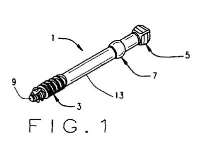
FIG. 1. is a view in perspective of one illustrative screw according to an
embodiment of the present invention.
FIG. 2 is a view in side elevation of the screw of FIG. 1.
FIG 3 is a top plan view of the screw of FIGS. 1 and 2.
FIG. 4 is a view in axial cross-section of the screw of FIGS. 2-3.
FIG. 5 is a detail in axial cross-section of a head part of the screw of FIGS.
2-
4.
FIG. 6 is a detail view of a distal tip part of the screw of FIGS. 1-5.
CA 02842324 2014-01-16
WO 2013/013218
PCT/US2012/047743
-11-
FIG. 7 is a detail view in axial cross-section of a screw thread part of the
screw of FIGS. 1-6.
FIG. 8 is a diagonal cross section taken along the line 8-8 of FIG. 3.
FIG. 9 shows a family of screws of the present invention, each screw having
the same nominal length, but with different overall lengths determined by a
head
spacing dimension "B."
FIG. 10 shows a family of screws of the present invention, one group of
screws having a "long" head spacing dimension "B" and a second group having a
short head spacing dimension "B", each screw in each group having different
nominal lengths and different thread lengths "C".
FIG. 11 is a view in side elevation of a screw driver according to an
embodiment of the present invention.
FIG. 12 is an end view of the driver of FIG. 11.
FIG. 13 is a fragmentary axial cross-section of a socket portion of the driver
of
FIGS. 11 and 12, taken along the line 13-13 of FIG. 12.
FIG. 14 is an end view of the fragment of FIG 13.
FIG. 15 is a view in side elevation of a handle part of the driver.
FIG. 16 is an end view of the handle of FIG. 15.
FIG. 17 is a fragmentary sectional view taken along line 1 7-1 7 of FIG 16.
FIGS. 18 and 19 are schematic views of the fixation of a fracture with the
screw 1 and driver 31 of the invention.
FIG. 20 is a cross-sectional view corresponding to FIG. 4 of another
embodiment of bone screw.
FIG. 21 is a cross-sectional view corresponding to FIGS. 4 and 19 of yet
another embodiment of bone screw.
Corresponding reference numerals indicate corresponding parts throughout
the several figures of the drawings.
DESCRIPTION OF THE PREFERRED EMBODIMENT
The following detailed description illustrates the invention by way of example
and not by way of limitation. The description clearly enables one skilled in
the art to
-12-
make and use the invention, describes several embodiments, adaptations,
variations,
alternatives, and uses of the invention, including what is presently believed
to be the
best mode of carrying out the invention.
As shown in FIGS. 1-8, in accordance with one embodiment, a bone screw 1
comprises a thread 3 at its distal end, a head 5 at its proximal end, and a
bone-engaging compression member 7 spaced distally from the head. The screw 1
may be made of any biocompatible material, but is preferably made of stainless
steel,
titanium, or titanium alloy. In this illustrative embodiment, it is made of
the titanium
alloy known as Ti6AI4V ELI, with an anodized finish in accordance with SAE
AMS2488D.
The screw 1 is first identified by its diameter and by its length, as in a
standard
lag bone screw. Diameter is defined as the major diameter of the screw thread
3, in
this illustrative embodiment 2.0 mm. The length A of the screw is measured
from its
distal end 9 to the largest diameter of the compression member 5. The size of
the
head 5 is nominally the same both across from face to face as it is tall (top
to bottom),
and both these dimensions are nominally the same as the screw diameter (major
thread diameter). In this embodiment, the head is 2.0 mm across and 1.9 mm
tall.
Unique to the screw of the invention is a dimension B measured from the
largest
diameter of the compression member 5 to the base 11 of the head 5, as
discussed
more fully hereinafter. The minor diameter of the thread 3 is equal to the
shaft
diameter of the screw shaft 13 between the thread 3 and the compression member
7
and between the compression member 7 and the head base 11. That dimension in
this embodiment is 0.75 times the major thread diameter, or 1.5 mm. The thread
9
has a length C that varies with the length of the screw 1. For a 16.0 mm long
screw,
the thread has a length of about 6 mm.
The thread 3 is self-tapping, but requires a pilot hole of about the diameter
of
the shaft 13.
The bone-engaging compression member 7 has a diameter from about 1.1 to
about 1.25 times the diameter of the screw. In this illustrative embodiment,
the
compression member 7 is a sphere having a diameter of 2.2 mm.
CA 2842324 2019-01-23
CA 02842324 2014-01-16
WO 2013/013218
PCT/US2012/047743
-13-
The head 5 in this illustrative embodiment is generally in the form of a cube
having a side nominally equal to the screw diameter. The top plan view (FIG.
3) of
the head 5 shows the sides as straight, but as viewed in side elevation (FIG.
2) or in
cross-section (FIGS. 4 and 5), the sides are sloped inward top and bottom at
an
angle of 16 from a maximum convex dimension 15, as indicated at 17 and 19
respectively.
The spacing distance B between the base 11 of the head 15 and the widest
part of the compression member may be chosen to suit the use of the screw. In
this
illustrative embodiment, as shown in FIG. 9, a set 23 of "long head" screws
have
spacing distance B of 3.5 mm, 5.0 mm, and 8.0 mm, respectively. For
applications
in which little room is available below the skin, a "short head" having a
spacing
distance of 1.0 mm, is provided.
The head 5 and compression member 7 are formed to be smooth, with no
sharp edges.
The illustrative screw is cannulated, having a central bore 21 of 0.75 (+.05)
mm. A solid screw would look the same, but without the central cannula.
As indicated in FIG. 10, each family includes screws of different lengths,
measured from the distal end of the screw to the maximum diameter of the
compression member, ranging from 12 mm to 60 mm in two millimeter increments.
Each length of each family in turn includes different compression member-to-
head
base dimensions "B": 1.0 mm ("short head"), 3.5 mm, five millimeters, and
eight
millimeters. Because the size of the screw head in each family is constant,
the
overall length of the screws of a given nominal length vary based on the
length of the
compression member to head spacing. As shown in FIG. 10, different lengths of
screw 1 will have different thread lengths "C".
An illustrative screw driver 31 of the invention is shown in FIGS. 11-17. The
driver 31 comprises a simple open box socket 33 formed integrally at the
distal end
of a driver shaft 35 axially aligned with a handle 37, as shown in FIGS. 11-
17. The
shaft 35 and socket 33 have an overall length of about 125 mm. The shaft and
socket are formed from a single 0.375 mm diameter rod of 17-4PH H900 stainless
CA 02842324 2014-01-16
WO 2013/013218
PCT/US2012/047743
-14-
steel and passivated per ASTM A967. At its distal end, the round tube is
squared
and routered to form inside walls 39 of the socket. The inside surfaces of the
walls
39 are flat and parallel and have a side "D" of 2.07 +/- .01, just larger than
the sides
of the screw head 5. The depth of the socket is 2.0 mm, about 0.1 mm deeper
than
the height of the screw head. A shallow well 41 at the bottom of the socket 33
acts
as a guide for a K-wire to enter a 0.90 +/- 0.05 mm cannula 43 extending
through the
driver shaft 35. The exterior of the socket 33 is smooth, and the wall
thickness of the
socket is as thin as is consistent with strength, to minimize the amount an
incision
must be spread.
The shaft 35 of the driver 31 has welded to its proximal end a standard
adapter 45 sized to fit a hollow 47 in the handle 37, to which it is attached.
The
handle 37 is formed of polyphenylsulfone (Radele R5500, Solvay Advanced
Polymers L.L.C). In this embodiment, the handle 37, shaft 35, and socket 33
are
packaged as a single unit, with handles of different colors signifying
different socket
sizes.
A kit of screwdrivers in this embodiment consists of eight screwdrivers, each
with a handle 37 secured to a shaft/socket of an appropriate size for each
diameter
of screw, in this illustrative embodiment a 2.0, 2.5, 3.0, 3.5, 4.0, 5.0, 6.0,
and 7.0 mm
size.
An example of the use of the screw, driver, and method of the present
invention to treat a bunion using the Akin procedure was conducted as follows:
A chevron style osteotomy was made through and into the head of the first
metatarsal.
Next, a .062 Kirschner wire was utilized to drill a pilot hole through both
sides
of the osteotomy.
Next, a depth gauge was utilized to determine the appropriate screw length.
Next a countersink was performed at the proximal entry point of the k-wire.
Next, an appropriately sized bone screw was screwed into the osteotomy and
compression was noted and achieved to two finger tightness.
CA 02842324 2014-01-16
WO 2013/013218
PCT/US2012/047743
-15-
Next capsular closure was performed over the head of the screw creating a
mild tenting effect directly over this.
Upon the completion of capsular closure the screw head was palpated and a
small stab incision was made into the capsule whereby the head of the screw
was
pushed through the capsule exposing the head.
Next the sub-cuticular layer was closed once again over the head of the screw
creating a mild tenting.
Upon the completion of the sub-cuticular closure the screw head was again
palpated and a small stab incision was made to expose the screw head through
this
layer.
Finally, the epidermal layer was closed utilizing absorbable 5-0 Vicryl
suture.
On completion of this closure it is noted that there is no tenting of the
epidermis due
to screw head prominence. Palpation of the screw head can, however, be
appreciated through the epidermis.
After an appropriate period, removal was conducted as follows:
The patient was brought into the operating room and prepped and draped in
the usual aseptic manner.
Next, attention was directed to the dorsal aspect of the foot which was
palpated locating the head of the previously applied cortical removable bone
screw.
Next, approximately 1.5 mL of lidocaine one percent was utilized to achieve
anesthesia.
Next, a stab incision was made with a number eleven blade directly over the
head of the previously mentioned screw.
Next, the driver was placed into the wound and the screw head was located
and securely contact fitted around the driver head. The driver was tilted at
approximately 15 to create the appropriate pulling effect as the screw was
removed.
Once the screw head was noted to exit the small epidermal incision, a small
hemostat was utilized to secure the skin around the screw head and allow
further
secure removal of the screw.
CA 02842324 2014-01-16
WO 2013/013218
PCT/US2012/047743
-16-
Upon complete removal, the remaining incision was closed with a single 5.0
nylon suture.
Another example of the use of the present invention is shown schematically in
FIGS 18 and 19. An incision is made through the epidermis 57 and subdermal
tissue 59, and they are retracted. The fracture in bone 53 is reduced by
manipulation, and drilled with a K-wire through both a proximal fragment 56
and a
distal fragment 54 at an angle to the fracture chosen for acceptably
compressing the
bone fragments in accordance with standard practice, and in order to place the
head
in an accessible position that will provide minimum patient discomfort. The K-
wire is
removed, and the bone fragment 56 is mildly countersunk. The depth of the hole
is
measured with a depth gauge. A proper size cannulated screw 1 is chosen based
on angle of entry and concavity of bone surface and distance from bone surface
to
skin surface. A K-wire having both ends tapered is inserted, and the screw 1
is
tightened over it to two finger tightness and good compression using the
screwdriver
31. The K-wire is removed. The capsule 59 is closed over the head 5, tenting
the
capsule. The capsule is then incised directly over the screw head 5 to expose
the
head. Any subcutaneous tissue is likewise closed over the head 5, incised to
expose the head, and closed. The epidermis 57 is then closed and sutured as
indicated at 61.
The screw 1 is removed in the same way as in the previous example,
requiring only a small stab incision to expose the screw head 5 and allow
removal of
the screw.
Numerous variations, within the scope of the appended claims will occur to
those skilled in the art in light of the foregoing description. Merely by way
of
example, although standard thread count and spacing will typically be used,
the
thread count, spacing, or both may be changed from screw to screw, without
departing from the scope of the present invention. The shape of the head and
its
spacing from the compression member may be varied widely.
The head may even be made with a conventional hex socket and driven with
a conventional hex-head driver as shown in FIG. 20, corresponding to FIG. 4.
This
-17-
approach allows a slightly more rounded head, but it suffers from the problems
of
ingrowth into the hex socket and possible difficulties in removing the screw.
Leaving
a neck below the head and above the compression member, however, is highly
advantageous in providing purchase for aiding in the extraction of the screw.
The
slight angulation of the driver allows for the retrograde force frequently
needed to
remove the screw.
As shown in FIG. 21, it is even possible to obtain some of the advantages of
the present invention by melding the compression ball and the head into a
single
body. That approach may utilize either the head of the embodiment of FIG. 20,
as
shown, or the head of the embodiment of FIGS. 1-8. That approach does elevate
the
head to a more reachable position, but it is believed to lack most of the
other
advantages of the illustrative embodiments.
Screw head shape can be square, triangular, rectangular, oblong, or any other
shape that will facilitate this process. It is believed at present that having
at least two
convex opposed faces is advantageous for positive driving of the screw in both
directions and for lifting the screw as it is removed. The shape of the
compression
member may also be varied. A ball shape, whether spherical or flattened, is
preferred
because of its lack of edges, and because it distributes stresses efficiently.
The size
of the compression member may be varied; it is believed that a somewhat larger
ball,
perhaps one millimeter larger than presently preferred, may give somewhat
improved
results.
As various changes could be made in the above constructions without
departing from the scope of the invention, it is intended that all matter
contained in
the above description or shown in the accompanying drawings shall be
interpreted
as illustrative and not in a limiting sense.
CA 2842324 2019-01-23