Note: Descriptions are shown in the official language in which they were submitted.
CA 02842420 2014-02-07
= f f.
METHOD AND SYSTEM FOR PROCESSING DATA FROM AMBULATORY
PHYSIOLOGICAL MONITORING
1. FIELD OF THE ITiVENTI9N
[00021 The present invention relates to the field of processing signals
obtained from non-
invasive physiological monitoring, and especially from non-invasive monitoring
which
gathers multiple physiological parameter/ while permitting relatively free
subject motion.
The present invention provides improved, robust systems and methods for
processing such
signals.
2. pACKGROUND OF THE INVENTION
[0003] Monitoting a subject's physiological parameters is routine in the
clinic and in the
hospital. Ber2IIISC of the interdependence of physiological and other bodily
processes,
monitoring of multiple related physiological parameters (referred to herein as
"multiple
parameter monitoring" or "MPM") is advantageous is known in these
environments..
[1/0041 Recent developments in sensors and sensor systems now increasingly
allow single
and multiple parameter physiological monitoring to move out of the controlled
environments of the Clinic or hospital and into less constrained environments
where the
subject rnay engage in normal activities. MIN is now possible in the subject's
nonnal
environments where the subject is carrying out normal activities with little
or no constraint.
"Ambulatory monitoring", as such monitoring is known in the art, thereby
encompasses the
monitoring of physiological parameters during normal daily activities,
including work
activities, and also encompasses monitoting during unconstrained sleep. For
example,
during "ambulatory monitoring", a subject may be wa]king, running, generally
exercising,
engaging in athletics, and the like; a subject.may also be working at either
sedentary or
active tasks; a sulleer may also be testing, sitting, reclining, sleeping, and
the like. ln this
application, thc term "ambulatory monitoring" is used and understood to refer
to monitoring
- 1 -
CA 02842420 2014-02-07
physiological parameters dunng the broad range ot suhject activities, and the
term
ambulatory multiple parameter monitoring" (or "ambulatory MPM") is and to
refer
monitoring multiple physiological parameters during such activities.
[0005] A recent example of an ambulatory monitoring system is described in US
patent no.
6,551,252 Bl, issued April 23, 2003. This patent describes monitoring systems
and
methods comprising comfortable garments that serve as platforms for sensors of
multiple
physiological parameters. Ambulatory monitoring then merely requires a subject
to wear
such a comfortable garment.
[0006] However, processing signals recorded during ambulatory MPM signals to
extract
useful physiological information during is considerably often more difficult
than similar
processing of signals obtained during in-clinic or in-hospital monitoring. For
example,
characteristics such as frequency spectrum and amplitude of the signals
recorded during
ambulatory monitoring can vary unpredictably as the monitored subject's
activity varies
unpredictably. Processing must be capable of handling such unpredictable
signal
characteristics. In addition, unconstrained subject activities can introduce
considerable
artifact and noise in ambulatory monitoring signals which is also of variable
characteristics.
Further, non-invasive sensors usually used for ambulatory monitoring often
output signals
sensitive to multiple physiological systems or processes. In contrast, few is
any of these
problems arise during controlled in-clinic or in-hospital monitoring. Signal
recording
during the latter monitoring usually have only limited variability with
limited artifact and
noise, and sensors used can be designed for sensitivity to single
physiological systems or
processes.
[0007] A recent example of the complexities of ambulatory signal processing is
US patent
no. 6,783,498 B2. This patent describes systems and methods for determining
cardiac
function from signals obtained using non-invasive sensors during ambulatory
monitoring.
Because the cardiac signals of interest arc usually have small amplitude and
are usually
obscured by considerably larger amplitude respiratory- and other undesired
signals, careful
processing is required to extract useful cardiac information.
[00081 Accordingly, the art is in need of improved processing techniques
broadly applicable
to signals from ambulatory MPM monitoring that provide robust and reliable
extraction of'
useful physiological information from such signals.
- 2 -
CA 02842420 2015-09-14
100091 A number of references are cited herein.
None of these references, regardless of how characterized above,
is admitted as prior to the invention of the subject matter claimed herein.
3. SUMMARY OF THE INVENTION
[MHO] Objects of this invention include systems and methods for improved
robust and
reliable extinction of physiological infomuttion from signals gathernd during
eoneurrent
monitoring of multiple physiological (MPM) parameters of a subject, especially
MPM
monitoring when the subject is carrying out normal waking and sleeping
activities.
[0011) Concurrent monitoring of multiple physiological parameters is
advantageous (even
if only one physiological system is of interest) because of the known
interdependence of the
body's physiological systems. And if two or more physiological systems or
their
interactions are of interest, monitoring multiple parameters is necessary.
Ambulatory
monitoring is also advantageous. For patients with disease, ambulatory
monitoring can aid
a physician in their tracking and treatment Ambulatory monitoring is also
useful in
diagnosis of, for example, sleep disorders. Also, even for subjects without
disease, minute-
by-minute physiological monitoring can be useful. For example, monitoring of
individuals
or workers exposed to physiological strms or danger, such as rescue personnel,
emergency
response personnel, military personnel, and the like, can help prevent their
injury. For
athletes and for those seeking general fitness, ambulatory monitoring can hack
the progress
funning programs and guide future effort. Additional applications are known
intim art
and are likely to be developed in the figure.
100121 However, processing ambulatory signals presents novel problems arising
in part
because these signals can be far from the ideal that normally expected in
controlled and
sedentary in-clinic or in-hospital monitoring. For example, monitoring during
normal
subject activities without the attention of specialized personnel requires
that sensors and
monitoring systems generally be chosen or designed to meet subject concerns,
such as
subject acceptability, unobtrusiveness (to the extent that a subject can
become unaware of
their presence), ease of use (so that the subject can use them without trained
assistance), and
the like. Meeting these subject concerns may preclude the use of mein:deafly
optimal but
often invasive sensors.
-3 -
CA 02842420 2014-02-07
10013] Therefore ambulatory lvf1"M signals oltcri include significant artitact
and/or noise,
such as mofion artifacts generated during subject activity. Further, a single
ambulatory
MPM sensor signal often includes mixed contributions from several
physiological systems
or processes. Extracting useful physiological information then requires
separation of such
mixed components, which is often difficult because the contributing components
may have
differing amplitudes and/or overlapping frequency spectrums. Moreover, MPM
signal
characteristics, such as frequency spectra are usually not stationary, but
vary with subject
activity level. Signal processing techniques with fixed parameters selected
for signals with
expected characteristics, for example a bank of fixed frequency filters, may
work only at a
few activity levels but not at most other activity levels.
[0014] This invention solves these problems by jointly processing signals from
multiple
(two or more) sensors using signal processing techniques that adapt to
unpredictable and
changing signal characteristics. Multiple input signals cach with mixed
physiological
components are jointly processed into output signals each with a single
physiological
component. Motion and other artifacts are minimized by jointly processing
sensor signals
and "artifact" signals correlated with the artifact sources. Adaptive
techniques also avoid
the need to know signal characteristics in advance, as these characteristics
may instead be
learned during a brief initialization period. It has been found important for
improved
adaptive processing peril-in-fiance that the signals being jointly processed
by periodically
sampled and the same frequency, and even coincidentally sampled if possible.
Further, the
adaptive techniques used in this invention are preferably configured with
response times to
varying signal characteristics adequate to physiological systems being
monitored. Signals
arising from normal ambulatory activities generally vary over periods from
several seconds
(15 secs or 30 secs) to minutes (1 min or 2 min) or more. Since adaptation
rates depend on
sampling frequency, filter memory times, rates of convergence, and the like,
the signal and
filter characteristics are selected in individual cases for adequate
physiological response.
[0015] The present invention may be applied to monitoring in sedentary, or
controlled,
environments, as well as to monitoring which does not constrain subject
activities. Such
non-constraining monitoring systems allow substantially free subject motion
during waking
and sleeping. The present invention may be applied to signals generated by a
variety of
sensors, preferably non-invasive sensors suitable for ambulatory, unassisted
monitoring in a
variety of environments. Sensors are preferably sufficiently accurate and
precise so that
contributions of the multiple physiological systems and/or processes each have
useful signal
to noise ratios. For example, if one input signal includes a first system's
signals with only
- 4 -
CA 02842420 2014-02-07
5% of the amplitude of a second system, then a useful sensor will have a
relative accuracy
andhir precision of I %, and preferably 0.5%, and more preferably 0.1% or
0.05% or lower.
For input to the present invention, sensor signals are digitized at periodic,
preferably fixed.
Sample rates, amplitude quantization, and the like are chosen as is known in
the arts so that
the digitized signal represents measured signal in a predictable and fixed
manner, preferably
without sliming spectra or amplitudes.
[0016] In one embodiment the invention includes a method for processing sensor
signals
arising from a plurality of sensors sensitive to a plurality of physiological
systems or
processes of a monitored subject, the method comprising adaptively enhancing
desired
physiological components relative to undesired artifact components in one or
more sensor
signals monitored from said subject during periods comprising unconstrained
activity; and
adaptively enhancing components sensitive to desired physiological systems or
processes
relative to components sensitive to other undesired physiological systems or
processes in
one or more of the sensor signals that have adaptively enhanced physiological
components.
[1:10171 Further aspects of this embodiment include: retrieving onc or tnore
sensor signals
from a wearable construction comprising one or more sensors; that the
physiological
systems or processes include one or more of respiratory activity, or cardiac
mechanical
activity, or cardiac electrical activity, or electroencephalographic activity;
or motion
activity; that the physiological systems or processes include one or more of
temperature
activity, or blood saturation activity, or vocal activity, or electro-
oculogram activity, or
electro-myogram activity; that enhancing components in sensor signals further
includes
processing said sensor signals jointly with one or more reference sensor
signals, wherein
said sensor signals and said reference sensor signals arc sampled and/or re-
sampled at a
single common sampling rate; and that the one or more reference signals
include signals
sensitive to subject motion activity.
[0018] Further aspects of this embodiment include: that the one or more
reference signals
sensitive to said undesired physiological systems or processes; that the
reference sensor
signals include components correlating with said undesinxi components in said
gensor
signals, and said sensor signals and said reference signals being sampled
and/or re-sampled
at a single common sampling rate; further including re-sampling one or more
sensor signals
at a single common sampling rate; that enhancing components sensitive to said
desired
physiological systems or processes includes joint processing with onc or more
reference
sIgnals sensitive to said undesired other physiological systems or processes;
and that
- 5 -
CA 02842420 2014-02-07
enhancing components sensitive to desired physiological components or
processes in one or
rnore sensor signals Ruttier includes generating additional signals in which
are enhanced
components sensitive to said other undesired physiological systems or
processes, whereby
said desired and said undesired physiological components are enhanced in
separate output
signals.
[00191 In one embodiment the invention includes a system for processing
physiological
sensor signal data comprising: a wearable construction comprising one or more
sensors
sensitive to one or more physiological systems or processes including motion
activity; and
computer memory comprising computer instructions to retrieve a plurality of
physiological
sensor signals from said wearable construction when wom by a monitored subject
during
periods comprising unconstrained activities, said retrieved sensor signals
comprising
reference signals sensitive to subject motion activity; and to enhance desired
physiological
components relative to undesired motion artifact components in one or more
retrieved
sensor signals, said enhancing comprising adaptively processing said sensor
signals jointly
with one or more of said reference signals in order to reduce an error signal.
[0020] Further aspects of this embodiment include: that the reference sensors
include one
or more accelerometers; further including de-trending one or more of said
sensor signals
and/or said reference signals; and that the error signal is a difference
between processed
retrieved sensor signals and processed reference sensor signals.
[0021] In one embodiment the invention includes a system for processing
physiological
sensor signal data comprising a wearable construction comprising one or more
sensors
sensitive to physiological systems or processes comprising cardiac pulsation
activity and
respiratory activity; and computer memory comprising computer instructions to
retrieve
sensor signals from said wearable construction when wom by a monitored subject
during
periods comprising unconstrained activities, said retrieved sensor signals
comprising
cardiac signals with cardiac pulsation components and respiratory signals with
respiratory
activity components; and to enhance desired cardiac components relative to
undesired
respiratory components in said cardiac signals, said enhancing comprising
adaptively
processing said cardiac signals jointly with said respiratory signals in order
to reduce an
error signal.
[0022] Further aspects of this embodiment include: that the said cardiac
signals include
cardiac pulsation components and respiratory activity components with relative
anaplitudes
larger than relative amplitudes of cardiac pulsation components and
respiratory activity
- 6 -
CA 02842420 2014-02-07
4,
components in said respiratory signals; that the error signal is a difference
between
processed cardiac signals and processed respiratory signals; that the
instructions further time
domain filter said enhanced cardiac signals; that the dine domain filtering
includes
ensemble averaging timed by electrocardiographic R waves; that a value of said
ensemble
averaged signal at a current time sample includes an average of a current
value of said
cardiac signal and of values of said cardiac signal at one or more prior time
samples, all
averaged samples having the same relative position in the cardiac cycle; that
the relative
position in the cardiac cycle is determined from R-R intervals; that the
SenSOrS include at
least one size sensor for monitoring respiratory signals and at least one size
sensor at a pre-
cordial mid-thorax level for monitoring cardiac pulsation signals; that the
instructions
further extract one= or more indicia of cardiac functioning from said enhanced
cardiac signal;
and that the indicia of cardiac functioning include stroke volume, or cardiac
output, or pre-
ejection period, or peak ejection rate, or time to peak ejection rate.
[0023j In one embodiment the invention includes a system for processing
physiological
sensor signal data comprising a wearable construction comprising one or more
season
sensitive to physiological systems or processes comprising
electroencephalographic (EEG)
activity and respiratory activity; and computer memory comprising computer
instructions to
retrieve sensor signals from said wearable construction when worn by a
monitored
subject during periods comprising unconstrained activities, said retrieved
sensor signals
comprising EEG signals and respiratory signals; to estimate respiratory
components in said
EEG signal by adaptively processing said EEG signals jointly with said
respiratory signals
in order to reduce an error signal; and to enhance desired EEG components
relative to
undesired respiratory components in said EEG signals in dependence on said
estimated
respiratory components.
[0024] Further aspects of this embodiment include: 63. The system of claim 61
wherein
said joint processing includes low pass filtering said EEG signals using a low
pass filter that
passes at least those frequencies in the range of frequencies present in
respiratory signals;
and that enhancing includes removing said estimated respiratory components
from said
retrieved and unprocessed EEG signal; that reraoiring includes subtraction.
100251 In one embodiment the invention includes a system for processing
physiological
sensor signal data comprising: a wearable construction comprising one or more
sensors
sensitive to physiological systems or processes comprising
electrocardiographic (ECG)
activity and respiratory activity; and computer memory comprising computer
instructions to
-7_
CA 02842420 2014-02-07
. .
retrieve sensor signals nom sato wearable construction when worn by a
monitored
stabjeccdIrring periods comArlsing unconstrained activities, said retrieved
sensor signals
compriiing ECG signals and respiratory signals; to generate an RR interval
signal from said
ECG signal comprising data describing successive intervals between successive
R-waves;
and to estimate respiratory components in said ECG signal by adaptively
processing said
ECG signals jointly with said respinnory signals in order to reduce an error
signal, wherein
a high frequency heart rate variability (HF HRV) signal includes said
estimated respiratory
components, a low frequency heart rate variability (LF HRV) signal include-s
said error
signal.
[00261 Further aspects of this embodiment include: that tlae instructions
further de-trend
said HF HRV signal and de-trend said LF HRV signal or further de-trend said HF
HRV and
de-trend said RR interval signal prior to said estimating; that the
instructions further
spectrally analyze said LF HRV signal and/or said HF FIRV signal; that the
respiratory
signal includes a tidal volume. (Vt) signal; that the retrieved respiratory
signals include at
least one signal from a size sensor at a rib cage (RC) level and at least one
signal from a size
sensor at an abdominal (AB) level, and wherein the instructions further
determine said Vt
signal by combining said RC signal and said AB signal; that the instructions
further low
pass filter said respiratory signal using a lowpass filter that passes at
least those frequencies
in the range of frequencies normally present in respiratory signals, for
example, passing
signals less than approximately 1.5 Hz; that the error signal is a difference
between said
processed ECG signals and said processed respiratory signals.
[0027] In one embodiment the invention includes a system for processing
physiological
sensor signal data comprising: a wearable construction comprising one or more
sensors
sensitive to physiological systems or processes comprising
electrocardiographic (ECG)
activity rind respiratory activity; and computer memory comprising computer
instructions to
retrieve sensor signals from said wearable construction when worn by a
monitored subject
during periods comprising unconstrained activities, said retrieved sensor
signals comprising
ECG signals and respiratory signals; to generate an RR interval signal from
said ECG signal
comprising data describing intervals between successive It-waves; to estimate
respiratory
components in said ECG signal by adaptively processing said ECG signals
jointly with said
respiratory signals in order to reduce an error signal, wherein a low
frequency heart rate
variability (LF HRV) signal includes said error signal; s.nd to estimate one
or more
corrected QT intervals independence on QT intervals measured in said ECG
signal and on
said LF 1-IRV
-8-.
CA 02842420 2014-02-07
, =
[00281 Further aspects of this embodiment include: that tlic respiratory
signal Victories a
tidal volume (Vt) signal, that the error signal is a difference between said
processed ECG
sigeRis and said processed respiratory signals; and that the corrected QT
intervals arc
estiraated using a formula substantially similar to:
QT
Q7; =
RR
or substantially similar to:
Q21,' = Qt + 0.154 (1¨ RR) ;
[0029) In one embodiment the invention includes a computer memory comprising
computer
instructions for processing sensor signals arising from a plurality of sensors
sensitive to a
plurality of physiological systenis or processes of a monitored subject, by
performing:
adaptively enhancing desired physiological components relative to undesired
artifact
components in one or more sensor signals monitored from said subject during
periods
comprising unconstrained activity; and adaptively enhancing components
sensitive to
desired physiological systems or processes relative to components sensitive to
other
undesired physiological systems or processes in one or more of the sensor
signals that have
adaptively enhanced physiological components. In further aspects the computer
memory
further includes one or more CD-ROMS or memories accessible to one or more
processors.
100301 Further aspects of most embodiments includes one of more of: that the
wearable
construction includes a hand for encircling a body part, or a garment for all
or part of the
trunk, or a garment for all or part of the trunk and all or part of one or
more extremities, or
two or more of said bands or said garments, and/or includes one or more
inductive
plethysmographie sensors; that said activities include one or more of
standing, or walking,
or running, or climbing, or sitting, or lying, or sleeping, normal daily
activities of said
subject, cr unconstrained by said monitoring; that the functioning of one or
more
physiological systems or processes varies during subject activity, and wherein
sensor
signals sensitive to said varying physiological systems or processes have
varying signal
characteristics; that the sensor signals include size sensor signals sensitive
to a rib cage size,
or to a mid-thorax size, or to an abdominal size, or to an extremity size;
that artifact
components include motion artifacts arising from subject activity or
electromagnetic
interference artifacts;
-9-
CA 02842420 2014-02-07
=.
[00311 Further aspects of adaptively enhancing includes one of more of joint
processing of
twd dr more sew& srgrrals sampled and/or re-sampled at a single common
sampling rate;
re-sampling one or more sensor signals to a common sampling rate; reducing an
error signal
that is a difference between said processed sensor signals and said processed
reference
sensor signals; adjusting weights of a finite impulse response filter by a
least means squares
technique; joint processing with one or more reference signals sensitive to
subject motion
activity.
[0032] Further aspects of most embodiments includes one of more of: that the
functioning
of one or more physiological systems or processes varies during subject
activity, and
wherein sensor signals sensitive to said varying physiological systems or
processes have
varying signal characteristics; that the retrieved sensor signals are
adaptively processed to
enhance desired components relative to artifact components; that the
instructions further
determine an R-R interval signal by detecting R waves in an electrocardiogram
signal
sensitive to cardiac electrical activity; discarding detected R waves that
occur in an ectopic
temporal location; deterrnining said R-R interval signal, and/or interpolating
a constructed R
wave at the expected temporal position of a discarded ectopic R. wave.
[0033] Specific embodiments of this invention will be appreciated from the
following
detailed descriptions and attached figures, and various of the described
embodiments are
recited in appended claims. In the following, and in the application as a
whole, headings arc
used for clarity and convenience only.
4. BRIEF DESCRIPTION OF THE DRAWINGS
[0034] The present invention may be understood more fully by reference to the
following
detailed description of preferred embodiments of the present invention,
illustrative examples
of specific embodiments of the invention, and the appended figures in which:
[00351 Figs. 1A-D illustrate exemplary ambulatory multiple parameter
monitoring systems
of this invention;
[00361 Fig. 2 illustrates methods of this invention;
[00371 Figs. 3A-B illustrate methods of motion artifact removal and an example
of motion
artifact removal.
100381 Fig. 4 illustrates methods of separating respiratory tmd cardiac
signals;
- 10-
CA 02842420 2014-02-07
. = =
[0039] Figs. 5A-E illustrate an example separating respiratory and cardiac
signals;
[0040] Figs. 6A-11 illustrate methods of separating respiratory and EEG
signals and an
example of separating respiratory and EEG signals;
[0041] Fig 7 illustrates methods of analysis of heart rate variability (HRV);
[00421 Figs. 8A-B illustrate an example of HRV analysis;
(0043] Figs. 9A-3 illustrate a further example of HRV analysis;
[0044] Figs. 10A-B illustrate a further exainple of IIRV analysis; and
10045] Fig. 11 illustrates an exemplary ECG,
5. DETAILED DESCRIPTION OF THE PREFERRED EMBODIMENTS
[0046] Preferred and/or illustrative embodiments of the present invention are
described
herein. However, the inventive principles of the present invention are not
limited to these
preferred and/or illustrative embodiments. These principles can be applied
more broadly
and/or adapted to future technological developments as will be apparent to one
of ordinary
skill in the art. The present should be understood to include such additional
embodiments.
[0047] This section describes, fust, preferred classes of ambulatory MPM
signals input to
this invention and illustrative systems for their capture, described next are
preferred
processing methods, beginning with a preferred integration of individual
methods and
followed by the individual methods and examples.
5.1 PREFERRED SIGNALS
[0048] Preferred embodiments of the present invention monitor a subject'
moment-by-
moment cardiac and pulmonary functioning, activity level, and associated or
other
physiological systems or processes. Particular embodiments may monitor fewer
physiological systems, while other embodiments may monitor additional
physiological
systems depending on the availability of ambulatory, non-invasive sensors.
[0049] Respiratory sensors gather signals sensitive to respiratory rate and/or
tidal volume.
Such sensors m.sy directly measure air flows or volumes at the mouth and nose
using one of
the many known technologies for such measurements. Preferably, the respiratory
sensors
are less intrusive. A preferred class of such sensors, relying on thc known
two-
11 -
CA 02842420 2014-02-07
compartment model of breathing, measure indicia of thorax and abdominal sizes,
such as
Votuines, cross sectional areas, circumferences, diameters, and the like, and
obtain an
overall tidal volume signal from combinations of these two size signals. These
sizes can be
measuring by sensors based on one of the many known technologies for such
measurements, such plethysmography and especially inductive plethysmogaphy
("LP").
Illustrative IP respiratory sensors are subsequently described.
[0050] Cardiac sensors gather signals sensitive to the electrical and/or
mechanical
functioning of the heart. Electrical functioning can be routinely recorded by
one, two, or
more electrocardiographic (ECG) leads conductively affixed to the subject.
Mechanical
functioning is extracted from non-invasively gathered signals sensitive to
moment-by-
moment volumes of one or more of the cardiac charnbers ("cardiac pulsation"
signals). A
preferred class of such sensors measures chest pulsations arising chiefly from
functioning of
the left ventricle. Such chest pulsations are known to clinicians and are
usually maximum
in the mid-thorax at the level of the xiphoid process, and can accordingly be
measured by
sensors sensitive to indicia of mid-thorax size, such as volume, a cross
sectional area,
circumference, diameter, and the like. However, most chest wall motion is
produced by
respiration, and cardiac-derived pulsations represent no more that 1-5% of
total signal
amplitude. Illustrative cardiac sensors based on IP technology are
subsequently described.
[0051] Activity level signals can be processed for the physiological content.
In this
invention, they also advantageously provide a reference for artifacts in
signals from other
sensors generated by subject motion. Subject accelerations are often reflected
in non-
invasive sensor signals, especially in signals from sensors sensitive to
indicia of subject
sizes such as thorax or abdominal sizes. Accordingly, moment-by-moment
activity levels
signals preferably gathered by one or more accelerometers sensitive to total
subject
acceleration provide a usefully accurate reference for such motion artifacts.
Alternatively,
individual sensors can include individual accelerometers sensitive to
accelerations local to
the sensor, and the reference signal generated will more accurately remove
motion artifacts
present in the individual sensor signals. Additional sources of artifacts may
be present in
some environments, and if sensors sensitive to these additional artifact
sources are
available, their output can provide a reference for such additional artifact
signals. For
example, electromagnetic interference can generate artifacts, and may possibly
be
monitored by signals gathered by conducting or magnetic "antenna"
arrangements.
- 12 -
CA 02842420 2014-02-07
[00521 Many associated or other phy5iolo0eal systems or processes may be
useful in
partitular-emboctiments, and their sensors can be useful in MPM monitoring.
For example,
temperatures measured by thermistors or similar devices and/or blood oxygen
saturation (or
blood saturation activity) measured by pulse oximeters can often usefully
associated with
parameters of cardio-respiratory functioning. Additionally,
electroencephalogram ("EEG")
signals (or cerebral electrical activity) are often useful, and can be
measured by one, two, or
more leads conductively affixed to the patient's head. EEG signals can be used
to monitor
general subject alertness, to monitor sleep stages during sleep studies, and
for other
purposes. Bleetro-oculogram ('EOG") signals or electro-myogram ("EMG") signals
can be
usefully gathered along with EEG signals.
[0053] Additional input signals can bc selected from the variety of known
preferably non-
invasive physiological sensors, and include, without linaitation, skin
conductanc.c signals,
and electrical andlor magnetic impedance signals sensitive to the functioning
of internal
systems such as respiratory or cardiac systems, sound and ultrasound signals,
and the like.
5.2 EXEMPLARY SYSTEMS
[00541 Exemplary systems can be conceptually divided for descriptive purposes
into
monitoring subsystems, which include the sensors that gather signals for
processing, and
processing subsystems, which provide platforms for executing this invention's
processing
methods.
[00551 Turning first to exemplary monitoring subsystems, and in particular to
their included
sensors, one of ordinary skill will appreciate that these sensors can be
constructed according
to the many known technologies useful for non-invasive physiological sensing.
It is routine
that selected sensors should have sufficient accuracy and precision, both in
amplitude and
response time (bandwidth), so that signals gathered actually reflect the
physiological
systems and processes of interest in an embodiment. Preferably, the sensors
havo
confirmed accuracies and precisions.
[00561 Specifically, several signals gathered in preferred embodiments of this
invention
arise from sensors measuring indicia of subject sizes, such as cross sectional
areas,
circumferences, diameters, or geometrically sitnilar indicia, of seleete.d
portions of the
subject's torso, neck, extremities, or other body part. Such sensors are
simply referred to
herein as "cross sectional size sensors" or as "size sensors". Size sensors
are known that are
based on diverse technologies, including magnetometers; strain gauges using
magnetic,
- 13 -
CA 02842420 2014-02-07
mechanical or optical means; optical techniques including interterometry;
electrical
linpedance; surface electrical or magnetic activity; PlettlYsmogranhy,
inductive
plethysmography, ultrasonic and doppler measurements of body wall motions or
body
diameters; and so forth. Such sensors are useful for the present invention.
Exemplary size
sensots based on inductive plethysmographic (IP) technology are summarized
subsequently.
[00571 This invention is directed to monitoring subsystems configured so that
a subject is
not constrained and can perform their normal daily waking and sleeping
activities (referred
to herein as "ambulatory monitoring subsystems"). Preferably, the monitoring
subsystems
arc also configured for subject usc without assistance by medical or other
trained personnel.
An exemplary monitoring subsystem configuration is as a wearable item, for
example, a
garments, a bands, a patch, and the like, into which sensors are incorporated.
[00581 Exemplary wearable monitoring subsystems are illustrated in Figs. 1A-C.
Fig. IA
illustrates band 19 that can be worn about a subject's torso pennitting
vigorous,
unconstrained activity, and that can incorporate size Sell-SOTS sensitive to
respiratory and/or
cardiac pulsation activity, accelerometers, ECG sensors, temperature sensors,
and so forth.
Signals gathered by band 19 are locally transmitted to and buffered in wrist-
mounted local
unit 21. From unit 21 they are transmitted for analysis. Local unit 21 may
also perform
methods of this invention.
100591 Fig. 1B illustrates shirt 11 that incorporates two or more size sensors
13 two lead
ECG 15, and optionally additional sensors, such as accelerometers, pulse
oximeters,
CO2sensors, EEG (and EOG and EMG) sensors, temperature sensors, and the like.
The
size sensors are preferably sensitive at least to rib cage (RC) and abdomen
(AB) sizes so
that tidal volume may be determined according to a two-component Itmg model.
Local unit
17 is a handheld computer for buffering signals, re-transmitting signals,
perfbnning certain
methods, allowing user feedback and interaction, and the like.
[0060] Finally, Fig. IC illustrates garment 23 equipped with a more extensive
array of size
capable of measuring venous and arterial pulsations, individual lung function,
and the like,
as well as other sensors. In particular, size sensor 29 at the mid-thorax
level of the xiphoid
prccess returns signals with cardiac pulsation components. This embodiment is
provided
with two buffering and/or processing units, local unit 25 and nearby unit 27.
[0061j Signals gathered by monitoring systems for use by this invention are
processed
according to the method's of this invention on the processing subsystem, which
can include
- 14-
CA 02842420 2014-02-07
. . õ
one or more analysis computers provuling processing capability that may be
variously
located or distilbuted. In one embodiment, basic signal processing, e.g.,
filtering and
digitization, is performed on units local to the monitoring subsystem, such as
local units 17,
21, and 25. Complete processing by this invention's methods generally requires
processing
capabilities similar to those of a modem desktop PC with, for example, a 2 Ghz
or more
processor, 256 Mb or more of main memory, 10 Gb or more of peripheral storage,
standard
interface units, and the like. In one embodiment, nearby unit 27 provides this
capability in
the vicinity of the monitored subject, while in another embodiment illustrated
in Fig. 1D
this capability is provided by remotely located system 33. Signal data
gathered is
transferred system 31 (and to unit 27) by routine means, for example,
wirelessly using
private wireless networks or the public cellular phone system; by means of a
memory
= device such as a micro hard disk or a flash memory card, and the like.
10062] This invention's methods are routinely coded in standard computer
languages, such
as C++, or in known higher level languages, such as Matlab and Matlab
toolboxes (Math
Works, Natick, MA), and then translated or compiled into executable computer
instructions.
These instructions are typically loaded into the processing subsystems from
computer
readable media (such as CD ROMS, flash cards, etc.), across network
connections, and the
like.
SUMMARY OF INDUCTIVE PLETHYSMOGRAPHY
[0063] An exemplary (non-limiting) technology for implementing size sensors is
inductive
plethysmography (IP), and the following summarize IP technology. IP sensors
determine
indicia of size by measuring the self-inductance of a conductive loop
configured about the
= subject in a plane of interest. The conductive loop is wearably
configured, such as by
incorporation in elastic band, to closely follow size changes of the enclosed
body part by
= corresponding changes in the loop's self-inductance, which is then
measured by
incorporating the loop in a resonant circuit and measuring changes in resonant
frequency,
for example, by counting oscillating current pulses in known periods of time.
[00641 Respiration data is preferably gathered by two IP size sensors about
the ribcage
("RC") and abdomen ("AB"). This data can be combined to yield a lung volume
and/or
tidal volume signal. Clinical studies comparing IP determined tidal volumes
with pneumo-
tachographic airflow measurements have reported correlation accuracies of r=-
0.96 and
greater. Cardiac pulsation data may be gathered by an IP sensor about the mid-
thorax that
returns signals, although dominated by respiratory components does include
extractable
- 15-
CA 0 2 8 42 42 0 2 015 - 0 9-14
cardiac components, from which indicia of moment-by-moment cardiac volumes,
cardiac
output, and ventricular wall motion, and the like can be extracted. LP sensors
about
extremities or neck return signals reflecting arterial and venous pulses.
(00651 Details oill) technologies may be found in numerous issued US patents
and pending
US patent applications.
Sec, for examp(e, patent no. 6,783,498 issued August 31, 2004 for
determining ventricular volumes; patent no. 6,551,252 issued April 22, 2003
for an
ambulatory IP system; patent no. 6,413,225 issued July 2, 2002 for calibrating
tidal
volumes; patent no. 6,341,504 issued January 29, 2002 for stretchable
conductive fabric;
patent no. 6,047,203 issued April 4, 2000 for an ambniatory IP system. Also
see, for
example, patent no. 5,331,968 issued July 26, 1994 for 111 sensors and
circuitry; patent no.
5,301,678 issued April 12, 1994 for IP transducer; patent no. 5,178,15 I
issued January 12,
1993 for IP measurement of cardiac output; patent no. 5,159,935 issued
November 3, 1992
for IP measurement of measuring individual lung function; patent no. 5,040,540
issued
August 20, 1991 for IP measurement of central venous pressure.
[00661 Additional less current information may be found in patent no.
4,986,277 issued
January 22, 1991 for IP measurement of central venous pressure; patent no.
4,834,109
issued May 30, 1989 for calibrating tidal volumes; patent no. 4,815,473 issued
March 28,
1989 for monitoring respiration; patent no. 4,807,640 issued February 28, 1989
for IP
transducer; patent no. 4,456,015 issued June 26, 1984 for IP measurement of
neck volume;
patent no. 4,452,252 issued June 5, 1984 for LP measurement of cardiac
parameters from
neck volumes); patent no. 4,373,534 issued February 15, 1983 for calibrating
tidal volumes;
patent no, 4,308,872 issued hutuary 5, 1982 for monitoring respiration.
100671 Monitoring subsystems based. on IP sensor technology usefig in the
present
invention are available from VivoMetrics, Inc., Ventura, CA.
5.3 PREFERRED PROCESSING METHODS
100681 In preferred embodiments, several of this invention's individual
processing methods
arc linked into an integrated system that processes MPM signal from a
monitoring
subsystem primarily directed to eartlio-respiratory monitoring. The integrated
arrangement
is described first, and is followed by detailed descriptions of Its component
steps.
[0069] In particulu embodiment gathering fewer signals, portions of the
integrated system
are not needed and may be dispensed with. In other particular embodiments,
additional
- 16-
CA 02842420 2014-02-07
. .
Classes of physiological signals may be gathered, and the integrated system
may be
expahded to process the additional classes in a manner analogous to the cardio-
respiratory
classes. Further, one of skill in the art will appreciate that the detailed
interconnections to
be described may be altered while still achieving the intent of this
invention.
5.3.2 INTEGRATION OF PROCESSING STEPS
100701 Fig. 2 illustrates a preferred processing arrangement useful for cardio-
respiratory,
ambulatory, MPM monitoring. In this figure, processing steps are indicated by
boxes; data
flow is indicated by lines; and steps that may be eliminated or bypassed are
indicated in
dashed outline. Ambulatory monitoring subsystem 43 gathers a basic (and
exemplary) set
of cardio-respiratory monitoring signals (MPM) from monitored subject 41,
Respiratory
signals gathered include two size sensor signals preferably from the subject's
RC and AB
(labeled "respiration" in Fig. 2); cardiac signals gathered include size
sensor signal from the
mid-thorax having a cardiac component (Iabeled 'thorax"); activity level
signals include a
one to three axis accelerometer signal used in part as a reference for motion
artifacts
(labeled "motion"); and an EEG signal from a single EEG sensitive lead
(labeled "EEG").
100711 Filtering and preprocessing 45 generally represents the preliminary
processing of
raw sensor signals, such as analog filtering, sampling and re-sampling,
digital filtering, and
the like. This pre-processing is configured as known in the arts in order to
output digital
signals free of aliasing and with a bandwidth and quanti7ation sufficient to
represent
intended physiological systems tutd/or processes. Some substantive processing
may also be
performed at this stage, for example, the two-component respiratory signals
can be
combined to yield a third respiratory signals sensitive to tidal voluine (Vt).
Alternatively,
all substantive processing may be delayed until after motion artifact removal.
IP-derived
signal preprocessing is described in detail in the previously referenced
patents relating to IP
technology.
100721 Experience with ambulatory MPM has taught that sufficiently vigorous
subject
activity usually generates significant motion artifact in many of all sensor
signals, especially
in size respiratory and cardiac size sensor signals. Motion artifact may also
be occasionally
present even in EEG signals. Because the motion artifact component may almost
completely swamp physiological components, it is preferably removed prior to
any further
processing by, e.g., step 53 for respiratory signals, by step 55 for thorax
(cardiac) signals,
and by optional step 57 for EEG signals.
- 17-
CA 02842420 2014-02-07
[0073] Motion artifacts are removed from an individual signal by jointly
processing the
fratvidnal signal along a motion artifact reference signal that represents the
causative
subject motions. In a preferred embodiment, the motion artifact reference
signal is derived
from one or more accelerometers worn by the subject. This signal can
optionally be high
and low pass filtered to separate out motion signals from posture signals,
respectively
(described in detail in several of the IP patent previously included). The
filtered motion
signals are used as the motion artifact reference, while the posture signals
can be separately
useful physiological data. Alternately, separate accelerometers may be mounted
with
sensors and their signals used to remove motion artifacts only from the
associated sensors.
100741 In certain embodiments, sensor signals may contain additional
artifacts, and if a
representative "artifact" signal; e.g., signal 47, is available, it can be
combined 49 with the
motion artifact signal so that these additional artifacts may also be removed.
Alternately
separate processing steps may be dedicated to removing additional artifacts
using their
reference signals. Electromagnetic interference is a frequent source of such
an additional
artifacts.
[0075] Experience with tunbulatory MEM has also taught that the non-invasive
sensors
used often return signals having contributions from two or more physiologeal
systems or
processes. It is usually physiological useful to separate these signals -into
data primarily
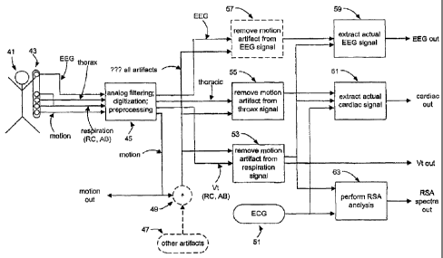
reflective of the functioning of individual physiological systems or
processes. But
separation by inspection or even by conventional filtering of single signals
fails is often not
possible because the individual contributions combined in the sensor signals
have widely
differing amplitudes and/or overlapping spectrums. However, it has been
discovered that
separation of such signals is usefully possible by joint processing of two or
more such
signals, each signal having different relative contributions of each of the
two or more
physiological systems or processes using adaptive processing techniques.
Accordingly,
such signal separation steps are additionally included following artifact
remeval.
100761 In particular, respiration often produces large amplitude movements,
and sensors
often return signals including undesired respiration components along with
components of
interest. Signals are often mixed with signals from other physiological
systems.
Accordingly, processing step 61 jointly processes respiration and thorax
signals to separate
signals primarily reflective of actual cardiac pulsation activity. Respiration
components can
also be present in EEG signals and can optionally be removed by processing
step 59.
Further, signals from other sensors (not illustrated) may be similarly
processed if undesired
- 18 -
CA 02842420 2014-02-07
respiration components are present. In other emnomments, sensor signals may
include
combinations of other pnysiblergicai processes and/or systems, and if so,
their joint
processing is advantageous to produce more useful physiological data. Methods
used arc
similar to those to be subsequently described for removing respiratory
components.
[0077] Furthermore, joint processing of two or more such signals, each signal
having
different relative contributions of each of two or more physiological systems
or processes
using adaptive processing techniques is useful in cases where two or more
physiological
systems or processes interact with each other in physiologically significant
manners (instead
of simply leading to sensor signals with undesired components). Joint
processing in such
cases can produce data in which such interactions are more deafly apparent. In
particular,
heart rate variability ("HRV") and/or respiratory sinus arrhyttunia ("RSA") is
an example
such an interaction arising in cardio-respiratory monitoring, and its
identification 63 is
subsequently described in detail. These interactions can be clearly identified
in the spectra
output from this step. Joint processing of other physiological interactions
often must be
specially designed in view of the particular interaction to be identified, but
such design is
routine in view of principles to be described in connection with HRV and RSA.
5.3.3 INDIVIDUAL PROCESSING STEPS
100781 Individual processing steps 53, 55, 57, 59, 61, and 63 (Fig. 2), and
examples of their
functioning, are now described in detail.
REMOVAL OF MOTION ARTIFACTS FROM RESPIRATORY SIGNALS
[00791 Figs. 3A-B illustrate separating respiratory signals from motion
artifacts by adaptive
processing of the respiratory signals along with a reference signal sensitive
to subject
motion. These figures illustrate RC signal processing; processing of other
respiratory
signals, e.g., AB or Vt signals, is closely similar.
[0080] For irnproved separation, it has been discovered advantageous that all
signals being
jointly processed in a single filtering step be sampled at the same sampling
rate and/or at
coincident sampling times. Since input (preprocessed) sensor signals are often
sampled at
rates specific to the different sensor types, re-sampling steps are
advantageously precede
tittering. Preferably, this re-sampling is to the lowest sampling rate among
the input signals
as long as any down-sampling of any input signal does not result in loss of
relevant
information. In the illustrated embodiment, "RC in" (rib cage size signals)
signals are
sampled at 50 Hz, while "ACC in" (accelerometer motion reference signals) are
sampled at
- 19-
CA 02842420 2014-02-07
Hz. Thus, stain down-sameles "ItC in by a tactor of 5 to Hz to the sampling
rate
Of ACC ïn.
[00811 Further, removal of signal mean values from certain signals has also
been discovered
to improve separation. For respiratory processing, removal of the mean from
the RC and
the ACC signals is advantageous, and steps 73 and 79 are interposed to remove
these
means. In this case, signal means have been found to only slowly vary when the
subject
maintains a single posture, and can removed by simply subtracting a running
average for a
time typical of a single posture, e.g., 30 see.
[0082] Steps 77 and 83 actually separate motion artifacts present in "RC in".
Adaptive
filtering is used in many individual methods of this invention, and is now
generally
described in detail. Specific implementations of adaptive filtering for the
separate methods
are described in connection with the methods themselves.
[0083] Adaptive .filtering process a primary signal having desired components
mixed with
undesired components in order to enhance the desired components at the expense
of the
undesired components. Preferably, an output signal from an adaptive filter is
dominated by
the desired components. Importantly, the filter does not need to be adjusted
in advance to
the expected characteristics of the desired and undesired components, but
instead "learns"
these characteristics from the input signals. The reference signal
specifically "teaches"
characteristics of the undesired components, and therefore preferably
(strongly) correlates
with these component in the primary signal.
10084] Specifically, the input reference signal is linearly filtered so that
it is similar to the
undesired components in the primary signal, and then combined with (subtracted
from) the
primary signal to yield an error signal. The adaptive filer adjusts the linear
filter
coefficients (weights), preferably sample-by-sample, to minimize the error
signal. Since the
minimized error signal is the primary signal from which the filtered reference
signal has
been removed, it contains the desired signal components with enhanced
amplitude.
Conversely, the filtered reference signal, as stated, closely resembles the
undesired
components present in the input primary signal. Either the filtered reference
signal or the
error signal (that is the corrected primary signal) can be further processed.
See, e.g.,
Widrow et al., 1985. Adaptive Signal Processing, Pearson Education Inc.
(included herein
by reference in its entirety for all purposes).
- 20 -
CA 02842420 2014-02-07
(00851 The linear filter may be finite impulse response (FIR.) type or
infinite impulse
=Pon= type (111t); in this invention FIR tillers are preferred because,
compared to IIR
filters, FIR filters are phase linear and allow weight adjustment that are
more stable with
less computational requirements. However, UR filters may be used in other
embodiments
where the computational resources are adequate. Although FIR filters are not
usually phase
linear, they can provide sharper filtering with fewer coefficients than FIR
filters.
10086] Many methods are known for adjusting FIR filter coefficients (weights).
See, e.g.,
Widrow et al., chaps. 6 and 8. A preferred but non-limiting method is known as
the least
mean square (LMS) method, which is a practical approach to adjusting filter
coefficient in
real-time without computationally-intensive matrix inversions (and without
requiring any
prior statistical knowledge of the signals). Specifically, the LMS method
computes filter
coefficients that minimize the mean squared error (MSE) of the error signal by
a steepest
decent method where all filter weights are updated time-sample-by-time-sample.
At each
iteration, the LMS method reduces the MSE. The LMS method determines
coefficients that
both converge from an initial estimate and also adjust to time variations in
primary and
reference signal characteristics.
[0087] In detail, the LMS method proceeds as follows. At each time sample, k,
k> O. the
filtered reference signal, yõ , is determined as usual for a FIR filter:
11-1
Y k =EW(0 irk-a (1)
1-0
Here, the input reference signal at time k-i < k is r,_õ w(k), are the
coefficients, i = 1..N,
at time k, and Nis the filter length. The filtered reference signal is then
subtracted from the
printery signal, Pk, to yield the error signal, z, .
zr = Pk ¨Y.e (2)
Either of both of põ and z, is then further processed. The set of N filter
coefficients is
usually initialized to zero at the first time sample: õ where N is the length
of Frit filter, are
initialized usually to zero:
=t) (3)
At subsequent time samples, all filter coefficients are then updated according
to:
w(k + I), = w(k), +2itzkpir_, (4)
-21 -
CA 02842420 2014-02-07
where # is a convergence parameter that controls the rate of convergence and
stability of the
LMS method.
postil Generally, filter length, N, is chosen in dependence on the amount of
memory
available for the filter, the desired convergence rate, the desired filter
characteristics
(sharpness, etc.), signal bandwidths, and the like. Longer filters take longer
to converge and
can excessively smooth the desired signal component, whilst shorter filters
may not
properly filter a reference signal to remove a sufficient amount of the
undesired signal
component. /n this invention, filter length are generally from approximately
15 to 140
depending on the signals being jointly processed. A typical filter length for
respiratory
signals is approximately 20, which can be adjusted but was found to me more
than
adequate. The convergence parameter, p, can be manually chosen based on
observing
adaptive filter performance or can be chosen automatically by methods known in
the art. It
has been found that once the convergence parameter is properly chosen, the
adaptive filter is
stable and converges in a number of sampling times approximately 1.3 times the
length of
the FIR filter. The parameter ir can be as small as approximately 10 when
processing
signal that have not been normalized to comparable ranges. When the input and
reference
signals are normalized by dividing the signal by its maximum sample for that
time segment,
convergence factors oft = 0.01 to 0.001 provided good convergence rates.
100891 Continuing now with the details of motion artifact removal from
respiratory signals,
respiratory signal 75 including motion artifact resulting from the subject's
motion is the
primary input signal, and accelerometer 81 signal (motion signal) is the
reference signal.
The reference signal is filtered by adaptive filter 83, and then filtered
reference signal 85 is
combined 77 with primary signal 75 resulting in error signal 87. The fihcr
weights are
adapted so that the error signal is minimized, in other words, so that as much
as possible of
the motion aififact is subtracted front the primary signal. The error signal
with enhanced
respiratory components is as "RC out".
(0090] Fig. 3B illustrates motion artifact removal from respiratory signals
monitored from a
strenuously sprinting subject. Here, the subject is sprinting at approximately
3 steps per
second and taking approximately 3 steps per breath (approximately 1 breath per
second).
The first signal band in Fig. 3B illustrates a portion of the "RC in" signal
75 iri which
motion artifact virtually completely swamps the respiration signal. Clearly,
manually
separating the respiratory component frorn the motion artifact is difficult if
not impossible.
- 22 -
CA 02842420 2014-02-07
The second signffligand illustrates a corresponding portion
(Airlift/accelerometer signal 81
reflecting subject motion. Each positive spike in this signal identifies each
step of the
subject as the subject's foot leaves the ground and causes a sudden upward
acceleration.
[00911 The third signal band illustrates adaptively filtered acceleration
signal 85. Close
examination show that the adaptive filter has caused a varying phase lag from
of the filtered
reference signal from the input reference signal, but has left signal spectrum
largely
unchanged. The fourth signal band illustrates signal 87 the "RC out" signal,
which is the
input primary signal with the filtered reference signal subtracted. It can be
appreciated that
most of the undesired motion artifact has been eliminated leaving a resulting
signal with the
subject's respiratory rib cage motions considerably enhanced and clearly
apparent. The fifth
signal band, with "RC in" and "RC out" superimposed, illustrates how the
respiratory
component is almost completely swamped by motion artifact component.
100921 This exarnple illustrates the effectiveness of this motion artifact
removal method.
REMOVAL OF MOTION ARTIFACTS FROM OTHER SIGNALS
[00931 Ivlotion artifacts are removed from other signals, in particular the
thorax signal in
step 55 and the EEG signal in step 57, with techniques substantially similar
to those
described above for motion artifact removal from respiratory signals.
[00941 In situations of less strenuous subject motion, motion artifacts may
have such a
reduced amplitude in EEG signal that their removal in step 57 may be bypassed.
Bypassing
motion artifact removal can be automatically controlled. For example, if a
running average
of power in the motion reference signal falls below a pre-determined threshold
value,
artifact removal can be bypassed. If the power is above the threshold,
artifact removal is
performed. The threshold can be pre-determined differently for different
monitoring signal
inputs.
CARDIAC SIGNAL EXTRACTION
[0095] Thorax signals, "THORAX in" preferably from a mid-thorax size sensor,
often have
desired cardiac pulsation components with amplitudes no more 1% to 5% of the
amplitudes
of the undesired respiratory components. It has been discovered that reliable
extraction of
this relatively small cardiac component requires consideration of two
reference signals: a
respiratory reference signal and an ECG reference signal. The respiratory
reference signal
is "RC in", because signals from a rib cage size sensor have been to correlate
most closely
- 23 -
CA 02842420 2014-02-07
with the undersired respiratory component in "THORAX in". The ECG signal is
processed
to extract an R wave signal.
[0096] The rightmost portion of Fig. 4 illustrates ECG signal processing.
First, R waves are
identified 113 using software or hardware means known in the art, for example,
the Pan and
Tomkins QRS detection algorithm. Prior to R wave identification 113, the "ECG
in" signal,
which can arise from onc or more ECG leads, is interpolated and up-sampled to
1 kHz from
its input sampling rate, which is often 200 Hz.
[0097] Next, it is preferred but optional to discard R waves 115 that can be
identified as
ectopic or artifact. An R wave is identified as ectopic if it occurs in an
unexpected temporal
relation with respect to the adjacent R. waves by being, e.g., more than a
threshold time
interval before or after the expected time of R wave occurrence determined
from a recent
mean heart rate. Mean heart rate can be determined from a running average of R-
R interval
lengths, e.g., from an average of the prior 10 sec of R-R intervals lengths. A
preferred
threshold time interval threshold is approximately 100 mcec. If motion
artifact is not
removed from "ECG in" signals, an R wave is also identified as ectopic if it
occurs during
sufficiently intense subject motion, e.g., when an accelerometer motion sensor
signal
exceeds a threshold value, preferably 0.5-1.5 g. Alternatively, motion
artifact may be
removed from the ECG as described above (either at all times, or only when the
acceleration exceeds the above threshold). Identified ectopic R wave are
discarded from the
R wave signal. Optionally a synthetic R wave is interpolated into the R wave
signal at the
expected R wave occurrence time.
[0098] Lastly, an output R wave signal is constructed. For cardiac filter
extraction, the
output R wave signal preferably identifies the times of R wave occurrences.
For IIRV
analysis, the output R wave signal preferably identifies time intervals
between sequential R
waves. Both output signal are advantageously sampled at 50 Hz.
[0099] The leftmost portion of Fig. 4 illustrates cardiac signal extraction.
Pre-processing 45
of the thorax signal preferably includes band pass filtering with a lower
corner frequency of
approximately 0.4 Hz (range 0.2 - 0.5 Hz) and an upper corner frequency of
approximately
10-15 Hz (range 10 - 30 Hz). This filtering rejects low and high frequency non-
cardiac
components. Next, motion artifact is removed (55 in Fig. 2) from the thorax
signal (and
from the respiratory signal) generating "THORAX in".
- 24 -
CA 02842420 2014-02-07
[0100] Since it again has been found that filtering is improved if all signals
are sampled at
the same rate and/or coincidently, re-sampling step 101 performs any necessary
re-
sampling. In the illustrated embodiment, 'THORAX in" is down-sampled to 50 Hz
from an
original 200 Hz sampling rate in order to match "RC in" and the processed R
wave signal
which are both sampled at 50 Hz. Optionally, the extracted cardiac signal can
be re-
sampled 107, e,g., back to 200 Hz, prior to output. Other common re-sampling
frequencies,
e.g., 100 Hz instead of 50 Hz, may be also be used.
101011 After any necessary re-sampling, adaptive filter 103 processes the
"THORAX in"
primary signal using "RC in' as a reference signal. The adaptive filter is
preferably a FIR
filter with length approximately 120 (range 60-140) and weights adjusted
according to the
LIvIS method as previously described in detail. Different embodiment employ
alternative
adaptive filters (IIR filters, lattice filters, and the like) and di fferent
weight adaptation
methods. Filter output is the error signal in which differences between the
primary thoracic
size sensor signal and the filtered motion reference signal are minimized so
that desired
cardiac components are enhanced while undesired respiratory components are
decreased. In
many situations, the cardiac components in the error signal have been
sufficiently enhanced
so that the error signal is useful without additional processing. In these
situations, further
processing 105 may be bypassed, and the error signal itself may be up-sampled,
output,
and/or input to cardiac feature extraction 111.
[01021 In many other situations, the error signal must be further processed
because it still
contains significant artifact. Because this remaining artifact often does not
strongly
correlate, or correlate at all, with available reference signals, e.g., motion
signals, other
artifact signals, respiratory signals, other sensor signals, and so forth,
further adaptive
processing as described is not advantageous. However, although the remaining
undesired
artifact components also do not correlate with the R-wave occurrence signal
output by ECG
processing, the desired cardiac components naturally do strongly correlated
with these It
wave occurrence ti:nes. Thus, the R-wave signal may he used to identify and
select the
cardiac components from the artifact components (instead of, as in the
adaptive processing
above, using a reference signal to identify and select the undesired artifact
components). A
preferred identification and selection method is ensemble averaging, which has
been found
to usually largely eliminate all remaining artifact.
101031 In detail, ensemble averaging 105 uses the R-wave occurrence signal
output by ECG
processing as a reference "clock" according to which physiologically
corresponding times in
-25 -
CA 02842420 2014-02-07
previous cardiac cycles can be identified and the signals at these
corresponding t
seleCted and averaged since the undesired artifact at these times are not
correlated while
the desired cardiac signal is strongly correlated at these times, ensemble
averaging will
further enhance the desired cardiac components while minimizing remain
artifact
components. Relatively simple ensemble averaging over 5 - 50 prior cardiac
cycles with
constant weights is preferred when its performance is adequate. The following
equation
describes the preferred ensemble averaging:
=
A
(5)
where Ra is the R-wave occurrence nearest to sample time t, and R,. , are the
M-1
previous R-waves in reverse temporal order.
[0104] Lastly, the processed signal may be re-sampled 107 to the desired
output sampling
frequency. Since the extracted cardiac signal usually closely corresponds to
the actual
cardiac volume, parameters of cardiac functioning can be determined 111. For
example, the
amplitude of the extracted cardiac signal is an indicia of stroke voltune; the
amplitude times
heart rate provides an indicia of cardiac output. The time from an R-wave peak
to the
subsequent amplitude maximum of the extracted cardiac signal provides an
indicia pre-
ejection period. The minimum of the derivative of the extracted cardiac signal
(remembering that as heart contracts, its volume decreases) provides an
indicia of the peak
ejection rate. Other cardiac parameters known in the art may similarly be
determined.
Further, the extracted cardiac signal can be used to detect and track
ventricular motion
abnormalities, and changes in detected ventricular motion over time can
provide much of
the same clinical information now detectable only with imaging techniques such
as
ultrasound.
101051 Figs. 5A-F illustrate an actual example cardiac signal extraction.
Figs. SA-C and F
are 25 sec concurrent samples of the "THORAX in" signal (prior to adaptive
filtering), the
"RC in signal, the "ECG in" signal, and the "CARDIAC out" signal (subsequent
to
ensemble averaging), respectively. Figs. 5D is a 150 sec. trace of respiratory
signal after
filtering by the adaptive FIR filter; and Fig. 5E is a 35 sec. trace of the
output of adaptive
filter 103 (prior to ensemble averaging) output. In this example, the TCCi
signal was down-
sampled to 50 Hz prior to adaptive filtering; the adaptive FIR filter had 120
stages; the
convergence factor was it 2 x10⁻¹ ; and the filter coefficients converged from
zero initial
values in about 150 time cycles, or 3 sec at 50Hz.
- 26 -
CA 02842420 2014-02-07
. .
101061 First, comparing the "THORAX in" %gnat of Fig. 5A with the "RESP. (RC)
in
siguai of Fig. 58 it is apparent that: the cardiac motion component appears at
most as small
irregularities in the "THORAX in" signal superimposed on the considerably
larger
respiratory motion component and roughly coincident with the R-waves in the
ECG signal;
and the respiratory motion components of the "THORAX in" and "RC in signals
are
similar. Comparing the unfiltered "RC in" signal of Fig. 5B with the FIR
filtered "RC in"
signal of Fig. 5D, it is apparent that the adaptive FIR filter makes the "RC
in" more similar
to "THORAX in" signal's respiratory component.
[0107] Next, comparing the adaptive-filter output of Fig, 5E with the input
"THORAX in"
signal of Fig. SA, it is apparent that: the adaptive filter has removed nearly
all of the
undesired respiratory component, leaving artifact, noise, and residual
respiratory
component; and cardiac motion (particularly systolic contractions) is more
readily apparent.
Comparing the adaptive-filter output of Fig. SE with the ensemble average
output of Fig.
SF, it is apparent that the ensemble average has eliminated nearly all of the
remaining
undesired components. The cardiac component can bc seen to generally comprise
periodic
beats with slower diastolic inflow ibllowed by rapid systolic outflow, Even
liner details of
the cardiac motion component are also apparent.
(0108j For ease of further comparison, temporally coincident gridlines have
been placed in
Figs. 5A-C and F at approximately 14 secs. In the "THORAX in" signal of Fig.
SA at 121,
a very small, but perceptible, irregularity due to the cardiac motion
component in
superimposed on the respiratory motion component. However, close examination
of the
"RC in" signal of Fig. 51) at 123 reveals that no cardiac motion component is
apparent. The
extracted cardiac motion signal of Fig. SF illustrates peak of systolic
ejection at coincident
time 127, while the ECG of Fig. SC at 125 shows that an R wave, mpresenting
ventricular
depolarization, slightly precedes mechanical ejection at coincident time 508,
measuring the
delay between the R wave and mechanical systole known in the art.
EEG SIGNAL EXTRACTION
101091 Studying subject sleep is one application of ambulatory multiple
parameter
physiological monitoring systems. Although a sleeping subject is not
"ambulatory" in the
sense of actively walking, etc., tnonitoring during unconstrained, normal
sleep can provide a
more realistic record of sleep activities. Thus, ambulatory MPM system are of
use even in
sleep monitoring.
- 2'7 -
CA 02842420 2014-02-07
101101 Sleep monotoring (also other monitoring tasks) is benefited by data
from which a
subjeees mental state can be classified as either awake, or drowsy, or sleep,
and sleep can be
classified as either stage I or II or III sleep or REM sleep. Since mental
state monitoring is
routinely performed be examining electroencephalogram (EEG) records (and
optionally
electro-oculogram (EGG) electro-myogram (EMG) records), processing EEG signals
from
ambulatory MPM systems is described. Processing of EGG and EMG signal is
similar to
EEG signal processing.
[0111] Such processing is preferably configured in view of the following EEG
signal
characteristics. EEG signals frequently contain undesired artifact at least
because the
recorded electrical activity has small amplitudes, often no more than 10's of
micro-volts,
more readily contaminated with undesired components such as noise and
influences from
other physiological systems. In particular, EEG signals often contain
respiratory
components of amplitudes similar to or greater than the EEG signal amplitudes.
On the
other hand, EEG signals processing should be substantially amplitude and phase
linear over
the range ofphysiologically significant frequencies from less than 4 Hz to
greater than 30
Hz (referred to in order of increasing frequency as delta, theta, alpha, and
beta waves).
Additionally, mainly during sleep, EEO signals may include brief higher
frequency bursts
(spindles, K complexes, and the like) that should be linearly processed.
[0112] Figs. 6A illustrates one preferred method for separating undesired
respiratory
components from desired EEG components in signals recorded from one or more
EEG
leads. To preserve frequencies, amplitudes, and phases, "EECi in" signal 161
passes directly
without processing to output stage 157 where "EEG out" signal 159 is
constructed by
subtracting estimated respiratory components 153. Adaptive FIR filter 151
determines the
estimated respiratory components by filtering respiratory reference signal
149, and the
adaptive filter weights are adjusted to minimize an error signal 155
determined as the
difference between EEG primary signal 143 and filtered reference signal 153.
When error
signal 155 is minimized, the filtered respiratory reference signal represents
the respiratory
components as closely as possible. In summary, the respiratory components,
represented by
the adaptively filtered respiratory reference signal, are used to construct
the adaptive filter
error signal from a filtered EEG primary signal and also to construct 'EEG
out" from an
unfiltered EEG reference signal. The error signal, representing the
respiratory k,ompolients
in thc filtered EEG reference signal, is not output. Adaptive processing is
preferred to fixed
bandwidth filters because the frequency spectrum of the undesired respiratory
component
can overlap the lower end of the EEG signal frequency spectnun.
- 28 -
CA 02842420 2014-02-07
,
. .
[01131 The primary arc reference sivat are processed before use by adaptive
filter 151.
Trre respiratory refererrce signal-preferably arises from a rib cage size
sensor, as the RC
signal best c,orrelates with the undesired respiratory components in the EEG
signal. In the
illustrated embodiment, the "RC in" is sampled at 50 Hz and the "EEG in"
signal is sampled
at 128 Hz. To have adaptive filter inputs coincidently sampled as is
preferred, the "RC in"
is up-sampled 147 to 128 Hz; down-sampling "EEG in" is undesirable since
significant high
titquency information can be lost. The up-sampled respiratory reference signal
149 is then
input to the adaptive filter.
10114] It has been found that filter performance is improved by low-pass
filtering 141 the
primary EEG signal to block higher frequency EEG components while passing all
components in the frequency range of the respiratory reference signal.
Accordingly, the
"EEG in" is first passed through low pass filter 141 that allows most of the
respiratory
signal to pass while blocking much of the EEG signal. Filter 141 preferably
has an upper
corner frequency at approximately 1.4 Hz (range 1 - 2 Hz). Such low pass
filtering of the
primary signal is believed to improve filter performance because, first, it
increases the
power of the undesired respiratory component in the primary input signal, and
second, such
increased power permits the adaptive filter to more accurately minimize the
error signal
because the relative decrease of the error signal is greater
[01151 Fig. 6B is an example of removing respiratory components from an EEG
signal.
The first signal band in Fig. 6B is "EEG in" signal 161, which is seen to
contain lower-
amplitude, higher-frequency components superimposed on larger-amplitude, lower-
frequency components. The second signal band is RC reference signal 149, which
is seen to
reflect largely steady breathing at about 15 breaths per minute. The third
signal band is the
adaptively filtered RC reference signal 153 which predicts the actual
respiratory component
in "EEG in". The fifth signal band superimposes the "EEG in" signal with
predicted
respiratory component 153. It can now be appreciated that much of the larger-
amplitude,
lower-frequency components in "EEG in" are of respiratory origin, while all
the lower-
amplitude, higher-frequency components are of EEG origin. The fourth band is
"EEG out"
signal 159 which include the higher-frequency EEG components without
detectable
respiratory components.
ANALYSIS OFIIRV AND RSA
[01161 Heart rate variability (I-IRV) refers to alterations in heart rate
often measured from
variations of RR intervals. HRV has many physiological uses, and is further
useful in
29
CA 02842420 2014-02-07
assessing cardiovascular disease. Generally, heart rate is infiuenced by the
autonomic
nervous system (AN), in particular by the fluctuating balance between the
sympathetic and
parasympathetic (vagal) branches of the ANS. Chemoreceptor processes,
thermoregulation,
and the rennin-angiotensin system are believed to cause very low frequency
HRV, below
approximately 0.04 Hz. A low frequency (LF) component between 0.04 Hz and 0.15
Hz is
believed to reflect the balance of sympathetic and parasympathetic branches of
the ANS.
Finally, direct vagal (parasympathetic) modulation of the sino-atrial node
causes high-
frequarcy band (HT') IIRV between approximately 0.15 to approximately 0.4 Hz
or higher.
Vagal activity is usually strongly influenced by respiration, and the
resulting HF modulation
found predominantly at respiratory frequencies is known as respiratory sinus
arrhythmia
(RSA).
[0117J Heretofore, HRV and RSA amplitude have determined by time donaain,
phase
domain, and frequency domain approaches. In time-domain approaches, .maximum
and
minimum values of R-R time intervals are measured within the bounds of each
breath. In
phase domain approaches, RSA is determined by analyzing heart rate dynamics
with respect
to respiratory phase. Finally, HRV is often spectrally analyzed by Fourier
transforming
appropriately windowed time R-R interval time series (windows reduce spectral
leakage and
filter random noise). None of these approaches is entirely satisfactory, often
because
absence of respiratory reference signals makes difficult determining the
origin of measured
HRV components.
[0118J Fig. 6A illustrates improved HRV and RSA analysis methods and systems
of this
invention which adaptively filter and R-R interval primary signal using a
respiratory
reference signal. The primary signal is "RR in" 173, an R-R interval signal
derived, e.g., by
processing an ECG signal by methods described with respect to Fig. 4. An RR
interval
signal is piecewise constant, each constant piece representing the length
immediately
previous R-R interval. In the illustrated embodiraent, "RR in is sampled at 50
Hz and
down-sampled (decimated) 175 by a factor of 10 to 5 Hz. Since HRV frequencies
of
interest are generally less than 0.5 Hz of less, a 5 Hz sampling rate provides
an adequate
signal representation. It has been found that the adaptive filter convergence
is improved if
its baseline (zero frequency component) is at zero. Accordingly, the baseline
may be
removed 177 (also referred to as "de-trending") after down-sampling and prior
to the
adaptive filter by subtracting the mean of a preferably 5 minute signal
segment using a best
straight line fit However, for particular uses, it may be advantageous to
retain the average
RR interval value (zero frequency component). In these cases, de-trending 177
is bypassed.
- 30 -
CA 02842420 2014-02-07
,
101191 The preferred secondary reference signal is the tidal volume, Vt, which
has been
found to correiate m.ost closely with respiratory modulation of heart rate.
This signal can be
derived, as explained in one or more of the above referenced US patent
relating to IP
technology, from a combination of signals of rib cage and abdomen size sensor
signals, and
in the embodiment is sampled at 50 Hz. Adaptively filtered signals are
preferably sampled
coincidently on at the same frequency, and Vt in is accordingly down-sampled
(decimated)
185 to 5 Hz to match the RR sampling frequency. Prior to down-sampling
extraneous
signal components are removed by low pass filter 171, which has preferred
upper corner
frequency of approximately 1.4 Hz (range 1 - 2 Hz). As above, this processing
does not
lose physiological information as frequencies of interest in HRV analysis are
generally less
than 0.5 Hz of less.
[01201 Next, signals 179 and 187 are adaptively filtered 189 and 181,
preferably with a FIR
type filter employing LMS method for weight adjustment. Error signal 191
results from
subtracting the adaptively filtered Vt signal from the RR signal. An adaptive
filter length of
approximately 20 has been found adequate for the illustrated HRV analysis.
Varying filter
length from 20 did not significantly improve HRV analysis. The filter
convergence
parameter, a, was advantageously in the range of approximately 2.5x1 0-9 to
approximately
2x10⁴, the low values resulting from use non-normalized input signals. As
input signal
characteristics change, filters parameters can be adjusted either manually or
automatically
(here and in the previously described LMS adaptive-filtering examples) with
programmable
features common in most signal processing software packages.
101211 As explained, the highest frequency (HF) components in the HRV spectra
are
usually due to respiratory modulation (RSA), the other components are usually
of lower
frequencies LF) and are due to other influences. Since adaptively filtered Vt
signal 193 is
as close as possible to the respiratory components in the RR signal, this
signal primarily
contains the HP HRV components, that is the RSA components. Conversely
minimized
crror signal 191, having had the }IF components subtracted 181, primarily
contains the LF
IIRV components believed to be due to other than respiratory influences.
f01221 In embodiments where an RR interval signal retaining the LF components
of
variability including the zero frequency component, the average RR interval,
preliminary
de-trending 177 is bypassed, and signal 191 is output. Then de-trending is
performed 177"
just prior to spectral analysis. In other embodiments, de-treading 177 is
performed since is
often results in more rapid filter convergence, while de-trending 177" is
bypassed. In all
- 31 -
CA 02842420 2014-02-07
cases, RIC signaller-trending is preferably prior to spectral analysis and may
be entirely
bypassed is spectral analysis is omitted).
[0123] Spectral analysis of the LF signal 195 and the HF signal 197 may be
performed by
many of the spectral analysis techniques known in the art. A preferred
techniques is
Welch's averaged, modified periodogram method, which analyzes a signal section-
by-
section with adjacent sections overlapping by 50% overlap. Prior to Fourier
transforming,
each section is windowed, preferably with a Hamming window although other
known
window functions may be used, e.g., Blackman-Harris, or Nutal, or the like.
Rectangular
windows are not preferred as they normally introduce spectral leakage. The
combined
results of the LF and IV spectra, signals 199 and 201, respectively, jointly
represent the full
spectrum of HRV, and the HF spectra alone represents RSA. Optionally, the
power in the
LF tmd HF frequency bands may be calculated across the ranges 0.04-0.15 and
0.14-0.4 Hz,
respectively.
[0124] Figs. 8A-1013 illustrate three examples of the above described HRV
analysis. Each
example has an analysis of separate 6-7 min segments of data extracted from a
continuous
16 hour ambulatory IV1PM record obtained with LifeShirtn" (VivoMetrics, Inc.,
Ventura,
CA). For 8 hours of the 16 hour period, the subject was awake, ambulatory, and
performing
normal daily tasks. For the remaining 8 hours, the subject was sleeping
without any
constraint on motions during sleep. A tidal volume signal, Vt, was obtained
from
monitored RC and AB signals and an RR signal was derived as described above.
[0125] Figs. 8A-B, Figs. 9A-B, and 10A-B all illustrate similar signals. In
Figs, 8A, 9A,
and 10A, the first two signal bands are input Vt signal 183 and input RR
signal 173 sampled
at 50 Hz (and, for the Vt signal, low pass filtered), respectively, while the
second two signal
bands are these signals down-sampled to 5 Hz, that is signals 187 and 179
(and, for the RR
signal, linearly de-trended), respectively. The fifth signal band is
adaptively filtered Vt
signal, which is the component of the RR signal predicted to correlate with
breathing, that is
the RSA component. The sixth signal band is RR signal 191 with the HF
component
subtracted but retaining the LF FIRV components. In Figs. 8B, 9B, and 10B, the
top two
spectra are of RR signal 179 and Vt signal 187, while the bottom two spectra
are of HF
(RSA) component 201 and of LF component 199.
[0126] It can be appreciated by examining the illustrated spectra that HRV
differs in each of
the three examples; in particular, the relative amount of RSA compared to LF
variability can
vary considerably. Further, it is readily apparent that in all examples RSA
has been cleanly
- 32 -
CA 02842420 2014-02-07
separated from the LF HRV due to other causes. In tact, exceptIlli perhaps a
tew percent
of spectral leakage, the RSA and LF variability have been completely
separated.
Additionally, little or no smoothing has occurred; the spectral details of the
separated
signals preserve well the details of the spectra of the original RR signal.
QT INTERVAL CORRECTION
[0127] Turing to Fig .11, which is a schematic of an ECG cardiac cycle, the QT
interval in
the ECG is the titne interval (usually specified in milliseconds) between a Q
wave (the first
component of the QRS complex) and the immediately following T wave. Electro-
physiologically, the QRS complex represent systolic depolarization of the
ventricles; the T
wave represents ventricular re-polarization; and the QT interval is the
represents an
approximate plateau period of ventricular depolarization. This interval is of
considerable
importance: its prolongation is associated with increased risk for malignant
ventricular
arrhythmia and sudden cardiac death in post myocardial infarction patients;
and new drug
evaluations must now include assessment of a drug's effects on the QT length.
[0128] Determining the QT interval is complicated by its strong dependence
upon the
length of the preceding cardiac cycle (e.g., the preceding RR interval). A
useful
determination of the QT interval should correct for this RR interval effects
and many
techniques for making this correction and determining a corrected QT interval,
QTc, have
been described in the art. One common technique is due to Bazett and is simply
expressed
by the following equation:
QT
QT = (6)
.12/t
where: QTc is the QT interval corrected for heart rate; QT is the measured
interval between
the Q wave and the tbllowing T wave; and RR is the measured RR interval
(specified in
seconds). See, e.g., Bazett, 1920, An analysis of time-relations of
electrocardiogram. Heart
7:353-370. Another common technique for QT interval correction uses a linear
regression
model expressed by the following equation:
Q71, Qt +0.154(1¨ RR) , (7)
where: QT1..c is the linearly corrected QT interval, and RR is the measured RR
interval.
See, e.g., Sagie et al., 1992, An improved method for adjusting the QT
interval for heart rate
- 33 -
CA 02842420 2014-02-07
(the FramingharMeart Study). Am .1 Cardiol ;70:797-801. Limitations of these
teelmiques
include aver-correction of the QT interval at high heart rates, or lack or
verification in
ambulatory conditions or limited predictive power (the linear regression model
accounting
for only approximately 46%, r = 0.68, of QT interval variance).
[01291 This invention provides improved systems and method for QT correction
that utilize
the results of thc previously described HRV and RSA analysis. Although direct
parasympathetic (vagal) modulation oldie QT interval is believed to be
relatively
unimportant, indirect effects due to the variations in heart rate (HRV)
because of
parasympathetic input to the sino-atrial node are significant, often to the
extent of obscuring
the baseline QT interval and its changes. As described., a major component of
HRV is due
to respiratory influences (RSA), and this invention's methods and systems
clearly separate
HRV into RSA and non-respiratory LF components.
[0130) In a first improved correction method, the QT interval is corrected
according to the
known corrections techniques but previous RR interval measures are replaced by
RR
interval signal 203 (Fig. 7) with LF HRV but excluding HF RSA variability.
Thereby, QTc
reflects LF HRV with obscuring effects from RSA eliminated. Far example, the
previously
mentioned QTc correction techniques become:
QT
(8)
VBR2o3
and
Vric = + 0. I 54 (1 ¨ Rk2õ ) (9)
where RR203 refers to signal 203 in Fig. 7.
[01311 In another improved correction method, the QT can be corrected by
performing on a
QT interval signal the same processing as illustrated in Fig. 7 for the RR
interval signal. A
QT interval signal may be derived from an ECG signal in the same manner as the
RR signal
is derived. The LF QT variability spectra represents slower variations in the
QT interval not
related to respiratory influences on heart rate. The HF QT variability spectra
represents the
indirect respiratory influences on the QT intervaL Also, use of respiratory
variables in
addition to RSA will allow for a more informed and accurate estimation of the
QTc
correction.
- 34-
CA 02842420 2014-02-07
101321 Use of either of these improved QT correction techniques permits a more
accurate
QTc estimate, and accordangly a more accurate estimate of other influences,
e.g., an
administered drug, on the QT interval.
[0133] The invention described and claimed herein is not to be limited in
scope by the
preferred embodiments herein disclosed, since these embodiments are intended
as
illustrations of several aspects of the invention. Indeed, various
modifications of the invention in
addition to those shown and described herein will become apparent to those
skilled in the art
from the foregoing description.
The scope of the claims should not be limited by the preferred embodiments or
the examples, but
should be given the broudest interpretation consistent with the description as
a whole.
-35-