Note: Descriptions are shown in the official language in which they were submitted.
MULTI-SPOT LASER SURGICAL PROBE USING
FACETED OPTICAL ELEMENTS
Field of the Invention
This invention relates to optical surgical probes and, more particularly, to a
multi-spot
laser surgical probe using faceted optical elements.
Background of the Invention
Optical surgical probes deliver light to a surgical field for a variety of
applications. In
some applications, it may be useful to deliver light to multiple spots in the
surgical field. For
example, in pan-retinal photocoagulation of retinal tissue, it may be
desirable to deliver laser
light to multiple spots so as to reduce the time of the pan-retinal
photocoagulation procedure.
Various techniques have been employed to produce multiple beams for a multi-
spot pattern. For
example, one approach uses a diffractive beam splitter element to divide an
incoming beam into
multiple spots that are coupled into multiple optical fibers that deliver the
multiple spots to the
retina. But it is also desirable to have a multi-spot generator that can be
placed at a distal end of
the optical surgical probe to more easily produce multiple spots from a single
input beam, so
that the multi-spot generator can more easily be used with existing laser
sources without the
need for additional components to align the laser surgical probe with the
sources.
Difficulties can arise in the use of a diffractive beam splitter element at a
distal end of
the optical surgical probe. As one example, a diffractive beam splitter
element produces a
multitude of higher diffraction orders, and while these orders are relatively
lower in light
intensity as compared to the primary spot pattern, they may not always be
negligible in terms of
their effects. As another example, a diffractive element may not perform
identically in different
refractive media. For example, if the diffractive beam splitter element is
placed into a medium
other than air, such as saline solution or oil, the recessed portions of the
microscopic surface
relief structure of the diffractive beam splitter element can be filled with
material having a
different refractive index than air, which can ruin the spot pattern. As yet
another example, the
spacing between the spots can vary for different wavelengths, which can be
problematic when
an aiming beam is of a certain color while a treatment beam is of a different
color. Lastly,
diffractive elements are frequently expensive and difficult to produce, and
this is particularly
the case when the diffractive element must be constructed to fit into a small
area, such as a distal
1
CA 2842474 2018-10-03
tip of a surgical probe for surgical instruments that are 23-gauge or smaller.
Thus, there remains
a need for an optical surgical probe that can produce multiple spots at a
target area using optical
elements at a distal end of the surgical probe.
Brief Summary of the Invention
Certain exemplary embodiments can provide a optical surgical probe comprising:
a
handpiece comprising a metal cannula, the metal cannula located at a distal
end of the
handpiece; a light guide extending within the metal cannula; the light guide
configured to carry
a light beam from a light source through the metal cannula; a multi-spot
generator formed within
a distal opening of the metal cannula and configured to seal the distal
opening of the metal
cannula, the multi-spot generator comprising: a faceted end surface spaced
from a distal end of
the light guide facing proximally within the metal cannula, the faceted end
surface including at
least one facet oblique to a path of the light beam; and a ball lens located
distal to the faceted
end surface; and a high-conductivity ferrule surrounding the distal end of the
light guide and
being in thermal contact with the metal cannula, the high-conductivity ferrule
comprising a side-
shield portion extending beyond the distal end of the light guide and
configured to shield the
cannula from a portion of the light beam reflected by the faceted end surface
of the multi-spot
generator.
Other embodiments provide a thermally robust optical surgical probe including
a multi-
spot generator with a faceted optical adhesive element. In particular
embodiments, an optical
surgical probe includes a handpiece configured to optically couple to a light
source and a
cannula at a distal end of the handpiece. The probe further includes at least
one light guide
within the handpiece. The light guide is configured to carry a light beam from
the light source
to a distal end of the handpiece. The probe also includes a multi-spot
generator in the cannula
that includes a faceted optical adhesive with a faceted end surface spaced
from a distal end of
the light guide. The faceted end surface includes at least one facet oblique
to a path of the light
beam. In some embodiments, the probe also includes a high-conductivity ferrule
at the distal
end of the light guide. In other embodiments, the cannula is formed from a
transparent material.
Other objects, features and advantages of the present invention will become
apparent
with reference to the drawings, and the following description of the drawings
and claims.
2
CA 2842474 2018-10-03
Brief Description of the Drawings
FIGURE 1 illustrates a multi-spot generator with a highly thermally conductive
ferrule
according to a particular embodiment of the present invention;
FIGURE 2 illustrates a multi-spot generator with a transparent cannula
according to a
particular embodiment of the present invention; and
FIGURE 3 illustrates a multi-spot generator with a transparent cannula
according to an
alternative embodiment of the present invention.
Detailed Description of the Preferred Exemplary Embodiments of the Invention
U.S. Patent No. 8,764,261 (filed December 3, 2010 and issued July 1, 2014)
commonly
assigned with the present Application, describes a multi-spot
2a
CA 2842474 2018-10-03
CA 02842474 2014-01-20
WO 2013/022693
PCT/US2012/049297
optical surgical probe using faceted optical adhesive. Various embodiments of
the
present invention provide additional features to facilitate the use of faceted
optical
adhesive in optical surgical probes. In particular, certain embodiments of the
present
invention provide a thermally robust optical surgical probe using faceted
optical
s adhesive. As
described in detail below, particular embodiments of the present
invention incorporate additional features to reduce the likelihood that "hot
spots" will
develop in the surgical probe that could cause the faceted optical adhesive or
the
adhesive joining the ferrule and the cannula to degrade and/or fail.
In certain embodiments of the present invention, a ferrule located within the
is distal end of
the probe is modified to improve its ability to conduct heat away from
the distal tip of the probe. The first modification is to change the material
from the
typically-used, low-thermal-conductivity stainless steel to a material with a
much
higher thermal conductivity such as copper or silver. The ferrule material
need not
necessarily be biocompatible since it is physically isolated from the outside
of the
s probe. This
permits the selection of a non-biocompatible material such as copper or
silver that has much higher thermal conductivity than any available
biocompatible
materials. The higher thermal conductivity enables more efficient conduction
of heat
away from the distal end of the probe. The second modification is to add to
the
cylindrical ferrule a distal side-shield that prevents light reflected off of
the adhesive
20 facets from
illuminating and being absorbed by the cannula. Instead, reflected light
illuminates and is substantially absorbed by the high-thermal-conductivity
ferrule that
efficiently conducts the heat away from the distal end of the probe.
In an alternative embodiment of the present invention, the absorptive cannula
is replaced with a transparent cannula that transmits reflected light from the
adhesive
25 facets into the
ambient region outside of the cannula. This results in a significant
reduction in the temperature of the distal end of the probe. Since high
intensity
transmitted light directed toward the surgeon may interfere with his view of
the retina,
various means are available to block or dissipate this light, including a
reflective,
diffusive or translucent layer on the outside of the transparent cannula and
an opaque
30 cylindrical
cannula outside of the transparent cannula and physically separated from it
by an insulating air gap. This opaque cannula need not necessarily be made
from a
highly thermally conductive material but it can be made from a stiff and
strong
material such as stainless steel which provides added structural strength to
the distal
end of the probe.
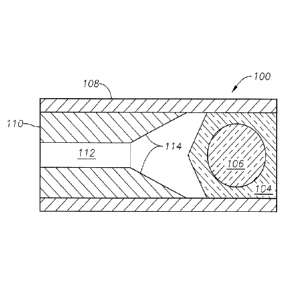
35 FIGURE 1
illustrates a multi-spot generator 100 according to a particular
embodiment of the present invention suitable for placement at a distal end of
an
optical surgical probe. In the depicted embodiment, a faceted optical adhesive
104
having a ball lens 106, such as a sapphire ball lens, is located within a
cannula 108.
3
CA 02842474 2014-01-20
WO 2013/022693
PCT/US2012/049297
Within the cannula 108 is a high-conductivity ferrule 110 holding an optical
fiber 112.
The optical fiber 112 delivers light, such as laser light, from an
illumination source
(not shown).
The high-thermal-conductivity ferrule 110 (hereinafter referred to as "high-
s conductivity
ferrule") is formed from a material with a thermal conductivity
significantly higher than the stainless steel material ordinarily used in
optical surgical
probes, which is typically around 15 W/m-K. For purposes of this
specification,
"high-conductivity" will refer to materials having thermal conductivity in
excess of
100 W/m-K. Suitable examples include copper (conductivity of 372 W/m-K),
sterling
io silver (410 W/m-
K), or pure silver (427 W/m-K). Because the high-conductivity
ferrule 110 is encapsulated within the cannula 108 by the faceted optical
adhesive
104, the high-conductivity ferrule 110 need not be made of a biocompatible
material,
which allows consideration of high-conductivity materials that are not
ordinarily used
in optical surgical probes.
15 The high-
conductivity ferrule 110 includes a side-shield 114 extending distally
past the optical fiber 112. The side-shield 114 is oriented to receive light
reflected
from facets of the faceted optical adhesive 104. While reflections from the
faceted
optical adhesive 104 are relatively low in energy compared to the incident
beam
(approximately 5% of incident energy), such reflections can nonetheless
produce "hot
zo spots" on the
cannula 108. Given that the cannula 108 is ordinarily formed from
stainless steel or other relatively poorly conducting material that is also
not highly
reflective, this can result in laser energy being absorbed, which in turn
creates the
potential for excess heat to accumulate near the faceted optical adhesive 104
or near
the adhesive that bonds the ferrule 110 to the cannula 114 (not shown). This
can
zs degrade the
performance of the optical surgical probe. The side-shield 114 intercepts
the reflected beams to prevent them from reaching the cannula, and because the
material of the ferrule 110 is highly conductive, any heat produced by
absorption of
the reflected beams in the ferrule 110 is rapidly dispersed, preventing the
equilibrium
temperature of the ferrule 110 from being significantly raised.
30 FIGURE 2
illustrates an alternative embodiment of a thermally robust multi-
spot generator according to the present invention. In the depicted embodiment,
cannula 108 is formed of a transparent material. The material is preferably
biocompatible, but if it is not, the outer surface cannula 108 may also be
coated or
treated to improve biocompatibility. The transparent cannula 108 allows the
reflected
35 light to pass
through the cannula 108 to avoid forming hot spots. In particular
embodiments, the cannula 108 is diffusive, so that the escaping light does not
form
visible light spots that could be distracting for a surgeon. For example, the
surface of
the cannula 108 could be formed from a translucent material, could be
chemically or
4
CA 02842474 2014-01-20
WO 2013/022693
PCT/US2012/049297
mechanically frosted (such as by scraping or acid etching), or could be coated
with a
diffusive coating. In alternative embodiments, the cannula 108 is transparent,
but
surrounded with a reflective coating, such as silver. The reflective coating
prevents
light from escaping into the surgeon's field of view while still reflecting
the light
away from the faceted optical adhesive 104, allowing the heat to be conducted
away
from the tip easily. The outer surface of the silver or reflective coating can
be
oxidized or coated to improve biocompatibility.
FIGURE 3 illustrates an alternative embodiment of the transparent cannula
108. In the depicted embodiment, an opaque outer cannula 120 surrounds the
io transparent
cannula 108. The opaque outer cannula 120 may be formed from
conventional biocompatible materials, and it is preferably relatively
absorptive,
although it need not be highly conductive. The outer cannula 120 and the
transparent
cannula 108 are separated by an air gap. The air gap provides thermal
insulation
between the transparent cannula 108, and the transparent cannula 108 may also
be
formed from an insulative material like glass. Whatever heat is produced in
the outer
cannula 120 may be conducted away into other parts of the probe or the
biological
material surrounding the outer cannula 120. The thermal insulation between the
outer
cannula 120 and the faceted optical adhesive 104 reduces the likelihood of
excess heat
from accumulating near the faceted optical adhesive 104.
The present invention is illustrated herein by example, and various
modifications may be made by a person of ordinary skill in the art. Although
the
present invention is described in detail, it should be understood that various
changes,
substitutions and alterations can be made hereto without departing from the
scope of
the invention as claimed.
5