Note: Descriptions are shown in the official language in which they were submitted.
CA 02843097 2014-02-20
STENT APPARATUSES FOR TREATMENT VIA BODY LUMENS AND
METHODS OF USE
FIELD OF THE INVENTION
Apparatuses and methods are provided for treatment and/or support via body
lumens and/or other hollow organs.
BACKGROUND OF THE INVENTION
A stenosis is a stricture of a canal or duct. In the context of the vascular
system
a stenosis is a narrowing of the lumen of a blood vessel. A stenosis can
severely
restrict blood flow and promote thrombosis which can lead to myocardial
infarction or
stroke, for example. A common type of primary stenosis is caused by a buildup
of
atherosclerotic plaque.
Several therapeutic methods have been developed to improve circulation and
homeostasis in stenotic vessels including by-pass surgery and
revascularization
procedures. Revascularization procedures (e.g. balloon angioplasty, bare metal
stents
as well as drug eluting stents, atherectomy, rotary ablation (rotablation))
serve to
improve blood flow by reducing or removing the stenosis. However, these
procedures
frequently injure the blood vessel. The biological response to the injury is a
multifactorial fibro-proliferative process that is similar to wound healing,
and includes
the elaboration of growth factors from a variety of cell types, infiltration
of leukocytes,
migration and proliferation of smooth muscle cells, the production of
extracellular
matrix and tissue remodeling. The process can result in the formation of a
thick
neointima within the vessel wall which reduces the lumina( area of the vessel
(e.g.
restenosis). Various levels of restenosis occur following about 20-50% of
coronary
angioplasty procedures.
Attempts have been made at reducing restenosis following vascular
intervention procedures by, for example, placing endovascular stents at the
location of
the stenosis. At present, this treatment sometimes itself causes restenosis.
Stents are
typically implanted within a vessel in a contracted state and expanded when in
place in
the vessel in order to maintain integrity of the vessel and to allow fluid
flow through
the vessel. Typically, implantation of sterns is accomplished by mounting the
stent on
CA 02843097 2014-02-20
the balloon portion of a catheter, positioning the stent in a vascular lumen,
and
expanding the stent to an expanded state by inflation of the balloon within
the stent.
The stent can then be left in place by deflating the balloon and removing the
catheter.
One problem with stenting according to this widely used procedure, however, is
that as
the stent expands, it engages relatively brittle plaque lining the arterial
tissues
surrounding the stent, not the arterial tissue itself In doing so, the
expanding stent
cracks the plaque to produce debris. This debris, in an untended condition,
then enters
the blood stream and occasionally injures the patient further by causing a
vessel
blockage downstream. This debris release is exacerbated by the fact that
conventional
stent structure contains large gaps, enabling the debris to move freely into
the
bloodstream. This debris creating effect is especially problematic when
stenting in the
carotid arteries, where the downstream blood flow leads directly to the brain
and debris
can cause strokes. In coronary arteries, debris is particularly dangerous
because it can
lead to heart attacks.
Currently, protection against this debris is carried out during the stenting
procedure by using a downstream embolic shower protection device. This sort of
device acts as a filter which traps debris of a predetermined size from
transiting
through the cardiovascular system. There are a number of drawbacks with using
these
embolic shower protection devices as they exist currently. One drawback is
that they
often encompass using another device, in addition to the balloon catheter
which must
be inserted into the patient, adding time and potential danger to the
procedure. Another
drawback is that the protection device must be downstream of the stent
location,
therefore, some additional stretch of vasculature must be available in order
to properly
position the protection device. Yet another drawback is that the embolic
shower
protection device is removed at the conclusion of the stenting procedure and
therelbre
does not provide any protection after that point, despite the fact that post
procedure
debris can become potentially dislodged as a result of the procedure. Yet
another
drawback is that the embolic shower protection device is placed some distance
from
the stent, thus possibly leaving some close side branches unprotected.
Another common practice in use with stenting procedures is the use of stents
for administering pharmacologic agents to treat restenosis and other body
ailments
through the lumen wall. Because of the mechanical strength that is required to
properly
support vessel walls, stents are typically constructed of metallic struts.
However, these
2
CA 02843097 2014-02-20
struts are often constructed to be thin because, in general, foreign material
in the body
is to be avoided and because of the need to obtain a stein that can be
crimped, flexible
and conform with the blood vessel anatomy. Arterial stents are built to cover
a
minimum amount of the blood vessel's walls, while still having a high radial
force in
order to avoid collapsing and thus keeping the lumen open. Typically, the
metal struts
cover only about 10% of the total covered area, and the stent somewhat
resembles a
cylindrical fishing net. However, one drawback with these steins is that
pharmaceuticals are placed only on the stent struts, which cover only a small
portion of
the blood vessel's wall, and they do not cover the apertures in the stent.
Thus the
therapeutic effects of the drug are achieved only on a small portion of the
injured
tissue. Since some pharmaceuticals are comprised of large molecules, with a
very high
molecular weight, and/or complicated and/or wide stereochemistry, and which
have
limited diffusion capabilities, there is a large area of tissue which is not
effectively
treated. Another drawback of current drug eluting stents is that in attempts
to
overcome the diffusion issues, an excessive amount of drug must be eluted in
the
hopes that it will permeate to the target tissue. In some cases, this causes
undesirable
overdosing of the tissue areas closest to the stent struts in addition to the
added
expense of using copious amounts of the drug. Furthermore, there are design
limits
which prevent increasing the amount of drug embedded and thus, eluted from the
stent.
U.S. Patent Publication No. 2004/0030377 to Dubson et al. describes a stent
assembly which is designed to deliver pharmaceuticals to a blood vessel after
implantation while encouraging endothelial growth.
Today's drug eluting stents suffer from higher incidences of sub acute
thrombosis than the previous generations of bare metal stents. Longer
administration
period of anticoagulant drugs like Plavix® is needed, with additional cost
and
more side effects for the patients. The main reason for the sub acute and the
chronic
thrombosis is sudden exposure of a small area of the stent strut to the blood
stream.
The small area of exposed stent typically happens when several adjacent
endothelial
cells fall from the stent strut surface leaving an exposed area of the strut
structure and
3
CA 02843097 2014-02-20
producing a site on which blood platelets can clot. Even if the patient is
being treated
with anticoagulants, there is a very high risk that the platelets will stick
to the exposed
stem and cause clotting. This phenomenon may lead to a total occlusion of the
blood
vessel and to an immediate myocardial infarction. Drug eluting stents are more
susceptible to such incidences since the conformity and the integrity of the
endothelial
cells covering the polymer is not as good as when they are covering a bare
metal stent.
SUMMARY OF THE INVENTION
An aspect of some embodiments of the invention relates to providing an
enhanced stent apparatus which includes at least one porous structure and
optionally a
support clement (e.g. a stern) at least partially covered by the porous
structure. In an
embodiment of the invention, the porous structure has a thickness of less than
100
microns. Optionally, the porous structure has a thickness of less than 20
microns.
Optionally, the porous structure has a thickness of less than 10 microns.
Optionally,
the porous structure is of varying thickness. In an embodiment of the
invention, the
porous structure is comprised of at least one fiber whose thickness is less
than 100
microns. Optionally, the porous structure is comprised of at least one fiber
whose
thickness is less than 20 microns. In an embodiment of the invention, the
porous
structure is comprised of at least one fiber whose thickness is less than 10
microns. in
some exemplary embodiments of the invention, the porous structure is comprised
of at
least one fiber whose thickness is in the range of 40 nm - 40 microns. In some
embodiments of the invention, the fiber thickness is in the order of the
thickness of
the porous structure. For example, the fiber thickness is at least half the
thickness of
the porous structure.
In some exemplary embodiments of the invention, the porous structure is
placed on the exterior of the stent support element, "exterior" meaning
between the
support element and a body lumen wall. In some embodiments, a porous structure
is
placed on the interior of the stern support element. Optionally, a porous
structure is
placed both on the exterior and the interior of the stent. In some embodiments
of the
invention, the porous structure and/or the support element are used to treat
the lumen
with pharmaceuticals. In some embodiments of the invention, the porous
structure is
at least temporarily secured to the support element. For example, to
facilitate insertion
and deployment of the enhanced stent apparatus in a patient.
4
CA 02843097 2014-02-20
in an exemplary embodiment of the invention, at least one fiber's thickness is
less than the diameter of an endothelial cell. In some exemplary embodiments
of the
invention, the porous structure's thickness is less than the diameter of an
endothelial
cell. In some exemplary embodiments of the invention, the fiber diameter is on
the
same approximate order of size (e.g. diameter or French (circumference)) as a
typical
endothelial cell (about 100 microns square, e.g. 3x30 microns).
In some embodiments of the invention, the porous structure is comprised of at
least one super-fiber which is comprised of a plurality of bundled fibers. In
an
embodiment of the invention, the super-fiber has an overall thickness of less
than 100
microns. Optionally, the super-fiber has an overall thickness of less than 20
microns.
Optionally, the super-fiber has an overall thickness of less than 10 microns.
In some embodiments of the invention, the porous structure and/or the stent
arc placed over a balloon-type catheter, likc an angioplasty balloon and are
balloon
expandable. In some embodiments of the invention, the porous structure and/or
the
stent are self-expandable. In some embodiments of the invention, the porous
structure
expands with the support element during deployment, whether with the
angioplasty
balloon or via self-expansion.
In an embodiment of the invention, at least the porous structure is made of a
rcsorbable polymer. Optionally, the stent is made of a resorbablc polymer.
Optionally,
the porous structure is made out of a resorbable and/or degradable polymer. In
some
embodiments of the invention, at least one polymer is used as a cover for the
stent
and/or porous structure. In some embodiments of the invention, the stent
and/or the
porous structure are made up a plurality of layers which exhibit different
performance
characteristics depending on the desired result. For example, some layers do
not
include pharmaceutical agents, where as some optionally do.
In an exemplary embodiment of the invention, the apertures and/or the stent's
struts and/or the fiber thicknesses of the porous structure arc sized to
encourage
growth of endothelial culls therethrough but to prevent transmission of
particulate
debris greater than a predetermined size, thereby also providing embolic
shower
protection. In an exemplary embodiment of the invention, the fiber diameter
and/or
French size is used as a measure for choosing at least one fiber for
constructing the
porous structure. The apertures sizes are optionally designed to capture and
hold any
plaque, greater than a predetermined size, which may be dislodged from the
lumen
5
CA 02843097 2014-02-20
wall. The size of the apertures may vary in some exemplary embodiments of the
invention. Optionally, the porous structure is comprised of apertures
averaging no
greater than 80 microns in diameter. Optionally, the porous structure is
comprised of
apertures averaging no greater than 200 microns in diameter. Optionally, the
porous
structure is comprised of apertures between 200 and 1500 microns in diameter
(see
description of diameter as being approximate or used in lieu of French,
herein).
In an embodiment of the invention, under 30% of the porous structure surface
area is dedicated to structure, leaving the remaining 70% of' the porous
structure
surface area as empty space, or apertures. In embodiments where the porous
structure
is placed on a support element, this means at the most, 30% of the support
element is
covered, or in other words, the coverage area of the porous structure is 30%.
Optionally, the coverage area of the porous structure is less than 204.14.
Optionally, the
coverage area of the porous structure is less than 15%. Optionally, the
coverage area
of the porous structure is less than 5%. Coverage area in the above described
context
should not be confused with embodiments wherein the porous structure is placed
on
only a portion of the support clement, which would reduce the "coverage area"
of the
porous structure even further, depending on how much of the support element on
which the porous structure is placed. For example, the porous structure
extends the
entire length of the support element and provides less than 30% coverage area,
due to
being comprised of 70% apertures. If the same 30% coverage area porous
structure is
overlaid only on half of the support structure, then the coverage area would
be
reduced to 15%, due to half of the support structure being completely
uncovered and
only 30% of the other half of the support structure being covered. In an
embodiment
of the invention, the coverage area of the porous structure is adapted to be
minimi7ed
while still performing an intended lumen treating function, such as those
described
herein.
In some exemplary embodiments of the invention, the coverage area, fibers
and/or apertures are sized in order to allow easy diffusion of endothelial
cells through
the porous structure and/or to facilitate growth of the endothelial cell
layer.
In an exemplary embodiment of the invention, taking various factors into
account such as fiber thickness in diameter (or French), porous structure
thickness,
aperture size, and/or coverage area, the porous structure resembles a fishing
net in
6
CA 02843097 2014-02-20
configuration. Optionally, the porous structure resembles a web configuration.
Optionally, the porous structure resembles a mesh configuration.
In some embodiments or the invention, the porous structure is constructed to
reduce the likelihood of endothelial cells falling off the porous structure,
thereby
reducing the chance of blood clots, ernbolization and exposure of the
structure to body
substances (e.g. blood) within the lumen. Optionally, endothelial cells are
encouraged
to remain on the porous structure by making the porous structure thickness on
the
order of or less than an endothelial cell.
In some exemplary embodiments of the invention, the porous structure is used
to control the local pressure exerted by the enhanced stent apparatus on the
body
lumen wall. For example, by increasing or decreasing the surface area of the
porous
structure as it at least partially covers the stent, the pressure exerted by
the enhanced
stem apparatus per unit area can be altered. In an embodiment of the
invention,
pressure control is used to reduce the likelihood of the enhanced stent
apparatus
IS causing plaque
to break off of the lumen wall. In some embodiments of the invention,
local pressure control is used to reduce the likelihood of tissue trauma
caused by stem
implants, thereby enhancing protection against stenosis/restenosis and/or
scarring of
the lumen tissue. In an embodiment of the invention, local pressure control
also
permits the support clement struts to be reduced in Si7C.
An aspect of some embodiments of the invention relates to exemplary methods
of manufacturing a porous structure, which is optionally coaxial to a lumen
and/or a
support clement, which has small apertures (aperture sizes described herein).
In some
exemplary embodiments of the invention, the porous structure is knitted and/or
woven
and/or braided and/or interlaced.
in some exemplary embodiments of the invention, the porous structure is
comprised of at least one substantially inelastic fiber, however the knitted
and/or
woven and/or braided and/or interlaced and/or porously dipped and/or
clectrospun
structure of the porous structure is adapted and constructed to be at least
partially
elastic. In an embodiment of the invention, this means that some or all the
elasticity of
the porous structure is achieved due to the structure of the interlaced and/or
crimped
and/or textured fibers rather than through the elastic properties of an
elastomer. For
example, a knitted porous structure may be expanded even if it is made of a
non-
stretchable material like metal. In an embodiment of the invention, a crimped
stent
7
CA 02843097 2014-02-20
can expand from as little as .3mm up to 3mm and from 1mm to as much as 8mm,
for
example.
In an exemplary embodiment, the porous structure is made by a knitting
method, made out of at least one fiber, having a diameter up to 100 microns
(and/or
French up to .1). In an embodiment of the invention, the structure of the
porous
structure is controlled by modifying the density of the needles in the
manufacturing
head used to manufacture the knitted porous structure. In an embodiment of the
invention, the manufacturing head used to manufacture the porous structure has
between 20 and 35 needles. Optionally, the head used has between 30 and 45
needles.
Optionally, the head used has between 35 and 80 needles. In some embodiments
of
the invention, the structure of the porous structure is controlled by
controlling the
tension (Le. slack) on the at least one fiber being used to manufacture the
porous
structure. For example, loop or eye shape and size are controllable by
increasing or
reducing the inlet and/or outlet tension on the fiber, or the stitch length,
being used for
manufacturing the porous structure.
In some exemplary embodiments of the invention, the porous structure is
manufactured by knitting a porous structure, from a fine polymer thread and/or
from a
fine metal wire. In some exemplary embodiments of the invention, the metal
wire is
coated with at least one layer of a polymer. In an embodiment of the
invention, the
polymer layer is 2 microns thick. In an embodiment of the invention, the
polymer
layer contains at least one pharmaceutical. Optionally, more than one polymer
layer is
used.
In some exemplary embodiments of the invention, at least one fiber used to
construct the porous structure is combined with a more durable fiber which
allows
handling, knitting and/or the production of the porous structure. Optionally,
the
durable fiber is dissolvable or degradable, thus after producing a desired
porous
structure (by knitting for example), the porous structure is put into a
substance for
dissolving or degrading the more durable fiber, leaving only original fiber in
the
porous structure. This allows production of the porous structure with
materials that by
themselves would not be durable to survive the manufacturing process.
In some embodiments of the invention, some individual eyes or loops of the
porous structure are secured to itself in order to prevent "run outs" and/or
unraveling.
8
CA 02843097 2014-02-20
For example, if one loop becomes unraveled, the next loop in the chain does
not
follow suit due to it being stitched closed.
In some exemplary embodiments or the invention, the porous structure is
manufactured by a dipping technique, using a polymeric solution mixed with an
inorganic salt such as sodium chloride, in order to have controlled sized
apertures
after dissolving the salt out. In some exemplary embodiments of the invention,
laser
cutting is used to control the aperture size of the polymer. In some exemplary
embodiments of the invention, the polymeric solution may include
pharmaceuticals.
In some exemplary embodiments of the invention, the porous structure is
manufactured with at least one pharmaceutically coated and/or imbued fiber.
Optionally, at least one pharmaceutical agent is added after porous structure
manufacture. Optionally, the porous structure does not contain any
pharmaceutical
agents. Optionally, at least one pharmaceutical agent is added to a polymer
used for
coating the porous structure.
In some exemplary embodiments of the invention, the porous structure is
manufactured for use without an underlying support clement. In some
embodiments
of the invention, the porous structure is manufactured separately from the
support
element and is added to the support element subsequently.
In an embodiment of the invention, the porous structure is wound and/or spun
onto a support element. In some exemplary embodiments of the invention, the
porous
structure is placed on the support element in a pattern resembling a helical
coil.
Optionally, the porous structure is placed on the support element in a pattern
resembling a plurality of intertwined helical coils.
In some exemplary embodiments of the invention, other methods are used to
manufacture an enhanced stent apparatus for use, such as manufacturing the
porous
structure and/or the support element and then placing them on an angioplasty
balloon.
Optionally, portions of the balloon are not covered by the porous structure.
Optionally, at least the porous structure is longer than the balloon.
Optionally, at least
the porous structure is placed on an at least partially inflated balloon.
Optionally, at
least the porous structure is placed on a deflated balloon. Optionally, the
porous
structure is manufactured around a shaped flexible core which can be
subsequently
removed. In some embodiments of the invention, the porous structure is
electrospun
9
CA 02843097 2014-02-20
onto the balloon. Optionally, portions of the balloon are masked to prevent
clectrospinning of the porous structure on undesirable areas of the balloon.
In an exemplary embodiment or the invention, the diameter of at least the
porous structure is reduced during or after manufacture in order to facilitate
delivery
of the enhanced stent apparatus to the treatment site. In an embodiment of the
invention, the diameter of a polymer porous structure is reduced by heat
shrinking it
onto the support element. Heat shrinking is optionally performed between the
Ts and
the Tõ, of at least one polymer material in the porous structure. In an
embodiment of
the invention, the porous structure is folded to reduce its diameter for
insertion into a
patient. Optionally, the porous structure is folded into "n" folds.
An aspect of some exemplary embodiments of the invention relates to securing
a porous structure to a support element, such as a stent, thereby creating an
enhanced
stern apparatus. In some exemplary embodiments of the invention, a porous
structure
and the support element are secured together to prevent the porous structure
from
becoming dislodged from the support element during navigation through a lumen
during delivery. In some embodiments of the invention, the porous structure
and the
support element are only temporarily secured together, for example by dipping
the
porous structure into an albumin solution. Optionally, the solution contains
bovine
serum albumin.
In some exemplary embodiments of the invention, the porous structure and the
support element arc provided with at least one common or partially common
polymer
coating which is cured at the same time, thereby attaching the porous
structure to the
support element. Optionally, curing is performed with one or more of heat
and/or
pressure. Optionally, an adhesive is used to at least temporarily secure the
porous
structure and the support element together. In some exemplary embodiments of
the
invention, the support element is situated between external and internal
polymer-
coated porous structures, wherein the porous structures are cured together
thereby
securing the support element between them.
In some embodiments of the invention, the porous structure and the support
element are attached together using an adhesive.
In some embodiments of the invention, the porous structure and the support
element are stitched together in at least one location. Optionally, the porous
structure
and the support element are stitched together at least at the ends of the
support
CA 02843097 2014-02-20
element. Optionally, the porous structure and the support element are stitched
together
at least at the center of the support element. In some embodiments of the
invention,
the porous structure and the support element are stitched together temporarily
with
biodegradable and/or bioresorbable fibers. For example, the porous structure
and the
support clement may only be fastened together for hours or days. Optionally,
they are
secured together long enough for implantation of the enhanced stent apparatus.
Temporary fastening of the porous structure to the support element allows for
navigation through the patient to the treatment site without the dislodgement
of the
porous structure from the support element, while ensuring that the fastener
(e.g.
stitches) does not irritate the lumen surface for an extended period of time.
It should
be noted, that other temporary and/or bioresorbable and/or or biodegradable
fasteners
such as glue, clips, flexible rings, polymer layers and the like, could be
used to secure
the porous structure and the support clement together for delivery.
In some embodiments of the invention, the porous structure and the support
element are attached together at a plurality of locations, using for example
sliding
connections, such as they do not restrict the expandability of the knitted
porous
structure. In some embodiments of the invention, the porous structure and
support
clement are attached together along the circumference of the support element,
optionally along the circumference at the end of the support clement.
In some exemplary embodiments of the invention, the porous structure
includes an additive material which improves the stiffness or other mechanical
properties of the porous structure, at least temporarily until it arrives at a
treatment
site in a lumen. Optionally the material is fibrogane. Optionally, the
material is
albumin fibrogane helonic acid. Optionally, the material is dissolved in the
course of
hours or a few days by naturally occurring substances in the body, such as
enzymes.
In some exemplary embodiments of the invention, a porous structure is used in
conjunction with a support element and an angioplasty balloon. In some
exemplary
embodiments of the invention, the porous structure is provided with an
adhesive
material adapted to allow porous structure to adhere to the lumen being
treated but not
to the balloon, for removal of the balloon from the lumen but not the porous
structure.
For example, adhesive is optionally provided on the lumen, or exterior, side
of the
porous structure but not the balloon side.
II
CA 02843097 2014-02-20
In some exemplary embodiments of the invention, the porous structure is
provided around a balloon wherein it is attached securely enough to not become
dislodged during delivery but to not remain attached to balloon at time
oldeployment.
Optionally, porous structure is coated with a material which has a higher
affinity for
the interior surface of a lumen than the balloon upon the application of an
implantation pressure. Optionally, an implantation pressure is no greater than
20 atm
hut the porous structure still adheres to the lumen due to the coating.
An aspect of some exemplary embodiments of the invention relates to eluting
pharmaceuticals from an enhanced stent apparatus assisted by endothelial cell
growth,
optionally enabling greater control of administered pharmaceutical dosage. In
some
exemplary embodiments of the invention, pharmacological therapy commences or
continues after the enhanced stent apparatus is expected to be encapsulated by
endothelial cell growth. Optionally, pharmacological treatment commences after
some
endothelial cell growth is expected through and/or around the enhanced gent
apparatus. Optionally, pharmacological treatment begins upon implantation
without
regard to endothelial cell growth. In an embodiment of the invention,
endothelial cell
layer growth is expected to substantially cover the porous structure within
hours of
enhanced stent apparatus implantation.
In some exemplary embodiments of the invention, the enhanced stent
apparatus is adapted and constructed to time-release pharmaceuticals in
accordance
with a predetermined treatment schedule. Optionally, the predetermined
treatment
schedule accommodates anticipated and/or actual endothelial cell growth rates
and/or
reduces inflammatory response by utilizing a coating with a predetermined
breakdown rate. In some exemplary embodiments of the invention, delivered
pharmaceuticals are at least: antimicrobial, antibiotic, anti-proliferative,
anti-
thrombotic, anti-coagulant, and/or anti-platelet. Optionally, delivered
pharmaceuticals
include growth factors, tissue engineering, material and/or liposomes.
In some exemplary embodiments of the invention, only a porous structure,
without a support element, is used to perform pharmacological treatment
Optionally,
the porous structure contains pharmaceuticals. Optionally, the support clement
(e.g.
stent) contains pharmaceuticals. Optionally, the porous structure and the
support
element contain pharmaceuticals.
12
CA 02843097 2014-02-20
An aspect of some exemplary embodiments of the invention relates to
enabling the use of large molecule and/or complex stereochemistry
pharmaceuticals
with a lumen treating porous structure. Due to the poor diffusion abilities of
large
sized and/or complex stereochemistry molecules the widely spaced stent struts
are
typically not sufficient to provide adequate large molecule diffusion. Usage
of large
and/or complex stereochemistry molecules is optionally encouraged by reducing
the
average distance between the tissue to be treated and the pharmaceutical
source (e.g.
the porous structure). In an exemplary embodiment of the invention, this is
optionally
achieved by placing a porous structure on the exterior surface of a lumen
treating
stent, where the porous structure has smaller aperture sizes than the stent
and overlaps
at least a portion of the apertures of the stent. In an exemplary embodiment
of the
invention, the increased drug eluting surface area abutting the lumen wall
provides a
shorter travel distance for the pharmaceutical from the porous structure to
the lumen
and optionally beyond into the rest of the patient's body over the travel
distance that
would have existed from the widely spaced stent struts. Optionally, diffusion
of large
sized and/or complex stereochemistry molecules is improved using a porous
structure
with a coverage area of less than 25%.
An aspect of some exemplary embodiments of the invention relates to uses of
a porous structure. In some exemplary embodiments of the invention, the porous
structure is implanted in a lumen without a support element (e.g. a stent). In
some
exemplary embodiments of the invention, the porous structure is implanted at a
lumen
treatment site and then optionally a support element is implanted subsequently
at the
treatment site. Optionally, the porous structure is implanted on the interior
of the
support element.
In an exemplary embodiment of the invention, angioplasty balloons are used
to implant the porous structure to provide long-term clinical effects.
Typically,
angioplasty balloon treatments suffer from the fact that their influences are
limited to
acute, short period effects.
In some exemplary embodiments of the invention, an enhanced stent apparatus
including the porous structure and a stem is used to reduce the likelihood of
stenosis
and/or restenosis in a previously treated area.
13
CA 02843097 2014-02-20
In an exemplary embodiment of the invention, usage of the porous structure
offers treatment to small blood vessels (smaller than 2 millimeters) where
stcnts can
not be used, for example in the small coronaries like diagonals.
In some exemplary embodiments of the invention, an enhanced stent apparatus
is used to treat areas with potential vulnerable plaque rupture.
In some exemplary embodiments of the invention, the porous structure is
longer than the support clement, thus allowing therapeutic coverage over a
larger area
offered by porous structure.
In some exemplary embodiments of the invention, the porous structure is used
In some exemplary embodiments of the invention, the porous structure is used
to allow treatments through the brain blood barrier ("BBB"). Optionally,
treatment
through the BBB is performed using at least one pharmaceutical eluted from the
porous structure. In an embodiment of the invention, the pharmaceutical
locally opens
the BBB so systemic and/or locally applied pharmaceuticals, such as cancer
treating
pharmaceuticals, can cross.
In some embodiments of the invention, carotid arteries arc treated using at
In some exemplary embodiments of the invention, bile ducts are treated using
at least a porous structure as described herein. For example, the bile ducts
often
In some embodiments of the invention, renal arteries are treated using at
least
a porous structure as described herein. For example, embolic shower protection
is
In some embodiments of the invention, veins are treated using at least a
porous
structure as described herein. For example, embolic shower protection is
optionally
14
CA 02843097 2014-02-20
provided for veins below the knee wherein debris as small as 70 microns in
diameter
is caught by porous structure.
In some exemplary embodiment of the invention, at least the porous structure
as described herein is used to treat aneurisms. Optionally, brain aneurisms
are treated
using the porous structure such that the structure prevents an embolic coil
from
projecting into a lumen proximal to the aneurism. In some embodiments of the
invention, treatment of aneurisms is performed without the use of a scrambled
wire
mass, wherein the porous structure restricts flow to the aneurism and/or
instigates the
creation of a clot within the aneurism which eventually solidifies it.
In an exemplary embodiment of the invention, the porous structure as
described herein is used to treat embolic showers instigated by stent
deployment.
An aspect of some exemplary embodiments of the invention relates to
reducing the likelihood of endothelial cells, or clumps of cells, falling off
the porous
structure, thereby reducing the chance of embolization and reducing the risk
of
exposure of the support structure to body substances (e.g. blood) within the
lumen. In
an embodiment of the invention, endothelial cells arc encouraged to remain on
the
porous structure by making the porous structure thickness on the order of an
endothelial cell or smaller, allowing an individual endothelial cell, and the
endothelial
cell layer as a whole, to be well-anchored to the basalintimal layer.
Optionally, the
linear nature and/or single cell width dimension of the endothelial cell layer
reduces
the likelihood of embolization occurring on an exposed section of the porous
structure. Optionally, the design of porous structure which encourages rapid
endothelial cell growth replaces endothelial cells lost before embolization
can occur.
There is thus provided in accordance with an exemplary embodiment of the
invention an enhanced stent apparatus, comprising: a support element, wherein
said
support element is constructed to be positioned in a body lumen; and, a porous
structure, said porous structure located on a surface of said support element,
and
wherein the porous structure is comprised of at least one fiber under 30
microns in
diameter, has a coverage area of less than 30% and is provided with apertures.
Optionally, the porous structure is comprised of at least one fiber under 20
microns in
diameter and has a coverage area of less than 20%. Optionally, the apertures
have an
average diameter of less than 200 microns. Optionally, the apertures have an
average
diameter of less than 100 microns. Optionally, the porous structure has a
thickness
CA 02843097 2014-02-20
less than 100 microns. Optionally, the porous structure has a thickness less
than 20
microns. Optionally, the apertures of said porous structure pass through the
porous
structure. Optionally, the porous structure is adapted to elute a
pharmacological agent
therefrom. Optionally, at least one fiber and the apertures are sized to
encourage the
15 temporarily secured to said support element. Optionally, the porous
structure is at
least temporarily secured to said support element using an adhesive.
Optionally, the
porous structure is at least temporarily secured to said support element using
at least
one stitch. Optionally, at least one stitch is loose, permitting porous
structure to slide
in relation to said support element. Optionally, the porous structure is at
least
20 temporarily secured to said support element using a biodegradable material.
Optionally, the porous structure is temporarily secured to said support
element for less
than 7 days. Optionally, the porous structure is temporarily secured to said
support
element for less than 4 days. Optionally, the porous structure is temporarily
secured to
said support element for less than a day. Optionally, the porous structure is
16
CA 02843097 2014-02-20
degradation time. Optionally, the performance characteristic is a
pharmaceutical to be
cluted. Optionally, the performance characteristic is a surface modification
treatment.
Optionally, the perfonnance characteristic is stickiness to the lumen.
Optionally, at
least the porous structure of the enhanced stent apparatus is attached to a
catheter
using a material that is less adhesive to at least the porous structure of an
enhanced
stent apparatus than at least the porous structure of an enhanced stent
apparatus is to
an interior surface of said body lumen upon the application of pressure.
Optionally, an
external polymer coating has a degradation time timed to the growth of an
endothelial
cell layer. Optionally, at least one of said support element and said porous
structure
elute at least one pharmaceutical into the lumen. Optionally, at least one of
the
support element and the porous structure are at least one of biodegradable or
bioresorbable.
There is thus provided in accordance with an exemplary embodiment of the
invention, a method of stenting a body lumen, comprising: removably placing at
least
the porous structure of an enhanced stent apparatus on a catheter adapted for
insertion
into said body lumen; navigating at least the porous structure of an enhanced
stent
apparatus to an area within said body lumen; lodging at least the porous
structure of
an enhanced stent apparatus at said area within said body lumen; detaching at
least the
porous structure of an enhanced stem apparatus from said catheter; and,
removing said
catheter. Optionally, the catheter is a balloon catheter. Optionally,
removably placing
includes attaching at least the porous structure of an enhanced stent
apparatus to said
catheter to prevent said porous structure slipping off of the catheter during
said
navigating. Optionally, at least the porous structure of an enhanced stent
apparatus is
removably placed on said catheter using an adhesive. Optionally, removably
placing
comprises attaching at least the porous structure of an enhanced stent
apparatus to said
catheter using a material that is less adhesive to at least the porous
structure of an
enhanced stent apparatus than at least the porous structure of an enhanced
stent
apparatus is to an interior surface of said body lumen upon the application of
pressure.
Optionally, at least the porous structure of the enhanced stent apparatus is
stiffened
during navigation by treating the porous structure with a stiffening
biodegradable
material. Optionally, at least the porous structure of the enhanced stent
apparatus is
stiffened using fibrogane. Optionally, at least the porous structure of the
enhanced
stent apparatus is stiffened using albumin fibrogane helonic acid. Optionally,
the body
17
CA 02843097 2014-02-20
lumen is a carotid artery. Optionally, the body lumen is a coronary artery.
Optionally,
the body lumen is a cerebral artery. Optionally, the body lumen is a renal
artery.
Optionally, the body lumen is a saphertous vein graft. Optionally, the body
lumen is a
vein. Optionally, the body lumen is a bile duct.
There is thus provided in accordance with an exemplary embodiment of the
invention a method of treating a lumen, comprising: implanting at least a
pharmaceutical-eluting porous structure within said lumen; waiting for
sufficient
endothelial cell overgrowth; and, eluting the pharmaceutical from said porous
structure. In an embodiment of the invention, the method further comprises
stimulating endothelial cell overgrowth between said implanting and said
waiting.
Optionally, endothelial cell overgrowth is around and through said porous
structure.
Optionally, sufficient endothelial cell overgrowth fully encapsulates said
porous
structure. Optionally, sufficient endothelial cell overgrowth at least
partially
encapsulates porous structure. Optionally, a determination of sufficient
growth is
related to the pharmaceutical being eluted. Optionally, the pharmaceutical
provides
one or more of the following effects: cell growth modification, encourages
endothelial
cell growth, reduces neointima growth, is anti-proliferative, is anti-
thrombotic, is anti-
coagulant, is anti-inflammatory, is anti-platelet, is a tissue engineering
factor, is an
immunomodulator, is antioxidant, is an antisense oligonueleotide, is a
collagen
inhibitor, is hydrophobic, or is hydrophilic. Optionally, the pharmaceutical
is at least
one of sirolimus, 7olimus, zotarolimus, paclitaxel, a taxane, tacrolimus,
everolimus,
vincritine, viblastine, a I-IMG-CoA reductase inhibitor, doxorubicin,
colchicinc,
actinomycin D, mitomycin C. cycloporine, mycophenolic acid, tnazolopyrimidine,
a
tria7olopyrimidine derivative, intrapidene, dexamethasone,
methylprecinisolone,
gamma interferon, heparin, a heparin-like dextran derivative, acid citrate
dextrose,
coumadin, warfarin, streptokinase, anistreplase, tissue plasminogen activator
(tPA),
urok incase, abciximab, probucol, tranilast, angiopcptin, c-myc, c-myb,
halofuginone,
batimistat, a liposome, gemcitabine, Rapamycin, VEGF, FGF-2, a micro carriers
containing endothelial cells, a genes, DNA, an endothelial cell seed, or a
hydrogel
containing endothelial cells. Optionally, the pharmaceutical is comprised of
at least
one of a steroid or a statin.
There is thus provided in accordance with an exemplary embodiment of the
invention a method of treating from a lumen, comprising: imbuing at least the
porous
I 8
CA 02843097 2014-02-20
structure of the enhanced stent apparatus with a large molecular weight
pharmaceutical; implanting at least the porous structure within said lumen;
wherein
said large molecular weight pharmaceutical has a molecular weight greater than
700
Daltons. Optionally, the molecular weight is greater than 3,000 Daltons.
Optionally,
the molecular weight is up to 50,000 nations. En an exemplary embodiment of
the
invention, the method further comprises waiting for sufficient endothelial
cell
overgrowth over said porous structure prior to eluting said large molecule
pharmaceutical. Optionally, sufficient overgrowth fully encapsulates said
porous
structure. Optionally, sufficient overgrowth at least partially encapsulates
porous
structure. Optionally, endothelial cell overgrowth is around and through said
porous
structure. Optionally, said large molecular weight pharmaceutical is comprised
of
liposomes. Optionally, said large molecular weight pharmaceutical is comprised
of
steroids. Optionally, said large molecular weight pharmaceutical is comprised
of
statins. Optionally, said large molecular weight pharmaceutical is comprised
of
anticoagulants. Optionally, said large molecular weight pharmaceutical is
comprised
of gemeitabine. Optionally, said large molecular weight pharmaceutical is
comprised
of at least one of a zolimus or zotarolimus pharmaceutical.
There is thus provided in accordance with an exemplary embodiment of the
invention a method of implanting an apparatus in a lumen, comprising: dilating
an
area desired to be treated within said lumen; and, implanting at least the
porous
structure an enhanced stent apparatus at said area after said dilating.
Optionally, the
dilating is performed by an angioplasty balloon. In an exemplary embodiment of
the
invention, the method further comprises providing embolic shower protection
during
the implanting using the enhanced stent apparatus.
There is thus provided in accordance with an exemplary embodiment of the
invention a method of delivering pharmaceutical therapy through a blood brain
barrier
with an impermeable endothelial cell layer, comprising: imbuing at least the
porous
structure of the enhanced stent apparatus with at least pharmaceutical;
implanting at
least the porous structure in proximity to said blood brain barrier, waiting
for a new
layer of endothelial cells to overgrow at least the porous structure of the
enhanced
stent apparatus; and, eluting the pharmaceutical after the impermeable
endothelial cell
layer is reabsorbed. Optionally, at least one pharmaceutical reduces the
ability of the
blood brain barrier to resist transmission of substances therethough.
Optionally, the
19
CA 02843097 2014-02-20
fiber diameter is minimized and aperture size is maximized in order to improve
endothelial cell overgrowth of at least the porous structure.
There is thus provided in accordance with an exemplary embodiment of the
invention a method of treating an aneurism in a body lumen, comprising:
implanting
an embolization coil with the aneurism; and, implanting an enhanced stent
apparatus
of claims 1-45 at a treatment site in the body lumen proximal to the aneurism,
such
that the embolization coil does not protrude into said lumen. Optionally, the
lumen is
a cerebral artery.
There is thus provided in accordance with an exemplary embodiment of the
invention a method of treating an aneurism in a body lumen, comprising:
implanting
an enhanced stent apparatus at a treatment site in the body lumen proximal to
the
aneurism, such that the enhanced stent apparatus restricts flow into the
aneurism.
Optionally, the lumen is a cerebral artery. Optionally, the method of treating
an
aneurism is performed in less than an hour and thirty minutes. Optionally, the
method
of treating an aneurism is performed in less than an hour.
There is thus provided in accordance with an exemplary embodiment of the
invention a method of treating vulnerable plaque in a body lumen, comprising:
identifying an at risk area within the lumen for vulnerable plaque; implanting
at least
the porous structure of the enhanced stent apparatus at a treatment site in
the body
lumen proximal to the vulnerable plaque, such that at least the porous
structure of the
enhanced stent apparatus traps and holds at least some of the vulnerable
plaque
between the apparatus and a wall of the lumen.
There is thus provided in accordance with an exemplary embodiment of the
invention a method of improving the pharmacokinetics of a luminal stent,
comprising:
imbuing at least the porous structure of the enhanced stent apparatus with a
pharmaceutical; implanting at least the porous structure of the enhanced stent
apparatus in a body lumen to be treated, wherein the average distance traveled
between the body lumen to be treated and the pharmaceutical is smaller than if
only
the support element of the enhanced stent apparatus had been implanted.
There is thus provided in accordance with an exemplary embodiment of the
invention a method for treatment of small body lumens, comprising: implanting
at
least the porous structure of the enhanced stent apparatus at a treatment site
in the
small body lumen, wherein the small body lumen is smaller than 2mm.
Optionally,
CA 02843097 2014-02-20
the implanting is performed by a balloon. Optionally, the treatment is long
term.
Optionally, long term is on the order of months. Optionally, long term is on
the order
or weeks.
There is thus provided in accordance with an exemplary embodiment of the
invention a method for reducing thrombosis development in a body lumen,
comprising: implanting at least the porous structure of the enhanced stent
apparatus in
the lumen. Optionally, the porous structure is placed over at least a portion
of a
support element between the support element and an interior of the body lumen.
Optionally, the support element is a drug eluting stent. Optionally, the
support
element is a bare metal stent.
BRIEF DESCRIPTION OF THE DRAWINGS
Exemplary non-limiting embodiments of the invention are described in the
following description, read with reference to the figures attached hereto. In
the
figures, identical and similar structures, elements or parts thereof that
appear in more
than one figure are generally labeled with the same or similar references in
the figures
in which they appear. Dimensions of components and features shown in the
figures
are chosen primarily for convenience and clarity of presentation and are not
necessarily to scale. The attached figures are:
Fig. I is a perspective view of an enhanced stent apparatus, in an open, non-
crimped mode, in accordance with an exemplary embodiment of the invention;
Fig. 2 is a cross-sectional side view of an enhanced stent apparatus, in
accordance with an exemplary embodiment of the invention;
Fig. 3 is an illustration of an enhanced stent apparatus in an open mode in
situ,
in accordance with an exemplary embodiment of the invention;
Fig. 4 is a perspective view or an enhanced stent apparatus, with multiple
helical coils in an open mode, in accordance with an exemplary embodiment of
the
invention;
Fig. 5 is a perspective view of an enhanced stent apparatus, in a crimped,
closed mode, in accordance with an exemplary embodiment of the invention;
Fig, 6A is a perspective view of a knitted porous structure enhanced stent
apparatus in an open mode, in accordance with an exemplary embodiment of the
invention;
21
CA 02843097 2014-02-20
Fig. 613 is a detailed view of a knitted porous structure, in accordance with
an
exemplary embodiment of the invention;
Fig 7 is a perspective view of a braided porous structure enhanced stent
apparatus, in accordance with an exemplary embodiment of the invention;
Fig. 8 is a perspective view of an enhanced stent apparatus on an angioplasty
balloon, in accordance with an exemplary embodiment of the invention;
Fig. 9 is a perspective view of an enhanced stent apparatus, provided with
longitudinal non-stretchable wires, and horizontal stretchable elastomers in
accordance with an exemplary embodiment of the invention;
Fig. 10 is a perspective view of an enhanced stent apparatus, wherein porous
structure is longer than the support element, in accordance with an exemplary
embodiment of the invention;
Fig. II is a perspective view of an enhanced stent apparatus, wherein porous
structure is significantly greater in diameter than a crimped support element,
and is
folded on itself for insertion into a lumen, in accordance with an exemplary
embodiment or the invention;
Fig. 12 is a perspective view of a porous structure significantly greater in
diameter than an at least partially deflated balloon wherein the porous
structure is
folded on itself for insertion into a lumen, in accordance with an exemplary
embodiment of the invention;
Fig. 13 illustrates the use of a funnel to reduce the diameter of at least a
porous
structure, in accordance with an exemplary embodiment of the invention;
Fig. 14 illustrates using a stretchable rubber tube for manufacturing a
compressed porous structure, in accordance with an exemplary embodiment of the
invention;
Fig. 15 is a graph showing fiber thickness vs. percentage of porous structure
surface area that is structure, in accordance with an exemplary embodiment of
the
invention;
Fig. 16 is a detailed illustration of a threading method for securing a porous
structure to a support element, in accordance with an exemplary embodiment of
the
invention;
22
CA 02843097 2014-02-20
Fig. 17 is a detailed illustration of a knotting method for securing a porous
structure to a support element, in accordance with an exemplary embodiment of
the
invention;
Fig. 18 is a cross-section view of an enhanced stent apparatus showing a
porous structure folding technique, in accordance with an exemplary embodiment
of
the invention;
Fig. 19 is a schematic showing a method for manufacturing a porous structure,
in accordance with an exemplary embodiment of the invention;
Fig. 20A is an illustration of a typical aneurism;
Fig. 2014 is an illustration of a prior art technique for treating an
aneurism;
Fig. 20C is an illustration of a technique Ibr treating an aneurism, in
accordance with an exemplary embodiment of the invention;
Fig. 21A is a cross-sectional view of a slip ring in a reduced profile
configuration, in accordance with an exemplary embodiment of the invention;
Fig. 21B is a cross-sectional view of a slip ring in a deployed configuration,
in
accordance with an exemplary embodiment of the invention;
Fig 22 is cross sectional view of a porous structure with an endothelium cell
layer overgrowing it, in accordance with an exemplary embodiment of the
invention;
and,
Fig. 23 is an illustration of a prior art situation in which a clump of
endothelial
cells detaches from a stent strut.
DETAILED DESCRIPTION OF EXEMPLARY EMBODIMENTS
The instant application is divided into a number of labeled sections which
generally include, in order, descriptions of apparatuses (e.g. porous
structures, stems,
etc.), materials and methods for manufacturing the apparatuses, the usage of
pharmaceuticals with the apparatuses and methods of using the apparatuses. It
should
be understood that the section headings are for clarity only, and are not
intended to
limit the subject matter described therein. Furthermore, some of the subject
matter
described in a particular section may belong in more than one section and
therefore,
some of the material could overlap between sections.
Overview of Exemplary Enhanced Stent Apparatus
In an exemplary embodiment of the invention, an apparatus is provided which
includes a porous structure and, optionally, an underlying support element,
such as a
23
CA 02843097 2014-02-20
stent (wherein underlying means the porous structure is between the support
element
and a lumen wall).
In some exemplary embodiments of the invention, an enhanced stent
apparatus, including the porous structure and a stent is used to treat
stenosis and/or
restenosis. In some exemplary embodiments of the invention, the enhanced stent
apparatus furnishes at least one of a multiplicity of benefits over a
conventional
arterial stent For example, the enhanced stem apparatus is optionally used to
prevent
plaque from getting into the blood stream to cause embolism, since the porous
structure is made with small enough apertures (sizes indicated below) to hold
detached plaque in place. In an embodiment of the invention, use of a porous
structure
replaces the use or an embolism protection device during stent implantation.
Optionally, the "umbrella" type embolism protection device is not used.
Optionally,
the porous structure is used in conjunction with an embolism protection device
for
enhanced protection over the method of using an embolism protection device
during
the implantation of a conventional arterial stent. In an embodiment of the
invention,
the enhanced stem apparatus delivers more comprehensive pharmacological
assistance to a treated area than conventional sterns. In some embodiments of
the
invention, the enhanced stent apparatus is optimized to encourage endothelial
cell
growth and/or migration.
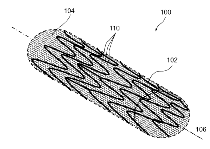
Fig. 1 shows a perspective view of an enhanced stent apparatus 100, in an
exemplary embodiment of the invention. A support element 102 is designed and
constructed to expand a blood vessel in a radial fashion from a central axis
106 of the
enhanced stent apparatus 100. Optionally, support element 102 is tubular in
shape. In
some exemplary embodiments of the invention, support element 102 is
constructed of
a flexible, biocompatible material. Optionally, support element 102 is
constructed of
stainless steel, nitinol, and/or cobalt chromium and/or other metal alloys
(e.g.
magnesium alloy). Optionally, support element 102 is constructed of polymer
either
biostable or bioresorbable. In some exemplary embodiments of the invention,
support
element 102 is a vascular stem, such as those made by Cordis*, Boston
Scientific
and/or MedtronicsO, for example.
In an exemplary embodiment of the invention, support element 102 is covered
by at least one porous structure 104. Optionally, support element 102 acts as
a support
structure for porous structure 104, for example to provide radial support and
or to
24
CA 02843097 2014-02-20
maintain a desired shape of porous structure 104. Fig. 2, shows a cross-
sectional view
of an enhanced stent apparatus. In this embodiment, support clement 102
supplies
structural support to porous structure 104, which is located on the exterior
of support
element 102.
In some exemplary embodiments of the invention, porous structure 104 is laid
on the exterior of support element 102 and thereby overlaps gaps in support
element
102 (making the aperture sues of the device as a whole smaller, for example
150
microns), since conventional stent construction usually results in multiple
gaps in the
structure of the stent, typically several millimeters. In other exemplary
embodiments
of the invention, porous structure 104 covers only a portion of support
element 102.
For example, only a portion of support element 102 is covered to avoid
restricting
luminal flow to a branching vessel.
In some exemplary embodiments of the invention, porous structure 104
extends past at least one end of support element 102. This can, for example,
better
treat the inside surface of a blood vessel at an edge of enhanced stent
apparatus 100,
where it is more likely to have restenosis. In an exemplary embodiment of the
invention, porous structure 104 pads and/or treats trauma caused by the edge
of
support element 102 by extending past at least one end of support element 102.
Optionally, porous structure 104 extends no more than 1 mm past the end of
support
element 102. Optionally, porous structure 104 extends over 1 mm past the end
of
support element 102. Optionally, porous structure 104 extends past only one
end or
both ends (as shown in Fig. 10) of support clement 102.
In some exemplary embodiments of the invention, porous structure 104 is
attached to support element 102 to prevent porous structure 104 from
unraveling
and/or causing tissue irritation and/or avoiding dislodgment of the porous
structure
from the support element during deployment. Optionally, the end of porous
structure
104 is folded over the end of support clement 102 and attached, providing
padding to
a potentially trauma causing edge. Optionally, the end of porous structure 104
is
folded under itself and is held folded due to the pressure between the support
element
and the lumen. In an embodiment of the invention, a treatment, such as heat,
is used to
make the fold sharp and/or permanent.
It should be understood that while an exemplary configuration of enhanced
stent apparatus is shown in Figs. 1 and 2, other configurations could possibly
be used,
CA 02843097 2014-02-20
including: a porous structure 104 over a pharmaceutical eluting support
element; a
pharmaceutical eluting porous structure over a support element 102; a
pharmaceutical
eluting porous structure over a pharmaceutical eluting support element; a
support
element in between at least two porous structures, optionally some or all
eluting
pharmaceuticals; and, an enhanced stent comprised of a plurality or layers
which
exhibit different optional characteristics such as degradation time and/or
pharmaceutical elution. It should be understood that any of the above
configurations
include biodegradable and/or bioresorbable materials. Optionally,
configurations arc
chosen for specific treatment regimens indicated by the condition of the
patient.
In some exemplary embodiments of the invention, porous structure 104 is used
to control the local pressure exerted by the enhanced stent apparatus on the
body
lumen wall. For example, by increasing or decreasing the coverage area of the
porous
structure as it at least partially covers the stcnt, the pressure exerted by
the enhanced
stern apparatus per unit area can be altered. In some embodiments of the
invention,
modification of the coverage area considers factors such as the stiffness of
support
element 102 and the geometry and/or coverage area of the support struts of
support
element 102. In an embodiment of the invention, pressure control is used to
reduce the
likelihood of the enhanced stent apparatus causing plaque to break off of the
lumen
wall. In some embodiments of the invention, pressure control is used to reduce
tissue
trauma typically caused by stent implants, thereby enhancing protection
against
stenosis/restenosis. Furthermore, in some embodiments of the invention,
support
element 102 struts which could not be used previously due to the likelihood of
trauma
to the lumen tissue can optionally be used in combination with porous
structure 104.
In some exemplary embodiments of the invention, bile ducts are treated using
at least a porous structure as described herein. For example, the bile ducts
often
become congested with debris (e.g. cholesterol) which restricts flow.
Treatment of the
bile ducts using enhanced stcnt apparatus may increase the diameter of the
bile ducts,
improving their operation.
It is known that varying types of body lumens possess varying surface
textures, both varying from each other, and sometimes within one type of
lumen.
Thus, in some exemplary embodiments of the invention, different porous
structures
with varying surface texture configurations are manufactured and/or used
depending
on the interior surface texture of a lumen being treated. For example, peaks
and
26
CA 02843097 2014-02-20
valleys in a body lumen are fitted with counter peaks and valleys of a porous
structure
(i.e. porous structure counter peak goes into lumen valley and porous
structure
counter valley accepts lumen peak). Optionally, the counter peaks and valleys
are of
the same magnitude as the peaks and valleys found in the lumen being treated.
It should be understood that the aperture size, the porous structure
thickness,
the fiber thickness (or French), and/or the coverage area are varied for
different
applications. For example when treating the carotids, debris of more than 100
microns
should be prevented from reaching the brain, thus the porous structure is
designed
such that when stent is expanded, usually to about 8 millimeters, the majority
of
aperture sizes are less than 100 microns. As another example, when treating
the
coronaries larger debris (>100 microns) is not as problematic, while the
endotheliazation process and the non-restriction of flow to side branches is
more
important. Thus for coronary artery applications, when the support element 102
is in
an expanded position, usually about 3 millimeters in diameter, the apertures
in the
porous structure are optionally larger than 100 microns and below 300 microns.
In
some embodiments of the invention, the rate of endothelium cell growth over
porous
structure may be modified by increasing and/or decreasing fiber thickness and
porous
structure thickness.
In some exemplary embodiments of the invention, porous structure 104 is used
with a balloon expandable support element 102. In some exemplary embodiments
of
the invention, porous structure 104 is placed directly on an expandable
balloon 802
without or with support element 102, for example as shown in Fig. S. In some
exemplary embodiments of the invention, the balloon catheter may extend past a
proximal and/or distal end of support element 102. it is optionally desirable
to provide
porous structure 104 which extends past the end of support element 102 to
provide a
buffer between the balloon and the blood vessel, and optionally to provide
pharmaceutical treatment to regions to which the underlying support element
102 does
not extend, but may be exposed to the balloon.
In an exemplary embodiment of the invention, porous structure 104 is under
100 microns in thickness. In some exemplary embodiments of the invention, the
porous structure is less than 30 microns thick. Optionally, the porous
structure is less
than 10 microns in thickness. For example, the porous structure is less than 5
microns
or 1 micron thick. Porous structure 104 is optionally comprised of at least
one fine,
27
CA 02843097 2014-02-20
thread-like fiber. In some exemplary embodiments of the invention, porous
structure
104 is comprised of at least one fiber that is 40 nm to 40 microns thick.
Optionally,
the fiber thickness is similar to or less than the diameter or an endothelial
cell to
encourage endothelial cell growth between fibers and/or around at least one
fiber. In
apertures 110 in porous structure 104. Optionally, the porous structure is
woven in an
even pattern. Optionally, the porous structure is constructed so that the
fibers are
randomly positioned in porous structure 104. Optionally, polymer fibers are
used to
construct porous structure 104. Optionally, polymer coverings are applied to
porous
structure 102 and/or support element 102. Exemplary porous structure
manufacture is
described in more detail in the "Methods of Manufacture" section below.
In an exemplary embodiment of the invention, the polymer covered porous
structure 104 is optionally made out of a closed interlocked design and/or an
open
interlocked design, or semi open design, similar to typical support clement
102
designs. The open interlocked design has an advantage when side branching is
needed. When treating a junction of two blood vessels, there is sometimes a
need to
introduce one stent through the side of another one. An open interlocked
design
allows such a procedure, and when the porous structure is made of metal mesh,
an
open interlocked design is utilized in order to allow easy side branching
sterns.
Optionally, using a biodegradable polymer coating on a non-biodegradable
support
element 102 leaves the support element 102 embedded after the biodegradable
polymer has degraded.
In an exemplary embodiment of the invention, porous structure 104 is crimped
to a small diameter while still maintaining its flexibility, to enable
successful
maneuverability through a patient's blood vessels to the site where enhanced
stent
apparatus 100 is to be implanted. In an exemplary embodiment of the invention,
porous structure 104 is expandable to enable expansion of porous structure 104
with
support element 102 upon deployment at a treatment site within a patient's
blood
CA 02843097 2014-02-20
vessel. Optionally, expansion of porous structure 104 along the longitudinal
axis
matches the expansion of support clement 102 along the longitudinal axis.
In an exemplary embodiment of the invention, at least porous structure 104 is
expandable without significant foreshortening or elongation of the length of
porous
structure 104. Optionally, porous structure 104 expands differently than
support
element 102, for example using sliding connections described herein. As
described
elsewhere herein, in a knitted embodiment of porous structure 104, expansion
occurs
at least partially as a result of the knitted structure, and not necessarily
because of the
elasticity of the fiber used in constructing porous structure 104. In an
embodiment of
the invention, at least one fiber which comprises porous structure 104 is
provided with
slack during manufacture to provide additional fiber material when porous
structure
104 expands. Fig. 9 shows a perspective view of an enhanced stent apparatus
900.
Enhanced stent apparatus 900 is provided with non-stretchable wires 902, and
stretchable elastomer fibers 904, in accordance with an exemplary embodiment
of the
invention. Such an embodiment assists with the preservation of overall
apparatus 900
length while allowing expandability and flexibility during implantation.
In an exemplary embodiment of the invention, an enhanced stent apparatus is
provided which is comprised of at least an expandable support element and an
expandable porous structure. The support clement is optionally a stent,
examples of
which are known in the art for providing treatment to a wide range of body
lumens. In
an embodiment of the invention, the porous structure has structure which
resembles to
fishing net. In an embodiment of the invention, the porous structure is
knitted from a
fiber approximately 15-20 microns in diameter, has a coverage area of less
than 20%,
and which has aperture sizes approximately 150x200 microns. In some
embodiments
of the invention, the porous structure is at least temporarily attached to
support struts
of the support element by stitching. Optionally, the stitches are loose,
allowing the
porous structure to slide on the support struts, for example to provide extra
expandability as described herein with respect to Fig. 16. In some embodiments
of the
invention, the stitching is biodegradable. In some embodiments of the
invention, the
support element and/or the porous structure are adapted to elute
pharmaceutical agents
into the body lumen being treated.
In some embodiments of the invention, different characteristics of the
enhanced stent apparatus are chosen based on the intended use or treatment to
be
29
CA 02843097 2014-02-20
rendered. For example, aperture sizes are optionally chosen based on a desire
to
provide embolic shower protection against debris of a certain 5i7C. As another
example, coverage area is optionally selected for modifying local pressure on
the
lumen being treated. Many of these characteristics are interrelated, as
described herein
and shown in Fig. 15, for example.
In an embodiment of the invention, porous structure 104 is flexible to allow
the lumen to naturally change its diameter, to account for pressure changes in
the
lumen and/or to respond to muscular activity. In some embodiments of the
invention,
the porous structure 104 divided into a plurality of semi-independent sectors,
which
react differently to stimuli within or from the lumen. Optionally, the sectors
are used
to prevent banding cube lumen across the entire length of the porous structure
104.
F.xemplary Characteristics and Performance of Porous Structure
Manufacturing techniques, described in more detail below, such as knitting
which provide slack to individual fibers, or sections, of porous structure
104, enable
porous structure 104 to optionally expand upon deployment up to 10 times its
diameter at insertion (insertion diameter is described in more detail below),
in an
embodiment of the invention. For example, in coronary applications porous
structure
104 may expand from lmm to 3mm in diameter. In other examples, porous
structure
104 may expand from 2mm to 8mm in carotid applications, while in brain
applications porous structure 104 may expand from .3mm to 2.5mm. These numbers
are approximate and are by way of example only. In an embodiment of the
invention,
expansion of porous structure 104 is effectuated in at least one of three
ways: 1) the
knitted/braided/woven structure of porous structure 104 (including slack in
the fibers
and curly fibers); 2) the fiber from which the porous structure 104 is made is
at least
slightly elastic; 3) sliding connections (described below) between porous
structure
104 and support element 102 permit shifting of porous structure 104 during
expansion
with respect to support clement 102, within certain limits. In an embodiment
of the
invention, the fiber from which porous structure 104 is made is comprised of
between
2% and 80% of non-elastic materials. In some embodiments of the invention, the
elastic material of the fiber from which porous structure 104 is made allows
for
expansion up to 1000% its original size.
In some exemplary embodiments of the invention, porous structure 104
exhibits a high durability when subjected to twisting, turning, compression
and/or
CA 02843097 2014-02-20
elongation, which allows porous structure 104 to withstand the delivery
process
through the patient's vasculature to a treatment site. In an embodiment of the
invention, porous structure 104 is loosely attached to the balloon at several
locations
and folded for insertion into a lumen, such as depicted in Fig. 12; such a
configuration
is optionally used in conjunction with supportive element 102. The folded
porous
structure 104 provides a reduced diameter apparatus for easier insertion into
body
lumens of the patient. As described below, porous structure 104 is at least
temporarily
fastened to balloon to prevent porous structure 104 from becoming dislodged
during
implantation.
In some exemplary embodiments of the invention, 20% of the total area of
porous structure 104 is comprised or apertures having an approximate diameter
no
greater than 50, 200 or more than 200 microns in an expanded configuration. It
is
recognized that during the course of manufacturing the porous structure, for
example
with certain manufacturing techniques like electrospinning and/or knitting,
apertures
created within the porous structure may overlap. This overlap effectively
creates an
aperture size which is smaller than specified. However, in some exemplary
embodiments of the invention, the effective, nominal aperture size is no
greater than
50, 200 or more than 200 microns in diameter. In some embodiments of the
invention,
aperture sizes arc selected to encourage endothelial cell overgrowth at a
certain rate.
It should be noted that shapes of apertures are likely to vary at least
somewhat
as a result of manufacture and/or desired properties of porous structure 104.
For
example, in a knitted porous structure, apertures are most likely to be
roughly square.
In contrast, use of a weaving technique to manufacture porous structure likely
produce
square and/or rectangular shaped apertures whereas a braided porous structure
is
likely to exhibit quadrilateral shaped apertures, such as in Fig. 7. In
describing an
approximate "diameter" of an aperture, it should be recognized that all, some
or none
of the apertures will be actual circles, squares, rectangles and/or
quadrilaterals capable
of simplistic area measurement using diameter. Therefore, description using
diameter
is merely an approximation to convey exemplary aperture sizes. For example,
"diameter" could be the distance between two parallel sides of a
quadrilateral, such as
a square or rectangle.
In some parts of the following description, aperture sizes described herein
are
in reference to their size upon porous structure deployment in a lumen. In
other parts,
31
CA 02843097 2014-02-20
the sizes refer to the aperture sizes when crimped. Sometimes, the aperture
sizes
described herein refer to their size in a state intermediate a crimped and
deployed
configuration. In context, it should be easily perceived which of the above
configurations applies, however, in the event it is not clear the aperture
sizes could be
considered as applying to expanded, crimped or intermediate configurations.
When a
fiber diameter is referred to, it relates to the fiber used to construct the
porous
structure 104. For example, if porous structure 104 is constructed from a
super-fiber
comprised of a bundle of 10 fibers each 2 microns in diameter, the overall
super-fiber
diameter is about 20 microns. Furthermore, it should be understood that
references to
fiber diameter arc for approximation and convenience only and does not imply
that
the fiber is necessarily round. Optionally, fiber sizes are measured in French
sizes, for
example .003 Fr.
Referring to Fig. IS, a graph 1500 is shown which correlates fiber thickness
of
porous structure with percentage of the coverage area of a support element,
for a
porous structure with a fishing net type configuration. It can be seen that
the general
trend is that as the Fiber sizes get thinner, the amount of porous structure
surface area
dedicated to structure is reduced. In an embodiment of the invention, it is
desirable to
have under 2.5% coverage area. Optionally, porous structure 104 exhibits less
than
20% coverage area. In an embodiment of the invention, the coverage area of the
porous structure is adapted to be minimized while still performing an intended
lumen
treating function, such as those described herein. In some embodiments of the
invention, the coverage area of porous structure 104 is minimized in order to
avoid
undesirable clinical side effects. For example, lumen tissue irritation and
pyrogenic
effects are considerations for minimizing the coverage area and optionally
other
characteristics such as aperture size, porous structure thickness and/or fiber
thickness,
of porous structure 104.
In some exemplary embodiments of the invention, the proportion of structure
to apertures or porous structure 104, fiber size and/or apertures are sized in
order to
allow easy diffusion through porous structure 104 and to facilitate growth of
endothelial cells. Since the fiber 2202 diameters used in construction or
porous
structure 104 are on the order of the size of the endothelial cells 2204, or
smaller, as
shown in Fig. 22, the integrity of the cells grown over the porous structure
will be
much better than what is achieved in the prior art. An individual cell,
statistically, will
32
CA 02843097 2014-02-20
have a firm connection to the blood vessel wall, since it is of the same order
or larger
than the fiber diameter, thus anchoring itself, in an embodiment of the
invention, in
more than one location to its native basalamina intimal layer and enabling
better
growth conditions. It is thus expected that the chance of late or sub-acute
thrombosis
can be reduced over what is currently achieved when treatment is performed
using a
pharmaceutical eluting stent. In addition, porous structure 104 effectively
acts as an
embolic shower protection device, holding detached plaque in place, preventing
it
from traveling from the vessel wall into the blood stream. It should be noted
that
porous structure 104 is configured, accounting for fiber thickness, porous
structure
thickness and/or aperture si7c such that endothelial cells will overgrow
porous
structure 104, and optionally support element 102, in order to secure the
enhanced
stem in place and/or insulate the foreign material of support element 102
and/or
porous structure 104 from the bloodstream. In an exemplary embodiment of the
invention, endothelial cell layer overgrowth of porous structure 104 is
established
within hours of implantation. In an embodiment of the invention, overgrowth is
accomplished within this time frame due to characteristics of porous structure
104 as
they relate to endothelial cells, for example, the overall thickness being on
the same
order of, or smaller, than an individual endothelial cell. In some embodiments
of the
invention, it is conceived that a patient's average stay in the hospital after
a stunting
procedure can be reduced as a result of the speed of endotheliazation using
enhanced
stein apparatus. In addition, the speed and efficacy of pharmaceutical
treatment can
expected to be enhanced as a result of the rapid endotheliazation over at
least porous
structure 104 of enhanced stern apparatus 100.
Fig. 23 shows a disadvantage of using prior art drug eluting stunts wherein a
clump of endothelium cells have become detached from the stent strut 2304,
revealing
an exposed "island" 2302 of the stent. Sometimes a clump falls off strut 2304
due to
poor adhesion of the endothelium cells to the polymer coating of the strut
2304.
Contributing to this poor adhesion, the stent strut 2304 is typically an order
of
magnitude larger than a single endothelium cell, thereby necessitating the
creation of
a large endothelium cell bridge to cross the strut. The exposed island 2302
can serve
as a seed for thrombosis development. In some embodiments of the invention,
porous
structure 104 is constructed to reduce the likelihood of endothelial cells
falling from
the porous structure, thereby reducing the chance of development of late or
sub-acute
33
CA 02843097 2014-02-20
thrombosis and exposure of support element 102 to substances within the lumen.
For
example, endothelial cell retention is optionally encouraged by constructing
porous
structure 104 from at least one fiber of a thickness and with aperture sizes
(to permit
growth of endothelial cell therethrough), such as described herein.
Optionally,
endothelial cells arc encouraged to remain on the porous structure by using a
fiber
layer of a thickness such as those described herein. Optionally, the linear
nature of a
single fiber porous structure 104, such as shown in Fig. 22, reduces the
possibility of a
large clump of endothelial cells becoming dislodged. In an embodiment of the
invention, porous structure 104, optionally imbued with a pharmaceutical, is
placed
on the interior of a bare metal or drug eluting support element in order to
reduce the
thrombogenicity of the support element. For example, by encouraging
endothelial cell
growth thereover and/or by reducing the exposed surface area of the support
element
by covering a portion of it up.
As suggested above, using a thin fiber whose thickness is similar to, or
smaller
than, the diameter of an endothelial cell enables an endothelial cell layer to
grow over
porous structure 104 while still being closely tied to the basalintima layer
at least at
two points of the endothelial cell, one point on each side of porous structure
104. In
an embodiment of the invention, the anchoring effect of this basalintima layer
on the
endothelial cell layer reduces the chance of parts of the endothelial cell
layer breaking
off and entering the lumen. This, in effect, reduces the chances of embolism
in the
patient and/or also reduces the likelihood of foreign bodies (e.g. the stent
and the
porous structure) coming into contact and reacting with the contents of the
lumen
being treated. In the event that a clump of several endothelium cells fall
from porous
structure 104, exposing a piece of the fiber, it is believed that there is a
reduced
chance of harm to the patient since the linear, single endothelial cell width
geometry
is not as thrombogenic as that shown in Fig. 23, where a clump of endothelial
cells at
least several cells in diameter has fallen off. In addition, the re-
endotheliazation will
be faster on an exposed porous structure 104 than on the exposed strut 2304
for at
least the reason that in the case of the porous structure 104, an endothelial
cell layer is
formed when just one endothelial cell overgrows the endothelial cell sized
fiber used
to construct the porous structure. In contrast, endothelial cell layer
overgrowth is only
accomplished after multiple endothelial cells have covered the exposed island.
34
CA 02843097 2014-02-20
In an embodiment of the invention, the reduced risk of late or sub-acute
thrombosis by using porous structure 104 for pharmaceutical elution optionally
allows
for a duration and/or dosage reduction in the use of anti-coagulants by the
patient.
In some exemplary embodiments of the invention, fiber thickness, porous
structure thickness and/or aperture size are all separately varied depending
on the
application of porous structure 104 and the needs of the patient. For example,
in the
coronary arteries it is sometimes helpful to provide for good pharmaceutical
dispersion. In such an example, fibers comprising porous structure 104 are
optionally
located closer together in order to allow for more complete transmission of a
pharmaceutical to patient.
In some exemplary embodiments, when large molecule drugs, which have a
poor diffusion into the tissue, are used to fight restenosis, the porous
structure can be
soaked and/or imbued with an appropriate drug in order to better diffuse it.
The
maximum concentrations of most of the drugs used are rather limited due to
side
effects and over toxicity, and at the same time the concentration is not
enough in order
to allow the optimum pharrnacokinetics in areas not covered by the stent
struts. The
porous structure mesh, having a better geometrical cover of the stent area,
provides a
better and more optimum pharmacokinetics to the whole area covered by the
stem.
For example, when high Dalton-large molecule drugs arc used, or when liposomes
arc
the carriers or the treatment agents, or when the stereo-chemical structure of
the drug
is large and/or complicated, and/or when the drug is hydrophobic, relatively
even
distribution of the drug is highly desirable. In some exemplary embodiments of
the
invention, pharmacokinetics are also optimized because the drug is located
on/in the
fibers of porous structure 104, and is covered and sealed within an
endothelium layer,
which helps the drug from being washed away by the blood.
In some exemplary embodiments of the invention, such as in a bypass vein
graft, side branching is not an issue, therefore aperture sizes arc optionally
made
smaller, but not so small as to prevent endothelial cell growth therethrough.
In another
exemplary embodiment of the invention, such as in the carotid arteries, side
branching
is not generally considered a problem, but catching debris is. Therefore, in
some
exemplary embodiments of the invention, the aperture sizes of porous structure
104
are decreased to as little as 20 microns in diameter. In other applications,
the aperture
CA 02843097 2014-02-20
size can be increased to 50, 100, 200 or even more then 200 microns, depending
on
the application of enhanced stent apparatus 100.
In some exemplary embodiments of the invention, a plurality of porous
structures is
used. Optionally, at least one porous structure is located on the interior of
support
element 102, inside the lumen of support element 102. Optionally, more then
one
porous structure is located on the exterior surface of support element 102. In
some
exemplary embodiments of the invention, at least some of the porous structures
located on support element 102 are configured to be "in-phase" where the
apertures of
the porous structures coincide with one another. Optionally, the porous
structures are
"out-of-phase" where the apertures are configured to not coincide with one
another. In
an exemplary embodiment of the invention, an "out-of-phase" configuration is
used to
improve contact surface area between porous structures and the lumen interior
surface. In an embodiment of the invention, increased contact surface arca can
improve pharmacokinetics, reduce local pressure exerted by porous structure
104 on
the lumen wall, improve embolic shower protection and/or realize other
advantageous
effects. In some exemplary embodiments of the invention, porous structure 104
is
constructed in the same shape and pattern as support element 102, but on a
smaller
scale.
Exemplary Materials of Manufacture
It should be noted that in some exemplary embodiments of the invention, a
stretchable and/or expandable porous structure 104 is desired. Therefore, in
some
embodiments of the invention, materials are chosen which arc either a)
stretchable
and/or b) can be used to manufacture a porous structure which is stretchable
(e.g. a
knitted structure). In some exemplary embodiments of the invention,
biodegradable
(i.e. are broken down by the body) and/or bioresorbable (i.e. are absorbed
into the
body) materials are used. In addition, blends of materials are used in
accordance with
some embodiments of the invention. In an embodiment of the invention, a
material is
chosen because it exhibits durability during manufacture, deployment and/or
use
despite being thin. In an embodiment of the invention, other considerations
for the
material to be used are their biocompatibility, toxicity, hemocompatibility,
and
trombogenic ity.
Exemplary materials for manufacturing porous structure 104 include natural-
based materials such as modified cellulose and/or collagen. In some
embodiments of
36
CA 02843097 2014-02-20
the invention, metal fibers are used to construct porous structure, optionally
constructed of stainless steel, and/or CoCr and/or CoNi alloy among other
possibilities. Optionally, the metal fibers used are coated with at least one
polymer. In
some embodiments of the invention, porous structure is manufactured from a
shape
memory alloy, such as nitinol. Optionally, carbon fiber is added to porous
structure
104 in order to improve strength characteristics of porous structure 104.
Optionally,
glass fiber is added to porous structure 104 in order to improve strength
characteristics
of porous structure 104. Optionally, a durable, resorbable and/or degradable
fiber is
added to porous structure 104 in order to improve strength and durability
characteristics of the fiber during manufacture, which is degraded or resorbed
or
washed away to leave a thinner porous structure 104.
In an embodiment of the invention, some polymer fibers are chosen for use in
constructing porous structure 104 because they are elastic, biocompatible,
hemocompatible, can be made not to stick to an expandable angioplasty-type
balloon
catheter, to stick to endothelium tissue, are selectably bio-stable and/or
biodegradable,
exhibit the requisite mechanical strength, are sterilizable, have a high
temperature
transformation zone (solid and non sticky at 37'C), are capable of hosting an
effective
amount of pharmaceuticals, and/or can release embedded pharmaceuticals at a
controlled rate. In some exemplary embodiments of the invention, other
materials
which exhibit some or all of these properties are optionally used to construct
porous
structure 104. Optionally, coatings arc put on porous structure 104, comprised
of
materials which exhibit some or all of these properties.
Polymer fibers are optionally made out of any of the following materials:
thermoplastic polymers for example polyethylene terephthalate (PET),
polyolefin,
oxidized acrylic, PTFE, polyethylene co-vinyl acetate, polyethylene elastomer,
PEO-
PBT, PEO-PLA, PBMA, polyurethane, Carbosil (PIG product), medical grade
polycarbonatc urethanes, Nylon, PEEK-Optima, carboxylic acid moiety comprising
one or more of a poly acrylic acid, a poly methacrylic acid, a maleic acid, a
helonic
acid, a taconic acid and/or combinations and/or esters of these monomers,
thermoplastic polymers, thermosetic polymers, polyolefin elastomers,
polyesters,
polyurethanes, polyfluoropolymers, and/or nylon. Optionally, the fibers are
constructed of an elastomer. Optionally, the fibers are constructed of a
coated fiber
with a drug and polymer coating mixed to get a predetermined drug release
37
CA 02843097 2014-02-20
characteristic, either coating over a metal and/or over a polymer fiber.
Optionally, the
fibers are constructed of other materials than the exemplary materials listed
above.
Exemplary polymers which are optionally used for this purpose are manufactured
by
Cordis1), Surrnodix , Boston Scientific, Abbott and lIemoteq Polymers.
Optionally, these polymers are selected for at least one of the reasons
specified in the
paragraph above. Optionally, the coating is used to facilitate the elution of
pharmaceuticals from porous structure 104.
In some embodiments of the invention, the porous structure is made out of a
resorbable/degradable polymer such as poly lactic-co-polyglycolic ("PLGA")
copolymers, or any other degradable copolymeric combination, such as
polycaprolactone ("PCL"), polygluconate, polylactic acid-polyethylene oxide
copolymers, poly(hydroxybutyrate), polyanhydride, poly-phosphoester,
poly(amino
acids), poly-L-lactide, poly-D-lactide, polyglycolide, poly(alpha-hydroxy
acid) and
combinations thereof.
In some embodiments of the invention, porous structure 104 is comprised of a
material which plastically or elastically deforms when a sufficient amount of
radial
pressure is applied to it, for example by an angioplasty balloon.
Exemplary Methods of Manufacture
Many of the methods and orientations described herein are designed to provide
a porous structure which exhibits at least some expandable quality. Porous
structure
104 is optionally adapted and constructed to stretch as it is being deployed
within the
lumen being treated. In some exemplary embodiments of the invention, porous
structure 104 is provided with stretchability in order to ease positioning
porous
structure 104 on a balloon and/or support element 102.
In an exemplary embodiment of the invention, weaving, braiding and/or
knitting results in some or all the elasticity of the porous structure being
achieved due
to the structure of the interlaced and/or crimped and/or textured fibers
(curly, slack).
This can be achieved by material elongation properties securing the porous
structure
to the stent In some exemplary embodiments of the invention, a porous
structure is
made by combining several interlacing techniques such as knitting over a
braided
porous structure or braiding over a knitted porous structure. In some
embodiments of
the invention, multiple layers are combined and/or created using these
techniques. In
some exemplary embodiments of the invention, a warp knitted porous structure
with
38
CA 02843097 2014-02-20
"laid in" yarns is used. In some exemplary embodiments of the invention, a
porous
structure is woven using elastomeric or crimped weft to obtain radial
elasticity.
In some exemplary embodiments of the invention, the porous structure is
manufactured by combining several techniques such as knitting over a braided
porous
structure or braiding over a knitted porous structure. In some exemplary
embodiments
of the invention, a weft knitted porous structure with "laid in" yarns is
used. In some
exemplary embodiments of the invention, a porous structure is woven using
elastomerie and/or crimped weft to obtain radial elasticity. Optionally,
porous
structure is comprised of at least one fiber oriented generally parallel to
the support
element's longitudinal axis.
In an exemplary embodiment of the invention, a manufactured porous
structure is added as a cover to support element 102. Optionally, porous
structure 104
is used separately from support element 102, which is optionally not used for
stenting.
In some exemplary embodiments of the invention, porous structure 104 is
manufactured directly onto support element 102.
In an exemplary embodiment of the invention, porous structure 104 is
manufactured by a knitting technique known to those skilled in the knitting
art for
non-analogous arts, such as clothing manufacturing and textiles. Knitting of
porous
structure 104 is optionally performed by heads having between 20 and 35
needles.
Optionally, the head used has between 30 and 45 needles. Optionally, the head
used
has between 35 and 80 needles. An example of the effect of head size on the
porous
structure can be seen in Fig. 15, described above, in which a 22 Size head and
a 35
size head are graphed. In Fig. 15, the needle gauges are 40.
In some embodiments of the invention, the shape and/or size of the knit is
controlled by controlling the tension on the fiber being used for knitting.
For example
to create a knit with larger eyes, slack is provided to the fiber during
knitting.
Optionally, the fiber is controlled during knitting to achieve a circular
shaped eye
when porous structure 104 is expanded. In an embodiment of the invention, pre-
tension on the fiber during knitting is approximately 10-20 grams. In some
embodiments of the invention, post-tension on the fiber during knitting is 15-
25
grams. The stitch length is between 300 and 400 microns, in an exemplary
embodiment of the invention. In some embodiments of the invention, the
knitting
machine is run at a relatively slow speed. For example, the knitting machine
is run at
39
CA 02843097 2014-02-20
10% of speed capacity using a Lamb Knitting Machine Corp. System Model WK6
with a special modification of speed operation measured by percentage
In an exemplary embodiment of the invention, a fiber or a super-fiber yarn
with a specific fineness, or a range of fineness, between 5 and 100 microns is
used to
manufacture a knit porous structure. Optionally, yarn with a fineness of 10 to
20
microns is used to manufacture a knit porous structure. Optionally, the yarn
is finer
than 5 microns. Yarn fineness is often referred to in textile terms by "Tex".
This is the
weight in grams of 1000 meters of the yarn. In an exemplary embodiment of the
invention, yarn ranging from 0.3 Tex to 10 Tex is used to manufacture porous
structure. In some embodiments of the invention, a specific yarn fineness is
chosen
based on the desired porous structure 104 characteristics. For example, a 0.5
Tex yarn
using a 22 gauge needle head will, in some embodiments, produce a porous
structure
with approximately 12% coverage area.
An exemplary resulting porous structure using the above components and
techniques, should have 5 to 50 courses per cm. Optionally, 20 to 45 courses
per cm
arc manufactured. Optionally, a porous structure with 30-35 courses per cm is
manufactured. Fig. 5 illustrates a knitted porous structure 104 and support
element
102 in a crimped, closed position. Fig. 6A illustrates a knitted porous
structure 104
laid on top of a support clement 102 in an open position. Fig. 613 shows
exemplary
knitting in detail.
In another exemplary embodiment of the invention, weaving techniques are
used to manufacture porous structure 104. Narrow needle looms as well as
conventional narrow looms can be configured to produce woven tubular
structures. In
weaving, at least two layers of warp yarns are interlaced with intersecting
fill yarns.
By carrying the fill yarn alternately back and forth across two layers or warp
yarns, a
tubular shape is created. The sire and shape of the weave are optionally
controlled by
determining the warp and/or fill density, the interlacing pattern and/or
frequency, the
yarn tension and/or the yarn dimensions and/or elastic properties. The types
of weaves
used for a porous structure are optionally one of "plain", "basket", "twill",
"sateen",
"leno" and/or "jacquard". Optionally, all of the fibers of porous structure
are the
same. Alternatively, warp and weft fibers of a weave are not constructed of
the same
materials. Optionally, different materials are used to take advantage of the
inherent
properties or the different materials, for example one material may be elastic
and a
CA 02843097 2014-02-20
different material may have a high tensile strength. Optionally, warp fibers
are coated
and/or arc pharmaceutical eluting while the well fibers are not, or vice
versa.
In another exemplary embodiment of the invention, braiding techniques are
used to manufacture porous structure 104, for example as described in Knitting
Technology, D.J. Spencer ¨ ed., Woodhead Publishing Limited, Abington Hall,
Abington, Cambridge, CBI 6A1I, England, the disclosure of which is
incorporated
herein by reference. Braiding machines are optionally used to interlace yarns
at a
variety of intersecting angles. In braiding, multiple yarns are fed to an
interlacing
zone. Interlacing is optionally achieved by rotation of the yarn spools or by
a
reciprocating needle bed. The size and shape of the braid is optionally
controlled by
the number of yarns, the interlacing pattern and/or angle and/or the yarn
dimensions
and/or elastic properties. Optionally, all of the fibers of porous structure
are the same.
Optionally, warp and well fibers of a braid arc not constructed of the same
materials,
for example where weft fibers are used to provide strength and warp fibers are
used to
provide stretchability of the braid.
In another exemplary embodiment of the invention, porous structure 104 is
manufactured by an electrospinning process. Electrospinning is a technique
which
utilizes a charged polymer solution (or melt) that is fed through a small
opening or
nozzle (usually a needle or pipette tip). Because of its charge, the solution
is drawn
toward a grounded collecting plate (usually a metal screen, plate, or rotating
mandrel),
typically 5 30 cm away, as a jet. Optionally, support element 102 is placed on
a
delivery catheter which is used as a mandrel. During the jet's travel, the
solvent
gradually evaporates, and a charged polymer fiber is left to accumulate on the
grounded target. The charge on the fibers eventually dissipates into the
surrounding
environment. The resulting product is a non-woven fiber porous structure that
is
composed of tiny fibers with diameters between approximately 40 nanometers and
40
microns (e.g. a felt), depending on the sin of the fibers input into the
system. If the
target is allowed to move with respect to the nozzle position, such as by
rotating
and/or moving the mandrel along its longer axis, specific fiber orientations
(parallel
alignment or a random alignment, as examples) can be achieved. In some
exemplary
embodiments of the invention, porous structure 104 is spun in a helical coil
pattern
onto the mandrel or support element 102. Optionally, porous structure 104 is
comprised of a plurality of helical coil patterns, constructed by moving the
mandrel
41
CA 02843097 2014-02-20
back and forth, such as depicted in FIG. 4. Optionally, porous structure 104
is
constructed with fibers oriented substantially parallel to central axis 106.
Optionally,
porous structure 104 is constructed with fibers oriented substantially
perpendicular to
central axis 106. Optionally, porous structure 104 is constructed with fibers
oriented in
a combination of any of the orientations described or suggested herein. The
mechanical
properties of the porous structure are optionally altered by varying the fiber
diameter
and orientation depending on the requirements for treating a patient. For
example, in
some embodiments of the invention, a laser is used to cut specific aperture
sizes and/or
to ensure that the apertures traverse from the exterior side of porous
structure 104 to
the interior side of porous structure 104. Optionally, solvent is used to
modify aperture
sizes.
Optionally, portions of catheter are masked in order to prevent accidental
coverage of the delivery catheter by porous structure 104. Optionally, support
element
102 is coated with an adhesive and/or a pharmaceutical agent prior to putting
the
porous structure 104 on the top of support element 102. In some exemplary
embodiments of the invention, the material used to produce the porous
structure 104 is
imbued with pharmaceutical agents. Optionally, pharmaceutical agents are
embedded
in the material coating the porous structure 104. In an exemplary embodiment
of the
invention, porous structure 104 is comprised of at least one inner coating
proximal to
supporting structure 102 which exhibits different properties than an external
coating
proximal to patient's blood vessel. For example, the inner coating is
optionally
configured to avoid adhesion to the delivery catheter and/or support
structure.
Optionally, inner coating is configured to adhere to support element 102, but
not to
delivery catheter.
75 In some embodiments of the invention, porous structure 104 is designed
to be
less sensitive to foreshortening and elongation forces as porous structure 104
expands
upon deployment. This is in part due to the knitted nature of porous structure
104, in
some embodiments. This property allows porous structure 104 to be secured to
support
element 102 at its ends, rather than in another location, such as the middle
as described
in U.S. App. No. 2005/0038503 to Greenhalgh et al.
In some exemplary embodiments of the invention, a porous structure is
manufactured in an at least partially open, wide diameter. condition. In some
42
CA 02843097 2014-02-20
exemplary embodiments of the invention, the at least partially stretched
porous
structure is reduced to a smaller diameter, by heat-setting, crimping and/or
folding,
after manufacture.
In an embodiment of the invention, the diameter of the at least partially
stretched porous structure is reduced mechanically. Optionally, a funnel 1304
shown
in Fig. 13, is used to reduce the diameter of the knitted porous structure
1302, during
the manufacture the porous structure. A knitted porous structure 1302 is drawn
down
from the knitting zone into a narrowing bore, funnel 1304. This results in a
final
porous structure diameter that is controllably smaller than the diameter of
the needle
bed. Fig. 14, illustrates how a porous structure is manufactured using a
stretched
rubber tube 1402. In this method, the porous structure 1400 is tightly
inserted unto a
pre-radially stretched tube 1402, and then the tube is relaxed, compressing
the porous
structure and creating a smaller aperture sized porous structure, the size of
which is
controlled by the stretch ratio of the rubber tube.
Referring to Fig. 1 8, an embodiment is shown in which porous structure 104 is
folded in "n" substantially folds, the folds used to reduce the overall
diameter of
enhanced stent apparatus 100 for easier insertion and navigation through the
patient.
Optionally, the folds are towards the same direction. In an embodiment of thc
invention, a folded porous structure 104 is at least temporarily secured to
support
element 102. Fig. 11 shows an alternate folded configuration from a
perspective view.
An additional or alternative embodiment to folding includes heat setting a
polymer comprised porous structure 104 to support structure 102. In an
embodiment
of the invention, heat setting is used when porous structure 104 is comprised
of at
least one polymer material. Determination of heat setting conditions is
related to the
polymer's heat transition temperatures, in an embodiment of the invention.
Heat
setting is performed in the temperature range between TA and Tin of the
polymer. At
this range the polymer becomes amorphous and is shrunk to support element 102,
establishing an overall enhanced stein apparatus 100 radius that is not much
more than
the support element 102 radius. For example, porous structure 104 adds less
than 10
microns in diameter total to support clement 102 which is lmm in diameter, in
some
embodiments of the invention. At T. the polymer turns into a viscous liquid
which
loses its mechanical integrity and will stick to support element 102 surface.
For
example, polyethyleneteraphthalate (PET) has a Ts of 70 C and a Tn, or 265 C,
43
CA 02843097 2014-02-20
therefore the heat set temperature somewhere within that range, in an
embodiment of
the invention, is 200 C. Using temperatures higher than Tõ, for heat setting
can cause
thermal degradation, which results in polymeric chain scission, unzipping of
the
polymer and/or producing a large array of oligomeric material that changes the
mechanical properties of porous structure 104 and/or releases poisonous and/or
non-
biocompatible materials, causing an inflammatory reaction in the patient.
Other
exemplary polymers which can be used in heat setting are below in Table 1 (not
an
exhaustive list):
Material name T, Tõ, set temp.
PP -18 C 165 C 140 C
NYLON 6,6, 80 C 265 C 210 C
PTFE _________________________ 150 C 330 C 300 C
PVA 100 C 230 C 190 C
Polyurethanes 70 C 120 C 100 C
MLA 60 C 175 C 100 C
Table 1: Exemplary polymers and temperatures for heat setting
Another additional or alternative embodiment to folding includes crimping
using at least a crimped support element 102 in combination with porous
structure
104. In some embodiments of the invention, porous structure 104 is crimped
with
support element 102. In an embodiment of the invention, crimping of porous
structure
104 and support element 102 is performed when it is desirable to reduce the
overall
diameter of enhanced stent apparatus 100. For example, a reduced diameter
enhanced
stent apparatus 100 allows for easier insertion and navigation of the
apparatus to the
treatment site. In some embodiments of the invention, at least a crimped
support
element 102 provides an object with relatively stable mechanical properties
for more
predictable movement during insertion and navigation.
Optionally, porous structure 104 is made on a non-crimped support element
102. A non-crimped support element 102 can be expanded or semi-expanded during
manufacture. In an exemplary embodiment of the invention, porous structure 104
and
support element 102 are crimped together. Optionally, excess porous structure
104
material, which is created as a result of reducing the profile of support
element 102
during crimping, is folded with support clement 102, such as shown in Fig. 18.
In
some exemplary embodiments of the invention, porous structure 104 is made on a
crimped or partially crimped support structure 102. When manufacturing a
porous
44
CA 02843097 2014-02-20
structure for placement on an already crimped support structure, consideration
may be
given to providing a porous structure which is sufficiently stretchable to
expand with
the radial expansion of the support structure, when implanted at a treatment
site
within a lumen. In some embodiments of the invention, a porous structure is
placed on
a support element already positioned on an angioplasty balloon.
In electrospinning embodiments of the invention, the procedure for
manufacturing a porous structure on a balloon is similar to manufacturing on a
stent
or mandrel. For example, varying the motion of the balloon with respect to the
electrospinning device allows the manufacture of specific fiber orientations.
Exemplary Methods for Coating a Fiber
In another exemplary embodiment of the invention, a manufacturing technique
is used to coat a fiber that porous structure 104 is comprised of with at
least one
polymer layer. For example, a dipping technique, shown in Fig. 19, using a
biocompatible, hemocompatible, biostable and/or biodegradable polymer
dissolved in
an organic solvent is utilized to create a dipping solution 1906 for use in
coating the
fiber comprising porous structure 104. The fiber to be coated is optionally
placed in a
spool 1902, from which the fiber 1904 is drawn to form porous structure 104.
Additives such as drugs, biological components, enzymes, growth factors,
and/or any
other additive mentioned herein or known in the art, may be incorporated into
fiber
1904 during the manufacturing process, for example by placing them in solution
1906
and passing fiber 1904 through solution 1906. In an embodiment of the
invention, at
least one layer is used in order to control the drug/biological additive's
release. For
example, more than one solution tank may be provided for fiber 1904 to pass
through
during manufacture. Fiber 1904 is optionally moved into a drying oven 1908
with an
operational temperature range from 37 70 degrees C (in some embodiments of the
invention) depending on the drug used, to dry solution 1906 onto fiber 1904.
In an
exemplary embodiment of the invention, fiber 1904 is then used by a knitting
system
1910 to manufacture porous structure 104. Optionally, knitting system 1910 is
the
Lamb Knitting Machine Corp. System Model WK6. Optionally, porous structure 104
is coated with a polymer layer after it has been manufactured.
In some exemplary embodiments of the invention, support element 102 and
porous structure 104 are coated with an additional substance. Optionally, the
CA 02843097 2014-02-20
additional substance is a polymer. Optionally, the additional substance is
drug eluting.
Optionally, the coating is hyaloronic acid. Alternatively or additionally, the
coating is
hyaluronan. Optionally, a different non-woven technology such as wet and/or
dry
spinning is used to manufacture porous structure 104. In some embodiments of
the
invention, additional coatings are added to achieve different effects, for
example
timed release of pharmaceutical agents and/or release of a plurality of agents
at
different times.
Exemplary Methods for Mountine Porous Structure to Support Element
In some embodiments of the invention, porous structure 104 is at least
temporarily secured to support clement 102. Advantages of securing porous
structure
104 to support element 102, at least temporarily, include: prevention of
unraveling
and/or run out of fiber from the porous structure weave, dislodgement and/or
slipping
of porous structure 104 with respect to support clement 102 during insertion,
delivery
and/or deployment. Optionally, support element 102 and porous structure 104
are not
secured together using an adhesive and/or other securing means despite being
in a
coaxial and proximate relationship in many embodiments.
In some exemplary embodiments of the invention, where support element 102
is optionally coated with a polymer, support element 102 and porous structure
104 are
attached together by curing at the same time the polymer support element
coating and
the polymer comprised porous structure, and/or coated porous structure, thus
adhering
the polymers together. Optionally, pressure and/or heat is used to adhere a
polymer
coated support element 102 to a non-coated or polymer coated porous structure
104,
for example when they are both hot. In some exemplary embodiments of the
invention, porous structure 104 is comprised of two components, an external
component and an internal component, relative to support element 102. Upon the
simultaneous curing of the external and internal components, the polymers of
which
both are comprised to adhere together, thereby securing porous structure 104
to
support element 102, which is located between the components.
In an exemplary embodiment of the invention, porous structure 104 is secured
to support clement 102 in order to avoid porous structure migration, but not
limit
porous structure 104 and/or support element 102 expandability.
In some exemplary embodiments of the invention, an adhesive is used to bond
support element 102 and porous structure 104 together. Optionally, porous
structure
46
CA 02843097 2014-02-20
104 is glued to support element 102 utilizing any natural and/or synthetic
biocompatible adhesive, such as cyanocrylate, thcrmo plastic elastomers,
silancs,
laminin, albumin and/or fibrinogen and/or PEG-PEG adhesive, and/or
polyurethane
adhesive and/or any other suitable compatible polymeric material. Optionally,
when
porous structure 104 is glued to support clement 102, wherein the support
element
102 is a drug eluting stent, the same polymer as used for the elution of the
drug is
used for attachment of porous structure 104 to support element 102.
In an embodiment of the invention, porous structure 104 is attached to support
element 102 at a plurality of points. Optionally, the plurality of points
defines a
pattern, such as a line or zigrag of points. Optionally, porous structure 104
compresses onto support element 102 to maintain an attachment to support
element
102. Optionally, the porous structure is held in place on support element at
least
partially by frictional forces. Optionally, porous structure 104 is sewn
and/or
mechanically entangled onto support element 102. Optionally, heating,
pressure, laser
welding, IN curing and/or ultrasound are used as techniques to secure porous
structure 104 to support element 102. Optionally, a primer, such as parylene,
is used
on support element 102 prior to adhering porous structure 104 to it in order
to enhance
cohesion.
In some exemplary embodiments of the invention, elastic and/or expandable o-
and/or c-rings are used to hold porous structure 104 on support element 102.
Optionally, c-rings are used to avoid hampering expandability of porous
structure 104.
Optionally, the rings arc used to at least temporarily secure and/or apply
friction to
each end of porous structure 104 to support element 102. Optionally, the rings
are
coated and/or embedded with pharmaceuticals for elution, such as described
herein.
Optionally, the rings are constructed of a polymer based material. In some
exemplary
embodiments of the invention, porous structure 104 is tied to support element
102,
optionally using fibers of porous structure 104. In an exemplary embodiment of
the
invention, a slip ring 2102 is used to secure porous structure 104 to support
element
102, as shown in Figs. 21A and 13. Slip ring 2102 is adapted to expand with
porous
structure 104 and support element 102 when they are expanded upon deployment
at a
lumen treatment site. Optionally, slip ring 2102 is flexible but is rigid
enough to
secure porous structure 104 to support element 102. In an embodiment of the
invention, slip ring 2102 is coiled around enhanced stent apparatus 100 when
it is in a
47
CA 02843097 2014-02-20
reduced profile configuration such that slip ring 2102 overlaps itself at
least partially.
Upon deployment, shown in Fig. 21 R, support clement 102 and porous structure
104
are expanded to provide treatment to the lumen. in an embodiment or the
invention,
slip ring 2102 expands with them while maintaining sufficient pressure on
porous
structure 104 to retain it to support element 102. In an embodiment of the
invention,
the overlapping portion of slip ring 2102 is reduced as a result of the
overall increase
in diameter of the slip ring 2102. Optionally, slip ring 2102 is comprised of
a
biodegradable and/or bioresorbable material. In some embodiments of the
invention,
slip ring 2102 is under 25 microns thick. Optionally, slip ring 2102 is under
15
microns thick. Optionally, slip ring 2102 is under 10 microns thick.
Referring to Fig. 16, an embodiment of the invention is shown in which
porous structure 104 is attached to support element 102 using a sliding
connection
1602. In an exemplary embodiment of the invention, the sliding connection is
established by attaching at least one loop 1604 of porous structure 104 to
support
element 102 in a condition that prevents the two from becoming separated, hut
is
loose enough to allow sliding of porous structure 104 with respect to support
clement
102. In an embodiment of the invention, a loose stitch is used to attach
porous
structure 104 to support clement 102 in a sliding connection. In an embodiment
of the
invention, expansion of porous structure 104 is assisted by utili7ing the
sliding nature
of the connection 1602. For example, porous structure 104 is secured to the
outermost
strut 1606 of support element 102 at its most outlying position 1608. In an
embodiment of the invention, on the other side of support element 102 porous
structure 104 is also attached to the outermost strut at its most outlying
position.
When support element 102 and porous structure 104 are expanded during
deployment,
porous structure 104 is afforded additional expandability, in relation to a
pre-
expanded configuration, as the sliding connection 1602 moves from the most
outlying
position 1608 on outermost strut 1606 to an innermost position 1610. The
distance
1612, about 1mm to 6rnm in an embodiment of the invention, from the most
outlying
position 1608 to innermost position 1610 provides additional expandability to
porous
structure 104. Optionally, sliding is prevented by securing porous structure
104 to
strut 1606 at innermost position 1610 as well as most outlying position 1608.
Optionally, sliding is prevented by tightening the connection between the two,
for
example by providing a tighter stitch.
48
CA 02843097 2014-02-20
In some embodiments of the invention, porous structure 104 is tied to support
element 102 using any one of thumb, square, reef, or double surgeon's knots.
Optionally, the at least one fiber used to construct porous structure 104 is
used to tie
porous structure 104 to support element 102. Fig. 17 shows an exemplary method
for
attaching porous structure 104 to support element 102 using knotting. It can
be seen
that a knotting fiber 1702 is used to secure porous structure 104 to support
element
102 at various points along a support element strut 1704. Optionally, knotting
fiber
1702 is threaded through a plurality of eyes 1706 and over support element
strut 1704
wherein a knot 1708 is tied to secure porous structure 104 to support element
102 at at
least some of the eyes.
As mentioned above, in some embodiments of the invention, securing porous
structure 104 to support element 102 is also used to reduce the likelihood of
run outs
and/or porous structure unraveling. In an embodiment or the invention, run
outs
and/or porous structure unraveling are to be avoided for at least the reasons
of:
avoiding porous structure protrusion into the lumen and/or rendering porous
structure
ineffective for intended treatment of the lumen. In an embodiment of the
invention,
porous structure 104 is secured to support element 102 at the ends of support
element
102, at at least some intersections where porous structure 104 and support
element
102 overlap, or both and/or every eye at both ends. Any of the methodologies
of
securing described above are optionally used to secure porous structure 104 to
support
element 102 to prevent run outs and/or unraveling.
In an exemplary embodiment of the invention, porous structure 104 is treated
to supply temporary enhanced adhesion to support element 102 during
implantation.
For example, enhanced stem apparatus 100 is optionally dipped in a liquid
which
causes porous structure 104 to adhere to support element 102. Optionally, this
adherence is due to surface tension of the liquid. Optionally, this adherence
is due to
temporary shrinkage of porous structure 104, which secures it to support
element 102
more tightly. In some exemplary embodiments of the invention, temporary
cohesion is
used to prevent porous structure 104 from slipping off of support element 102
as a
result of frictional stress experienced during navigation of the vasculature
during
implantation.
General Pharmacological Usage
49
CA 02843097 2014-02-20
Alternatively or additionally to the physical prevention of debris from
entering
the bloodstream, porous structure 104 optionally contains pharmaceuticals
designed to
u-eat a variety of ailments. In some exemplary embodiments of the invention,
pharmaceuticals are optionally provided including one or more pharmacological
agents for encouraging cell and/or liposomal growth and/or other endothelial
cell
growth factors, anti-proliferative, anti-thrombotic, anti-coagulant and/or
anti-platelet
effects, tissue engineering factors. immunomodulators, antioxidants, antisense
oligonueleotides, collagen inhibitors, hydrophobic pharmaceuticals,
hydrophilic
pharmaceuticals and/or endothelial cell seeding substances. Optionally,
pharmacological therapy rendered from a porous structure is used to accelerate
vein to
artery conversion. Specific examples of pharmaceuticals that are optionally
used with
porous structure 104 include: anti-proliferative agents like sirolimus,
zolimus or
zotarolimus (ABT-5780), paclitaxel and other taxancs, tacrolimus, everolimus,
vincritine, viblastine, IRvIG-CoA reductase inhibitors, doxorubicin,
colchicine,
actinomycin D, mitomycin C, cycloporine, and/or mycophenolic acid,
triazolopyrimidinc and its derivatives (i.e. Trapidil), a coronary
vasodilating drug);
intrapide, glucocorticoids like dexamethasone, methylpralnisolone, and/or
gamma
interferon; antithrombotics like heparin, heparin-like dextran derivatives,
acid citrate
dextrose, coumadin, warfarin, streptokinase, anistreplase, tissue plasminogen
activator
(IPA), urokinease and/or abciximab; antioxidants like probucol; growth factor
inhibitors like tranilast and/or angiopeptin; antisense oligonucleotides like
c-myc
and/or c-myb; collagen inhibitors like halofuginone and/or batimistat;
liposotnes;
gemcitabine Gemzarl)); steroids and corticosteroids for example
cortisone and
prednisone; cortisone, prednisone; Rapamycin10; statin drugs like
simva,statin,
lovastatin, and/or simvastatine (i.e. Zocor0); VEGF; FGF-2; micro carriers
containing
endothelial cells; genes; DNA; endothelial cell seeds; and/or hydrogels
containing
endothelial cells.
Typically, stents (i.e. support elements) which provide pharmaceutical
treatment only have the pharmaceutical embedded on the structure of the stent,
in
particular on the stcnt struts. This structure is typically minimized in order
to provide
flexibility and reduce cost, among other reasons. As a result of a minimized
support
element structure, the struts of the structure are usually spaced widely
apart. Thus,
when the stein is in situ, and pharmaceuticals are released into the patient
from the
CA 02843097 2014-02-20
stent, the pharmaceutical is only diffused from the widely spaced struts. This
prevents
even distribution of the pharmaceutical over the entire length of the stent.
In addition,
stent struts are typically large in relation to endothelial cells and
therefore formation
of a covering endothelial cell layer typically takes on the order of days or
weeks,
rendering pharmaceutical elution into body tissues delayed and/or ineffective
(due to a
number of reasons, including the pharmaceutical being washed away by fluids
flowing in the lumen before the endothelial cell layer covers the stent).
In contrast, usage of a pharmaceutical enhanced porous structure, such as
described herein, to cover the stent, including the struts, provides far more
surface
I 0 area in contact with the inner wall of the patient's blood vessel,
thereby enabling more
diffusion to take place. In comparison to conventional techniques for stent
delivered
pharmaceuticals, lower concentrations of pharmaceutical are optionally used
with the
present invention because of its improved therapy-rendering surface area. In
an
embodiment of the invention, improved delivery by the presently described
invention
allows for lower doses of pharmaceutical to be used in order to render the
same
relative amount of treatment, and reduce the overall dosage needed in order to
obtain
the same results, thus reducing possible side effects. For example, a
currently
recommended concentration of taxol on a drug eluting stent is around I pgimm2
of
stent surface. In contrast, a concentration of 0.514/mm2 is optionally used
with porous
structure 104, due to its increased treatment rendering surface area.
Optionally, the
concentration is less than 0.5 g/mtre.As another example, typical
concentrations of
rapamycin and 'limus drugs today arc around 140 g/cm2, however, using the
herein
described porous structure 104 a concentration of 80 g/cm2 is optionally used
to
achieve the same therapeutic effect. In some exemplary embodiments of the
invention, as little as lOug/cm2 is optionally used to achieve the same
therapeutic
effect. In some exemplary embodiments of the invention, concentrations of
pharmaceutical embedded on porous structure are up to 15 times less than
conventionally used today.
In conventional stents, at the struts, the pharmaceutical may not propagate
far
enough and/or without effect into the vascular wall, or may overdose a
particular
section of the vascular wall, without sufficient propagation laterally to the
rest of the
inner surface where it is needed. Having additional surface area, more evenly
covering the stent surface area, porous structure 104 can deliver drugs in a
more
51
CA 02843097 2014-02-20
locally homogenous way. Optionally, there is an axial profile change in
dosage. Since
distribution of the drug into the tissue is governed by diffusion, and since
the amount
of dosage concentration on the struts is limiting due to over toxicity and
side effects,
spreading the drug in a more even manner is very helpful for obtaining better
pharmacokinctic.s.
In an exemplary embodiment of the invention, pharmaceuticals to be
administered to patient are located in and/or on the fibers of porous
structure 104.
Examples of where and how pharmaceuticals are optionally located in and/or on
the
fibers of porous structure 104 and/or eluted include:
to 1. depositing pharmaceutical in the apertures of porous structure;
2. mixing pharmaceutical particles into fibers of porous structure at fiber
creation;
3. applying pharmaceutical topically to the porous structure, such as by
spraying;
4. dipping porous structure into a solution containing a pharmaceutical
additive, thereby depositing the additive on and/or in the fibers of the
porous structure;
5. encapsulating a pharmaceutical additive on porous structure, optionally
using a thermal process;
6. grafting a pharmaceutical additive onto porous structure using plasma
treatment;
7. etching a pharmaceutical additive into porous structure, for example via
spattering or coating;
8. transferring a pharmaceutical additive to porous structure using
concentration differences between the porous structure and an additive
containing substance, for example by adhering micro carriers containing
pharmaceutical additive to a porous structure allowing their migration
into the porous structure;
9. any method known to those skilled in the art, such as shown in U.S. Pat.
App. No. 2004/0030377 to Dubson et al., U.S. Pat. App No.
2005/0187140 to Hunter et aL., U.S. Pat. App. No. 2004/0236407 to
Fierens et al., U.S. Pat. 6,902,522, to Wash, et al., U.S. Pat. 6,669,961
to Kim, et al., U.S. Pat. 6,447,796 to Vook, et al., U.S. Pat. 6,369,039 to
52
CA 02843097 2014-02-20
Palasis et al., U.S. Pat. No. 6,939,374 to Banik, et al., and U.S. Pat. No.
6,919,100 to Narayanan;
10. elution of the drug from a polymer coating the porous structure fibers;
11. elution of the drug from the polymer from which the porous structure is
constructed; and,
12. incorporating the drug in a biodegradable polymer.
Optionally, embedding of the pharmaceutical occurs before (e.g. mixing
pharmaceutical particles into fibers of porous structure at fiber creation),
during (e.g.
using the dipping method of FIG. 19) and/or after (e.g. a spray on
pharmaceutical after
apparatus is made) the manufacture of enhanced stent apparatus 100. In some
exemplary embodiments of the invention, a pharmaceutically embedded porous
structure 104 is placed on top of a pharmaceutically treated support element
102. In
some exemplary embodiments of the invention, porous structure 104 is coated
with at
least a polymer. In some exemplary embodiments of the invention, a porous
structure
is provided with a polymer coating which contains a pharmaceutical which
elutes from
the coating.
Pharmaceuticals are optionally embedded into porous structure 104 such that
they are released into the patient over an approximate predetermined amount of
time.
For example, pharmaceuticals are optionally embedded into porous structure 104
for
release over the course of a week. Other pharmaceuticals are optionally
embedded into
porous structure 104 for release over the course of months. Factors which vary
according to the release schedule of the pharmaceutical include the type of
material
used to construct porous structure 104, the type of pharmaceutical being used,
the
manner in which porous structure 104 is constructed, and/or the amount of
coverage of
support element 102 that porous structure 104 provides.
In some exemplary embodiments of the invention, 1 microgram of
pharmaceutical per square centimeter of fiber surface coverage (not the area
of the
fiber themselves, but the area of the tissue it treats) area is embedded on
the fibers.
Optionally, up to 200 micrograms of pharmaceutical per square centimeter of
fiber
surface area is embedded on the fibers. Optionally, a higher or lower
concentration of
pharmaceutical is used depending on the therapeutic needs of the patient and
depending on the type of drug used.
53
CA 02843097 2014-02-20
Law and/or Complicated Stereochemistry Molecule Pharmaceutical Usage
In some exemplary embodiments of the invention, usage of porous structure
104 for enhanced pharmaceutical delivery allows for effective dispersion and
delivery
of large molecule and complex stereochemistry pharmaceuticals. Traditionally,
large
molecule pharmaceuticals are not used with drug eluting stents because they
don't
diffuse very well and the widely spaced struts of traditional gents do not
facilitate
even and/or widespread diffusion of the large molecule, as described above. In
contrast, use of a device with more extensive coverage of a vascular wall
would make
treatment using large molecule pharmaceuticals more feasible. This is
optionally
accomplished by providing porous structure 104 and/or support element 102 with
large molecule pharmaceuticals for elution and taking advantage of the
increased
vascular wall coverage of porous structure 104, due to the smaller aperture
sizes in
some exemplary embodiments. Alternatively or additionally, due to the
overgrowth of
porous structure 104 by cells from the body, large molecule pharmaceuticals
are more
efficiently delivered to the patient as pharmaceuticals are delivered into
tissue, rather
than being washed away in the blood stream, for example. Optionally,
pharmaceuticals larger than 700 Dalton, 1,000 Dalton, 3,000 Dalton or up to
50,000
Dalton arc dispersed and delivered evenly into patient's vasculaturc.
Optionally, liposomes arc eluted from at least one porous structure 104 and/or
support element 102. Optionally, steroids, statins, anticoagulants,
gemcitabine
(Gemzar1)), zolimus or zotarolimus (AHT-5780)), sirolimus (e.g. Rapamycin0),
taxol/paclitaxcl, and/or other large or complex molecule pharmaceuticals arc
eluted
from at least one porous structure 104 and/or support element 102. Referring
to Fig. 3,
pharmaceutical agents 406 are shown eluting from enhanced stent apparatus 100
into
artery 400 from lumen wall 404. Optionally, agents 406 elute from porous
structure
after at least some growth of endothelial cells 408 through enhanced stent
apparatus
100, for example the time determined by experimental endothelial cell growth
data.
As described elsewhere herein, porous structure 104 optionally acts to trap
debris 402
between the exterior surface of enhanced stent apparatus 100 and lumen wall
404.
Timed Release Pharmaceutical Usaae
In an exemplary embodiment of the invention, pharmaceuticals are eluted
from an enhanced stent apparatus into overgrown endothelial tissue and not
merely
into the interior surface of the lumen being treated. In an exemplary
embodiment of
54
CA 02843097 2014-02-20
the invention, pharmaceutical release is thus optimized by ensuring that only
a pre-
defined amount of drug is lost into thc bloodstream and/or into other non-
therapeutic
media. In some exemplary embodiments, including, for example, in conjunction
with
B1313 treatment as described below, endothelial cell growth can assist with
pharmaceutical therapy by providing a transfer medium for the pharmaceutical
from
an implanted stent to the body area being treated.
In some exemplary embodiments of the invention, pharmaceuticals are cluted
depending on the extent of endothelial tissue growth. Optionally,
pharmacological
treatment commences after some endothelial cell growth is exhibited through
and/or
around the enhanced stent apparatus. Optionally, pharmacological treatment
begins
upon implantation without regard to endothelial cell growth. In some exemplary
embodiments of the invention, the enhanced stent apparatus is adapted and
constructed to time-release pharmaceuticals in accordance with a predetermined
treatment schedule. Optionally, the predetermined treatment schedule
accommodates
anticipated and/or actual endothelial cell growth rates by utilizing a coating
with a
predetermined breakdown rate. Optionally, release of pharmaceuticals is
determined
by time in situ. For example, if it is estimated that it would take 8 hours
for
endothelial cell growth to completely encapsulate the implanted stent,
pharmaceuticals located in the porous structure of the stent optionally have a
predetermined 8 hour delay prior to release and/or elute at a low rate to
prevent
inefficient or undesirable (i.e. toxic overdose) use of the pharmaceutical. In
an
embodiment of the invention, it takes only few hours for the endothelial cells
to cover
the thin porous structure, therefore the time release delay is adapted to
match. This
may be achieved by coating porous structure 104 with a "diffusion barrier"
layer that
inhibits the diffusion of drug for a predefined period. Optionally this may be
achieved
by using a controlled degradable matrix. Optionally, pharmaceutical release
occurs
after only partial growth of endothelial cells around and/or through porous
structure
and/or stent. Optionally, pharmaceuticals begin to elute immediately upon
insertion
and/or implantation into a body lumen. Optionally, it is sufficient for
pharmaceutical
therapy that porous structure 104 has any biological covering, such as mucus,
etc. In
some embodiments of the invention, delay is determined according to the
material that
is expected to overgrow porous structure 104.
CA 02843097 2014-02-20
In an exemplary embodiment of the invention, timed release of
pharmaceuticals is accomplished by coating and/or constructing porous
structure 104
and/or support element 102 of multiple biodegradable/rtorbable layers. By
using
layers which offer different performance characteristics (e.g. different
pharmaceutical,
different degradation time, stickiness to the body lumen, surface treatment
modifications (e.g. treatment to make it non-sticky to the lumen)), enhanced
stent
apparatus 100 can be tailored to perform a specific treatment schedule. For
example,
layer #1 (the external layer) is comprised of a material which degrades in 2
hours,
layer #2 (an inner layer) includes a pharmaceutical for elution into the
patient and
which degrades in 10 hours minutes, layer #3 (an inner layer) includes a
different
pharmaceutical for elution into the patient which degrades in 6 hours, and so
on.
Naturally, depending on the therapy desired for the patient, the layers and/or
performance characteristics of those layers are changed to provide the desired
treatment. It should be noted that a biodegradable layer can be placed in the
outermost
position which is timed to the expected endothelial cell growth, as described
above. In
such an embodiment, the degradation of the outermost layer is completed at
approximately the same time as the completion of the endothelial cell layer
overgrowth of enhanced stent apparatus 100, enabling a pharmaceutical to be
cluted
directly into endothelial tissue from a second layer of enhanced stent
apparatus 100.
In an exemplary embodiment of the invention, support element 102 elutes
pharmaceuticals, but treatment is assisted by porous structure 104 which
encourages
endothelial cell growth over support clement 102. Optionally, the
pharmaceutical
located on support element 102 elutes slowly to allow for endothelial cell
growth. In
some embodiments of the invention, the rate of elution depends on the local
concentration and the anticipated diffusion rate of the pharmaceutical through
the
surrounding body tissue.
In some embodiment of the invention, a first pharmaceutical agent is cluted,
which is designed to encourage endothelial cell overgrowth, followed by a
second
pharmaceutical agent designed to treat a malady of the patient.
In some embodiments of the invention, at least porous structure 104 is
attached to the lumen using an adhesive which is impermeable to the
pharmaceutical
in porous structure 104. However, timed release is achieved by allowing the
endothelial layer to overgrow porous structure 104, such that the
pharmaceutical will
56
CA 02843097 2014-02-20
elute into the endothelial layer that is not proximal to the adhesive.
Optionally, the
adhesive is biodegradable and/or biorcsorbable and merely delays elution.
Blood Brain Barrier (BBB) Therapy
The BBB is the specialized system of capillary endothelial cells that protects
the brain from harmful substances in the blood stream, while supplying the
brain with
the required nutrients for proper function. Unlike peripheral capillaries that
allow
relatively free exchange of substance across/between cells, the BBB strictly
limits
transport into the brain through both physical (tight junctions) and metabolic
(enzyme) barriers. Thus the BBB is often the rate-limiting factor in
determining
permeation of therapeutic drugs into the brain.
In some exemplary embodiments of the invention, a pharmaceutical eluting
porous structure is used to enable treatments through the BBB. As described
herein,
pharmaceutical therapy is often enhanced by endothelial cell growth through
and/or
around an implanted drug eluting stent. Use of porous structure 104 in brain
arteries,
allows the endothelium cells to grow over the porous structure 104, thus
embedding
porous structure 104 into the arterial tissue. The end result, after the
previous
endothelial cell layer has been absorbed by the body is that porous structure
104,
which contains a brain treating pharmaceutical, is on the other side of the
endothelium
layer, thus on the other side of thc BBB, with no significant impediment
between
porous structure 104 and the brain tissue. In addition, some exemplary
embodiments
of porous structure 104 are suitably sized to be used in the narrow lumens
found in the
brain. Exemplary pharmaceuticals suitable for use with porous structure 104 in
treating through the BBB include gemcitabine (Gemz.an)), and enzastamin,
dopamine
and dopamine derivatives, and anti-cancer drugs. In some embodiments of the
invention, porous structure 104 elutes anti-BBB materials for lowering
resistance to
transmission of substances through the BBB.
Pharmaceutical Treatment of Small lumens
Currently, small lumens such as small coronary or brain arteries are treated
only with a balloon type catheter. These treatments are short term and do not
lend
themselves to rendering pharmaceutical treatment to the lumen, as is sometimes
desired. Traditional stenting is not often performed at the very least due to
the
difficulty of navigating a stent into the small spaces of these arteries. in
an exemplary
embodiment of the invention, lumens smaller than 2mm in diameter are treated
with
57
CA 02843097 2014-02-20
pharmaceuticals using at least a pharmaceutical eluting porous structure 104,
and
optionally a support element. Optionally, the support element is a stent.
Optionally,
the support element is a balloon on which porous structure 104 is placed. In
an
exemplary embodiment of the invention, a balloon-type catheter is used to
insert
porous structure 104 in a small lumen. The balloon is expanded to cause porous
structure 104 expansion and to instigate contact between porous structure 104
and the
lumen wall to be treated. In an embodiment of the invention, porous structure
104 at
least partially adheres to the lumen wall. Optionally, a biocompatible
adhesive is used
to adhere porous structure 104 to the lumen wall. In some embodiments of the
invention, porous structure 104 is self-expandable and does not need, or only
partially
relies on the balloon for expansion. In an embodiment of the invention, the
balloon is
removed once porous structure has been deployed within the small lumen.
In some embodiments of the invention, small lumens are treated long term,
which is not performed currently. For example, by implanting at least the
porous
structure of an enhanced stent apparatus 100, treatment can last on the order
of
months (e.g. a month or more). Optionally, treatment can last on the order of
weeks
(e.g. a week or more).
Exemplary Treatment Methods
In an exemplary embodiment of the invention, enhanced stent apparatus 100 is
used for treating, dilating, drugging and/or supporting body lumens, such as
blood
vessels. In some exemplary embodiments of the invention, enhanced stent
apparatus
100 is used for treatment of disorders in the carotid arteries. In some
embodiments of
the invention, enhanced stent apparatus 100 is used for treatment of disorders
in the
coronary arteries. As described above, treatment can be rendered through the
BBB.
Stem apparatus 100 can be either a balloon expandable stent or a self-
expandable
stent, or use any other expansion method. Optionally, support element 102
and/or
porous structure 104 are self-expandable. Optionally, pharmaceuticals are used
to
treat a patient via body lwnens, for example, as described herein. In some
embodiments of the invention, enhanced stent apparatus 100 is used for
treatment of
aneurisms (described below), for example in the brain. In some embodiments of
the
invention, enhanced stent apparatus 100 is used for preventative treatment of
vulnerable plaque.
58
CA 02843097 2014-02-20
In operation, enhanced stent apparatus 100 is navigated to the area in a body
lumen 400, as shown in Fig. 3, where the enhanced stent apparatus 100 is to be
emplaced, using techniques known in the art. In some exemplary embodiments of
the
invention, enhanced stent apparatus 100 can be expanded within body lumen 400
using a balloon. Optionally, enhanced stem apparatus 100 can be expanded
within
body lumen 400 using self-expandable techniques known in the art. Optionally,
support element 102 and/or porous structure 104 are constructed of a thermo-
sensitive, shape memory alloy which, when exposed to a patient's natural body
temperature, assumes an expanded shape within body lumen 400 at some time
after
situation in the appropriate location to render treatment. Alternatively,
super-elastic or
elastic release is used for placing a stunt in a treatment area. In some
exemplary
embodiments of the invention, a balloon is used to pre-dilate body lumen 400
at a
treatment area prior to implantation of enhanced stent apparatus 100 at that
area, in an
at least a two step (1. pre-dilate, 2. implant apparatus 100) procedure.
Optionally, only
porous structure 104 is implanted and not the whole enhanced stent apparatus
100. In
some exemplary embodiments of the invention, a balloon is used to post-dilate
body
lumen 400 at a treatment area after implantation of enhanced stent apparatus
100 at
that area, in an at least a two step (1. implant apparatus 100, 2. post-
dilate) procedure.
This kind of procedure is commonly used when implant apparatus 100 is a self-
expandable stent, such as for carotid applications.
In some exemplary embodiments of the invention, the porous structure mesh is
tilled with a material which improves the stiffness of the porous structure
temporarily
until it arrives at a treatment site in a lumen. In some embodiments of the
invention,
the material is dissolved by naturally occurring substances in the body, such
as
enzymes. Optionally, the dissolving is timed to the anticipated overgrowth of
porous
structure 104 by the endothelial cell layer. Optionally the material is
fibrogane.
Optionally, the material is albumin fibrogane hclonic acid laminin.
Exemplary Treatment of Erribolie Showers at Insertion and/or Deployment
It is commonplace in stenting procedures to use an embolic shower protection
device which is situated only during the stenting procedure downstream from
the
treatment area, the idea being that the protection device will trap debris
which falls
from the blood vessel walls during the stenting procedure. In an exemplary
embodiment of the invention, usage of enhanced stent apparatus 100 with porous
59
CA 02843097 2014-02-20
structure 104 obviates the need for an embolic shower protection device. The
small
aperture size of porous structure 104 is designed to trap arterial wall plaque
402 and
other debris of a particular size that becomes dislodged during and/or after
the
stenting procedure, between porous structure 104 and the lumen wall 404. In an
exemplary embodiment of the invention, debris greater than the size of the
apertures
in diameter is prevented from entering the bloodstream in this manner.
An additional advantage of using implanted porous structure 104 instead of a
conventional embolic shower protection device is that it remains in place
after the
procedure. That is, debris which becomes dislodged at some time after the
stenting is
performed still becomes trapped by porous structure 104. This is an
improvement over
the embolic shower protection device conventionally used, which is removed at
the
conclusion of the stenting procedure. Optionally, enhanced stent apparatus 100
is used
with an embolic shower protection device as reassurance during the stealing
procedure. Optionally, porous structure 104 filters a particular type or types
of debris
while support element 102 filters another type or types.
It should be noted further that in an exemplary einbodiment of the invention
the aperture sizes of the porous structure 104 are designed and constructed to
permit
the passage of blood therethrough. This prevents the "jailing" of branching
blood
vessels which prevents the passage of critical blood components, such as red
blood
cells from passing into the branching vessel. In an exemplary embodiment of
the
invention, the aperture size of porous structure 104 is larger than the
average Si7e of a
red blood cell, or about 7 microns, allowing throughput aired blood cells
without the
risk of producing significant hemolysis. In some exemplary embodiments of the
invention, the approximate aperture diameters are greater than 20 microns. In
some
exemplary embodiments or the invention, the approximate aperture diameters are
smaller than 100 microns thus allowing blood to flow through while holding
large
debris (>100 microns) in place.
Carotid stenting is rarely performed currently, due to the high risk of debris
becoming dislodged during the stenting procedure. This dislodged debris then
travels
to the brain where it often causes serious injury to the patient. In order to
combat this
problem of dislodged debris, enhanced stent apparatus 100, which includes
porous
structure 104, is used for stenting in the carotid arteries in some exemplary
embodiments of the invention.
CA 02843097 2014-02-20
Exemplary Treatment of Aneurisms
Referring to Fig. /OA, a typical aneurism volume 2002 is depicted
promulgating from a body lumen 2004. Fig. 2013 shows a current method of
treating
an aneurism called coil embolization. Coil embolization is particularly
indicated for
treatment of cerebral aneurisms. Coil embolization of cerebral aneurisms
involves the
insertion of a catheter through the groin with a small microcatheter navigated
to the
aneurism itself through the cerebral arteries. A coil 2006 is then deployed
into the
aneurism filling it from within and thus disturbing the blood flow in the
aneurism
volume. This effect that leads to the creation of blood clot, which is trapped
in
aneurism volume 2002 and which eventually turns into a more solid structure,
thus
reducing the risk of rupture of the aneurism. In some treatments, a stern 2008
is also
used in order to keep coil 2006 from falling out of aneurism volume 2002 and
into the
blood stream. However, in some cases parts of coil 2006 protrude through stem
2008
and are therefore exposed to the blood flow within the lumen 2004.
Additionally, safe
insertion of coil 2006 into aneurism volume 2002 can be a complicated
procedure.
Additionally, the blood clot produced might grow through the stent struts into
the
blood vessel lumen, narrowing it possibly to the point of complete occlusion.
Referring to Fig. 20C, an embodiment of the invention is shown in which
porous structure 104 located on enhanced stent apparatus 100 is used to treat
an
aneurism while preventing coil 2006 from protruding into lumen 2004.
Optionally, a
cerebral aneurism is treated by this method. Porous structure 104 is adapted
to have
aperture sizes which arc small enough to prevent coil 2006 from protruding
into
lumen 2004, in accordance with an embodiment of the invention. Optionally, a
plurality of porous structures are used at least slightly out of phase in
order to prevent
at least a portion of coil 2006 from protruding into lumen 2004. In some
exemplary
embodiments of the invention, coil 2006 is covered with a porous structure
(separate
from porous structure 104), thereby creating more surface area for the blood
to stick
to, enhancing the creation of a blood clot within the aneurism volume 2002. In
some
embodiments of the invention, porous structure 104 is manufactured using an
electrospinning technique.
In an exemplary embodiment of the invention, enhanced stent apparatus 100 is
used to treat an aneurism without the need for coil 2006. In some embodiments
of the
invention, porous structure 104 is adapted to restrict blood flow into
aneurism volume
61
CA 02843097 2014-02-20
2002, thus causing the trapped blood in aneurism volume 2002 to clot, which in
time
will solidify and create a solid tissue structure thus reducing the likelihood
of
aneurism rupture or expansion as a result of increased blood flow thereto. For
example, the aperture sizes in porous structure 104 may be small or spaced
widely
apart. Optionally, a plurality of "out of phase" porous structures arc used
together to
restrict blood flow into aneurism volume 2002. Optionally, porous structure
104 has
apertures smaller than 20 microns. Eliminating the need of coil 2006 is
advantageous,
as it makes the procedure faster, safer, and simplifies the delivery catheter
that can be
used to perform the procedure.
In some exemplary embodiments of the invention, porous structure 104 is
shorter than support element 102. A shorter porous structure 104 is optionally
used so
that only the aneurism is treated and not a healthy portion of the lumen.
Optionally, a
shorter porous structure 104 is used to avoid restricting blood flow to a
branching
vessel. Optionally, porous structure 104 has small aperture sizes on the
aneurism side
for restricting flow therethmugh, while the other side has larger apertures to
avoid
restricting blood flow to a branching vessel.
In some exemplary embodiments of the invention, porous structure 104 is
comprised of a self-expanding material, such as nitinol, having enough radial
force to
hold itself in place within the lumen. Optionally, a support clement 102 is
not used at
all and porous structure 104 provides the necessary treatment to the aneurism.
Optionally, the radial pressure applied by porous structure 104 is equivalent
to about I
atmosphere. Optionally, the aperture diameters for aneurism treatments are
smaller
than 30 microns.
In some embodiments of the invention, porous structure 104 also prevents
blood clots and/or other embolism-causing debris from entering the lumen 2004
from
aneurism volume 2002.
Exemplary Treatment of Vulnerable Plaque
Identification of vulnerable plaque areas allows prophylactic treatment of
these areas before they can create problems for the patient. in an embodiment
of the
invention, an enhanced stern apparatus 100 is used to preemptively treat lumen
areas
expected to trigger problematic conditions for the patient in the future. For
example,
plaque often builds up in blood vessels which in some cases breaks off in a
clump or
partially tears, causing a thrombosis. The downstream movement of the plaque
or
62
CA 02843097 2014-02-20
thrombosis is a potential cause of a heart attack, stroke or other malady in
the patient.
In some embodiments of the invention, an enhanced stcnt apparatus, including
at least
porous structure 104 is implanted at a potentially problematic location within
a lumen,
preventing the plaque from rupturing and, thus, from entering the bloodstream.
In
In some exemplary embodiments of the invention, porous structure 104 is
positioned on a catheter, such as an expandable balloon, for implantation in a
lumen
separately from or without support element 102. Treatment with a catheter is
optionally provided by using the catheter to implant porous structure 104
adapted and
During delivery of porous structure 104 to a treatment site within a lumen, a
balance is optionally struck between securing porous structure 104 to the
catheter
63
CA 02843097 2014-02-20
embodiments of the invention, delivery lasts for 6 hours or less. Optionally,
delivery
lasts for 3 hours or less. Optionally, delivery lasts for 1 hour or less.
In some exemplary embodiments of the invention, the catheter is treated with
an anti-sticking agent such as Parylene c, silicon coating and/or Teflon
coating, to
help prevent porous structure 104 from staying fastened to catheter after
deployment
at treatment site. Optionally, a thin film is coated onto the catheter which
secures
porous structure 104 to the catheter during the delivery, but dissolves upon
an
approximate lapsing of time, allowing porous structure 104 to be removed from
the
catheter. Optionally, the thin film is comprised of albumin fibrogene helonic
acid
laminin. Optionally, the thin film layer is up to a few microns thick.
Alternatively, the
thin film layer is 0.1 microns in thickness.
In some exemplary embodiments of the invention, the mesh-like structure of
porous structure 104 is filled and/or encapsulated with a gel type material,
such as
fibrogane, fibrinogen and/or hyaluronic acid and/or luminin. The gel material
stiffens
porous structure 104 for delivery, however, upon extended exposure to infra-
lumen
conditions, the gel dissolves leaving only porous structure 104 after some
period of
time, for example a few hours or days.
In an embodiment of the invention, an adhesive material which is sensitive to
a certain threshold (e.g. 1 atm. up to 20 atm.) of pressure is placed on
porous structure
104 such that when the balloon pressures porous structure 104 against the
lumen,
porous structure 104 adheres to the lumen. In an exemplary embodiment of the
invention, when porous structure 104 is coated with the pressure sensitive
adhesive it
is only coated on the lumen side of porous structure 104. Optionally, porous
structure
104 is covered with a selectively adhesive material which has a high affinity
for
adhering to body tissue, for example fibrin sealant, biological glue,
collagen,
hydrogel, hydrocolloid, or collagen algirate, but limited affinity for
adhesion to other
substances, such as a delivery catheter or balloon.
Upon arrival at a lumen treatment site, the balloon is expanded in order to
place porous structure 104, in accordance with an exemplary embodiment of the
invention. As described above, porous structure 104 is optionally placed on
the
balloon such that when the balloon is expanded, porous structure 104 is
expanded
correspondingly. In some exemplary embodiments of the invention, the balloon
is
expanded until it begins to apply pressure to the internal surface of the
lumen being
64
CA 02843097 2014-02-20
treated. The amount of pressure exerted by the balloon is variable depending
on the
purpose and technique used to carry out the treatment. In some exemplary
embodiments of the invention, the balloon is expanded to press porous
structure 104
against an interior surface of the lumen being treated. Optionally, the
expansion
pressure is used to overcome a stenosis being treated. Optionally, porous
structure 104
is at least temporarily fastened to the interior surface of the lumen with the
assistance
of an adhesive. In some exemplary embodiments of the invention, porous
structure
104 is at least temporarily attached using at least one barb or pin located on
an
exterior surface of porous structure 104 facing the inside surface of the
blood vessel.
Optionally, the adhesive is applied to the exterior surfaces of porous
structure 104
prior to insertion into the lumen. In some exemplary embodiments of the
invention,
once porous structure 104 is placed at the treatment site within the lumen, a
support
element 102 is implanted at the same site interior of porous structure 104 in
relation to
the interior surface of the lumen, thus sandwiching porous structure 104
between
support element 102 and the lumen.
In some exemplary embodiments of the invention, porous structure 104
provides mechanical support to a blood vessel wall. Optionally, porous
structure 104
support is in addition to support rendered by support element 102.
Alternatively,
porous structure support 104 is in lieu of support rendered by support clement
102. In
some exemplary embodiments of the invention, support element 102 provides no
or
minimal support to the blood vessel wall while supporting porous structure
104.
Optionally, porous structure 104 provides pharmacological treatment to blood
vessel
while providing no or minimal support to blood vessel. Optionally, porous
structure
104 is implanted along with support element 102, however support element 102
degrades in situ, leaving porous structure 104. Optionally, porous structure
104
prevents support structure 102 from falling apart in large pieces, permitting
release of
piece of support structure 102 only when below a certain threshold sin, for
example
under 20 microns in diameter. Optionally, porous structure 104 is implanted
along
with support element 102, however porous structure 102 degrades in situ,
leaving
support element 102. This last configuration is sometimes indicated when
porous
structure 104 is made of a polymer containing a pharmaceutical. Eliminating
the
polymer and the pharmaceutical after period of time has an advantage because
it
CA 02843097 2014-02-20
reduces the likelihood of long term side effects such as thrombosis associated
with the
presence of the polymer and the pharmaceutical.
The present invention has been described using non-limiting detailed
descriptions of embodiments thereof that are provided by way of example and
are not
intended to limit the scope or the invention. It should be understood that
features
and/or steps described with respect to one embodiment may be used with other
embodiments and that not all embodiments of the invention have all of the
features
and/or steps shown in a particular figure or described with respect to one of
the
embodiments. Variations of embodiments described will occur to persons of the
art.
Furthermore, the terms "comprise," "include," "have" and their conjugates,
shall
mean, when used in the disclosure and/or claims, "including but not
necessarily
limited to".
It is noted that some of the above described embodiments may describe the
best mode contemplated by the inventors and therefore may include structure,
acts or
details of structures and acts that may not be essential to the invention and
which are
described as examples. Structure and acts described herein arc replaceable by
equivalents, which perform the same function, even if the structure or acts
are
different, as known in the art. Therefore, the scope of the invention is
limited only by
the elements and limitations as used in the claims.
66