Note: Descriptions are shown in the official language in which they were submitted.
CA 02843145 2014-01-24
WO 2013/016056
PCT/US2012/046964
DEVICES AND METHODS FOR TRANSNASAL DILATION AND IRRIGATION
OF THE SINUSES
FIELD OF THE INVENTION
The present invention relates, in general, to medical devices and, in
particular,
to medical devices and related methods for the treatment of sinus conditions.
BACKGROUND OF THE INVENTION
The paranasal sinuses are hollow cavities in the skull connected by small
openings, known as ostia, to the nasal canal. Each ostium between a paranasal
sinus and the nasal cavity is formed by a bone covered by a layer of mucosa!
tissue.
Normally, air passes into and out of the paranasal sinuses through the ostia.
Also,
mucus is continually formed by the mucosal lining of the sinuses and drains
through
the ostia and into the nasal canal.
Sinusitis is a general term that refers to inflammation in one or more of the
paranasal sinuses. Acute sinusitis can be associated with upper respiratory
infections or allergic conditions, which may cause tissue swelling and
temporarily
impede normal trans-ostial drainage and ventilation of the sinuses, thereby
resulting
in some collection of mucus and possibly infection within the sinus cavities.
Chronic
sinusitis is a long term condition characterized by persistent narrowing or
blockage of
one or more sinus ostia, resulting in chronic infection and inflammation of
the
sinuses. Chronic sinusitis is often associated with longstanding respiratory
allergies,
nasal polyps, hypertrophic nasal turbinates and/or deviated internasal septum.
While acute sinusitis is typically caused by infection with a single pathogen
(e.g., one
type of bacteria, one type of virus, one type of fungus, etc.), chronic
sinusitis is often
associated with multiple pathogen infections (e.g., more than one type of
bacteria or
more than one genus of micro-organism).
Chronic sinusitis, if left untreated, can result in irreparable damage to the
tissues and/or bony structures of the paranasal anatomy. The initial treatment
of
chronic sinusitis usually involves the use of drugs such as decongestants,
steroid
nasal sprays and antibiotics (if the infection is bacterial). In cases where
drug
1
CA 02843145 2014-01-24
WO 2013/016056
PCT/US2012/046964
treatment alone fails to provide permanent relief, surgical intervention may
be
indicated.
The most common surgical procedure for treating chronic sinusitis is
functional endoscopic sinus surgery (FESS). FESS is commonly performed using
an
endoscope and various rigid instruments inserted through the patient's
nostril. The
endoscope is used to visualize the positioning and use of various rigid
instruments
used for removing tissue from the nasal cavity and sinus ostia in an attempt
to
improve sinus drainage.
A technique known as the Balloon SinuplastyTM procedure and a system for
performing the procedure has been developed by Acclarent Inc, of Menlo Park,
CA
for the treatment of sinusitis. A number of US patents and patent applications
including US Patent Nos. 7645272, 7654997, and 7803150 describe various
embodiment of the Balloon SinuplastyTM procedure as well as various devices
useable in the performance of such procedure. In the Balloon Sinuplasty TM
procedure, a guide catheter is inserted into the nose and positioned within or
adjacent to the ostium of the affected paranasal sinus. A guidewire is then
advanced
through the guide catheter and into the affected paranasal sinus. Thereafter,
a
dilation catheter having an expandable dilator (e.g. an inflatable balloon) is
advanced
over the guidewire to a position where the dilator is positioned within the
ostium of
the affected paranasal sinus. The dilator is then expanded, causing dilation
of the
ostium and remodelling of bone adjacent to the ostium, without required
incision of
the mucosa or removal of any bone. The catheters and guidewire are then
removed
and the dilated ostium allows for improved drainage from and ventilation of
the
affected paranasal sinus.
After performing a FESS or Balloon Sinuplasty TM procedure, it may be useful
or necessary to irrigate the paranasal sinus. A device described in US
2008/0183128 may be used for irrigating a paranasal sinus. The irrigation
catheter
may be advanced through a guide catheter and into an ostium or the sinus for
purposes of, for example irrigation, suctioning, substance delivery and
culture
retrieval.
2
CA 02843145 2014-01-24
WO 2013/016056
PCT/US2012/046964
There is a continuing need for improved methods and devices for treating the
paranasal sinus. Although the irrigation catheter described above is easy to
use, it
would be useful to provide for irrigation of the sinuses during the Balloon
Sinuplasty TM procedure.
SUMMARY OF THE INVENTION
Accordingly, in one aspect, the current invention is directed to a medical
device for the treatment of a sinus opening, the medical device having a
proximal
end, a distal end, and a shaft system having an inflation lumen and an
irrigation
lumen between the proximal end and distal end. The shaft system has a proximal
shaft section and a distal shaft section, an inflatable balloon on the distal
shaft
section and proximal to the distal end, and an irrigation tip on the distal
shaft section,
distal to the inflatable balloon. The irrigation tip has a tip opening and one
or more
radially facing openings.
In one embodiment, the medical device may have 3 radially facing openings.
The radially facing openings may have a diameter of between 0.020 inches and
0.050 inches or of 0.026 inches.
In another embodiment, the inflation lumen and the irrigation lumen of the
medical device are adjacent lumens. In further embodiments, the medical device
includes a guide element lumen.
In still another embodiment, the irrigation tip has an irrigation tip lumen
proximal of the atraumatic tip. The irrigation tip lumen has an irrigation tip
lumen
diameter, the tip opening has a tip opening diameter, and the irrigation tip
lumen
diameter is greater than the tip opening diameter. In another embodiment, the
tip
opening diameter is 0.037 inches and the irrigation lumen diameter is 0.042
inches.
In a further embodiment, the proximal shaft section of the medical device
includes a stiffening member. In another embodiment, the stiffening member is
a
hypotube.
In another aspect, the current invention is directed to a system for
accessing,
dilating and irrigating a sinus, the system having a sinus guide catheter, a
guiding
3
CA 02843145 2014-01-24
WO 2013/016056
PCT/US2012/046964
element; and a medical device. The medical device has an inflation lumen, an
irrigation lumen, an inflatable balloon and an irrigation tip. The inflation
lumen and
the irrigation lumen are adjacent lumens and the irrigation tip has a tip
opening and
at least one radially facing opening.
In one embodiment the medical device of the system has one or more direct
visualization markers or one or more radiographic markers.
In another embodiment the medical device of the system has 3 radially facing
openings. The radially facing openings may have a diameter of between 0.020
inches and 0.050 inches or of 0.026 inches.
In another embodiment, the system guiding element is selected from the
group consisting of a guidewire or a sinus illumination system. In further
embodiments, the medical device of the system includes a guide element lumen.
In other embodiments, the medical device of the system has an irrigation tip
with an irrigation tip lumen proximal of the atraumatic tip. The irrigation
tip lumen
has an irrigation tip lumen diameter, the tip opening has a tip opening
diameter, and
the irrigation tip lumen diameter is greater than the tip opening diameter. In
another
embodiment, the tip opening diameter is 0.037 inches and the irrigation tip
lumen
diameter is 0.042 inches.
In a further embodiment, the medical device of the system has a proximal
shaft section that includes a stiffening member. In another embodiment, the
stiffening member comprises a hypotube.
In another aspect, the invention is directed to a packaged kit for treating a
sinus opening. The kit comprises a medical device having an inflation lumen,
an
irrigation lumen, an inflatable balloon and an irrigation tip, the inflation
lumen and the
irrigation lumen being adjacent lumens and the irrigation tip having at least
one
radially facing opening, a balloon insertion stylet for insertion of the
medical device
into a sinus guide catheter, and irrigation tubing for connecting the medical
device to
a source of irrigation fluid.
In still another aspect, the invention is directed to a method for treating a
4
CA 02843145 2014-01-24
WO 2013/016056
PCT/US2012/046964
target space in the nasal anatomy. The method includes providing a medical
device
having an inflation lumen, an irrigation lumen, an inflatable balloon and an
irrigation
tip. The inflation lumen and the irrigation lumen are adjacent lumens and the
irrigation tip has a tip opening and at least one radially facing opening. The
method
includes inserting the medical device into a sinus guide catheter, inserting a
guiding
element into the medical device through the irrigation lumen, positioning the
guide
catheter in the nasal anatomy, advancing the guiding element into the target
space
of the nasal anatomy, advancing the medical device over the guiding element
into
the target space of the nasal anatomy, inflating the balloon to dilate a sinus
opening,
deflating the balloon, withdrawing the guiding element from the medical
device,
connecting irrigation tubing to the medical device, and delivering fluid to
the target
space though the tip opening and the at least one radially facing opening.
In one embodiment delivering the fluid occurs at a flow rate of between 50
ml/min and 200 ml/min or at a flow rate of between 75 ml/min and 125 ml/min
and
the sinus opening may be frontal sinus opening, a maxillary sinus opening, an
ethmoid sinus opening and a sphenoid sinus opening.
In another embodiment the fluid may be water, saline, contrast agents,
antimicrobial agents anti-inflammatory agents, decongestants , mucous thinning
agents, anesthetic agents, analgesic agents, anti-allergenic agents,
allergens, anti-
proliferative agents, hemostatic agents, cytotoxic agents, and biological
agents or
combinations of any of the above.
The novel features of the invention are set forth with particularity in the
appended claims. A better understanding of the features and advantages of the
present invention will be obtained by reference to the following detailed
description
that sets forth illustrative embodiments, in which the principles of the
invention are
utilized, and the accompanying drawings, in which like numerals indicate like
elements.
BRIEF DESCRIPTION OF THE DRAWINGS
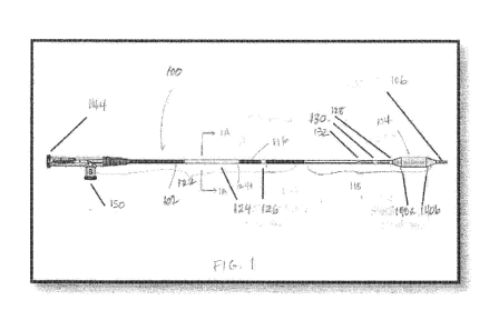
FIG. 1 is a simplified side view of a medical device according to an
embodiment of the present invention.
CA 02843145 2014-01-24
WO 2013/016056
PCT/US2012/046964
FIG. 1A is a cross section view through line 1A-1A of FIG. 1.
FIG 1B is an alternative embodiment of a cross section view through
line 1A-1A of FIG. 1.
FIG. 2 is an enlarged view of the distal end of the medical device
shown in FIG. 1.
FIG. 3 shows a collection of sinus guide catheters useful for positioning
of the sinus balloon catheters of the invention.
FIG. 4 shows a stylet for positioning the medical devices of the
invention.
FIG. 5 shows irrigation tubing useful with the medical devices
according to the invention.
FIG. 6 is a perspective view of a guidewire for use with the medical
devices of the invention.
FIG. 7 is a perspective view of a sinus illumination system for use with
the medical devices of the invention.
FIG. 8 is a side view of a medical device according to an embodiment
of the present invention.
DETAILED DESCRIPTION
The following detailed description should be read with reference to the
drawings, in which like elements in different drawings are identically
numbered. The
drawings, which are not necessarily to scale, depict exemplary embodiments for
the
purpose of explanation only and are not intended to limit the scope of the
invention.
The detailed description illustrates by way of example, not by way of
limitation, the
principles of the invention. This description will clearly enable one skilled
in the art to
make and use the invention, and describes several embodiments, adaptations,
variations, alternatives and uses of the invention, including what is
presently believed
to be the best mode of carrying out the invention.
6
CA 02843145 2014-01-24
WO 2013/016056
PCT/US2012/046964
As used herein, the terms "about" or "approximately" for any numerical values
or ranges indicate a suitable dimensional tolerance that allows the part or
collection
of components to function for its intended purpose as described herein.
Medical devices according to embodiments of the present invention are
beneficial in that, for example, their configuration provides for a
particularly efficient
preparation and treatment of a patient's sinus opening and is mechanically
simple.
Moreover, the simplicity of the medical devices provides for them to be
manufactured
in a cost effective manner. In addition, the medical device according to
embodiments of the present invention is sufficiently stiff that it can be
beneficially
employed to access sinus anatomy followed by a convenient remodeling and
irrigation of the sinus.
FIG. 1 is a simplified side view of a medical device 100 for the treatment of
a
sinus opening (for example a frontal sinus opening, maxillary sinus opening,
ethmoid
sinus opening or sphenoid sinus opening) according to an embodiment of the
present invention. Although described with regard to the sinus opening, the
inventions described herein may also be useful for the dilation of the
Eustachian
tube, repair of endo-cranial fractures, airway procedures such as subglottic
stenosis
dilation and other procedures of the ear, nose and throat. The medical device
100 is
a sinus remodeling and irrigation catheter with an integrated shaft system 102
and a
high pressure balloon 104 near the irrigation tip 106. The shaft system 102
contains
adjacent dual lumen tubing (see FIG. 1A). By adjacent dual lumen tubing is
intended
that the lumens are next to each other but are spaced apart, one from the
other. The
inflation lumen 108 is used for inflation of the balloon with water, contrast
medium or
saline through inflation port 150, and the irrigation lumen 110 permits
passage of a
guidewire or sinus illumination system to facilitate advancement of the
medical
device 100 to the target site and, further, to allow for the flow of
irrigation fluid (water
or saline) to the target site. In an alternative embodiment, there may be
provided a
third lumen, a guide element lumen 111, such lumen being adjacent to the
inflation
lumen 108 and the irrigation lumen 110 (see FIG. 1B). The irrigation lumen 110
and
the guide element lumen 111 merge into a single irrigation lumen 110 in the
distal
shaft portion 118 of the device, proximal to the balloon 104. The medical
device 100
has an irrigation tip 106 with both a forward facing tip opening 114 and
radially facing
7
CA 02843145 2014-01-24
WO 2013/016056
PCT/US2012/046964
openings 112a, 112b and 112c to facilitate irrigation delivery through the
irrigation
lumen 110. The medical device 100 is intended to dilate sinus ostia and spaces
within the paranasal sinus cavities and to provide a means to irrigate from
within a
target sinus for diagnostic and therapeutic purposes. The medical device 100
is
designed to irrigate the sinus through the tip opening 114 and three radially
facing
openings 112a, 112b and 112c in the irrigation tip 106, by delivering fluid
via the
irrigation lumen 110 for delivery before, during, or after dilation of the
sinus ostia or
spaces within the paranasal sinus cavities. Further, instead of delivering
fluid
through the irrigation lumen 110, a vacuum may be applied and a culture may be
obtained by suctioning through the tip opening 114 or the radially facing
openings
112a, 112b and 112c. By radially facing openings is intended that the flow
through
the openings may be at 90 degrees from the flow through the tip opening, but
is may
also be at 30, 45 or 60 degrees or other angles between 0 and 90 degrees, and
the
openings may be round or non-round such as oval or slot-shaped.
The sinus balloon 104 is designed to be non-compliant or semi-compliant.
The diameter of the non-compliant balloon does not vary significantly with
inflation
pressure and that of the semi-compliant balloon will vary only to the extent
that it will
"hourglass" or "dog-bone" about a target region. The balloon itself may be any
shape such as round, triangular, oval or square. In the embodiment shown in
FIG. 1,
the balloon is round and semi-compliant. A stiffening member (in this case a
hypotube 116) is incorporated on the proximal end of the medical device (at
the
distal end of the proximal shaft portion 122) to provide rigidity during
insertion
through a sinus guide catheter, as further described below.
As shown in FIG.1 in some embodiments, direct visualization markers and/or
radiographic markers may be disposed along the integrated shaft system 102.
Generally, "direct visualization markers" refers to markers that may be viewed
during
use with the naked eye or by use of an endoscope, while radiographic markers
include radiopaque material and are viewed using a radiographic device such as
intra-operative fluoroscopy. In one embodiment, at the distal end, there is a
first
distal radiographic marker 120a, which has a proximal edge aligned with the
location
where the proximal taper 140a of the balloon 104 meets the proximal end of the
effective length 142 of the balloon 104. There is also a second distal
radiographic
8
CA 02843145 2014-01-24
WO 2013/016056
PCT/US2012/046964
marker 120b, which has a distal edge aligned with the location where the
distal taper
140b meets the distal end effective length 142 of the balloon 104. The
distance
across the outside edges of the distal markers 120a and 120b represents the
effective length 142 of the balloon 104. The distal markers 120a and 120b may
be
platinum marker bands. In this embodiment, the distal markers help to ensure
that
the medical device 100 is in a straight position inside the guide during the
device
loading and preparation. Additional radiographic markers may be included along
the shaft of the catheter and/or at the distal tip.
Direct visualization markers can be positioned in a number of locations
along the integrated shaft system 102. Although one embodiment is described
here
with reference to FIGs. 1 and 2, other variations may be substituted in
alternative
embodiments. In one embodiment, shaft system 102 may have a dark color, such
as
black, dark blue, dark grey or the like, and markers may have a light color,
such as
white, green, red or the like. In some embodiments, markers may have different
colors and/or different widths to facilitate distinguishing the markers from
one
another during use. This contrast in colors may facilitate viewing the markers
in a
darkened operation room and/or when using an endoscope inside a patient in the
presence of blood.
In one embodiment, there may be a first distal shaft marker 128 (or
"endoscopic marker," since it is typically viewed during use via an endoscope)
disposed on the distal shaft portion 118 of the shaft system 102 at a location
such
that its distal edge aligns with the location where the proximal taper 140a of
the
balloon 104 meets the shaft system 102. The extended balloon neck 134 allows
the
first endoscopic marker 128 to be placed on the shaft and away from any
adhesive
bonding used to secure the proximal end of the balloon neck to the shaft. The
first
endoscopic marker 128 indicates to the user the ending location of the balloon
104
and indicates that the balloon has exited the guide during a procedure. In one
embodiment, the first endoscopic marker 128 may be about 2 mm wide.
A second distal shaft marker 130 is disposed on the shaft system102 such
that the distal edge of the marker is 1 cm. Ø2 cm from the location where
the
proximal taper 140a of the balloon 104 meets the shaft system 102. This marker
indicates to the user that the shaft location is 1 cm away from the end of the
balloon
9
CA 02843145 2014-01-24
WO 2013/016056
PCT/US2012/046964
indicating that the balloon has extended from the guide during the procedure.
In one
embodiment, the second distal shaft marker may be about 2 mm wide and white in
color, while the first marker is about 2 mm and green in color. Of course, any
of a
number of different size and color combinations may be used alternatively.
A third distal shaft marker 132 is disposed on the shaft system 102 such
that the distal edge of the marker is 1 cm. Ø1 cm from the distal edge of
the second
distal shaft marker 130. As shown in FIG. 1, the third distal shaft marker is
a double
marker to distinguish the second and third distal shaft markers 130 and 132
one from
one another. The third distal shaft marker 132 indicates the shaft location 2
cm away
from the end proximal end of the balloon 104, thus indicating the distance the
balloon has extended from the guide during the procedure. In one embodiment,
the
two markers forming the third distal shaft marker 132 are each 0.75 mm wide
and
white in color, however, the size and color of the marker can be changed in
alternative embodiments. The differences in the first, second and third distal
shaft
markers' color, length and number of marks give the indication of the relative
location
proximal to the balloon under endoscopic visibility. Using an endoscope, the
physician user can identify the length of catheter that has been advanced and
retracted out of a guide catheter and/or can approximate a location of the
balloon
104 relative to patient anatomy such as a paranasal sinus ostium, other
paranasal
sinus opening, or other openings in the ear, nose or throat. This
approximation of
balloon position may be very useful in circumstances when the balloon 104 has
been
advanced far enough into an anatomical location that the balloon 104 can no
longer
be viewed via endoscope. For example, using the three endoscopic markers, the
user is able to endoscopically gauge the distance the catheter has advanced
into the
frontal recess once the proximal portion of the balloon is no longer visible.
Of course,
in alternative embodiments, distal shaft markers having different numbers,
sizes,
colors and positions along the catheter shaft may be used.
In some embodiments, in addition to one or more distal shaft markers, one or
more proximal shaft markers may be disposed along the proximal portion 122 of
shaft system 102. In general, such proximal shaft markers may be viewed
directly by
a physician, without using an endoscope, to indicate to the physician a
location of
the balloon 104 of the medical device 100 relative to a guide catheter (see
i.e.
CA 02843145 2014-01-24
WO 2013/016056
PCT/US2012/046964
catheter 200a in FIG. 3) through which the medical device 100 is being
advanced.
As with the distal shaft markers, the proximal shaft markers may have any
suitable
width, color, number, position and the like. In one embodiment, for example,
as
shown in FIG. 1, two proximal shaft markers 124, 126 may have a light color to
contrast with a dark colored shaft system 102 and increase visibility in a
darkened
operating room. The more proximal of the proximal markers 124 (or the "first
proximal shaft marker") may indicate that a tip of the medical device 100 is
at a distal
end of the guide catheter 200 and that the balloon 104 has exited the distal
end 202
of the guide catheter as the marker 124 passes into the proximal end 204 of
the
guide catheter. The more distal of the proximal markers 126 (or the "second
proximal
shaft marker") may indicate to a user that the balloon 104 is just proximal to
a curve
206 in a guide catheter when marker 126 is located at the proximal end 204 of
the
guide catheter.
In one embodiment, the first proximal shaft marker 124 is disposed on the
shaft system 102 such that the length from the proximal end of the proximal
balloon
taper 140a to the proximal end of the first shaft marker 124 is 13.1 cm. Ø2
cm.. The
length of the first proximal shaft marker 124 can vary depending on the size
of the
balloon catheter and may be determined by adding the length of the irrigation
tip
106, the effective or working length 142 of the balloon 104, and the lengths
of the
two balloon taper sections 140a and 140b. Also, the first proximal shaft
marker 124
is preferably white in color, however, other light colors, such as grey, can
be used as
well.
The second proximal shaft marker 126 is disposed on the shaft system 102
distally from the first proximal shaft marker 124. The second proximal shaft
marker
126 is positioned such that the irrigation tip 106 of the medical device 100
is 11.4
cm. Ø2 cm from the distal edge of the second proximal shaft marker 126.
Also, the
second proximal shaft marker 126 has a length of 3 mm. 2 mm. It is preferred
that
the second shaft proximal marker 126 is white in color, however, other light
colors,
such as grey, can be used as well.
When the medical device 100 is inserted into a guide catheter 200a, a user
may visualize the first and second proximal shaft markers 124 and 126 to
determine
the position of the irrigation tip 106 and the balloon 104 of the medical
device 100
11
CA 02843145 2014-01-24
WO 2013/016056
PCT/US2012/046964
relative to the sinus guide catheter 200a. For instance, when the second
proximal
shaft marker 126 is aligned with the proximal opening 204 of the guide
catheter, the
user will know that the balloon 104 is proximal to the curve 206 of the guide
catheter.
The position of the second proximal shaft marker 126 helps to visually ensure
that
the medical device 100 is properly loaded into the sinus guide catheter 200a.
When
the distal edge of the first proximal shaft marker 124 is aligned with the
proximal
opening 204 of the guide catheter 200a, the user knows that the irrigation tip
106 of
the medical device 100 is beginning to exit the guide catheter 200a, and when
the
proximal edge of the first proximal shaft marker is aligned with the proximal
opening
204 of the guide catheter 200a, the user knows that the balloon is completely
out of
the guide catheter 200a.
The visible markers 124, 126, 128, 130 and 132 are preferably light in color,
such as white as indicated above, to contrast with a dark color of the shaft
system
102, which is preferably black. The high contrast between these visible
markers and
the shaft helps view the markers in a low light environment. Also, the high
contrast
allows the user to view directly with an endoscope the markers and know where
the
balloon 104 is located relative to a sinus ostium. Furthermore, the color
contrast is
useful during the procedure when the field is full of blood and/or mucus to
view the
markers and know the position of the balloon. Of course, any other suitable
contrasting color combination may be used. In one embodiment, for example, the
shaft system 102 may be light colored, and the markers 124, 126, 128, 130 and
132
may be dark colored.
FIG. 3 shows a series of sinus guide catheters 200a-200f that may be used in
conjunction with the medical device 100. These guide catheters 200a-200f are
substantially rigid and each has a preset distal curve of 0 degrees (200a), 30
degrees (200b), 90 degrees (200d), 70 degrees (200c) or 110 degrees (200e and
200f). Different curvatures are useable to access the ostia of different
sinuses. For
example, a 70 degree guide is typically used to access the ostium of a frontal
sinus,
a 90 or 110 degree guide is typically used to access the ostium of a maxillary
sinus,
etc. Each of these guide catheters 200a-200f has a length of 12.7 cm. These
sinus
guide catheters are described in U.S. patent application Ser. Nos. 10/944,270
and
11/355,512 and U.S. patent Nos. 7,654,997 and 7,803,150 which are hereby
12
CA 02843145 2014-01-24
WO 2013/016056
PCT/US2012/046964
incorporated by reference, and are commercially available as Relieva TM sinus
guide
catheters from Acclarent, Inc., Menlo Park, Calif.
The medical device 100 is packaged with a balloon insertion stylet 300 (see
FIG. 4) and irrigation tubing 400 (see FIG. 5). The stylet 300 comprises a
rounded
distal tip 302, a support shaft 304 and a proximal loop 306. The insertion
stylet 300
assists with insertion of the medical device 100 into the sinus guide catheter
200a
and is removed from the device 100 prior to advancement of the medical device
100
into the patient anatomy. The irrigation tubing 400 incorporates standard luer
connectors 402 and 404 on each end and is used to attach a sterile syringe to
the
irrigation port 144 of the medical device 100 for sinus irrigation.
Additionally, as
shown in FIG. 8, a ring 800 is provided that may be operated by the thumb or
finger
of a user to aid in insertion of the medical device 820.
In the following description, the sinus guide catheter will be referred to as
200a, but any of the guide catheters 200b-f shown in FIG. 3 may be used.
Following
insertion of the medical device 100 into the sinus guide catheter 200a, a
guiding
element such as a sinus guidewire 500 (i.e. Relieva Vigor Sinus Guidewire
manufactured by Acclarent Inc, Menlo Park, CA and shown in FIG. 6) or sinus
illumination system 600 (i.e. Relieva Luma SentryTM Sinus Illumination System
shown manufactured by Acclarent Inc, Menlo Park, CA and shown in FIG. 7) is
inserted through the irrigation port 144 of the medical device 100 and to the
distal tip
of the sinus guide catheter 200a. Sinus access is achieved by positioning the
sinus
guide catheter 200a in the nasal anatomy, and advancing the sinus guidewire
500 or
sinus illumination system 600 into the target sinus. Once sinus access has
been
achieved, the medical device 100 is advanced over the sinus guidewire 500 or
sinus
illumination system 600 and into the target space. The endoscopic markers on
the
balloon catheter can be used to assist with placement. The medical device 100
is
then inflated to dilate the sinus ostia. Following dilation, the balloon is
deflated. The
guidewire 500 or sinus illumination system 600 is removed from the nasal
anatomy.
A standard syringe is connected to the irrigation tubing 400, which is
connected to
the irrigation port 144 of the medical device 100. Fluid is manually delivered
to the
sinus through the irrigation tip 106 via the distal tip opening 114 and three
radially
facing openings 112a, 112b and 112c of the medical device 100, each side port
13
CA 02843145 2014-01-24
WO 2013/016056
PCT/US2012/046964
having a diameter of 0.026 inches. Upon completion, the medical device 100 is
retracted into the sinus guide catheter 200a and removed from the anatomy. The
medical device 100 can be prepared for additional sinus dilations and/or
irrigations in
the same patient. Alternatively, a suction system such as a standard syringe
or
other vacuum source such as a vacuum pump may be connected to the irrigation
port 144 either directly and through a tubing system and the target sinus may
be
suctioned either before or after treatment thereof.
The medical device 100 sizes may be 3.5mm x 12mm, 6mm x 16mm or 7mm
x 24mm, although others are within the scope of the invention, including, but
not
limited to 5mm x 16mm, 5mm x 24mm or 7mm x 16mm. The distal shaft portion or
section 118 of the device has an inner diameter of 0.037 inches and the
proximal
shaft portion or section 122 of the device has an inner diameter of 0.042
inches. The
distal edge 138 of the first endoscopic marker 128 is located 10 mm from the
proximal edge 136 of the proximal balloon taper 140a, the length from the
medical
device tip opening 114 to the distal end 124a of the first shaft marker 124 is
114 mm
and the distance from the proximal end 136 of the proximal balloon taper 140a
to the
proximal end 124a of the shaft marker 124 is 131 mm. The total length of the
3.5
mm medical device is 250mm and of the other medical devices is 252mm. The
balloon inflated diameters for the medical devices are as follows: 3.5 mm for
the
3.5mm x 12mm, 6 mm for the 6mm x 16mm and 7 mm for the 7mm x 24mm. The
balloon inflated working lengths for the medical devices are as follows: 12 mm
for the
3.5mm x 12mm, 16 mm for the 6mm x 16mm and 24 mm for the 7mm x 24mm. The
maximum outer shaft diameter is 0.086 inches. The deflation time of the
balloon
catheter is preferably seconds. The irrigation flow rate is approximately
100
ml/min and may between 50 and 200 ml/min or 75 and 125 ml/min with a maximum
flow rate of 250 ml/min.
The balloon 104 is made of any suitable material known in the art for
inflation
balloons and may be constructed of compliant, semi-compliant or non-compliant
materials such as nylon (semi-compliant) and polyethylene terepththalate (PET)
(non-compliant). In a particular embodiment, the balloon is constructed of
semi-
compliant material such as nylon. The atraumatic tip portion 146 is also made
of
nylon and is soft with a durometer of less than approximately 55D (often
14
CA 02843145 2014-01-24
WO 2013/016056
PCT/US2012/046964
approximately 40D). The remainder of the irrigation tip is less soft (with a
durometer
greater than about 55D, often about 70D) than the tip portion 146 and is
flexible with
a longer length than prior art balloons tips in order to accommodate the
radially
facing openings 112a, 112b and 112c. In this way, the medical device 100 is
more
easily inserted into the guide catheter 200a and through the tortuous sinus
anatomy.
The atraumatic tip portion 146 may further contain a marker that is nylon with
20%
barium sulfate and is approximately 1 mm in length or may contain any other
type of
radiopaque marker for fluoroscopic visualization or colored marker for direct
visualization of the patient anatomy. In the particular embodiment shown in
Figure
1A, the outer shaft 148 of the medical device 100 is made of pebax. The first
inner
shaft 150 (comprising the inflation lumen) and the second inner shaft 152
(comprising the irrigation lumen) are made of nylon and pebax. The hypotube
shaft
116 that surrounds the outershaft 148 is 304 stainless steel. The combination
of
materials (the nylon balloon and the adjacent dual lumen design) provides for
ease
of insertion of the medical device into and removal from the guide catheter
200a (at
least in part due to the smaller profile of the nylon balloon) and navigation
through
the tortuous sinus anatomy. Insertion into the guide catheter 200a and
navigation
through the tortuous anatomy is also enhanced by the atraumatic tip that is
long, soft
and flexible.
Medical device 100 is configured to irrigate or suction fluids deep within the
sinuses, as well as other areas within the paranasal space. Medical device 100
is
sized appropriately to be delivered into adult as well as pediatric sinuses,
including
maxillary, sphenoid, ethmoid and frontal sinuses. Further, the devices of the
invention may be useful for the treatment of the Eustachian tube or through an
incision to access the middle ear. Medical device 100 can also be used to
deliver
diagnostic or therapeutic substances into the sinuses or other areas in the
paranasal
space. Examples of such diagnostic or therapeutic substances include, but are
not
limited to: contrast agents, pharmaceutically acceptable salt or dosage form
of an
antimicrobial agent (e.g., antibiotic, antiviral, anti-parasitic, antifungal,
etc.), a
corticosteroid or other anti-inflammatory (e.g., an NSAID), a decongestant
(e.g.,
vasoconstrictor), a mucous thinning agent (e.g., an expectorant or mucolytic),
an
anesthetic agent with or without vasoconstrictor (e.g., Xylocaine with or
without
epinephrine, Tetracaine with or without epinephrine), an analgesic agent, an
agent
CA 02843145 2014-01-24
WO 2013/016056
PCT/US2012/046964
(anti-allergenic agent) that prevents of modifies an allergic response (e.g.,
an
antihistamine, cytokine inhibitor, leucotriene inhibitor, IgE inhibitor,
immunomodulator), an allergen or another substance that causes secretion of
mucous by tissues, anti-proliferative agents, hemostatic agents to stop
bleeding,
cytotoxic agents e.g. alcohol, and biological agents such as protein
molecules, stem
cells, genes or gene therapy preparations.
Referring now to FIG. 1, in one embodiment, medical device may include a
forward facing tip opening 114 three radially facing openings 112 a, 112b, and
112c,
on irrigation tip 106 spaced 120 degrees apart, with the inner diameter of the
forward
facing tip opening being 0.037 inches and each of the side openings having a
inner
diameter of 0.026 inches and the inner diameter of the irrigation lumen
proximal of
the atraumatic tip is about 0.042 inches. Alternative embodiments may include
any
suitable alternative number of side openings distributed in any suitable
pattern such
as a helical pattern. In one embodiment, a first side opening may be placed at
about
2.5 mm from the distal end of medical device 100, a second side opening may be
placed at about 3.5 mm from the distal end of medical device 100, and a third
side
opening may be placed at about 4.5 mm from the distal end of medical device
100,
with each of these measurements being from the distal end to approximately the
center of each side opening. The length of the irrigation tip from the distal
end of the
medical device 100 to the distal end of the balloon 104 is approximately 7 mm.
Each
side opening may have any suitable diameter in various alternative
embodiments.
For example, in one embodiment, each side opening may have a diameter of
between about 0.020 inches and about 0.050 inches and or between about 0.030
inches and about 0.040 inches and or about 0.033 inches, so long as the
diameter of
the irrigation lumen of the irrigation tip proximal of the atraumatic tip is
larger than the
diameter of the forward facing tip opening.
In an alternative embodiment, the medical device 100 may contain an
integrated guidewire such that there is no irrigation from the distal end of
the device,
but only from the radially facing openings.
The invention has been described with reference to certain examples or
embodiments of the invention, but various additions, deletions, alterations
and
modifications may be made to those examples and embodiments without departing
16
CA 02843145 2014-01-24
WO 2013/016056
PCT/US2012/046964
from the intended spirit and scope of the invention. For example, any element
or
attribute of one embodiment or example may be incorporated into or used with
another embodiment or example, unless otherwise specified or if to do so would
render the embodiment or example unsuitable for its intended use. Also, where
the
steps of a method or process have been described or listed in a particular
order, the
order of such steps may be changed unless otherwise specified or unless doing
so
would render the method or process unworkable for its intended purpose. All
reasonable additions, deletions, modifications and alterations are to be
considered
equivalents of the described examples and embodiments and are to be included
within the scope of the following claims.
17